Article

Assessment of Liver Regeneration in Patients Who Have Undergone Living Donor Hepatectomy for Living Donor Liver Transplantation

1
Department of Surgery and Liver Transplant Institute, Inonu University Faculty of Medicine, Malatya 244280, Turkey
2
Department of Biochemistry, Inonu University Faculty of Pharmacy, Malatya 244280, Turkey
3
Department of Biostatistics, and Medical Informatics, Inonu University Faculty of Medicine, Malatya 44280, Turkey
*
Author to whom correspondence should be addressed.
Vaccines 2023, 11(2), 244; https://doi.org/10.3390/vaccines11020244
Submission received: 1 January 2023 / Revised: 17 January 2023 / Accepted: 19 January 2023 / Published: 21 January 2023

Abstract

:
Background: Inflammation and the associated immune pathways are among the most important factors in liver regeneration after living donor hepatectomy. Various biomarkers, especially liver function tests, are used to show liver regeneration. The aim of this study was to evaluate the course of liver regeneration following donor hepatectomy (LDH) by routine and regeneration-related biomarkers. Method: Data from 63 living liver donors (LLDs) who underwent LDH in Inonu University Liver Transplant Institute were prospectively analyzed. Serum samples were obtained on the preoperative day and postoperative days (POD) 1, 3, 5, 10, and 21. Regenerative markers including alfa-fetoprotein (AFP), des carboxy prothrombin (DCP), ornithine decarboxylase (ODC), retinol-binding protein 4 (RBP4), and angiotensin-converting enzyme isotype II (ACEII) and liver function tests including alanine aminotransferase (ALT), aspartate aminotransferase (AST), gamma-glutamyl transferase (GGT), alkaline phosphatase (ALP) and total bilirubin levels were all analyzed. Results: The median age of the LLDs was 29.7 years and 28 LLDs were female. Eight LLDs developed postoperative complications requiring relaparotomy. The routine laboratory parameters including AST (<0.001), ALT (<0.001), ALP (<0.001), and total bilirubin (<0.001) showed a significant increase over time until postoperative day (POD) 3. For the regeneration-related parameters, except for the RBP4, all parameters including ACEII (p = 0.006), AFP (p = 0.002), DCP (p = 0.007), and ODC (p = 0.002) showed a significant increase in POD3. The regeneration parameters showed a different pattern of change. In right-lobe liver grafts, ACEII (p = 0.002), AFP (p = 0.035), and ODC (p = 0.001) showed a significant increase over time. DCP (p = 0.129) and RBP4 (p = 0.335) showed no significant changes in right-lobe liver grafts. Conclusions: Regenerative markers are increased in a sustained fashion following LDH. This is more prominent following right-lobe grafts which are indicative of progenitor-associated liver regeneration.

1. Introduction

Liver regeneration has been known for centuries and has been subject to many myths. The regenerative capacity of the liver determines the success of many surgical procedures such as anatomic and non-anatomic or living donor hepatectomy (LDH), which is an important part of living donor liver transplantation (LDLT) [1]. Furthermore, this ability of the liver protects organisms from many injurious effects [2]. There are two main liver regeneration mechanisms: typical and progenitor cell-dependent regeneration. Both mechanisms are triggered during any injury to the liver, however, the dominance of either mechanism depends on the amount of liver tissue that is affected by the injurious event. The studies on experimental models have shown that the regeneration of the liver has several stages including priming, proliferation, and termination, all providing hepatocyte proliferation and hypertrophy [3,4,5].
The process of regeneration has been studied extensively in preclinical models and factors such as the hepatocyte growth factor (HGF), interleukin-6 (IL-6), tumor necrosis factor-α (TNF- α), and signal transducer and activator of transcription 3 (STAT3), whilst Notch and Yap pathways have been found to be important in the regenerative process. During the era of COVID-19, the role of angiotensin-converting enzyme II receptors (ACEII) has been emphasized [6,7,8,9]. Studies have shown that ACEII is over-expressed early after partial hepatectomy and is sustained until the termination of the regenerative process [6,7,8,9]. Studies regarding the ACEII levels following liver resection in humans are lacking. Retinol binding protein (RBP) and especially RBP4 have been shown to play a role in glucose homeostasis in the liver in various pathological processes such as non-alcoholic fatty liver disease [10]. However, its role in the regenerating liver where hypoglycemia is prevalent has not been studied. Ornithine decarboxylase (ODC) is an important enzyme in the catabolism of ornithine to polyamines which is very important in the stabilization of the deoxyribonucleic acid (DNA) structure. ODC can be considered as an indirect marker for DNA synthesis [11]. There is a lack of literature analyzing the role of ODC in liver regeneration in liver transplantation. Alfa Feto Protein (AFP) is mainly involved in hepatoblasts and embryonic hepatic progenitor cells [12]. These cells are shown to be conditionally activated following various injurious effects in preclinical models [12]. Therefore, it can be stated that AFP is associated with hepatic progenitor cell-dependent liver regeneration such as those seen in massive hepatic resections and toxic liver injury [12]. The association of changes in AFP levels needs to be analyzed with the regenerative process in LDLT. Des-gamma-carboxy prothrombin (DCP) is a known marker for the prognostication and diagnosis of hepatocellular carcinoma [13]. However, changes in the levels of DCP in hepatic regeneration have not been analyzed before.
As can be seen clearly, there is a lack of comprehensive studies regarding the regenerative process following living donor hepatectomy (LDH) and in the recipients following the transplantation of the partial liver graft. Furthermore, the majority of our knowledge regarding liver regeneration following partial hepatectomy originates from preclinical experimental models [14].
We are a center of excellence in LDLT and we perform an average of 300 cases of LDLT annually. Therefore, in the present study, we aimed to evaluate the process of liver regeneration in living liver donors (LLDs), who underwent LDH by evaluating serum biomarkers of regeneration including AFP, DCP, ACEII, ODC, and RBP4.

2. Materials and Methods

2.1. Study Population and Design

This is a prospective cohort study. We performed a power analysis to determine the minimum number of subjects that should be included in the present study. Power analyses showed that, with the study power being 0.95, the alpha coefficient being 0.05, and the effect size being (d) = 0.5, the minimum number of subjects required for the study was calculated to be 44. The power analysis was performed using G*Power 3.1.9.7 (Düsseldorf, Germany). We prospectively analyzed and followed up with 63 LLDs who received LDH between 2020 and 2022 in Inonu University Liver Transplant Institute and included in our study. Our LLDs evaluation algorithm, LDH technique, and postoperative follow-up protocol have been previously defined [15]. Verbal and written informed consent were obtained from all LLDs.

2.2. Study Parameters

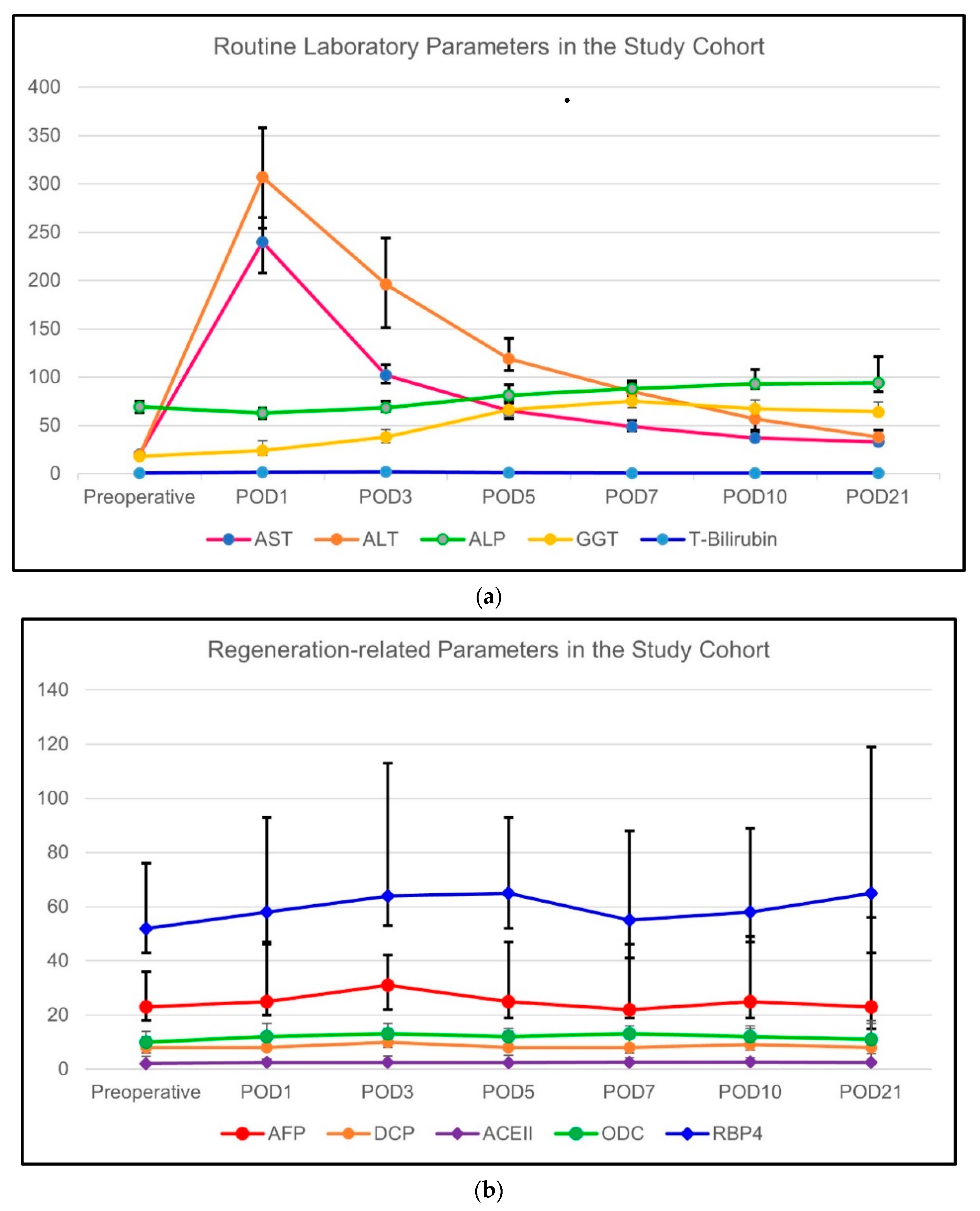
The demographic characteristics including age, sex, body mass index (BMI), habits (smoking and alcohol consumption), blood type (ABO and Rh), operative parameters including the type of liver graft resected, graft weight, remnant liver volume, postoperative complications requiring relaparotomy (such as bleeding and biliary peritonitis) and clinical variables including the routine laboratory parameters on preoperative and postoperative days (PODs) 1, 3, 5, 7, 10 and 21 were collected. Specific laboratory values including alfa-fetoprotein (AFP), des carboxy prothrombin (DCP), ornithine decarboxylase (ODC), retinol binding protein 4 (RBP4), and angiotensin converting enzyme isotype II (ACEII) levels were measured in the same time intervals.
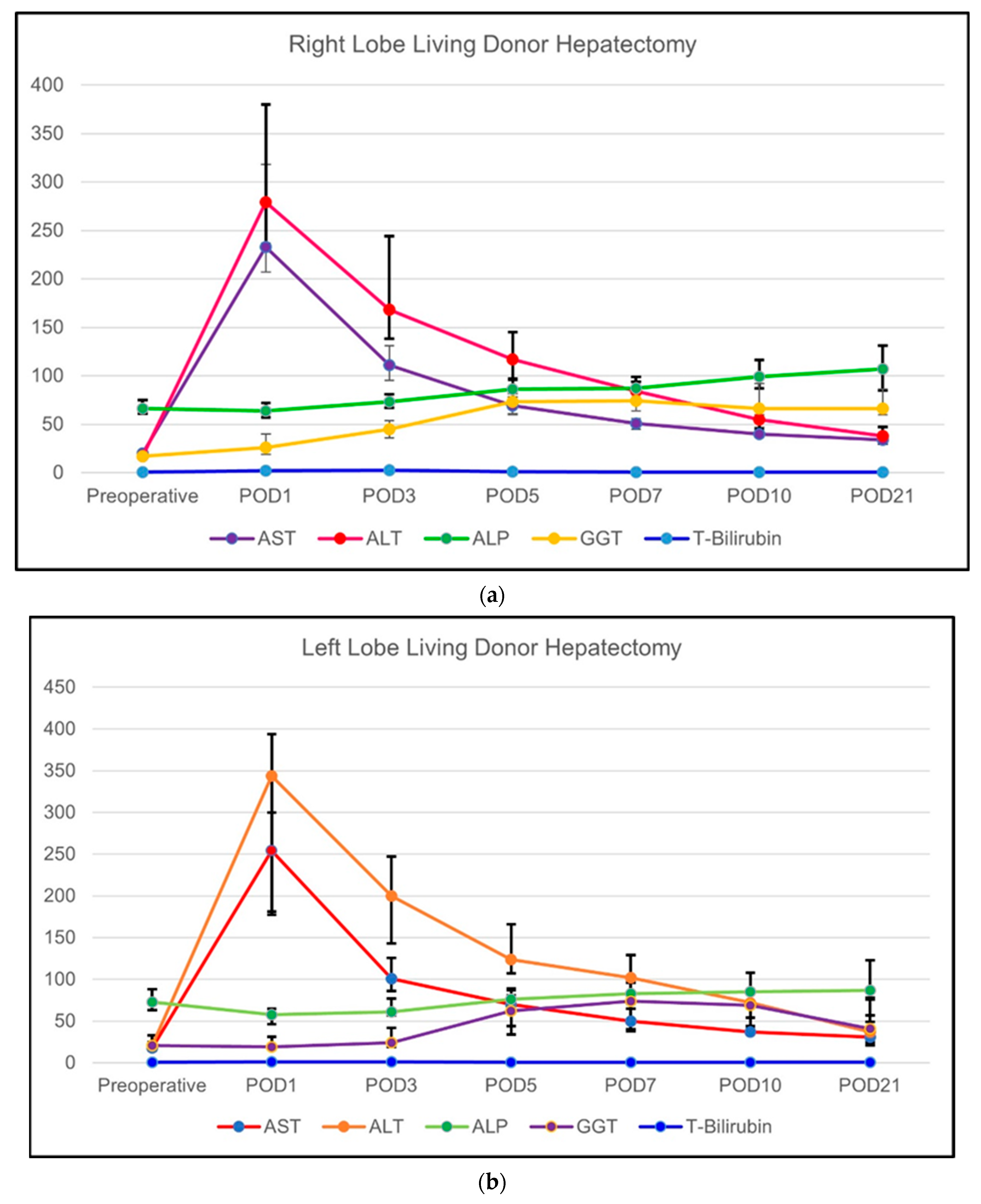
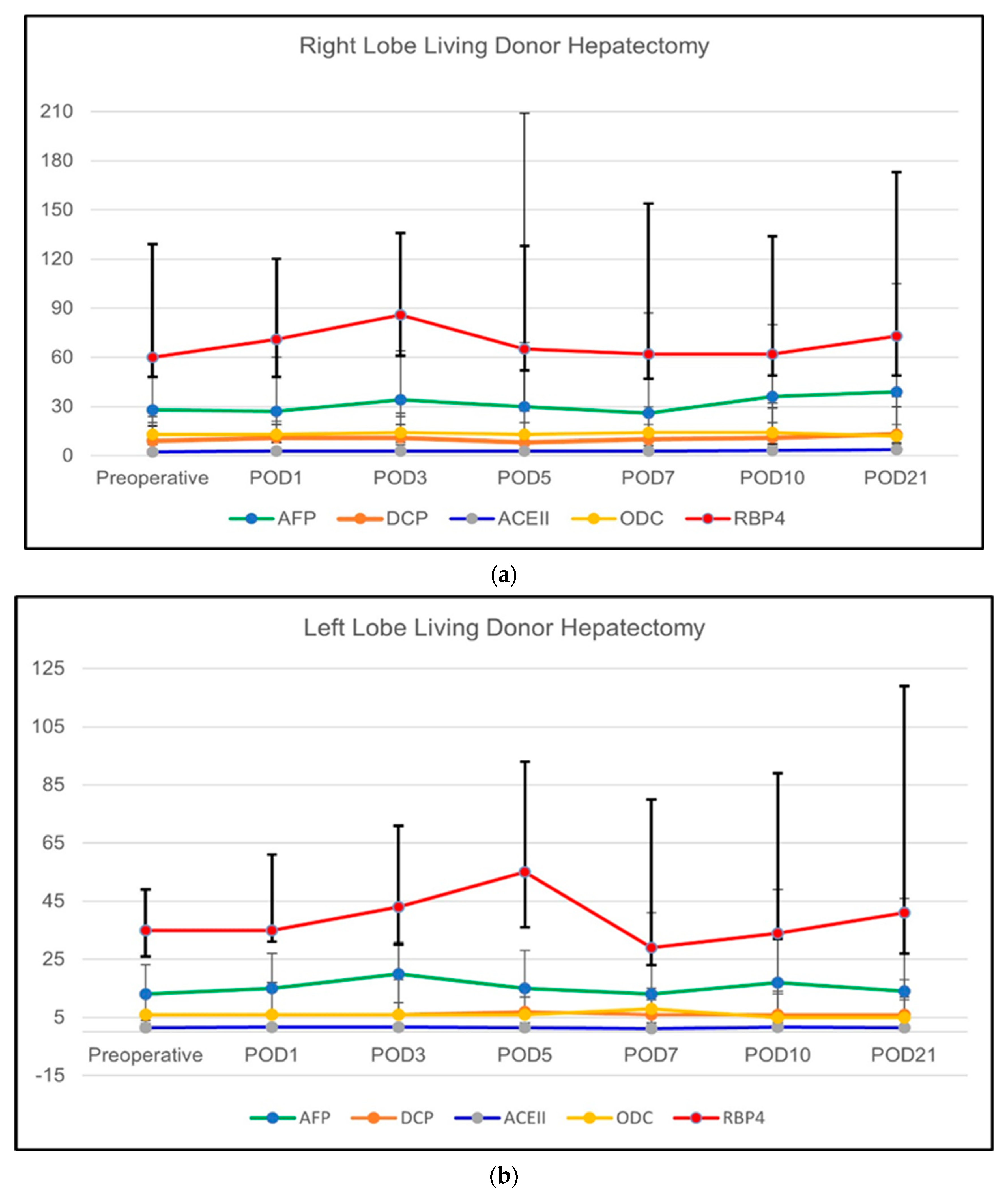
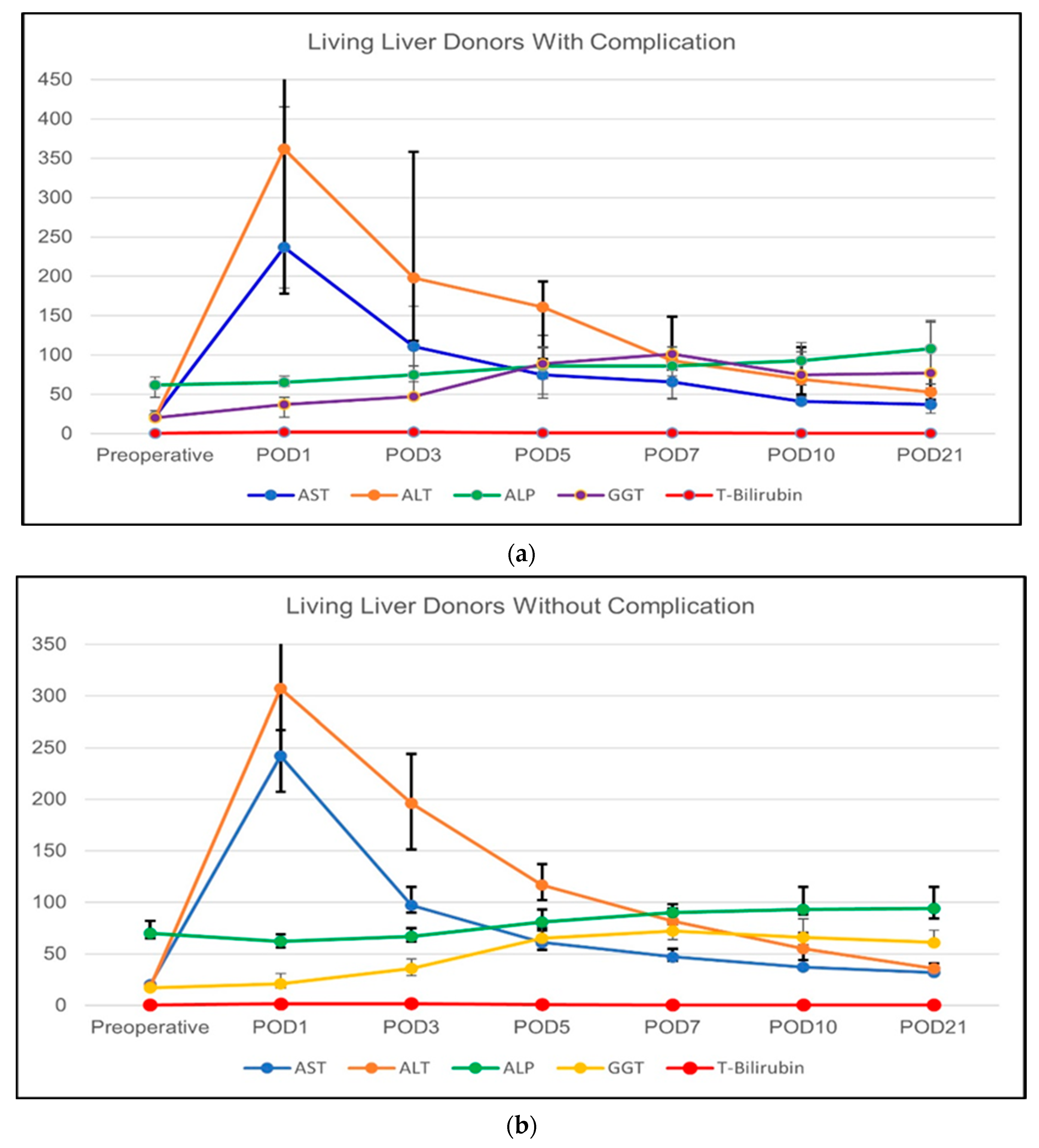
All routine laboratory evaluations including liver function tests, complete blood count, and coagulation studies were obtained from the hospital database. The demographic and operative parameters were obtained from the electronic patient registry. Five milliliters of extra blood was drawn from the subjects and transferred to Inonu University Liver Transplant Institute Hepatology Research Laboratories and all the samples were centrifuged at 2000 rpm at 4 °C for 10 min and serum obtained from the blood samples were divided into four aliquots and stored at −80 °C until the experiments were performed. The comparison of these variables was performed according to different study subgroups; (i) the routine and specific laboratory variables were compared according to sex (male versus females); (ii) according to LDH type (right-lobe LDH versus left-lobe LDH (including left-lobe lateral segment)); and (iii) according to the presence or absence of complications requiring relaparotomy.

2.3. Measurement of the Specific Laboratory parameters

All routine and specific biochemical analysis were performed at Inonu University Liver Transplant Institute Hepatology Research Laboratories. All specific measurements including AFP, DCP, ACEII, ODC, and RBP4 were performed using enzyme-linked immunosorbent assay (ELISA). The measurements were performed by using Human AFP ELISA Kit (BT-LAB, Cat No: E1630Hu), Human DCP ELISA Kit (BT-LAB, Cat No: E4012Hu), Human ACEII ELISA Kit (BT-LAB, Cat No: E3169Hu), Human ODC ELISA Kit (BT-LAB, Cat No: E0845Hu), and Human RBP4 ELISA Kit (BT-LAB, Cat No: E1206Hu) according to manufacturer’s instructions. For all ELISA measurements, after pipetting 40 µL of serum samples to the wells of the precoated ELISA plate, 10 µL of the biotinylated antibody of AFP, DCP, ACEII, ODC, and RBP4 were added to sample wells, respectively. Then, 50 µL of Streptavidin-HRP was added and incubated for 1 h at 37 °C. At the end of the incubation period, the wells were washed thoroughly, 50 µL of both Solution A and B were added and incubated for 10 min at 37 °C in the dark. Then, 50 µL of Stop Solution was pipetted and the color of the solution turned yellow. Absorbance was determined by using Biotek Synergy H1m™ microplate reader (BioTek Instruments Inc., Winooski, VT, USA) at 450 nm within 10 min.

2.4. Ethics Committee Approval and Financial Support

The ethics committee approval was obtained from the Malatya Clinical Research Ethics Committee (Approval no: 2020/170). All stages of the study were carried out according to the guidelines of the Declaration of Helsinki. The financial support was received from the Inonu University Scientific Research Projects Coordination Unit (Project ID: TSA-2021-2382).

2.5. Statistical Analysis

The categorical variables were expressed as the number of individuals and percentage of the study population. Continuous variables are expressed as mean ± standard deviation or median (95% lower CL for median; 95% upper CL for median). The normality of continuous variables was tested using the Kolmogorov–Smirnov and Shapiro–Wilk tests. The Friedman, Mann–Whitney U, and Kruskal–Wallis tests were used to compare the dependent variables among the study groups. Any p-value less than 0.05 was considered statistically significant. All analyses were performed using the Statistical Software Package for Social Sciences version 26.0 (IBM, SPSSv26.0, Armonk, NY, USA).

3. Results

3.1. General Assessment of Demographic and Clinical Variables

In total, 63 LLDs who received LDH were included in the present study. The median age of the LLDs were 29 (95% CI = 28–34) years. There were 28 female (44.4%) and 35 male (55.6%) subjects. The median BMI of the LLDs was 24.7 kg/m2 (95% CI = 23.3–25.9). The blood groups of the LLDs were group 0 in 33 (52.4%), group A in 20 (31.8%), and group B in 10 (15.9%) subjects. Twenty (31.8%) LLDs were smokers and five (7.9%) consumed alcohol on regular basis. Right-lobe LDH was performed in 43 subjects (68.2%), and the left-lobe LDH was performed in 10 LLDs (15.9%), and left lateral segmentectomy was performed in 10 (15.9%) subjects. The median future remnant liver volume of the LLDs was 32 (95% CI = 32–35). The median liver graft volume was 700 cc 95% CI = 670–770). Eight LLDs (12.7%) suffered from early perioperative complications requiring emergency laparotomy. We observed no postoperative mortality in any of the LLDs. The demographic and some clinical variables are summarized in Table 1.

3.2. Analysis of the Biochemical Parameters in Whole Study Group

3.2.1. Change of the Routine Biochemical Parameters

The changes in routine biochemical markers over the designated time period are summarized in Table 2. These biochemical results are also graphically shown in Figure 1a,b. The routine laboratory parameters including AST (<0.001), ALT (<0.001), ALP (<0.001), and total bilirubin (<0.001) of the whole study cohort showed a significant increase over time starting from the preoperative period until the postoperative day (POD) 3. Thereafter, the routine laboratory values returned to the normal range until the POD21. The only exception was the GGT (<0.001) which remained elevated starting from the POD1 until the end of the study period.

3.2.2. Change of Regeneration-Related Biochemical Parameters

The course of regeneration-related biochemical markers over the designated time period is summarized in Table 2. For regeneration-related parameters, except for the RBP4, all parameters including ACEII (p = 0.006), AFP (p = 0.002), DCP (p = 0.007), and ODC (p = 0.002) showed a modest increase in POD3 which was statistically significant. In RBP4 (p = 0.084), there was a tendency towards an increase in the serum levels in POD 3 but it did not reach statistical significance.

3.3. Analysis of the Biochemical Parameters in Selected Study Subgroups

3.3.1. Comparison of LLDs with Right-Lobe versus Left-Lobe LDH

Change of the Routine Biochemical Parameters

The changes in routine biochemical markers over the designated time period among subgroups (right-lobe LDH versus left-lobe LDH) are summarized in Table 3. These biochemical results are also graphically shown in Figure 2a,b. The type of LDH did not have a significant effect on the routine laboratory values in AST, ALT, GGT, and ALP at any time point. The only exception was AST in the POD5 (p = 0.034) and POD10 (p = 0.039) which were significantly higher in the right-lobe when compared to left-lobe grafts. However, the total bilirubin was significantly higher in the LLDs who underwent right-lobe LDH in POD1 (<0.001), POD3 (<0.001), POD5 (<0.001), POD7 (<0.001), POD10 (<0.001), and POD21 (p = 0.004). Similarly, the group analysis of the routine parameters in the individual subgroups showed a similar pattern of changes as in the general population.

Change of the Regeneration-Related Biochemical Parameters

The course of regeneration-related biochemical markers over the designated time period among subgroups (right-lobe LDH versus left-lobe LDH) are summarized in Table 4. These biochemical results are also graphically shown in Figure 3a,b. The regeneration-related parameters showed a different pattern of change. In the right-lobe LDH subgroup, ACEII (p = 0.002), AFP (p = 0.035), and ODC (p = 0.001) showed a significant increase starting from the POD1 until the POD21. DCP (p = 0.129) and RBP (p = 0.335) showed no significant changes in the right-lobe LDH subgroup. On the other hand, only DCP showed a significant increase in the left-lobe LDH subgroup throughout the designated time periods (p = 0.035). The comparison between the right-lobe LDH and left-lobe LDH groups showed that ACEII was significantly higher in the right-lobe LDH group during all time periods except POD10 (p = 0.058). Similarly, AFP was significantly higher in all time periods in the right-lobe LDH group except for the POD10 (p = 0.079). The comparison of DCP showed that the right-lobe LDH group had significantly higher levels during the preoperative period (p = 0.010) and POD1 (p = 0.008), POD3 (p = 0.021), and POD7 (p = 0.008). RBP was significantly higher in right-lobe LDH group during the preoperative period (p = 0.044), POD1 (p = 0.022), POD3(p = 0.015), POD7 (p = 0.009), and POD21 (p = 0.039). The ODC was significantly higher in the right-lobe LDH group in preoperative period (p = 0.05) and POD5 (p = 0.021), POD10 (p = 0.02), and POD21 (p = 0.015).

3.3.2. Comparison of LLDs with and without Postoperative Complications

Change of the Routine Biochemical Parameters

The routine biochemical markers of LLDs with and without complication were compared and the obtained results are given in Table 5. In LLDs with postoperative complications, the change of AST (p < 0.001), ALT (p < 0.001), GGT (p < 0.001), ALP (p < 0.001), and total bilirubin (p < 0.001) significantly increased over time towards POD5 and then returned to normal ranges. Similarly, LLDs without postoperative complications showed the same trend of change in AST (p < 0.001), ALT (p < 0.001), GGT (p < 0.001), ALP (p < 0.001), and total bilirubin (p < 0.001). When the two subgroups were compared in terms of these routine biochemical markers, there seemed to be no difference in most of the parameters at any time point. However, the total bilirubin was slightly higher in the LLDs with complications in POD7 (p = 0.017) and POD21 (p = 0.009). The results of these analyses are summarized in Figure 4a,b.

Change of the Regeneration-Related Biochemical Parameters

The regeneration-related biochemical markers were also compared among the LLDs with and without postoperative complications and the results are given in Table 6. In LLDs with postoperative complications, we did not observe any significant difference over time in any of these parameters. In LLDs without postoperative complications, ACEII (p = 0.027), AFP (p = 0.003), DCP (p = 0.026), and ODC (p = 0.027) showed a slight increase over time which was statistically significant. RBP showed a trend towards an increase but did not reach significance in LLDs without complications. The comparison of the two subgroups did not show any significant difference at any time point. The results of the analyses are summarized in Figure 5a,b.

3.3.3. Comparison of LLDs by Sex

In our study, there were 28 female LLDs and 8 of these (28.5%) underwent left- or left-lateral-lobe donor hepatectomy. There were 35 male LLDs and 12 (35.3%) that underwent left- or left-lateral-segment LDH. There were no significant differences in terms of donor hepatectomy types between the two groups (p = 0.47).

Change of the Routine Biochemical Parameters

Similar to the general population, in the female patients, serum AST (p < 0.001), ALT (p < 0.001), GGT (p < 0.001), ALP (p < 0.001), and TBil (p < 0.001) levels showed a steady and significant increase starting from the POD1 until POD 5 and then steadily decreased to normal levels throughout the rest of the follow-up period. Similar results were obtained in the male LLDs in the course of serum levels of AST (p < 0.001), ALT (p < 0.001), GGT (p < 0.001), ALP (p < 0.001), and TBil (p < 0.001). We analyzed the course of serum levels of liver function tests within and among the sex subgroups in the designated time intervals. Only AST and ALT showed changes between the male and female LLDs. The serum AST levels of the male LLDs were significantly higher than the female LLDs on POD7 (p = 0.019) and POD21 (p = 0.035). Serum ALT values were significantly higher in the male donor on POD1 (p = 0.040), POD7 (p = 0.012), POD10 (p = 0.005), and POD21(p < 0.001). There were no significant differences in the remaining liver function tests between the male and female LLDs. The comparisons of the liver functions tests within the sex groups and between the male and female patients are summarized in Table 7.

Change of the Regeneration-Related Biochemical Parameters

The changes in the serum levels of the regeneration-related biochemical parameters were analyzed according to the sex of the LLDs. The results of the analyses of the regeneration-related biochemical parameters according to the sex of the LLDs are summarized in Table 8. In female LLDs, changes in the serum levels of DCP (p = 0.004) and ODC (p = 0.015) showed a significant and sustained increase in designated time intervals. On the other hand, the serum levels of ACEII, AFP and RBP did not show a significant change between the different time intervals, although there is a tendency for all three parameters to show a sustained increase in the postoperative periods.
On the other hand, male patients showed a sustained and significant increase in serum levels of ACEII (p = 0.027) and AFP(p = 0.025) throughout the follow-up period. RBP, DCP, and ODC showed no significant change in the designated time intervals in male patients. The comparison of the regeneration-related biomarkers among the two sexes did not show any difference in any time interval.

3.3.4. Comparison of LLDs in Terms of Remnant Liver Volume (RLV) (%)

LLDs were divided into three subgroups based on the percentage of RLV: RLV≤ 30%, RLV = 31–35% and RLV ≥ 36%. The groups were compared in terms of changes in regeneration-related biochemical parameters and the results are summarized in Table 9.

4. Discussion

Liver regeneration is a crucial component that determines the success of major hepatic resections such as formal hepatectomy, informal hepatectomy, and LDH. The regenerative capacity of the liver enables us to perform major liver resections and if this regenerative process is disturbed in either way, post-hepatectomy liver failure or a small-for-size syndrome is observed [16]. After major hepatectomy series, the incidence of post-hepatectomy liver failure is reported as high as 32% [17]. In the present study, we have found that markers of regeneration change significantly after LDH in LLDs. This phenomenon is more prominent in right-lobe liver grafts. The change in the serum levels of the markers is not prominently observed in LLDs with complication who require re-laparotomy. To our knowledge, this is the only study analyzing the markers of regeneration in LLDs.
Conventional markers for liver and hepatocyte damage are ALT, AST, and ALP [18]. However, these are not sufficient to evaluate the regenerative process of the liver [19]. There is clearly a need for additional markers to evaluate the regenerative capacity of the liver. Furthermore, there is a need for the evaluation of the normal postoperative course of these markers so that the deviation from the normal course can be evaluated under complicated conditions. In the present study, the markers of hepatocellular markers such as AST, ALT, ALP, and GGT have been elevated towards the POD3 and POD5. Thereafter, a decline in the levels of these enzymes has been observed. This pattern of change was observed in both right, left, and left lateral liver grafts. However, in terms of remnant liver volume, right-lobe liver grafts are associated with lower remnant liver volumes when compared to [20]. This has been reflected in the postoperative course of the enzymes and AST, ALT, ALP, and GGT have been significantly higher in the right-lobe grafts in various postoperative time intervals when compared to left- and left-lateral-liver grafts. Similarly, ALT and AST levels of the male LLDs were higher than the female in postoperative periods. This may be related with the liver mass and the amount of liver tissue resected from the male patients. However, we could not find any data in the literature to explain the differences in liver function tests in LLDs according to sex.
We previously found that the total bilirubin was an important factor in determining the prognosis of the patient’s acute liver failure who has undergone liver transplantation [21]. Studies have shown that the bilirubin levels are indicative of the function of the liver and it is correlated with the severity of liver dysfunction [22,23]. In the present study, we found that the total bilirubin levels of the right-lobe liver grafts have been significantly higher than the other liver grafts which may be related with the larger mass of the liver removed and the associated decrease in the function initially observed in LLDs. Furthermore, in complicated patients, the postoperative bilirubin levels tended to be higher during the postoperative period, although this was not statistically significant. However, this finding shows the impact of any postoperative period on the function of the liver.
Historically, Rao et al. [24] showed that, in chloroform poisoning, serum markers such as GGT, DCP, and AFP were elevated and their combined analysis with the markers of hepatocellular damage would give accurate prognostic information for the affected patients. Later on, the same team evaluated the prognostic significance of these markers in mushroom poisoning [18]. Both studies have shown that, in patients who survived, regenerative markers increased gradually while the markers of liver damage decreased. Living donor hepatectomy and especially harvesting right-lobe grafts has a risk of post-hepatectomy liver failure [25]. Furthermore, there is a certain level of liver dysfunction in the early postoperative period following living donor hepatectomy [26]. For this reason, we wanted to evaluate the course of regenerative markers and the markers of hepatocellular damage in LLDs. We found that the markers of hepatocellular damage have gradually increased towards the POD3 and POD5 and gradually decreased thereafter. On the other hand, we observed a sustained elevation of AFP, DCP, ACEII, ODC, and GGT. Furthermore, this trend in the levels of regeneration markers did not occur in left-lobe liver grafts (except for DCP elevation); however, the right-lobe liver grafts showed a pattern of sustained AFP, DCP, ACEII, and ODC elevation. This proves that, after the removal of right-lobe liver grafts, progenitor oval cells actively take part in the regeneration of the remnant liver. The data regarding the serum ACEII levels following the living donor hepatectomy is lacking. Our study is the first study to emphasize the regeneration-related role of ACEII and it is mainly enhanced in right-lobe liver grafts. Furthermore, our study showed a sex difference between the serum levels of ACEII, which was previously studied in patients with hypertension and cardiac failure [27,28]; however, not in a liver transplant setting. The results of the present study show that there is a difference between the regenerative capacity of the female and male LLDs. It has been emphasized in a limited number of studies that the livers of female patients have a higher regenerative potential [29,30,31]. Unfortunately, our data are limited in terms of their ability to explain this observation, however, we can clearly state that it is not related to the type of graft because the frequency of the right- and left-lobe liver grafts were similar among the groups. The evaluation of the regeneration-related biomarkers and their changes according to the sex of the LLDs requires further studies with higher patient numbers.
However, in left and left lateral liver grafts, the changes in the levels of the enzymes AST, ALT, ALP, and GGT were like the general population. Therefore, evaluating the regenerative process according to elevated liver enzymes may be misleading. The DCP levels in left-lobe liver grafts changed mildly but it was statistically significant. Although our literature search did not yield any relationship between DCP and progenitor cells, our results also suggest that DCP may be indicative of the hepatocyte activity after partial hepatectomy rather than a progenitor cell marker. AFP, ACEII, and ODC seem to be better markers for progenitor-assisted liver regeneration.
We only had eight patients with early postoperative complications requiring relaparotomy. The regenerative markers tend to be elevated in these patients and the fluctuations in the level of the liver enzymes were similar to the general population. We believe that we did not observe any significant changes in the serum levels of the regenerative markers because there was a small group of complicated LLDs.
Our study is the first study to address liver regeneration in LLDs. Furthermore, it gives valuable information regarding the changes in the levels of these biomarkers and the transaminases. However, the current study has some limitations. The first one is the low number of patients. Although we reached the minimum number of patients calculated in a power analysis, the regenerative markers showed wide variation. Therefore, we believe that studies with a larger number of patients will reduce the margin of error. Furthermore, we tried to perform CT volumetry as a part of the follow-up of these LLDs; however, the compliance of the LLDs was low and there were a lot of missing data. Performing volumetric analysis and correlating the results with the levels of the regenerative markers would yield valuable information.
In conclusion, there is dual hepatic regeneration and the dominant mechanism depends on the volume of the resected liver. In right-lobe liver grafts, the remnant is small and this triggers progenitor cell-mediated regeneration. However, in left-lobe and left-lateral-lobe grafts, progenitor-related regenerative markers are not elevated. AFP, ACEII, and ODC are good markers for the surveillance of regeneration following living donor hepatectomy.

Author Contributions

Conceptualization, S.A., B.S. and T.T.S.; Methodology, S.A., A.T., Y.D. and Z.O.; Software, S.A.; Validation, S.A., T.T.S. and Z.K.; Formal Analysis, S.A. and Z.K.; Investigation, S.A.; Resources, S.A. and T.T.S.; Data Curation, S.A., A.T. and B.S.; Writing—Original Draft Preparation, S.A., T.T.S. and B.S.; Writing—Review and Editing, S.A., B.S. and S.Y.; Visualization, S.A. and T.T.S.; Supervision, S.A. and S.Y.; Project Administration, S.A.; Funding Acquisition, this study was supported and funded by the Inonu University Scientific Research Projects Coordination Unit. All authors have read and agreed to the published version of the manuscript.

Funding

The study was supported and funded by the Inonu University Scientific Research Projects Coordination Unit (Project ID: TSA-2021-2382). We would like to thank İbrahim Türkmen, The Scientific Research Projects Unit Coordinator, and all members of the coordination committee for their all support.

Institutional Review Board Statement

Not applicable.

Informed Consent Statement

Verbal and written informed consent was obtained from the voluntary participants.

Data Availability Statement

The datasets analyzed during the current study are available from the corresponding author upon reasonable request.

Acknowledgments

We would like to commend all healthcare professionals who were always on the frontline. They took the courage and responsibility of treating all patients during these hard times despite risking their own lives. They are real heroes.

Conflicts of Interest

The authors declare no conflict of interest.

Ethics Committee Approval

The study was conducted in accordance with the Declaration of Helsinki and approved by the Inonu University institutional review board for non-interventional studies (protocol code: 170 and date of approval: 170).

References

  1. Kiseleva, Y.V.; Antonyan, S.Z.; Zharikova, T.S.; Tupikin, K.A.; Kalinin, D.V.; Zharikov, Y.O. Molecular pathways of liver regeneration: A comprehensive review. World. J. Hepatol. 2021, 13, 270–290. [Google Scholar] [CrossRef] [PubMed]
  2. Michalopoulos, G.K.; Bhushan, B. Liver regeneration: Biological and pathological mechanisms and implications. Nat. Rev. Gastroenterol. Hepatol. 2021, 18, 40–55. [Google Scholar] [CrossRef] [PubMed]
  3. Gilgenkrantz, H.; Collin de l’Hortet, A. Understanding Liver Regeneration: From Mechanisms to Regenerative Medicine. Am. J. Pathol. 2018, 188, 1316–1327. [Google Scholar] [CrossRef] [PubMed] [Green Version]
  4. Lopez-Luque, J.; Fabregat, I. Revisiting the liver: From development to regeneration—what we ought to know! Int. J. Dev. Biol. 2018, 62, 441–451. [Google Scholar] [CrossRef] [PubMed] [Green Version]
  5. Wirth, K.M.; Kizy, S.; Steer, C.J. Liver Regeneration in the Acute Liver Failure Patient. Clin. Liver Dis. 2018, 22, 269–287. [Google Scholar] [CrossRef] [PubMed]
  6. Guan, G.W.; Gao, L.; Wang, J.W.; Wen, X.J.; Mao, T.H.; Peng, S.W.; Zhang, T.; Chen, X.M.; Lu, F.M. Exploring the mechanism of liver enzyme abnormalities in patients with novel coronavirus-infected pneumonia. Zhonghua Gan Zang Bing Za Zhi 2020, 28, 100–106. [Google Scholar] [CrossRef]
  7. Guan, W.J.; Ni, Z.Y.; Hu, Y.; Liang, W.H.; Ou, C.Q.; He, J.X.; Liu, L.; Shan, H.; Lei, C.L.; Hui, D.S.C.; et al. Clinical Characteristics of Coronavirus Disease 2019 in China. N. Engl. J. Med. 2020, 382, 1708–1720. [Google Scholar] [CrossRef]
  8. Li, J.; Fan, J.G. Characteristics and Mechanism of Liver Injury in 2019 Coronavirus Disease. J. Clin. Transl. Hepatol. 2020, 8, 13–17. [Google Scholar] [CrossRef] [Green Version]
  9. Sahin, T.T.; Akbulut, S.; Yilmaz, S. COVID-19 pandemic: Its impact on liver disease and liver transplantation. World J. Gastroenterol. 2020, 26, 2987–2999. [Google Scholar] [CrossRef]
  10. Francisco, V.; Sanz, M.J.; Real, J.T.; Marques, P.; Capuozzo, M.; Ait Eldjoudi, D.; Gualillo, O. Adipokines in Non-Alcoholic Fatty Liver Disease: Are We on the Road toward New Biomarkers and Therapeutic Targets? Biology 2022, 11, 1237. [Google Scholar] [CrossRef]
  11. Sanchez-Sevilla, L.; Mendieta-Condado, E.; Hernandez-Munoz, R. Putrescine treatment reverses alpha-tocopherol-induced desynchronization of polyamine and retinoid metabolism during rat liver regeneration. J. Transl. Med. 2016, 14, 307. [Google Scholar] [CrossRef] [PubMed] [Green Version]
  12. Kuhlmann, W.D.; Peschke, P. Hepatic progenitor cells, stem cells, and AFP expression in models of liver injury. Int. J. Exp. Pathol. 2006, 87, 343–359. [Google Scholar] [CrossRef] [PubMed]
  13. Feng, H.; Li, B.; Li, Z.; Wei, Q.; Ren, L. PIVKA-II serves as a potential biomarker that complements AFP for the diagnosis of hepatocellular carcinoma. BMC Cancer 2021, 21, 401. [Google Scholar] [CrossRef] [PubMed]
  14. Miyaoka, Y.; Miyajima, A. To divide or not to divide: Revisiting liver regeneration. Cell Div. 2013, 8, 8. [Google Scholar] [CrossRef] [Green Version]
  15. Yilmaz, S.; Akbulut, S.; Usta, S.; Ozsay, O.; Sahin, T.T.; Sarici, K.B.; Karabulut, E.; Baskiran, A.; Gonultas, F.; Ozdemir, F.; et al. Diagnostic and therapeutic management algorithm for biliary complications in living liver donors. Transpl. Int. 2021, 34, 2226–2237. [Google Scholar] [CrossRef]
  16. Eshkenazy, R.; Dreznik, Y.; Lahat, E.; Zakai, B.B.; Zendel, A.; Ariche, A. Small for size liver remnant following resection: Prevention and management. Hepatobiliary Surg. Nutr. 2014, 3, 303–312. [Google Scholar] [CrossRef]
  17. Guglielmi, A.; Ruzzenente, A.; Conci, S.; Valdegamberi, A.; Iacono, C. How much remnant is enough in liver resection? Dig. Surg. 2012, 29, 6–17. [Google Scholar] [CrossRef]
  18. Horn, K.D.; Wax, P.; Schneider, S.M.; Martin, T.G.; Nine, J.S.; Moraca, M.A.; Virji, M.A.; Aronica, P.A.; Rao, K.N. Biomarkers of liver regeneration allow early prediction of hepatic recovery after acute necrosis. Am. J. Clin. Pathol. 1999, 112, 351–357. [Google Scholar] [CrossRef] [Green Version]
  19. O’Grady, J.G.; Alexander, G.J.; Hayllar, K.M.; Williams, R. Early indicators of prognosis in fulminant hepatic failure. Gastroenterology 1989, 97, 439–445. [Google Scholar] [CrossRef]
  20. Mohapatra, N.; Sinha, P.K.; Sasturkar, S.V.; Patidar, Y.; Pamecha, V. Preoperative Alanine Aminotransferase and Remnant Liver Volume Predict Liver Regeneration After Live Donor Hepatectomy. J. Gastrointest. Surg. 2020, 24, 1818–1826. [Google Scholar] [CrossRef]
  21. Sahin, T.T.; Sarici, K.B.; Kilci, B.; Koc, C.; Otan, E.; Karakas, S.; Kutluturk, K.; Aydin, C.; Kayaalp, C.; Yilmaz, S. Factors Influencing Mortality Following Liver Transplant in Acute Liver Failure: A Single Center Experience. Exp. Clin. Transplant 2019. [Google Scholar] [CrossRef]
  22. Campani, C.; Bamba-Funck, J.; Campion, B.; Sidali, S.; Blaise, L.; Ganne-Carrie, N.; Demory, A.; Sutter, O.; Larrey, E.; Evain, M.; et al. Baseline ALBI score and early variation of serum AFP predicts outcomes in patients with HCC treated by atezolizumab-bevacizumab. Liver Int. 2022. [Google Scholar] [CrossRef] [PubMed]
  23. Farmer, D.G.; Anselmo, D.M.; Ghobrial, R.M.; Yersiz, H.; McDiarmid, S.V.; Cao, C.; Weaver, M.; Figueroa, J.; Khan, K.; Vargas, J.; et al. Liver transplantation for fulminant hepatic failure: Experience with more than 200 patients over a 17-year period. Ann. Surg. 2003, 237, 666–675. [Google Scholar] [CrossRef] [PubMed]
  24. Rao, K.N.; Virji, M.A.; Moraca, M.A.; Diven, W.F.; Martin, T.G.; Schneider, S.M. Role of serum markers for liver function and liver regeneration in the management of chloroform poisoning. J. Anal. Toxicol. 1993, 17, 99–102. [Google Scholar] [CrossRef] [PubMed]
  25. Zuckerman, J.; Gorgen, A.; Acuna, S.A.; Abreu, P.; Goldaracena, N.; Galvin, Z.; Cattral, M.S.; Ghanekar, A.; McGilvray, I.D.; Lilly, L.B.; et al. Outcomes of Highly Selected Live Donors With a Future Liver Remnant Less Than or Equal to 30%: A Matched Cohort Study. Transplantation 2021, 105, 2397–2403. [Google Scholar] [CrossRef]
  26. Pamecha, V.; Patil, N.S.; Parthasarathy, K.; Sinha, P.K.; Mohapatra, N.; Rastogi, A.; Rudrakumar, K.; Mukund, A.; Chaudhary, A.; Kanal, U. Expanding donor pool for live donor liver transplantation: Utilization of donors with non-alcoholic steatohepatitis after optimization. Langenbecks Arch. Surg. 2022, 407, 1575–1584. [Google Scholar] [CrossRef]
  27. Fernandez-Atucha, A.; Izagirre, A.; Fraile-Bermudez, A.B.; Kortajarena, M.; Larrinaga, G.; Martinez-Lage, P.; Echevarria, E.; Gil, J. Sex differences in the aging pattern of renin-angiotensin system serum peptidases. Biol. Sex Differ. 2017, 8, 5. [Google Scholar] [CrossRef] [Green Version]
  28. Salah, H.M.; Mehta, J.L. Hypothesis: Sex-Related Differences in ACE2 Activity May Contribute to Higher Mortality in Men Versus Women With COVID-19. J Cardiovasc. Pharmacol. Ther. 2021, 26, 114–118. [Google Scholar] [CrossRef]
  29. Bizzaro, D.; Crescenzi, M.; Di Liddo, R.; Arcidiacono, D.; Cappon, A.; Bertalot, T.; Amodio, V.; Tasso, A.; Stefani, A.; Bertazzo, V.; et al. Sex-dependent differences in inflammatory responses during liver regeneration in a murine model of acute liver injury. Clin. Sci. 2018, 132, 255–272. [Google Scholar] [CrossRef]
  30. Marcos, R.; Correia-Gomes, C.; Miranda, H.; Carneiro, F. Liver gender dimorphism--insights from quantitative morphology. Histol. Histopathol. 2015, 30, 1431–1437. [Google Scholar] [CrossRef]
  31. Sutti, S.; Tacke, F. Liver inflammation and regeneration in drug-induced liver injury: Sex matters! Clin. Sci. 2018, 132, 609–613. [Google Scholar] [CrossRef] [PubMed]
Figure 1. (a) The course of routine biochemical blood parameters of the LLDs in the study group; and (b) the course of regeneration-related biochemical parameters of the LLDs in the study group.
Vaccines 11 00244 g001
Figure 2. (a) The course of routine biochemical blood parameters of LLDs who underwent right-lobe LDH; and (b) the course of routine biochemical blood parameters of LLDs who underwent left (including left lateral segment)-lobe LDH.
Vaccines 11 00244 g002
Figure 3. (a) The course of regeneration-related biochemical parameters of LLDs who underwent right-lobe LDH. (b) The course of regeneration-related biochemical parameters of LLDs who underwent left (including left lateral segment)-lobe LDH.
Vaccines 11 00244 g003
Figure 4. (a) The course of routine biochemical blood parameters of LLDs with postoperative complications (requiring relaparotomy); and (b) the course of routine biochemical blood parameters of LLDs without postoperative complications.
Vaccines 11 00244 g004
Figure 5. (a) The course of regeneration-related biochemical parameters of LLDs with postoperative complications (requiring relaparotomy); and (b) the course of regeneration-related biochemical parameters of LLDs without postoperative complications.
Vaccines 11 00244 g005
Table 1. Demographic and some clinical data of the LLDs included in the study group.
VariablesResults
Age (Median (95% CI))29 (28–34)
BMI (Median (95% CI))24.7 (23.3–25.9)
Graft volume (Median (95% CI))
 Volume (Overall)700 (670–770)
 Volume (Right-lobe graft)770 (740–850)
 Volume (Left-lobe)530 (400–690)
 Volume (Left-lobe lateral segment)300 (280–330)
RLV(%)(Median (95% CI))32 (32–35)
Sex (n;%)
 Male28 (44.4)
 Female35 (55.6)
Blood groups (n;%)
 033 (52.4)
 A20 (31.8)
 B10 (15.9)
Smoking (n;%)
 Yes20 (31.8)
 No43 (68.2)
Alcohol use (n;%)
 Yes5 (7.9)
 No58 (92.1)
Donor hepatectomy (n;%)
 Right lobe43 (68.2)
 Left lobe10 (15.9)
 Left lateral segment10 (15.9)
Complications (n;%)
 Yes8 (12.7)
 No55 (87.3)
Table 2. Routine and regeneration-related biochemical parameters of the LLDs included in the study group.
Variables (Median (95% CI))ResultsVariables (Median (95% CI))Results
 ACEII Preop2.1 (1.7–4.7) AST Preop20 (19–22)
 ACEII POD12.5 (1.9–3.7) AST POD1240 (208–265)
 ACEII POD32.5 (2.3–4.9) AST POD3102 (94–113)
 ACEII POD52.4 (2.1–5.1) AST POD565 (57–73)
 ACEII POD72.6 (1.9–4.2) AST POD749 (44–55)
 ACEII POD102.7 (2.1–4.2) AST POD1037 (37–43)
 ACEII POD212.5 (1.6–5.7) AST POD2133 (31–36)
p **0.006p **<0.001
 AFP Preop23 (18–36) ALT Preop19 (17–21)
 AFP POD125 (20–47) ALT POD1307 (254–358)
 AFP POD331 (22–42) ALT POD3196 (151–244)
 AFP POD525 (19–47) ALT POD5119 (107–140)
 AFP POD722 (19–46) ALT POD785 (81–94)
 AFP POD1025 (19–49) ALT POD1057 (45–70)
 AFP POD2123 (15–56) ALT POD2138 (32–45)
p **0.002p **<0.001
 DCP Preop8 (6–12) GGT Preop18 (17–22)
 DCP POD18 (7–13) GGT POD124 (19–34)
 DCP POD310 (8–15) GGT POD338 (32–46)
 DCP POD58 (7–14) GGT POD566 (60–76)
 DCP POD78 (6–12) GGT POD775 (68–83)
 DCP POD109 (7–15) GGT POD1067 (60–76)
 DCP POD218 (7–18) GGT POD2164 (60–74)
p **0.007p **<0.001
 RBP Preop52 (43–76) ALP Preop69 (63–75)
 RBP POD158 (46–93) ALP POD163 (57–68)
 RBP POD364 (53–113) ALP POD368 (64–75)
 RBP POD565 (52–93) ALP POD581 (76–92)
 RBP POD755 (41–88) ALP POD788 (83–96)
 RBP POD1058 (47–89) ALP POD1093 (88–108)
 RBP POD2165 (43–119) ALP POD2194 (85–121)
p **0.084p **<0.001
 ODC Preop10 (6–14) TBil Preop0.5 (0.5–0.6)
 ODC POD112 (7–17) TBil POD11.8 (1.5–2.2)
 ODC POD313 (8–17) TBil POD31.9 (1.6–2.6)
 ODC POD512 (9–15) TBil POD50.9 (0.8–1.2)
 ODC POD713 (8–16) TBil POD70.6 (0.6–0.8)
 ODC POD1012 (7–16) TBil POD100.5 (0.4–0.7)
 ODC POD2111 (7–17) TBil POD210.5 (0.5–0.6)
p **0.002p **<0.001
** Friedman test.
Table 3. Comparison of study subgroups created according to type of hepatectomy (right-lobe LDH vs. left-lobe LDH) type in terms of routine biochemical parameters.
Variables (Median (95% CI))Type of LDHp *
Right LobeLeft Lobe ***
AST Preop20 (19–22)20 (16–22)0.976
AST POD1233 (207–318)246 (192–270)0.935
AST POD3111 (95–131)95 (77–115)0.163
AST POD569 (61–78)53 (43–81)0.034
AST POD751 (45–56)44 (38–58)0.674
AST POD1040 (37–47)35 (28–40)0.039
AST POD2134 (32–37)28 (23–36)0.083
p **<0.001<0.001
ALT Preop17 (15–21)20 (19–28)0.236
ALT POD1279 (236–380)344 (244–394)0.570
ALT POD3168 (138–244)213 (155–272)0.136
ALT POD5117 (97–145)125 (107–166)0.330
ALT POD784 (75–94)88 (69–114)0.333
ALT POD1055 (45–67)67 (39–86)0.794
ALT POD2138 (30–47)38 (21–53)0.914
p **<0.001<0.001
GGT Preop17 (15–20)19 (17–27)0.090
GGT POD126 (19–40)20 (16–31)0.315
GGT POD345 (36–54)28 (23–41)0.045
GGT POD573 (60–81)64 (41–76)0.396
GGT POD774 (64–86)77 (55–85)0.785
GGT POD1066 (60–92)71 (57–84)0.725
GGT POD2166 (60–80)60 (27–73)0.050
p **<0.001<0.001
ALP Preop66 (61–75)74 (59–86)0.337
ALP POD164 (57–72)61 (52–68)0.451
ALP POD373 (67–81)62 (59–70)0.043
ALP POD586 (75–96)77 (75–92)0.425
ALP POD787 (75–99)92 (73–100)0.959
ALP POD1099 (87–116)92 (82–119)0.497
ALP POD21107 (85–131)87 (78–107)0.199
p **<0.001<0.001
TBil Preop0.5 (0.5–0.7)0.5 (0.5–0.6)0.629
TBil POD12.2 (1.8–2.5)1.1 (1.0–1.3)<0.001
TBil POD32.5 (2.0–3.1)1.0 (0.9–1.2)<0.001
TBil POD51.1 (0.9–1.4)0.6 (0.5–0.7)<0.001
TBil POD70.8 (0.7–1.0)0.4 (0.4–0.5)<0.001
TBil POD100.7 (0.5–0.8)0.3 (0.3–0.5)<0.001
TBil POD210.5 (0.5–0.6)0.4 (0.4–0.6)0.004
p **<0.001<0.001
* Mann–Whitney U test; ** Friedman test; *** Left or left-lob lateral segment.
Table 4. Comparison of study subgroups created according to type of hepatectomy (right-lobe LDH vs. left-lobe LDH) type in terms of regeneration-related biochemical parameters.
Variables (Median (95% CI))Donor Hepatectomyp *
Right LobeLeft Lobe ***
ACEII Preop2.2 (1.7–6.4)1.5 (1.3–2.1)0.065
ACEII POD12.8 (2.2–4.8)1.6 (1.4–2.0)0.018
ACEII POD32.9 (2.4–6.6)1.7 (1.4–2.7)0.010
ACEII POD52.8 (2.1–6.1)1.5 (1.0–3.2)0.012
ACEII POD72.8 (2.1–6.2)1.1 (0.9–3.1)0.042
ACEII POD103.2 (2.1–5.6)1.6 (1.2–3.1)0.058
ACEII POD213.7 (1.8–7.2)1.5 (1.0–4.3)0.042
p **0.0020.994
AFP Preop28 (20–61)13 (12–23)0.008
AFP POD127 (21–60)15 (14–27)0.045
AFP POD334 (24–64)20 (18–31)0.050
AFP POD530 (20–69)15 (12–28)0.026
AFP POD726 (19–87)13 (11–41)0.028
AFP POD1036 (20–80)17 (13–49)0.079
AFP POD2139(19–105)14 (11–46)0.046
p **0.0350.119
DCP Preop9 (7–18)6 (3–7)0.010
DCP POD111 (8–19)6(5–6)0.008
DCP POD311 (8–19)6 (5–10)0.021
DCP POD58 (7–209)7 (8–14)0.098
DCP POD710 (6–27)6 (3–11)0.008
DCP POD1011 (7–29)6 (4–14)0.058
DCP POD2113 (7–30)6 (4–18)0.076
p **0.1290.035
RBP Preop60 (48–129)35 (26–49)0.044
RBP POD171 (48–120)35 (31–61)0.022
RBP POD386 (61–136)43 (30–61)0.015
RBP POD565 (52–128)55 (36–93)0.102
RBP POD762 (47–154)29 (23–80)0.009
RBP POD1062 (49–134)34 (32–89)0.039
RBP POD2173 (49–173)41 (27–119)0.104
p **0.3350.125
ODC Preop13 (7–24)6 (4–12)0.050
ODC POD113 (10–26)6 (5–17)0.059
ODC POD314 (8–26)6 (5–18)0.056
ODC POD513 (9–27)6 (6–12)0.021
ODC POD714 (9–30)8 (6–15)0.081
ODC POD1014 (9–32)5 (4–13)0.020
ODC POD2112 (8–36)5 (4–12)0.015
p **0.0010.580
* Mann–Whitney U test; ** Friedman test; *** Left or left-lobe lateral segment.
Table 5. Comparison of the study subgroups created according to the postoperative complications status in terms of routine biochemical parameters.
Variables (Median (95% CI))Postoperative Complications ***p *
YesNo
AST Preop22 (16–24)20 (19–22)0.748
AST POD1237 (185–415)242 (207–267)0.536
AST POD3111 (110–162)97 (90–115)0.231
AST POD575 (45–125)61 (54–73)0.215
AST POD766 (44–73)47 (43–55)0.064
AST POD1041 (37–47)37 (35–44)0.413
AST POD2137 (26–58)32 (30–36)0.243
p **<0.001<0.001
ALT Preop20 (17–23)18 (15–20)0.426
ALT POD1362 (178–847)307 (244–358)0.239
ALT POD3198 (118–358)196 (151–244)0.502
ALT POD5161 (95–193)117 (102–137)0.160
ALT POD793 (81–149)82 (72–94)0.109
ALT POD1069 (50–110)55 (44–70)0.184
ALT POD2153 (43–75)36 (31–41)0.056
p **<0.001<0.001
GGT Preop20 (18–29)17 (15–20)0.426
GGT POD137 (21–46)21 (17–31)0.239
GGT POD347 (44–107)36 (29–45)0.502
GGT POD589 (50–110)65 (58–76)0.160
GGT POD7101 (45–104)72 (64–83)0.109
GGT POD1075 (62–104)66 (59–84)0.184
GGT POD2177 (63–142)61 (58–73)0.056
p **<0.001<0.001
ALP Preop62 (58–72)70 (65–82)0.457
ALP POD165 (60–73)62 (56–69)0.302
ALP POD375 (66–86)67 (62–75)0.457
ALP POD586 (70–109)81 (75–93)0.542
ALP POD786 (68–110)90 (79–98)0.975
ALP POD1093 (74–116)93 (88–115)0.719
ALP POD21108 (81–144)94 (84–115)0.571
p **<0.001<0.001
TBil Preop0.7 (0.3–0.9)0.5 (0.5–0.6)0.217
TBil POD12.0 (1.8–3.5)1.6 (1.4–2.1)0.154
TBil POD32.3 (1.3–4.4)1.8 (1.4–2.5)0.287
TBil POD51.1 (0.7–2.3)0.9 (0.8–1.2)0.272
TBil POD71.1 (0.6–1.7)0.6 (0.6–0.8)0.017
TBil POD100.7 (0.5–1.8)0.5 (0.4–0.7)0.114
TBil POD210.7 (0.5–0.8)0.5 (0.5–0.6)0.009
p **<0.001<0.001
* Mann–Whitney U test; ** Friedman test; *** complications requiring relaparotomy.
Table 6. Comparison of the study subgroups created according to the postoperative complications status in terms of regeneration-related biochemical parameters.
Variables (Median (95% CI))Postoperative Complications ***p *
YesNo
ACEII Preop1.5 (1.5–12.5)2.2 (1.8–6.4)0.585
ACEII POD12.2 (1.7–12.5)2.6 (1.9–4.8)0.801
ACEII POD32.4 (1.5–12.5)2.5 (2.3–5.4)0.556
ACEII POD52.1 (1.5–12.5)2.4 (2.1–5.3)0.556
ACEII POD72.2 (1.6–12.5)2.8 (1.8–4.7)0.747
ACEII POD102.4 (2.1–12.5)2.7 (1.8–4.3)0.695
ACEII POD211.9 (1.1–12.5)2.6 (1.6–6.2)0.713
p **0.0720.027
AFP Preop20 (17–126)24 (18–59)0.801
AFP POD122 (14–126)25 (20–51)0.780
AFP POD332 (13–126)31 (22–58)0.547
AFP POD522 (16–126)27 (19–53)0.856
AFP POD718 (118–126)26 (20–55)0.476
AFP POD1016 (13–126)28 (20–50)0.510
AFP POD2114 (12–126)34 (16–69)0.444
p **0.5290.003
DCP Preop5 (3–33)9 (6–14)0.275
DCP POD16 (4–33)9 (7–14)0.356
DCP POD36 (5–33)11 (8–17)0.146
DCP POD56 (5–33)8 (7–15)0.300
DCP POD76 (4–33)9 (6–17)0.213
DCP POD107 (3–34)9 (7–16)0.467
DCP POD215 (4–34)10 (7–21)0.298
p **0.4020.026
RBP Preop57 (48–251)49 (36–89)0.510
RBP POD148 (38–251)61 (45–117)0.812
RBP POD364 (43–251)64 (49–122)0.989
RBP POD551 (42–251)65 (54–120)0.664
RBP POD753 (36–251)55 (36–118)0.944
RBP POD1057 (49–251)59 (45–115)0.758
RBP POD2165 (50–251)64 (41–134)0.526
p **0.7630.116
ODC Preop6 (2–42)11 (7–16)0.245
ODC POD16 (5–42)13 (10–17)0.281
ODC POD37 (5–42)14 (9–19)0.233
ODC POD56 (6–42)12 (9–18)0.555
ODC POD76 (5–42)13 (10–19)0.493
ODC POD106 (4–42)13 (10–18)0.449
ODC POD216 (5–42)12 (8–18)0.445
p **0.1420.027
* Mann–Whitney U test; ** Friedman test; *** complications requiring relaparotomy.
Table 7. Comparison of study subgroups created according to sex in terms of routine biochemical parameters.
Variables (Median (95% CI))Sexp *
FemaleMale
AST Preop18 (17–22)20 (19–22)0.123
AST POD1206 (179–247)246 (218–313)0.208
AST POD393 (84–111)111 (95–131)0.100
AST POD565 (53–78)65 (57–77)0.803
AST POD743 (35–51)53 (47–58)0.019
AST POD1037 (37–45)42 (37–54)0.062
AST POD2132 (24–35)33 (32–40)0.035
p **<0.001<0.001
ALT Preop15 (14–20)21 (20–30)0.001
ALT POD1233 (177–326)357 (262–408)0.040
ALT POD3149 (117–223)208 (172–258)0.064
ALT POD5108 (80–137)126 (111–156)0.137
ALT POD771 (54–85)91 (85–111)0.012
ALT POD1044 (40–56)70 (61–88)0.005
ALT POD2127 (23–32)48 (38–57)<0.001
p **<0.001<0.001
GGT Preop15 (12–18)20 (18–27)0.001
GGT POD117 (16–34)27 (21–36)0.086
GGT POD336 (24–47)41 (32–54)0.319
GGT POD565 (54–76)69 (60–84)0.240
GGT POD767 (57–77)82 (72–98)0.078
GGT POD1066 (59–92)69 (62–94)0.262
GGT POD2161 (49–67)71 (60–89)0.057
p **<0.001<0.001
ALP Preop64 (59–72)72 (65–83)0.148
ALP POD157 (52–64)65 (62–72)0.098
ALP POD373 (62–80)67 (62–73)0.740
ALP POD588 (77–99)77 (70–92)0.459
ALP POD794 (86–100)84 (75–99)0.668
ALP POD10102 (88–127)92 (82–101)0.394
ALP POD2194 (78–123)94 (87–123)0.263
p **<0.001<0.001
TBil Preop0.5 (0.5–0.7)0.5(0.5–0.6)0.093
TBil POD11.7 (1.4–1.9)1.85(1.4–2.4)0.515
TBil POD31.8 (1.4–2.3)2(1.4–2.7)0.709
TBil POD50.9 (0.8–1.3)0.9(0.7–1.3)0.787
TBil POD70.7 (0.6–0.8)0.6(0.6–0.9)0.568
TBil POD100.5 (0.4–0.7)0.5(0.5–0.8)0.682
TBil POD210.6 (0.5–0.6)0.5(0.5–0.6)0.154
p **<0.001<0.001
* Mann–Whitney U test; ** Friedman test.
Table 8. Comparison of study subgroups created according to sex in terms of regeneration-related biochemical parameters.
Variables (Median (95% CI))Sexp *
FemaleMale
ACEII Preop1.7 (1.4–6.4)2.2 (2.0–5.1)0.418
ACEII POD12.0 (1.7–4.5)2.6 (2.2–10.1)0.552
ACEII POD32.4 (2.1–6.7)2.8 (2.4–4.9)0.482
ACEII POD52.2 (1.8–6.1)2.8 (2.0–5.3)0.520
ACEII POD72.2 (1.6–6.8)2.8 (2.2–4.8)0.329
ACEII POD102.4 (1.5–5.6)2.8 (2.1–4.3)0.675
ACEII POD211.9 (1.4–6.4)3.7 (1.7–7.9)0.493
p **0.0950.027
AFP Preop21 (15–61)24 (18–48)0.514
AFP POD127 (18–51)21 (20–55)0.938
AFP POD324 (18–69)32 (25–49)0.325
AFP POD522 (17–61)27 (20–49)0.489
AFP POD721 (13–66)22 (19–57)0.402
AFP POD1021 (16–80)28 (20–56)0.385
AFP POD2120 (14–93)34 (19–105)0.382
p **0.0850.025
DCP Preop6 (4–14)9 (7–15)0.182
DCP POD16 (6–17)10 (8–16)0.216
DCP POD39 (6–17)11 (9–19)0.355
DCP POD57 (6–19)8 (7–15)0.793
DCP POD76 (5–19)9 (6–18)0.202
DCP POD108 (6–29)9 (7–15)0.612
DCP POD217 (5–30)13 (7–28)0.396
p **0.0040.260
RBP Preop48 (31–129)56 (42–82)0.441
RBP POD161 (42–102)53 (46–120)0.489
RBP POD361 (44–134)78 (57–115)0.605
RBP POD555 (44–128)65 (61–120)0.344
RBP POD747 (36–126)70 (46–139)0.441
RBP POD1049 (39–134)62 (49–89)0.458
RBP POD2152 (41–159)73 (50–134)0.930
p **0.1270.634
ODC Preop10 (5–16)11 (6–24)0.459
ODC POD112 (5–20)12 (9–17)0.315
ODC POD312 (6–19)13 (8–19)0.482
ODC POD512 (6–23)12 (9–18)0.488
ODC POD713 (6–24)11 (9–19)0.559
ODC POD1010 (5–32)13 (9–17)0.532
ODC POD2111 (6–36)12 (8–37)0.641
p **0.0150.133
* Mann–Whitney U test; ** Friedman test.
Table 9. Comparison of the study subgroups created according to the percentage of remnant liver volume in terms of regeneration-related biochemical parameters.
Variables (Median (95% CI))Remnant Liver Volume (%)p *
≤3031–35≥36
ACEII Preop2.2 (1.4–6.6)4.3 (1.7–7.2)1.5 (1.3–2.1)0.062
ACEII POD12.6 (2.3–4.5)3.7 (2.0–8.9)1.6 (1.4–2.0)0.029
ACEII POD32.7 (2.4–5.1)3.9 (2.3–9.4)1.7 (1.4–2.7)0.014
ACEII POD52.4 (2.1–5.1)4.2 (2.1–9.1)1.5 (1.0–3.2)0.014
ACEII POD72.7 (1.6–4.5)4.0 (1.9–8.6)1.1 (0.9–3.1)0.031
ACEII POD102.7 (1.8–5.6)3.8 (2.4–9.9)1.6 (1.2–3.1)0.042
ACEII POD212.6 (1.5–7.9)5.6 (1.6–12.5)1.5 (1.0–4.3)0.040
p **0.4700.0090.994
AFP Preop26 (14–59)34 (21–103)13 (12–23)0.005
AFP POD126 (19–51)31 (21–93)15 (14–27)0.039
AFP POD331 (18–64)37 (24–98)20 (18–31)0.039
AFP POD529 (18–61)33 (21–77)15 (12–28)0.034
AFP POD724 (19–55)36 (18–111)13 (11–41)0.041
AFP POD1031 (18–50)36 (20–91)17 (13–49)0.064
AFP POD2139 (19–126)46 (14–126)14 (11–46)0.062
p **0.1080.1320.119
DCP Preop8 (4–14)12 (8–30)6 (3–7)0.009
DCP POD110 (7–17)13 (8–29)6 (5–6)0.010
DCP POD311 (8–17)14 (7–27)6 (5–10)0.029
DCP POD58 (6–15)14 (7–26)7 (7–14)0.070
DCP POD79 (6–16)12 (6–30)6 (3–11)0.008
DCP POD109 (7–17)12 (7–30)6 (4–14)0.048
DCP POD2110 (6–34)15 (7–33)6 (4–18)0.072
p **0.1060.5860.035
RBP Preop50 (22–89)76 (54–190)35 (26–49)0.013
RBP POD168 (42–100)93(46–175) 35 (31–61)0.032
RBP POD382 (44–122)113 (62–174)43 (30–61)0.015
RBP POD565 (46–107)80 (51–197)55 (36–93)0.088
RBP POD758 (36–99)88 (53–196)29 (23–80)0.011
RBP POD1056 (45–134)65 (52–224)34 (32–89)0.064
RBP POD2169 (36–136)79 (49–251)41 (27–119)0.109
p **0.0590.6580.125
ODC Preop10 (5–16)14 (10–33)6 (4–12)0.042
ODC POD111 (5–20)14 (10–37)6 (5–17)0.050
ODC POD311 (8–19)15 (8–42)6 (5–18)0.041
ODC POD513 (8–23)15 (10–41)6 (6–12)0.027
ODC POD712 (7–19)15 (9–35)8 (6–15)0.076
ODC POD1015 (6–23)14 (9–38)5 (4–13)0.020
ODC POD2115 (7–42)12 (7–41)5 (4–12)0.019
p **0.0110.0420.580
* Kruskal–Wallis test; ** Friedman test.
Disclaimer/Publisher’s Note: The statements, opinions and data contained in all publications are solely those of the individual author(s) and contributor(s) and not of MDPI and/or the editor(s). MDPI and/or the editor(s) disclaim responsibility for any injury to people or property resulting from any ideas, methods, instructions or products referred to in the content.

