Article

Maternal Hydroxytyrosol Supplementation Enhances Antioxidant Capacity and Immunometabolic Adaptations in Nutrient-Restricted Beef Cows and Their Offspring

by
Nieves Escalera-Moreno
1,*,
Javier Álvarez-Rodríguez
2,
Leire López de Armentia
3,
Alba Macià
4,5,
Maria José Martín-Alonso
1,
Ester Molina
1,
Daniel Villalba
1,
Albina Sanz
3 and
Beatriz Serrano-Pérez
1,5,*
1
Animal Science Department, University of Lleida, Av. Rovira Roure 191, 25198 Lleida, Spain
2
Departamento de Producción Animal y Ciencia de los Alimentos, Escuela Politécnica Superior de Huesca, Universidad de Zaragoza-CITA-IA2, Carretera de Cuarte s/n, 22071 Huesca, Spain
3
Centro de Investigación y Tecnología Agroalimentaria de Aragón (CITA) (IA2-UNIZAR), Avda. Montañana 930, 50059 Zaragoza, Spain
4
Food Technology, Engineering and Science Department, XaRTA-TPV, University of Lleida, Av. Rovira Roure 191, 25198 Lleida, Spain
5
AGROTECNIO-CERCA Center, University of Lleida, 25198 Lleida, Spain
*
Authors to whom correspondence should be addressed.
Antioxidants 2025, 14(9), 1097; https://doi.org/10.3390/antiox14091097 (registering DOI)
Submission received: 23 July 2025 / Revised: 29 August 2025 / Accepted: 2 September 2025 / Published: 8 September 2025
(This article belongs to the Special Issue Novel Antioxidants for Animal Nutrition—2nd Edition)

Abstract

The impact of maternal dietary restriction and hydroxytyrosol (HT) supplementation during the last third of gestation on plasma malondialdehyde (MDA) concentration, total antioxidant capacity (ABTS assay), and peripheral blood gene expression related to antioxidant defence, immune response, and energy metabolism was evaluated in beef cows and calves. Two feeding treatments in late gestation (T100% vs. T60% of nutrient requirements) and two HT levels (Control vs. HT at 180 mg/kg of diet) were evaluated during gestation (n = 46 cows) and lactation (n = 37 cows and calves). In pregnant cows, undernutrition led to inhibition of glucose oxidation (PDK4), decreased lipid synthesis (HMGCS1 and SCD) and TLR signalling; T60% cows showed higher plasma MDA (p < 0.05) with no positive effect of HT on antioxidant capacity. Contrarily, during lactation, earlier HT supplementation upregulated antioxidant capacity and modulated antioxidant gene expression (p < 0.05). In calves, there was an increase in SOD1, CAT, and GPX1, especially in the T60%-HT group (p < 0.05). Interestingly, HT supplementation increased glucose transport (SCLA2A1/GLUT1) during pregnancy and lactation (p < 0.05). However, it caused different effects on immunometabolic regulation in both dams and calves, depending on maternal diet. Overall, maternal HT supplementation under restricted nutritional conditions promoted postpartum antioxidant capacity and modulated immune and metabolic gene expression in cows and calves.

1. Introduction

The main challenges in beef cattle production systems are improving productivity and maximizing nutrient efficiency to minimize feed costs and reduce environmental impact. However, minimizing feed costs through dietary restrictions or the use of low-quality pasture resources can negatively impact reproduction [1]. In cattle, a high metabolic challenge during gestation affects cows’ ability to physiologically adapt to the homeorhetic demands associated with fetal growth, parturition, and lactogenesis. Consequently, challenged cows may develop metabolic stress, a condition characterized by excessive lipomobilization, immune dysfunction, inflammation, and oxidative stress, resulting from intense catabolic responses [2,3]. Metabolic adaptations necessary for lactation begin at the end of gestation, a critical period that not only influences maternal well-being but also the development and postnatal health of calves. The onset of lactation dramatically alters the metabolism of many maternal organs so that the mammary gland is supplied with nutrients necessary for the synthesis of milk.
Metabolic reprogramming reflects cell responses to critical changes in the environment that govern the nature of the immune response [4]. Immune cells possess signalling pathways that detect nutritional and energetic status, adjusting metabolism towards anabolic pathways, which are more antigenic or immunogenic, or catabolic pathways, which are more tolerogenic, respectively [5,6]. Enhanced glycolysis is a metabolic change in immune cells that have undergone rapid activation to carry out their particular effector functions in response to the stimulation of pattern recognition receptors, cytokine receptors, or antigen receptors (e.g., Toll-like receptors (TLRs)), which involves nuclear factor kappa B (NF-κB) activation with a more pro-inflammatory effector phenotype. Inflammatory stimuli in both innate and adaptive immune systems also trigger the fatty acid synthesis pathway that allows cells to generate lipids that are necessary for cellular growth, membrane biosynthesis and proliferation. On the contrary, cells involved in the resolution of immune responses and inflammation are adapted to function and slow growth in tissue environments where nutrients are more limiting and efficiency is crucial. Oxidative phosphorylation and a reliance on fatty acid oxidation have been observed in non-inflammatory and regulatory/tolerogenic cells with increased cellular lifespans [7]. However, recent studies have highlighted the heterogeneity in activation states [8,9]. Oxidative stress, proposed as a link between the metabolic and immune systems of cows, arises from an increase in catabolic pathways generating energy from lipids and amino acids, leading to lipid peroxidation [6,10].
Furthermore, maternal nutritional factors, particularly during late gestation, have been shown to significantly impact fetal development, endocrine regulation, and overall calf physiology [11,12,13], even when dietary restriction occurs early in pregnancy [14,15]. The first month of life in calves represents a critical period characterized by intense redox, immunological, and metabolic changes that impact their development and survival. As in cows, redox balance is crucial for the prevention of oxidative stress. Metabolic demands and the quality of the diet influence the animal antioxidant capacity [16]. Immunologically, calves initially rely on passive immunity acquired through maternal colostrum. However, during this period, the progressive activation of their own immune system begins [17]. They become vulnerable to various diseases due to immunosuppressive or inflammatory responses, as a result of the immaturity of their immune systems, when facing environmental challenges or pathogens [18]. Metabolically, calves undergo a significant transition as they adapt to a milk-based diet, which involves changes in carbohydrate and lipid metabolism. This phase is essential for establishing a solid metabolic foundation and healthy growth [19,20].
Among the strategies to mitigate the effects of undernutrition is the supplementation of antioxidants in the prepartum diet [21,22]. These compounds, present in many foods, may help neutralize free radicals and reduce oxidative stress in undernourished cows. Specifically, the use of phenolic compounds, such as hydroxytyrosol (HT), a natural compound that can be found in olive leaves [23], has demonstrated remarkable antioxidant activity in animal plasma, acting as an effective neutralizer of reactive oxygen species (ROS). Its catechol-like structure enables it to donate hydrogen atoms that stabilize free radicals present in the plasma milieu, thereby reducing lipid peroxidation and oxidative damage to plasma proteins. Furthermore, its ability to chelate metal ions, such as Fe2+ and Cu2+, limits metal-catalyzed prooxidant reactions [24]. Compared to other popular antioxidants, HT exhibits greater antioxidant activity [25] and demonstrates rapid absorption into the bloodstream, reaching detectable plasma concentrations within 5 to 10 min of ingestion [26]. Although current evidence on the use of HT in cattle is limited, its effects have been positively documented. In swine, maternal supplementation with HT during gestation improves fetal energy metabolism, as well as antioxidant and immune capacity, by modifying the lipid composition of tissues, protecting polyunsaturated fatty acids (PUFAs) from oxidation, and modulating the inflammatory balance during gestation [27,28].
This study evaluated the effects of undernutrition and supplementation with HT during the last third of gestation on malondialdehyde (MDA) concentration, total antioxidant capacity (ABTS assay) in blood plasma, and the expression of genes involved in antioxidant status, immune regulation, and metabolism pathways in cows during late pregnancy, as well as in cows and their respective calves after parturition.

2. Materials and Methods

2.1. Animal Ethics

The experiment was conducted in accordance with the requirements of the Spanish Policy for Animal Protection RD 53/2013 (BOE, 2013), which meets the European Union Directive 2010/63/EU on the protection of animals used for experimental and other scientific purposes. In addition, the in-house Ethical Committee of the ‘Centro de Investigación y Tecnología Agroalimentaria de Aragón (CITA)’ approved the animal procedure protocol (protocol number CEEA-04 2021-09).

2.2. Animals and Husbandry

The experiment was conducted at the experimental facilities of La Garcipollera Research Station in the mountain area of the central Pyrenees (northeastern Spain, 945 m a.s.l.), in Aragón (northeastern Spain), from November 2021 to March 2022. Cows were recruited from a large experimental suckler cattle herd composed exclusively of beef breeds, which were evenly represented. A full description of the animals included in this study can be found in [29]. The study animal sample comprised 46 prepartum cows (T100%-CTROL, n = 11; T100%-HT, n = 10; T60%-CTROL, n = 14; T60%-HT, n = 11) and 37 postpartum cows (aged 6.7 ± 1.5 years and initial body weight of 672 ± 48.5 kg; means ± standard deviations) with their nursing calves (T100%-CTROL, n = 10; T100%-HT, n = 9; T60%-CTROL, n = 9; T60%-HT, n = 9).

2.3. Dietary Treatments and Performance Recordings

Briefly, at 28 ± 0.7 weeks of gestation (twelve weeks before calving), healthy dams, free from any reproductive and clinical disease, were stratified according to expected calving date and age and then randomly allocated into 4 groups based on the starting date of gestation, within a 2 × 2 factorial design to evaluate the effects of feeding level. The groups included a diet that met 100% of nutrient requirements versus one that met only 60% (T100% vs. T60%) and HT supplementation as a total mixed ration (TMR), i.e., 0 mg HT/kg of TMR (CTROL) vs. 180 mg HT/kg of TMR (HT), added in the form of mash (100 g of HT with a 10% purity/per L of solution, and 18 L of solution per ton of diet) (Econatur, La Carlota, Córdoba, España). The restricted feeding level was based on commercial winter season practices [1] for pregnant cows, while the HT dose was selected based on health claims and no adverse effects in other animal species [30].
Feed was provided at 08:00 a.m., and the cows were tied up for a maximum of 3 h until they finished the assigned amount of feed (10.5 kg/day to T100% or 7.0 kg/day to T60%). The TMR was formulated to meet the nutritional requirements of late-gestation dairy cows and was offered as a mash. A detailed composition of the TMR ingredients, nutrient supply, and energy value is provided in the Supplementary Materials. The TMR composition was identical across the dietary treatments, except for the inclusion of HT. According to the INRA nutritive value calculations [31], the estimated energy value was 1240 kcal of Net Energy for lactation/kg of DM. At all times, the cows had additional access to mineral blocks, which were licked at an individual rate of approximately 75–80 g/day.
To ensure maximal uniformity, 37 pairs of cows and calves born consecutively from unassisted (eutocic) parturitions were selected. After calving, the cows were fed to cover 100% of their postpartum requirements and did not receive any type of polyphenol supplementation. The lactation diet had the same feed composition as the one used during gestation, but all dietary treatments were provided with 10.5 kg of TMR/day. All the cows and calves were loose-housed, with straw as indoor bedding and outdoor access. Each dietary treatment was replicated twice, with a total of eight group pens. The calves were nursed by the cows twice daily (two 30 min periods at 7:00 and 14:00 h). The dams were weighed at weeks −12, −9, and −3 before calving, just after calving, and at week 5 postpartum. The average daily gain (ADG) during the last stages of pregnancy and after parturition was calculated by linear regression.

2.4. Blood and Colostrum Sample Collection

Samples were collected at weeks −9, −6, and −3 before calving from the pregnant cows, and at weeks 1 and 5 after calving from both the cows and their nursed calves (Figure 1). Two blood samples were individually collected from each cow through coccygeal puncture, and from each calf through jugular vein puncture, within a time frame between 8:00 a.m. and 10:00 a.m. The first blood sample was collected in heparin tubes for MDA and ABTS analysis. These samples were immediately centrifuged (2500 × g for 10 min) to obtain the plasma and then stored at −20 °C until analysis. The second blood sample was collected into TempusTM Blood RNA Tubes (Applied Biosystems, Foster City, CA, USA) for whole-blood gene expression analysis, according to the manufacturer’s instructions. These samples were shaken after sample collection and then stored at −20 °C until use. For the analysis of HT metabolites, serum and colostrum samples were collected in a selection of 24 dams (6 dams/group). Blood samples were collected in empty tubes at weeks −12 and −3, and colostrum samples were manually collected during the first 12 h after calving, frozen at −20 °C, and subsequently lyophilized.

2.5. Sample Analysis

2.5.1. Determination of Malondialdehyde and Total Antioxidant Activity (ABTS Assay) in Plasma

MDA and ABTS (2,2′-azino-bis(3-ethylbenzothiazoline-6-sulfonic acid)) were analysed as pro- and anti-oxidative markers in plasma, respectively. The MDA levels were measured using a gas chromatography method that separately quantifies free MDA (FMDA) and protein-bound MDA (PBMDA) to ultimately determine total MDA. This method has been previously validated in bovine plasma [33]. The ABTS radical cation (μmol Trolox-eq./mL) was obtained by diluting the plasma to 1/60, mixing it with the ABTS solution, and subsequently calibrating it with Trolox as the reference standard, as a proven method to measure antioxidant capacity in the ruminant plasma [34]. Twelve replicates of each extract were prepared [35]. To minimize inter-operator variability, all assays were performed by the same trained technician under standardized laboratory conditions.

2.5.2. HT Metabolite Analysis

The presence of specific phase II metabolites was assessed in both plasma and colostrum matrices. Among the potential metabolic products of HT, HT sulphate (HTS) and alcohol homovanillic sulphate (AHVS) were selected as compliance signature biomarkers, based on their specificity, detectability, and established role as metabolites of HT in mammalian systems. The tentative quantification of these phenolic metabolites was carried out by UPLC coupled to tandem mass spectrometry (MS/MS) [36], reaching a detection limit of 30 ng/mL and 15 ng/mL for HTS and AHVS, respectively.

2.5.3. RNA Extraction, cDNA Synthesis, and qPCR Analysis

Total RNA was extracted from TempusTM Blood RNA Tubes (Applied Biosystems, Foster City, CA, USA) with a TempusTM Spin RNA Isolation Kit (Applied Biosystems, Foster City, CA, USA) according to the manufacturer’s recommendations. The concentration and purity of the total RNA were determined spectrophotometrically using a UV/Vis Implen NanoPhotometer N50 (Thermo Scientific, Waltham, MA, USA). The samples were treated with RNase-free DNase I (Thermo Scientific, Waltham, MA, USA) to eliminate contaminating genomic DNA. First-strand cDNA synthesis was performed with 0.3 µg of total RNA and random hexamer primers using the Maxima RevertAid H Minus First Strand cDNA Synthesis Kit (Thermo Scientific, Waltham, MA, USA) according to the manufacturer’s recommendations. The samples were stored at −80 °C for subsequent gene expression analysis.
To prevent genomic contamination, whenever possible, the primers were designed to span an intron. A standard curve was created for each gene by amplifying serial dilutions of a control cDNA to ensure linearity between the initial template concentration and the cycle threshold (Ct) values. Amplification was performed in a QuantStudio™ 7 Flex Real-Time PCR System sequence detector (Applied Biosystems, Foster City, CA, USA). The qPCR conditions included an initial activation and denaturation step of 10 min at 95 °C, followed by 40 cycles of 10 s at 95 °C and 1 min at 60 °C.
Messenger RNA expression was determined by qPCR. Genes selected for transcript analysis were those involved in antioxidant status (superoxide dismutase 1 (SOD1), superoxide dismutase 2 (SOD2), catalase (CAT), glutathione peroxidase 1 (GPX1), nuclear factor erythroid 2-related factor 2 (NRF2)), immune regulation (5-lipoxygenase (ALOX5), tumor necrosis factor alpha (TNFA), nuclear factor kappa B (NFKB), toll-like receptor 4 (TLR4), peroxisome proliferator-activated receptor delta (PPARD)) and energy metabolism (pyruvate dehydrogenase kinase isoenzyme 4 (PDK4), the gene encoding solute carrier family 2 member 1 (SLC2A1/GLUT1), insulin-like growth factor 1 receptor (IGF1R), acyl-CoA dehydrogenase (ACADVL), stearoyl-CoA desaturase (SCD), fatty acid synthase (FASN), sterol regulatory element-binding protein 1 (SREBF1), 3-hydroxy-3-methylglutaryl-CoA synthase 1 (HMGCS1)). The expression levels of the target gene were normalized to those of the combination of the two most stable reference genes (ACTB and RPL19), which were validated using NormFinder [37]. The sequences and sources of the primers are shown in Table 1. PCR reactions were run using 3 μL of 300-fold diluted cDNA as the template in a total volume of 8 μL. The reaction mixture contained 2.5 μL of PowerTrack SYBR Green Master Mix (Thermo Scientific, Waltham, MA, USA), 2 μL of H2O, and forward and reverse primer concentrations, as reported in Table 1 (125 nM–400 nM). Each measurement was carried out in triplicate, and the average was used to calculate the relative gene amount. Data were normalized and analysed with the 2−ΔΔCt method using the mean Ct value obtained for the two reference genes and the Ct values for each sample.

2.6. Data Analysis

The data obtained were analysed using JMP Pro version 18 software (SAS Institute Inc., Cary, NC, USA). Blood MDA concentration, total antioxidant capacity (ABTS assay), and gene expression at weeks −9, −6 and −3 before calving in the pregnant cows, and at weeks 1 and 5 after calving in the cows and their nursed calves, were analysed in separate data sets by means of mixed models with repeated measures. The fixed effects included feeding level (T100% vs. T60%), HT supplementation (CTROL vs. HT), sampling week (weeks −9, −6, and −3 before calving, or weeks 1 and 5 after calving), as well as their interactions. Each cow was considered as the experimental unit and included as a random effect to account for individual variability among animals. Starting body weight at week −12 before calving was considered as a covariate in all models. The interactions between fixed effects are only reported in Section 3 if they are significant (p < 0.05). Blood gene expression data were log-transformed to meet the assumption of normality and homoscedasticity. Least square means and their standard errors are presented. Normalized gene expression values were statistically analysed in log form but expressed as relative quantification (RQ). The separation of means was carried out with Tukey’s test. Chi-square tests were used to assess associations between HT metabolite detection in serum (weeks −12 and −3) and colostrum (<12 h parturition) and factors of interest (feeding level and HT supplementation). The level of significance was set at p-value < 0.05.

3. Results

3.1. Cow Performance and Circulating HT Metabolites

During the last third of pregnancy, the feeding level affected (p < 0.001) the cow ADG, such that the T100% cows showed greater ADG during the prepartum period than the T60% cows (Table 2). Consequently, the BW after calving was greater (p < 0.001) in the T100% cows than in the T60% cows. The loss of ADG during the first month of lactation was greater (p < 0.05) in the T100% cows than in the T60% cows. However, the gestation dietary HT supplementation did not affect (p > 0.10) the cow ADG either during the last third of pregnancy or the first month of lactation (Table 2).

3.2. MDA Concentration and Total Antioxidant Capacity in Plasma

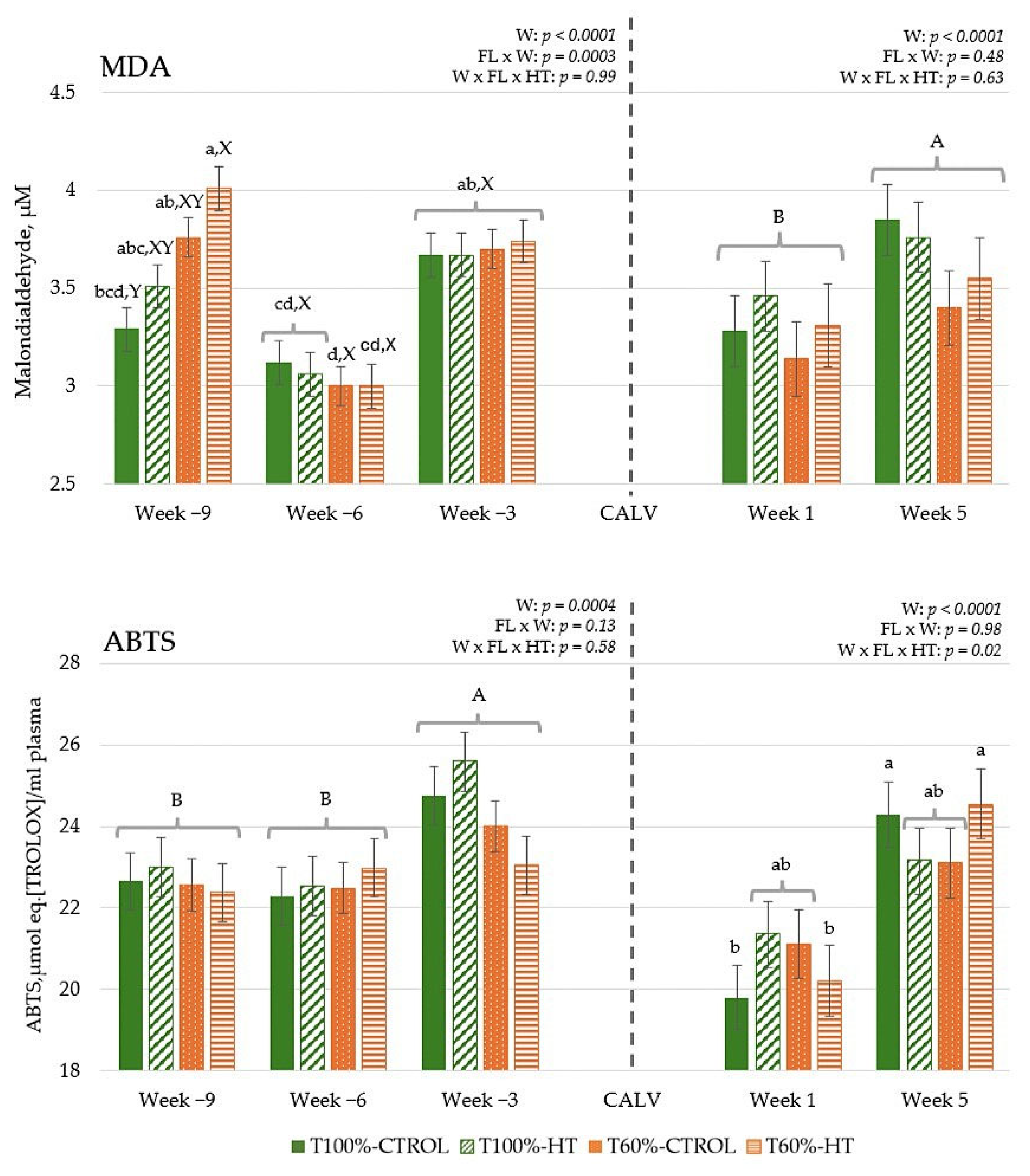
During gestation, the total MDA concentration was higher (p < 0.001) at week −9 in T60%-HT than in T100%-CTROL, although no differences were observed between the treatments in the rest of the sampling weeks (Figure 2). MDA values decreased at week −6 and increased again at week −3 (p < 0.001). After calving, total MDA increased as lactation progressed and was significantly higher at week 5 compared to week 1 (p < 0.05) (Figure 2). During gestation, the levels of ABTS were significantly increased at week −3 compared to weeks −6 and −9 (p < 0.01). During lactation, total antioxidant capacity was significantly increased for T100%-CTROL and T60%-HT at week 5 compared to the other groups (p < 0.05). Total antioxidant capacity was also significantly increased at week 5 as compared to week 1 (p < 0.001) (Figure 2).
In the calves, MDA levels were also significantly increased at week 5 as compared to week 1 (p < 0.001) (Figure 3). Higher total antioxidant capacity was observed in the calves from T60%-HT at week 1 dams compared to the T60%-CTROL group at the same week and the T100%-CTROL group at week 5 (p < 0.05).

3.3. Gene Expression

3.3.1. Genes Related to Antioxidant Response

The results during gestation did not reveal a significant effect of undernutrition or HT supplementation, but the week affected all genes (Figure 4). In general, antioxidant defences, namely, NRF2, SOD2, CAT, and GPX1, showed the lowest expression at week −9 and peaked at week −6, followed by a decrease at week −3 (p < 0.0001). However, SOD1 expression increased linearly as gestation progressed to week −3 (p < 0.05). After calving, there were differences in the interaction among week, feeding level, and HT supplementation prepartum in SOD1 expression, which was the lowest in the T60%-HT cows compared with the rest of the dietary treatments (p < 0.05). Meanwhile, there was lower NRF2 and SOD2 expression in the HT cows compared to the CTROL cows during the lactation period (p < 0.05). Regarding the week effect during the lactation period, antioxidant defences (NRF2, SOD2, CAT, and GPX1) decreased significantly in expression at week 5 compared to week 1 (p < 0.0001).
In the calves, antioxidant genes were greater in the HT-supplemented and higher-fed groups during the first week, with some antioxidant responses decreasing and others increasing by week 5, indicating that both diet and age influenced oxidative status (Figure 5). The interaction of feeding level with HT supplementation significantly affected transcription factor NRF2, with high expression levels observed in the T100%-CTROL and T60%-HT groups compared with the T100%-HT and T60%-CTROL groups (p < 0.05). The interaction of feeding level per week of age affected SOD1 expression, which showed greater expression in the T100%-HT group than the T60%-CTROL group at week 1 and greater expression in the T60%-HT group compared to the T60%-CTROL at week 5 (p < 0.05). Accordingly, greater GPX1 expression levels were observed in the T60%-HT group compared to the T60%-CTROL group at week 5 (p < 0.05). Meanwhile, the gene expression of antioxidant enzymes (SOD1, CAT, and GPX1) was greater in the HT cows compared with the CTROL groups, regardless of the feeding level (p < 0.05). The expression of NRF2 was decreased at week 5 as compared to week 1, contrary to CAT, which showed an increase at week 5 (p < 0.05).

3.3.2. Genes Related to Immune Response

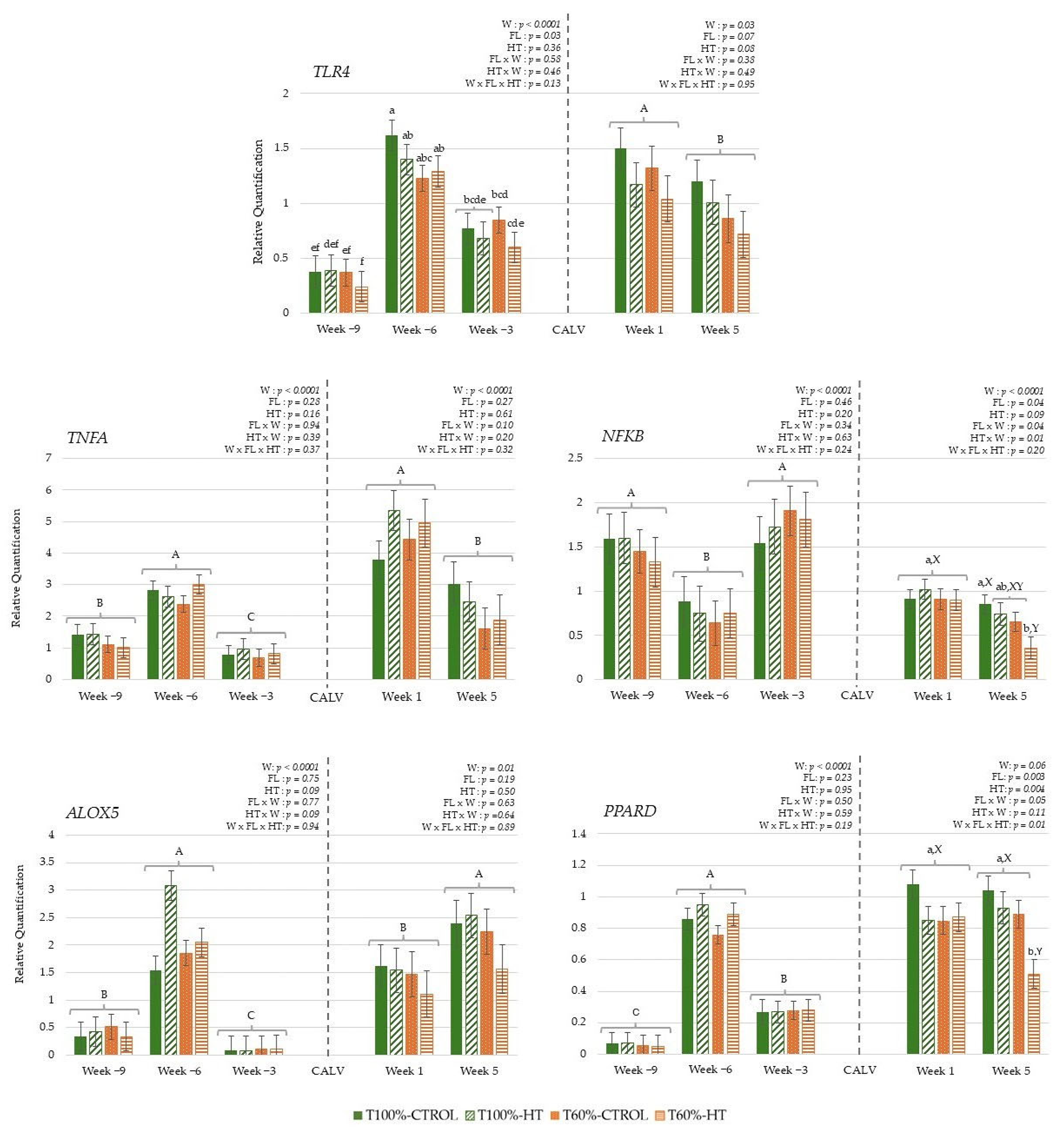
The results during gestation did not reveal differences based on HT supplementation, but they did reveal differences based on feeding level; lower overall TLR4 expression was observed in the T60% cows compared to the T100% cows during gestation (p < 0.05). Regarding the week effect, immune genes, namely, TLR4, PPARD, TNFA, and ALOX5, showed lower expression at week −9 relative to calving and increased subsequently at week −6, followed by a decrease at week −3. In contrast, transcription factor NFKB gene expression decreased at week −6 and then increased at week −3, reaching similar levels to those observed at week −9 (Figure 6). During lactation, the gene expression of transcription factors PPARD and NFKB was affected by the interaction among week postpartum, prepartum feeding level, and HT supplementation. At week 5, the T60%-HT group exhibited the lowest PPARD expression levels among all cow groups, and these cows also showed significantly lower NFKB expression compared with the T100%-CTRL group (p < 0.05). Some genes showed differences in their expression based on the week effect, with decreased TLR4 and TNFA expression at week 5 compared to week 1, but increased ALOX5 expression at week 5 compared to week 1 (p < 0.05) (Figure 6).
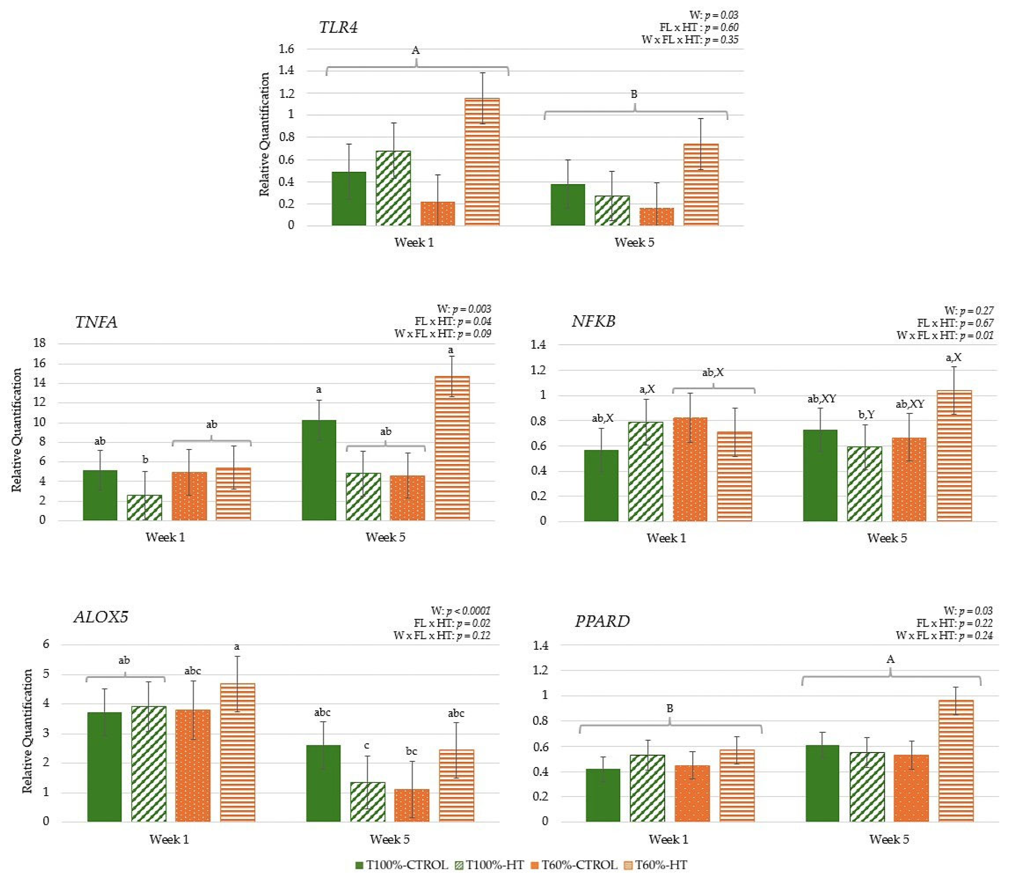
In the calves, pro-inflammatory immune-related pathways were influenced by the interaction between week, feeding level, and HT supplementation, with early increases in pro-inflammatory genes followed by later changes in regulatory pathways, reflecting dynamic diet- and time-dependent effects. At week 5, NFKB expression levels were significantly lower in the T100%-HT group compared to the T100%-HT group at week 1 and the T60%-HT group at week 5 (p < 0.05) (Figure 7). The expression of pro-inflammatory immune genes TNFA and ALOX5 was also significantly affected by the interaction between feeding level and supplementation, with greater TNFA and ALOX5 expression observed in the T100%-CTROL and T60%-HT groups (p < 0.05). TLR4 and ALOX5 expression was significantly decreased at week 5; meanwhile, the expression of TNFA and PPARD was significantly increased at week 5 as compared to week 1 (p < 0.05).

3.3.3. Genes Related to Energy Metabolism

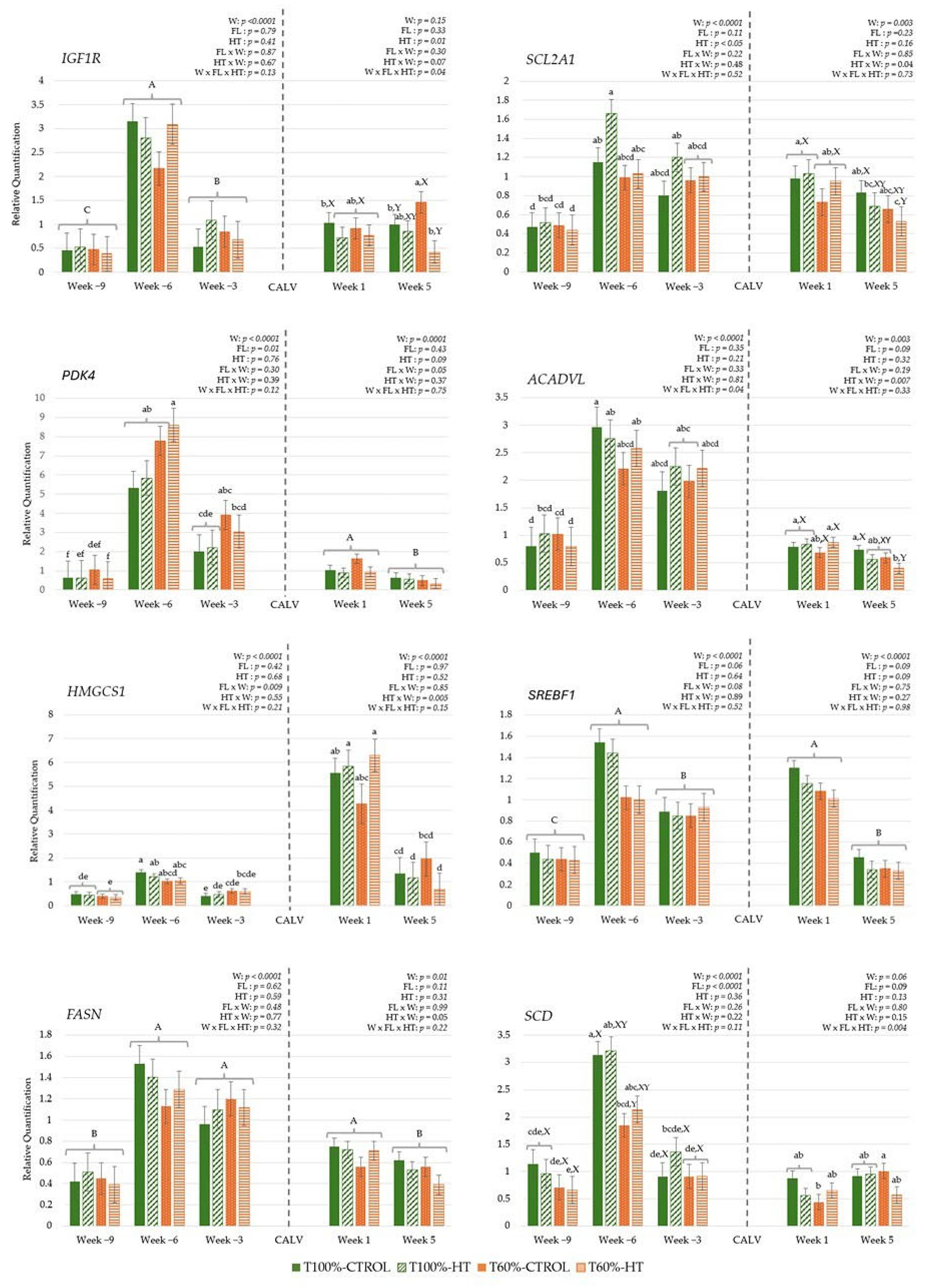
The interaction between week, feeding level, and HT supplementation during gestation showed that the long-chain fatty acid oxidation key enzyme ACADVL increased less in the T60%-CTROL group compared to the rest of the cow groups from week −9 to −6 (Figure 8). Moreover, at week −6, the expression levels in the T60%-HT group were statistically equal to those in the T100%-CTROL group (p > 0.05). Regarding feeding level, significantly greater gene expression of the cholesterol biosynthesis enzyme HMGCS1 was observed in the T100% cows at week −6 (p < 0.05) compared to the T100% and T60% cows at week −9 and week −3. Additionally, greater gluconeogenic (PDK4) but lower fatty acid desaturation enzyme (SCD) pathways were observed in the T60% cows compared to the T100%-cows during gestation (p < 0.05). Regarding HT supplementation, glucose transporter, SCLA2A1/GLUT1, gene expression was greater in the HT cows compared to the CTROL cows (p < 0.05). SLC2A1/GLUT1 and fatty acid synthase enzyme FASN gene expression also increased from week −9 to week −6 and then remained constant until week −3. Likewise, IGF signalling (IGF1R), gluconeogenic (PDK4) and lipid synthesis pathways (SCD, SREBF1) showed increased expression levels up to week −6, followed by a decrease in week −3 (p < 0.0001).
During lactation, the interaction among week, feeding level, and HT supplementation revealed greater IGF1R expression in the cows from the T60%-CTROL group at week 5 compared to the cows from the T100%-CTROL and T60%-HT groups. Meanwhile, higher SCD expression was observed in the T60%-CTROL cows at week 5 compared to the T60%-CTROL cows at week 1 (p < 0.05). Regarding HT supplementation per week, there was greater glucose transporter (SLC2A1/GLUT1), long-chain fatty acid oxidation (ACADVL) and lipid biosynthesis (HMGCS1) gene expression in the HT cows at week 1 compared to the CTROL cows at the same week, but there was lower expression in the HT cows at week 5 compared to the CTROL cows at week 5 (p < 0.05). Some genes also showed differences in their expression based on the week effect, with significantly decreased PDK4, SREBF1, and FASN expression at week 5 compared to week 1 (p < 0.05).
In the calves, maternal HT supplementation and feeding level influenced early metabolic activity in blood cells, enhancing glucose metabolism in the first week, while lipid-related pathways decreased over time, reflecting diet- and time-dependent effects. The interaction between feeding level and HT supplementation across weeks showed greater PDK4 expression in T100%-HT and T60%-HT at week 1 compared to the T60%-CTROL group at week 5 (p < 0.05). On the other hand, the interaction between feeding level at supplementation was significantly affected in T60%-HT, with greater glucose transport SCL2A1/GLUT1 expression compared to T60%-CTROL and T100%-CTROL, mainly at week 1 (p < 0.05). The expression of IGF1R, long-chain fatty acid oxidation enzyme (ACADVL), and fatty acid desaturation enzyme (SCD) decreased at week 5 as compared to week 1 (p < 0.05) (Figure 9).

4. Discussion

This study was designed to characterize the effects of undernutrition and HT supplementation during the last third of gestation on oxidative status, immune regulation, and metabolic pathways in cows during late pregnancy. It also assessed the effects in cows and their calves just after parturition (week 1) and in the early postpartum period (week 5). Our main findings were that in pregnant cows, undernutrition favoured gluconeogenic pathways (PDK4) and decreased lipid synthesis (HMGCS1 and SCD) and TLR signalling. This suggests an enhanced catabolic/tolerogenic function in blood cells in response to dietary nutrient restriction in late pregnancy. After parturition, T60% cows still sustained SCD inhibition of saturated fatty acids, with diminished antioxidant SOD1 expression in blood cells. However, a recovery was observed in the early postpartum period, with increased lipid desaturation and IGF signalling. HT supplementation increased glucose transport (SLC2A1/GLUT1) during late pregnancy. After parturition (week 1), prenatal HT supplementation increased glucose transport (SLC2A1/GLUT1) and cholesterol synthesis (HMGCS1) and blunted antioxidant responses (NRF2 and SOD2) in cows but increased antioxidant defences (SOD1, CAT) in calves. Interestingly, HT supplementation caused different effects depending on maternal metabolic status. In T60%-cows, HT increased long-chain fatty acid beta-oxidation (ACADVL) at week −6 and after parturition, while early postpartum was characterized by diminished ACADVL, IGF signalling (IGF-R), immune regulation (NFKB, PPARD), and antioxidant responses (SOD1) and improved antioxidant capacity (increased ABTS). In calves from T60%-group, HT supplementation increased glucose transport (SLC2A1/GLUT1) and gluconeogenic pathways (PDK4), which pointed to enhanced immune responses (TNFA, ALOX5 (at week 1), and NFKB (week 5)) and antioxidant capacity (ABTS, GPX1 (week 1), and NRF2 (week 5)) during their first month of life.
Late pregnancy is marked by endocrine adjustments that prepare the cow for parturition and lactogenesis [49]. Both milk production and immune defences require reprograming to ensure glucose availability to the respective cell types with increased rates of lipid mobilization and decreased rates of lipid synthesis pathways in adipose tissue. During this period, leukocyte function must undergo an intracellular metabolic shift from oxidative phosphorylation, associated with anti-inflammatory responses during pregnancy, to mainly aerobic glycolysis, associated with a pro-inflammatory response during parturition [50,51]. Late pregnancy (week −6) was characterized by a marked immunoactivation with an increase in energy supply, with the transcription of major regulators of lipid metabolism, IGF signalling, gluconeogenic and glycolytic genes, and antioxidant defences that seemed to support the requirements of a pro-inflammatory response. However, an altered immune response before parturition [52] caused a slight decrease in immune function and glycolytic pathways for energy supply at the end of the study period (week −3). These immunometabolic patterns were associated with changes in the somatotropic axis and glucocorticoid response. The ‘uncoupling of the somatotropic axis’ reduces insulin sensitivity in peripheral tissues and favours the supply of glucose to lactocytes [53]. Thus, circulating immune cells rely on the rate of GLUT expression during activation to maintain glucose disposal and shift from insulin-dependent (GLUT4) to insulin-independent glucose transporter expression (GLUT1/GLUT3) [54]. Circulating blood cells probably rely on SLC2A1/GLUT1 expression since it increased in week −6 and was maintained during the study period. In this trial, undernutrition in the last third of pregnancy caused fat mobilization in dams, which was reflected in higher plasma NEFA and cholesterol, and lower IGF-1, glucose, and fructosamine in T60% than T100% cows. These energy metabolism responses prioritized calf fetus growth, since calf body weight at birth was similar, regardless of maternal feeding level [29]. Direct contact between cells and circulating metabolites during this period influenced gene expression in circulating leukocytes during late pregnancy. Accordingly, maternal undernutrition promoted higher expression of PDK4 during pregnancy in T60% cows, a key gene in gluconeogenesis that encodes a kinase that inhibits pyruvate metabolism in the TCA cycle and favours fatty acid oxidation. In addition, decreased expression rates of pathways stimulating lipid synthesis and TLR signalling were observed in T60% dams. Increased fatty acid synthesis is necessary for proper immune cell differentiation during TLR activation to facilitate signalling in response to external stimuli [55,56]. TLR4 is mainly expressed in neutrophils and macrophages, playing a key role in the activation of the inflammatory response, associated with the expression of TNFA, a pro-inflammatory cytokine involved in the regulation of inflammation, immune cell recruitment, and apoptosis [57]. Recent studies have shown that the type of fatty acid being synthesized governs the type of cytokines being made by macrophages [56] and T helper cells [58]. Thus, overfeeding leads to increased inflammatory activity in various species, as adipose tissue is pro-inflammatory by nature, which results in greater activation of TLR4 [59,60]. Hence, the inhibition of TLR4 signalling is likely to be related to an increase in oxidative phosphorylation and consumption of fatty acids in mitochondria, related to an anti-inflammatory phenotype. Consequently, maternal undernutrition during pregnancy enhanced metabolism to catabolic pathways and decreased genes involved in lipid synthesis in blood leukocytes, with decreased expression of HMGCS1, the cytosolic enzyme 3-hydroxy-3-methylglutaryl-coenzyme A synthase 1 involved in endogenous cholesterol biosynthesis [61], and decreased SCD expression. Decreased SCD expression could be related to lower activity of its upstream regulators, such as sterol regulatory element-binding protein (SREBP). However, in the present study, no significant differences were observed in the expression of this gene. The observed immunometabolic pathways in response to diet restriction are consistent with previous studies performed in ruminants [5,19,62].
After parturition, dams experienced physiological inflammation linked to the inherent stress of parturition [51,63], with a marked increase in pro-inflammatory response (TLR4, TNFA, NFKB) and enhanced metabolism of lipid biosynthesis (SREBF1, HMGCS1, FASN), long-chain fatty acid oxidation (ACADVL) and gluconeogenic (PDK4) pathways, and antioxidant defences (NRF2, SOD2, CAT and GPX1) in blood cells. In oxidative stress conditions, Nrf2, involved in mitochondrial biogenesis, regulates antioxidant enzymes such as SOD2, which catalyzes the dismutation of superoxide into oxygen and H2O2 within mitochondria; CAT, which degrades H2O2 into water and oxygen; and GPX1, which reduces lipid hydroperoxides to alcohol and also converts H2O2 into water [64,65]. Oxidative stress is generally believed to be a mediator of insulin resistance, and it can be associated with a slight increase in MDA levels, a stable end-product of lipid peroxidation, before and after parturition, both in cows and calves, and with an increase in ROS production and NEFA mobilization [66]. ROS are continuously produced by normal metabolic processes, but the production rate might increase significantly under conditions of high metabolic demand [67]. At the beginning of the last third of gestation (week −9), MDA levels were influenced by feeding level, with undernourished cows having the highest values [68,69,70]. This suggests a great mobilization of body fat tissues, at the cost of, among other factors, more weight and body condition loss during this period and later, such as early lactation, to meet the high metabolic demands [29], reaching optimal or normal MDA levels before parturition [71,72,73]. As lactation progressed, all dams exhibited an increase in MDA but also in antioxidant capacity. In general, an immunometabolic pro-inflammatory phenotype decreased, but an increase in ALOX5 was observed, which encodes the enzyme 5-lipoxygenase and participates in the biosynthesis of leukotrienes, in addition to being an indicator of leukocyte maturation in cows [74]. After parturition (week 1), when both lactation and immune response impose high glucose demands, T60% cows still sustained catabolic metabolism in blood cells with diminished fatty acid desaturation (SCD) and lower SOD1 expression, which converts the reactive superoxide radical into H2O2 within the cytoplasmic environment, probably associated with a lower immune–redox response. Subsequent metabolic recovery and probably cell activation capacity were observed in early postpartum (week 5) with increased fatty acid desaturation and IGF signalling in these dams. These patterns reflect a shift from catabolic to anabolic pathways; however, these changes can induce immunosuppressive responses and inflammatory states that affect the immune system’s ability to respond effectively to infections, often without clinical manifestation [75,76].
The benefits of olive polyphenol supplementation in mitigating oxidative and immunometabolism stress caused by undernutrition and the physiological demands of pregnancy were confirmed in early lactation. A large body of published work has demonstrated the antioxidant and anti-inflammatory properties of HT and olive oil [77]. However, the redox reactions produced in living cells following HT supplementation are poorly understood. Although there was no antioxidant effect during pregnancy, possibly due to the immunosuppressive effect of the progesterone environment in pregnant dams [78], HT diminished NRF2 and SOD2 gene expression after calving, probably thanks to its ability to scavenge free radicals. HT has been demonstrated to reduce ROS generation by promoting mitophagy and Nrf2 modulation [79,80]. The Kelch-like ECH-associated protein 1 (Keap1)/nuclear factor erythroid 2-related factor 2 (Nrf2)/antioxidant response element (ARE) signalling pathway serves as a crucial endogenous cellular antioxidant system, regulating the expression of downstream antioxidant enzymes to alleviate oxidative stress-induced damage, and provides a mechanistic context for the observed modulation of antioxidant defences by HT [81]. In murine models, HT also alters the molecular regulators of glucose uptake through epigenetic mechanisms or redox reactions [82,83]. In this study, HT supplementation promoted insulin-independent glucose uptake in blood cells, with increased gene expression of glucose transport (SLC2A1/GLUT1) during late pregnancy and just after parturition (week 1). During periods of oxidative stress, plasma glucose levels tend to increase, as an adaptation mechanism, leading to a decrease in SLC2A1/GLUT1 expression in some animal species [84,85]. According to our results, SLC2A1/GLUT1 expression was not affected by diet during the peripartum period, as observed in monocytes in peripartal dairy cows with different dietary energy supplies [54]. Hence, HT would help to mitigate oxidative stress and probably stabilize cell glucose levels, leading to the positive regulation of this gene. Interestingly, prepartum HT supplementation affected the peripheral blood immunometabolic and redox pathways differently depending on the feeding treatment and the period relative to calving. In T60%-cows, with decreased lipid synthesis, HT increased long-chain fatty acid beta-oxidation (ACADVL) at week −6 and just after parturition (week 1), highlighting a compensatory mechanism in response to maternal undernutrition [61,86] that was not observed in T100%-HT cows. Metabolic rewiring towards enhanced long-chain fatty acid oxidation seems to support not only the anti-inflammatory phenotype in macrophages and regulatory T cell formation, but also pro-inflammatory functions in monocytes during glucose deprivation in in vitro models [87]. Accordingly, a moderate increase in NEFA and cholesterol levels and a decrease in IGF-1 levels were observed in T60%-HT [29]. In early postpartum (week 5), T60%-HT still sustained an improved total antioxidant capacity with respect to the week after parturition. However, in this period, when most of the total body glucose is directed to the mammary gland, a decrease in SLC2A1/GLUT1 expression was observed that entailed diminished pro-inflammatory immune regulation (NFKB, PPARD), long-chain fatty acid oxidation (ACADVL), IGF signalling (IGF1R), and antioxidant responses (SOD1). This response was not observed, however, in T60%-CTROL dams. Eger et al. [54] reported that the monocyte glucose transporter was negatively correlated with lactose yield and, therefore, with milk production, in accordance with our results, as the peak of lactogenesis in beef cows occurs later than the conclusion of our study, at 6 to 8 weeks postpartum [88]. These results point to a better immune status in HT-supplemented dams that corresponded with higher plasma IgG concentrations, and higher colostrum IgM and IgG concentrations [29].
The improved maternal metabolic status in HT-supplemented dams was reflected in calves that were heavier at birth [29]. The first weeks of life of calves constitute a critical and highly dynamic period from a metabolic standpoint, requiring its own homeostatic regulation. Young calves exhibit bioenergetic pathways that involve both mitochondrial respiration and glycolysis [89]. Accordingly, the first week of life of calves was characterized by activation of pro-inflammatory pathways (TLR4 and ALOX5) and fatty acid biosynthesis pathways (SCD), which could promote cell membrane formation [63], supported by IGF signalling [90,91], long-chain fatty acid oxidation (ACADVL), and stabilizing pyruvate pathways (PDK4). This indicates the preservation of glucose oxidation, favouring gluconeogenesis and the need to obtain energy through oxidation of very long-chain fatty acids [92,93,94]. Higher expression of NRF2, a signal of an enhanced antioxidant response and mitochondrial biosynthesis, was also recorded as a response to the oxidative stress generated at birth and the rapid growth and immune activation of the calf [16,64]. In five-week-old calves, higher expressions of TNFA and PPARD were observed in the progeny from T60%-HT dams, suggesting an enhancement in mitochondrial function and cellular defence systems [95]. In addition to this, there was an increase in the expression of CAT, probably counteracting the increase in lipid peroxidation (higher MDA levels) [64]. In this period, glucose use is initially prioritized in other tissues, followed by a subsequent progression towards controlled inflammation, regulation, and maturation of the immune response, antioxidant system, and metabolism [17,96].
The benefits of maternal olive polyphenol supplementation, depending on maternal dietary treatment, were also confirmed in calves. Our results suggest that maternal undernutrition, without HT supplementation, could have rewired calf immunometabolism towards catabolic and less inflammatory pathways [94,97]. Accordingly, changes in calf endocrine regulation were observed as elevated plasma cortisol levels in newborn calves from T60% dams [29]. HT supplementation during the last third of gestation had a favourable modulatory effect on T60% calves, with higher expression of TNFA, TLR4, and ALOX5, in comparison with the levels reached by T100%-CTROL. Metabolically, total antioxidant capacity, glucose transport (SLC2A1/GLUT1), especially for T60%-HT calves compared to T60%-CTROL calves, and gluconeogenic pathways were observed in calves born from mothers receiving HT. Similar results have been observed with other types of antioxidants in various tissues [98,99,100]. HT supplementation caused a similar high expression of NRF2 in T60%-HT and T100%-CTROL and a high expression of CAT in calves whose mothers received HT, regardless of the feeding level, suggesting an improved redox environment in blood cells [101,102]. In summary, these results point to better antioxidant regulation, more stable insulin levels, and better glucose uptake and transport, reinforcing the growth and development of T60% calves [98,103]. However, this was not the case for the T100% group, where lessened regulation of these markers was observed during the first week [85], and others, such as NRF2, TNFA, and ALOX5, throughout the study period. It is likely that the availability of exogenous antioxidants obtained through colostrum reduced the need to activate pathways involved in the endogenous production of antioxidants [104,105,106]. In five-week-old calves, modulation of maternal HT supplementation still affected T60% calves, with higher levels of NFKB, unlike T100% calves, who showed a decrease in this gene at week 5. Additionally, T60%-HT calves also showed higher expression levels of antioxidant enzymes SOD1 and GPX1 at week 5 compared to T60%-CTROL calves, suggesting more intense activation of the antioxidant system, as an adaptive measure against oxidative stress and immunometabolic response [72,73,102].
This study was limited by its relatively short postpartum follow-up (5 weeks), moderate sample size, and lack of functional immune or health performance outcomes in calves. Future studies should explore whether the immunometabolic modulation observed here translates into improved long-term growth or lifespan disease resistance.

5. Conclusions

In conclusion, nutrient restriction during the last third of pregnancy caused immunometabolic adaptations towards catabolic pathways in peripheral blood, with limited long-term effects after parturition. Dietary HT promoted a higher gene expression of glucose transport, metabolic regulation, and long-chain fatty acid beta-oxidation, highlighting a compensatory mechanism in response to maternal undernutrition in nutrient-restricted dams and their calves. Interestingly, dietary HT also improved plasma antioxidant activity and modulated the key genes involved in pro-inflammatory immune responses in the offspring. Our findings may contribute to identifying the potential benefits of olive polyphenol supplementation in mitigating oxidative and immunometabolism stress caused by undernutrition and the physiological demands of pregnancy and early lactation. These results suggest that HT may be a useful additive in low-input beef systems to improve offspring resilience under maternal nutritional stress.

Supplementary Materials

The following supporting information can be downloaded at https://www.mdpi.com/article/10.3390/antiox14091097/s1. Table S1: Ingredient composition of the Total Mixed Ration (TMR) (g/kg of diet, as-fed basis); Table S2: Chemical composition and nutritive value of the Total Mixed Ration (TMR) (g/kg DM, unless otherwise stated).

Author Contributions

Conceptualization, J.Á.-R., A.S. and B.S.-P.; methodology, N.E.-M., L.L.d.A., A.M., M.J.M.-A., E.M., D.V. and B.S.-P.; validation, N.E.-M. and B.S.-P.; formal analysis, N.E.-M., D.V. and J.Á.-R.; investigation, N.E.-M., J.Á.-R., L.L.d.A., A.S. and B.S.-P.; data curation, N.E.-M., J.Á.-R. and B.S.-P.; writing—original draft preparation, N.E.-M., J.Á.-R. and B.S.-P.; writing—review and editing, N.E.-M., J.Á.-R., L.L.d.A., A.M., M.J.M.-A., E.M., D.V., A.S. and B.S.-P.; supervision, J.Á.-R. and B.S.-P.; project administration, A.S. and B.S.-P.; funding acquisition, A.S. and B.S.-P. All authors have read and agreed to the published version of the manuscript.

Funding

This work was part of the project PID2020-113617RR-C22, funded by MICIU/AEI/10.13039/501100011033 (FETALNUT, the Spanish Ministry of Science, Innovation and Universities, Government of Spain), the Government of Aragon (grant research group INPASS A25_23R), and the Government of Catalunya (2021 SGR 01361). N. Escalera-Moreno received a PhD grant from University of Lleida, and L. López de Armentia received a grant FPI-AE.

Institutional Review Board Statement

The animal study protocol was approved by the Animal Ethics Committee of the Centro de Investigación y Tecnología Agroalimentaria de Aragón (CITA), Spain (protocol number CEEA-04 2021-09; approved on 18 October 2021). The care and use of animals were conducted in accordance with the European Parliament and Council of the European Union on the protection of animals used for experimental and other scientific purposes (Directive 2010/63/EU).

Informed Consent Statement

Not applicable.

Data Availability Statement

All of the data is contained within the article and the Supplementary Materials.

Acknowledgments

We greatly appreciate the technical assistance of Teresa Giró, Anna Ñaco, Joan Carles Melo and La Garcipollera staff during sample collection and processing.

Conflicts of Interest

The authors declare no conflicts of interest.

References

  1. Sanz, A.; Bernués, A.; Villalba, D.; Casasús, I.; Revilla, R. Influence of management and nutrition on postpartum interval in Brown Swiss and Pirenaica cows. Livest. Prod. Sci. 2004, 86, 179–191. [Google Scholar] [CrossRef]
  2. Sordillo, L.M.; Raphael, W. Significance of metabolic stress, lipid mobilization, and inflammation on transition cow disorders. Vet. Clin. Food Anim. Pract. 2013, 29, 267–278. [Google Scholar] [CrossRef]
  3. Abuelo, A.; Hernández, J.; Benedito, J.L.; Castillo, C. Redox biology in transition periods of dairy cattle: Role in the health of periparturient and neonatal animals. Antioxidants 2019, 8, 20. [Google Scholar] [CrossRef] [PubMed]
  4. O’Neill, L.A.; Pearce, E.J. Immunometabolism governs dendritic cell and macrophage function. J. Exp. Med. 2016, 213, 15–23. [Google Scholar] [CrossRef] [PubMed]
  5. Bouvier-Muller, J.; Allain, C.; Tabouret, G.; Enjalbert, F.; Portes, D.; Noirot, C.; Rupp, R.; Foucras, G. Whole blood transcriptome analysis reveals potential competition in metabolic pathways between negative energy balance and response to inflammatory challenge. Sci. Rep. 2017, 7, 2379. [Google Scholar] [CrossRef]
  6. Griffiths, H.R.; Gao, D.; Pararasa, C. Redox regulation in metabolic programming and inflammation. Redox Biol. 2017, 12, 50–57. [Google Scholar] [CrossRef]
  7. O’Neill, L.A.; Kishton, R.J.; Rathmell, J. A guide to immunometabolism for immunologists. Nat. Rev. Immunol. 2016, 16, 553–565. [Google Scholar] [CrossRef]
  8. Bidault, G.; Virtue, S.; Petkevicius, K.; Jolin, H.E.; Dugourd, A.; Guénantin, A.-C.; Leggat, J.; Mahler-Araujo, B.; Lam, B.Y.H.; Ma, M.K.; et al. SREBP1-induced fatty acid synthesis depletes macrophages antioxidant defences to promote their alternative activation. Nat. Metab. 2021, 3, 1150–1162. [Google Scholar] [CrossRef]
  9. Kabat, A.M.; Pearce, E.L.; Pearce, E.J. Metabolism in type 2 immune responses. Immunity 2023, 56, 723–741. [Google Scholar] [CrossRef]
  10. Celi, P.; Gabai, G. Oxidant/antioxidant balance in animal nutrition and health: The role of protein oxidation. Front. Vet. Sci. 2015, 2, 48. [Google Scholar] [CrossRef]
  11. Larson, D.M.; Martin, J.L.; Adams, D.C.; Funston, R.N. Winter grazing system and supplementation during late gestation influence performance of beef cows and steer progeny. J. Anim. Sci. 2009, 87, 1147–1155. [Google Scholar] [CrossRef]
  12. LeMaster, C.T.; Taylor, R.K.; Ricks, R.E.; Long, N.M. The effects of late gestation maternal nutrient restriction with or without protein supplementation on endocrine regulation of newborn and postnatal beef calves. Theriogenology 2017, 87, 64–71. [Google Scholar] [CrossRef]
  13. Maresca, S.; Lopez Valiente, S.; Rodriguez, A.M.; Long, N.M.; Pavan, E.; Quintans, G. Effect of protein restriction of bovine dams during late gestation on offspring postnatal growth, glucose-insulin metabolism and IGF-1 concentration. Livest. Sci. 2018, 212, 120–126. [Google Scholar] [CrossRef]
  14. Noya, A.; Serrano-Pérez, B.; Villalba, D.; Casasús, I.; Molina, E.; López-Helguera, I.; Sanz, A. Effects of maternal subnutrition during early pregnancy on cow hematological profiles and offspring physiology and vitality in two beef breeds. Anim. Sci. J. 2019, 90, 857–869. [Google Scholar] [CrossRef]
  15. Noya, A.; Casasús, I.; Ferrer, J.; Sanz, A. Long-term effects of maternal subnutrition in early pregnancy on cow-calf performance, immunological and physiological profiles during the next lactation. Animals 2019, 9, 936. [Google Scholar] [CrossRef] [PubMed]
  16. Sordillo, L.M.; Weaver, J.A.; Cao, Y.Z.; Corl, C.; Sylte, M.J.; Mullarky, I.K. Enhanced 15-HPETE production during oxidant stress induces apoptosis of endothelial cells. Prostaglandins Other Lipid Mediat. 2005, 76, 19–34. [Google Scholar] [CrossRef]
  17. Godden, S.M.; Haines, D.M.; Konkol, K.; Peterson, J. Improving passive transfer of immunoglobulins in calves. II: Interaction between feeding method and volume of colostrum fed. J. Dairy Sci. 2009, 92, 1758–1764. [Google Scholar] [CrossRef] [PubMed]
  18. Chase, C.C.; Hurley, D.J.; Reber, A.J. Neonatal immune development in the calf and its impact on vaccine response. Vet. Clin. N. Am. Food Anim. Pract. 2008, 24, 87–104. [Google Scholar] [CrossRef]
  19. Loor, J.J.; Everts, R.E.; Bionaz, M.; Dann, H.M.; Morin, D.E.; Oliveira, R.; Lewin, H.A. Nutrition-induced ketosis alters metabolic and signaling gene networks in liver of periparturient dairy cows. Physiol. Genom. 2007, 32, 105–116. [Google Scholar] [CrossRef]
  20. Naeem, A.; Drackley, J.K.; Stamey, J.; Loor, J.J. Role of metabolic and cellular proliferation genes in ruminal development in response to enhanced plane of nutrition in neonatal Holstein calves. J. Dairy Sci. 2012, 95, 1807–1820. [Google Scholar] [CrossRef] [PubMed]
  21. Parraguez, V.H.; Sales, F.; Peralta, O.; De Los Reyes, M.; Gonzalez-Bulnes, A. Oxidative stress and fetal growth restriction set up earlier in undernourished sheep twin pregnancies: Prevention with antioxidant and nutritional supplementation. Antioxidants 2022, 11, 1287. [Google Scholar] [CrossRef]
  22. Zhang, C.; Li, C.; Shao, Q.; Meng, S.; Wang, X.; Kong, T.; Li, Y. Antioxidant monoammonium glycyrrhizinate alleviates damage from oxidative stress in perinatal cows. J. Anim. Physiol. Anim. Nutr. 2023, 107, 475–484. [Google Scholar] [CrossRef]
  23. Rigacci, S.; Stefani, M. Nutraceutical properties of olive oil polyphenols. An itinerary from cultured cells through animal models to humans. Int. J. Mol. Sci. 2016, 17, 843. [Google Scholar] [CrossRef]
  24. Britton, J.; Davis, R.; O’Connor, K.E. Chemical, physical and biotechnological approaches to the production of the potent antioxidant hydroxytyrosol. Appl. Microbiol. Biotechnol. 2019, 103, 5957–5974. [Google Scholar] [CrossRef] [PubMed]
  25. Chimi, H.; Ciliard, J.; Ciliard, P.; Rahmani, M. Peroxyl and hydroxyl radical scavenging activity of some natural phenolic antioxidants. J. Am. Oil Chem. Soc. 1991, 68, 307–312. [Google Scholar] [CrossRef]
  26. Granados-Principal, S.; Quiles, J.L.; Ramirez-Tortosa, C.L.; Sanchez-Rovira, P.; Ramirez-Tortosa, M.C. Hydroxytyrosol: From laboratory investigations to future clinical trials. Nutr. Rev. 2010, 68, 191–206. [Google Scholar] [CrossRef]
  27. Vázquez-Gómez, M.; Garcia-Contreras, C.; Torres-Rovira, L.; Pesantez, J.L.; Gonzalez-Añover, P.; Gomez-Fidalgo, E.; Sánchez-Sánchez, R.; Óvilo, C.; Isabel, B.; Astiz, S.; et al. Polyphenols and IUGR pregnancies: Maternal hydroxytyrosol supplementation improves prenatal and early-postnatal growth and metabolism of the offspring. PLoS ONE 2017, 12, e0177593. [Google Scholar] [CrossRef] [PubMed]
  28. Garcia-Contreras, C.; Vazquez-Gomez, M.; Pardo, Z.; Heras-Molina, A.; Encinas, T.; Torres-Rovira, L.; Astiz, S.; Nieto, R.; Ovilo, C.; Gonzalez-Bulnes, A.; et al. Polyphenols and IUGR pregnancies: Effects of maternal hydroxytyrosol supplementation on hepatic fat accretion and energy and fatty acids profile of fetal tissues. Nutrients 2019, 11, 1534. [Google Scholar] [CrossRef] [PubMed]
  29. de López Armentia, L.; Noya, A.; Álvarez-Rodríguez, J.; Villalba, D.; Serrano-Pérez, B.; Casasús, I.; Alabart, J.L.; Sanz, A. Effects of undernutrition and hydroxytyrosol supplementation in late pregnancy on cow-calf performance, metabolic and immune status, and newborn vitality in beef herds. Animal, 2025; submitted. [Google Scholar]
  30. EFSA Panel on Dietetic Products, Nutrition and Allergies (NDA); Turck, D.; Bresson, J.L.; Burlingame, B.; Dean, T.; Fairweather-Tait, S.; Heinonen, M.; Hirsch-Ernst, K.I.; Mangelsdorf, I.; McArdle, H.J.; et al. Scientific opinion on safety of hydroxytyrosol as a novel food pursuant to Regulation (EC) No 258/97. EFSA J. 2017, 15, 4728. [Google Scholar] [CrossRef]
  31. INRA. INRA Feeding System for Ruminants; Wageningen Academic Publishers: Wageningen, The Netherlands, 2018. [Google Scholar]
  32. BioRender. BioRender Scientific Illustration Software. 2025. Available online: https://biorender.com/ (accessed on 16 July 2025).
  33. Orquera-Arguero, K.G.; Casasús, I.; Ferrer, J.; Blanco, M. Beef cows’ performance and metabolic response to short nutritional challenges in different months of lactation. Res. Vet. Sci. 2023, 159, 26–34. [Google Scholar] [CrossRef] [PubMed]
  34. Celi, P. Biomarkers of oxidative stress in ruminant medicine. Small Rumin. Res. 2010, 89, 233–240. [Google Scholar] [CrossRef] [PubMed]
  35. Jiménez-Escrig, A.; Dragsted, L.O.; Daneshvar, B.; Pulido, R.; Saura-Calixto, F. In vitro antioxidant activities of edible artichoke (Cynara scolymus L.) and effect on biomarkers of antioxidants in rats. J. Agric. Food Chem. 2003, 51, 5540–5545. [Google Scholar] [CrossRef]
  36. Rubió, L.; Farràs, M.; de La Torre, R.; Macià, A.; Romero, M.-P.; Valls, R.M.; Solà, R.; Farré, M.; Fitó, M.; Motilva, M.-J. Metabolite profiling of olive oil and thyme phenols after a sustained intake of two phenol-enriched olive oils by humans: Identification of compliance markers. Food Res. Int. 2014, 65, 59–68. [Google Scholar] [CrossRef]
  37. Andersen, C.L.; Jensen, J.L.; Ørntoft, T.F. Normalization of real-time quantitative reverse transcription-PCR data: A model-based variance estimation approach to identify genes suited for normalization, applied to bladder and colon cancer data sets. Cancer Res. 2004, 64, 5245–5250. [Google Scholar] [CrossRef]
  38. Serrano-Pérez, B.; Hansen, P.J.; Mur-Novales, R.; García-Ispierto, I.; de Sousa, N.M.; Beckers, J.F.; Almería, S.; López-Gatius, F. Crosstalk between uterine serpin (SERPINA14) and pregnancy-associated glycoproteins at the fetal–maternal interface in pregnant dairy heifers experimentally infected with Neospora caninum. Theriogenology 2016, 86, 824–830. [Google Scholar] [CrossRef]
  39. Monteiro, P.L.J.; Ribeiro, E.S.; Maciel, R.P.; Dias, A.L.G.; Sole, E.; Lima, F.S.; Bisinotto, R.; Thatcher, W.; Sartori, R.; Santos, J. Effects of supplemental progesterone after artificial insemination on expression of interferon-stimulated genes and fertility in dairy cows. J. Dairy Sci. 2014, 97, 4907–4921. [Google Scholar] [CrossRef]
  40. Lesage-Padilla, A.; Forde, N.; Poirée, M.; Healey, G.D.; Giraud-Delville, C.; Reinaud, P.; Eozenou, C.; Vitorino Carvalho, A.; Galio, L.; Raliou, M.; et al. Maternal metabolism affects endometrial expression of oxidative stress and FOXL2 genes in cattle. PLoS ONE 2017, 12, e0189942. [Google Scholar] [CrossRef]
  41. Bühler, S.; Frahm, J.; Tienken, R.; Kersten, S.; Meyer, U.; Huber, K.; Dänicke, S. Effects of energy supply and nicotinic acid supplementation on serum anti-oxidative capacity and on expression of oxidative stress-related genes in blood leucocytes of periparturient primi- and pluriparous dairy cows. J. Anim. Physiol. Anim. Nutr. 2018, 102, e87–e98. [Google Scholar] [CrossRef]
  42. Sassu, E.L.; Kangethe, R.T.; Settypalli, T.B.K.; Chibssa, T.R.; Cattoli, G.; Wijewardana, V. Development and evaluation of a real-time PCR panel for the detection of 20 immune markers in cattle and sheep. Vet. Immunol. Immunopathol. 2020, 227, 110092. [Google Scholar] [CrossRef] [PubMed]
  43. Gu, F.F.; Jiang, L.Y.; Wang, D.M.; Zhao, F.Q.; Liu, J.X. Supplementation with N-carbamoylglutamate during the transition period improves the function of neutrophils and reduces inflammation and oxidative stress in dairy cows. J. Dairy Sci. 2022, 105, 5786–5795. [Google Scholar] [CrossRef]
  44. Moyes, K.M.; Graugnard, D.E.; Khan, M.J.; Mukesh, M.; Loor, J.J. Postpartal immunometabolic gene network expression and function in blood neutrophils are altered in response to prepartal energy intake and postpartal intramammary inflammatory challenge. J. Dairy Sci. 2014, 97, 2165–2177. [Google Scholar] [CrossRef]
  45. Gu, M.; Cosenza, G.; Iannaccone, M.; Guo, Y.; Di Stasio, L.; Pauciullo, A. The single nucleotide polymorphism g.133A>C in the stearoyl CoA desaturase gene (SCD) promoter affects gene expression and quali-quantitative properties of river buffalo milk. J. Dairy Sci. 2019, 102, 442–451. [Google Scholar] [CrossRef]
  46. Pfaffl, M.W.; Georgieva, T.M.; Georgiev, I.P.; Ontsouka, E.; Hageleit, M.; Blum, J.W. Real-time RT-PCR quantification of insulin-like growth factor (IGF)-1, IGF-1 receptor, IGF-2, IGF-2 receptor, insulin receptor, growth hormone receptor, IGF-binding proteins 1, 2 and 3 in the bovine species. Domest. Anim. Endocrinol. 2002, 22, 91–102. [Google Scholar] [CrossRef]
  47. Graber, M.; Kohler, S.; Kaufmann, T.; Doherr, M.G.; Bruckmaier, R.M.; van Dorland, H.A. A field study on characteristics and diversity of gene expression in the liver of dairy cows during the transition period. J. Dairy Sci. 2010, 93, 5200–5215. [Google Scholar] [CrossRef]
  48. Jermann, P.M.; Fritsche, D.; Wagner, L.A.; Wellnitz, O.; Bruckmaier, R.M.; Gross, J.J. Effect of Different Dietary Regimens at Dry-Off on Performance, Metabolism, and Immune System in Dairy Cows. J. Dairy Sci. 2022, 105, 4624–4642. [Google Scholar] [CrossRef]
  49. Bauman, D.E.; Currie, W.B. Partitioning of nutrients during pregnancy and lactation: A review of mechanisms involving homeostasis and homeorhesis. J. Dairy Sci. 1980, 63, 1514–1529. [Google Scholar] [CrossRef]
  50. Liu, S.; Zhang, S.; Hong, L.; Diao, L.; Cai, S.; Yin, T.; Zeng, Y. Characterization of progesterone-induced dendritic cells in metabolic and immunologic reprogramming. J. Reprod. Immunol. 2023, 159, 104128. [Google Scholar] [CrossRef] [PubMed]
  51. Minuti, A.; Jahan, N.; Lopreiato, V.; Piccioli-Cappelli, F.; Bomba, L.; Capomaccio, S.; Loor, J.J.; Ajmone-Marsan, P.; Trevisi, E. Evaluation of circulating leukocyte transcriptome and its relationship with immune function and blood markers in dairy cows during the transition period. Funct. Integr. Genom. 2020, 20, 293–305. [Google Scholar] [CrossRef] [PubMed]
  52. Horst, E.A.; Kvidera, S.K.; Baumgard, L.H. Invited review: The influence of immune activation on transition cow health and performance—A critical evaluation of traditional dogmas. J. Dairy Sci. 2021, 104, 8380–8410. [Google Scholar] [CrossRef]
  53. Habel, J.; Sundrum, A. Mismatch of Glucose Allocation between Different Life Functions in the Transition Period of Dairy Cows. Animals 2020, 10, 1028. [Google Scholar] [CrossRef]
  54. Eger, M.; Hussen, J.; Koy, M.; Danicke, S.; Schuberth, H.-J.; Breves, G. Glucose transporter expression differs between bovine monocyte and macrophage subsets and is influenced by milk production. J. Dairy Sci. 2016, 99, 2276–2287. [Google Scholar] [CrossRef] [PubMed]
  55. Everts, B.; Amiel, E.; Huang, S.C.; Smith, A.M.; Chang, C.H.; Lam, W.Y.; Redmann, V.; Freitas, T.C.; Blagih, J.; van der Windt, G.J.; et al. TLR-driven early glycolytic reprogramming via the kinases TBK1-IKKɛ supports the anabolic demands of dendritic cell activation. Nat. Immunol. 2014, 15, 323–332. [Google Scholar] [CrossRef]
  56. Carroll, R.G.; Zasłona, Z.; Galván-Peña, S.; Koppe, E.L.; Sévin, D.C.; Angiari, S.; Triantafilou, M.; Triantafilou, K.; Modis, L.K.; O’Neill, L.A. An unexpected link between fatty acid synthase and cholesterol synthesis in proinflammatory macrophage activation. J. Biol. Chem. 2018, 293, 5509–5521. [Google Scholar] [CrossRef] [PubMed]
  57. Iwasaki, A.; Medzhitov, R. Control of adaptive immunity by the innate immune system. Nat. Immunol. 2015, 16, 343–353. [Google Scholar] [CrossRef]
  58. Wang, C.; Yosef, N.; Gaublomme, J.; Wu, C.; Lee, Y.; Clish, C.B.; Kaminski, J.; Xiao, S.; Meyer Zu Horste, G.; Pawlak, M.; et al. CD5L/AIM Regulates Lipid Biosynthesis and Restrains Th17 Cell Pathogenicity. Cell 2015, 163, 1413–1427. [Google Scholar] [CrossRef]
  59. Shi, H.; Kokoeva, M.V.; Inouye, K.; Tzameli, I.; Yin, H.; Flier, J.S. TLR4 Links Innate Immunity and Fatty Acid–Induced Insulin Resistance. J. Clin. Investig. 2006, 116, 3015–3025. [Google Scholar] [CrossRef]
  60. Vailati-Riboni, M.; Kanwal, M.; Bulgari, O.; Meier, S.; Priest, N.V.; Burke, C.R.; Trevisi, E.; Loor, J.J. Body Condition Score and Plane of Nutrition Prepartum Affect Adipose Tissue Transcriptome Regulators of Metabolism and Inflammation in Grazing Dairy Cows during the Transition Period. J. Dairy Sci. 2016, 99, 758–770. [Google Scholar] [CrossRef]
  61. Van Dorland, H.A.; Richter, S.; Morel, I.; Doherr, M.G.; Castro, N.; Bruckmaier, R.M. Variation in Hepatic Regulation of Metabolism during the Dry Period and in Early Lactation in Dairy Cows. J. Dairy Sci. 2009, 92, 1924–1940. [Google Scholar] [CrossRef]
  62. Faulconnier, Y.; Chilliard, Y.; Torbati, M.B.; Leroux, C. The Transcriptomic Profiles of Adipose Tissues Are Modified by Feed Deprivation in Lactating Goats. Comp. Biochem. Physiol. Part D Genom. Proteom. 2011, 6, 139–149. [Google Scholar] [CrossRef]
  63. Loor, J.J. Genomics of Metabolic Adaptations in the Peripartal Cow. Animal 2010, 4, 1110–1139. [Google Scholar] [CrossRef]
  64. Suzuki, T.; Motohashi, H.; Yamamoto, M. Toward Clinical Application of the Keap1-Nrf2 Pathway. Trends Pharmacol. Sci. 2013, 34, 340–346. [Google Scholar] [CrossRef]
  65. Schogor, A.L.B.; Palin, M.F.; dos Santos, G.T.; Benchaar, C.; Lacasse, P.; Petit, H.V. Mammary Gene Expression and Activity of Antioxidant Enzymes and Oxidative Indicators in the Blood, Milk, Mammary Tissue and Ruminal Fluid of Dairy Cows Fed Flax Meal. Br. J. Nutr. 2013, 110, 1743–1750. [Google Scholar] [CrossRef]
  66. Castillo, C.; Hernandez, J.; Valverde, I.; Pereira, V.; Sotillo, J.; Alonso, M.L.; Benedito, J.L. Plasma Malonaldehyde (MDA) and Total Antioxidant Status (TAS) during Lactation in Dairy Cows. Res. Vet. Sci. 2006, 80, 133–139. [Google Scholar] [CrossRef]
  67. Bell, A.W. Regulation of Organic Nutrient Metabolism during Transition from Late Pregnancy to Early Lactation. J. Anim. Sci. 1995, 73, 2804–2819. [Google Scholar] [CrossRef]
  68. Bernabucci, U.; Ronchi, B.; Lacetera, N.; Nardone, A. Markers of oxidative status in plasma and erythrocytes of transition dairy cows during hot season. J. Dairy Sci. 2002, 85, 2173–2179. [Google Scholar] [CrossRef]
  69. Gao, F.; Liu, Y.; Li, L.; Li, M.; Zhang, C.; Ao, C.; Hou, X. Effects of maternal undernutrition during late pregnancy on the development and function of ovine fetal liver. Anim. Reprod. Sci. 2014, 147, 99–105. [Google Scholar] [CrossRef]
  70. Xue, Y.; Guo, C.; Hu, F.; Zhu, W.; Mao, S. Undernutrition-induced lipid metabolism disorder triggers oxidative stress in maternal and fetal livers using a model of pregnant sheep. FASEB J. 2020, 34, 6508–6520. [Google Scholar] [CrossRef]
  71. Tsiplakou, E.; Abdullah, M.A.M.; Mavrommatis, A.; Chatzikonstantinou, M.; Skliros, D.; Sotirakoglou, K.; Flemetakis, E.; Labrou, N.E.; Zervas, G. The effect of dietary Chlorella vulgaris inclusion on goat’s milk chemical composition, fatty acids profile and enzymes activities related to oxidation. J. Anim. Physiol. Anim. Nutr. 2018, 102, 142–151. [Google Scholar] [CrossRef]
  72. Leduc, A.; Souchet, S.; Gelé, M.; Le Provost, F.; Boutinaud, M. Effect of feed restriction on dairy cow milk production: A review. J. Anim. Sci. 2021, 99, skab130. [Google Scholar] [CrossRef]
  73. Barrio, E.; Hervás, G.; Gindri, M.; Friggens, N.C.; Toral, P.G.; Frutos, P. Relationship between feed efficiency and resilience in dairy ewes subjected to acute underfeeding. J. Dairy Sci. 2023, 106, 6028–6040. [Google Scholar] [CrossRef]
  74. Cattaneo, L.; Mezzetti, M.; Lopreiato, V.; Piccioli-Cappelli, F.; Trevisi, E.; Minuti, A. Gene network expression of whole blood leukocytes in dairy cows with different milk yield at dry-off. PLoS ONE 2021, 16, e0260745. [Google Scholar] [CrossRef] [PubMed]
  75. Goff, J.P.; Horst, R.L. Physiological Changes at Parturition and Their Relationship to Metabolic Disorders. J. Dairy Sci. 1997, 80, 1260–1268. [Google Scholar] [CrossRef] [PubMed]
  76. Sordillo, L.M.; Contreras, G.A.; Aitken, S.L. Metabolic Factors Affecting the Inflammatory Response of Periparturient Dairy Cows. Anim. Health Res. Rev. 2009, 10, 53–63. [Google Scholar] [CrossRef]
  77. Robles-Almazan, M.; Pulido-Moran, M.; Moreno-Fernandez, J.; Ramirez-Tortosa, C.; Rodriguez-Garcia, C.; Quiles, J.L.; Ramirez-Tortosa, M.C. Hydroxytyrosol: Bioavailability, Toxicity, and Clinical Applications. Food Res. Int. 2018, 105, 654–667. [Google Scholar] [CrossRef]
  78. Scheibl, P.; Zerbe, H. Einfluss von Progesteron auf das Immunsystem mit Berücksichtigung der bovinen Retentio secundinarum (Effect of progesterone on the immune system in consideration of bovine placental retention). Dtsch. Tierarztl. Wochenschr. 2000, 107, 221–227. [Google Scholar]
  79. Dong, Y.; Yu, M.; Wu, Y.; Xia, T.; Wang, L.; Song, K.; Zhang, C.; Lu, K.; Rahimnejad, S. Hydroxytyrosol Promotes the Mitochondrial Function through Activating Mitophagy. Antioxidants 2022, 11, 893. [Google Scholar] [CrossRef] [PubMed]
  80. Wen, X.; Tang, S.; Wan, F.; Zhong, R.; Chen, L.; Zhang, H. The PI3K/Akt-Nrf2 Signaling Pathway and Mitophagy Synergistically Mediate Hydroxytyrosol to Alleviate Intestinal Oxidative Damage. Int. J. Biol. Sci. 2024, 20, 4258–4276. [Google Scholar] [CrossRef]
  81. Lee, S.; Hu, L. Nrf2 activation through the inhibition of Keap1-Nrf2 protein-protein interaction. Med. Chem. Res. 2020, 29, 846–867. [Google Scholar] [CrossRef]
  82. Casuso, R.A.; Al Fazazi, S.; Ruiz-Ojeda, F.J.; Plaza-Díaz, J.; Rueda-Robles, A.; Aragón-Vela, J.; Huertas, J.R. Hydroxytyrosol modifies skeletal muscle GLUT4/AKT/Rac1 axis in trained rats. J. Cell. Physiol. 2021, 236, 489–494. [Google Scholar] [CrossRef]
  83. Casuso, R.A.; Al Fazazi, S.; Plaza-Díaz, J.; Ruiz-Ojeda, F.J.; Rueda-Robles, A.; Aragón-Vela, J.; Huertas, J.R. Physiological Doses of Hydroxytyrosol Modulate Gene Expression in Skeletal Muscle of Exercised Rats. Life 2021, 11, 1393. [Google Scholar] [CrossRef]
  84. Sun, X.; Zhang, H.; Sheikhahmadi, A.; Wang, Y.; Jiao, H.; Lin, H.; Song, Z. Effects of heat stress on the gene expression of nutrient transporters in the jejunum of broiler chickens (Gallus gallus domesticus). Int. J. Biometeorol. 2015, 59, 127–135. [Google Scholar] [CrossRef]
  85. Abbas, Z.; Sammad, A.; Hu, L.; Fang, H.; Xu, Q.; Wang, Y. Glucose metabolism and dynamics of facilitative glucose transporters (GLUTs) under the influence of heat stress in dairy cattle. Metabolites 2020, 10, 312. [Google Scholar] [CrossRef]
  86. Loor, J.J.; Dann, H.M.; Guretzky, N.A.J.; Everts, R.E.; Oliveira, R.; Green, C.A.; Litherland, N.B.; Rodriguez-Zas, S.L.; Lewin, H.A.; Drackley, J.K. Plane of nutrition prepartum alters hepatic gene expression and function in dairy cows as assessed by longitudinal transcript and metabolic profiling. Physiol. Genom. 2006, 27, 29–41. [Google Scholar] [CrossRef]
  87. Raulien, N.; Friedrich, K.; Strobel, S.; Rubner, S.; Baumann, S.; von Bergen, M.; Körner, A.; Krueger, M.; Rossol, M.; Wagner, U. Fatty Acid Oxidation Compensates for Lipopolysaccharide-Induced Warburg Effect in Glucose-Deprived Monocytes. Front. Immunol. 2017, 8, 609. [Google Scholar] [CrossRef] [PubMed]
  88. National Research Council. Nutrient Requirements of Beef Cattle, 8th ed.; National Academies Press: Washington, DC, USA, 2016. [Google Scholar] [CrossRef]
  89. Eder, J.M.; Sacco, R.E. Ex vivo activated CD4+ T cells from young calves exhibit Th2-biased effector function with distinct metabolic reprogramming compared to adult cows. Vet. Immunol. Immunopathol. 2022, 248, 110418. [Google Scholar] [CrossRef] [PubMed]
  90. Bühler, C.; Hammon, H.; Rossi, G.L.; Blum, J.W. Small intestinal morphology in eight-day-old calves fed colostrum for different durations or only milk replacer and treated with long-R3-insulin-like growth factor I and growth hormone. J. Anim. Sci. 1998, 76, 758–765. [Google Scholar] [CrossRef] [PubMed]
  91. Blum, J.W. Nutritional Physiology of Neonatal Calves. J. Anim. Physiol. Anim. Nutr. 2006, 90, 1–11. [Google Scholar] [CrossRef]
  92. Von Knethen, A.; Brüne, B. PPARgamma—An Important Regulator of Monocyte/Macrophage Function. Arch. Immunol. Ther. Exp. 2003, 51, 219–226. [Google Scholar] [PubMed]
  93. Sumner-Thomson, J.M.; Vierck, J.L.; McNamara, J.P. Differential Expression of Genes in Adipose Tissue of First-Lactation Dairy Cattle. J. Dairy Sci. 2011, 94, 361–369. [Google Scholar] [CrossRef]
  94. Osorio, J.S.; Ji, P.; Drackley, J.K.; Luchini, D.; Loor, J.J. Supplemental Smartamine M or MetaSmart during the Transition Period Benefits Postpartal Cow Performance and Blood Neutrophil Function. J. Dairy Sci. 2013, 96, 6248–6263. [Google Scholar] [CrossRef]
  95. Hao, J.; Shen, W.; Yu, G.; Jia, H.; Li, X.; Feng, Z.; Wang, Y.; Weber, P.; Wertz, K.; Sharman, E.; et al. Hydroxytyrosol promotes mitochondrial biogenesis and mitochondrial function in 3T3-L1 adipocytes. J. Nutr. Biochem. 2010, 21, 634–644. [Google Scholar] [CrossRef] [PubMed]
  96. Weaver, D.M.; Tyler, J.W.; VanMetre, D.C.; Hostetler, D.E.; Barrington, G.M. Passive Transfer of Colostral Immunoglobulins in Calves. J. Vet. Intern. Med. 2000, 14, 569–577. [Google Scholar] [CrossRef] [PubMed]
  97. Carlson, H.; Cullens-Nobis, F.M.; Owczarzak, E.J.; Abuelo, A. Effect of Parenteral Micronutrient Supplementation at Birth on Immunity, Growth, and Health in Preweaning Dairy Heifers. J. Dairy Sci. 2024, 107, 4926–4941. [Google Scholar] [CrossRef]
  98. Osorio, J.S.; Jacometo, C.B.; Zhou, Z.; Luchini, D.; Cardoso, F.C.; Loor, J.J. Hepatic Global DNA and Peroxisome Proliferator-Activated Receptor Alpha Promoter Methylation Are Altered in Peripartal Dairy Cows Fed Rumen-Protected Methionine. J. Dairy Sci. 2016, 99, 234–244. [Google Scholar] [CrossRef]
  99. Bender, C.; Straßmann, S.; Heidrich, P. Cellular Antioxidant Effects and Bioavailability of Food Supplements Rich in Hydroxytyrosol. Appl. Sci. 2021, 11, 4763. [Google Scholar] [CrossRef]
  100. Castillo, C.; Pereira, V.; Abuelo, Á.; Hernández, J. Effect of Supplementation with Antioxidants on the Quality of Bovine Milk and Meat Production. Sci. World J. 2013, 2013, 616098. [Google Scholar] [CrossRef]
  101. Novoselec, J.; Klir Šalavardić, Ž.; Đidara, M.; Novoselec, M.; Vuković, R.; Ćavar, S.; Antunović, Z. The Effect of Maternal Dietary Selenium Supplementation on Blood Antioxidant and Metabolic Status of Ewes and Their Lambs. Antioxidants 2022, 11, 1664. [Google Scholar] [CrossRef] [PubMed]
  102. Xiong, L.; Lin, T.; Yue, X.; Zhang, S.; Liu, X.; Chen, F.; Guan, W. Maternal Selenium-Enriched Yeast Supplementation in Sows Enhances Offspring Growth and Antioxidant Status through the Nrf2/Keap1 Pathway. Antioxidants 2023, 12, 2064. [Google Scholar] [CrossRef]
  103. Komatsu, T.; Itoh, F.; Kushibiki, S.; Hodate, K. Changes in Gene Expression of Glucose Transporters in Lactating and Nonlactating Cows. J. Anim. Sci. 2005, 83, 557–564. [Google Scholar] [CrossRef]
  104. Wachter, C.M.; McDaniel, B.T.; Whitlow, L.W.; Pettyjohn, S. Genetics of antioxidant activity in Holsteins and Jerseys: Associations with various traits. J. Dairy Sci. 1999, 82, 31. [Google Scholar]
  105. Rtibi, K.; Jabri, M.A.; Selmi, S.; Souli, A.; Sebai, H.; El-Benna, J.; Marzouki, L. Carob Pods (Ceratonia siliqua L.) Inhibit Human Neutrophils Myeloperoxidase and In Vitro ROS-Scavenging Activity. RSC Adv. 2015, 5, 84207–84215. [Google Scholar] [CrossRef]
  106. Pelegrin-Valls, J.; Álvarez-Rodríguez, J.; Martín-Alonso, M.J.; Aquilué, B.; Serrano-Pérez, B. Impact of Carob (Ceratonia siliqua L.) Pulp Inclusion and Warm Season on Gastrointestinal Morphological Parameters, Immune-Redox Defences and Coccidiosis in Concentrate-Fed Light Lambs. Res. Vet. Sci. 2023, 163, 104969. [Google Scholar] [CrossRef] [PubMed]
Figure 1. Schedule of blood sample collection dates before and after calving (created with [32]).
Antioxidants 14 01097 g001
Figure 2. Interaction between feeding level (FL) and hydroxytyrosol (HT) supplementation on total plasma malondialdehyde (MDA) concentration and total antioxidant capacity (ABTS assay) at given weeks (W) relative to calving (CALV) in cows during gestation (T100%-CTROL, n = 11; T100%-HT, n = 10; T60%-CTROL, n = 14; T60%-HT, n = 11) and during lactation (T100%-CTROL, n = 10; T100%-HT, n = 9; T60%-CTROL, n = 9; T60%-HT, n = 9). Means with different letters differ at p < 0.05. If only the week had a significant effect, its letters (A, B) are shown. If there are any other significant effects, letters from the triple interaction (a, b, c, d) are used, and if any interactions between feeding level or supplementation with week occur, differences within the same week (X, Y) are also included. Groups represented with the same letters between gestation and lactation are not related.
Antioxidants 14 01097 g002
Figure 3. Interaction between maternal feeding level (FL) and hydroxytyrosol (HT) supplementation on total plasma malondialdehyde (MDA) concentration and total antioxidant capacity (ABTS assay) in calves at given weeks (W) relative to birth (T100%-CTROL, n = 10; T100%-HT, n = 9; T60%-CTROL, n = 9; T60%-HT, n = 9). Means with different letters differ at p < 0.05. If there are any other significant effects, letters from the triple interaction (a, b) are used.
Antioxidants 14 01097 g003
Figure 4. Peripheral gene expression related to antioxidant status (nuclear factor erythroid 2-related factor 2 (NRF2), superoxide dismutase 1 (SOD1), superoxide dismutase 2 (SOD2), catalase (CAT), glutathione peroxidase 1 (GPX1)), according to week (W) relative to calving (CALV), feeding level (FL), and hydroxytyrosol (HT) supplementation in cows, during gestation (T100%-CTROL, n = 11; T100%-HT, n = 10; T60%-CTROL, n = 14; T60%-HT, n = 11) and during lactation (T100%-CTROL, n = 10; T100%-HT, n = 9; T60%-CTROL, n = 9; T60%-HT, n = 9). Means with different letters differ at p < 0.05. If only the week has a significant effect, its letters (A, B, C) are shown. If there are any other significant effects, letters from the triple interaction (a, b, c, d) are used, and if any interactions between feeding level or supplementation with week occur, differences within the same week (X, Y) are also included. If no significant effects are detected, no letters are shown. Groups represented with the same letters between gestation and lactation are not related.
Antioxidants 14 01097 g004
Figure 5. Interaction between maternal feeding level (FL) and hydroxytyrosol (HT) supplementation on peripheral gene expression related to antioxidant status (nuclear factor erythroid 2-related factor 2 (NRF2), superoxide dismutase 1 (SOD1), superoxide dismutase 2 (SOD2), catalase (CAT), glutathione peroxidase 1 (GPX1)), in calves at given weeks (W) relative to birth (T100%-CTROL, n = 10; T100%-HT, n = 9; T60%-CTROL, n = 9; T60%-HT, n = 9). Means with different letters differ at p < 0.05. If there are any other significant effects, letters from the triple interaction (a, b, c, d) are used. If interactions between maternal treatment and week occur, differences within the same week (X, Y) are also included. If no significant effects are detected, no letters are shown.
Antioxidants 14 01097 g005
Figure 6. Peripheral gene expression related to immune response (toll-like receptor 4 (TLR4), tumor necrosis factor alpha (TNFA), nuclear factor kappa B (NFKB), 5-lipoxygenase (ALOX5), peroxisome proliferator-activated receptor delta (PPARD)), according to week (W) relative to calving (CALV), feeding level (FL), and hydroxytyrosol (HT) supplementation in cows, during gestation (T100%-CTROL, n = 11; T100%-HT, n = 10; T60%-CTROL, n = 14; T60%-HT, n = 11) and during lactation (T100%-CTROL, n = 10; T100%-HT, n = 9; T60%-CTROL, n = 9; T60%-HT, n = 9). Means with different letters differ at p < 0.05. If only the week has a significant effect, its letters (A, B, C) are shown. If there are any other significant effects, letters from the triple interaction (a, b, c, d, e, f) are used, and if any interactions between feeding level or supplementation with week occur, differences within the same week (X, Y) are also included. If no significant effects are detected, no letters are shown. Groups represented with the same letters between gestation and lactation are not related.
Antioxidants 14 01097 g006
Figure 7. Interaction between maternal feeding level (FL) and hydroxytyrosol (HT) supplementation on peripheral gene expression related to immune response (toll-like receptor 4 (TLR4), tumor necrosis factor alpha (TNFA), nuclear factor kappa B (NFKB), 5-lipoxygenase (ALOX5), peroxisome proliferator-activated receptor delta (PPARD)), in calves at given weeks (W) relative to birth (T100%-CTROL, n = 10; T100%-HT, n = 9; T60%-CTROL, n = 9; T60%-HT, n = 9). Means with different letters differ at p < 0.05. If only the week has a significant effect, its letters (A, B) are shown. If there are any other significant effects, letters from the triple interaction (a, b, c) are used. If interactions between maternal treatment and week occur, differences within the same week (X, Y) are also included. If no significant effects are detected, no letters are shown.
Antioxidants 14 01097 g007
Figure 8. Peripheral gene expression related to energy metabolism (insulin-like growth factor 1 receptor (IGF1R), the gene encoding solute carrier family 2 member 1 (SLC2A1/GLUT1), pyruvate dehydrogenase kinase isoenzyme 4 (PDK4), acyl-CoA dehydrogenase (ACADVL), 3-hydroxy-3-methylglutaryl-CoA synthase 1 (HMGCS1), sterol regulatory element-binding protein 1 (SREBF1), fatty acid synthase (FASN), stearoyl-CoA desaturase (SCD)), according to week (W) relative to calving (CALV), feeding level (FL), and hydroxytyrosol (HT) supplementation in cows, during gestation (T100%-CTROL, n = 11; T100%-HT, n = 10; T60%-CTROL, n = 14; T60%-HT, n = 11) and during lactation (T100%-CTROL, n = 10; T100%-HT, n = 9; T60%-CTROL, n = 9; T60%-HT, n = 9). Means with different letters differ at p < 0.05. If only the week has a significant effect, its letters (A, B, C) are shown. If there are any other significant effects, letters from the triple interaction (a, b, c, d, e, f) are used, and if any interactions between feeding level or supplementation with week occur, differences within the same week (X, Y) are also included. If no significant effects are detected, no letters are shown. Groups represented with the same letters between gestation and lactation are not related.
Antioxidants 14 01097 g008
Figure 9. Interaction between maternal feeding level (FL) and hydroxytyrosol (HT) supplementation on peripheral gene expression related to energy metabolism (insulin-like growth factor 1 receptor (IGF1R), the gene encoding solute carrier family 2 member 1 (SLC2A1/GLUT1), pyruvate dehydrogenase kinase isoenzyme 4 (PDK4), acyl-CoA dehydrogenase (ACADVL), 3-hydroxy-3-methylglutaryl-CoA synthase 1 (HMGCS1), sterol regulatory element-binding protein 1 (SREBF1), fatty acid synthase (FASN), stearoyl-CoA desaturase (SCD)), in calves at given weeks (W) relative to birth (T100%-CTROL, n = 10; T100%-HT, n = 9; T60%-CTROL, n = 9; T60%-HT, n = 9). Means with different letters differ at p < 0.05. If only the week has a significant effect, its letters (A, B) are shown. If there are any other significant effects, letters from the triple interaction (a, b, c) are used. If interactions between maternal treatment and week occur, differences within the same week (X, Y) are also included. If no significant effects are detected, no letters are shown.
Antioxidants 14 01097 g009
Table 1. Primer sequences and sources.
GeneGeneral Gene RoleForward and Reverse Primer (5′–3′)Base PairAccess. No.Efficiency (%)nMSource
RPL19HousekeepingF: GATCCGGAAGCTGATCAAAG
R: ATTCGAGCATTGGCAGTACC
147NM_001040516.197.5200[38]
ACTBHousekeepingF: CTGGACTTCGAGCAGGAGAT
R: GATGTCGACGTCACACTTC
207AY141970105200[39]
SOD1Antioxidant enzymeF: CACCATCCACTTCGAGGCAA
R: GCACTGGTACAGCCTTGTGT
126NM_174615.295.1300[40]
SOD2Antioxidant enzymeF: GGATCCCCTGCAAGGAACAA
R: TGGCCTTCAGATAATCGGGC
110NM_201527.298.6300[40]
CATAntioxidant enzymeF: TCACTCAGGTGCGGACTTTC
R: GGATGCGGGAGCCATATTCA
162NM_001035386.2111.1300[40]
GPX1Antioxidant enzymeF: GAGCCCTTCAACCTGTCCTC
R: GCGTTTTCCTGATGCCCAAAC
179NM_174076.395.5250[41]
NRF2Transcription factorF: AGCTCAGCATGATGGACTTGGA
R: CAGCTCATGCTCCTTCTGTCG
152NM_001011678.2101.1333[41]
TLR4Immune responseF: TCCCCGACAACATCCCCATA
R: AAAGGCTCCCCAGGCTAAAC
224NM_174198103.2125[42]
NFKBTranscription factorF: CGGGGACTACGACCTGAATG
R: GCCTGGTCCCGTGAAATACA
250NM_00108024295.5250[42]
TNFAImmune responseF: CCAGAGGGAAGAGCAGTCC
R: GGCTACAACGTGGGCTACC
112NM_173966.389.9125[42]
ALOX5Inflammation regulationF: GCAGGAAGACCGCATGTTTG
R: GTTCCCTTGCTCGATCTCCT
163NM_001192792107.2200[43]
PPARDTranscription factorF: TGTGGCAGCCTCAATATGGA
R: GACGGAAGAAGCCCTTGCA
100NM_001083636.1106.2400[44]
PDK4Energy metabolismF: TGTATCCCAAGCAAGGAACC
R: TTTGATCCCTTAGCGTGTCC
86NM_001101883.199.9400[5]
SCDEnergy metabolismF: CAGCGGAAGGTCCCGA
R: CAAGTGGGCCGGCATC
157NM_173959.490.5400[45]
IGF1REnergy metabolismF: TTAAAATGGCCAGAACCTGAG
R: ATTATAACCAAGCCTCCCAC
314NM_001244612.1104400[46]
SREBF1Energy metabolismF: CCAGCTGACAGCTCCATTGA
R: TGCGCGCCACAAGGA
67NM_00111330295.7400[47]
FASNEnergy metabolismF: CTGAGTCGGAGAACCTGGAG
R: ACAATGGCCTCGTAGGTGAC
232NM_00101266990.3400[47]
HMGCS1Energy metabolismF: TGTACGGCTCCCTGGCTTCTG
R: CATGTTCCTTCGAAGAGGGAATC
313BC_10285093.5200[47]
SLC2A1/GLUT1Energy metabolismF: GCTTCTCCAACTGGACTTCG
R: ACAGCTCCTCAGGTGTCTTG
225NM_17460299.8250[48]
ACADVLEnergy metabolismF: TCCCCAAACTGGCATCTGGG
R: ATGGGTGACGCCGCCAAAGC
275BC_10310493.2400[47]
Table 2. Maternal feeding level (FL) and hydroxytyrosol (HT) supplementation effects on cow performances and circulating HT metabolite uptake to serum and colostrum 1.
p-Values
T100%-CTROLT100%-HTT60%-CTROLT60%-HTFLHTFL × HT
Gestation (n)11101411
BW at −12 weeks before calving (kg)672 ± 15.3683 ± 16.4669 ± 13.5668 ± 15.30.560.760.69
ADG 2 during 12 weeks prepartum (kg/day)0.62 ± 0.08 a0.43 ± 0.08 a0.03 ± 0.07 b0.15 ± 0.08 b<0.0010.660.05
BW after calving (kg) 679 ± 6.4 a666 ± 6.5 a625 ± 5.3 b624 ± 6.2 b<0.0010.240.34
Postpartum (n)10999
ADG (5 weeks postpartum (kg/day)) −0.71 ± 0.23 a−0.66 ± 0.24 a−0.06 ± 0.25 b−0.30 ± 0.25 b0.040.690.56
Cows with key HT metabolites detected
in serum at week −3 before calving 3 (n)
6666
Hydroxytyrosol sulphate (HTS)0/6 a5/6 b0/6 a5/6 b1.00<0.0011.00
Alcohol homovanillic sulphate (AHVS)0/6 a5/6 b0/6 a5/6 b1.00<0.0011.00
Both AHVS and HTS0/6 a4/6 b0/6 a4/6 b1.00<0.0011.00
Cows with key HT metabolites detected
in colostrum (<12 h postpartum) (n)
6666
Hydroxytyrosol sulphate (HTS)0/6 a5/6 b0/6 a3/6 b1.00<0.0010.39
Alcohol homovanillic sulphate (AHVS)0/6 a4/6 b0/6 a4/6 b1.00<0.0011.00
Both AHVS and HTS0/6 a4/6 b0/6 a2/6 b1.00<0.0010.35
1 Within each row, a different letter (a, b) denotes statistical differences across treatments (p < 0.05). 2 ADG during the prepartum period was calculated with the BW of the gestating cows, including gravid uterus and conceptus. The BW of the cows after calving did not consider the conceptus or placenta weight. 3 HT metabolites in serum were not detected in any cow at week −12 before calving.
Disclaimer/Publisher’s Note: The statements, opinions and data contained in all publications are solely those of the individual author(s) and contributor(s) and not of MDPI and/or the editor(s). MDPI and/or the editor(s) disclaim responsibility for any injury to people or property resulting from any ideas, methods, instructions or products referred to in the content.