Review

Targeting Triple-Negative Breast Cancer by the Phytopolyphenol Carnosol: ROS-Dependent Mechanisms

1
General Requirement Department, Fatima College of Health Sciences, Al Ain P.O. Box 24162, United Arab Emirates
2
Department of Biology, College of Science, United Arab Emirates University, Al Ain P.O. Box 15551, United Arab Emirates
*
Author to whom correspondence should be addressed.
These authors contributed equally to this work.
Antioxidants 2023, 12(7), 1349; https://doi.org/10.3390/antiox12071349
Submission received: 24 April 2023 / Revised: 29 May 2023 / Accepted: 1 June 2023 / Published: 27 June 2023

Abstract

:
Triple-negative breast cancer (TNBC), which lacks the expression of the three hormone receptors (i.e., estrogen receptor, progesterone receptor, and human epidermal growth factor receptor), is characterized by a high proliferative index, high invasiveness, poor prognosis, early relapse, and a tendency to be present in advanced stages. These characteristics rank TNBC among the most aggressive and lethal forms of breast cancer. The lack of the three receptors renders conventional hormonal therapy ineffective against TNBC. Moreover, there are no clinically approved therapies that specifically target TNBC, and the currently used chemotherapeutic agents, such as cisplatin, taxanes, and other platinum compounds, have a limited clinical effect and develop chemoresistance over time. Phytochemicals have shown efficacy against several types of cancer, including TNBC, by targeting several pathways involved in cancer development and progression. In this review, we focus on one phytochemical carnosol, a natural polyphenolic terpenoid with strong anti-TNBC effects and its ROS-dependent molecular mechanisms of action. We discuss how carnosol targets key pathways and proteins regulating the cell cycle, growth, epigenetic regulators, invasion, and metastasis of TNBC. This review identifies carnosol as a potential novel targeting protein degradation molecule.

1. Introduction

Over the past five decades, deaths from heart disease, stroke, and pneumonia have dropped because of the development of treatment and preventive approaches based on a profound understanding of their risk factors and pathogenesis. However, during the same period, cancer mortality has remained relatively unchanged [1]. We are at a turning point in history as cancer-related deaths currently exceed those from cardiovascular diseases [2,3,4,5].
In 2020, breast cancer was the leading cause of death among women globally. Breast cancer remains the fifth most common cancer in terms of incidence and mortality, accounting for 11.7% of total cancer incidence and 6.9% of total cancer-related deaths [6,7]. Breast cancer is a heterogeneous disease, which makes it challenging to diagnose and treat [8]. Currently, breast cancer is classified based on the proliferation index (Ki67) and expression of hormone receptors, namely estrogen receptor (ER), progesterone receptor (PR), and human epidermal growth factor receptor-2 (HER2). Accordingly, there are four molecular subtypes of breast cancer: luminal A (ER+/PR+/Ki67 low < 14% or Ki67 intermediate 14–19%); luminal B (ER+/PR+/HER2−/+/Ki67 intermediate 14–19% or Ki67 high > 20%); basal-like or triple-negative (ER/PR/HER2/Ki67); and HER2 overexpressing (ER/PR/HER2+/Ki67) [9,10,11,12].
The triple-negative breast cancer (TNBC) subtype accounts for approximately 15–20% of all breast cancer cases [13]. TNBC frequently occurs in younger women (<50 years) and is more prevalent in women of Hispanic and African American origin [14]. TNBC exhibits increased proliferation markers, mitotic activity, high-grade nuclear atypia, scant stromal content, a high nuclear-cytoplasmic ratio, central necrosis, multiple apoptotic cells, a high invasive capacity, and stromal lymphocytic infiltration [15,16]. Additionally, TNBC is characterized by a rapid growth rate, a higher grade compared to other breast cancer subtypes, lymph node progression, and metastasis to other organs, mostly the lungs and brain [17]. TNBC is also reported to be very aggressive and consistently has a poor prognosis, a high reoccurrence rate, and shorter survival [8,9,18,19]. The lack of expression of the three hormonal receptors renders TNBC unresponsive to conventional targeted hormonal therapies (e.g., tamoxifen, aromatase inhibitors) and HER2-directed therapies (e.g., trastuzumab) [20]. The current treatment of TNBC mainly relies on chemotherapy; however, treatment resistance and metastasis still occur [21].
The currently used and approved chemotherapeutic agents for TNBC include cisplatin, anthracycline, taxanes, paclitaxel, tamoxifen, and other platinum compounds [22,23,24]. However, in addition to other conventional therapies, these chemotherapeutic approaches are associated with severe side effects and the development of chemoresistance over time [24]. Identifying a novel anticancer drug for the treatment of TNBC is considered a relatively expensive approach. Currently, cancer research is oriented toward testing plant-derived compounds (i.e., phytochemicals) as an anticancer treatment approach. Plant-derived compounds are convenient to use, are cost-effective, exhibit less toxicity, are generally more effective, and are associated with fewer side effects compared to conventional chemotherapeutic agents, making them a promising option for treating breast cancer, more specifically TNBC.
Several phytochemicals have been reported to display anticancer activity against cancer, including TNBC, such as resveratrol, indole-3-carbino, fisetin, 6-gingerol, curcumin, capsaicin, quercetin, ursolic acid, ailanthone, and cordycepin [25,26,27,28,29,30,31]. These phytochemicals reportedly target several molecular pathways implicated in carcinogenesis, including apoptosis, proliferation, inflammation, invasion, metastasis, angiogenesis, and chemoresistance [25,26,27,28,29,30,31]. Multiple cellular signaling pathways implicated in tumor development and metastasis, such as TNF-α, Notch, Wnt-β, TGF-β, Ras, EGFR, INF-γ, hedgehog, TLR/NF-kB/NLR, PLK, CCNB1, PI3K/AKT, IL-1β, RANKL, SLUG, TWIST1, SNAIL1, and ZEB1, have been modulated by phytochemicals [28,29,30,31,32].
Carnosol, a polyphenolic phytochemical of the terpenoid class, was reported to exert a wide range of biological effects, including anti-inflammatory [33], antioxidant [34], neuroprotective [35], antimicrobial [36], and anticancer effects [28,37,38]. Carnosol reportedly exerts its anticancer effect by modulating various signaling pathways involved in cellular processes, including apoptosis (Bax/Bcl2/caspases), survival and proliferation (Akt/mTOR/MAPK), and transcription factors such as NF-κB and STAT3-6, among others [38,39,40,41]. In the current review, we summarize the recent findings reporting the anti-cancer effect of carnosol against TNBC along with its molecular mechanisms of action.

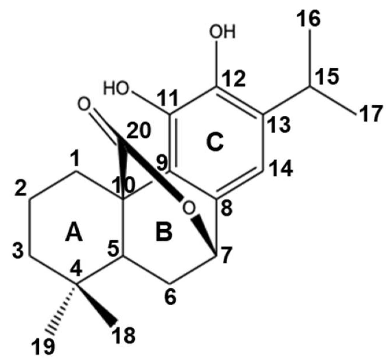
2. Carnosol: Sources, Chemistry, and Structural Characterization

Rosemary and sage are Mediterranean herbs used for culinary purposes and have been known to contain various bioactive compounds, including polyphenols such as carnosol, carnosic acid, rosmanol, and rosmarinic acid. Carnosol was first isolated from sage (Salvia carnosa) in 1942, and its chemical structure (C20H28O4) (Figure 1) was characterized by Brieskorn et al. in 1964 [42]. Carnosol is an ortho-diphenolic diterpene with an abietane carbon skeleton with hydroxyl groups at positions C-11 and C-12 and a lactone moiety across the B ring and is considered to be a byproduct of the oxidative degradation of carnosic acid [43,44]. Carnosol is soluble in various organic solvents, such as dimethyl sulfoxide (250 mg/mL), ethanol (8 mg/mL), and dimethyl formamide (35 mg/mL) [43]. In recent years, carnosol has received increasing attention for its various health-promoting properties.

3. Safety and Toxicological Studies on Carnosol

Toxicological studies are essential in the development of novel therapeutic drugs for their potential clinical use. Several animal studies have suggested that the daily oral consumption of carnosol is safe. Johnson et al. reported that carnosol at a dose of 30 mg/kg was well tolerated in mice [37]. Similarly, Sprague–Dawley rats fed with an AIN-76A diet supplemented with 1% carnosol for two weeks showed no changes in body weight [45]. Importantly, the European safety authority has recognized carnosol-enriched rosemary extracts as “Generally Recognized as Safe” (GRAS). In a study testing the toxicity of several rosemary extracts, rats were fed rosemary extracts for 90 days, and the no-observed-adverse-effect level (NOAEL) values were reported to range between 140 and 400 mg extract/kg body weight/day equivalent to 20–60 mg/kg body weight/day of carnosol and carnosic acid [46]. In a more recent study, Phipps et al. investigated the toxicity of carnosol and carnosic acid by feeding male and female mice rosemary extract for 90 days and reported no adverse side effects even at a high dose of 195 mg/kg body weight/day, which is equivalent to 64 mg/kg body weight/day of carnosol and carnosic acid [47].

4. Carnosol as a Potent ROS Generator in Cancer Cells

Reactive oxygen species (ROS), such as 1O2, O2−, H2O2, OH•, and HOCl, are byproducts of several cellular processes that can act as second messengers for some important signaling pathways that regulate various cellular processes, including gene expression in both normal and cancer cells [48,49]. Although low to moderate levels of ROS were reported to activate cancer cell proliferation, drug resistance, migration, invasion, and angiogenesis, high concentrations of ROS, on the other hand, induce severe cellular damage, resulting in cell cycle arrest and activation of programmed cell death in cancer cells [48,49]. Indeed, increasing evidence suggests that anticancer drugs that increase intracellular ROS induce cell death in various types of cancer. Thus, anticancer strategies based on the induction of ROS are increasingly considered a promising approach for the treatment of cancer.
Although carnosol was previously described as an antioxidant phytopolyphenol, it was reported to be a potent generator of ROS in MDA-MB-231 and Hs578T TNBC cells [38,50]. Indeed, carnosol induced a concentration-dependent accumulation of ROS, which was detectable as early as 1 h post-treatment and preceded cell death that occurred 24 h post-treatment [38]. In agreement with these findings, carnosol was also reported to induce ROS production in other cancer cells, such as colon (HCT116) [51] and osteosarcoma (MG-63) [52] cells. Inhibition of ROS by the ROS scavengers tiron or N-acetylcysteine (NAC) abrogated carnosol-induced cell death in MDA-MB-231 [38,53], HCT116 [51], and MG-63 [52] cells, thus strongly suggesting that the anticancer activity of carnosol might be mediated mainly through ROS production.

5. Anticancer Activities of Carnosol against TNBC

Despite the large number of studies reporting the anticancer activity of carnosol, only a few reports investigated its activity against TNBC. Interestingly, carnosol was shown to inhibit TNBC by targeting diverse pathways involved in proliferation, growth, migration, invasion, and epigenetic regulation. Table 1 summarizes the major targets of carnosol in TNBC.

5.1. Carnosol Decreases Cellular Viability and Induces G2 Arrest in TNBC

Aberrant cell cycle progression leading to uncontrolled cellular proliferation is a characteristic of cancer cells. These result from mutations in upstream signaling pathways or in genes encoding cell cycle proteins. Cyclin-dependent kinases (CDKs) are known to be aberrantly activated in many cancers [57]. Therefore, inhibitors of CDKs and cell cycle regulators are attractive targets in cancer therapy. Most of the currently used anticancer drugs, such as tamoxifen and methotrexate [58], limit tumor progression by hindering one or more cell cycle phases and their transition points, thereby attenuating cancer cell proliferation. Cell cycle arrest by these drugs may also be associated with the induction of apoptosis, a type of programmed cell death [59]. Several studies reported that the anticancer effects of carnosol are mediated via the inhibition of cancer cell proliferation either via cell cycle arrest and/or induction of cell death [37,41,60]. Our team was one of the first to explore the anticancer activity of carnosol against TNBC [38]. Carnosol was reported to significantly reduce the cellular viability, in a concentration- and time-dependent manner, of various TNBC cell lines, including MDA-MB-231 [38,53,54,55], MDA-MB-435 [54], HBL-100 [54], and Hs578T (RI, unpublished data). Carnosol at 50 and 100 µM was shown to induce cell cycle blockade at the G2 phase [46]. This G2 block seems to be mediated through the overexpression of the CDK inhibitor p21 [38]. Indeed, p21 was shown to be upregulated only at concentrations that induced cell arrest [38]. Carnosol was also reported to cause G2 arrest in other cancer cell lines, such as MG-63 (osteosarcoma) [52], CaCo-2 (colon cancer) [60], and PC (prostate cancer) [37]. Interestingly, G2 block in PC3 cells was also associated with upregulation of p21. Thus, induction of G2 arrest is a common mechanism of action of carnosol against various types of cancer.

5.2. Carnosol Induces Programmed Cell Death (PCD) I and II in TNBC Cells via a ROS-Dependent Mechanism

Deregulation of apoptosis is implicated in several pathological conditions, including cancer [61]. Usually, cancer cells resist and escape apoptosis through the overexpression of antiapoptotic proteins and the repression of tumor suppressor genes, leading to uncontrolled proliferation and consequently to cancer development, progression, and treatment resistance [62,63,64].
Therefore, a better understanding of the molecular mechanisms underlying the resistance of TNBC to apoptosis will probably provide the foundations for developing targeted molecular therapies. TNBC is one of the most aggressive cancer types and is known to evade apoptosis and develop resistance to currently used therapeutic modalities, including chemotherapy and radiotherapy. Hence, inducing apoptosis in apoptosis-resistant cancer cells remains one of the key strategies in cancer treatment [65]. Al Dhaheri et al. showed that carnosol at 50 and 100 μM led to a concentration-dependent increase in the population of Annexin V-positive cells and accumulation of cleaved caspase 3 and cleaved PARP [38] in MDA-MB-231 treated for 24 h. Carnosol was shown to activate both the intrinsic and extrinsic apoptotic pathways in MDA-MB-231 cells. Indeed, active caspase 8 and caspase 9 were both detected in carnosol-treated cells [38] (Figure 2). Carnosol modulated the level of the anti-apoptotic/pro-apoptotic regulatory proteins Bcl-2/Bax and induced mitochondrial membrane potential depolarization, which are associated with the activation of the intrinsic pathway [38]. In addition, carnosol upregulated TNF-α (RI, unpublished data), a protein involved in the activation of the extrinsic pathway. More recently, Gonzalez–Cardenete et al. showed that novel semi-synthetic carnosol analogs inhibited cellular viability and induced apoptosis in MDA-MB-231 after 72 h with an IC50 between 1.3 and 18.7 μM [66]. However, the mechanism of action of these derivatives was not investigated. Still, these analogs could serve as a foundation for the development of novel anticancer compounds targeting TNBC.
Autophagy is a biological process that maintains cellular homeostasis by trafficking damaged or unwanted cellular components to the lysosome, thereby facilitating cellular survival. In the context of cancer, cancer cells can use autophagy as a cytoprotective mechanism to prevent the accumulation of cellular damages caused by chemotherapeutics, leading to the promotion of temporal survival and the development of chemoresistance [67]. Contrary to its cytoprotective role, autophagy is also known to induce cell death stimulated by excessive cellular stress, termed type II programmed cell death (PCDII) [68]. More importantly, cell death mediated by autophagy can involve the induction of apoptosis [69,70]. Consistently, inhibition of autophagy has been reported to reduce apoptosis in some tumors [71]. In this regard, both cell death mechanisms, apoptosis, and autophagy, may interplay with each other and share common mechanisms that drive the cellular death response. Carnosol, at a non-cytotoxic concentration (25 μM), was shown to induce autophagy in two TNBC cell lines, namely MDA-MB-231 [38,53] and Hs578-T (RI, unpublished data). Autophagy was confirmed by transmission electron microscopy (TEM), accumulation of lipidized LC3II, and decreased expression of p62 (SQSMT1) [38,53]. Interestingly, carnosol-mediated autophagy was independent of Beclin-1 [38]. Indeed, siRNA knockdown of Beclin-1 did not inhibit autophagy in carnosol-treated MDA-MB-231 cells. TEM showed that carnosol induced both mitophagy, characterized by swollen and damaged mitochondria, and reticulophagy [38,53], characterized by the accumulation of large numbers of swollen endoplasmic reticulum. Carnosol-induced autophagy seems to be mediated through downregulation of the mTOR pathway, a negative regulator of autophagy. Indeed, carnosol induced a dramatic decrease not only in the level of phosphorylated mTOR but also in the total mTOR protein [53] (Figure 2).
Considering that autophagy and apoptosis were both induced by carnosol in TNBC cells, Alsamri et al. examined the relationship between these two processes [53]. They found that carnosol-induced autophagy and apoptosis occurred independently of each other in TNBC. They showed that inhibition of autophagy by 3-methyladenine (3-MA) or chloroquine (CQ) only modestly rescued MDA-MB-231 from cell death. Similarly, inhibition of apoptosis by the pan-caspase inhibitor Z-VAD-FMK only had a slight effect on cellular viability. In contrast, a combination of autophagy and apoptosis inhibitors fully rescued carnosol-treated MDA-MB-231 cells from cell death [53]. This finding clearly shows that both programmed cell death mechanisms, apoptosis (PCDI) and autophagy (PCDII), were activated independently of each other in TNBC by carnosol. This was further confirmed by the accumulation of cleaved PARP and the persistence of the loss of mitochondrial membrane potential when autophagy was inhibited. The induction of autophagy and apoptosis in TNBC by carnosol was shown to be ROS-dependent, as inhibition of ROS by either tiron [38] or NAC [53] not only abolished the cytotoxic effect of carnosol on TNBC but also significantly reduced the level of LC3II (marker of autophagy) and cleaved PARP (marker of apoptosis) and restored the level of mTOR protein.

5.3. Carnosol Induces ER Stress via Activation of the p38-MAPK Pathway through a ROS-Dependent Mechanism

The unfolded protein response (UPR) is a highly regulated cascade system that is responsible for protein folding and trafficking and maintaining other cellular functions [72]. The UPR involves three key sensors located on the endoplasmic reticulum (ER) membrane, namely (i) protein kinase R-like ER kinase (PERK), (ii) transcription factor 6 (ATF6), and (iii) inositol-requiring enzyme 1α (IRE1α) [73]. Accumulating evidence suggests that the UPR signaling cascades are activated in response to ROS accumulation or oxidative triggers [74], which induce a cellular condition called ER stress, which is characterized by an accumulation of unfolded or misfolded proteins. ER stress leads to the activation of the UPR for the sake of restoring metabolic homeostasis, thus promoting cancer cell survival [75]. However, during severe ER stress, as a consequence of elevated or persistent oxidative stress, the UPR system triggers cancer cell death through activation of PCDI and/or PCDII [76]. Hence, finding or developing new drugs that stimulate ROS production and ultimately lead to UPR activation should be considered as an approach for new effective anticancer therapeutic approaches.
Recently, Alsamri et al. showed that carnosol triggers the activation of the three UPR sensors (PERK, IRE1α, and ATF-6) in MDA-MB-231 [53] and Hs578T cells (RI, unpublished data). Indeed, carnosol induced the phosphorylation of IRE1α and EIF2 α, upregulated the protein levels of XBP-1s, cleaved ATF6, ATF4, and CHOP, and downregulated PDI and Ero-1 α, which are enzymes that promote proper protein folding in the ER [53]. These biochemical data were in agreement with TEM data that showed dilated and multilamellar ER [53]. The activation of UPR sensors by carnosol seems to depend solely on ROS accumulation (Figure 2). Indeed, NAC pretreatment was sufficient to completely abolish the activation of the three UPR sensors by carnosol and restore the normal levels of PDI and Ero-1 and, thus, the protein folding function of the ER [53].
The p38 MAPK pathway translates extracellular signals to the intracellular machinery that regulates various cellular processes, such as cell cycle, cell death, differentiation, and senescence. Depending on the kinetics of activation, downstream signaling pathways are consequently activated [77,78,79]. Several studies showed that p38 plays a dual role in the UPR cascade [80,81]. p38 can be activated as a consequence of IRE-1α and PERK oligomerization [81,82]. On the other hand, prolonged activation of p38 induces ER stress and activates the UPR response pathway by regulating the expression of UPR components such as CHOP [39], ATF6 [39], and XBP-1s [83]. Moreover, studies showed that in the presence of ER stress, p38 can promote cell death through the induction of autophagy and/or apoptosis in various types of cancer, depending on the type and duration of the stimulus. Carnosol was shown to activate p38MAPK in MDA-MB-231 cells in a ROS-dependent manner [53] (Figure 2). Time course analysis revealed that the phosphorylation of p38MAPK occurred concomitantly with ROS generation, i.e., 1 h post-carnosol treatment [53]. However, the activation of p38MAPK was shown to be dependent on ROS since NAC pretreatment was sufficient to block the activation of p38MAPK [53]. Interestingly, p38MAPK activation was the earliest event induced by carnosol; it preceded the activation of the UPR pathways, induction of autophagy, and activation of the apoptotic pathway, which occurred at 3, 6, and 24 h post-treatment, respectively. Thus, p38 seems to play a central role in the anti-TNBC effect of carnosol. Indeed, chemical inhibition of p38MAPK activation by either of the two p38 inhibitors, SB202190 or SB203580, was sufficient to partially reduce the cytotoxic effect of carnosol, as well as efficiently block the activation of the IRE-1α and ATF6 pathways and induction of autophagy [53] (Figure 2). Carnosol-mediated apoptosis, however, seems to occur independently of p38MAPK. Considering the fact that carnosol induces sustained DNA damage [38] in addition to protein misfolding [53], we hypothesize that the activation of apoptosis is a secondary response to excessive cellular damage due to prolonged exposure to carnosol.

5.4. Carnosol Inhibits p300 Acetyl Transferase

Acetylation of histones and non-histone proteins is the most abundant modification that is involved in various key cellular processes, such as cellular proliferation, cell-cycle progression, differentiation, apoptosis, dosage compensation, hormonal signaling, gene transcription, DNA damage repair, protein folding, autophagy, and metabolism [84,85,86,87,88]. The dynamic and reversible acetylation of histone and non-histone proteins is a major epigenetic mechanism that regulates gene transcription, and deregulation of this process has been implicated in a wide variety of human diseases, including cancer [89,90,91,92,93,94,95]. Thus, proteins involved in the regulation of acetylation status are considered an attractive therapeutic target. Acetylation of histones and non-histone proteins is regulated by a class of enzyme family members, the histone acetyltransferases (HATs) [96,97]. One such enzyme, p300 HAT, has been associated with tumor development and progression [98,99,100,101]. Deregulation of HAT activity is particularly linked to the formation and progression of different types of cancer, including breast cancer [102,103]. Indeed, p300 was reported to be upregulated in invasive breast cancer [104,105]. Carnosol was reported to induce a concentration-dependent decrease in the overall expression of histone H3 and H4 in MDA-MB-231 and Hs578T cells [56]. In silico docking analysis revealed that carnosol can bind to the acetyl-CoA binding pocket of the catalytic domain of p300, thus potentially blocking its activity [56]. This was confirmed by an in vitro HAT assay in which carnosol was able to completely inhibit the ability of p300 to bind to histone H3 and histone H3K56, a preferred substrate of p300. This inhibition could only be partially relieved by a large excess of acetyl-CoA (400 μM), thus suggesting competitive inhibition of p300 by carnosol [56]. Given that p300 was targeted for proteasomal degradation by carnosol in TNBC, it is hard to appreciate the extent of this specific inhibition on the anti-TNBC of carnosol. It is hence worth testing the effect of p300 inhibition in other cancer cell types where carnosol-mediated p300 degradation does not occur.

5.5. Carnosol, a Potential Targeted Protein Degradation Molecule, Targets Key Proteins Regulating Cancer Growth and Metastasis through a p38MAPK-Dependent Mechanism

Targeted protein degradation (TPD) via the proteasome via stimulation of the ubiquitin proteasome system is gaining attention as a novel and promising anticancer therapeutic approach [106]. There are at least ten TPD molecules in clinical trials against various types of cancer [107]. For example, ARV-471, now in a phase II clinical trial, was shown to specifically target the ER, a regulator of breast cancer pathogenesis [107]. KT-333 is another PTD molecule in a phase I clinical trial that was shown to specifically target STAT3 in multiple xenograft mouse models [107]. Interestingly, carnosol was reported to possess PTD activity against several key regulator proteins, including STAT3, mTORC1, p300, and PCAF. Indeed, carnosol targeted these proteins for proteasomal degradation. It is noteworthy to mention that carnosol also induced an increased level of overall protein ubiquitination in MDA-MB-231 cells. Protein polyubiquitination and degradation of STAT3, mTORC1, p300, and PCAF occurred through a ROS-dependent mechanism, as NAC pretreatment completely reversed this effect [50,53,56]. Interestingly, the targeting of these four proteins for proteasomal degradation is dependent upon the activation of p38MAPK (Figure 2). Indeed, chemical inhibition of p38MAPK by SB202190 not only reduced the level of protein polyubiquitination in MDA-MB-231 but also inhibited the degradation of STAT3, mTORC1, p300, and PCAF [53]. Hence, the mechanism through which p38MAPK targets STAT3, mTORC1, p300, and PCAF for proteasomal degradation warrants further investigation.

5.6. Carnosol Inhibits Tumor Growth and Metastasis in TNBC

Cancer metastasis is a complex process involving several steps. Invasion is a critical step in cancer metastasis that involves proteolytic degradation of the extracellular matrix. It has been reported that elevated levels of matrix metalloproteinases (MMPs) are linked to the aggressiveness and metastatic potential of breast cancer [108,109,110]. Inhibiting the expression or activity of these MMPs is considered a potential therapeutic approach against breast cancer. Alsamri et al. reported that carnosol significantly and efficiently inhibited the migration and invasion potential of TNBC cells both in vitro and in vivo [50]. It also significantly inhibited tumor growth of MDA-MB-231 in vivo, as demonstrated with the chick embryo tumor growth and metastasis assay [50]. The anti-metastatic activity of carnosol involved the inhibition of both the expression and activity of MMP-9, as demonstrated with gelatin zymography, ELISA, and RT-PCR assays [50]. Similar findings were reported when carnosol was used against the highly metastatic mouse melanoma cell line B16/F10 [111]. Thus, inhibition of the expression and activity of MMP-9 seems to be a general mechanism underlying carnosol-mediated inhibition of cancer cell invasion. Interestingly, inhibition of the invasion of TNBC cells involved inhibition of the STAT3 signaling pathway, which is known to regulate the expression of MMP-9. The inhibition of the STAT3 pathway by carnosol was mediated through the ROS-dependent targeting of STAT3 protein to proteasomal degradation in TNBC MDA-MB-231 and Hs578T cells, and also in non-TNBC MCF-7 and T47D breast cancer cells [50]. While screening for potential new STAT3 inhibitors, Yanagimichi et al. reported that carnosol induced a dramatic decrease in the level of STAT 3 protein in HepG2 hepatocellular carcinoma cells [112]. Intriguingly, in HCT116 colon cancer cells, although carnosol inhibited STAT3 phosphorylation, it did not affect the level of total cellular STAT3 [41,51]. It is also noteworthy to mention that inhibition of MMP-9 expression and, thus, the inhibition of invasion of the highly metastatic mouse melanoma B16/F10 cells occurred independently of STAT3 and was attributed to the inhibition of the ERK 1/2, AKT, p38, JNK, and NF-kB signaling pathways [111]. Hence, the cell-type-specific effect of carnosol on STAT3 warrants further investigation.

5.7. Synergetic Anticancer Effects of Carnosol in an In Vitro Combination Assay

Despite the increasing interest in the anticancer effect of carnosol, specifically with respect to cancer initiation and progression, few studies have shown the efficacy of using diterpenoids in combination with other phytochemicals or chemotherapeutic agents [113,114,115]. Vergara et al. investigated the cytotoxic effect of carnosol alone and in combination with other phytochemicals or commonly used chemotherapeutic drugs on several human cancer cell lines, including TNBC (HBL-100, MDA-231, and MDA-435) cells [54]. Vergara et al. treated TNBC cells with carnosol (50 μM) and curcumin (70 μM) for 4 h and assessed their potential synergetic effects using western blotting. Combination therapy reduced the expression of Bcl-2, cyclin D1, and survival, and increased the expression of p27 more efficiently than mono-treatment. These findings highlight the promising synergetic potential of carnosol and curcumin against TNBC by decreasing viability and inducing apoptosis and cell cycle blockade [54]. These findings warrant further investigation of the effect of the combination of carnosol with currently used anticancer chemotherapeutic drugs against TNBC.

6. Conclusions

Carnosol, a phytochemical abundant in rosemary and sage extracts, has long been recognized for its many biological and medicinal properties, including antioxidant, antimicrobial, anti-inflammatory, neuroprotective, and anticancer effects. The anticancer effect of carnosol against various types of cancer has gained much interest in the recent past. Interestingly, carnosol has proven its effectiveness against TNBC through the modulation of several biochemical pathways (Figure 2). Indeed, carnosol exerts its effect against TNBC by targeting and modulating several cancer hallmarks. We should take advantage of the fact that carnosol can be isolated from widely abundant perennial plants, and therefore a constant supply of this agent can be secured for further studies. Further, increasing evidence from recent preclinical studies strongly suggests that carnosol is a potential promising chemopreventive and chemotherapeutic agent against TNBC. Therefore, more focus should be given to in vivo studies with carnosol, which are still dramatically lacking.

Author Contributions

Conceptualization: H.A., Y.A.D. and R.I.; writing original draft: H.A. and R.I.; review and editing: H.A., Y.A.D. and R.I. All authors have read and agreed to the published version of the manuscript.

Funding

This research received no external funding.

Data Availability Statement

Not applicable.

Conflicts of Interest

The authors declare no conflict of interest.

References

  1. Ritchie, H.; Roser, M. Causes of Death. Our World Data 2018. Published online at OurWorldInData.org. Available online: https://ourworldindata.org/causes-of-death (accessed on 6 June 2019).
  2. Song, M. Cancer Overtakes Vascular Disease as Leading Cause of Excess Death Associated with Diabetes. Lancet Diabetes Endocrinol. 2021, 9, 131–133. [Google Scholar] [CrossRef] [PubMed]
  3. Weir, H.K.; Anderson, R.N.; Coleman King, S.M.; Soman, A.; Thompson, T.D.; Hong, Y.; Moller, B.; Leadbetter, S. Heart Disease and Cancer Deaths—Trends and Projections in the United States, 1969–2020. Prev. Chronic Dis. 2016, 13, E157. [Google Scholar] [CrossRef] [Green Version]
  4. Harding, M.C.; Sloan, C.D.; Merrill, R.M.; Harding, T.M.; Thacker, B.J.; Thacker, E.L. Transitions From Heart Disease to Cancer as the Leading Cause of Death in US States, 1999–2016. Prev. Chronic Dis. 2018, 15, E158. [Google Scholar] [CrossRef] [PubMed] [Green Version]
  5. Definition of Cancer—NCI Dictionary of Cancer Terms—National Cancer Institute. Available online: https://www.cancer.gov/publications/dictionaries/cancer-terms/def/cancer (accessed on 8 April 2021).
  6. Cancer Today—Population Fact Sheets. International Agency for Research on Cancer. Available online: http://gco.iarc.fr/today/home (accessed on 8 April 2021).
  7. Sung, H.; Ferlay, J.; Siegel, R.L.; Laversanne, M.; Soerjomataram, I.; Jemal, A.; Bray, F. Global Cancer Statistics 2020: GLOBOCAN Estimates of Incidence and Mortality Worldwide for 36 Cancers in 185 Countries. CA Cancer J. Clin. 2021, 71, 209–249. [Google Scholar] [CrossRef] [PubMed]
  8. Hutchinson, L. Breast Cancer: Challenges, Controversies, Breakthroughs. Nat. Rev. Clin. Oncol. 2010, 7, 669–670. [Google Scholar] [CrossRef] [Green Version]
  9. Greenwalt, I.; Zaza, N.; Das, S.; Li, B.D. Precision Medicine and Targeted Therapies in Breast Cancer. Surg. Oncol. Clin. N. Am. 2020, 29, 51–62. [Google Scholar] [CrossRef]
  10. Harris, E.E.R. Precision Medicine for Breast Cancer: The Paths to Truly Individualized Diagnosis and Treatment. Int. J. Breast Cancer 2018, 2018, 4809183. [Google Scholar] [CrossRef] [Green Version]
  11. Maisonneuve, P.; Disalvatore, D.; Rotmensz, N.; Curigliano, G.; Colleoni, M.; Dellapasqua, S.; Pruneri, G.; Mastropasqua, M.G.; Luini, A.; Bassi, F.; et al. Proposed New Clinicopathological Surrogate Definitions of Luminal A and Luminal B (HER2-Negative) Intrinsic Breast Cancer Subtypes. Breast Cancer Res. 2014, 16, R65. [Google Scholar] [CrossRef] [Green Version]
  12. Sørlie, T.; Perou, C.M.; Tibshirani, R.; Aas, T.; Geisler, S.; Johnsen, H.; Hastie, T.; Eisen, M.B.; van de Rijn, M.; Jeffrey, S.S.; et al. Gene Expression Patterns of Breast Carcinomas Distinguish Tumor Subclasses with Clinical Implications. Proc. Natl. Acad. Sci. USA 2001, 98, 10869–10874. [Google Scholar] [CrossRef] [Green Version]
  13. Chodosh, L.A. Breast Cancer: Current State and Future Promise. Breast Cancer Res. 2011, 13, 113. [Google Scholar] [CrossRef]
  14. Morris, G.J.; Naidu, S.; Topham, A.K.; Guiles, F.; Xu, Y.; McCue, P.; Schwartz, G.F.; Park, P.K.; Rosenberg, A.L.; Brill, K.; et al. Differences in Breast Carcinoma Characteristics in Newly Diagnosed African-American and Caucasian Patients: A Single-Institution Compilation Compared with the National Cancer Institute’s Surveillance, Epidemiology, and End Results Database. Cancer 2007, 110, 876–884. [Google Scholar] [CrossRef] [PubMed]
  15. Dawson, S.J.; Provenzano, E.; Caldas, C. Triple Negative Breast Cancers: Clinical and Prognostic Implications. Eur. J. Cancer 2009, 45 (Suppl. S1), 27–40. [Google Scholar] [CrossRef] [PubMed]
  16. Irvin, W.J.; Carey, L.A. What Is Triple-Negative Breast Cancer? Eur. J. Cancer 2008, 44, 2799–2805. [Google Scholar] [CrossRef] [PubMed]
  17. Collett, K.; Stefansson, I.M.; Eide, J.; Braaten, A.; Wang, H.; Eide, G.E.; Thoresen, S.Ø.; Foulkes, W.D.; Akslen, L.A. A Basal Epithelial Phenotype Is More Frequent in Interval Breast Cancers Compared with Screen Detected Tumors. Cancer Epidemiol. Biomark. Prev. 2005, 14, 1108–1112. [Google Scholar] [CrossRef] [PubMed] [Green Version]
  18. Fulford, L.G.; Reis-Filho, J.S.; Ryder, K.; Jones, C.; Gillett, C.E.; Hanby, A.; Easton, D.; Lakhani, S.R. Basal-like Grade III Invasive Ductal Carcinoma of the Breast: Patterns of Metastasis and Long-Term Survival. Breast Cancer Res. 2007, 9, R4. [Google Scholar] [CrossRef] [Green Version]
  19. Dent, R.; Trudeau, M.; Pritchard, K.I.; Hanna, W.M.; Kahn, H.K.; Sawka, C.A.; Lickley, L.A.; Rawlinson, E.; Sun, P.; Narod, S.A. Triple-Negative Breast Cancer: Clinical Features and Patterns of Recurrence. Clin. Cancer Res. 2007, 13 Pt 1, 4429–4434. [Google Scholar] [CrossRef] [Green Version]
  20. Mahtani, R.; Kittaneh, M.; Kalinsky, K.; Mamounas, E.; Badve, S.; Vogel, C.; Lower, E.; Schwartzberg, L.; Pegram, M.; Breast Cancer Therapy Expert Group (BCTEG). Advances in Therapeutic Approaches for Triple-Negative Breast Cancer. Clin. Breast Cancer 2021, 21, 383–390. [Google Scholar] [CrossRef]
  21. Lyons, T.G. Targeted Therapies for Triple-Negative Breast Cancer. Curr. Treat. Options Oncol. 2019, 20, 82. [Google Scholar] [CrossRef]
  22. Jhan, J.-R.; Andrechek, E.R. Triple-Negative Breast Cancer and the Potential for Targeted Therapy. Pharmacogenomics 2017, 18, 1595–1609. [Google Scholar] [CrossRef] [Green Version]
  23. Kim, H.; Samuel, S.L.; Zhai, G.; Rana, S.; Taylor, M.; Umphrey, H.R.; Oelschlager, D.K.; Buchsbaum, D.J.; Zinn, K.R. Combination Therapy with Anti-DR5 Antibody and Tamoxifen for Triple Negative Breast Cancer. Cancer Biol. Ther. 2014, 15, 1053–1060. [Google Scholar] [CrossRef] [Green Version]
  24. Loi, S.; Pommey, S.; Haibe-Kains, B.; Beavis, P.A.; Darcy, P.K.; Smyth, M.J.; Stagg, J. CD73 Promotes Anthracycline Resistance and Poor Prognosis in Triple Negative Breast Cancer. Proc. Natl. Acad. Sci. USA 2013, 110, 11091–11096. [Google Scholar] [CrossRef] [PubMed] [Green Version]
  25. Ijaz, S.; Akhtar, N.; Khan, M.S.; Hameed, A.; Irfan, M.; Arshad, M.A.; Ali, S.; Asrar, M. Plant Derived Anticancer Agents: A Green Approach towards Skin Cancers. Biomed Pharm. 2018, 103, 1643–1651. [Google Scholar] [CrossRef] [PubMed]
  26. Kapinova, A.; Stefanicka, P.; Kubatka, P.; Zubor, P.; Uramova, S.; Kello, M.; Mojzis, J.; Blahutova, D.; Qaradakhi, T.; Zulli, A.; et al. Are Plant-Based Functional Foods Better Choice against Cancer than Single Phytochemicals? A Critical Review of Current Breast Cancer Research. Biomed Pharm. 2017, 96, 1465–1477. [Google Scholar] [CrossRef] [PubMed]
  27. Ham, S.L.; Nasrollahi, S.; Shah, K.N.; Soltisz, A.; Paruchuri, S.; Yun, Y.H.; Luker, G.D.; Bishayee, A.; Tavana, H. Phytochemicals Potently Inhibit Migration of Metastatic Breast Cancer Cells. Integr. Biol. 2015, 7, 792–800. [Google Scholar] [CrossRef] [PubMed] [Green Version]
  28. Fakhri, S.; Moradi, S.Z.; Yarmohammadi, A.; Narimani, F.; Wallace, C.E.; Bishayee, A. Modulation of TLR/NF-ΚB/NLRP Signaling by Bioactive Phytocompounds: A Promising Strategy to Augment Cancer Chemotherapy and Immunotherapy. Front. Oncol. 2022, 12, 834072. [Google Scholar] [CrossRef]
  29. Zhang, Y.; Ma, X.; Li, H.; Zhuang, J.; Feng, F.; Liu, L.; Liu, C.; Sun, C. Identifying the Effect of Ursolic Acid Against Triple-Negative Breast Cancer: Coupling Network Pharmacology With Experiments Verification. Front. Pharm. 2021, 12, 685773. [Google Scholar] [CrossRef]
  30. Wei, C.; Khan, M.A.; Du, J.; Cheng, J.; Tania, M.; Leung, E.L.-H.; Fu, J. Cordycepin Inhibits Triple-Negative Breast Cancer Cell Migration and Invasion by Regulating EMT-TFs SLUG, TWIST1, SNAIL1, and ZEB1. Front. Oncol. 2022, 12, 898583. [Google Scholar] [CrossRef]
  31. Wang, Y.; Zhong, Z.; Ma, M.; Zhao, Y.; Zhang, C.; Qian, Z.; Wang, B. The Role Played by Ailanthone in Inhibiting Bone Metastasis of Breast Cancer by Regulating Tumor-Bone Microenvironment through the RANKL-Dependent Pathway. Front. Pharm. 2022, 13, 1081978. [Google Scholar] [CrossRef]
  32. Dandawate, P.; Padhye, S.; Ahmad, A.; Sarkar, F.H. Novel Strategies Targeting Cancer Stem Cells through Phytochemicals and Their Analogs. Drug Deliv. Transl. Res. 2013, 3, 165–182. [Google Scholar] [CrossRef] [Green Version]
  33. Subbaramaiah, K.; Cole, P.A.; Dannenberg, A.J. Retinoids and Carnosol Suppress Cyclooxygenase-2 Transcription by CREB-Binding Protein/P300-Dependent and -Independent Mechanisms. Cancer Res. 2002, 62, 2522–2530. [Google Scholar]
  34. Satoh, T.; Izumi, M.; Inukai, Y.; Tsutsumi, Y.; Nakayama, N.; Kosaka, K.; Shimojo, Y.; Kitajima, C.; Itoh, K.; Yokoi, T.; et al. Carnosic Acid Protects Neuronal HT22 Cells through Activation of the Antioxidant-Responsive Element in Free Carboxylic Acid- and Catechol Hydroxyl Moieties-Dependent Manners. Neurosci. Lett. 2008, 434, 260–265. [Google Scholar] [CrossRef] [PubMed]
  35. Kim, S.-J.; Kim, J.-S.; Cho, H.-S.; Lee, H.J.; Kim, S.Y.; Kim, S.; Lee, S.-Y.; Chun, H.S. Carnosol, a Component of Rosemary (Rosmarinus Officinalis L.) Protects Nigral Dopaminergic Neuronal Cells. Neuroreport 2006, 17, 1729–1733. [Google Scholar] [CrossRef] [PubMed]
  36. Weckesser, S.; Engel, K.; Simon-Haarhaus, B.; Wittmer, A.; Pelz, K.; Schempp, C.M. Screening of Plant Extracts for Antimicrobial Activity against Bacteria and Yeasts with Dermatological Relevance. Phytomedicine 2007, 14, 508–516. [Google Scholar] [CrossRef] [PubMed]
  37. Johnson, J.J.; Syed, D.N.; Suh, Y.; Heren, C.R.; Saleem, M.; Siddiqui, I.A.; Mukhtar, H. Disruption of Androgen and Estrogen Receptor Activity in Prostate Cancer by a Novel Dietary Diterpene Carnosol: Implications for Chemoprevention. Cancer Prev. Res. 2010, 3, 1112–1123. [Google Scholar] [CrossRef] [PubMed] [Green Version]
  38. Al Dhaheri, Y.; Attoub, S.; Ramadan, G.; Arafat, K.; Bajbouj, K.; Karuvantevida, N.; AbuQamar, S.; Eid, A.; Iratni, R. Carnosol Induces ROS-Mediated Beclin1-Independent Autophagy and Apoptosis in Triple Negative Breast Cancer. PLoS ONE 2014, 9, e109630. [Google Scholar] [CrossRef] [Green Version]
  39. Wang, X.Z.; Ron, D. Stress-Induced Phosphorylation and Activation of the Transcription Factor CHOP (GADD153) by P38 MAP Kinase. Science 1996, 272, 1347–1349. [Google Scholar] [CrossRef] [Green Version]
  40. Yan, M.; Vemu, B.; Veenstra, J.; Petiwala, S.M.; Johnson, J.J. Carnosol, a Dietary Diterpene from Rosemary (Rosmarinus officinalis) Activates Nrf2 Leading to Sestrin 2 Induction in Colon Cells. Integr. Mol. Med. 2018, 5, 1–7. [Google Scholar] [CrossRef] [Green Version]
  41. O’Neill, E.J.; Hartogh, D.J.D.; Azizi, K.; Tsiani, E. Anticancer Properties of Carnosol: A Summary of in Vitro and In Vivo Evidence. Antioxidants 2020, 9, 961. [Google Scholar] [CrossRef]
  42. Brieskorn, C.H.; Fuchs, A.; Bredenberg, J.B.; McChesney, J.D.; Wenkert, E. The Structure of Carnosol. J. Org. Chem. 1964, 29, 2293–2298. [Google Scholar] [CrossRef]
  43. Gajhede, M.; Anthoni, U.; Per Nielsen, H.; Pedersen, E.J.; Christophersen, C. Carnosol. Crystal Structure, Absolute Configuration, and Spectroscopic Properties of a Diterpene. J. Crystallogr. Spectrosc. Res. 1990, 20, 165–171. [Google Scholar] [CrossRef]
  44. Birtić, S.; Dussort, P.; Pierre, F.-X.; Bily, A.C.; Roller, M. Carnosic Acid. Phytochemistry 2015, 115, 9–19. [Google Scholar] [CrossRef] [PubMed] [Green Version]
  45. Singletary, K.; MacDonald, C.; Wallig, M. Inhibition by Rosemary and Carnosol of 7,12-Dimethylbenz[a]Anthracene (DMBA)-Induced Rat Mammary Tumorigenesis and in Vivo DMBA-DNA Adduct Formation. Cancer Lett. 1996, 104, 43–48. [Google Scholar] [CrossRef] [PubMed]
  46. Phipps, K.R.; Lozon, D.; Baldwin, N. Genotoxicity and Subchronic Toxicity Studies of Supercritical Carbon Dioxide and Acetone Extracts of Rosemary. Regul. Toxicol. Pharm. 2021, 119, 104826. [Google Scholar] [CrossRef] [PubMed]
  47. Use of Rosemary Extracts as a Food Additive—Scientific Opinion of the Panel on Food Additives, Flavourings, Processing Aids and Materials in Contact with Food|EFSA. Available online: https://www.efsa.europa.eu/en/efsajournal/pub/721 (accessed on 17 April 2023).
  48. Nakamura, H.; Takada, K. Reactive Oxygen Species in Cancer: Current Findings and Future Directions. Cancer Sci. 2021, 112, 3945–3952. [Google Scholar] [CrossRef] [PubMed]
  49. Perillo, B.; Di Donato, M.; Pezone, A.; Di Zazzo, E.; Giovannelli, P.; Galasso, G.; Castoria, G.; Migliaccio, A. ROS in Cancer Therapy: The Bright Side of the Moon. Exp. Mol. Med. 2020, 52, 192–203. [Google Scholar] [CrossRef] [PubMed]
  50. Alsamri, H.; El Hasasna, H.; Al Dhaheri, Y.; Eid, A.H.; Attoub, S.; Iratni, R. Carnosol, a Natural Polyphenol, Inhibits Migration, Metastasis, and Tumor Growth of Breast Cancer via a ROS-Dependent Proteasome Degradation of STAT3. Front. Oncol. 2019, 9, 743. [Google Scholar] [CrossRef] [PubMed] [Green Version]
  51. Park, K.-W.; Kundu, J.; Chae, I.-G.; Kim, D.-H.; Yu, M.-H.; Kundu, J.K.; Chun, K.-S. Carnosol Induces Apoptosis through Generation of ROS and Inactivation of STAT3 Signaling in Human Colon Cancer HCT116 Cells. Int. J. Oncol. 2014, 44, 1309–1315. [Google Scholar] [CrossRef] [Green Version]
  52. Lo, Y.-C.; Lin, Y.-C.; Huang, Y.-F.; Hsieh, C.-P.; Wu, C.-C.; Chang, I.-L.; Chen, C.-L.; Cheng, C.-H.; Chen, H.-Y. Carnosol-Induced ROS Inhibits Cell Viability of Human Osteosarcoma by Apoptosis and Autophagy. Am. J. Chin. Med. 2017, 45, 1761–1772. [Google Scholar] [CrossRef]
  53. Alsamri, H.; Alneyadi, A.; Muhammad, K.; Ayoub, M.A.; Eid, A.; Iratni, R. Carnosol Induces P38-Mediated ER Stress Response and Autophagy in Human Breast Cancer Cells. Front. Oncol. 2022, 12, 911615. [Google Scholar] [CrossRef]
  54. Vergara, D.; Simeone, P.; Bettini, S.; Tinelli, A.; Valli, L.; Storelli, C.; Leo, S.; Santino, A.; Maffia, M. Antitumor Activity of the Dietary Diterpene Carnosol against a Panel of Human Cancer Cell Lines. Food Funct. 2014, 5, 1261–1269. [Google Scholar] [CrossRef]
  55. Ling, T.; Tran, M.; González, M.A.; Gautam, L.N.; Connelly, M.; Wood, R.K.; Fatima, I.; Miranda-Carboni, G.; Rivas, F. (+)-Dehydroabietylamine Derivatives Target Triple-Negative Breast Cancer. Eur. J. Med. Chem. 2015, 102, 9–13. [Google Scholar] [CrossRef] [PubMed]
  56. Alsamri, H.; Hasasna, H.E.; Baby, B.; Alneyadi, A.; Dhaheri, Y.A.; Ayoub, M.A.; Eid, A.H.; Vijayan, R.; Iratni, R. Carnosol Is a Novel Inhibitor of P300 Acetyltransferase in Breast Cancer. Front. Oncol. 2021, 11, 664403. [Google Scholar] [CrossRef] [PubMed]
  57. Otto, T.; Sicinski, P. Cell Cycle Proteins as Promising Targets in Cancer Therapy. Nat. Rev. Cancer 2017, 17, 93–115. [Google Scholar] [CrossRef] [PubMed] [Green Version]
  58. Dembic, Z. Antitumor Drugs and Their Targets. Molecules 2020, 25, 5776. [Google Scholar] [CrossRef] [PubMed]
  59. Kashyap, D.; Mittal, S.; Sak, K.; Singhal, P.; Tuli, H.S. Molecular Mechanisms of Action of Quercetin in Cancer: Recent Advances. Tumor Biol. 2016, 37, 12927–12939. [Google Scholar] [CrossRef] [PubMed]
  60. Visanji, J.M.; Thompson, D.G.; Padfield, P.J. Induction of G2/M Phase Cell Cycle Arrest by Carnosol and Carnosic Acid Is Associated with Alteration of Cyclin A and Cyclin B1 Levels. Cancer Lett. 2006, 237, 130–136. [Google Scholar] [CrossRef]
  61. Hanahan, D.; Weinberg, R.A. Hallmarks of Cancer: The next Generation. Cell 2011, 144, 646–674. [Google Scholar] [CrossRef] [Green Version]
  62. Mohammad, R.M.; Muqbil, I.; Lowe, L.; Yedjou, C.; Hsu, H.-Y.; Lin, L.-T.; Siegelin, M.D.; Fimognari, C.; Kumar, N.B.; Dou, Q.P.; et al. Broad Targeting of Resistance to Apoptosis in Cancer. Semin Cancer Biol. 2015, 35 (Suppl.), S78–S103. [Google Scholar] [CrossRef]
  63. Plati, J.; Bucur, O.; Khosravi-Far, R. Dysregulation of Apoptotic Signaling in Cancer: Molecular Mechanisms and Therapeutic Opportunities. J. Cell. Biochem. 2008, 104, 1124–1149. [Google Scholar] [CrossRef] [Green Version]
  64. Carafa, V.; Altucci, L. Deregulation of Cell Death in Cancer: Recent Highlights. Cancers 2020, 12, 3517. [Google Scholar] [CrossRef]
  65. Tamm, I.; Schriever, F.; Dörken, B. Apoptosis: Implications of Basic Research for Clinical Oncology. Lancet Oncol. 2001, 2, 33–42. [Google Scholar] [CrossRef] [PubMed]
  66. González-Cardenete, M.A.; González-Zapata, N.; Boyd, L.; Rivas, F. Discovery of Novel Bioactive Tanshinones and Carnosol Analogues against Breast Cancer. Cancers 2023, 15, 1318. [Google Scholar] [CrossRef] [PubMed]
  67. Mathew, R.; Karantza-Wadsworth, V.; White, E. Role of Autophagy in Cancer. Nat. Rev. Cancer 2007, 7, 961–967. [Google Scholar] [CrossRef] [PubMed]
  68. Wang, S.; Yu, Q.; Zhang, R.; Liu, B. Core Signaling Pathways of Survival/Death in Autophagy-Related Cancer Networks. Int. J. Biochem. Cell Biol. 2011, 43, 1263–1266. [Google Scholar] [CrossRef]
  69. Li, X.; Wu, W.K.K.; Sun, B.; Cui, M.; Liu, S.; Gao, J.; Lou, H. Dihydroptychantol A, a Macrocyclic Bisbibenzyl Derivative, Induces Autophagy and Following Apoptosis Associated with P53 Pathway in Human Osteosarcoma U2OS Cells. Toxicol. Appl. Pharm. 2011, 251, 146–154. [Google Scholar] [CrossRef]
  70. Maiuri, M.C.; Zalckvar, E.; Kimchi, A.; Kroemer, G. Self-Eating and Self-Killing: Crosstalk between Autophagy and Apoptosis. Nat. Rev. Mol. Cell Biol. 2007, 8, 741–752. [Google Scholar] [CrossRef]
  71. Wang, W.; Fan, H.; Zhou, Y.; Duan, P.; Zhao, G.; Wu, G. Knockdown of Autophagy-Related Gene BECLIN1 Promotes Cell Growth and Inhibits Apoptosis in the A549 Human Lung Cancer Cell Line. Mol. Med. Rep. 2013, 7, 1501–1505. [Google Scholar] [CrossRef] [Green Version]
  72. Cao, S.S.; Kaufman, R.J. Endoplasmic Reticulum Stress and Oxidative Stress in Cell Fate Decision and Human Disease. Antioxid. Redox Signal. 2014, 21, 396–413. [Google Scholar] [CrossRef] [Green Version]
  73. Rutkowski, D.T.; Kaufman, R.J. A Trip to the ER: Coping with Stress. Trends Cell Biol. 2004, 14, 20–28. [Google Scholar] [CrossRef]
  74. Zeeshan, H.M.A.; Lee, G.H.; Kim, H.-R.; Chae, H.-J. Endoplasmic Reticulum Stress and Associated ROS. Int. J. Mol. Sci. 2016, 17, 327. [Google Scholar] [CrossRef] [Green Version]
  75. Madden, E.; Logue, S.E.; Healy, S.J.; Manie, S.; Samali, A. The Role of the Unfolded Protein Response in Cancer Progression: From Oncogenesis to Chemoresistance. Biol. Cell 2019, 111, 1–17. [Google Scholar] [CrossRef] [PubMed] [Green Version]
  76. Limonta, P.; Moretti, R.M.; Marzagalli, M.; Fontana, F.; Raimondi, M.; Montagnani Marelli, M. Role of Endoplasmic Reticulum Stress in the Anticancer Activity of Natural Compounds. Int. J. Mol. Sci. 2019, 20, 961. [Google Scholar] [CrossRef] [Green Version]
  77. Coulthard, L.R.; White, D.E.; Jones, D.L.; McDermott, M.F.; Burchill, S.A. P38(MAPK): Stress Responses from Molecular Mechanisms to Therapeutics. Trends Mol. Med. 2009, 15, 369–379. [Google Scholar] [CrossRef] [PubMed] [Green Version]
  78. Lee, J.; Sun, C.; Zhou, Y.; Lee, J.; Gokalp, D.; Herrema, H.; Park, S.W.; Davis, R.J.; Ozcan, U. P38 MAPK-Mediated Regulation of Xbp1s Is Crucial for Glucose Homeostasis. Nat. Med. 2011, 17, 1251–1260. [Google Scholar] [CrossRef] [PubMed] [Green Version]
  79. Darling, N.J.; Cook, S.J. The Role of MAPK Signalling Pathways in the Response to Endoplasmic Reticulum Stress. Biochim. Biophys. Acta 2014, 1843, 2150–2163. [Google Scholar] [CrossRef] [Green Version]
  80. Wagner, E.F.; Nebreda, A.R. Signal Integration by JNK and P38 MAPK Pathways in Cancer Development. Nat. Rev. Cancer 2009, 9, 537–549. [Google Scholar] [CrossRef]
  81. Koeberle, A.; Pergola, C.; Shindou, H.; Koeberle, S.C.; Shimizu, T.; Laufer, S.A.; Werz, O. Role of P38 Mitogen-Activated Protein Kinase in Linking Stearoyl-CoA Desaturase-1 Activity with Endoplasmic Reticulum Homeostasis. FASEB J. 2015, 29, 2439–2449. [Google Scholar] [CrossRef]
  82. Kim, I.; Xu, W.; Reed, J.C. Cell Death and Endoplasmic Reticulum Stress: Disease Relevance and Therapeutic Opportunities. Nat. Rev. Drug Discov. 2008, 7, 1013–1030. [Google Scholar] [CrossRef]
  83. Qu, M.; Liu, Y.; Xu, K.; Wang, D. Activation of P38 MAPK Signaling-Mediated Endoplasmic Reticulum Unfolded Protein Response by Nanopolystyrene Particles. Adv. Biosyst. 2019, 3, e1800325. [Google Scholar] [CrossRef]
  84. Santos-Rosa, H.; Caldas, C. Chromatin Modifier Enzymes, the Histone Code and Cancer. Eur. J. Cancer 2005, 41, 2381–2402. [Google Scholar] [CrossRef]
  85. Glass, C.K.; Rosenfeld, M.G. The Coregulator Exchange in Transcriptional Functions of Nuclear Receptors. Genes Dev. 2000, 14, 121–141. [Google Scholar] [CrossRef] [PubMed]
  86. Tang, Y.; Luo, J.; Zhang, W.; Gu, W. Tip60-Dependent Acetylation of P53 Modulates the Decision between Cell-Cycle Arrest and Apoptosis. Mol. Cell 2006, 24, 827–839. [Google Scholar] [CrossRef] [PubMed]
  87. Marmorstein, R.; Zhou, M.-M. Writers and Readers of Histone Acetylation: Structure, Mechanism, and Inhibition. Cold Spring Harb. Perspect. Biol. 2014, 6, a018762. [Google Scholar] [CrossRef] [PubMed] [Green Version]
  88. Narita, T.; Weinert, B.T.; Choudhary, C. Functions and Mechanisms of Non-Histone Protein Acetylation. Nat. Rev. Mol. Cell Biol. 2019, 20, 156–174. [Google Scholar] [CrossRef] [PubMed]
  89. Mullighan, C.G.; Zhang, J.; Kasper, L.H.; Lerach, S.; Payne-Turner, D.; Phillips, L.A.; Heatley, S.L.; Holmfeldt, L.; Collins-Underwood, J.R.; Ma, J.; et al. CREBBP Mutations in Relapsed Acute Lymphoblastic Leukaemia. Nature 2011, 471, 235–239. [Google Scholar] [CrossRef] [Green Version]
  90. Pasqualucci, L.; Dominguez-Sola, D.; Chiarenza, A.; Fabbri, G.; Grunn, A.; Trifonov, V.; Kasper, L.H.; Lerach, S.; Tang, H.; Ma, J.; et al. Inactivating Mutations of Acetyltransferase Genes in B-Cell Lymphoma. Nature 2011, 471, 189–195. [Google Scholar] [CrossRef] [Green Version]
  91. Zhao, D.; Zou, S.-W.; Liu, Y.; Zhou, X.; Mo, Y.; Wang, P.; Xu, Y.-H.; Dong, B.; Xiong, Y.; Lei, Q.-Y.; et al. Lysine-5 Acetylation Negatively Regulates Lactate Dehydrogenase A and Is Decreased in Pancreatic Cancer. Cancer Cell 2013, 23, 464–476. [Google Scholar] [CrossRef] [Green Version]
  92. Debes, J.D.; Sebo, T.J.; Lohse, C.M.; Murphy, L.M.; Haugen, D.A.L.; Tindall, D.J. P300 in Prostate Cancer Proliferation and Progression. Cancer Res. 2003, 63, 7638–7640. [Google Scholar]
  93. Shi, J.; Wang, Y.; Zeng, L.; Wu, Y.; Deng, J.; Zhang, Q.; Lin, Y.; Li, J.; Kang, T.; Tao, M.; et al. Disrupting the Interaction of BRD4 with Diacetylated Twist Suppresses Tumorigenesis in Basal-like Breast Cancer. Cancer Cell 2014, 25, 210–225. [Google Scholar] [CrossRef] [Green Version]
  94. Houtkooper, R.H.; Pirinen, E.; Auwerx, J. Sirtuins as Regulators of Metabolism and Healthspan. Nat. Rev. Mol. Cell Biol. 2012, 13, 225–238. [Google Scholar] [CrossRef] [Green Version]
  95. Falkenberg, K.J.; Johnstone, R.W. Histone Deacetylases and Their Inhibitors in Cancer, Neurological Diseases and Immune Disorders. Nat. Rev. Drug Discov. 2014, 13, 673–691. [Google Scholar] [CrossRef]
  96. Dwarakanath, B.S.; Verma, A.; Bhatt, A.N.; Parmar, V.S.; Raj, H.G. Targeting Protein Acetylation for Improving Cancer Therapy. Indian J. Med. Res. 2008, 128, 13–21. [Google Scholar] [PubMed]
  97. Gujral, P.; Mahajan, V.; Lissaman, A.C.; Ponnampalam, A.P. Histone Acetylation and the Role of Histone Deacetylases in Normal Cyclic Endometrium. Reprod. Biol. Endocrinol. 2020, 18, 84. [Google Scholar] [CrossRef] [PubMed]
  98. Dekker, F.J.; Haisma, H.J. Histone Acetyl Transferases as Emerging Drug Targets. Drug Discov. Today 2009, 14, 942–948. [Google Scholar] [CrossRef] [PubMed]
  99. Iyer, N.G.; Xian, J.; Chin, S.-F.; Bannister, A.J.; Daigo, Y.; Aparicio, S.; Kouzarides, T.; Caldas, C. P300 Is Required for Orderly G1/S Transition in Human Cancer Cells. Oncogene 2007, 26, 21–29. [Google Scholar] [CrossRef] [PubMed] [Green Version]
  100. Zhong, J.; Ding, L.; Bohrer, L.R.; Pan, Y.; Liu, P.; Zhang, J.; Sebo, T.J.; Karnes, R.J.; Tindall, D.J.; van Deursen, J.; et al. P300 Acetyltransferase Regulates Androgen Receptor Degradation and PTEN-Deficient Prostate Tumorigenesis. Cancer Res. 2014, 74, 1870–1880. [Google Scholar] [CrossRef] [PubMed] [Green Version]
  101. Wang, L.; Gural, A.; Sun, X.-J.; Zhao, X.; Perna, F.; Huang, G.; Hatlen, M.A.; Vu, L.; Liu, F.; Xu, H.; et al. The Leukemogenicity of AML1-ETO Is Dependent on Site-Specific Lysine Acetylation. Science 2011, 333, 765–769. [Google Scholar] [CrossRef] [Green Version]
  102. Gojis, O.; Rudraraju, B.; Gudi, M.; Hogben, K.; Sousha, S.; Coombes, R.C.; Cleator, S.; Palmieri, C. The Role of SRC-3 in Human Breast Cancer. Nat. Rev. Clin. Oncol. 2010, 7, 83–89. [Google Scholar] [CrossRef]
  103. Nandy, D.; Rajam, S.M.; Dutta, D. A Three Layered Histone Epigenetics in Breast Cancer Metastasis. Cell Biosci. 2020, 10, 52. [Google Scholar] [CrossRef] [Green Version]
  104. Xiao, X.-S.; Cai, M.-Y.; Chen, J.-W.; Guan, X.-Y.; Kung, H.-F.; Zeng, Y.-X.; Xie, D. High Expression of P300 in Human Breast Cancer Correlates with Tumor Recurrence and Predicts Adverse Prognosis. Chin. J. Cancer Res. 2011, 23, 201–207. [Google Scholar] [CrossRef] [Green Version]
  105. Yokomizo, C.; Yamaguchi, K.; Itoh, Y.; Nishimura, T.; Umemura, A.; Minami, M.; Yasui, K.; Mitsuyoshi, H.; Fujii, H.; Tochiki, N.; et al. High Expression of P300 in HCC Predicts Shortened Overall Survival in Association with Enhanced Epithelial Mesenchymal Transition of HCC Cells. Cancer Lett. 2011, 310, 140–147. [Google Scholar] [CrossRef] [PubMed]
  106. Zhao, L.; Zhao, J.; Zhong, K.; Tong, A.; Jia, D. Targeted Protein Degradation: Mechanisms, Strategies and Application. Signal Transduct. Target. Ther. 2022, 7, 113. [Google Scholar] [CrossRef] [PubMed]
  107. Mullard, A. Targeted Protein Degraders Crowd into the Clinic. Nat. Rev. Drug Discov. 2021, 20, 247–250. [Google Scholar] [CrossRef]
  108. Li, H.; Qiu, Z.; Li, F.; Wang, C. The Relationship between MMP-2 and MMP-9 Expression Levels with Breast Cancer Incidence and Prognosis. Oncol. Lett. 2017, 14, 5865–5870. [Google Scholar] [CrossRef] [Green Version]
  109. Li, C.; Guo, S.; Shi, T. Role of NF-ΚB Activation in Matrix Metalloproteinase 9, Vascular Endothelial Growth Factor and Interleukin 8 Expression and Secretion in Human Breast Cancer Cells. Cell Biochem. Funct. 2013, 31, 263–268. [Google Scholar] [CrossRef]
  110. Cancemi, P.; Buttacavoli, M.; Roz, E.; Feo, S. Expression of Alpha-Enolase (ENO1), Myc Promoter-Binding Protein-1 (MBP-1) and Matrix Metalloproteinases (MMP-2 and MMP-9) Reflect the Nature and Aggressiveness of Breast Tumors. Int. J. Mol. Sci. 2019, 20, 3952. [Google Scholar] [CrossRef] [PubMed] [Green Version]
  111. Huang, S.-C.; Ho, C.-T.; Lin-Shiau, S.-Y.; Lin, J.-K. Carnosol Inhibits the Invasion of B16/F10 Mouse Melanoma Cells by Suppressing Metalloproteinase-9 through down-Regulating Nuclear Factor-Kappa B and c-Jun. Biochem. Pharm. 2005, 69, 221–232. [Google Scholar] [CrossRef]
  112. Yanagimichi, M.; Nishino, K.; Sakamoto, A.; Kurodai, R.; Kojima, K.; Eto, N.; Isoda, H.; Ksouri, R.; Irie, K.; Kambe, T.; et al. Analyses of Putative Anti-Cancer Potential of Three STAT3 Signaling Inhibitory Compounds Derived from Salvia officinalis. Biochem. Biophys. Rep. 2021, 25, 100882. [Google Scholar] [CrossRef]
  113. Şakalar, Ç.; İzgi, K.; İskender, B.; Sezen, S.; Aksu, H.; Çakır, M.; Kurt, B.; Turan, A.; Canatan, H. The Combination of Thymoquinone and Paclitaxel Shows Anti-Tumor Activity through the Interplay with Apoptosis Network in Triple-Negative Breast Cancer. Tumor Biol. 2016, 37, 4467–4477. [Google Scholar] [CrossRef]
  114. Jafri, S.H.; Glass, J.; Shi, R.; Zhang, S.; Prince, M.; Kleiner-Hancock, H. Thymoquinone and Cisplatin as a Therapeutic Combination in Lung Cancer: In Vitro and in Vivo. J. Exp. Clin. Cancer Res. 2010, 29, 87. [Google Scholar] [CrossRef] [Green Version]
  115. Islam, M.T. Diterpenes and Their Derivatives as Potential Anticancer Agents. Phytother. Res. 2017, 31, 691–712. [Google Scholar] [CrossRef] [PubMed]
Figure 1. Chemical structure of carnosol.
Antioxidants 12 01349 g001
Figure 2. Mechanisms of action of carnosol against TNBC. Carnosol inhibits the cellular proliferation of TNBC by blocking the cell cycle at G2 phase via the upregulation of the cyclin-dependent kinase inhibitor p21. Carnosol stimulates ROS generation, leading to the activation of p38MAPK. Activated p38MAPK induces ER stress and activates the unfolded protein response (UPR) response pathway sensors IRE1α and ATF-6. Consequently, proteins such as mTOR, p300, PCAF, and STAT3 are targeted for proteasome degradation. The degradation of mTOR leads to the induction of excessive autophagy, which ultimately triggers autophagy-dependent cell death. ROS also activates, through a p38-independent mechanism, the PERK-ATF4-CHOP sensor, which might contribute to the activation of the intrinsic apoptosis pathway by inducing pro-apoptotic genes and downregulating the anti-apoptotic protein Bcl-2. CHOP could also contribute to the activation of the extrinsic pathway by upregulating the expression of death receptors. Excessive generation of ROS by prolonged exposure to carnosol leads to oxidative DNA damage that could also contribute to the activation of the programmed cell death pathways. Carnosol could also suppress TNBC migration and invasion through inhibition of the STAT3 pathway, leading to downregulation of MMP-9. Inhibition of the STAT3 pathway involves the targeting of STAT3 protein to proteasome degradation by the ROS-p38MAPK-ER stress cascade. Carnosol-mediated downregulation of p300 and PCAF protein levels, along with the resulting histone hypoacetylation, might also contribute to the inhibition of tumor growth and metastasis in triple-negative breast cancer.
Antioxidants 12 01349 g002
Table 1. Summary of the anti-TNBC effect of carnosol.
Cell LineCarnosol Experimental ModelTargeted ProteinsReference
AloneIn CombinationIn VitroIn VivoIn SilicoUpregulatedDownregulated
HBL-100
MDA-231
MDA-435
12.5–200 μM;
4 h, 24 h, 48 h and 72 h
Carnosol 50 μM + Curcumin 70 μM; 4 h--p27Cyclin D1
Bcl2
Survivin
[54]
MDA-MB-23125–100 μM;
24 h and 48 h
---p21/WAF1
Bax
LC3II
pERK1/2
γH2AX
p27
PARP
Cleaved Caspases 3,8,9
Bcl2
p62 (SQSTM1)
[38]
MDA-MB-231EC50 < 9 μM--- [55]
MDA-MB-231
Hs578T
25–100 μM-- MMP-9
STAT3
[50]
MDA-MB-231 Hs578T25–100 μM;
24 h
-- Ac-H3K14
Ac-H3K56
Ac-H4K9
Ac-H4K16
Ac-H4K5
P300
PCAF
[56]
MDA-MB-231 Hs578T50 and 100 μM;
24 h
---PARP
LC3 I/II
IRE1α
XBP-1s
Cl. ATF6
eIF2α
ATF4
CHOP
pP38
Ubiquitination
mTOR
PDI
Ero1α
PCAF
STAT3
[53]
Disclaimer/Publisher’s Note: The statements, opinions and data contained in all publications are solely those of the individual author(s) and contributor(s) and not of MDPI and/or the editor(s). MDPI and/or the editor(s) disclaim responsibility for any injury to people or property resulting from any ideas, methods, instructions or products referred to in the content.

