Review

NO and Heme Proteins: Cross-Talk between Heme and Cysteine Residues

1
Institute of Biosciences and BioResources (IBBR), National Research Council (CNR), Via Pietro Castellino 111, 80131 Napoli, Italy
2
Department of Marine Biotechnology, Stazione Zoologica Anton Dohrn (SZN), Villa Comunale, 80121 Napoli, Italy
3
Department of Food and Drug, University of Parma, 43124 Parma, Italy
4
Biopharmanet-TEC, University of Parma, 43124 Parma, Italy
*
Author to whom correspondence should be addressed.
Antioxidants 2023, 12(2), 321; https://doi.org/10.3390/antiox12020321
Submission received: 23 December 2022 / Revised: 19 January 2023 / Accepted: 24 January 2023 / Published: 30 January 2023
(This article belongs to the Section ROS, RNS and RSS)

Abstract

:
Heme proteins are a diverse group that includes several unrelated families. Their biological function is mainly associated with the reactivity of the heme group, which—among several other reactions—can bind to and react with nitric oxide (NO) and other nitrogen compounds for their production, scavenging, and transport. The S-nitrosylation of cysteine residues, which also results from the reaction with NO and other nitrogen compounds, is a post-translational modification regulating protein activity, with direct effects on a variety of signaling pathways. Heme proteins are unique in exhibiting this dual reactivity toward NO, with reported examples of cross-reactivity between the heme and cysteine residues within the same protein. In this work, we review the literature on this interplay, with particular emphasis on heme proteins in which heme-dependent nitrosylation has been reported and those for which both heme nitrosylation and S-nitrosylation have been associated with biological functions.

1. NO and Heme Proteins

Heme proteins (or hemeproteins or hemoproteins) are a structurally and functionally diverse group of metalloproteins exhibiting the heme moiety—an iron-coordinated porphyrin ring—as the prosthetic group [1,2]. The most abundant heme types, differing in the substituents of the porphyrin ring, are heme b (or protoheme IX), found in globins, peroxidases, and cyclooxygenases; heme c, typical of cytochrome c; heme a, found in cytochrome c oxidase; and the bacterial heme o [1] (Figure 1). All hemes contain an iron ion coordinated to the porphyrin pyrrol rings (Figure 1). The iron is typically Fe(II) (ferrous) or Fe(III) (ferric), with Fe(IV) (ferryl) being involved in some catalytic intermediates. One or two side chains of the protein can bind the iron ion as axial ligands (Figure 1). The heme is said to be pentacoordinated and hexacoordinated, respectively. Heme proteins encompass a wide variety of folds [2], ranging from all α-helical folds, such as globins [3,4], to all β-barrel and mixed α-helical-β-barrel structures, such as nitrophorins (NPs), α1-microglobulin, and nitrobindins [5]. Despite the large variability in their structure, the function of heme proteins is associated with one of the many intrinsic reactivities of the heme group, ultimately linked with its ‘electron sink’ nature, which makes it capable of delocalizing radical electrons. This general reactivity can lead to different functions, including electron transport, catalysis, or the reversible binding of the gaseous ligands, nitric oxide (or nitrogen oxide or nitrogen monoxide, NO), oxygen (O2), and carbon monoxide (CO).
Evolutionary unrelated heme proteins can play a similar physiological role, as in the case of cytochrome a, cytochrome b, and cytochrome c—all involved in electron transport—or nitrobindins and NPs—both involved in NO transport [6]. Conversely, heme proteins belonging to the same family can be associated with different functions, as in the case of globins, whose biological roles range from NO scavenging to O2 transport [7,8]. Therefore, the protein fold does not necessarily define the reactivity and biological function of heme proteins. It is the protein structure, the axial ligands that coordinate the heme iron, the redox potential, the heme accessibility from the bulk solution, and the environment around the heme that dictate the functional properties of the specific protein under physiological conditions and, hence, its biological role [2,9]. This ‘promiscuity’ can sometimes hinder the identification of the relevant physiological role in newly discovered proteins. Examples are neuroglobin (Ngb) and cytoglobin (Cygb), identified more than 20 years ago but still with no definitive indication of their prevalent function [10].
Among the typical reactivities of heme proteins is the binding to NO, followed by either dissociation or chemical modification [11] (Figure 2a). NO is a free radical that reacts with biological molecules with unpaired orbital electrons, including transition metal ions such as the heme iron [11]. The interest in these reactions has greatly increased since NO was identified as the previously elusive endothelium-derived relaxing factor (EDRF), a vertebrate biological messenger that plays a role in various biological processes [12]. The discovery that NO, CO, and H2S are essential in signaling pathways is a relatively recent development [13]. The biology of NO should be considered in parallel with H2S and CO since these small gaseous molecules exert similar signaling or messenger roles, with increasing evidence of a cross-talk between them [13].
NO is biosynthesized endogenously by heme proteins named NO synthases (NOSs), which oxidize the guanidino group of L-arginine into L-citrulline and NO in the presence of O2 and reduced nicotinamide-adenine-dinucleotide phosphate (NADPH) [14]. It is now accepted that NO can also be produced in vivo from nitrite (NO2) by heme proteins exhibiting the nitrite reductase activity (NiR) [15] (see below), which has been identified in vitro for at least ten human heme proteins [16]. Therefore, heme proteins are central in NO biosynthesis.
Once generated, NO can freely diffuse across biological membranes far from the site of its generation [17,18]. Overall, the half-life of NO ranges from milliseconds to seconds depending on the tissue and its oxygenation [19]. It is limited by the interaction of NO with surrounding radicals, biomolecules—particularly heme proteins (see below) —and with O2 (Reaction 1). However, the latter reaction has an estimated rate constant of around 106 M−2 s−1 and at physiological concentrations of NO (10 nM–1 µM) is very slow (9–900 min) [20].
2 NO + O2 → 2 NO2
NO can have different biological fates, usually terminating with the interaction with proteins involved in intracellular signaling, triggering the activation of a wide variety of pathways. These interactions result in intracellular effects ranging from neurotransmission to immune responses [21]. Soluble guanylate cyclase (sGC), also a heme protein, is considered the primary target of NO.
In addition to heme binding, NO and its derivatives can react with amino acid side chains, a reactivity not shared by the other heme ligands, CO and O2 (Figure 2b). Particularly, cysteine (Cys) nitrosation (or S-nitrosylation, as the reaction is now more commonly known) to nitroso Cys (Cys-SNO) is a post-translational modification that regulates cellular mechanisms ranging from enzyme activity to apoptosis, cellular metabolism, membrane trafficking, protein phosphorylation, transcription-factor activity, receptors, and ion-channels activity [22]. This reaction is chemically different from the binding of NO to the heme group, since it requires the transfer of an electron to an acceptor. Indeed, S-nitrosylation is formally the reaction of the nitrosonium ion (NO+) with Cys thiolate (-S) or the reaction of the Cys thiyl radical (-S·) with NO, although the latter mechanism is unlikely since the thiyl radical rapidly reacts with O2 [23]. However, NO can be oxidized non-enzymatically to Cys-reactive derivatives, allowing S-nitrosylation.
In this review, we will focus on the dual reactivity of heme proteins with NO. These two reactivities are worth investigating jointly for heme proteins for several reasons:
(i) Metal-to-Cys S-autonitrosylation involves both the heme group and a specific Cys residue of the same protein, making concerted nitrosylation and S-nitrosylation a complex mechanism for post-translational modifications.
(ii) Allosteric communication between the heme moiety and the reactive Cys residue has been observed, making NO-based chemistry a dual source of conformational and functional complexity for heme proteins.
(iii) NO, a signaling molecule with several roles, can be freed by heme proteins either through dissociation from the heme group or through S-transnitrosylation from Cys-SNO. The two effects are difficult to investigate separately, especially in vivo, where relative concentrations of NO and other reactive nitrogen compounds are challenging to measure. For instance, there is an ongoing debate regarding the molecular source of NO in red blood cells (RBCs), which is responsible for vasodilation under hypoxic conditions. It was proposed to either derive from hemoglobin (Hb) Cysβ93-SNO or through the reduction in NO2 at the Hb heme (see below).
(iv) In heme proteins, the NO derivatives freed by Cys-SNO encounter a heme group in their proximity—and vice versa—raising the possibility of immediate scavenging and poor diffusion outside of the protein matrix. When assessing NO delivery by heme proteins, the reactivity of both the heme group and the Cys residues need to be characterized both in vitro and in vivo.
(v) Experimentally, NO and its derivatives are relatively unstable and can undergo several chemical and biochemical reactions, thus making it challenging to dissect the reactivity of the heme group and Cys residues within the same protein. Indeed, some ambiguity about the specific reaction between the protein and NO is present in the less recent literature, even in in vitro studies. The physiological occurrence and relevance of either reaction at the heme group and Cys residues still need to be assessed for each heme protein.

2. General Reactivity of Heme Proteins with NO

2.1. Reactivity of NO with the Heme Group

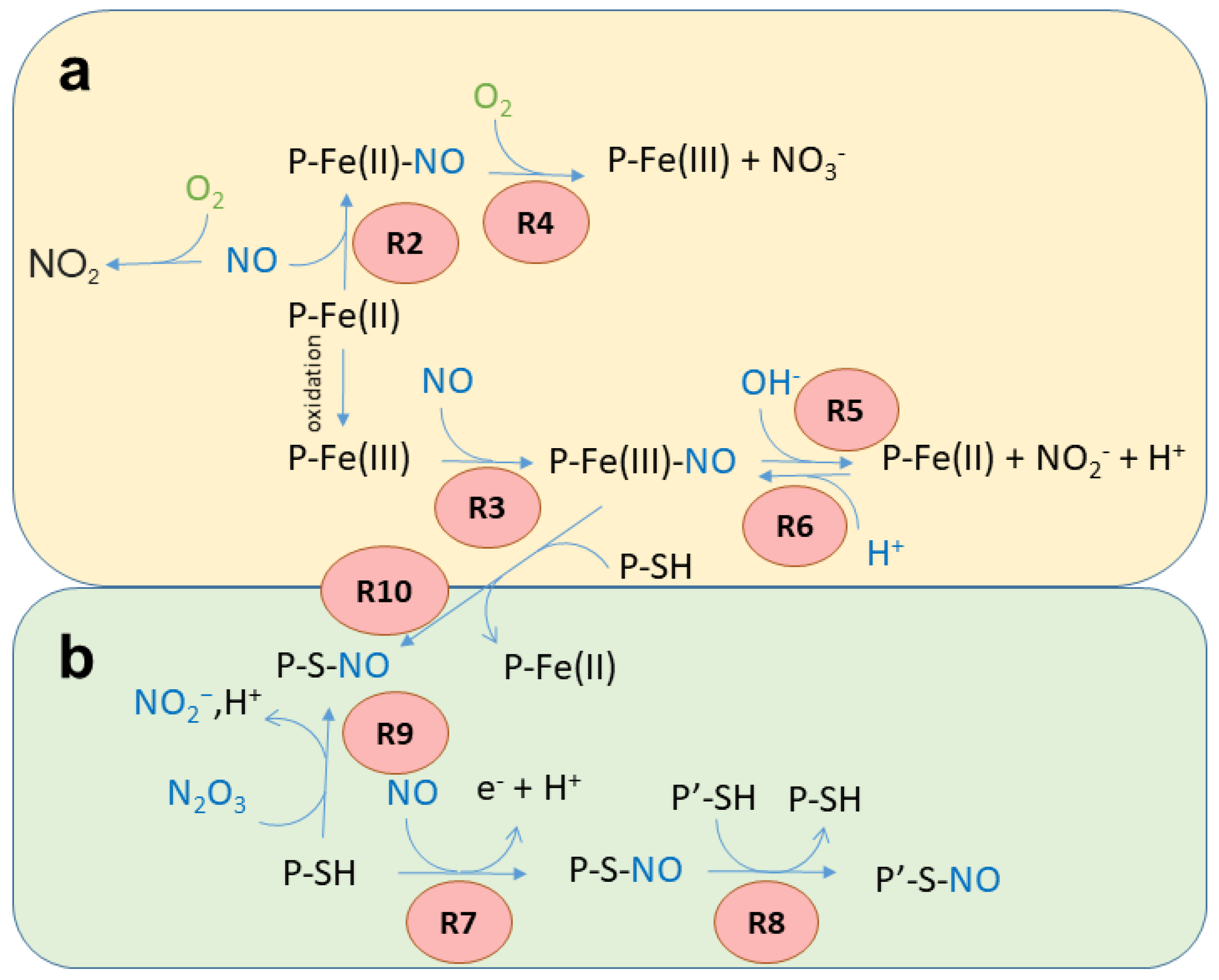
Recent reviews on heme-binding proteins extensively cover the complex reactivity of NO with the metal center of the heme group [24,25]. For several heme proteins, the interaction or the reactivity with NO is widely accepted as physiologically relevant, whereas for others—although intrinsically reactive with NO in vitro—the relevance is under debate. For circulating proteins such as Hb, several reactions might be relevant in vivo at different times, depending on the local microenvironment they encounter within the bloodstream. The main reactivities that heme proteins exhibit are reported below and in Figure 3a.
Reversible binding to ferrous heme. Under anaerobic conditions, Fe(II) heme proteins rapidly bind NO to produce a stable nitrosyl complex (Reaction 2, R2 in Figure 3a). For instance, Hb exhibits a kNO of 60 µM−1s−1 [26]. The binding is reversible, but the dissociation rate constants are typically very small, with koff < 10-3 s−1 [27,28].
P-Fe(II) + NO → P-Fe(II)-NO
In vivo, this reaction is associated with NO scavenging only under anaerobic conditions, since the presence of heme-bound O2 would quickly result in NO dioxygenation (see Reaction 4).
Reversible binding to ferric heme. The Fe(III) heme group forms bonds 6–7 orders of magnitude weaker with NO in comparison with Fe(II) (Reaction 3, R3 in Figure 3a) [24,25]. For Mb, the kNO for this reaction is 0.070 µM−1s−1 [29,30], with koff typically >10 s−1.
P-Fe(III) + NO → P-Fe(III)-NO
Some heme proteins play a physiological role based on this reaction. For instance, NPs are heme-binding lipocalins found in the saliva of the bloodsucking Reduviidae and Cimicidae families of Heteroptera. They are stable in the Fe(III) state, and the structure of the distal side of the heme group stabilizes the NO complex and prevents reactions with O2, water, hydroxides, or thiols, thus allowing their function as transporters of NO [31]. NO is then released at the feeding site, where it induces vasodilation.
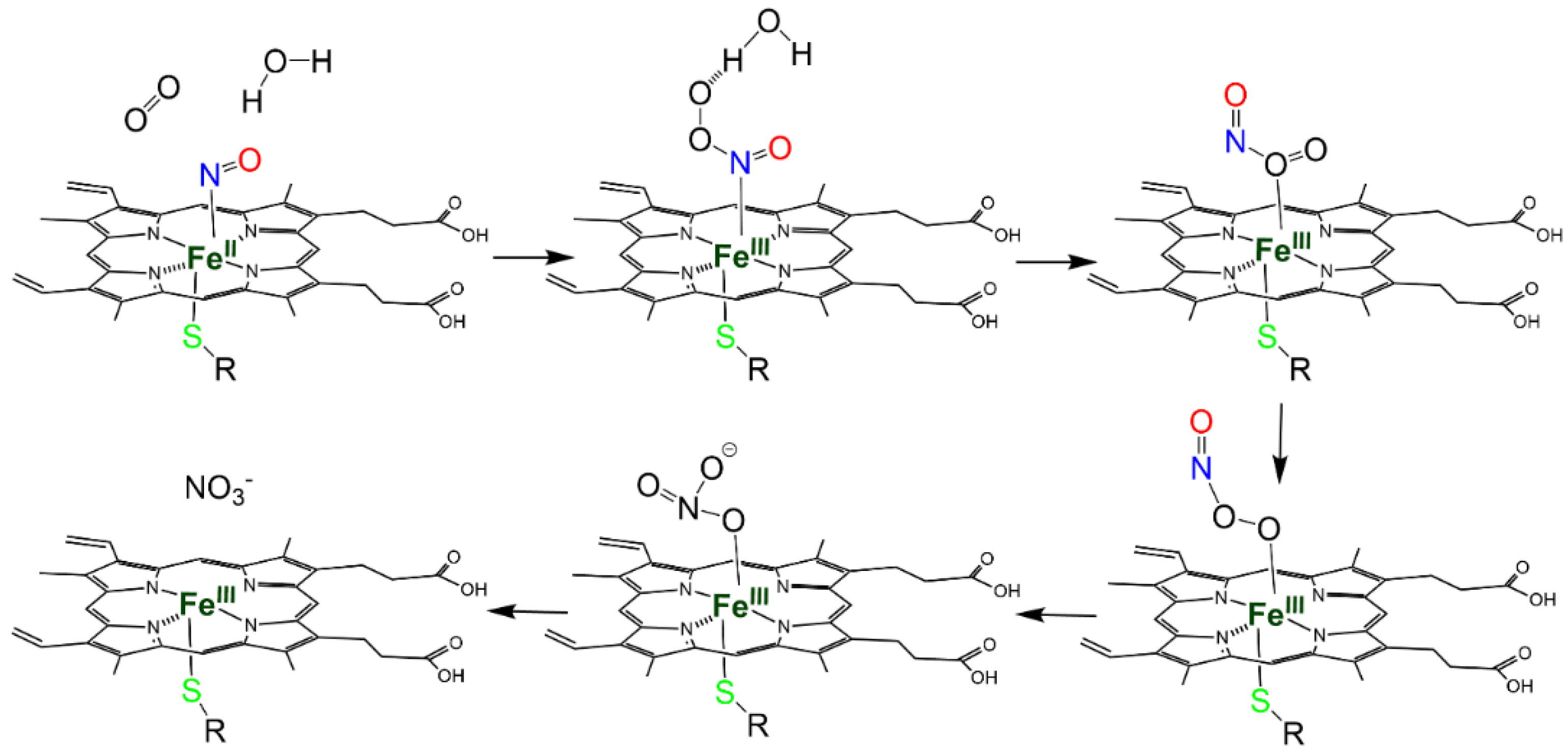
NO dioxygenation. In the presence of O2, several Fe(II) heme proteins, and particularly globins, are known to catalyze the NO-dioxygenase (NOD) reaction, where NO is oxidized to nitrate (NO3) via a peroxynitrite intermediate (Reaction 4, R4 in Figure 3a):
P-Fe(II)- O2 + NO → P-Fe(III) N(O)OO → P-Fe(III) + NO3
This reaction, which results in heme oxidation, is very fast (Hb kNOD 60–80 µM−1s−1 [26]) when the heme group is bound to O2 and exposed to NO. When the heme protein is bound to NO and exposed to O2, the reaction occurs at a much slower rate, since NO has to dissociate to allow O2 binding before the reaction can take place. The NOD reaction in vivo is typically associated with NO scavenging under aerobic conditions [26], but also, as in the case of Ascaris Hb, to O2 scavenging under anaerobic conditions [32]. It has been noted that catalytic reactions that lead to heme oxidation can be physiologically relevant only if efficient mechanisms for its reduction are in place to restore the Fe(II) form [33].
Autoreduction in Fe(III)–NO complexes. Fe(III) heme proteins undergo ‘reductive nitrosylation’ (or ‘autoreduction’) in the presence of NO (Reaction 5, R5 in Figure 3a).
P-Fe(III) + NO + OH → P-Fe(II)-NO2H → P-Fe(II) + NO2 + H+
The reaction occurs in two steps and is pH-dependent, since it usually requires the displacement of the intermediate by hydroxide [34] (Scheme 1). Since the affinity of heme proteins in the Fe(III) form for NO is usually low (see Reaction 3), this reaction requires NO concentrations that afford a significant binding.
Nitrite reduction. The reverse of Reaction 5, i.e., the reduction in NO2 (NiR) by Fe(II) heme in the absence of O2 (Reaction 6, R6 in Figure 3a), has been extensively investigated in mammals because of its potential to produce NO [35].
P-Fe(II) + NO2 + H+→ P-Fe(III) + NO + OH
At least ten mammalian heme proteins have been characterized in vitro for NiR, i.e., Hb, myoglobin (Mb), Ngb, Cygb, cytochrome c oxidase, cytochrome bc1, cytochrome c, endothelial (eNOS), cytochrome P450, and indoleamine 2,3-dioxygenase 1 [8,16,36,37]. For proteins that are physiologically in the Fe(II) state and exhibit high concentrations (i.e., Mb and Hb in muscle tissue and red blood cells, respectively), the extent of NO production through this reaction is under debate, since newly produced NO would tightly bind the nearby Fe(II) hemes (Reaction 1).
NO2 concentration in mammalian tissues is fairly high (0.1–10 μM [38]), confirming that the NiR reaction can physiologically take place in vivo. NO2 can have a dietary origin [39] or can be produced from nitrate (NO3) by bacterial Mo-containing nitrate reductases [40], suggesting a NO3-NO2-NO pathway for NO biosynthesis that involves the oral microbiome and enteral symbiotic bacteria for the initial step [41,42].

2.2. S-Nitrosylation

S-nitrosylation is a Cys post-translational modification that reversibly regulates protein function and is a fundamental regulatory mechanism in NO-related signaling [22,43]. It consists of the covalent binding of a nitroso group to a Cys residue to form the SNO. S-nitrosylation may produce novel protein–protein interactions by producing changes in the surface-charge or by allosterically generating solvent-exposed binding sites [44]. The current view is that S-nitrosylation plays a role equivalent to phosphorylation or acetylation to elicit the biological responses of target proteins, and the modification is often considered as an evolutionarily conserved signaling mechanism that involves many classes of proteins located in the cardiovascular system [45].
Regardless of the reaction responsible for S-nitrosylation, it is believed that not all exposed Cys residues can form stable SNOs. The identification of S-nitrosylation sites in the proteome [46] has allowed the identification of Cys environments that either promote the formation of SNOs or stabilize them once they are produced. Particularly, the presence of an acid-base motif in the proximity of the reactive Cys was associated with the formation of a thiolate, thus favoring the reaction [47]. Since the environment of Cys side chains can depend on the conformation of the protein within its conformational space, the residue can be reactive in one conformation but not in another. For example, it was proposed that in oxygenated Hb (R-state), the histidine (His) residue proximal to Cysβ93 favors the base-catalyzed S-nitrosylation, whereas, in the deoxygenated form (T-state), denitrosylation is facilitated by the proximal aspartic acid (Asp) residue [47]. In serine racemase, the reaction with either NO or nitroso donors is also conformation-dependent and occurs only in the conformation stabilized by its allosteric effector ATP [48]. Hydrophobic environments, such as biological membranes or environments formed by protein structures, favor the formation of SNOs ([47] and references herein).
In the last two decades, S-nitrosylation has drawn attention mostly for the biomedical implications for its direct effect on a variety of signaling pathways in physiological and pathological conditions [49]. In several neurodegenerative diseases, high levels of NO may contribute to mitochondrial dysfunction and protein misfolding via aberrant protein S-nitrosylation. Aberrant protein S-nitrosylation may particularly impact the quality control of protein folding, including molecular chaperones, autophagy, and the ubiquitin-proteasome system [49].
The main reactions in which Cys residues and NO are involved have been summarized in Figure 3b and detailed below.
S-nitrosylation associated with reduction. It is accepted that the direct reaction of the NO radical with thiols does not result in S-nitrosylation since the reaction requires the loss of an electron. However, in the presence of O2 or other oxidants, a redox reaction can occur, with the formation of the SNO through a thiyl radical or through a nitrosonium (NO+) ion [22] (Reaction 7, R7 in Figure 3b).
P-SH + NO → P-SNO + e + H+
S-nitrosylation by transnitrosylation. S-nitrosylation can also result from transnitrosylation by S-nitrosoglutathione (GSNO) [43] or S-nitrosylated proteins [50,51] (Reaction 8, R8 in Figure 3b).
P-S-NO+ P′-SH → P-SH + P′SNO
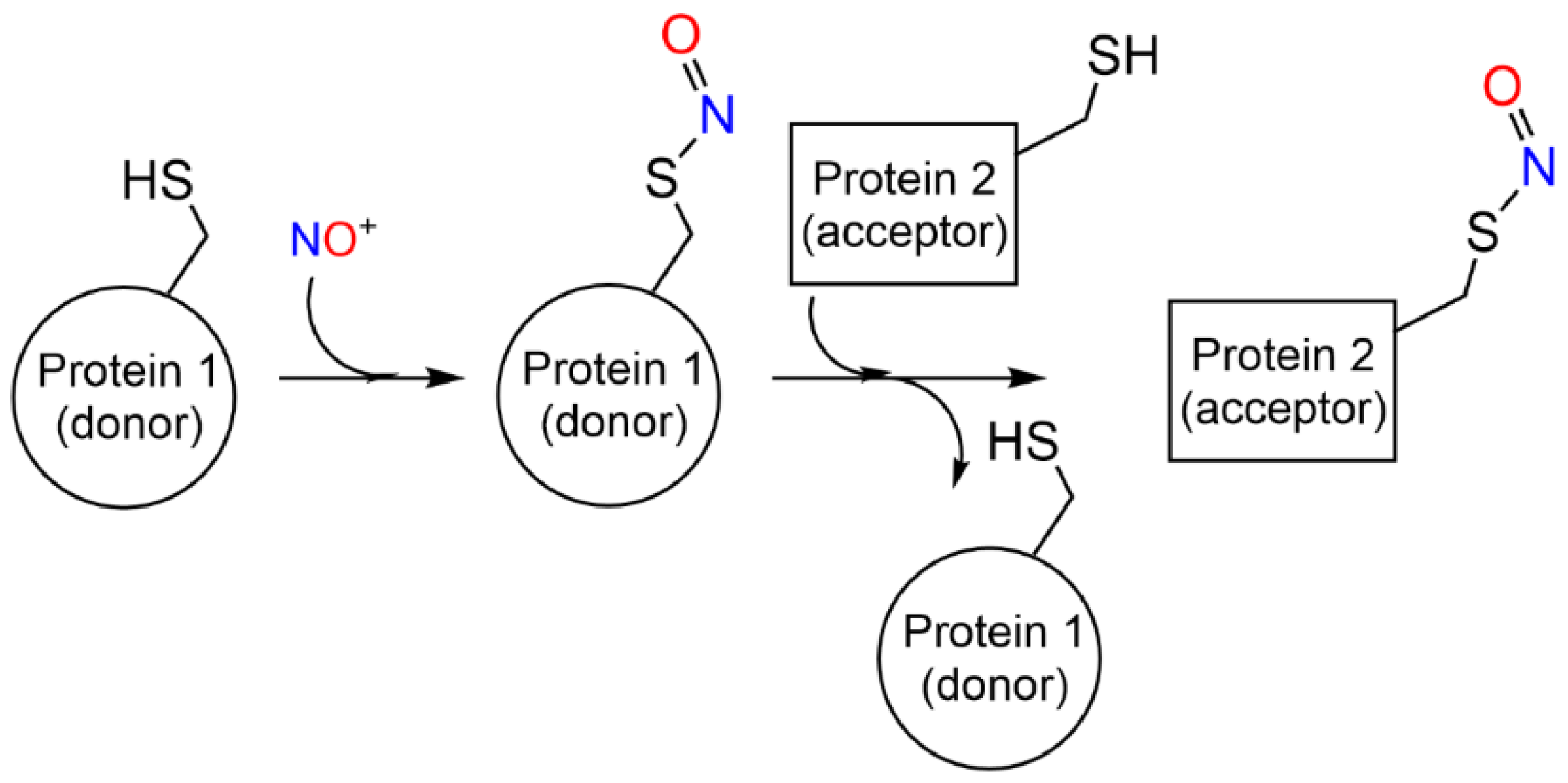
Transnitrosylation has been associated with site-specific protein–protein interactions mediated by specific recognition motifs [47] (and references herein) (Scheme 2). Enzymes that donate and accept nitroso groups from proteins are named S-nitrosylases and denitrosylases, respectively, and can have high-molecular weight final acceptors—as the thioredoxin/thioredoxin reductase system (Trx1/TrxR) —or low molecular weight acceptors, as the GSNO reductase and SNO-coenzyme A reductase systems [52,53]. The enzymatic control of S-nitrosylation is fundamental for the regulation of NO-based signal transduction and is dynamically regulated by the equilibrium between S-nitrosylated proteins and low-molecular-weight SNOs, which may be a cause or consequence of diseases, including asthma, cystic fibrosis, Parkinson’s disease, heart failure, and stroke [47].
S-nitrosylation by reaction with nitrous anhydride. S-nitrosylated Cys residues can also be formed by the reaction with dinitrogen trioxide (or nitrous anhydride, N2O3) (Reaction 9, R9 in Figure 3b). N2O3 partially dissociates, forming the nitrosonium ion (NO+), which reacts with thiolates (Reaction 8, transnitrosylation). N2O3 is formed through the reaction of NO with O2, particularly in hydrophobic microenvironments such as cell membranes [54].
P-S + N2O3 → P-SNO+ NO2
Metal-to-Cys S-nitrosylation. Metal-to-Cys S-nitrosylation (or heme-dependent S-nitrosylation or heme-assisted S-nitrosylation in the specific case of heme proteins) is a special case of S-nitrosylation associated with reduction (Reaction 7) and is of great relevance to heme proteins. It consists of the transfer of a nitroso group from a heme group to a nearby Cys residue, either within the same protein (auto S-nitrosylation) or in another protein or peptide (Reaction 10, R10 in Figure 3).
P-Fe(III)-NO+ P-SH → P-SNO + P-Fe(II) + H+
The NO+ ion is generated by the binding of NO to a Fe(III) heme (Reaction 3), which is reduced to Fe(II) (Scheme 3).

3. Heme Reactivity and S-Nitrosylation of Heme Proteins

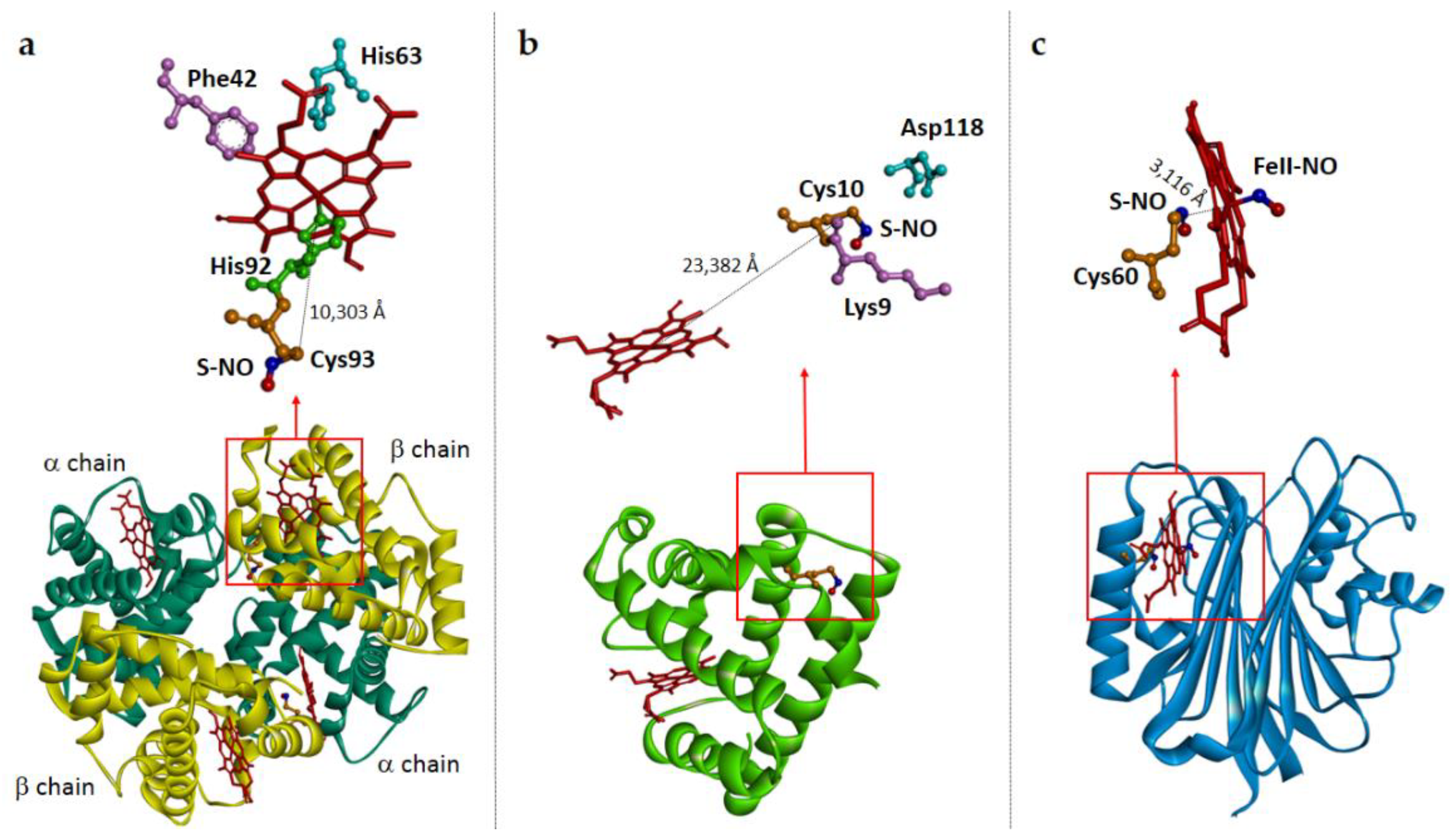
In this review, we will describe the reactivity of NO at the heme site and at Cys residues for heme proteins for which both have been described. An overview is in Table 1. In case they are available, the structures of S-nitrosylated proteins are depicted in Figure 3.

3.1. Hemoglobin

Hb is an O2 transporter contained in RBCs at a high concentration. It is a heterotetramer of paralogous subunits exhibiting the globin fold [103]. The subunits of the most abundant human form, HbA, are named α and β and each binds a Fe(II) heme coordinated to a His residue named proximal His (F8 in the globin nomenclature). The residues that coordinate and orient the heme group are among the few invariant residues among all Hbs [103]. Hb exhibits cooperative binding associated with the equilibrium between at least to conformations endowed with a different affinity for O2. The binding of O2 at one subunit shifts the proximal His with respect to the heme, triggering a modification of the network of non-covalent bonds at the α1β2 interface. These changes destabilize the conformation known as the “T-state”, endowed with low O2 affinity and stabilize the conformation known as the “R-state”, endowed with high O2 affinity [104].

3.1.1. Heme Reactivity with NO

Among heme proteins, the NO adducts of Hb are some of the most studied systems in biology. Hb in the Fe(II) state binds NO (Reaction 2, binding of NO to Fe(II)) with a much higher affinity in comparison with O2 and CO, particularly because of a slow dissociation rate, in the range 10−5–10−3 s−1 [55]. The binding of NO has been characterized in detail [56], indicating a cooperative process and estimating different dissociation rates for the two different subunits and the two quaternary states [56]. Although widely investigated in vitro [57], the formation of significant amounts of the Hb–NO complex in vivo is unlikely, given the high concentrations of the competing ligand, O2, to form Hb-O2.
Hb-Fe(II)-O2 quickly reacts with NO through the NOD reaction (Reaction 4) to yield Hb-Fe(III) and nitrate. This reaction has been associated with the scavenging of NO and vasoconstriction, as observed for hemolytic diseases [8,16,36,37,61]. Under physiological conditions, this reaction is limited by the confinement of Hb inside RBCs.
The autoreduction in Fe(III)-NO (Reaction 5) was observed for Hb in 1901 by Haldane, who prepared Hb-Fe(II)-NO by adding NO2 to Hb [62]. This activity, which was first characterized in cured meat [62], was later suggested to play an important role in delivering NO to tissues in the capillaries, where Hb—mostly in the deoxy form—can produce NO through the NiR (Reaction 6) [63].
Impaired reactivity of Hb with NO has been associated with pathology and was particularly studied in the field of Hb-based O2 carriers (HBOCs), a class of biotechnological therapeutics based on chemically or genetically modified heme proteins designed to deliver O2 to tissues as an alternative to RBCs [61,105]. One of the hurdles in this approach consists of the scavenging of NO by Hb derivatives when administered intravenously in a cell-free form. Indeed, HBOCs are not shielded by RBCs, which greatly reduce the interaction of Hb with NO [61]. This interaction—which can either occur through the binding of NO to Fe(II) (Reaction 2) or through the autoreduction in Fe(III)–NO complex (Reaction 5) depending on the saturation of Hb—interferes with the regulation of the vascular tone by NO signaling, typically causing life-threatening vasoconstriction [26]. NO scavenging has been addressed by producing sufficiently large polymeric Hbs to reduce extravasation and consequent reactions with NO or by creating recombinant Hbs with reduced rates for the NOD reaction (Reaction 4).

3.1.2. S-Nitrosylation

HbA is by far the better-characterized globin for which S-nitrosylation has been detected, with an extensive functional and structural characterization [106]. It was not only one of the first proteins for which S-nitrosylation was identified, but also the first one for which a physiologically relevant role for S-nitrosylation was proposed [107]. Indeed, it was hypothesized that Hb is S-nitrosylated in the lungs and releases the nitroso group in the capillaries, producing vasodilation and regulating blood pressure under hypoxic conditions, an effect long known but of unclear molecular origin [108].
All mammalian Hbs contain a reactive Cys at position β93, the primary site of Hb S-nitrosylation. Cysβ93 is solvent-accessible, close to the heme group, and is a known target for thiol reagents, which can allosterically modulate O2 affinity, cooperativity, and Hb sensitivity to pH [109,110]. Cysβ93 was implicated in several redox reactions (recently reviewed in [111]) and it was particularly proposed as a scavenger of reactive oxygen species (ROS). Consistently, mice inflammation models expressing the Cysβ93Ala variant of Hb showed a greater degree of hypotension and lung injury associated with the increased formation of reactive species in RBCs [112].
Hb S-nitrosylation of Cysβ93 was suggested to be possible through a heme-dependent mechanism in which either Fe(III)-NO heme mediates the metal-to-Cys S-nitrosylation of Cysβ93 (Reaction 10) or NO2 reacts with Fe(II) heme through the NiR (Reaction 6) to produce NO, which in turn can react with the –SH radical (Reaction 7) [58] (Figure 4a). This reaction was suggested to be conformation-dependent, since it was reported to take place at a higher rate in the lungs, where Hb is mostly in the R-state [59]. Indeed, the total NO bound to Hb was shown to be constant irrespective of pO2 during the arterial–venous cycling, whereas the relative amount of NO bound to hemes and Cysβ93 changes. It was concluded that NO is exchanged between hemes and thiols [59]. According to this model, upon the conformational transition to the T-state in the capillaries, NO is transnitrosylated to other target proteins or peptides [108] (Scheme 4). The physiological relevance of this reaction is under debate since it would require Hb in the Fe(III) state, usually maintained at low concentrations by the antioxidant systems known to operate within RBCs [113]. The His residue proximal to the Cys93 in the β subunit of Hb in the R-state was suggested to facilitate S-nitrosylation, whereas the proximal aspartic acid (Asp) in the deoxygenated T-state would favor denitrosylation [114]. Based on these observations, Stamler and colleagues proposed that SNO-Hb in erythrocytes serves as a physiological regulator of vascular tone by preserving the bioactivity of NO within the vasculature to modulate the blood flow [59].
Metal-to-Cys S-nitrosylation was shown to be absent in the Hb pathologenetic variant —HbS—responsible for sickle cell anemia, suggesting that defective NO processing might contribute to the disease [115]. Based on this model, a mixture of HBOCs with their S-nitrosylated derivative has been investigated, concluding that they might be useful in the treatment of conditions of acute hypoxia, such as tumor oxygenation, wound healing, hemorrhagic trauma, and sickle cell and hemolytic anemia [116].
Following the characterization of mice expressing the Cysβ93Ala Hb variant, the role of Cysβ93 S-nitrosylation as the mechanism responsible for NO-mediated vasodilation has been put into question. Some NO-mediated effects such as ex vivo cardioprotection, inhibition of platelet activation, or capillary relaxation were demonstrated to be intact in these mice [117,118], although hypoxic vasodilation was critically impaired. Altogether, these studies supported the hypothesis that NO bioactivity occurs independently of S-nitrosylation and that Reaction 6 (NiR reaction) better explains RBC-associated NO vasoactivity [117]. Particularly, it has been shown in several experimental settings that NO2 mediates the inhibition of platelet activation in the presence of RBCs and that NO scavengers abolish this effect [119].
The extent of heme-assisted S-nitrosylation in vivo has also been challenged by showing that Hb-SNO—although measurable in the human circulation—did not exhibit a significant arterial–venous gradient and that NO delivery from Hb-SNO was small [63]. Heme-assisted S-nitrosylation has also been disputed [60]. Indeed, it was shown that Hb is not capable of transferring NO from Cysβ93 to the heme—and vice versa—upon T–R conformational changes, neither in whole blood, nor in washed red cells or purified Hb [60].
The conflicting reports and models on NO release or production from RBCs [120] confirm the difficulty in dissecting the reactivity of the heme group of heme proteins with respect to the S-nitrosylation of Cys residues, calling for a thorough functional and structural characterization of heme proteins for which these reactivities are proposed as physiologically relevant.

3.2. Ascaris Hemoglobin

Ascaris lumbricodes is a parasitic nematode that, in the adult form, inhabits the O2-poor small intestine, leading an anaerobic life. It currently infects nearly 25% of the world’s human population [121]. The octameric Ascaris Hb, unlike tetrameric vertebrate Hbs, is free in solution in the perienteric fluid of the worm and it is endowed with an exceptionally high affinity for O2, 25,000 times higher than human Hb. The globin domain of Ascaris displays the classic globin-fold with minor differences [64]. The distal His residue, characteristic of many vertebrate Hbs, is replaced by a glutamine residue, which dramatically increases O2 affinity.

3.2.1. Heme Reactivity with NO

Ascaris Hb works as a dioxygenase by catalyzing NO3 production from O2 and NO through the NOD reaction (Reaction 4), thus maintaining the perienteric fluid hypoxic [65]. This reaction is uncommon since vertebrate globins usually scavenge NO through Reaction 4 in the presence of an excess of O2. In the O2-poor environment where Ascaris lives, the opposite occurs, with NO being produced in excess by NOS to detoxify O2 [32]. Upon oxidation, Ascaris Hb-Fe(III) subsequently binds NO through Reaction 3 [65].

3.2.2. S-Nitrosylation

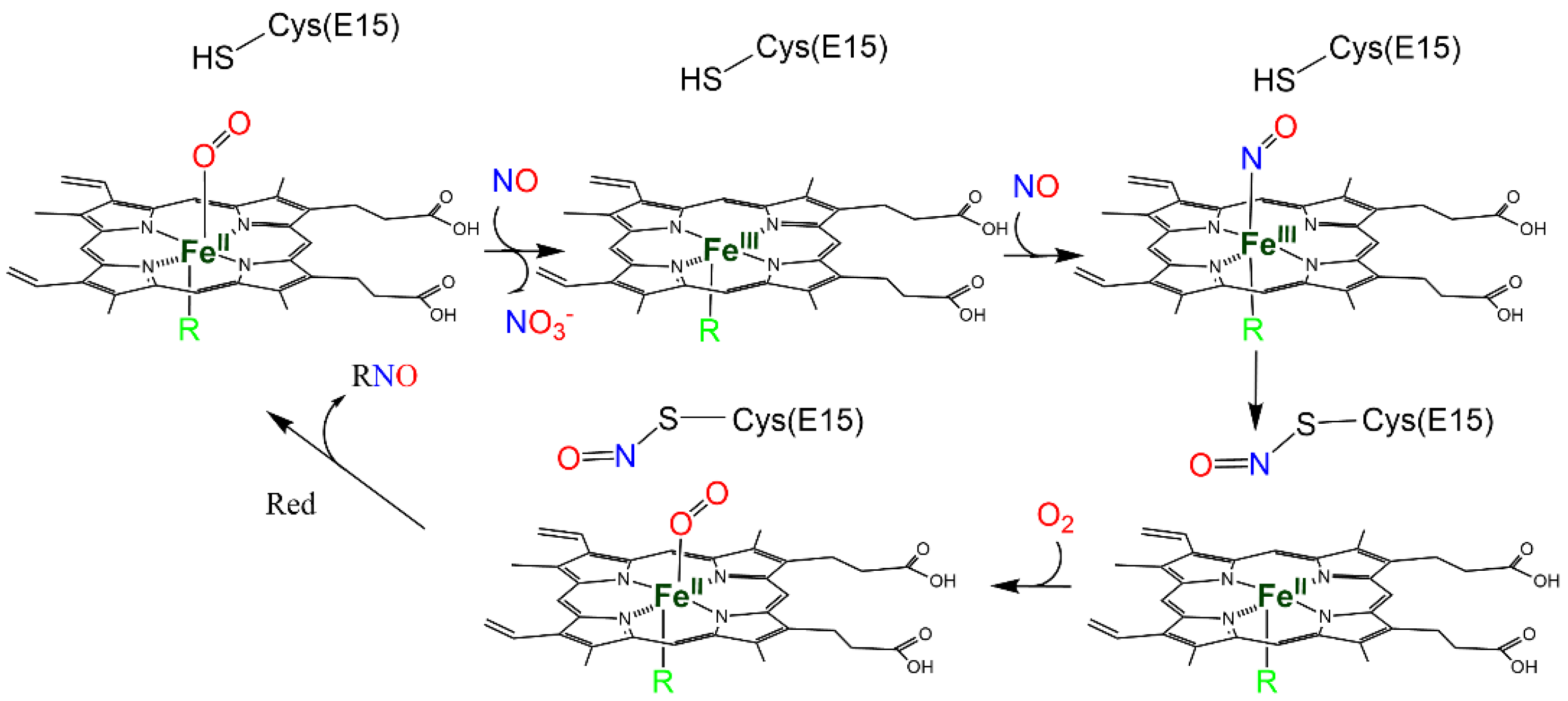
Cys72 (E15 in globin nomenclature) is positioned in the distal pocket, close to the heme [122] and is found mostly as nitrosothiol in the perienteric fluid of Ascaris. Unlike other globins, nitrosothiols oxidize the heme group of Ascaris Hb, suggesting that the heme and Cys72 constitute a redox couple [65]. Mutation of Cys72 blocks the production of Fe(III)-NO, further confirming the role of Cys. Based on absorption spectroscopy and stopped flow spectroscopy, it was suggested that the Fe(III)–NO complex formed through the NOD reaction (Reaction 4) followed by NO binding to the Fe(III) heme (Reaction 3) can produce heme-dependent S-nitrosylation of Cys72, with a concomitant reduction in the heme to the Fe(II) form (Reaction 10) followed by O2 binding [65]. The binding of O2 under almost anaerobic conditions and in the presence of relatively high concentrations of NO is only possible due to its unusually high affinity for Ascaris Hb. Finally, Cys-SNO can be reduced to Cys through a yet-unknown mechanism, thus allowing the enzyme cycle to proceed (Scheme 5).

3.3. Myoglobin

Mb is a monomeric globin present in the cardiac and aerobic skeletal muscles of almost all vertebrates, where it plays a role in O2 storage and NO homeostasis [123]. The high O2 affinity of Mb maintains a robust gradient in pO2 from blood to myocytes, thus channeling O2 from the bloodstream to the mitochondria for oxidative phosphorylation. Some fish species, such as threespine stickleback (Gasterosteus aculeatus), have lost the ability to produce Mb [124] and 6 of the 16 species of icefishes lack the expression of Mb in the heart ventricle due to 4 independent mutations [125]. Mb is also largely absent from the heart of the African butterflyfish (Pantodon buchholzi), but is highly expressed in the swim bladder [124]. The discovery of Mb in smooth muscle, endothelial, and tumor cells [126,127,128] and the characterization of Mb-knockout mice [129] have suggested that Mb might play a more complex and versatile role than previously thought.

3.3.1. Heme Reactivity with NO

The reactivity of mammalian Mbs with NO is one of the most studied systems in biochemistry. Under normoxic conditions, when the O2-bound form dominates, Mb scavenges NO producing Fe(III)-Mb and NO3 through the NOD reaction (Reaction 4) [67]. Under hypoxic conditions, where Mb becomes increasingly deoxygenated, Mb produces NO from NO2 through the NiR reaction (Reaction 6), faster than Hb [68]. Therefore, Mb has a dual role of NO scavenger and NO producer at different pO2 [130], thus regulating the activation of the sGC signaling pathway [131]. The NiR activity of Mb in the myocardium was shown to be the major source of NO [130] and has a relevant function under hypoxic conditions and in ischemia-reperfusion events [132]. Therefore, NO2 can act as a stable reservoir for supporting NO signalling in hypoxic or metabolic stress.

3.3.2. S-Nitrosylation

Human Mb has a single reactive Cys in position 110 [133] known to form Cys-SNO in vitro through metal-to-Cys S-nitrosylation, reacting with NO when the protein is in the Fe(III) form, similarly to Hb (Reaction 10) [67,134,135]. While S-nitrosylation in Hb was proposed by some authors to be dependent on the allosteric R–T transition, the degree of O2 saturation does not affect the accessibility of Cys110 for S-nitrosylation. Rayner et al. [135] demonstrated the S-nitrosylation of human Mb-Fe(II)-O2 in vivo, suggesting that the modified protein may stimulate aortic vessel relaxation similar to free NO. Mb-SNO may also be important in maintaining optimal NO concentrations in the human vasculature.
Teleosts Mbs, in which S-nitrosylation has been well documented [129,136,137], exhibit the same globin-fold as that of higher vertebrates but lack the D-helix [138]. Among teleosts, Antarctic notothenioids, carps, and tunas have a conserved Cys—Cys10—in the A helix, whereas the non-tuna scombroids, bonito, mackerel, and blue marlin lack this residue [137]. Cys10 is partially solvent-exposed and is located between the side chains of Asp118 and Lys9 at the protein surface, thus supporting the hypothesis that the “acid-base” motifs are important determinants for Cys S-nitrosylation [69]. The X-ray crystal structure of S-nitrosylated blackfin tuna Mb (PDB: 2NRM) (Figure 4b) indicates that S-nitrosylation at Cys10 causes reversible conformational changes [69]. Recently, the spectral, dynamical, and structural properties of unmodified blackfin tuna Mb and its S-nitrosylated derivative were studied by molecular dynamic simulations, suggesting that S-nitrosylation can also modulate local hydration. Indeed, the local hydration between the A and H helices and around the site of the S-nitrosylation is increased in S-nitrosylated Mb compared to the unmodified protein, with changes in the structure and dynamics of the protein [70].
Mbs of salmonids—among which rainbow trout and Atlantic salmon have been particularly investigated—were shown to undergo S-nitrosylation at a Cys residue different from Cys10 [71,72]. Indeed, in addition to Cys10, they exhibit a second Cys residue in position 107, located at the interface between the G and H helices, far from the heme pocket. The S-nitrosylation of these Mbs induces conformational changes in the tertiary structure associated with an increase in the O2 affinity [71,72]. Conversely, under hypoxic conditions, NO is released, improving myocardial efficiency during intense exercise [71,72].

3.4. Neuroglobin

Ngb is a globin predominantly expressed in neurons. Sequence conservation and its presence in several taxa point to an important role in the central nervous system, which, however, has not been fully elucidated [139]. Ngb has been associated with O2 supply to tissues, O2-scavenging, and sensing. It exhibits an hexacoordinated heme both in the Fe(II) and Fe(III) forms [140,141].

3.4.1. Heme Reactivity with NO

Ngb in the Fe(II) form binds NO under anaerobic conditions (Reaction 2) upon displacement of the distal His. The complex has an estimated dissociation rate constant of 2.9 × 10⁻³ s−1 and a KD of 7.7 × 10⁶ M−1, lower than that of Mb or Hb by a factor of 104–105. [74]. In the Fe(III) form, Ngb reacts with NO, leading to autoreduction (Reaction 5) followed by the binding of free NO to Fe(II) [73]. The NOD reactivity (Reaction 4) was also investigated, indicating that it occurs in a biphasic fashion consistent with the formation of a peroxynitrite intermediate [73]. It was also shown that Ngb efficiently catalyzes the conversion of NO2 to NO with a second-order rate constant of 5.1 M−1 s−1 (Reaction 6, NiR reaction), although the role of this reaction in the retina and brain is not clear [74].

3.4.2. S-Nitrosylation

The S-nitrosylation of Ngb has been observed for the murine ortholog upon reaction with NO2 [74], hinting at heme-dependent S-nitrosylation (NiR reaction, Reaction 6, followed by metal-to-Cys S-nitrosylation, Reaction 10). Murine Ngb has two Cys residues, Cys55—only ∼15 Å from the heme iron—and the solvent-exposed Cys120. Although the exact site of S-nitrosylation has not been defined, Cys55 was deemed the most likely one [74].
It was hypothesized that NO+ generated by NO reaction with Fe(III) heme may be a possible pathway of S-nitrosothiols formation [74]. Alternatively, the reaction might occur through N2O3 (Reaction 9), which could be accommodated in a large hydrophobic cavity present in Ngb and connecting the heme distal site to the solvent [74]. The levels of S-nitrosylated Ngb formed in vitro under physiological concentrations of NO2, however, were deemed too low to function as NO storage, but it was hypothesized that it could have a potential role in redox signalling pathways [74].

3.5. Cytoglobin

Cygb is a globin discovered 20 years ago [142] expressed at low concentrations in the cytoplasm of all tissues of vertebrates. It is upregulated under hypoxic conditions [143]. Like Ngb, Cygb is hexacoordinated, with two His residues coordinated at the proximal and distal sites [144]. Several functions have been proposed for Cygb, including NO scavenging [145], scavenging of ROS [146], lipid peroxidation [147], and synthesis of NO through the NiR activity [75].

3.5.1. Heme Reactivity with NO

Similarly to other globins and heme proteins in general, Cygb in the Fe(II) form binds NO under anaerobic conditions (Reaction 2) [75] and catalyzes the NOD reaction under aerobic conditions (Reaction 4), with a comparable rate as Ngb [33]. The Leu46Trp variant decreases NOD activity > 10,000-fold. After conflicting reports [74], it was shown that under anaerobic conditions Cygb generates NO through the NiR activity (Reaction 6), with the formation of Cygb-Fe(III) and Cygb-Fe(II)-NO [75]. It was suggested that this reaction occurs in vivo and is crucial in the activation of sGC under hypoxic conditions [75]. Cygb NiR activity is strongly dependent on the oxidation state of the two surface-exposed Cys38 and Cys83, with the formation of the disulfide bond increasing the activity 50-fold [7]. Among teleosts, the heme of Cygb1 of Danio rerio can efficiently reduce NO2 to form NO (Reaction 6, NiR reaction) at a rate of 14.2 M−1s−1, compared to 0.40 M−1s−1 of mammalian Cygb and 0.31 M−1s−1 of fish Cygb2 [76].

3.5.2. S-Nitrosylation

Since the redox state of Cys38 and Cys83 has been associated with the NiR activity of Cygb, it appears plausible that the S-nitrosylation of one or both residues could contribute to modulating the activity. Indeed, it was shown that Cygb undergoes S-nitrosylation upon incubation with NO2, albeit at lower levels in comparison with Ngb [74]. Both Cys38 and Cys83 are located in proximity to the internal cavity [148]. It was hypothesized that the formation of S-nitrosylated Cys may be heme-independent or that it could result from the reactivity with N2O3 [74]. As for Ngb, the levels of S-nitrosylated Cygb are likely too low to function as NO storage system [74].

3.6. NO Synthase

In metazoans, NO is produced by three isoforms of NO synthase (NOS) —the neuronal NOS (nNOS or NOS I), the inducible NOS (iNOS or NOS II), and the endothelial NOS (eNOS or NOS III). All contain a heme group. All NOS use L-arginine and O2 as substrates to generate L-citrulline and NO. NOS require NADPH, flavin adenine dinucleotide (FAD), flavin mononucleotide (FMN), and (6R)5,6,7,8-tetrahydrobiopterin (BH4) as cofactors [24,149]. NOS I is constitutively expressed in central and peripheral neurons, NOS II is expressed in many cells, and NOS III is mostly expressed in endothelial cells [150]. All vertebrate NOS are homodimers linked to a zinc ion by tetrahedral coordination. Each NOS monomer is endowed with an N-terminal oxygenase domain and a C-terminal reductase domain, both linked by a calmodulin (CaM) binding site. The Fe(II) heme binds O2, which oxidizes the guanidine group of the L-arginine to generate L-citrulline and NO in a two-step reaction:
2 L-arginine + 3 NADPH + 3 H+ + 4 O2 → 2 citrulline +2 NO + 4 H2O + 3 NADP+
The NOS-derived NO binds the Fe(II)-heme group of sGC. The formation of a NO-ferroheme–sGC complex induces the synthesis of the second messenger cyclic guanosine 3′,5′-monophosphate (GMP).

3.6.1. Heme Reactivity with NO

Similarly to other NO-hemoproteins, NOS may serve as a reservoir of bioactive NO [131]. Upon synthesis and before being released, the newly synthesized NO binds to the heme of NOS [151,152], forming the heme Fe(III)–NO complex (Reaction 3). This complex can release NO with a rate constant kd (productive cycle) or it can be reduced by the NOS reductase domain to Fe(II)–NO through the rate constant kr, comparable to kd. Fe(II)–NO can be oxidized in the presence of O2 (futile cycle) through the kox rate constant [77,153].
The kox constant, much higher in NOS enzymes than in any other hemoprotein [77], is characteristic of heme-thiolate enzymes and does not require NO dissociation from the heme before the NOD reaction can take place (Reaction 4). Indeed, it occurs by the direct reaction of O2 with the heme Fe(II)–NO complex, through an N-bound Fe(III)-N(O)OO- intermediate species [78,154,155] (Scheme 6).

3.6.2. S-Nitrosylation

Constitutive NOS (nNOS and eNOS) are S-nitrosylation targets of NO themselves [155,156], with eNOS constitutively S-nitrosylated in endothelial cells. S-nitrosylation reversibly inhibits eNOS activity and is responsible for its fine regulation [157,158], supporting the hypothesis that S-nitrosylation is a dynamic and physiologically relevant regulator of NO signaling pathways in the vascular endothelium. The S-nitrosylation of Cys98 is involved in the disruption of the zinc tetrathiolate cluster of the eNOS dimer [81].
In further studies of human eNOS, multiple Cys residues, all located on the surface of the protein, were identified as potential sites of S-nitrosylation. Among these, Cys93 and Cys98 are involved in the formation of eNOS homodimers through a Zn tetrathiolate cluster [80]. Other Cys residues (Cys660, Cys801, and Cys1113) located within regions known to bind FMN, FAD, and NADPH, as well as Cys852, Cys975/990, and Cys1047/1049 located in regions not yet identified in any known biochemical functions can undergo S-nitrosylation [80].

3.7. Guanylyl Cyclase

Soluble guanylyl cyclase (sGC), a heme-containing heterodimer composed of an α and a β subunit, is the main receptor of NO in mammals. Each subunit contains three domains: an N-terminal portion consisting of a heme–NO/O2 (HNOX) region with the heme present only in the β subunit; a “dimerization domain” with a Per/Arnt/Sim (PAS)-fold domain and a coiled-coil region (CC domain); and the C-terminal catalytic domain whose activation occurs when the α and β subunit are associated [159]. The HNOX and PAS domains form the N-terminal sensor module of sGC whereas the CC domains of both subunits form the transducer module that connects the N-terminal sensor module and the C-terminal catalytic module. NO has a dual effect on sGC: it binds at the heme and stimulates its catalytic activity and reacts with a Cys residue producing S-nitrosylation, which inhibits the enzyme.

3.7.1. Heme Reactivity with NO

When NO binds the heme of the β subunit, the domain of sGC catalyzes the cyclization of guanosine triphosphate (GTP) to the second messenger cyclic cGMP, initiating the signal transduction cascade [160,161]. In the HNOX domain of the β-subunit, the b-type heme is in the pentacoordinated Fe(II) state and exhibits a high affinity for NO (Reaction 2) [162]. The proximal ligand of the heme is His105, whose binding with Fe(II) is broken when NO binds the iron on the distal side of the heme [82,83]. Upon the binding of NO to the β heme of human HNOX, structural changes in the sensor module trigger a conformational switch of the transducer module from bending to straightening, inducing structural changes within the catalytic module and activating the enzyme [84].

3.7.2. S-Nitrosylation

In addition to the binding of heme, sGC can be S-nitrosylated, as observed in smooth muscle cells [86]. S-nitrosylation of sGC affects its sensitivity to NO at the heme group, resulting in decreased responsiveness. In particular, Cys122 in the β subunit and Cys243 in the α subunit were identified as potential targets for S-nitrosylation. Desensitization of sGC deriving from S-nitrosylation constitutes a mechanism of sGC modulation by negative feedback [86]. sGC can also undergo a reductive nitrosylation reaction (Reaction 5) coupled with the heme-dependent S-nitrosation of Cys residues [85].
Oxidation of the heme iron in sGC causes dysfunction in NO/cGMP signal transduction, with effects on cardiovascular physiology as it decreases the affinity for NO and the enzymatic response to NO [163]. Fe(III) sGC undergoes reductive nitrosylation with NO in two steps, requiring two equivalents of NO. The first NO reduces the iron from Fe(III) to Fe(II) (Reaction 5, autoreduction in Fe(III)-NO). The second equivalent of NO binds the heme iron, forming a Fe(II)–nitrosyl complex (Reaction 2, binding of NO to Fe(II)). Reductive nitrosylation is accompanied by a conformational change in the protein and a heme-dependent SNO formation of Cysβ78 and Cysβ122 with the NO desensitization of Fe(III) sGC [85]. The Trx1/TrxR system may be involved in restoring the basal activity of sGC and its NO sensitivity via a mixed disulfide exchange mechanism between sGC and Trx1 [164].
The modulation of sGC activity by thiol oxidation has been proposed as a therapeutic approach for pathologies associated with oxidative or nitrosative stress, such as cardiovascular diseases [159].

3.8. Cyclooxygenases

Cyclooxygenases (COX), previously known as prostaglandin H synthases, are homodimeric enzymes that catalyze the first, rate-limiting step of prostanoids biosynthesis from arachidonic acid through their dual peroxidase and dioxygenase activities [165]. COXs consist of three domains: the epidermal growth factor domain, the membrane-bound domain, and the heme-binding catalytic domain, which contains the cyclooxygenase and peroxygenase active sites [165]. The heme group is in the Fe(III) in the resting oxidation states of COXs. Their reaction is multistep. First, COXs oxygenate arachidonic acid to prostaglandin G2, which is then reduced to prostaglandin H2 through the peroxidase activity in the enzyme.
Two homologous COXs have been identified, COX1 and COX2. COX1 is expressed constitutively, whereas the expression of COX2 is upregulated by inflammatory and proliferative stimuli. The interaction between NO and COXs and the activation of recombinant COX2 by NO have long been known [166], although the dissection of the effects of S-nitrosylation and NO heme binding has been carried out more recently, pointing to the former as the main effect of NO on the protein function [167].

3.8.1. Heme Reactivity with NO

Early spectroscopic experiments suggested that NO does not bind the Fe(III) heme group of COX1 [168]. NO binding to the heme of COX1 was monitored by stopped-flow spectrophotometry and enzyme assays. A very high dissociation constant of 0.92 mM was estimated [87]. However, the reaction was much faster with the Fe(II) form [87]. Even upon binding, NO only produced a slight decrease in enzyme activity, ruling out the regulation by direct nitrosylation at the heme [87]. The limited binding of NO to the heme group was also observed for COX2 in the Fe(III) state [167].

3.8.2. S-Nitrosylation

COX undergoes several post-translational modifications [169]. Particularly, S-nitrosylation was observed in both COX1 and COX2, although only the latter undergoes activation as a consequence [167]. NO was found to increase COX2 activity by increasing the apparent Vmax 1.6-fold and by decreasing the KMO2 1.5-fold, only in the presence of O2 [167]. Consistently, the conformation of COX2—but not COX1—was shown to be altered by S-nitrosylation in circular dichroism experiments [167]. The Snyder group showed that COX2, when incubated with various nitroso donors, is S-nitrosylated and activated in HEK293T cells overexpressing COX2 [88]. By individually substituting the 13 Cys residues of COX2 to Ser, they concluded that Cys526 is the likely site of allosteric activation, although other Cys residues were also S-nitrosylated [88].
By investigating the truncated variants of COX2 and iNOS in a murine macrophage cell line, it was concluded that the two proteins directly interact, bringing the NO-producing enzyme in proximity to COX2 for S-nitrosylation [88]. It was also shown that COX2 also binds nNOS and that the disruption of the complex prevents N-methyl-D-aspartate (NMDA) neurotoxicity [170]. In other experiments, COX2 was shown to coimmunoprecipitate with iNOS but not with eNOS [171]. In rats, atorvastatin, a statin with cardioprotective activity, causes the S-nitrosylation of COX2 through iNOS [171]. Atorvastatin-induced S-nitrosylation of COX2 contributes to the production of omega-3 fatty acids-derived protective molecules known as 13-series resolvins [172]. Overall, the interaction of NOS and COX2 constitutes a layer of the complex interplay between prostaglandin biosynthesis and NO signaling [173].
No effect has ever been observed for COX1 in vitro when incubated with either NO or nitroso donors [87,167]. However, it was shown that bombesin might induce the S-nitrosylation and activation of COX1 in presympathetic spinally projecting neurons in a rat model, thus eliciting the secretion of catecholamines [174,175]. It was also shown that COX1, unlike COX2, does not coimmunoprecipitate with iNOS [176].

3.9. Nitrophorins (Cimex lectularius)

NPs are heme proteins discovered 30 years ago [177] in the saliva of blood-feeding insects. They include proteins form unrelated lineages in a typical case of convergent evolution. Rhodnius prolixus NP exhibits a lipocalin-like eight-stranded β-barrel structure, whereas Cimex lectularius NP has a β-sandwich motif with seven peripheral α-helices [6,178]. The heme iron of NPs is stable in the Fe(III) state and binds NO (Reaction 3) in a pH-dependent fashion [31]. Upon contact with blood, which exhibits a higher pH than the insect’s saliva, the NO–NP bond weakens, and NO is released in the capillaries, thus facilitating the insect’s feeding through vasodilation, reduced platelet aggregation, and reduced blood coagulation [179]. The heme of C. lectularius NP—the NP for which S-nitrosylation has been observed in addition to NO heme binding—is located between the β-sandwich and one of the α-helices, and it is coordinated to Cys60, forming a Fe(III)-thiolate complex. At the distal side, the heme is partially coordinated with a water molecule as a sixth ligand. Consistently, the crystal structure of C. lectularius NP shows a weakly bound water molecule on the distal side [90].

3.9.1. Heme Reactivity with NO

Since NO transport and delivery is the main physiological role of C. lectularius NP, the properties of the complex have been thoroughly investigated [31,89]. C. lectularius NP forms a stable heme Fe(III)–NO complex—with a Kd in the µM to nM range—and NO can, therefore, be stored for a long period of time in the salivary gland of the insect.

3.9.2. S-Nitrosylation

At high NO concentrations, it was observed that C. lectularius NP is S-nitrosylated both at the heme and at the proximal Cys, Cys60 [90]. At NO concentrations in the 200 μM–2 mM range, a first molecule of NO binds the Fe(III) heme, and, following the homolytic cleavage of the Fe(III)−S bond, a second molecule reacts with Cys60 (Scheme 7). C. lectularius NP can, therefore, reversibly bind two molecules of NO at two different sites. At lower NO concentrations, NO is first released from the Cys60-SNO adduct following the transfer of an electron from Fe(II). In parallel, Fe(II) is oxidized to Fe(III), which exhibits a lower affinity for NO—particularly at a higher pH—thus releasing the second molecule of NO [90]. The S-nitrosylation of Cys60 was also confirmed by X-ray crystallography (PDB: 1Y21) (Figure 4c) [180]. This reactivity is not shared by another Cys-coordinated heme protein, cytochrome P450, in which the proximal cysteinate ligand is protected by a network of hydrogen bonds [181].
The mechanisms proposed for Cys60 S-nitrosylation are essentially two [31]: (i) a concerted mechanism in which NO attacks the cysteinate concomitantly with the cleavage of the Fe(III)–S-Cys60 bond, or (ii) a two-step mechanism in which the homolytic cleavage transiently forms the thiyl radical (RS•), which then reacts with a second molecule of NO to form SNO. The latter mechanism is deemed less likely [31]. It is currently under debate if the release of NO by Cys60 is physiologically relevant, since it would require high NO concentrations. Moreover, the Fe(II)–NO heme, whose formation should be coupled with S-nitrosylation (Scheme 7), has not been detected in electron paramagnetic resonance (EPR) experiments on whole-gland homogenates at pH 7.0 [180].

3.10. Plant Ascorbate Peroxidase

Ascorbate peroxidase (or L-ascorbate peroxidase, APX) (EC 1.11.1.11) converts ascorbate to dehydroascorbate using a molecule of hydrogen peroxide as an electron donor. It is a central component of the ascorbate–glutathione cycle, and its activity was first reported in 1979 [182]. It is a member of the family of heme-containing peroxidases, which catalyze the H2O2-dependent oxidation of a wide range of substrates. APX is an important component of the enzymatic antioxidant defense system in plants in several organelles, including chloroplasts, cytosol, mitochondria, and peroxisomes [183].

3.10.1. Heme Reactivity with NO

NO is known to bind tobacco APX at the Fe(III) heme group (Reaction 3), inhibiting its activity in a reversible fashion [91]. The reaction of tobacco APX with a short-lived NO donor (NOC-9) demonstrated a maximum inhibition of 66% of APX after a 5-min incubation, whereas after 60 min from its removal fully restored its activity, providing evidence that NO inhibits APX in a reversible manner [91].

3.10.2. S-Nitrosylation

In plants, NO is involved in the regulation of various important processes, including hormone signaling, stress responses, and cell death [184]. In plants, many proteins are modified by S-nitrosylation, which directly regulates downstream the activity in the target proteins [184]. However, the mechanisms of NO production in photosynthetic organisms are still controversial since no NOS has been identified [185]. Therefore, the NO2-dependent pathway is probably the main source of NO production in plants [186].
The peroxidase activity in cytosolic APX is regulated by post-translational modifications such as S-nitrosylation, tyrosine (Tyr) nitration, and S-sulfhydration [187]. Proteomics data show that cytosolic APX is the main potential target of S-nitrosylation. The Arabidopsis genome contains nine genes encoding APX, of which APX1, APX2, and APX6 are cytosolic enzymes, and the other six enzymes are found in other subcellular compartments [188]. APX1 is involved in hydrogen peroxide detoxification using ascorbate as reducing equivalents. NO coordinates the intracellular level of ROS during stress responses through the S-nitrosylation-mediated boosting of the H2O2-scavenging activity of APX1 and attenuation of the ROS synthesis activity of NADPH oxidase [92]. Arabidopsis APX1 contains five Cys residues, two of which (Cys32 and Cys49) were identified as S-nitrosylated by biochemical and homology modeling studies. Homology modeling predicts that Cys32 and Cys49 are accessible, thus facilitating S-nitrosylation. The S-nitrosylation of APX1 at Cys32 was shown to enhance the enzymatic activity of the protein, leading to the increased resistance to oxidative stress, whereas the substitution of Cys32 with Ser causes a nearly 50% reduction in the S-nitrosylation of the APX1 mutant and, consequently, the reduction in ascorbate peroxidase activity [92]. Moreover, the S-nitrosylation of APX1 at Cys32 also plays an important role in regulating immune responses [93]. Recent evidence suggests that the S-nitrosylation of APX in Nicotiana tabacum may induce APX ubiquitination and degradation [93], an opposite behaviour compared to more recent data in Arabidopsis, which show that the S-nitrosylation of Cys32 causes an increase in APX activity, thus suggesting an interplay between NO and ROS metabolism.

3.11. Catalase

CATs catalyze the dismutation of H2O2 into H2O and O2. CATs are found in diverse organisms, including prokaryotes, fungi, animals, and plants, and are tetramers. CATs are highly expressed and constitute an integral part of the plant antioxidative system [189]. Mammalian CAT is a homotetrameric peroxisomal heme-containing enzyme essential for scavenging oxidative species. Each monomer contains a heme group with the iron in the Fe(III) state. All CATs contain the heme group essential for enzymatic activity with an absorption maximum around 404–406 nm [190]. As an APX, CATs are characterized by their high specificity for H2O2, and weak activity against organic peroxides. The second type of heme-dependent CAT is the bifunctional CAT-peroxidases found in some fungi and cyanobacteria [191]. Bifunctional CAT-peroxidases are more similar to APX and fungal cytochrome c peroxidase. Available genomic information suggests that most animals, including mammals, contain a single CAT gene, whereas the Arabidopsis genome is endowed with three genes (CAT1, CAT2, CAT3) [192]. CATs may also be grouped into classes. According to this grouping, Class I CATs are strongly expressed in photosynthetic tissues, while Class II CATs are associated with vascular tissues. Class III CATs are notably expressed in seeds and reproductive tissues.

3.11.1. Heme Reactivity with NO

CAT was the first antioxidant enzyme found to be modulated by NO donors and it was demonstrated that NO inhibits the activity in tobacco CAT, which is also irreversibly inhibited by peroxynitrite [91]. As in mammals [193,194], NO has been shown to reversibly inhibit CAT activity by directly interacting with the heme prostetic group. The reaction of tobacco CAT with NO donor (S-nitroso-N-acetylpenicillamine, SNAP) shows an initial absorption spectrum with a peak of around 405 nm, consistent with the enzyme in the Fe(III) state; the final spectrum, after incubation with SNAP for 3 h, shows an increased absorbance peak at 420–440 nm, due to the NO binding to the heme, accompanied by the inhibition of enzyme activity. After removing SNAP, there is a recovery of active CAT FeIII with a peak of 405 nm, demonstrating that the inhibition is reversible [91]. More recently, the X-ray structure of bovine liver CAT complexed with NO (CAT-NO) showed that, like Fe(III)-Mb-NO, CAT-NO is comparatively labile and that the heme iron in CAT may be endowed with some metastable Fe(II) form, despite being formally Fe(III) [195].

3.11.2. S-Nitrosylation

There is emerging evidence that plant CATs are S-nitrosylated in vitro [91,196], although the specific target residues have not yet been identified [197]. In Ganoderma lucidum, a well-known medicinal mushroom, the S-nitrosylation of CAT was potentially identified at three sites—Cys401, Cys642 and Cys653—by mass spectrometry [94]. The authors also showed that CAT is S-nitrosylated (in vitro and indirectly in vivo by mutants of GSNOR). These S-nitrosylated Cys residues may interfere with the CAT activity regulated by GSNOR. In Arabidopsis, GSNOR (AtGSNOR1) is the major regulator of the total cellular SNO levels in plants and the master regulator for intracellular reactive nitrogen species (RNS) levels. The repressor of GSNOR1 (ROG1), identified as the non-canonical CAT (CAT3), exhibits transnitrosylase activity that specifically modifies AtGSNOR1 at Cys10. ROG1 and GSNOR1 cooperate to modulate the intracellular RNS levels, thus regulating NO-based redox signaling in plants [198].

3.12. Cytochrome c

Cytochrome c is a highly conserved electron carrier, present in both prokaryotic and eukaryotic organisms. It is fine-regulated by many post-translational modifications that can alter its structure and function, leading to a variety of conformational states of biological relevance [199,200]. Cytochrome c is a small heme protein of 13 kDa, localized between the inner and outer mitochondrial membranes, where it is involved as an electron transfer mediator of the respiratory chain for the synthesis of ATP [201]. In its heme c, the central iron atom is hexacoordinated with four nitrogens from the protoporphyrin IX and two axial ligands: the His18 and the sulfur atom of a methionine (Met80) side chain [202,203]. These axial ligands provide the appropriate redox potential for electron transfer from the electron donor of complex III (cytochrome bc1 complex or CoQH2-cytochrome c reductase) to the electron acceptor of complex IV (cytochrome c oxidase) to cytochrome c oxidase in the respiratory chain [199].
Cytochrome c is also involved in apoptotic pathways. In fact, after an apoptotic stimulus, it is released from mitochondria into the cytoplasm, stimulating apoptosome formation and subsequent caspase-3 activation and, thus, leading to the propagation of the apoptotic cascade [201,204,205].

3.12.1. Heme Reactivity with NO

During apoptosis, Fe(III) cytochrome c can be heme-nitrosylated (Reaction 3) and then released into the cytoplasm. This heme nitrosylation increases caspase-3 activation whereas the inhibition of intracellular cytochrome c nitrosylation decreases the cascade of apoptosis [95]. Having a hexacoordinated heme less reactive with NO, during apoptosis, cytochrome c may be subjected to a conformational change resulting in a pentacoordinate heme [206]. In the Fe(II) form, the reaction of NO with cytochrome c induces the cleavage of an Fe–Met80 bond, allowing the binding of the Fe(II) heme to NO (Reaction 2) [96].

3.12.2. S-Nitrosylation

Since cytochrome c has no free Cys, it can be nitrosylated only on its heme. However, cytochrome c is a mediator of S-nitrosation in biological systems, under anaerobic conditions [97] and aerobic conditions in living cells [98]. In fact, Fe(III) cytochrome c can act as an electron acceptor allowing the formation of GSNO and Fe(II) cytochrome c, starting from GSH and NO. GSNO can participate in transnitrosation reactions and transfer a reversibly S-nitroso functional group to another thiol [207,208]. Lower SNO formation has been found in cytochrome c deficient mouse embryonic cells as compared to wild-type controls.

3.13. Cytochrome c Oxidase

Cytochrome c oxidase (CcOX), or Complex IV, is a transmembrane protein complex found in bacteria, archaea, and in the mitochondria of eukaryotes, where it is the terminal complex of the oxidative phosphorylation pathway. It receives electrons from four cytochrome c molecules and transfers them to one O2 molecule, producing two molecules of water. Concomitantly, it transports four protons across the membrane, contributing to the proton gradient used by ATP synthase to produce ATP. The subunit composition of the mitochondrial enzyme depends on the species and can include between 5 and 13 subunits. The three major subunits (I, II, III) are encoded by mitochondrial DNA, whereas the other smaller subunits are encoded by the nuclear genome and synthesized on cytoplasmic ribosomes. Subunits I and II are the catalytic core of the enzyme. Subunit I contains heme α, heme α3, and CuB, while subunit II contains CuA and the cytochrome c binding site. Subunit III and most of the nuclear subunits are essential for the assembly of a functional catalytic enzyme. Heme α is coordinated by two His residues, whereas heme α3 by one His residue [209].

3.13.1. Heme Reactivity with NO

NO is produced continuously in the mitochondrial membrane by mitochondrial NOS and it can inhibit the electron transport chain at multiple sites, the most sensitive of which is CcOX, with 50% reversible inhibition at NO concentrations of 0.1 µM [210]. According to Pearce et al. and Collman et al. [99,100], NO reacts with Fe(II) heme α3 to form an iron-nitrosyl complex with a high affinity (KD = 0.1 nM) [211]. NO released from the heme binds rapidly to the cupric (CuB2+) site to reduce it to the cuprous state (CuB+) and produces nitrite (NO2) [212]. Long-term exposure to pathophysiological concentrations of NO (>1 µM) leads to the persistent inhibition of cell respiration [213]. The physiological consequences of the interaction between CcOX and NO are dependent on the intracellular O2 concentration and the redox state of CcOX [214]. Inhibition clearly involves the competition between O2 and NO in binding to the bimetallic heme α3-CuB active site of the enzyme.

3.13.2. S-Nitrosylation

Due to the high amount of nitrosylating agents, mitochondrial proteins are subject to S-nitrosylation, which mostly inhibits their activities. Mitochondrial proteins targeted for S-nitrosylation include all the complexes of the electron transport chain (ETC): Complex I (NADH: ubiquinone oxidoreductase); Complex II (succinate dehydrogenase); Complex III (cytochrome b-c1 Complex); cytochrome c, Complex IV (CcOX), as well as adenosine triphosphate (ATP) synthase (Complex V) [101]. Zhang et al. demonstrated that the long-term exposure of pulmonary artery endothelial cells to high concentrations of NO causes the modification of Cys residues located in subunit II of CcOX, revealing a novel putative NO-sensitive motif containing Cys196 and Cys200 [102]. Site-directed mutagenesis of these two Cys reduced NO-mediated nitrosylation of complex IV [102].

4. Conclusions

The interaction of NO with biological systems is pervasive throughout the evolutionary history of life and the understanding of its role in physiology and pathology can lead to the development of therapies for human diseases. Since the discovery of NO as EDRF, there has been an intense interest in how NO is produced, distributed, scavenged, and how it interacts with target proteins. Heme proteins were found to play a central role in the regulation of NO metabolism since the heme group is capable of both reversibly binding NO and catalyzing complex redox reactions that can result in both its scavenging and its synthesis. The axial ligands and the heme environment can drastically change these reactivities and, therefore, the physiological role of the specific heme protein. As several Cys-containing proteins, heme proteins are capable of forming Cys-SNOs, which can lead to transient changes in protein function. Heme proteins are unique in exhibiting this dual reactivity toward NO, with increasing evidence of cross-reactivity between the heme and Cys residues within the same protein, particularly through a reaction known as “heme-dependent S-nitrosylation”. This cross-reactivity can finely regulate the reactivity of heme proteins toward NO and other nitrogen compounds. However, since most heme proteins exhibit various reactivities with NO and that several Cys residues can be S-nitrosylated in vitro, it is imperative to assess the extent of these reactions in vivo before concluding that they are physiological relevant. The current debate on the interaction of Hb with NO is a clear example of this complexity. We hope this review will contribute to highlight the importance of NO chemistry in heme proteins.

Author Contributions

Conceptualization, S.B.; writing—original draft preparation, C.V., D.G. and S.B.; writing—review and editing, C.V., D.G. and S.B.; supervision, S.B. All authors have read and agreed to the published version of the manuscript.

Funding

This study was granted support from the Italian National Program for Antarctic Research (PNRA) of the Ministry of University and Research (MUR) with project PNRA16_000226.

Acknowledgments

D.G. and C.V. wish to thank V. Brasiello and F. Segreti for administrative support and assistance.

Conflicts of Interest

The authors declare no conflict of interest.

References

  1. Granick, S.; Beale, S.I. Hemes, chlorophylls, and related compounds: Biosynthesis and metabolic regulation. Adv. Enzymol. Relat. Areas Mol. Biol. 1978, 46, 33–203. [Google Scholar] [CrossRef]
  2. Paoli, M.; Marles-Wright, J.; Smith, A. Structure-function relationships in heme-proteins. DNA Cell Biol. 2002, 21, 271–280. [Google Scholar] [CrossRef] [Green Version]
  3. Burmester, T.; Hankeln, T. Function and evolution of vertebrate globins. Acta Physiol. 2014, 211, 501–514. [Google Scholar] [CrossRef] [PubMed]
  4. Weber, R.E.; Vinogradov, S.N. Nonvertebrate hemoglobins: Functions and molecular adaptations. Physiol. Rev. 2001, 81, 569–628. [Google Scholar] [CrossRef] [PubMed] [Green Version]
  5. Smith, L.J.; Kahraman, A.; Thornton, J.M. Heme proteins--diversity in structural characteristics, function, and folding. Proteins 2010, 78, 2349–2368. [Google Scholar] [CrossRef]
  6. De Simone, G.; Ascenzi, P.; di Masi, A.; Polticelli, F. Nitrophorins and nitrobindins: Structure and function. Biomol. Concepts 2017, 8, 105–118. [Google Scholar] [CrossRef] [PubMed]
  7. Reeder, B.J.; Ukeri, J. Strong modulation of nitrite reductase activity of cytoglobin by disulfide bond oxidation: Implications for nitric oxide homeostasis. Nitric Oxide Biol. Chem. 2018, 72, 16–23. [Google Scholar] [CrossRef] [PubMed]
  8. Tejero, J.; Gladwin, M.T. The globin superfamily: Functions in nitric oxide formation and decay. Biol. Chem. 2014, 395, 631–639. [Google Scholar] [CrossRef] [Green Version]
  9. Rydberg, P.; Sigfridsson, E.; Ryde, U. On the role of the axial ligand in heme proteins: A theoretical study. J. Biol. Inorg. Chem. JBIC A Publ. Soc. Biol. Inorg. Chem. 2004, 9, 203–223. [Google Scholar] [CrossRef]
  10. Burmester, T.; Hankeln, T. What is the function of neuroglobin? J. Exp. Biol. 2009, 212, 1423–1428. [Google Scholar] [CrossRef]
  11. Ford, P.C.; Lorkovic, I.M. Mechanistic aspects of the reactions of nitric oxide with transition-metal complexes. Chem. Rev. 2002, 102, 993–1018. [Google Scholar] [CrossRef]
  12. Palmer, R.M.; Ferrige, A.G.; Moncada, S. Nitric oxide release accounts for the biological activity of endothelium-derived relaxing factor. Nature 1987, 327, 524–526. [Google Scholar] [CrossRef]
  13. Olson, K.R.; Donald, J.A.; Dombkowski, R.A.; Perry, S.F. Evolutionary and comparative aspects of nitric oxide, carbon monoxide and hydrogen sulfide. Respir. Physiol. Neurobiol. 2012, 184, 117–129. [Google Scholar] [CrossRef]
  14. Alderton, W.K.; Cooper, C.E.; Knowles, R.G. Nitric oxide synthases: Structure, function and inhibition. Biochem. J. 2001, 357, 593–615. [Google Scholar] [CrossRef] [PubMed]
  15. Wang, J.; Keceli, G.; Cao, R.; Su, J.; Mi, Z. Molybdenum-containing nitrite reductases: Spectroscopic characterization and redox mechanism. Redox Rep. Commun. Free Radic. Res. 2017, 22, 17–25. [Google Scholar] [CrossRef] [Green Version]
  16. Zheng, Y.; Deng, W.; Liu, D.; Li, Y.; Peng, K.; Lorimer, G.H.; Wang, J. Redox and spectroscopic properties of mammalian nitrite reductase-like hemoproteins. J. Inorg. Biochem. 2022, 237, 111982. [Google Scholar] [CrossRef]
  17. Lancaster, J.R., Jr. A tutorial on the diffusibility and reactivity of free nitric oxide. Nitric Oxide Biol. Chem. 1997, 1, 18–30. [Google Scholar] [CrossRef] [PubMed]
  18. Lancaster, J.R., Jr.; Gaston, B. NO and nitrosothiols: Spatial confinement and free diffusion. Am. J. Physiol. Lung Cell. Mol. Physiol. 2004, 287, L465–L466. [Google Scholar] [CrossRef] [PubMed] [Green Version]
  19. Thomas, D.D.; Liu, X.; Kantrow, S.P.; Lancaster, J.R., Jr. The biological lifetime of nitric oxide: Implications for the perivascular dynamics of NO and O2. Proc. Natl. Acad. Sci. USA 2001, 98, 355–360. [Google Scholar] [CrossRef] [PubMed]
  20. Wink, D.A.; Grisham, M.B.; Mitchell, J.B.; Ford, P.C. Direct and indirect effects of nitric oxide in chemical reactions relevant to biology. Methods Enzymol. 1996, 268, 12–31. [Google Scholar] [CrossRef] [PubMed]
  21. Lundberg, J.O.; Weitzberg, E. Nitric oxide signaling in health and disease. Cell 2022, 185, 2853–2878. [Google Scholar] [CrossRef] [PubMed]
  22. Hess, D.T.; Matsumoto, A.; Kim, S.O.; Marshall, H.E.; Stamler, J.S. Protein S-nitrosylation: Purview and parameters. Nat. Rev. Mol. Cell Biol. 2005, 6, 150–166. [Google Scholar] [CrossRef] [PubMed]
  23. Tamba, M.; Simone, G.; Quintiliani, M. Interactions of thiyl free radicals with oxygen: A pulse radiolysis study. Int. J. Radiat. Biol. Relat. Stud. Phys. Chem. Med. 1986, 50, 595–600. [Google Scholar] [CrossRef] [PubMed]
  24. Lehnert, N.; Kim, E.; Dong, H.T.; Harland, J.B.; Hunt, A.P.; Manickas, E.C.; Oakley, K.M.; Pham, J.; Reed, G.C.; Alfaro, V.S. The Biologically Relevant Coordination Chemistry of Iron and Nitric Oxide: Electronic Structure and Reactivity. Chem. Rev. 2021, 121, 14682–14905. [Google Scholar] [CrossRef]
  25. Foley, E.L.; Hvitved, A.N.; Eich, R.F.; Olson, J.S. Mechanisms of nitric oxide reactions with globins using mammalian myoglobin as a model system. J. Inorg. Biochem. 2022, 233, 111839. [Google Scholar] [CrossRef] [PubMed]
  26. Olson, J.S.; Foley, E.W.; Rogge, C.; Tsai, A.L.; Doyle, M.P.; Lemon, D.D. No scavenging and the hypertensive effect of hemoglobin-based blood substitutes. Free Radic. Biol. Med. 2004, 36, 685–697. [Google Scholar] [CrossRef]
  27. Wanat, A.; Schneppensieper, T.; Stochel, G.; van Eldik, R.; Bill, E.; Wieghardt, K. Kinetics, mechanism, and spectroscopy of the reversible binding of nitric oxide to aquated iron(II). An undergraduate text book reaction revisited. Inorg. Chem. 2002, 41, 4–10. [Google Scholar] [CrossRef]
  28. Kharitonov, V.G.; Sharma, V.S.; Magde, D.; Koesling, D. Kinetics of nitric oxide dissociation from five- and six-coordinate nitrosyl hemes and heme proteins, including soluble guanylate cyclase. Biochemistry 1997, 36, 6814–6818. [Google Scholar] [CrossRef] [PubMed]
  29. Tsai, A.-l. How does NO activate hemeproteins? FEBS Lett. 1994, 341, 141–145. [Google Scholar] [CrossRef] [Green Version]
  30. Eich, R.F.; Li, T.; Lemon, D.D.; Doherty, D.H.; Curry, S.R.; Aitken, J.F.; Mathews, A.J.; Johnson, K.A.; Smith, R.D.; Phillips, G.N., Jr.; et al. Mechanism of NO-induced oxidation of myoglobin and hemoglobin. Biochemistry 1996, 35, 6976–6983. [Google Scholar] [CrossRef]
  31. Walker, F.A. Nitric oxide interaction with insect nitrophorins and thoughts on the electron configuration of the FeNO6 complex. J. Inorg. Biochem. 2005, 99, 216–236. [Google Scholar] [CrossRef] [PubMed]
  32. Bowman, J.W.; Winterrowd, C.A.; Friedman, A.R.; Thompson, D.P.; Klein, R.D.; Davis, J.P.; Maule, A.G.; Blair, K.L.; Geary, T.G. Nitric oxide mediates the inhibitory effects of SDPNFLRFamide, a nematode FMRFamide-related neuropeptide, in Ascaris suum. J. Neurophysiol. 1995, 74, 1880–1888. [Google Scholar] [CrossRef] [PubMed]
  33. Smagghe, B.J.; Trent, J.T., 3rd; Hargrove, M.S. NO dioxygenase activity in hemoglobins is ubiquitous in vitro, but limited by reduction in vivo. PLoS ONE 2008, 3, e2039. [Google Scholar] [CrossRef] [Green Version]
  34. Hoshino, M.; Maeda, M.; Konishi, R.; Seki, H.; Ford, P.C. Studies on the Reaction Mechanism for Reductive Nitrosylation of Ferrihemoproteins in Buffer Solutions. J. Am. Chem. Soc. 1996, 118, 5702–5707. [Google Scholar] [CrossRef]
  35. Cosby, K.; Partovi, K.S.; Crawford, J.H.; Patel, R.P.; Reiter, C.D.; Martyr, S.; Yang, B.K.; Waclawiw, M.A.; Zalos, G.; Xu, X.; et al. Nitrite reduction to nitric oxide by deoxyhemoglobin vasodilates the human circulation. Nat. Med. 2003, 9, 1498–1505. [Google Scholar] [CrossRef]
  36. Gladwin, M.T.; Kim-Shapiro, D.B. The functional nitrite reductase activity of the heme-globins. Blood 2008, 112, 2636–2647. [Google Scholar] [CrossRef] [PubMed] [Green Version]
  37. Liu, X.; Tong, J.; Zweier, J.R.; Follmer, D.; Hemann, C.; Ismail, R.S.; Zweier, J.L. Differences in oxygen-dependent nitric oxide metabolism by cytoglobin and myoglobin account for their differing functional roles. FEBS J. 2013, 280, 3621–3631. [Google Scholar] [CrossRef] [Green Version]
  38. Bryan, N.S.; Fernandez, B.; Bauer, S.M.; Garcia-Saura, M.F.; Milsom, A.B.; Rassaf, T.; Maloney, R.E.; Bharti, A.; Rodriguez, J.; Feelisch, M. Nitrite is a signaling molecule and regulator of gene expression in mammalian tissues. Nat. Chem. Biol. 2005, 1, 290–297. [Google Scholar] [CrossRef]
  39. Tsuchiya, K.; Kanematsu, Y.; Yoshizumi, M.; Ohnishi, H.; Kirima, K.; Izawa, Y.; Shikishima, M.; Ishida, T.; Kondo, S.; Kagami, S.; et al. Nitrite is an alternative source of NO in vivo. Am. J. Physiol. Heart Circ. Physiol. 2005, 288, H2163–H2170. [Google Scholar] [CrossRef] [Green Version]
  40. Coelho, C.; Romao, M.J. Structural and mechanistic insights on nitrate reductases. Protein Sci. A Publ. Protein Soc. 2015, 24, 1901–1911. [Google Scholar] [CrossRef]
  41. DeMartino, A.W.; Kim-Shapiro, D.B.; Patel, R.P.; Gladwin, M.T. Nitrite and nitrate chemical biology and signalling. Br. J. Pharmacol. 2019, 176, 228–245. [Google Scholar] [CrossRef] [Green Version]
  42. Lundberg, J.O.; Weitzberg, E.; Gladwin, M.T. The nitrate-nitrite-nitric oxide pathway in physiology and therapeutics. Nat. Rev. Drug Discov. 2008, 7, 156–167. [Google Scholar] [CrossRef] [PubMed]
  43. Broniowska, K.A.; Diers, A.R.; Hogg, N. S-nitrosoglutathione. Biochim. Et Biophys. Acta 2013, 1830, 3173–3181. [Google Scholar] [CrossRef] [PubMed] [Green Version]
  44. Marino, S.M.; Gladyshev, V.N. Structural analysis of cysteine S-nitrosylation: A modified acid-based motif and the emerging role of trans-nitrosylation. J. Mol. Biol. 2010, 395, 844–859. [Google Scholar] [CrossRef] [Green Version]
  45. Wolhuter, K.; Eaton, P. How widespread is stable protein S-nitrosylation as an end-effector of protein regulation? Free Radic. Biol. Med. 2017, 109, 156–166. [Google Scholar] [CrossRef] [Green Version]
  46. Derakhshan, B.; Wille, P.C.; Gross, S.S. Unbiased identification of cysteine S-nitrosylation sites on proteins. Nat. Protoc. 2007, 2, 1685–1691. [Google Scholar] [CrossRef]
  47. Stomberski, C.T.; Hess, D.T.; Stamler, J.S. Protein S-Nitrosylation: Determinants of Specificity and Enzymatic Regulation of S-Nitrosothiol-Based Signaling. Antioxid. Redox Signal. 2019, 30, 1331–1351. [Google Scholar] [CrossRef] [PubMed]
  48. Marchesani, F.; Gianquinto, E.; Autiero, I.; Michielon, A.; Campanini, B.; Faggiano, S.; Bettati, S.; Mozzarelli, A.; Spyrakis, F.; Bruno, S. The allosteric interplay between S-nitrosylation and glycine binding controls the activity of human serine racemase. FEBS J. 2021, 288, 3034–3054. [Google Scholar] [CrossRef]
  49. Stamler, J.S.; Lamas, S.; Fang, F.C. Nitrosylation. the prototypic redox-based signaling mechanism. Cell 2001, 106, 675–683. [Google Scholar] [CrossRef] [PubMed] [Green Version]
  50. Jia, J.; Arif, A.; Terenzi, F.; Willard, B.; Plow, E.F.; Hazen, S.L.; Fox, P.L. Target-selective protein S-nitrosylation by sequence motif recognition. Cell 2014, 159, 623–634. [Google Scholar] [CrossRef]
  51. Nakamura, T.; Lipton, S.A. Emerging role of protein-protein transnitrosylation in cell signaling pathways. Antioxid. Redox Signal. 2013, 18, 239–249. [Google Scholar] [CrossRef] [PubMed] [Green Version]
  52. Stomberski, C.T.; Zhou, H.L.; Wang, L.; van den Akker, F.; Stamler, J.S. Molecular recognition of S-nitrosothiol substrate by its cognate protein denitrosylase. J. Biol. Chem. 2019, 294, 1568–1578. [Google Scholar] [CrossRef] [PubMed] [Green Version]
  53. Anand, P.; Stamler, J.S. Enzymatic mechanisms regulating protein S-nitrosylation: Implications in health and disease. J. Mol. Med. 2012, 90, 233–244. [Google Scholar] [CrossRef] [Green Version]
  54. Liu, X.; Miller, M.J.; Joshi, M.S.; Thomas, D.D.; Lancaster, J.R., Jr. Accelerated reaction of nitric oxide with O2 within the hydrophobic interior of biological membranes. Proc. Natl. Acad. Sci. USA 1998, 95, 2175–2179. [Google Scholar] [CrossRef] [Green Version]
  55. Cooper, C.E. Nitric oxide and iron proteins. Biochim. Et Biophys. Acta 1999, 1411, 290–309. [Google Scholar] [CrossRef] [Green Version]
  56. Azizi, F.; Kielbasa, J.E.; Adeyiga, A.M.; Maree, R.D.; Frazier, M.; Yakubu, M.; Shields, H.; King, S.B.; Kim-Shapiro, D.B. Rates of nitric oxide dissociation from hemoglobin. Free Radic. Biol. Med. 2005, 39, 145–151. [Google Scholar] [CrossRef] [PubMed]
  57. Gibson, Q.H.; Roughton, F.J. The kinetics and equilibria of the reactions of nitric oxide with sheep haemoglobin. J. Physiol. 1957, 136, 507–524. [Google Scholar] [CrossRef]
  58. Luchsinger, B.P.; Rich, E.N.; Gow, A.J.; Williams, E.M.; Stamler, J.S.; Singel, D.J. Routes to S-nitroso-hemoglobin formation with heme redox and preferential reactivity in the beta subunits. Proc. Natl. Acad. Sci. USA 2003, 100, 461–466. [Google Scholar] [CrossRef] [Green Version]
  59. Pezacki, J.P.; Ship, N.J.; Kluger, R. Release of Nitric Oxide from S-Nitrosohemoglobin. Electron Transfer as a Response to Deoxygenation. J. Am. Chem. Soc. 2001, 123, 4615–4616. [Google Scholar] [CrossRef]
  60. Xu, X.; Cho, M.; Spencer, N.Y.; Patel, N.; Huang, Z.; Shields, H.; King, S.B.; Gladwin, M.T.; Hogg, N.; Kim-Shapiro, D.B. Measurements of nitric oxide on the heme iron and beta-93 thiol of human hemoglobin during cycles of oxygenation and deoxygenation. Proc. Natl. Acad. Sci. USA 2003, 100, 11303–11308. [Google Scholar] [CrossRef]
  61. Kim, H.W.; Greenburg, A.G. Artificial Oxygen Carriers as Red Blood Cell Substitutes: A Selected Review and Current Status. Artif. Organs 2004, 28, 813–828. [Google Scholar] [CrossRef]
  62. Haldane, J. The Red Colour of Salted Meat. J. Hyg. 1901, 1, 115–122. [Google Scholar] [CrossRef]
  63. Gladwin, M.T.; Shelhamer, J.H.; Schechter, A.N.; Pease-Fye, M.E.; Waclawiw, M.A.; Panza, J.A.; Ognibene, F.P.; Cannon, R.O., 3rd. Role of circulating nitrite and S-nitrosohemoglobin in the regulation of regional blood flow in humans. Proc. Natl. Acad. Sci. USA 2000, 97, 11482–11487. [Google Scholar] [CrossRef] [PubMed] [Green Version]
  64. Yang, J.; Kloek, A.P.; Goldberg, D.E.; Mathews, F.S. The structure of Ascaris hemoglobin domain I at 2.2 A resolution: Molecular features of oxygen avidity. Proc. Natl. Acad. Sci. USA 1995, 92, 4224–4228. [Google Scholar] [CrossRef] [Green Version]
  65. Minning, D.M.; Gow, A.J.; Bonaventura, J.; Braun, R.; Dewhirst, M.; Goldberg, D.E.; Stamler, J.S. Ascaris haemoglobin is a nitric oxide-activated ‘deoxygenase’. Nature 1999, 401, 497–502. [Google Scholar] [CrossRef] [PubMed]
  66. Gardner, P.R. Nitric oxide dioxygenase function and mechanism of flavohemoglobin, hemoglobin, myoglobin and their associated reductases. J. Inorg. Biochem. 2005, 99, 247–266. [Google Scholar] [CrossRef]
  67. Rayner, B.S.; Wu, B.J.; Raftery, M.; Stocker, R.; Witting, P.K. Human S-nitroso oxymyoglobin is a store of vasoactive nitric oxide. J. Biol. Chem. 2005, 280, 9985–9993. [Google Scholar] [CrossRef] [Green Version]
  68. Shiva, S.; Huang, Z.; Grubina, R.; Sun, J.; Ringwood, L.A.; MacArthur, P.H.; Xu, X.; Murphy, E.; Darley-Usmar, V.M.; Gladwin, M.T. Deoxymyoglobin is a nitrite reductase that generates nitric oxide and regulates mitochondrial respiration. Circ. Res. 2007, 100, 654–661. [Google Scholar] [CrossRef] [PubMed] [Green Version]
  69. Schreiter, E.R.; Rodriguez, M.M.; Weichsel, A.; Montfort, W.R.; Bonaventura, J. S-nitrosylation-induced conformational change in blackfin tuna myoglobin. J. Biol. Chem. 2007, 282, 19773–19780. [Google Scholar] [CrossRef] [Green Version]
  70. Turan, H.T.; Meuwly, M. Spectroscopy, Dynamics, and Hydration of S-Nitrosylated Myoglobin. J. Phys. Chem. B 2021, 125, 4262–4273. [Google Scholar] [CrossRef]
  71. Helbo, S.; Gow, A.J.; Jamil, A.; Howes, B.D.; Smulevich, G.; Fago, A. Oxygen-linked S-nitrosation in fish myoglobins: A cysteine-specific tertiary allosteric effect. PLoS ONE 2014, 9, e97012. [Google Scholar] [CrossRef] [PubMed] [Green Version]
  72. Helbo, S.; Fago, A. Allosteric modulation by S-nitrosation in the low-O(2) affinity myoglobin from rainbow trout. Am. J. Physiol. Regul. Integr. Comp. Physiol. 2011, 300, R101–R108. [Google Scholar] [CrossRef] [PubMed] [Green Version]
  73. Herold, S.; Fago, A.; Weber, R.E.; Dewilde, S.; Moens, L. Reactivity studies of the Fe(III) and Fe(II)NO forms of human neuroglobin reveal a potential role against oxidative stress. J. Biol. Chem. 2004, 279, 22841–22847. [Google Scholar] [CrossRef] [PubMed] [Green Version]
  74. Petersen, M.G.; Dewilde, S.; Fago, A. Reactions of ferrous neuroglobin and cytoglobin with nitrite under anaerobic conditions. J. Inorg. Biochem. 2008, 102, 1777–1782. [Google Scholar] [CrossRef] [PubMed]
  75. Li, H.; Hemann, C.; Abdelghany, T.M.; El-Mahdy, M.A.; Zweier, J.L. Characterization of the Mechanism and Magnitude of Cytoglobin-mediated Nitrite Reduction and Nitric Oxide Generation under Anaerobic Conditions. J. Biol. Chem. 2012, 287, 36623–36633. [Google Scholar] [CrossRef] [PubMed] [Green Version]
  76. Corti, P.; Ieraci, M.; Tejero, J. Characterization of zebrafish neuroglobin and cytoglobins 1 and 2: Zebrafish cytoglobins provide insights into the transition from six-coordinate to five-coordinate globins. Nitric Oxide Biol. Chem. 2016, 53, 22–34. [Google Scholar] [CrossRef] [PubMed]
  77. Tejero, J.; Santolini, J.; Stuehr, D.J. Fast ferrous heme–NO oxidation in nitric oxide synthases. FEBS J. 2009, 276, 4505–4514. [Google Scholar] [CrossRef] [PubMed] [Green Version]
  78. Tejero, J.; Hunt, A.P.; Santolini, J.; Lehnert, N.; Stuehr, D.J. Mechanism and regulation of ferrous heme-nitric oxide (NO) oxidation in NO synthases. J. Biol. Chem. 2019, 294, 7904–7916. [Google Scholar] [CrossRef]
  79. Zhang, Y.; Hogg, N. Formation and stability of S-nitrosothiols in RAW 264.7 cells. Am. J. Physiol. Lung Cell. Mol. Physiol. 2004, 287, L467–L474. [Google Scholar] [CrossRef]
  80. Tummala, M.; Ryzhov, V.; Ravi, K.; Black, S.M. Identification of the cysteine nitrosylation sites in human endothelial nitric oxide synthase. DNA Cell Biol. 2008, 27, 25–33. [Google Scholar] [CrossRef]
  81. Taldone, F.S.; Tummala, M.; Goldstein, E.J.; Ryzhov, V.; Ravi, K.; Black, S.M. Studying the S-nitrosylation of model peptides and eNOS protein by mass spectrometry. Nitric Oxide Biol. Chem. 2005, 13, 176–187. [Google Scholar] [CrossRef] [PubMed]
  82. Dierks, E.A.; Hu, S.; Vogel, K.M.; Yu, A.E.; Spiro, T.G.; Burstyn, J.N. Demonstration of the Role of Scission of the Proximal Histidine−Iron Bond in the Activation of Soluble Guanylyl Cyclase through Metalloporphyrin Substitution Studies. J. Am. Chem. Soc. 1997, 119, 7316–7323. [Google Scholar] [CrossRef]
  83. Liu, R.; Kang, Y.; Chen, L. Activation mechanism of human soluble guanylate cyclase by stimulators and activators. Nat. Commun. 2021, 12, 5492. [Google Scholar] [CrossRef]
  84. Kang, Y.; Liu, R.; Wu, J.X.; Chen, L. Structural insights into the mechanism of human soluble guanylate cyclase. Nature 2019, 574, 206–210. [Google Scholar] [CrossRef] [PubMed]
  85. Fernhoff, N.B.; Derbyshire, E.R.; Underbakke, E.S.; Marletta, M.A. Heme-assisted S-nitrosation desensitizes ferric soluble guanylate cyclase to nitric oxide. J. Biol. Chem. 2012, 287, 43053–43062. [Google Scholar] [CrossRef] [PubMed] [Green Version]
  86. Sayed, N.; Baskaran, P.; Ma, X.; van den Akker, F.; Beuve, A. Desensitization of soluble guanylyl cyclase, the NO receptor, by S-nitrosylation. Proc. Natl. Acad. Sci. USA 2007, 104, 12312–12317. [Google Scholar] [CrossRef] [Green Version]
  87. Tsai, A.L.; Wei, C.; Kulmacz, R.J. Interaction between nitric oxide and prostaglandin H synthase. Arch. Biochem. Biophys. 1994, 313, 367–372. [Google Scholar] [CrossRef]
  88. Kim, S.F.; Huri, D.A.; Snyder, S.H. Inducible nitric oxide synthase binds, S-nitrosylates, and activates cyclooxygenase-2. Science 2005, 310, 1966–1970. [Google Scholar] [CrossRef] [Green Version]
  89. Christmann, R.; Auerbach, H.; Berry, R.E.; Walker, F.A.; Schünemann, V. Nitric oxide heme interactions in nitrophorin from Cimex lectularius. Hyperfine Interact. 2016, 237, 19. [Google Scholar] [CrossRef]
  90. Weichsel, A.; Maes, E.M.; Andersen, J.F.; Valenzuela, J.G.; Shokhireva, T.; Walker, F.A.; Montfort, W.R. Heme-assisted S-nitrosation of a proximal thiolate in a nitric oxide transport protein. Proc. Natl. Acad. Sci. USA 2005, 102, 594–599. [Google Scholar] [CrossRef]
  91. Clark, D.; Durner, J.; Navarre, D.A.; Klessig, D.F. Nitric oxide inhibition of tobacco catalase and ascorbate peroxidase. Mol. Plant-Microbe Interact. MPMI 2000, 13, 1380–1384. [Google Scholar] [CrossRef] [PubMed] [Green Version]
  92. Yang, H.; Mu, J.; Chen, L.; Feng, J.; Hu, J.; Li, L.; Zhou, J.M.; Zuo, J. S-nitrosylation positively regulates ascorbate peroxidase activity during plant stress responses. Plant Physiol. 2015, 167, 1604–1615. [Google Scholar] [CrossRef] [Green Version]
  93. de Pinto, M.C.; Locato, V.; Sgobba, A.; Romero-Puertas Mdel, C.; Gadaleta, C.; Delledonne, M.; De Gara, L. S-nitrosylation of ascorbate peroxidase is part of programmed cell death signaling in tobacco Bright Yellow-2 cells. Plant Physiol. 2013, 163, 1766–1775. [Google Scholar] [CrossRef] [PubMed] [Green Version]
  94. Liu, R.; Zhu, T.; Chen, X.; Wang, Z.; Yang, Z.; Ren, A.; Shi, L.; Yu, H.; Zhao, M. GSNOR regulates ganoderic acid content in Ganoderma lucidum under heat stress through S-nitrosylation of catalase. Commun. Biol. 2022, 5, 32. [Google Scholar] [CrossRef]
  95. Schonhoff, C.M.; Gaston, B.; Mannick, J.B. Nitrosylation of cytochrome c during apoptosis. J. Biol. Chem. 2003, 278, 18265–18270. [Google Scholar] [CrossRef] [Green Version]
  96. Ascenzi, P.; Coletta, M.; Santucci, R.; Polizio, F.; Desideri, A. Nitric oxide binding to ferrous native horse heart cytochrome c and to its carboxymethylated derivative: A spectroscopic and thermodynamic Study. J. Inorg. Biochem. 1994, 53, 273–280. [Google Scholar] [CrossRef] [PubMed]
  97. Basu, S.; Keszler, A.; Azarova, N.A.; Nwanze, N.; Perlegas, A.; Shiva, S.; Broniowska, K.A.; Hogg, N.; Kim-Shapiro, D.B. A novel role for cytochrome c: Efficient catalysis of S-nitrosothiol formation. Free Radic. Biol. Med. 2010, 48, 255–263. [Google Scholar] [CrossRef] [Green Version]
  98. Broniowska, K.A.; Keszler, A.; Basu, S.; Kim-Shapiro, D.B.; Hogg, N. Cytochrome c-mediated formation of S-nitrosothiol in cells. Biochem. J. 2012, 442, 191–197. [Google Scholar] [CrossRef] [Green Version]
  99. Pearce, L.L.; Kanai, A.J.; Birder, L.A.; Pitt, B.R.; Peterson, J. The catabolic fate of nitric oxide: The nitric oxide oxidase and peroxynitrite reductase activities of cytochrome oxidase. J. Biol. Chem. 2002, 277, 13556–13562. [Google Scholar] [CrossRef] [Green Version]
  100. Collman, J.P.; Dey, A.; Decreau, R.A.; Yang, Y.; Hosseini, A.; Solomon, E.I.; Eberspacher, T.A. Interaction of nitric oxide with a functional model of cytochrome c oxidase. Proc. Natl. Acad. Sci. USA 2008, 105, 9892–9896. [Google Scholar] [CrossRef]
  101. Chang, A.H.; Sancheti, H.; Garcia, J.; Kaplowitz, N.; Cadenas, E.; Han, D. Respiratory substrates regulate S-nitrosylation of mitochondrial proteins through a thiol-dependent pathway. Chem. Res. Toxicol. 2014, 27, 794–804. [Google Scholar] [CrossRef]
  102. Zhang, J.; Jin, B.; Li, L.; Block, E.R.; Patel, J.M. Nitric oxide-induced persistent inhibition and nitrosylation of active site cysteine residues of mitochondrial cytochrome-c oxidase in lung endothelial cells. Am. J. Physiol. Cell Physiol. 2005, 288, C840–C849. [Google Scholar] [CrossRef] [Green Version]
  103. Perutz, M.F. Relation between structure and sequence of haemoglobin. Nature 1962, 194, 914–917. [Google Scholar] [CrossRef]
  104. Perutz, M.F.; Wilkinson, A.J.; Paoli, M.; Dodson, G.G. The stereochemical mechanism of the cooperative effects in hemoglobin revisited. Annu. Rev. Biophys. Biomol. Struct. 1998, 27, 1–34. [Google Scholar] [CrossRef]
  105. Mozzarelli, A.; Ronda, L.; Faggiano, S.; Bettati, S.; Bruno, S. Haemoglobin-based oxygen carriers: Research and reality towards an alternative to blood transfusions. Blood Transfus. Trasfus. Del Sangue 2010, 8 (Suppl. S3), s59–s68. [Google Scholar] [CrossRef]
  106. Gladwin, M.T.; Ognibene, F.P.; Pannell, L.K.; Nichols, J.S.; Pease-Fye, M.E.; Shelhamer, J.H.; Schechter, A.N. Relative role of heme nitrosylation and beta-cysteine 93 nitrosation in the transport and metabolism of nitric oxide by hemoglobin in the human circulation. Proc. Natl. Acad. Sci. USA 2000, 97, 9943–9948. [Google Scholar] [CrossRef] [Green Version]
  107. Jia, L.; Bonaventura, C.; Bonaventura, J.; Stamler, J.S. S-nitrosohaemoglobin: A dynamic activity of blood involved in vascular control. Nature 1996, 380, 221–226. [Google Scholar] [CrossRef] [PubMed]
  108. Premont, R.T.; Stamler, J.S. Essential Role of Hemoglobin betaCys93 in Cardiovascular Physiology. Physiology 2020, 35, 234–243. [Google Scholar] [CrossRef] [PubMed]
  109. Roche, C.J.; Cassera, M.B.; Dantsker, D.; Hirsch, R.E.; Friedman, J.M. Generating S-nitrosothiols from hemoglobin: Mechanisms, conformational dependence, and physiological relevance. J. Biol. Chem. 2013, 288, 22408–22425. [Google Scholar] [CrossRef] [Green Version]
  110. Bonaventura, C.; Henkens, R.; Alayash, A.I.; Banerjee, S.; Crumbliss, A.L. Molecular controls of the oxygenation and redox reactions of hemoglobin. Antioxid. Redox Signal. 2013, 18, 2298–2313. [Google Scholar] [CrossRef]
  111. Alayash, A.I. betaCysteine 93 in human hemoglobin: A gateway to oxidative stability in health and disease. Lab. Investig. A J. Tech. Methods Pathol. 2021, 101, 4–11. [Google Scholar] [CrossRef]
  112. Vitturi, D.A.; Sun, C.W.; Harper, V.M.; Thrash-Williams, B.; Cantu-Medellin, N.; Chacko, B.K.; Peng, N.; Dai, Y.; Wyss, J.M.; Townes, T.; et al. Antioxidant functions for the hemoglobin beta93 cysteine residue in erythrocytes and in the vascular compartment in vivo. Free Radic. Biol. Med. 2013, 55, 119–129. [Google Scholar] [CrossRef] [Green Version]
  113. Kuhn, V.; Diederich, L.; Keller, T.C.S.t.; Kramer, C.M.; Luckstadt, W.; Panknin, C.; Suvorava, T.; Isakson, B.E.; Kelm, M.; Cortese-Krott, M.M. Red Blood Cell Function and Dysfunction: Redox Regulation, Nitric Oxide Metabolism, Anemia. Antioxid. Redox Signal. 2017, 26, 718–742. [Google Scholar] [CrossRef] [PubMed] [Green Version]
  114. Stamler, J.S.; Jia, L.; Eu, J.P.; McMahon, T.J.; Demchenko, I.T.; Bonaventura, J.; Gernert, K.; Piantadosi, C.A. Blood flow regulation by S-nitrosohemoglobin in the physiological oxygen gradient. Science 1997, 276, 2034–2037. [Google Scholar] [CrossRef] [Green Version]
  115. Pawloski, J.R.; Hess, D.T.; Stamler, J.S. Impaired vasodilation by red blood cells in sickle cell disease. Proc. Natl. Acad. Sci. USA 2005, 102, 2531–2536. [Google Scholar] [CrossRef] [PubMed] [Green Version]
  116. Irwin, D.; Buehler, P.W.; Alayash, A.I.; Jia, Y.; Bonventura, J.; Foreman, B.; White, M.; Jacobs, R.; Piteo, B.; TissotvanPatot, M.C.; et al. Mixed S-nitrosylated polymerized bovine hemoglobin species moderate hemodynamic effects in acutely hypoxic rats. Am. J. Respir. Cell Mol. Biol. 2010, 42, 200–209. [Google Scholar] [CrossRef] [Green Version]
  117. Sun, C.W.; Yang, J.; Kleschyov, A.L.; Zhuge, Z.; Carlstrom, M.; Pernow, J.; Wajih, N.; Isbell, T.S.; Oh, J.Y.; Cabrales, P.; et al. Hemoglobin beta93 Cysteine Is Not Required for Export of Nitric Oxide Bioactivity From the Red Blood Cell. Circulation 2019, 139, 2654–2663. [Google Scholar] [CrossRef]
  118. Isbell, T.S.; Sun, C.W.; Wu, L.C.; Teng, X.; Vitturi, D.A.; Branch, B.G.; Kevil, C.G.; Peng, N.; Wyss, J.M.; Ambalavanan, N.; et al. SNO-hemoglobin is not essential for red blood cell-dependent hypoxic vasodilation. Nat. Med. 2008, 14, 773–777. [Google Scholar] [CrossRef] [Green Version]
  119. Srihirun, S.; Sriwantana, T.; Unchern, S.; Kittikool, D.; Noulsri, E.; Pattanapanyasat, K.; Fucharoen, S.; Piknova, B.; Schechter, A.N.; Sibmooh, N. Platelet inhibition by nitrite is dependent on erythrocytes and deoxygenation. PLoS ONE 2012, 7, e30380. [Google Scholar] [CrossRef] [PubMed] [Green Version]
  120. Chen, K.; Popel, A.S. Nitric oxide production pathways in erythrocytes and plasma. Biorheology 2009, 46, 107–119. [Google Scholar] [CrossRef]
  121. Dold, C.; Holland, C.V. Ascaris and ascariasis. Microbes Infect. 2011, 13, 632–637. [Google Scholar] [CrossRef] [PubMed]
  122. Darawshe, S.; Tsafadyah, Y.; Daniel, E. Quaternary structure of erythrocruorin from the nematode Ascaris suum. Evidence for unsaturated haem-binding sites. Biochem. J. 1987, 242, 689–694. [Google Scholar] [CrossRef] [Green Version]
  123. Flogel, U.; Fago, A.; Rassaf, T. Keeping the heart in balance: The functional interactions of myoglobin with nitrogen oxides. J. Exp. Biol. 2010, 213, 2726–2733. [Google Scholar] [CrossRef] [PubMed] [Green Version]
  124. Gallagher, M.D.; Macqueen, D.J. Evolution and Expression of Tissue Globins in Ray-Finned Fishes. Genome Biol. Evol. 2017, 9, 32–47. [Google Scholar] [CrossRef] [Green Version]
  125. Moylan, T.J.; Sidell, B.D. Concentrations of myoglobin and myoglobin mRNA in heart ventricles from Antarctic fishes. J. Exp. Biol. 2000, 203, 1277–1286. [Google Scholar] [CrossRef] [PubMed]
  126. Cossins, A.R.; Williams, D.R.; Foulkes, N.S.; Berenbrink, M.; Kipar, A. Diverse cell-specific expression of myoglobin isoforms in brain, kidney, gill and liver of the hypoxia-tolerant carp and zebrafish. J. Exp. Biol. 2009, 212, 627–638. [Google Scholar] [CrossRef] [Green Version]
  127. Gorr, T.A.; Wichmann, D.; Pilarsky, C.; Theurillat, J.P.; Fabrizius, A.; Laufs, T.; Bauer, T.; Koslowski, M.; Horn, S.; Burmester, T.; et al. Old proteins—New locations: Myoglobin, haemoglobin, neuroglobin and cytoglobin in solid tumours and cancer cells. Acta Physiol. 2011, 202, 563–581. [Google Scholar] [CrossRef] [PubMed]
  128. Helbo, S.; Weber, R.E.; Fago, A. Expression patterns and adaptive functional diversity of vertebrate myoglobins. Biochim. Et Biophys. Acta 2013, 1834, 1832–1839. [Google Scholar] [CrossRef]
  129. Garry, D.J.; Ordway, G.A.; Lorenz, J.N.; Radford, N.B.; Chin, E.R.; Grange, R.W.; Bassel-Duby, R.; Williams, R.S. Mice without myoglobin. Nature 1998, 395, 905–908. [Google Scholar] [CrossRef]
  130. Hendgen-Cotta, U.B.; Kelm, M.; Rassaf, T. Myoglobin’s novel role in nitrite-induced hypoxic vasodilation. Trends Cardiovasc. Med. 2014, 24, 69–74. [Google Scholar] [CrossRef]
  131. Kleschyov, A.L. The NO-heme signaling hypothesis. Free Radic. Biol. Med. 2017, 112, 544–552. [Google Scholar] [CrossRef] [PubMed]
  132. Hendgen-Cotta, U.B.; Merx, M.W.; Shiva, S.; Schmitz, J.; Becher, S.; Klare, J.P.; Steinhoff, H.J.; Goedecke, A.; Schrader, J.; Gladwin, M.T.; et al. Nitrite reductase activity of myoglobin regulates respiration and cellular viability in myocardial ischemia-reperfusion injury. Proc. Natl. Acad. Sci. USA 2008, 105, 10256–10261. [Google Scholar] [CrossRef] [PubMed] [Green Version]
  133. Hubbard, S.R.; Hendrickson, W.A.; Lambright, D.G.; Boxer, S.G. X-ray crystal structure of a recombinant human myoglobin mutant at 2.8 A resolution. J. Mol. Biol. 1990, 213, 215–218. [Google Scholar] [CrossRef]
  134. Rayner, B.S.; Wu, B.J.; Raftery, M.; Stocker, R.; Witting, P.K. Regulation of vascular tone by S-nitroso-myoglobin. Redox Rep. Commun. Free Radic. Res. 2004, 9, 382–386. [Google Scholar] [CrossRef]
  135. Witting, P.K.; Douglas, D.J.; Mauk, A.G. Reaction of human myoglobin and nitric oxide. Heme iron or protein sulfhydryl (s) nitrosation dependence on the absence or presence of oxygen. J. Biol. Chem. 2001, 276, 3991–3998. [Google Scholar] [CrossRef] [PubMed] [Green Version]
  136. Godecke, A.; Flogel, U.; Zanger, K.; Ding, Z.; Hirchenhain, J.; Decking, U.K.; Schrader, J. Disruption of myoglobin in mice induces multiple compensatory mechanisms. Proc. Natl. Acad. Sci. USA 1999, 96, 10495–10500. [Google Scholar] [CrossRef] [Green Version]
  137. Marcinek, D.J.; Bonaventura, J.; Wittenberg, J.B.; Block, B.A. Oxygen affinity and amino acid sequence of myoglobins from endothermic and ectothermic fish. Am. J. Physiol. Regul. Integr. Comp. Physiol. 2001, 280, R1123–R1133. [Google Scholar] [CrossRef] [PubMed] [Green Version]
  138. Birnbaum, G.I.; Evans, S.V.; Przybylska, M.; Rose, D.R. 1.70 A resolution structure of myoglobin from yellowfin tuna. An example of a myoglobin lacking the D helix. Acta Crystallogr. Sect. D Biol. Crystallogr. 1994, 50, 283–289. [Google Scholar] [CrossRef]
  139. Exertier, C.; Montemiglio, L.C.; Freda, I.; Gugole, E.; Parisi, G.; Savino, C.; Vallone, B. Neuroglobin, clues to function and mechanism. Mol. Asp. Med. 2022, 84, 101055. [Google Scholar] [CrossRef]
  140. Abbruzzetti, S.; Faggiano, S.; Bruno, S.; Spyrakis, F.; Mozzarelli, A.; Dewilde, S.; Moens, L.; Viappiani, C. Ligand migration through the internal hydrophobic cavities in human neuroglobin. Proc. Natl. Acad. Sci. USA 2009, 106, 18984–18989. [Google Scholar] [CrossRef]
  141. Boron, I.; Capece, L.; Pennacchietti, F.; Wetzler, D.E.; Bruno, S.; Abbruzzetti, S.; Chisari, L.; Luque, F.J.; Viappiani, C.; Marti, M.A.; et al. Engineered chimeras reveal the structural basis of hexacoordination in globins: A case study of neuroglobin and myoglobin. Biochim. Et Biophys. Acta 2015, 1850, 169–177. [Google Scholar] [CrossRef] [PubMed]
  142. Burmester, T.; Ebner, B.; Weich, B.; Hankeln, T. Cytoglobin: A Novel Globin Type Ubiquitously Expressed inVertebrate Tissues. Mol. Biol. Evol. 2002, 19, 416–421. [Google Scholar] [CrossRef] [PubMed] [Green Version]
  143. Schmidt, M.; Gerlach, F.; Avivi, A.; Laufs, T.; Wystub, S.; Simpson, J.C.; Nevo, E.; Saaler-Reinhardt, S.; Reuss, S.; Hankeln, T.; et al. Cytoglobin is a respiratory protein in connective tissue and neurons, which is up-regulated by hypoxia. J. Biol. Chem. 2004, 279, 8063–8069. [Google Scholar] [CrossRef] [Green Version]
  144. Gabba, M.; Abbruzzetti, S.; Spyrakis, F.; Forti, F.; Bruno, S.; Mozzarelli, A.; Luque, F.J.; Viappiani, C.; Cozzini, P.; Nardini, M.; et al. CO rebinding kinetics and molecular dynamics simulations highlight dynamic regulation of internal cavities in human cytoglobin. PLoS ONE 2013, 8, e49770. [Google Scholar] [CrossRef] [PubMed] [Green Version]
  145. Liu, X.; Follmer, D.; Zweier, J.R.; Huang, X.; Hemann, C.; Liu, K.; Druhan, L.J.; Zweier, J.L. Characterization of the function of cytoglobin as an oxygen-dependent regulator of nitric oxide concentration. Biochemistry 2012, 51, 5072–5082. [Google Scholar] [CrossRef]
  146. McRonald, F.E.; Risk, J.M.; Hodges, N.J. Protection from intracellular oxidative stress by cytoglobin in normal and cancerous oesophageal cells. PLoS ONE 2012, 7, e30587. [Google Scholar] [CrossRef] [Green Version]
  147. Reeder, B.J.; Svistunenko, D.A.; Wilson, M.T. Lipid binding to cytoglobin leads to a change in haem co-ordination: A role for cytoglobin in lipid signalling of oxidative stress. Biochem. J. 2011, 434, 483–492. [Google Scholar] [CrossRef] [Green Version]
  148. de Sanctis, D.; Dewilde, S.; Pesce, A.; Moens, L.; Ascenzi, P.; Hankeln, T.; Burmester, T.; Bolognesi, M. Mapping protein matrix cavities in human cytoglobin through Xe atom binding. Biochem. Biophys. Res. Commun. 2004, 316, 1217–1221. [Google Scholar] [CrossRef]
  149. Forstermann, U.; Sessa, W.C. Nitric oxide synthases: Regulation and function. Eur. Heart J. 2012, 33, 829–837. [Google Scholar] [CrossRef] [Green Version]
  150. Gonzalez-Domenech, C.M.; Munoz-Chapuli, R. Molecular evolution of nitric oxide synthases in metazoans. Comp. Biochem. Physiol. Part D Genom. Proteom. 2010, 5, 295–301. [Google Scholar] [CrossRef]
  151. Negrerie, M.; Berka, V.; Vos, M.H.; Liebl, U.; Lambry, J.C.; Tsai, A.L.; Martin, J.L. Geminate recombination of nitric oxide to endothelial nitric-oxide synthase and mechanistic implications. J. Biol. Chem. 1999, 274, 24694–24702. [Google Scholar] [CrossRef] [PubMed] [Green Version]
  152. Abu-Soud, H.M.; Wang, J.; Rousseau, D.L.; Fukuto, J.M.; Ignarro, L.J.; Stuehr, D.J. Neuronal nitric oxide synthase self-inactivates by forming a ferrous-nitrosyl complex during aerobic catalysis. J. Biol. Chem. 1995, 270, 22997–23006. [Google Scholar] [CrossRef] [PubMed] [Green Version]
  153. Santolini, J.; Meade, A.L.; Stuehr, D.J. Differences in Three Kinetic Parameters Underpin the Unique Catalytic Profiles of Nitric-oxide Synthases, I., II, and III. J. Biol. Chem. 2001, 276, 48887–48898. [Google Scholar] [CrossRef] [Green Version]
  154. Moller, J.K.; Skibsted, L.H. Mechanism of nitrosylmyoglobin autoxidation: Temperature and oxygen pressure effects on the two consecutive reactions. Chemistry 2004, 10, 2291–2300. [Google Scholar] [CrossRef] [PubMed]
  155. Herold, S.; Rock, G. Mechanistic studies of the oxygen-mediated oxidation of nitrosylhemoglobin. Biochemistry 2005, 44, 6223–6231. [Google Scholar] [CrossRef] [PubMed]
  156. Ravi, K.; Brennan, L.A.; Levic, S.; Ross, P.A.; Black, S.M. S-nitrosylation of endothelial nitric oxide synthase is associated with monomerization and decreased enzyme activity. Proc. Natl. Acad. Sci. USA 2004, 101, 2619–2624. [Google Scholar] [CrossRef] [Green Version]
  157. Erwin, P.A.; Lin, A.J.; Golan, D.E.; Michel, T. Receptor-regulated dynamic S-nitrosylation of endothelial nitric-oxide synthase in vascular endothelial cells. J. Biol. Chem. 2005, 280, 19888–19894. [Google Scholar] [CrossRef] [Green Version]
  158. Erwin, P.A.; Mitchell, D.A.; Sartoretto, J.; Marletta, M.A.; Michel, T. Subcellular targeting and differential S-nitrosylation of endothelial nitric-oxide synthase. J. Biol. Chem. 2006, 281, 151–157. [Google Scholar] [CrossRef] [Green Version]
  159. Beuve, A. Thiol-Based Redox Modulation of Soluble Guanylyl Cyclase, the Nitric Oxide Receptor. Antioxid. Redox Signal. 2017, 26, 137–149. [Google Scholar] [CrossRef] [Green Version]
  160. Arnold, W.P.; Mittal, C.K.; Katsuki, S.; Murad, F. Nitric oxide activates guanylate cyclase and increases guanosine 3′:5′-cyclic monophosphate levels in various tissue preparations. Proc. Natl. Acad. Sci. USA 1977, 74, 3203–3207. [Google Scholar] [CrossRef]
  161. Derbyshire, E.R.; Marletta, M.A. Structure and regulation of soluble guanylate cyclase. Annu. Rev. Biochem. 2012, 81, 533–559. [Google Scholar] [CrossRef] [PubMed] [Green Version]
  162. Montfort, W.R.; Wales, J.A.; Weichsel, A. Structure and Activation of Soluble Guanylyl Cyclase, the Nitric Oxide Sensor. Antioxid. Redox Signal. 2017, 26, 107–121. [Google Scholar] [CrossRef] [Green Version]
  163. Zhao, Y.; Brandish, P.E.; Di Valentin, M.; Schelvis, J.P.; Babcock, G.T.; Marletta, M.A. Inhibition of soluble guanylate cyclase by ODQ. Biochemistry 2000, 39, 10848–10854. [Google Scholar] [CrossRef] [PubMed]
  164. Huang, C.; Alapa, M.; Shu, P.; Nagarajan, N.; Wu, C.; Sadoshima, J.; Kholodovych, V.; Li, H.; Beuve, A. Guanylyl cyclase sensitivity to nitric oxide is protected by a thiol oxidation-driven interaction with thioredoxin-1. J. Biol. Chem. 2017, 292, 14362–14370. [Google Scholar] [CrossRef] [Green Version]
  165. Rouzer, C.A.; Marnett, L.J. Cyclooxygenases: Structural and functional insights. J. Lipid Res. 2009, 50, S29–S34. [Google Scholar] [CrossRef] [PubMed] [Green Version]
  166. Salvemini, D.; Masferrer, J.L. Interactions of nitric oxide with cyclooxygenase: In vitro, ex vivo, and in vivo studies. In Methods in Enzymology; Academic Press: Cambridge, MA, USA, 1996; Volume 269, pp. 12–25. [Google Scholar]
  167. Qiao, J.; Ma, L.; Roth, J.; Li, Y.; Liu, Y. Kinetic basis for the activation of human cyclooxygenase-2 rather than cyclooxygenase-1 by nitric oxide. Org. Biomol. Chem. 2018, 16, 765–770. [Google Scholar] [CrossRef]
  168. Hajjar, D.P.; Lander, H.M.; Pearce, S.F.A.; Upmacis, R.; Pomerantz, K.B. Nitric oxide enhances prostaglandin-H synthase-1 activity by a heme-independent mechanism: Evidence implicating nitrosothiols. J. Am. Chem. Soc. 1995, 117, 3340–3346. [Google Scholar] [CrossRef]
  169. Alexanian, A.; Sorokin, A. Cyclooxygenase 2: Protein-protein interactions and posttranslational modifications. Physiol. Genom. 2017, 49, 667–681. [Google Scholar] [CrossRef]
  170. Tian, J.; Kim, S.F.; Hester, L.; Snyder, S.H. S-nitrosylation/activation of COX-2 mediates NMDA neurotoxicity. Proc. Natl. Acad. Sci. USA 2008, 105, 10537–10540. [Google Scholar] [CrossRef] [Green Version]
  171. Atar, S.; Ye, Y.; Lin, Y.; Freeberg, S.Y.; Nishi, S.P.; Rosanio, S.; Huang, M.H.; Uretsky, B.F.; Perez-Polo, J.R.; Birnbaum, Y. Atorvastatin-induced cardioprotection is mediated by increasing inducible nitric oxide synthase and consequent S-nitrosylation of cyclooxygenase-2. Am. J. Physiol. Heart Circ. Physiol. 2006, 290, H1960–H1968. [Google Scholar] [CrossRef]
  172. Dalli, J.; Chiang, N.; Serhan, C.N. Elucidation of novel 13-series resolvins that increase with atorvastatin and clear infections. Nat. Med. 2015, 21, 1071–1075. [Google Scholar] [CrossRef] [PubMed] [Green Version]
  173. Sorokin, A. Nitric Oxide Synthase and Cyclooxygenase Pathways: A Complex Interplay in Cellular Signaling. Curr. Med. Chem. 2016, 23, 2559–2578. [Google Scholar] [CrossRef] [PubMed]
  174. Tanaka, K.; Shimizu, T.; Higashi, Y.; Nakamura, K.; Taniuchi, K.; Dimitriadis, F.; Shimizu, S.; Yokotani, K.; Saito, M. Central bombesin possibly induces S-nitrosylation of cyclooxygenase-1 in pre-sympathetic neurons of rat hypothalamic paraventricular nucleus. Life Sci. 2014, 100, 85–96. [Google Scholar] [CrossRef] [PubMed]
  175. Tanaka, K.; Shimizu, T.; Lu, L.; Yokotani, K. Possible involvement of S-nitrosylation of brain cyclooxygenase-1 in bombesin-induced central activation of adrenomedullary outflow in rats. Eur. J. Pharmacol. 2012, 679, 40–50. [Google Scholar] [CrossRef]
  176. Xiao, C.Y.; Hara, A.; Yuhki, K.; Fujino, T.; Ma, H.; Okada, Y.; Takahata, O.; Yamada, T.; Murata, T.; Narumiya, S.; et al. Roles of prostaglandin I(2) and thromboxane A(2) in cardiac ischemia-reperfusion injury: A study using mice lacking their respective receptors. Circulation 2001, 104, 2210–2215. [Google Scholar] [CrossRef] [PubMed] [Green Version]
  177. Ribeiro, J.M.; Hazzard, J.M.; Nussenzveig, R.H.; Champagne, D.E.; Walker, F.A. Reversible binding of nitric oxide by a salivary heme protein from a bloodsucking insect. Science 1993, 260, 539–541. [Google Scholar] [CrossRef]
  178. Weichsel, A.; Andersen, J.F.; Champagne, D.E.; Walker, F.A.; Montfort, W.R. Crystal structures of a nitric oxide transport protein from a blood-sucking insect. Nat. Struct. Biol. 1998, 5, 304–309. [Google Scholar] [CrossRef]
  179. Mollace, V.; Salvemini, D.; Anggard, E.; Vane, J. Nitric oxide from vascular smooth muscle cells: Regulation of platelet reactivity and smooth muscle cell guanylate cyclase. Br. J. Pharmacol. 1991, 104, 633–638. [Google Scholar] [CrossRef] [Green Version]
  180. Valenzuela, J.G.; Walker, F.A.; Ribeiro, J.M. A salivary nitrophorin (nitric-oxide-carrying hemoprotein) in the bedbug Cimex lectularius. J. Exp. Biol. 1995, 198, 1519–1526. [Google Scholar] [CrossRef]
  181. Galinato, M.G.I.; Spolitak, T.; Ballou, D.P.; Lehnert, N. Elucidating the Role of the Proximal Cysteine Hydrogen-Bonding Network in Ferric Cytochrome P450cam and Corresponding Mutants Using Magnetic Circular Dichroism Spectroscopy. Biochemistry 2011, 50, 1053–1069. [Google Scholar] [CrossRef]
  182. Kelly, G.J.; Latzko, E. Soluble ascorbate peroxidase. Sci. Nat. 1979, 66, 617–618. [Google Scholar] [CrossRef]
  183. Anjum, N.A.; Sharma, P.; Gill, S.S.; Hasanuzzaman, M.; Khan, E.A.; Kachhap, K.; Mohamed, A.A.; Thangavel, P.; Devi, G.D.; Vasudhevan, P.; et al. Catalase and ascorbate peroxidase-representative H2O2-detoxifying heme enzymes in plants. Environ. Sci. Pollut. Res. Int. 2016, 23, 19002–19029. [Google Scholar] [CrossRef]
  184. Yu, M.; Lamattina, L.; Spoel, S.H.; Loake, G.J. Nitric oxide function in plant biology: A redox cue in deconvolution. New Phytol. 2014, 202, 1142–1156. [Google Scholar] [CrossRef] [PubMed]
  185. Frohlich, A.; Durner, J. The hunt for plant nitric oxide synthase (NOS): Is one really needed? Plant Sci. Int. J. Exp. Plant Biol. 2011, 181, 401–404. [Google Scholar] [CrossRef] [PubMed]
  186. Zaffagnini, M.; De Mia, M.; Morisse, S.; Di Giacinto, N.; Marchand, C.H.; Maes, A.; Lemaire, S.D.; Trost, P. Protein S-nitrosylation in photosynthetic organisms: A comprehensive overview with future perspectives. Biochim. Biophys. Acta 2016, 1864, 952–966. [Google Scholar] [CrossRef] [PubMed]
  187. Gonzalez-Gordo, S.; Rodriguez-Ruiz, M.; Lopez-Jaramillo, J.; Munoz-Vargas, M.A.; Palma, J.M.; Corpas, F.J. Nitric Oxide (NO) Differentially Modulates the Ascorbate Peroxidase (APX) Isozymes of Sweet Pepper (Capsicum annuum L.) Fruits. Antioxidants 2022, 11, 765. [Google Scholar] [CrossRef] [PubMed]
  188. Mittler, R.; Vanderauwera, S.; Gollery, M.; Van Breusegem, F. Reactive oxygen gene network of plants. Trends Plant Sci. 2004, 9, 490–498. [Google Scholar] [CrossRef]
  189. Mhamdi, A.; Queval, G.; Chaouch, S.; Vanderauwera, S.; Van Breusegem, F.; Noctor, G. Catalase function in plants: A focus on Arabidopsis mutants as stress-mimic models. J. Exp. Bot. 2010, 61, 4197–4220. [Google Scholar] [CrossRef] [Green Version]
  190. Padovani, D.; Hessani, A.; Castillo, F.T.; Liot, G.; Andriamihaja, M.; Lan, A.; Pilati, C.; Blachier, F.; Sen, S.; Galardon, E.; et al. Sulfheme formation during homocysteine S-oxygenation by catalase in cancers and neurodegenerative diseases. Nat. Commun. 2016, 7, 13386. [Google Scholar] [CrossRef]
  191. Regelsberger, G.; Jakopitsch, C.; Plasser, L.; Schwaiger, H.; Furtmüller, P.G.; Peschek, G.A.; Zámocký, M.; Obinger, C. Occurrence and biochemistry of hydroperoxidases in oxygenic phototrophic prokaryotes (cyanobacteria). Plant Physiol. Biochem. 2002, 40, 479–490. [Google Scholar] [CrossRef]
  192. Frugoli, J.A.; Zhong, H.H.; Nuccio, M.L.; McCourt, P.; McPeek, M.A.; Thomas, T.L.; McClung, C.R. Catalase is encoded by a multigene family in Arabidopsis thaliana (L.) Heynh. Plant Physiol. 1996, 112, 327–336. [Google Scholar] [CrossRef] [Green Version]
  193. Brown, G.C. Reversible binding and inhibition of catalase by nitric oxide. Eur. J. Biochem. 1995, 232, 188–191. [Google Scholar] [CrossRef] [PubMed]
  194. Hoshino, M.; Ozawa, K.; Seki, H.; Ford, P.C. Photochemistry of nitric oxide adducts of water-soluble iron(III) porphyrin and ferrihemoproteins studied by nanosecond laser photolysis. J. Am. Chem. Soc. 1993, 115, 9568–9575. [Google Scholar] [CrossRef] [Green Version]
  195. Purwar, N.; McGarry, J.M.; Kostera, J.; Pacheco, A.A.; Schmidt, M. Interaction of nitric oxide with catalase: Structural and kinetic analysis. Biochemistry 2011, 50, 4491–4503. [Google Scholar] [CrossRef]
  196. Ortega-Galisteo, A.P.; Rodríguez-Serrano, M.; Pazmiño, D.M.; Gupta, D.K.; Sandalio, L.M.; Romero-Puertas, M.C. S-Nitrosylated proteins in pea (Pisum sativum L.) leaf peroxisomes: Changes under abiotic stress. J. Exp. Bot. 2012, 63, 2089–2103. [Google Scholar] [CrossRef]
  197. Begara-Morales, J.C.; Sanchez-Calvo, B.; Chaki, M.; Valderrama, R.; Mata-Perez, C.; Padilla, M.N.; Corpas, F.J.; Barroso, J.B. Antioxidant Systems are Regulated by Nitric Oxide-Mediated Post-translational Modifications (NO-PTMs). Front. Plant Sci. 2016, 7, 152. [Google Scholar] [CrossRef] [PubMed] [Green Version]
  198. Wang, Y.; Chu, C. S-Nitrosylation Control of ROS and RNS Homeostasis in Plants: The Switching Function of Catalase. Mol. Plant 2020, 13, 946–948. [Google Scholar] [CrossRef]
  199. Hannibal, L.; Tomasina, F.; Capdevila, D.A.; Demicheli, V.; Tortora, V.; Alvarez-Paggi, D.; Jemmerson, R.; Murgida, D.H.; Radi, R. Alternative Conformations of Cytochrome c: Structure, Function, and Detection. Biochemistry 2016, 55, 407–428. [Google Scholar] [CrossRef]
  200. Guerra-Castellano, A.; Marquez, I.; Perez-Mejias, G.; Diaz-Quintana, A.; De la Rosa, M.A.; Diaz-Moreno, I. Post-Translational Modifications of Cytochrome c in Cell Life and Disease. Int. J. Mol. Sci. 2020, 21, 8483. [Google Scholar] [CrossRef]
  201. Ow, Y.P.; Green, D.R.; Hao, Z.; Mak, T.W. Cytochrome c: Functions beyond respiration. Nat. Rev. Mol. Cell Biol. 2008, 9, 532–542. [Google Scholar] [CrossRef]
  202. Schweitzer-Stenner, R.; Hagarman, A.; Verbaro, D.; Soffer, J.B. Chapter Six—Conformational Stability of Cytochrome c Probed by Optical Spectroscopy. In Methods in Enzymology; Academic Press: Cambridge, MA, USA, 2009; Volume 466, pp. 109–153. [Google Scholar]
  203. Schweitzer-Stenner, R. Cytochrome c: A Multifunctional Protein Combining Conformational Rigidity with Flexibility. New J. Sci. 2014, 2014, 484538. [Google Scholar] [CrossRef] [Green Version]
  204. Kluck, R.M.; Bossy-Wetzel, E.; Green, D.R.; Newmeyer, D.D. The release of cytochrome c from mitochondria: A primary site for Bcl-2 regulation of apoptosis. Science 1997, 275, 1132–1136. [Google Scholar] [CrossRef] [Green Version]
  205. Liu, X.; Kim, C.N.; Yang, J.; Jemmerson, R.; Wang, X. Induction of apoptotic program in cell-free extracts: Requirement for dATP and cytochrome c. Cell 1996, 86, 147–157. [Google Scholar] [CrossRef] [PubMed] [Green Version]
  206. Jemmerson, R.; Liu, J.; Hausauer, D.; Lam, K.P.; Mondino, A.; Nelson, R.D. A conformational change in cytochrome c of apoptotic and necrotic cells is detected by monoclonal antibody binding and mimicked by association of the native antigen with synthetic phospholipid vesicles. Biochemistry 1999, 38, 3599–3609. [Google Scholar] [CrossRef] [PubMed]
  207. Arnelle, D.R.; Stamler, J.S. NO+, NO, and NO- donation by S-nitrosothiols: Implications for regulation of physiological functions by S-nitrosylation and acceleration of disulfide formation. Arch. Biochem. Biophys. 1995, 318, 279–285. [Google Scholar] [CrossRef] [PubMed]
  208. Singh, R.J.; Hogg, N.; Joseph, J.; Kalyanaraman, B. Mechanism of nitric oxide release from S-nitrosothiols. J. Biol. Chem. 1996, 271, 18596–18603. [Google Scholar] [CrossRef] [PubMed] [Green Version]
  209. Capaldi, R.A. Structure and function of cytochrome c oxidase. Annu. Rev. Biochem. 1990, 59, 569–596. [Google Scholar] [CrossRef]
  210. Cleeter, M.W.; Cooper, J.M.; Darley-Usmar, V.M.; Moncada, S.; Schapira, A.H. Reversible inhibition of cytochrome c oxidase, the terminal enzyme of the mitochondrial respiratory chain, by nitric oxide. Implications for neurodegenerative diseases. FEBS Lett. 1994, 345, 50–54. [Google Scholar] [CrossRef] [Green Version]
  211. Stubauer, G.; Giuffre, A.; Brunori, M.; Sarti, P. Cytochrome c oxidase does not catalyze the anaerobic reduction of NO. Biochem. Biophys. Res. Commun. 1998, 245, 459–465. [Google Scholar] [CrossRef]
  212. Mason, M.G.; Nicholls, P.; Wilson, M.T.; Cooper, C.E. Nitric oxide inhibition of respiration involves both competitive (heme) and noncompetitive (copper) binding to cytochrome c oxidase. Proc. Natl. Acad. Sci. USA 2006, 103, 708–713. [Google Scholar] [CrossRef] [PubMed]
  213. Clementi, E.; Brown, G.C.; Feelisch, M.; Moncada, S. Persistent inhibition of cell respiration by nitric oxide: Crucial role of S-nitrosylation of mitochondrial complex I and protective action of glutathione. Proc. Natl. Acad. Sci. USA 1998, 95, 7631–7636. [Google Scholar] [CrossRef] [PubMed] [Green Version]
  214. Taylor, C.T.; Moncada, S. Nitric oxide, cytochrome C oxidase, and the cellular response to hypoxia. Arterioscler. Thromb. Vasc. Biol. 2010, 30, 643–647. [Google Scholar] [CrossRef] [PubMed]
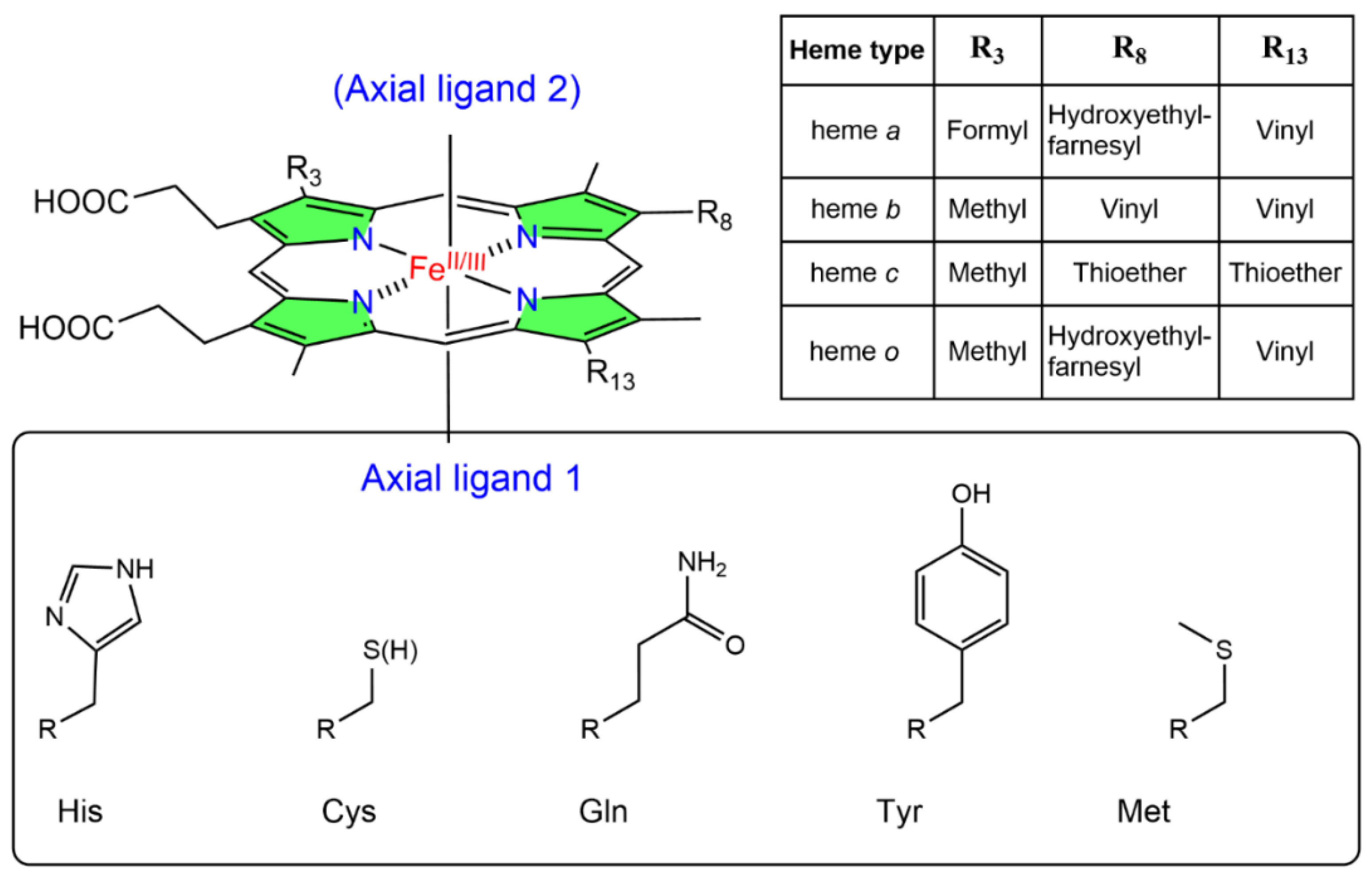
Figure 1. Structures of heme a, b, c, and o. The pyrrol rings are in green. The axial ligands mentioned in this work are reported.
Antioxidants 12 00321 g001
Figure 2. NO bound to an iron ion of a heme group (a) and to a Cys residue as nitrosothiol (SNO) (b).
Antioxidants 12 00321 g002
Figure 3. Possible reactions of NO with the heme group (a) and Cys residues (b). The reaction numbers (red circles) refer to the reactions reported in the text.
Antioxidants 12 00321 g003
Scheme 1. Hydroxide-mediated reductive nitrosylation of Fe(III) heme b.
Antioxidants 12 00321 sch001
Scheme 2. S-nitrosylation coupled with transnitrosylation of proteins.
Antioxidants 12 00321 sch002
Scheme 3. Metal-to-Cys S-nitrosylation.
Antioxidants 12 00321 sch003
Figure 4. Three-dimensional structures of S-nitrosylated forms of (a) liganded human Hb (PDB 1BUW) with the α chains in cyan, and the β chains in yellow; (b) blackfin tuna Mb (PDB 2NRM) with the globin chain in green; (c) C. lectularius NP (PDB 1Y21) with the protein chain in blue. In all figures, the heme is in red; the binding of NO with Cys is highlighted in stick and balls with Cys in orange, NO in blue and red. In the enlargements, the heme and the residues relevant to S-nitrosylation are reported; the distance between the iron and the Cys is measured in Å.
Antioxidants 12 00321 g004
Scheme 4. Simplified model of the conformation-dependent nitrosylation and denitrosylation of Hb according to Stamler and coworkers.
Antioxidants 12 00321 sch004
Scheme 5. Simplified model of the catalytic cycle of Ascaris Hb, proposed in [65].
Antioxidants 12 00321 sch005
Scheme 6. Proposed kox reaction of NOS. Simplified from [78].
Antioxidants 12 00321 sch006
Scheme 7. Reactions of NO with the heme group and proximal Cys of C. lectularius NP.
Antioxidants 12 00321 sch007
Table 1. List of heme proteins discussed in this review.
ProteinHeme-NO Binding
[Ref]
S-Nitrosylation: Cys Involved; Biological Effect [Ref]; PDB EntryHeme-Dependent SNO [Ref]
Mammalian HemoglobinReaction 2 [55,56,57]Cysβ93; NO release [58];
PDB 1BUW (Figure 4a)
Yes [59];
Disputed [60]
Reaction 4 [8,16,36,37,61]
Reaction 5 [62]
Reaction 6 [63]
Ascaris
Hemoglobin
Reaction 3 [64]Cys72 in vitro evidence; NO-related functions in dioxygenation and oxygenation [64]Yes [65]
MyoglobinReaction 4 [66,67]
Reaction 6 [68]
Human Hb: Cys110, in vitro evidence [67];
Thunnus atlanticus (blackfin tuna) Mb: Cys10, in vitro evidence; Changes in the structure and dynamics [69,70]; PDB 2NRM (Figure 4b);
Salmonid Mbs: Cys107, in vitro evidence; Increase in O2 affinity [71,72]
Yes (human) [67]
NeuroglobinReaction 2 [73]
Reaction 4 [73]
Reaction 5 [73]
Reaction 6 [74]
Murine Ngb: Cys55 [74]Yes [74]
CytoglobinReaction 2 [75]
Reaction 4 [33]
Reaction 6 [75,76]
Cys38 and Cys83 [74]Yes [74]
NO synthaseReaction 2
Reaction 3
Reaction 4 [77,78]
Cys101, in vitro evidence; Regulation of NO signaling pathways in the vascular endothelium [79]ND 1
Cys93-Cys98, in vitro evidence; Formation of eNOS homodimer [80,81]
Guanilyl cyclaseReaction 2 [82,83,84]βCys122 and αCys243; Desensitization of NO [85,86]Yes [85]
CyclooxygenaseReaction 2 [87]COX2: Cys526; Activation of the enzyme [88]ND 1
Nitrophorin (Cimex lectularius)Reaction 3 [31,89]Cys60; NO release [90]; PDB 1Y21 (Figure 4c)Yes [90]
Ascorbate peroxidase
(Plant)
Reaction 3 [91]Arabdopsis thaliana APX1: Cys32 in vivo and Cys49 in vitro evidence; Increase in the enzymatic activity; regulation of immune response [92,93]ND 1
CatalaseReaction 3 [91]Ganoderma lucidum: Cys401, Cys642, Cys653 in vivo and in vitro evidence; Alteration in catalase (CAT) activity [94]ND 1
Cytochrome cReaction 2 [95]
Reaction 3 [96]
Trans-nitrosation of other thiols; Mediator of S-nitrosation in biological systems [97,98]ND 1
Cytochrome c oxidaseReaction 2 [99,100]Key Cys residues in subunit II of CcOX in vitro evidence Cys196, Cys200; Inhibition of cell respiration [101,102]ND 1
1 Not determined.
Disclaimer/Publisher’s Note: The statements, opinions and data contained in all publications are solely those of the individual author(s) and contributor(s) and not of MDPI and/or the editor(s). MDPI and/or the editor(s) disclaim responsibility for any injury to people or property resulting from any ideas, methods, instructions or products referred to in the content.