
A Review of the Role of Curcumin in Metal Induced Toxicity

 ,
,  ,
,  , and
, and
Abstract
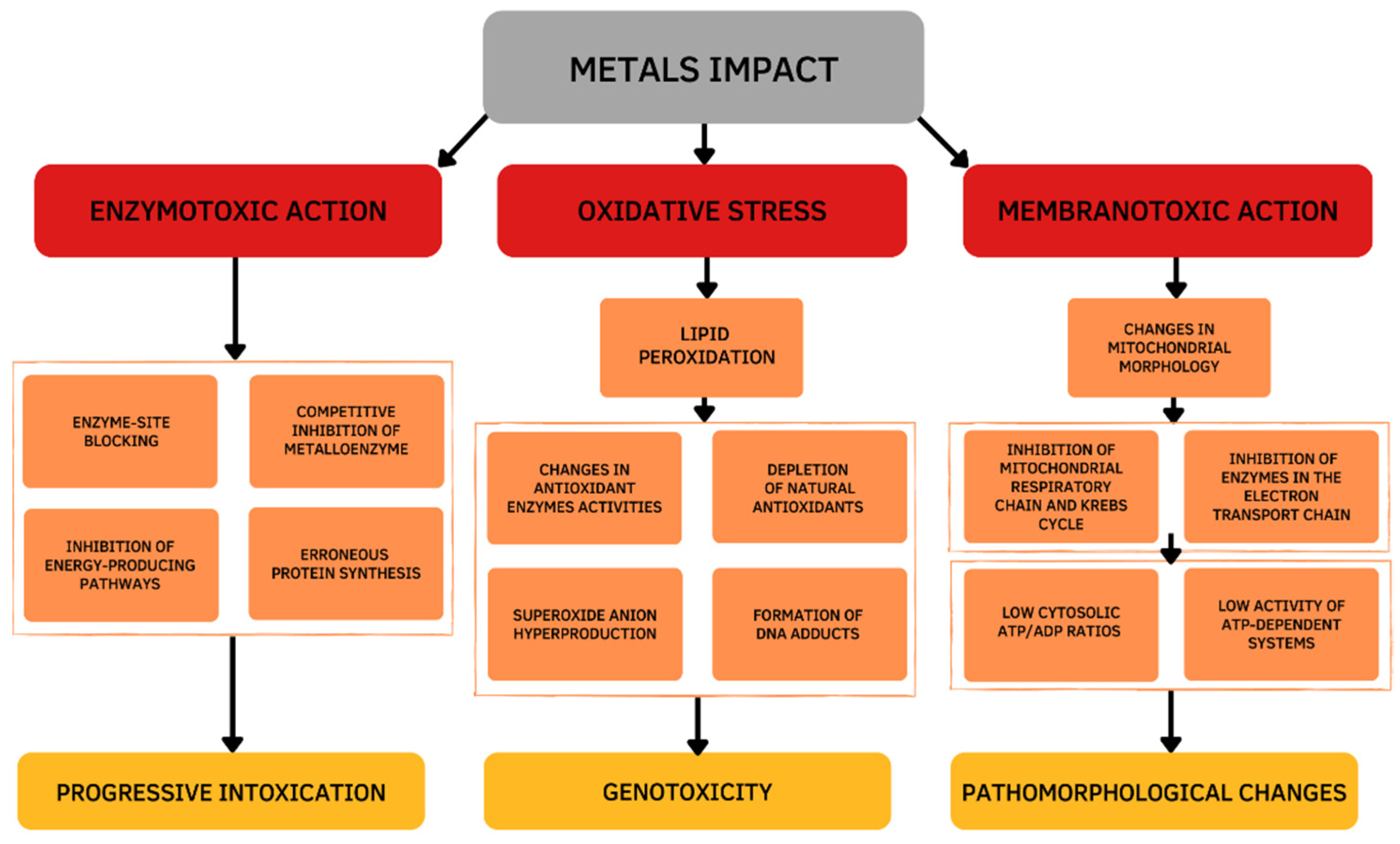
1. Introduction
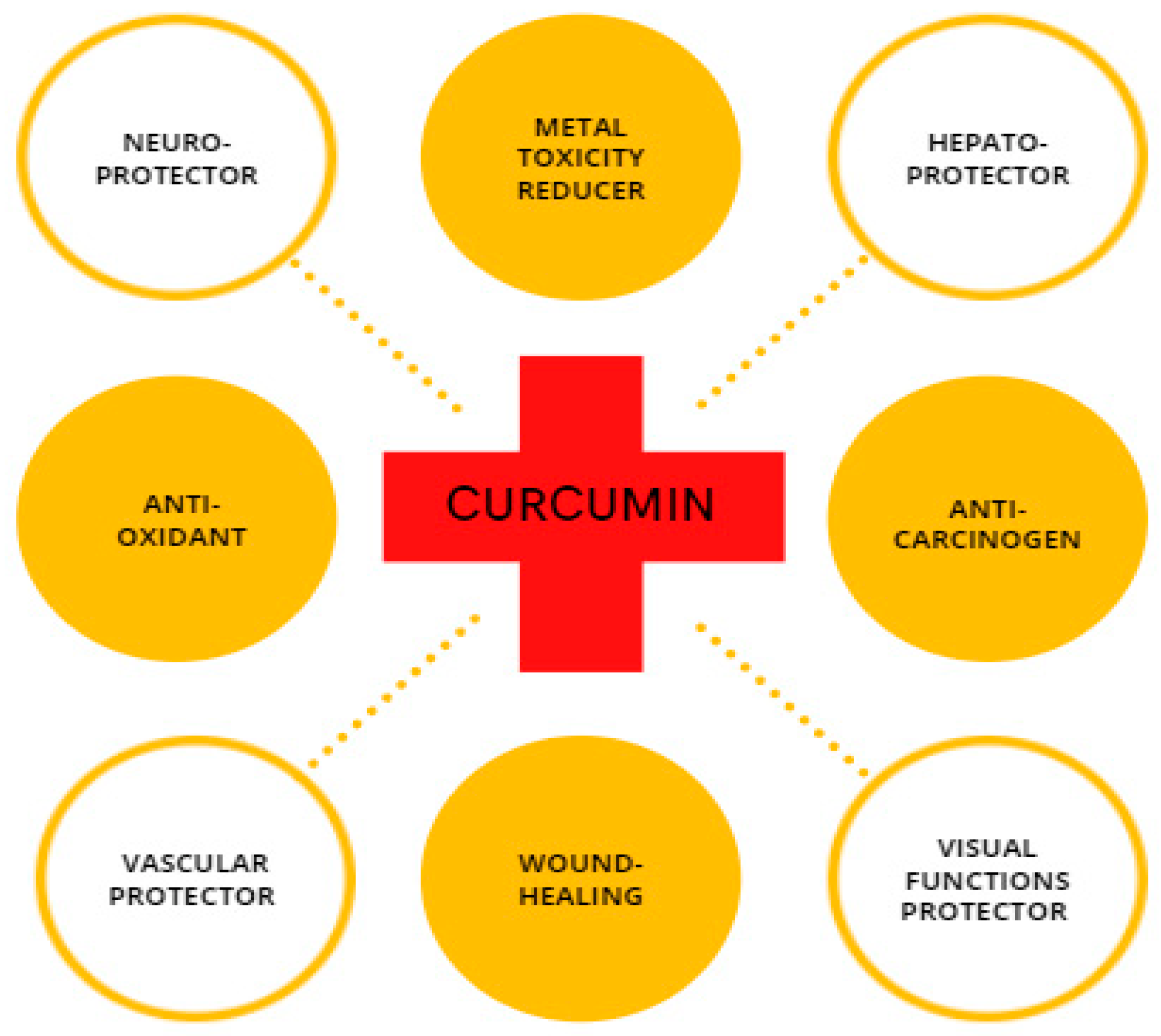
2. Curcumin as a Bioactive Compound in Turmeric Plant
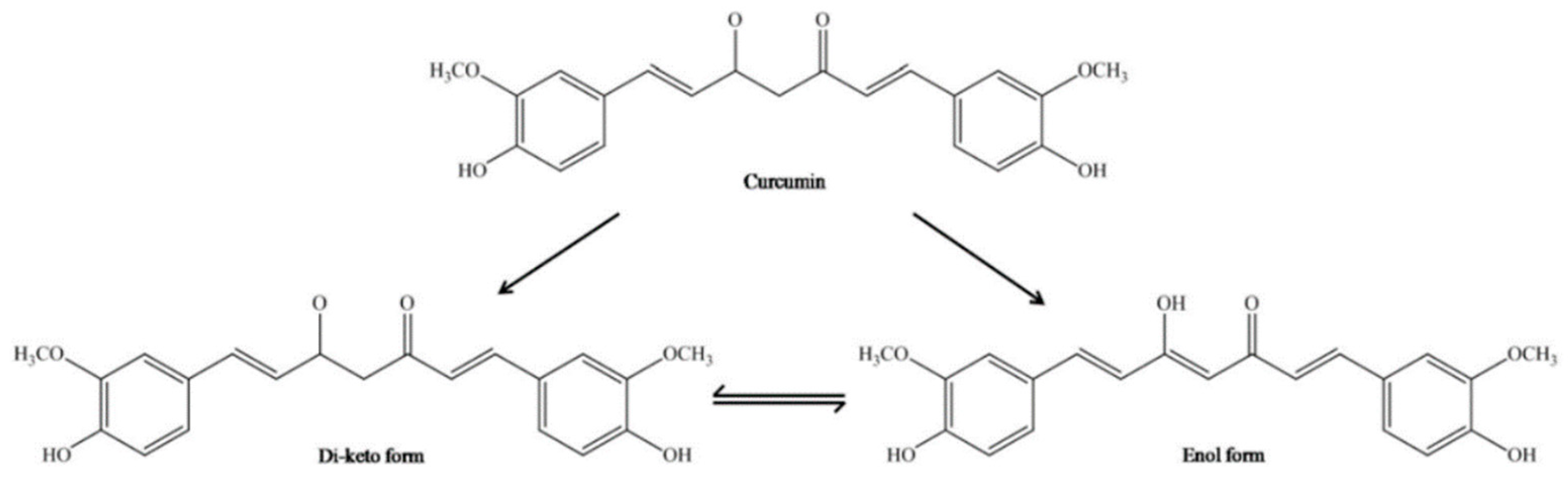
3. Chemical Properties of Curcumin
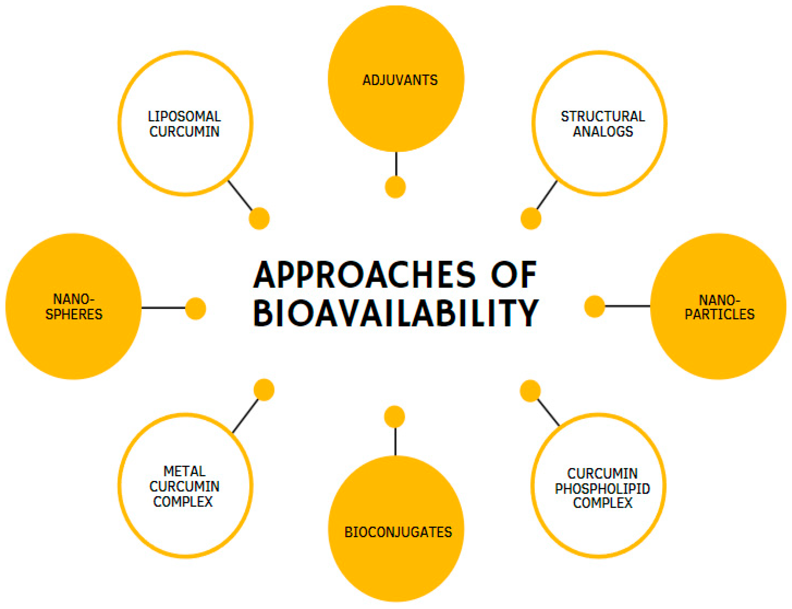
4. Bioavailability of Curcumin
5. General Perspective of Curcumin in the Protection of Metal Toxicity
6. Curcumin on Aluminum-Induced Toxicity
7. Curcumin on Arsenic-Induced Toxicity

| Dose/Concentration | Name of Animal Model/Cell Lines | Route of Exposure | Duration of Exposure/Treatment | Results | Source |
|---|---|---|---|---|---|
| Clinical Trial | |||||
| CUR + piperine (20:1) 2 × 500 mg/day | Chronically arsenic-exposed males or females | Orally | 6 months | ↓DNA damage, ↓ROS generation, ↓CAT, SOD enzymes, | [111] |
| CUR + piperine (100:1) 500 mg twice/day | Chronically arsenic-exposed males or females | Orally | 6 months | ↑expression of protein, mRNA of DNA-PK, DNA ligase IV, XRCC4, ↑BER and NHEJ repair pathways, ↓DNA-damaging effect in lymphocytes | [112] |
| In Vivo | |||||
| 5 mg/kg b.w. NaAsO2 + 15 mg/kg b.w. CUR | Male Wistar rats | NaAsO2-orally/CUR-orally | 30 days (co-administration) | ↓transaminases, phosphatases, glucose, urea, creatinine, bilirubin, TL, cholesterol, TG, plasma and brain ache, the levels of TP and Alb | [113] |
| 5 or 300 ppm NaAsO2 + 0.5 mg/kg b.w. nano-CUR | Male Swiss albino mice | NaAsO2-drinking water/CUR-orally | NaAsO2-7 days/CUR-14 days(post-treatment) | ↓histopathological alterations, ↓accumulation of acidic vesicles, ↓apoptotic cells in the thymus and spleen, ↓autophagy, ↓redox imbalance in immune cells | [115] |
| In Vitro | |||||
| 10 μM NaAsO2 + 0, 1, 2.5, 5, 10, 25, 50 or 100 μM CUR | PC12 cells | Cell line | 24 h | ↑membrane integrity, ↓DNA damage, apoptosis rate, ↑protein expressions, ↑cell viability, ↑cytoprotective effect, ↓oxidative stress | [114] |
8. Curcumin on Cadmium-Induced Toxicity
9. Curcumin on Copper-Induced Toxicity
10. Curcumin on Iron-Induced Toxicity
11. Curcumin on Lead-Induced Toxicity
| Dose/Concentration | Name of Animal Model/Cell Lines | Route of Exposure | Duration of Exposure/Treatment | Results | Source |
|---|---|---|---|---|---|
| In Vivo | |||||
| 50 mg/kg Pb(C2H3O2)2 + 100 mg/kg CUR 50 mg/kg Pb(C2H3O2)2 + 200 mg/kg CUR | Male Sprague–Dawley rats | Pb(C2H3O2)2, CUR- orogastric tube | Pb(C2H3O2)2-4 weeks/CUR-4 weeks (post-treatment) | ↓Pb concentration, ↓oxidative stress, ↓cerebellar damage in cerebellum, ↑motor coordination | [155] |
| 50 mg/kg Pb(C2H3O2)2 + 200 mg/kg CUR | Male Wistar rats | Pb(C2H3O2)2 -IP/CUR-orally | 7 days (co-administration) | ↓apoptosis, ↓oxidative stress, ↓inflammation, ↓liver injury, ↑AKT/GSK-3β signaling pathway | [157] |
| 20 mg/kg OD Pb(C2H3O2)2 + 30 mg/kg BD CUR | Male and female Wistar rats | Pb(C2H3O2)2, CUR- IP | 5 days (co-administration) | ↓damage neurons, ↓protein oxidation, lipid peroxidation, ↑GSH | [158] |
| 1 mg/L Pb + 15 g/kg CUR | Cyprinus carpio | Pb-aquarium water/CUR-orally | 8 weeks (co-administration) | ↓mRNA, expression of NF-kB p65, ↓AST, ALT, LDH, ↑protease activity, ↑SOD, GPx, GSH, G-Rd, GST, Nrf2, ↑lysozyme activity, C3, IgM, ↑intestinal microbial abundance, ↑growth parameters, ↑RBC, Hct, Hb, serum protein, albumin, ↑enzymatic activities, ↑IL-10, ↓MDA | [154] |
| In Vitro | |||||
| 10 μM Pb(C2H3O2)2 + 50, 100, or 150 μM CUR | Rat pups’ hippocampi | 3 h | ↓free radicals, ↑cell viability | [158] | |
12. Curcumin on Zinc-Induced Toxicity
| Dose/Concentration | Name of Animal Model/Cell Lines | Route of Exposure | Duration of Exposure/Treatment | Results | Source |
|---|---|---|---|---|---|
| In Vivo | |||||
| 5.6 mg/kg b.w. ZnONP + 200 mg/kg CUR | Male albino rats | ZnONP-IP/CUR-oral gavage | ZnONP-28 days (started on day 7, three times per week)/CUR-28 days (pre-treatment) | ↑cerebellum structure, ↓oxidative stress markers, ↑inflammatory response, ↓COX-2, P53 | [52] |
| 50 mg/kg ZnONP + 200 mg/kg CUR | Male Wistar rats | ZnONP, CUR-orally | ZnONP-14 days (started on day 7)/CUR-14 days (pre-treatment) | ↑body, kidney weight, ↓MDA in the renal tissue, ↑SOD, GPx, ↓histological changes, ↓apoptotic index | [162] |
| 50 mg/kg nano-ZnO + 200 mg/kg CUR | Male Wistar rats | ZnO-gavage method/ CUR- oral gavage | ZnO-21 days (started on day 7)/CUR-21 days (pre-treatment)2 | ↓lipid peroxidation, ↑SOD, GPx ↓ALT, AST, ALP, ↓histology changes, ↓apoptotic index of hepatocytes | [163] |
13. Curcumin on Mercury-Induced Toxicity
14. Curcumin on Selenium-Induced Toxicity
15. Curcumin on Chromium-Induced Toxicity
16. Conclusions and Future Outlook
Author Contributions
Funding
Data Availability Statement
Acknowledgments
Conflicts of Interest
References
- Islam, M.S.; Hossain, M.B.; Matin, A.; Sarker, M.S. Assessment of heavy metal pollution, distribution and source apportionment in the sediment from Feni River estuary, Bangladesh. Chemosphere 2018, 202, 25–32. [Google Scholar] [CrossRef] [PubMed]
- Ahmed, A.S.; Sultana, S.; Habib, A.; Ullah, H.; Musa, N.; Hossain, M.B.; Rahman, M.M.; Sarker, M.S.I. Bioaccumulation of heavy metals in some commercially important fishes from a tropical river estuary suggests higher potential health risk in children than adults. PLoS ONE 2019, 14, e0219336. [Google Scholar] [CrossRef] [PubMed]
- Hossain, M.B.; Ahmed, A.S.S.; Sarker, M.S.I. Human health risks of Hg, As, Mn, and Cr through consumption of fish, Ticto barb (Puntius ticto) from a tropical river, Bangladesh. Environ. Sci. Pollut. Res. 2018, 25, 31727–31736. [Google Scholar] [CrossRef] [PubMed]
- Engwa, G.A.; Ferdinand, P.U.; Nwalo, F.N.; Unachukwu, M.N. Mechanism and Health Effects of Heavy Metal Toxicity in Humans. In Poisoning in the Modern World-New Tricks for an Old Dog; BoD—Books on Demand GmbH: Norderstedt, Germany, 2019; Volume 10, pp. 70–90. [Google Scholar] [CrossRef]
- Sabath, E.; Robles-Osorio, M.L. Renal health and the environment: Heavy metal nephrotoxicity. Nefrología (Engl. Ed.) 2012, 32, 279–286. [Google Scholar] [CrossRef]
- Jomova, K.; Valko, M. Advances in metal-induced oxidative stress and human disease. Toxicology 2011, 283, 65–87. [Google Scholar] [CrossRef]
- Kim, J.J.; Kim, Y.S.; Kumar, V. Heavy metal toxicity: An update of chelating therapeutic strategies. J. Trace Elem. Med. Biol. 2019, 54, 226–231. [Google Scholar] [CrossRef]
- Deore, M.S.; Keerthana, S.; Naqvi, S.; Kumar, A.; Flora, S.J.S. Alpha-Lipoic Acid Protects Co-Exposure to Lead and Zinc Oxide Nanoparticles Induced Neuro, Immuno and Male Reproductive Toxicity in Rats. Front. Pharmacol. 2021, 12, 1210. [Google Scholar] [CrossRef]
- Perrone, D.; Ardito, F.; Giannatempo, G.; Dioguardi, M.; Troiano, G.; Lo Russo, L.; De Lillo, A.; Laino, L.; Muzio, L.L. Biological and therapeutic activities, and anticancer properties of curcumin (Review). Exp. Ther. Med. 2015, 10, 1615–1623. [Google Scholar] [CrossRef]
- Moniruzzaman, M.; Lee, S.; Park, Y.; Min, T.; Bai, S.C. Evaluation of dietary selenium, vitamin C and E as the multi-antioxidants on the methylmercury intoxicated mice based on mercury bioaccumulation, antioxidant enzyme activity, lipid peroxidation and mitochondrial oxidative stress. Chemosphere 2021, 273, 129673. [Google Scholar] [CrossRef]
- Gulcin, İ.; Alwasel, S.H. Metal Ions, Metal Chelators and Metal Chelating Assay as Antioxidant Method. Processes 2022, 10, 132. [Google Scholar] [CrossRef]
- WHO Technical Report Series. Evaluation of Certain Food Additives and Contaminants. Seventy-Fourth Report of the Joint FAO/WHO Expert Committee on Food Additives Food and Agriculture Organization of the United Nations. 2011. Available online: www.who.int (accessed on 26 October 2022).
- Cuciureanu, R.; Urzica, A.; Voitcu, M.; Antoniu, A. Assessment of daily aluminum intake by food consumption. Rev. Med. Chir. Soc. Med. Nat. Iasi 2000, 104, 107–112. Available online: https://pubmed.ncbi.nlm.nih.gov/12089908/ (accessed on 26 October 2022).
- EFSA European Food Safety Authority. Scientific Opinion on Arsenic in Food. EFSA J. 2009, 7, 1351. [Google Scholar] [CrossRef]
- EFSA European Food Safety Authority. Scientific Opinion on the public health hazards to be covered by inspection of meat (solipeds). EFSA J. 2013, 11, 3263. [Google Scholar] [CrossRef]
- EFSA European Food Safety Authority. Scientific Opinion on the risks to public health related to the presence of chromium in food and drinking water. EFSA J. 2014, 12, 3595. [Google Scholar] [CrossRef]
- Baars, A.J.; Theelen, R.M.C.; Janssen, P.J.C.M.; Meijerink, M.C.M.; Verdam, L.; Zeilmaker, M.J. Re-Evaluation of Human-Toxicological Maxi-Mum Permissible Risk Levels; Rijksinstituut voor Volksgezondheid en Milieu RIVM: Utrecht, The Netherlands, 2001.
- Yates, A.A.; Schlicker, S.A.; Suitor, C.W. Dietary Reference Intakes: The new basis for recommendations for calcium and related nutrients, B vitamins, and choline. J. Am. Diet. Assoc. 1998, 98, 699–706. [Google Scholar] [CrossRef]
- WHO Food Additives Series 18. Iron. Available online: https://inchem.org/documents/jecfa/jecmono/v18je18.htm (accessed on 26 October 2022).
- Alexander, J.; Benford, D.; Boobis, A.; Ceccatelli, S.; Cravedi, J.-P.; Di Domenico, A.; Doerge, D.; Dogliotti, E.; Edler, L.; Farmer, P.; et al. Scientific Opinion on Lead in Food. EFSA J. 2010, 8, 1570. [Google Scholar] [CrossRef]
- Chib, S.; Singh, S. Manganese and related neurotoxic pathways: A potential therapeutic target in neurodegenerative diseases. Neurotoxicol. Teratol. 2022, 94, 107124. [Google Scholar] [CrossRef]
- Aubrac, G.; Bastiansz, A.; Basu, N. Systematic Review and Meta-Analysis of Mercury Exposure among Populations and Environments in Contact with Electronic Waste. Int. J. Environ. Res. Public Health 2022, 19, 11843. [Google Scholar] [CrossRef]
- de Almeida, R.P.; Ferrari, R.G.; Kato, L.S.; Hauser-Davis, R.A.; Conte-Junior, C.A. A Systematic Review on Metal Dynamics and Marine Toxicity Risk Assessment Using Crustaceans as Bioindicators. Biol. Trace Elem. Res. 2022, 200, 881–903. [Google Scholar] [CrossRef]
- Johnson, L.J.; Meacham, S.L.; Kruskall, L.J. The antioxidants-vitamin C, vitamin E, selenium, and carotenoids. J. Agromedicine 2003, 9, 65–82. [Google Scholar] [CrossRef] [PubMed]
- Gera, M.; Sharma, N.; Ghosh, M.; Huynh, D.L.; Lee, S.J.; Min, T.; Kwon, T.; Jeong, D.K. Nanoformulations of curcumin: An emerging paradigm for improved remedial application. Oncotarget 2017, 8, 66680. [Google Scholar] [CrossRef] [PubMed]
- Zhou, H.; Beevers, C.S.; Huang, S. Targets of curcumin. Curr. Drug Targ. 2011, 12, 332–347. [Google Scholar] [CrossRef] [PubMed]
- Pescosolido, N.; Giannotti, R.; Plateroti, A.M.; Pascarella, A.; Nebbioso, M. Curcumin: Therapeutical potential in ophthalmology. Planta Med. 2014, 80, 249–254. [Google Scholar] [CrossRef] [PubMed]
- Aggarwal, B.B.; Sundaram, C.; Malani, N.; Ichikawa, H. Curcumin: The indian solid gold. Adv. Exp. Med. Biol. 2007, 595, 1–75. [Google Scholar]
- Anand, P.; Thomas, S.G.; Kunnumakkara, A.B.; Sundaram, C.; Harikumar, K.B.; Sung, B.; Thakaran, S.T.; Misra, K.; Priyadarsini, I.K.; Rajasekharan, K.N.; et al. Biological activities of curcumin and its analogues (Congeners) made by man and Mother Nature. Biochem. Pharmacol. 2008, 76, 1590–1611. [Google Scholar] [CrossRef]
- Obeid, M.A.; Alsaadi, M.; Aljabali, A.A. Recent updates in curcumin delivery. J. Liposome Res. 2022, 14, 1–12. [Google Scholar] [CrossRef]
- Soleimani, V.; Sahebkar, A.; Hosseinzadeh, H. Turmeric (Curcuma longa) and its major constituent (curcumin) as nontoxic and safe substances: Review. Phyther. Res. 2018, 32, 985–995. [Google Scholar] [CrossRef]
- Rahmani, A.H.; Al Zohairy, M.A.; Aly, S.M.; Khan, M.A. Curcumin: A Potential Candidate in Prevention of Cancer via Modulation of Molecular Pathways. Biomed. Res. Int. 2014, 2014, 761608. [Google Scholar] [CrossRef]
- Howells, L.M.; Iwuji, C.O.O.; Irving, G.R.B.; Barber, S.; Walter, H.; Sidat, Z.; Griffin-Teall, N.; Singh, R.; Foreman, N.; Patel, S.R.; et al. Curcumin combined with FOLFOX chemotherapy is safe and tolerable in patients with metastatic colorectal cancer in a randomized phase IIa trial. J. Nutr. 2019, 149, 1133–1139. [Google Scholar] [CrossRef]
- Gupta, S.C.; Patchva, S.; Aggarwal, B.B. Therapeutic roles of curcumin: Lessons learned from clinical trials. AAPS J. 2013, 15, 195–218. [Google Scholar] [CrossRef]
- Ramirez-Tortosa, M.C.; Ramirez-Tortosa, C.L.; Mesa, M.D.; Granados, S.; Gil, Á.; Quiles, J.L. Curcumin ameliorates rabbits’s steatohepatitis via respiratory chain, oxidative stress, and TNF-α. Free Radic. Biol. Med. 2009, 47, 924–931. [Google Scholar] [CrossRef]
- Quiles, J.L.; Mesa, M.D.; Ramírez-Tortosa, C.L.; Aguilera, C.M.; Battino, M.; Gil, Á.; Ramirez-Tortosa, M.C. Curcuma longa extract supplementation reduces oxidative stress and attenuates aortic fatty streak development in rabbits. Arterioscler. Thromb. Vasc. Biol. 2002, 22, 1225–1231. [Google Scholar] [CrossRef]
- Liu, L.; Li, Y.; Peng, H.; Liu, R.; Ji, W.; Shi, Z.; Shen, J.; Ma, G.; Zhang, X. Targeted exosome coating gene-chem nanocomplex as “nanoscavenger” for clearing α-synuclein and immune activation of Parkinson’s disease. Sci. Adv. 2020, 6, eaba3967. [Google Scholar] [CrossRef]
- Mythri, R.B.; Bharath, M.M.S. Curcumin: A Potential Neuroprotective Agent in Parkinson’s Disease. Curr. Pharm. Des. 2012, 18, 91–99. [Google Scholar] [CrossRef]
- Patil, R.; Gangalum, P.R.; Wagner, S.; Portilla-Arias, J.; Ding, H.; Rekechenetskiy, A.; Konda, B.; Inoue, S.; Black, K.L.; Ljubimova, J.Y.; et al. Curcumin Targeted, Polymalic Acid-Based MRI Contrast Agent for the Detection of Aβ Plaques in Alzheimer’s Disease. Macromol. Biosci. 2012, 15, 1212–1217. [Google Scholar] [CrossRef]
- Liu, Z.J.; Li, Z.H.; Liu, L.; Tang, W.X.; Wang, Y.; Dong, M.R.; Xiao, C. Curcumin attenuates beta-amyloid-induced neuroinflammation via activation of peroxisome proliferator-activated receptor-gamma function in a rat model of Alzheimer’s disease. Front. Pharmacol. 2016, 7, 261. [Google Scholar] [CrossRef]
- Tejada, S.; Manayi, A.; Daglia, M.F.; Nabavi, S.; Sureda, A.; Hajheydari, Z.; Gortzi, O.; Pazoki-Toroudi, H.; Nabavi, S.M. Wound Healing Effects of Curcumin: A Short Review. Curr. Pharm. Biotechnol. 2016, 17, 1002–1007. [Google Scholar] [CrossRef]
- Cheppudira, B.; Fowler, M.; McGhee, L.; Greer, A.; Mares, A.; Petz, L.; Devore, D.; Loyd, D.R.; Clifford, J.L. Curcumin: A novel therapeutic for burn pain and wound healing. Expert Opin. Investig. Drugs 2013, 22, 1295–1303. [Google Scholar] [CrossRef]
- Akbik, D.; Ghadiri, M.; Chrzanowski, W.; Rohanizadeh, R. Curcumin as a wound healing agent. Life Sci. 2014, 116, 1–7. [Google Scholar] [CrossRef]
- Huang, H.C.; Xu, K. Curcumin-mediated neuroprotection against amyloid-β-induced mitochondrial dysfunction involves the inhibition of GSK-3β. J. Alzheimers Dis. 2012, 32, 981–996. [Google Scholar] [CrossRef]
- Nam, S.M.; Choi, J.H.; Yoo, D.Y.; Kim, W.; Jung, H.Y.; Kim, J.W.; Yoo, M.; Lee, S.; Kim, C.J.; Yoon, Y.S.; et al. Effects of curcumin (Curcuma longa) on learning and spatial memory as well as cell proliferation and neuroblast differentiation in adult and aged mice by upregulating brain-derived neurotrophic factor and CREB signaling. J. Med. Food 2014, 17, 641–649. [Google Scholar] [CrossRef]
- Issuriya, A.; Kumarnsit, E.; Wattanapiromsakul, C.; Vongvatcharanon, U. Histological studies of neuroprotective effects of Curcuma longa Linn. on neuronal loss induced by dexamethasone treatment in the rat hippocampus. Acta Histochem. 2014, 116, 1443–1453. [Google Scholar] [CrossRef]
- Yu, S.Y.; Gao, R.; Zhang, L.; Luo, J.; Jiang, H.; Wang, S. Curcumin ameliorates ethanol-induced memory deficits and enhanced brain nitric oxide synthase activity in mice. Prog. Neuropsychopharmacol. Biol. Psychiatry 2013, 44, 210–216. [Google Scholar] [CrossRef]
- Nazari, Q.A.; Kume, T.; Izuo, N.; Takada-Takatori, Y.; Imaizumi, A.; Hashimoto, T.; Izumi, Y.; Akaike, A. Neuroprotective effects of curcumin and highly bioavailable curcumin on oxidative stress induced by sodium nitroprusside in rat striatal cell culture. Biol. Pharm. Bull. 2013, 36, 1356–1362. [Google Scholar] [CrossRef] [PubMed]
- Sookram, C.; Tan, M.; Daya, R.; Heffernan, S.; Mishra, R.K. Curcumin prevents haloperidol-induced development of abnormal oro-facial movements: Possible implications of Bcl-XL in its mechanism of action. Synapse 2011, 65, 788–794. [Google Scholar] [CrossRef] [PubMed]
- Katsidoni, V.; Alexiou, P.; Fotiadou, M.; Pelecanou, M.; Sagnou, M.; Panagis, G. Curcumin, demethoxycurcumin and bisdemethoxycurcumin differentially inhibit morphine’s rewarding effect in rats. Psychopharmacology 2014, 231, 4467–4478. [Google Scholar] [CrossRef] [PubMed]
- Jones, E.A.; Shahed, A.; Shoskes, D.A. Modulation of apoptotic and inflammatory genes by bioflavonoids and angiotensin II inhibition in ureteral obstruction. Urology 2000, 56, 346–351. [Google Scholar] [CrossRef] [PubMed]
- Amer, M.G.; Karam, R.A. Morphological and Biochemical Features of Cerebellar Cortex After Exposure to Zinc Oxide Nanoparticles: Possible Protective Role of Curcumin. Anat. Rec. 2018, 301, 1454–1466. [Google Scholar] [CrossRef]
- Hong, J.; Bose, M.; Ju, J.; Ryu, J.H.; Chen, X.; Sang, S.; Lee, M.J.; Yang, C.S. Modulation of arachidonic acid metabolism by curcumin and related β-diketone derivatives: Effects on cytosolic phospholipase A2, cyclooxygenases and 5-lipoxygenase. Carcinogenesis 2004, 25, 1671–1679. [Google Scholar] [CrossRef]
- Tanrikulu-Küçük, S.; Basaran-Küçükgergin, C.; Sögüt, I.; Tunçdemir, M.; Dogru-Abbasoglu, S.; Seyithanoglu, M.; Kocak, H.; Oner-Iyidogan, Y. Dietary curcumin and capsaicin: Relationship with hepatic oxidative stress and apoptosis in rats fed a high fat diet. Adv. Clin. Exp. Med. 2019, 28, 1013–1020. [Google Scholar] [CrossRef]
- Jain, S.K.; Rains, J.; Jones, K. Effect of curcumin on protein glycosylation, lipid peroxidation, and oxygen radical generation in human red blood cells exposed to high glucose levels. Free Radic. Biol. Med. 2006, 41, 92–96. [Google Scholar] [CrossRef]
- Dai, J.; Gu, L.; Su, Y.; Wang, Q.; Zhao, Y.; Chen, X.; Deng, H.; Li, W.; Wang, G.; Li, K. Inhibition of curcumin on influenza A virus infection and influenzal pneumonia via oxidative stress, TLR2/4, p38/JNK MAPK and NF-κB pathways. Int. Immunopharmacol. 2018, 54, 177–187. [Google Scholar] [CrossRef]
- Gupta, A.; Kumar, A.; Naqvi, S.; Flora, S.J.S. Chronic exposure to multi-metals on testicular toxicity in rats. Toxicol. Mech. Methods 2021, 31, 53–66. [Google Scholar] [CrossRef]
- Wright, L.E.; Frye, J.B.; Gorti, B.; Timmermann, B.N.; Funk, J.L. Bioactivity of Turmeric-Derived Curcuminoids and Related Metabolites in Breast Cancer. Curr. Pharm. Des. 2013, 19, 6218–6225. [Google Scholar] [CrossRef]
- Liang, G.; Yang, S.; Zhou, H.; Shao, L.; Huang, K.; Xiao, J.; Huang, Z.; Li, X. Synthesis, crystal structure and anti-inflammatory properties of curcumin analogues. Eur. J. Med. Chem. 2009, 44, 915–919. [Google Scholar] [CrossRef]
- Gupta, S.C.; Prasad, S.; Kim, J.H.; Patchva, S.; Webb, L.J.; Priyadarsini, I.K.; Aggarwal, B.B. Multitargeting by curcumin as revealed by molecular interaction studies. Nat. Prod. Rep. 2011, 28, 1937–1955. [Google Scholar] [CrossRef]
- Lee, W.H.; Loo, C.Y.; Bebawy, M.; Luk, F.; Mason, R.S.; Rohanizadeh, R. Curcumin and its Derivatives: Their Applications in Neuropharmacology and Neuroscience in the 21st Century. Curr. Neuropharmacol. 2013, 11, 338–378. [Google Scholar] [CrossRef]
- Ghorbani, Z.; Hekmatdoost, A.; Mirmiran, P. Anti-hyperglycemic and insulin sensitizer effects of turmeric and its principle constituent curcumin. Int. J. Endocrinol. Metab. 2014, 12, e18081. [Google Scholar] [CrossRef]
- Moniruzzaman, M.; Min, T. Curcumin, curcumin nanoparticles and curcumin nanospheres: A review on their pharmacodynamics based on monogastric farm animal, poultry and fish nutrition. Pharmaceutics 2020, 12, 447. [Google Scholar] [CrossRef]
- Wahlstrom, B.; Blennow, G. A study on the fate of curcumin in the rat. Acta Pharm. Toxicol. 1978, 43, 86–92. [Google Scholar] [CrossRef] [PubMed]
- Pawar, K.S.; Mastud, R.N.; Pawar, S.K.; Pawar, S.S.; Bhoite, R.R.; Bhoite, R.R.; Kulkarni, M.V.; Deshpande, A.R. Oral Curcumin With Piperine as Adjuvant Therapy for the Treatment of COVID-19: A Randomized Clinical Trial. Front. Pharmacol. 2021, 12, 669362. [Google Scholar] [CrossRef] [PubMed]
- Chiang, P.C.; Lin, S.C.; Pan, S.L.; Kuo, C.H.; Tsai, I.L.; Kuo, M.T.; Wen, W.C.; Chen, P.; Guh, J.H. Antroquinonol displays anticancer potential against human hepatocellular carcinoma cells: A crucial role of AMPK and mTOR pathways. Biochem. Pharmacol. 2010, 79, 162–171. [Google Scholar] [CrossRef] [PubMed]
- Maiti, K.; Mukherjee, K.; Gantait, A.; Saha, B.P.; Mukherjee, P.K. Curcumin-phospholipid complex: Preparation, therapeutic evaluation and pharmacokinetic study in rats. Int. J. Pharm. 2007, 330, 155–163. [Google Scholar] [CrossRef]
- Adams, B.K.; Ferst, E.M.; Davis, M.C.; Herold, M.; Kurtkaya, S.; Camalier, R.F.; Hollingshead, M.G.; Kaur, G.; Sausville, E.A.; Rickles, F.R.; et al. Synthesis and biological evaluation of novel curcumin analogs as anti-cancer and anti-angiogenesis agents. Bioorganic Med. Chem. 2004, 12, 3871–3883. [Google Scholar] [CrossRef]
- Kim, J.Y.; Min, T.; Lee, S.J. Nanospheres loaded with curcumin promote gut epithelial motility through F-actin-related migration signaling events. J. Nutr. Biochem. 2021, 88, 108555. [Google Scholar] [CrossRef]
- Kumari, M.; Sharma, N.; Manchanda, R.; Gupta, N.; Syed, A.; Bahkali, A.H.; Nimesh, S. PGMD/curcumin nanoparticles for the treatment of breast cancer. Sci. Rep. 2021, 11, 3824. [Google Scholar] [CrossRef]
- Mihoub, A.B.; Acherar, S.; Frochot, C.; Malaplate, C.; Yen, F.T.; Arab-Tehrany, E. Synthesis of new water soluble β-cyclodextrin@curcumin conjugates and in vitro safety evaluation in primary cultures of rat cortical neurons. Int. J. Mol. Sci. 2021, 22, 3255. [Google Scholar] [CrossRef]
- Marczylo, T.H.; Verschoyle, R.D.; Cooke, D.N.; Morazzoni, P.; Steward, W.P.; Gescher, A.J. Comparison of systemic availability of curcumin with that of curcumin formulated with phosphatidylcholine. Cancer Chemother. Pharmacol. 2007, 60, 171–177. [Google Scholar] [CrossRef]
- Muddineti, O.S.; Shah, A.; Rompicharla, S.V.K.; Ghosh, B.; Biswas, S. Cholesterol-grafted chitosan micelles as a nanocarrier system for drug-siRNA co-delivery to the lung cancer cells. Int. J. Biol. Macromol. 2018, 118, 857–863. [Google Scholar] [CrossRef]
- Das, R.K.; Kasoju, N.; Bora, U. Encapsulation of curcumin in alginate-chitosan-pluronic composite nanoparticles for delivery to cancer cells. Nanomed. Nanotechnol. Biol. Med. 2010, 6, 153–160. [Google Scholar] [CrossRef]
- Bhatia, A.; Flamer, D.; Shah, P.S.; Cohen, S.P. Transforaminal Epidural Steroid Injections for Treating Lumbosacral Radicular Pain from Herniated Intervertebral Discs: A Systematic Review and Meta-Analysis. Anesth. Analg. 2016, 122, 857–870. [Google Scholar] [CrossRef]
- Fonseca-Santos, B.; dos Santos, A.M.; Rodero, C.F.; Daflon Gremião, M.P.; Chorilli, M. Design, characterization, and biological evaluation of curcumin-loaded surfactant-based systems for topical drug delivery. Int. J. Nanomed. 2016, 11, 4553–4562. [Google Scholar] [CrossRef]
- Karthikeyan, A.; Senthil, N.; Min, T. Nanocurcumin: A Promising Candidate for Therapeutic Applications. Front. Pharmacol. 2020, 11, 487. [Google Scholar] [CrossRef]
- Dende, C.; Meena, J.; Nagarajan, P.; Nagaraj, V.A.; Panda, A.K.; Padmanaban, G. Nanocurcumin is superior to native curcumin in preventing degenerative changes in Experimental Cerebral Malaria. Sci. Rep. 2017, 7, 10062. [Google Scholar] [CrossRef]
- Shaikh, J.; Ankola, D.D.; Beniwal, V.; Singh, D.; Kumar, M.N.V.R. Nanoparticle encapsulation improves oral bioavailability of curcumin by at least 9-fold when compared to curcumin administered with piperine as absorption enhancer. Eur. J. Pharm. Sci. 2009, 37, 223–230. [Google Scholar] [CrossRef]
- Wanninger, S.; Lorenz, V.; Subhan, A.; Edelmann, F.T. Metal complexes of curcumin—Synthetic strategies, structures and medicinal applications. Chem. Soc. Rev. 2015, 44, 4986–5002. [Google Scholar] [CrossRef]
- Farzaei, M.H.; Zobeiri, M.; Parvizi, F.; El-Senduny, F.F.; Marmouzi, I.; Coy-Barrera, E.; Naseri, R.; Nobavi, S.M.; Rahimi, R.; Abdollahi, M. Curcumin in liver diseases: A systematic review of the cellular mechanisms of oxidative stress and clinical perspective. Nutrients 2018, 10, 855. [Google Scholar] [CrossRef]
- Sarawi, W.S.; Alhusaini, A.M.; Fadda, L.M.; Alomar, H.A.; Albaker, A.B.; Aljrboa, A.S.; Alotaibi, A.M.; Hasan, I.H.; Mahmoud, A.M. Curcumin and nano-curcumin mitigate copper neurotoxicity by modulating oxidative stress, inflammation, and akt/gsk-3β signaling. Molecules 2021, 26, 5591. [Google Scholar] [CrossRef]
- Kolev, T.M.; Velcheva, E.A.; Stamboliyska, B.A.; Spiteller, M. DFT and experimental studies of the structure and vibrational spectra of curcumin. Int. J. Quantum Chem. 2005, 102, 1069–1079. [Google Scholar] [CrossRef]
- Barik, A.; Mishra, B.; Shen, L.; Mohan, H.; Kadam, R.M.; Dutta, S.; Zhang, H.Y.; Priyadarsini, K.I. Evaluation of a new copper(II)-curcumin complex as superoxide dismutase mimic and its free radical reactions. Free Radic. Biol. Med. 2005, 39, 811–822. [Google Scholar] [CrossRef] [PubMed]
- Simunkova, M.; Alwasel, S.H.; Alhazza, I.M.; Jomova, K.; Kollar, V.; Rusko, M.; Valko, M. Management of oxidative stress and other pathologies in Alzheimer’s disease. Arch. Toxicol. 2019, 93, 2491–2513. [Google Scholar] [CrossRef] [PubMed]
- Borsari, M.; Ferrari, E.; Grandi, R.; Saladini, M. Curcuminoids as potential new iron-chelating agents: Spectroscopic, polarographic and potentiometric study on their Fe(III) complexing ability. Inorganic. Chim. Acta 2002, 328, 61–68. [Google Scholar] [CrossRef]
- Vajragupta, O.; Boonchoong, P.; Watanabe, H.; Tohda, M.; Kummasud, N.; Sumanont, Y. Manganese complexes of curcumin and its derivatives: Evaluation for the radical scavenging ability and neuroprotective activity. Free Radic. Biol. Med. 2003, 35, 1632–1644. [Google Scholar] [CrossRef]
- Priyadarsini, K.I.; Maity, D.K.; Naik, G.H.; Kumar, M.S.; Unnikrishnan, M.K.; Satav, J.G.; Mohan, H. Role of phenolic O-H and methylene hydrogen on the free radical reactions and antioxidant activity of curcumin. Free Radic. Biol. Med. 2003, 35, 475–484. [Google Scholar] [CrossRef]
- Jiao, Y.; Wilkinson, I.V.J.; Christine Pietsch, E.; Buss, J.L.; Wang, W.; Planalp, R.; Torti, F.M.; Torti, S.V. Iron chelation in the biological activity of curcumin. Free Radic. Biol. Med. 2006, 40, 1152–1160. [Google Scholar] [CrossRef]
- Zhao, X.Z.; Jiang, T.; Wang, L.; Yang, H.; Zhang, S.; Zhou, P. Interaction of curcumin with Zn(II) and Cu(II) ions based on experiment and theoretical calculation. J. Mol. Struct. 2010, 984, 316–325. [Google Scholar] [CrossRef]
- Schmitz, A.E.; De Oliveira, P.A.; De Souza, L.F.; Da Silva, D.G.H.; Danielski, S.; Santos, D.B.; de Almeira, E.A.; Prediger, R.D.; Fisher, A.; Farina, M.; et al. Interaction of curcumin with manganese may compromise metal and neurotransmitter homeostasis in the hippocampus of young mice. Biol. Trace Elem. Res. 2014, 158, 399–409. [Google Scholar] [CrossRef]
- Balasubramanian, K. Molecular orbital basis for yellow curry spice curcumin’s prevention of Alzheimer’s disease. J. Agric. Food Chem. 2006, 54, 3512–3520. [Google Scholar] [CrossRef]
- Jaruga, E.; Sokal, A.; Chrul, S.; Bartosz, G. Apoptosis-Independent Alterations in Membrane Dynamics Induced by Curcumin. Exp. Cell Res. 1998, 245, 303–312. [Google Scholar] [CrossRef]
- Rainey, N.E.; Moustapha, A.; Saric, A.; Nicolas, G.; Sureau, F.; Petit, P.X. Iron chelation by curcumin suppresses both curcumin-induced autophagy and cell death together with iron overload neoplastic transformation. Cell Death Discov. 2019, 5, 150. [Google Scholar] [CrossRef]
- Łukasz, B.; Rybakowska, I.M.; Krakowiak, A.; Anand, J.S. Skutki zdrowotne środowiskowej i zawodowej ekspozycji na glin. Med. Pr. 2020, 71, 79–88. [Google Scholar] [CrossRef]
- Lukiw, W.J.; Percy, M.E.; Kruck, T.P. Nanomolar aluminum induces pro-inflammatory and pro-apoptotic gene expression in human brain cells in primary culture. J. Inorg. Biochem. 2005, 99, 1895–1898. [Google Scholar] [CrossRef]
- Banasik, A.; Lankoff, A.; Piskulak, A.; Adamowska, K.; Lisowska, H.; Wojcik, A. Aluminum-induced micronuclei and apoptosis in human peripheral-blood lymphocytes treated during different phases of the cell cycle. Environ. Toxicol. 2005, 20, 402–406. [Google Scholar] [CrossRef]
- Walton, J.R. An aluminum-based rat model for Alzheimer’s disease exhibits oxidative damage, inhibition of PP2A activity, hyperphosphorylated tau, and granulovacuolar degeneration. J. Inorg. Biochem. 2007, 101, 1275–1284. [Google Scholar] [CrossRef]
- Drago, D.; Bettella, M.; Bolognin, S.; Cendron, L.; Scancar, J.; Milacic, R.; Ricchelli, F.; Casini, A.; Messori, L.; Tognon, G.; et al. Potential pathogenic role of β-amyloid1-42-aluminum complex in Alzheimer’s disease. Int. J. Biochem. Cell Biol. 2008, 40, 731–746. [Google Scholar] [CrossRef]
- Sood, P.K.; Nahar, U.; Nehru, B. Stress proteins and glial cell functions during chronic aluminium exposures: Protective role of curcumin. Neurochem. Res. 2012, 37, 639–646. [Google Scholar] [CrossRef]
- Cherny, R.A.; Barnham, K.J.; Lynch, T.; Volitakis, I.; Li, Q.X.; McLean, C.A.; Multhaup, G.; Beyreuther, K.; Tanzi, R.E.; Masters, C.L.; et al. Chelation and intercalation: Complementary properties in a compound for the treatment of Alzheimer’s disease. J. Struct. Biol. 2000, 130, 209–216. [Google Scholar] [CrossRef]
- Jiang, T.; Wang, L.; Zhang, S.; Sun, P.C.; Ding, C.F.; Chu, Y.Q.; Zhou, P. Interaction of curcumin with Al(III) and its complex strctures based on experiments and theoretical calculations. J. Mol. Struct. 2011, 1004, 163–173. [Google Scholar] [CrossRef]
- Ahmadi, F.; Alizadeh, A.A.; Shahabadi, N.; Rahimi-Nasrabadi, M. Study binding of Al-curcumin complex to ds-DNA, monitoring by multispectroscopic and voltammetric techniques. Spectrochim. Acta Part A Mol. Biomol. Spectrosc. 2011, 79, 1466–1474. [Google Scholar] [CrossRef]
- Kumar, A.; Dogra, S.; Prakash, A. Protective effect of curcumin (Curcuma longa), against aluminium toxicity: Possible behavioral and biochemical alterations in rats. Behav. Brain Res. 2009, 205, 384–390. [Google Scholar] [CrossRef] [PubMed]
- Zhang, H.; Zhao, W.; Malhotra, A. Efficacy of Curcumin in Ameliorating Aluminum- Induced Neurotoxicity. J. Environ. Pathol. Toxicol. Oncol. 2018, 37, 163–172. [Google Scholar] [CrossRef] [PubMed]
- Ibrahim, R.M.; Elaal, F.Z.A.; Sahar, Z. Effect of Curcumin and Nano-curcumin on Reduce Aluminum Toxicity in Rats. Int. J. Food Sci. Biotechnol. 2019, 4, 64–73. [Google Scholar] [CrossRef]
- Sharma, D.; Sethi, P.; Hussain, E.; Singh, R. Curcumin counteracts the aluminium-induced ageing-related alterations in oxidative stress, Na+, K+ ATPase and protein kinase C in adult and old rat brain regions. Biogerontology 2009, 10, 489–502. [Google Scholar] [CrossRef] [PubMed]
- Chung, J.Y.; Yu, S.D.; Hong, Y.S. Environmental source of arsenic exposure. J. Prev. Med. Public Health 2014, 47, 253–257. [Google Scholar] [CrossRef]
- Zhang, A.L.; Tang, S.; Fang, Y.; Li, C.Z.; Ding, X.J.; Zhao, H.; Wang, J.H.; Yang, G.H.; Li, J. Histone demethylase JHDM2A regulates H3K9 dimethylation in response to arsenic-induced DNA damage and repair in normal human liver cells. J. Appl. Toxicol. 2020, 40, 1661–1672. [Google Scholar] [CrossRef]
- Fatoki, J.O.; Badmus, J.A. Arsenic as an environmental and human health antagonist: A review of its toxicity and disease initiation. J. Hazard. Mater. Adv. 2022, 5, 100052. [Google Scholar] [CrossRef]
- Biswas, J.; Sinha, D.; Mukherjee, S.; Roy, S.; Siddiqi, M.; Roy, M. Curcumin protects DNA damage in a chronically arsenic-exposed population of West Bengal. Hum. Exp. Toxicol. 2010, 29, 513–524. [Google Scholar] [CrossRef]
- Roy, M.; Sinha, D.; Mukherjee, S.; Biswas, J. Curcumin prevents DNA damage and enhances the repair potential in a chronically arsenic-exposed human population in West Bengal, India. Eur. J. Cancer Prev. 2011, 20, 123–131. [Google Scholar] [CrossRef]
- Yousef, M.I.; El-Demerdash, F.M.; Radwan, F.M.E. Sodium arsenite induced biochemical perturbations in rats: Ameliorating effect of curcumin. Food Chem. Toxicol. 2008, 46, 3506–3511. [Google Scholar] [CrossRef]
- Rahaman, M.S.; Banik, S.; Akter, M.; Rahman, M.M.; Sikder, M.T.; Hosokawa, T.; Saito, T.; Kurasaki, M. Curcumin alleviates arsenic-induced toxicity in PC12 cells via modulating autophagy/apoptosis. Ecotoxicol. Environ. Saf. 2020, 200, 110756. [Google Scholar] [CrossRef]
- Jamal, Z.; Das, J.; Gupta, P.; Dhar, P.; Chattopadhyay, S.; Chatterji, U. Self Nano-Emulsifying Curcumin (SNEC30) attenuates arsenic-induced cell death in mice. Toxicol. Rep. 2021, 8, 1428–1436. [Google Scholar] [CrossRef]
- Ensibi, C.; Daly, Y.M.N. Toxicity assessment of cadmium chloride on planktonic copepods Centropages ponticus using biochemical markers. Toxicol. Rep. 2017, 4, 83–88. [Google Scholar] [CrossRef]
- Park, J.H.; Lee, B.M.; Kim, H.S. Potential protective roles of curcumin against cadmium-induced toxicity and oxidative stress. J. Toxicol. Environ. Health B Crit. Rev. 2021, 24, 95–118. [Google Scholar] [CrossRef]
- Mohajeri, M.; Rezaee, M.; Sahebkar, A. Cadmium-induced toxicity is rescued by curcumin: A review. Biofactors 2017, 43, 645–661. [Google Scholar] [CrossRef]
- Qayoom, A.; Kazmi, S.; Nadir, A.S. Turmeric Powder as a Natural Heavy Metal Chelating Agent: Surface Characterisation. Pak. J. Sci. Ind. Res. Ser. A Phys. Sci. 2017, 60, 1–8. [Google Scholar] [CrossRef]
- Mehmood, S.; Saeed, D.A.; Rizwan, M.; Khan, M.N.; Aziz, O.; Bashir, S.; Ibrahim, M.; Ditta, A.; Akmal, M.; Mumtaz, M.A.; et al. Impact of different amendments on biochemical responses of sesame (Sesamum indicum L.) plants grown in lead-cadmium contaminated soil. Plant Physiol. Biochem. 2018, 132, 345–355. [Google Scholar] [CrossRef]
- Eybl, V.; Kotyzova, D.; Koutensky, J. Comparative study of natural antioxidants—Curcumin, resveratrol and melatonin–In cadmium-induced oxidative damage in mice. Toxicology 2006, 225, 150–156. [Google Scholar] [CrossRef]
- Alghasham, A.; Salem, T.A.; Meki, A.R.M. Effect of cadmium-polluted water on plasma levels of tumor necrosis factor-α, interleukin-6 and oxidative status biomarkers in rats: Protective effect of curcumin. Food Chem. Toxicol. 2013, 59, 160–164. [Google Scholar] [CrossRef]
- Rennolds, J.; Malireddy, S.; Hassan, F.; Tridandapani, S.; Parinandi, N.; Boyaka, P.N.; Cormet-Boyaka, E. Curcumin regulates airway epithelial cell cytokine responses to the pollutant cadmium. Biochem. Biophys. Res. Commun. 2012, 417, 256–261. [Google Scholar] [CrossRef]
- El-Houseiny, W.; Khalil, A.A.; Abd-Elhakim, Y.M.; Badr, H.A. The potential role of turmeric and black pepper powder diet supplements in reversing cadmium-induced growth retardation, ATP depletion, hepatorenal damage, and testicular toxicity in Clarias gariepinus. Aquaculture 2019, 510, 109–121. [Google Scholar] [CrossRef]
- Deevika, B.; Asha, S.; Taju, G.; Nalini, T. Cadmium acetate induced nephrotoxicity and protective role of curcumin in rats. Asian J. Pharm. Clin. Res. 2012, 5, 186–188. [Google Scholar]
- Aktas, C.; Kanter, M.; Erboga, M.; Ozturk, S. Anti-apoptotic effects of curcumin on cadmium-induced apoptosis in rat testes. Toxicol. Ind. Health 2012, 28, 122–130. [Google Scholar] [CrossRef]
- Oguzturk, H.; Ciftci, O.; Aydin, M.; Timurkaan, N.; Beytur, A.; Yilmaz, F. Ameliorative effects of curcumin against acute cadmium toxicity on male reproductive system in rats. Andrologia 2012, 44, 243–249. [Google Scholar] [CrossRef] [PubMed]
- Eybl, V.; Kotyzová, D.; Bludovská, M. The effect of curcumin on cadmium-induced oxidative damage and trace elements level in the liver of rats and mice. Toxicol. Lett. 2004, 151, 79–85. [Google Scholar] [CrossRef]
- Tarasub, N.; Junseecha, T.; Tarasub, C.; Ayutthaya, W.D.N. Protective Effects of Curcumin, Vitamin C, or their Combination on Cadmium-Induced Hepatotoxicity. J. Basic Clin. Pharm. 2012, 3, 273. [Google Scholar] [CrossRef]
- Kukongviriyapan, U.; Pannangpetch, P.; Kukongviriyapan, V.; Donpunha, W.; Sompamit, K.; Surawattanawan, P. Curcumin protects against cadmium-induced vascular dysfunction, hypertension and tissue cadmium accumulation in mice. Nutrients 2014, 6, 1194–1208. [Google Scholar] [CrossRef]
- Daniel, S.; Limson, J.L.; Dairam, A.; Watkins, G.M.; Daya, S. Through metal binding, curcumin protects against lead- and cadmium-induced lipid peroxidation in rat brain homogenates and against lead-induced tissue damage in rat brain. J. Inorg. Biochem. 2004, 98, 266–275. [Google Scholar] [CrossRef]
- Brewer, G.J. Alzheimer’s disease causation by copper toxicity and treatment with zinc. Front. Aging Neurosci. 2014, 6, 92. [Google Scholar] [CrossRef]
- Montes, S.; Rivera-Mancia, S.; Diaz-Ruiz, A.; Tristan-Lopez, L.; Rios, C. Copper and copper proteins in Parkinson’s disease. Oxid. Med. Cell. Longev. 2014, 2014, 147251. [Google Scholar] [CrossRef]
- Uriu-Adams, J.Y.; Keen, C.L. Copper, oxidative stress, and human health. Mol. Asp. Med. 2005, 26, 268–298. [Google Scholar] [CrossRef]
- Bourassa, M.W.; Brown, H.H.; Borchelt, D.R.; Vogt, S.; Miller, L.M. Metal-deficient aggregates and diminished copper found in cells expressing SOD1 mutations that cause ALS. Front. Aging Neurosci. 2014, 6, 110. [Google Scholar] [CrossRef]
- Tegoni, M.; Valensin, D.; Toso, L.; Remelli, M. Copper chelators: Chemical properties and bio-medical applications. Curr. Med. Chem. 2014, 21, 3785–3818. [Google Scholar] [CrossRef]
- Balasubramanian, K. Quantum chemical insights into Alzheimer’s disease: Curcumin’s chelation with Cu(II), Zn(II), and Pd(II) as a mechanism for its prevention. Int. J. Quantum Chem. 2016, 116, 1107–1119. [Google Scholar] [CrossRef]
- Elkhateeb, S.A.; Ibrahim, T.R.; El-Shal, A.S.; Hamid, O.I.A. Ameliorative role of curcumin on copper oxide nanoparticles-mediated renal toxicity in rats: An investigation of molecular mechanisms. J. Biochem. Mol. Toxicol. 2020, 34, e22593. [Google Scholar] [CrossRef]
- Shen, L.; Zhang, H.Y.; Ji, H.F. A theoretical study on Cu(II)-chelating properties of curcumin and its implications for curcumin as a multipotent agent to combat Alzheimer’s disease. J. Mol. Struct. Theochem. 2005, 757, 199–202. [Google Scholar] [CrossRef]
- Abolaji, A.O.; Fasae, K.D.; Iwezor, C.E.; Aschner, M.; Farombi, E.O. Curcumin attenuates copper-induced oxidative stress and neurotoxicity in Drosophila melanogaster. Toxicol. Rep. 2020, 7, 261–268. [Google Scholar] [CrossRef]
- Hashish, E.A.; Elgaml, S.A. Hepatoprotective and Nephroprotective Effect of Curcumin Against Copper Toxicity in Rats. Indian J. Clin. Biochem. 2016, 31, 270–277. [Google Scholar] [CrossRef]
- Baum, L.; Ng, A. Curcumin interaction with copper and iron suggests one possible mechanism of action in Alzheimer’s disease animal models. J. Alzheimers Dis. 2004, 6, 367–377. [Google Scholar] [CrossRef]
- Silva, B.; Faustino, P. An overview of molecular basis of iron metabolism regulation and the associated pathologies. Biochim. Biophys. Acta Mol. Basis Dis. 2015, 1852, 1347–1359. [Google Scholar] [CrossRef]
- Moinipour, N.; Barati, M.; Sahebkar, A.; Iranshahy, M.; Shakeri, A. Protective Effects of Curcumin against Iron-induced Toxicity. Curr. Pharm. Biotechnol. 2021, 23, 1020–1027. [Google Scholar] [CrossRef] [PubMed]
- Stockwell, B.R.; Angeli, J.P.F.; Bayir, H.; Bush, A.I.; Conrad, M.; Dixon, S.J.; Fulda, S.; Gascon, S.; Hatzios, S.K.; Kagan, V.E.; et al. Ferroptosis: A Regulated Cell Death Nexus Linking Metabolism, Redox Biology, and Disease. Cell 2017, 171, 273–285. [Google Scholar] [CrossRef] [PubMed]
- Kose, T.; Vera-Aviles, M.; Sharp, P.A.; Latunde-Dada, G.O. Curcumin and (−)-epigallocatechin-3-gallate protect murine MIN6 pancreatic beta-cells against iron toxicity and erastin-induced ferroptosis. Pharmaceuticals 2019, 12, 26. [Google Scholar] [CrossRef] [PubMed]
- Manjunatha, H.; Srinivasan, K. Protective effect of dietary curcumin and capsaicin on induced oxidation of low-density lipoprotein, iron-induced hepatotoxicity and carrageenan-induced inflammation in experimental rats. FEBS J. 2006, 273, 4528–4537. [Google Scholar] [CrossRef]
- Messner, D.J.; Sivam, G.; Kowdley, K.V. Curcumin reduces the toxic effects of iron loading in rat liver epithelial cells. Liver Int. 2009, 29, 63–72. [Google Scholar] [CrossRef]
- Wani, A.L.; Ara, A.; Usmani, J.A. Lead toxicity: A review. Interdiscip. Toxicol. 2015, 8, 55–64. [Google Scholar] [CrossRef]
- Patra, R.C.; Rautray, A.K.; Swarup, D. Oxidative stress in lead and cadmium toxicity and its amelioration. Vet. Med. Int. 2011, 2011, 457327. [Google Scholar] [CrossRef]
- Gutteridge, J.M.C. Lipid peroxidation initiated by superoxide-dependent hydroxyl radicals using complexed iron and hydrogen peroxide. FEBS Lett. 1984, 172, 245–249. [Google Scholar] [CrossRef]
- Lee, J.W.; Choi, H.; Hwang, U.K.; Kang, J.C.; Kang, Y.J.; Kim, K.I.; Kim, J.H. Toxic effects of lead exposure on bioaccumulation, oxidative stress, neurotoxicity, and immune responses in fish: A review. Environ. Toxicol. Pharmacol. 2019, 68, 101–108. [Google Scholar] [CrossRef]
- Giri, S.S.; Yun, S.; Jun, J.W.; Kim, H.J.; Kim, S.G.; Kang, J.W.; Kim, S.W.; Han, S.J.; Sukumaran, V.; Park, S.C. Therapeutic effect of intestinal autochthonous Lactobacillus reuteri P16 against waterborne lead toxicity in Cyprinus carpio. Front. Immunol. 2018, 9, 1824. [Google Scholar] [CrossRef]
- Giri, S.S.; Kim, M.J.; Kim, S.G.; Kim, S.W.; Kang, J.W.; Kwon, J.; Lee, S.B.; Jung, W.J.; Sukumaran, V.; Park, S.C. Role of dietary curcumin against waterborne lead toxicity in common carp Cyprinus carpio. Ecotoxicol. Environ. Saf. 2021, 219, 112318. [Google Scholar] [CrossRef]
- Abubakar, K.; Mailafiya, M.M.; Danmaigoro, A.; Chiroma, S.M.; Rahim, E.B.A.; Zakaria, M.Z.A.B. Curcumin attenuates lead-induced cerebellar toxicity in rats via chelating activity and inhibition of oxidative stress. Biomolecules 2019, 9, 453. [Google Scholar] [CrossRef] [PubMed]
- Abubakar, K.; Mailafiya, M.M.; Chiroma, S.M.; Danmaigoro, A.; Zyoud, T.Y.T.; Rahim, E.A.; Zakaria, M.Z.A.B. Ameliorative effect of curcumin on lead-induced hematological and hepatorenal toxicity in a rat model. J. Biochem. Mol. Toxicol. 2020, 34, e22483. [Google Scholar] [CrossRef]
- Alhusaini, A.; Fadda, L.; Hasan, I.H.; Zakaria, E.; Alenazi, A.M.; Mahmoud, A.M. Curcumin ameliorates lead-induced hepatotoxicity by suppressing oxidative stress and inflammation, and modulating akt/gsk-3β signaling pathway. Biomolecules 2019, 9, 703. [Google Scholar] [CrossRef]
- Dairam, A.; Limson, J.L.; Watkins, G.M.; Antunes, E.; Daya, S. Curcuminoids, curcumin, and demethoxycurcumin reduce lead-induced memory deficits in male wistar rats. J. Agric. Food Chem. 2007, 55, 1039–1044. [Google Scholar] [CrossRef]
- Mocchegiani, E.; Romeo, J.; Malavolta, M.; Costarelli, L.; Giacconi, R.; Diaz, L.E.; Marcos, A. Zinc: Dietary intake and impact of supplementation on immune function in elderly. Age 2013, 35, 839–860. [Google Scholar] [CrossRef]
- Keerthana, S.; Kumar, A. Potential risks and benefits of zinc oxide nanoparticles: A systematic review. Crit. Rev. Toxicol. 2020, 50, 47–71. [Google Scholar] [CrossRef]
- Mary, C.P.V.; Vijayakumar, S.; Shankar, R. Metal chelating ability and antioxidant properties of Curcumin-metal complexes—A DFT approach. J. Mol. Graph. Model. 2018, 79, 1–14. [Google Scholar] [CrossRef]
- Heidai-Moghadam, A.; Khorsandi, L.; Jozi, Z. Curcumin attenuates nephrotoxicity induced by zinc oxide nanoparticles in rats. Environ. Sci. Pollut. Res. 2019, 26, 179–187. [Google Scholar] [CrossRef]
- Khorsandi, L.; Mansouri, E.; Orazizadeh, M.; Jozi, Z. Curcumin attenuates hepatotoxicity induced by zinc oxide nanoparticles in rats. Balk. Med. J. 2016, 33, 252–257. [Google Scholar] [CrossRef]
- Moniruzzaman, M.; Lee, J.H.; Lee, J.H.; Won, S.H.; Damusaru, J.H.; Bai, S.C. Interactive effect of dietary vitamin E and inorganic mercury on growth performance and bioaccumulation of mercury in juvenile olive flounder, Paralichthys olivaceus treated with mercuric chloride. Anim. Nutr. 2017, 3, 276–283. [Google Scholar] [CrossRef] [PubMed]
- Berlin, M. Mercury in dental amalgam: A risk analysis. Neurotoxicology 2020, 81, 382–386. [Google Scholar] [CrossRef] [PubMed]
- Bjørklund, G.; Antonyak, H.; Polishchuk, A.; Semenova, Y.; Lesiv, M.; Lysiuk, R.; Peana, M. Effect of methylmercury on fetal neurobehavioral development: An overview of the possible mechanisms of toxicity and the neuroprotective effect of phytochemicals. Arch. Toxicol. 2022, 96, 3175–3199. [Google Scholar] [CrossRef] [PubMed]
- Bridges, C.C.; Zalups, R.K. The aging kidney and the nephrotoxic effects of mercury. J. Toxicol. Environ. Health B Crit. Rev. 2017, 20, 55–80. [Google Scholar] [CrossRef] [PubMed]
- Agarwal, R.; Goel, S.K.; Behari, J.R. Detoxification and antioxidant effects of curcumin in rats experimentally exposed to mercury. J. Appl. Toxicol. 2010, 30, 457–468. [Google Scholar] [CrossRef]
- Zhao, G.; Qi, L.; Wang, Y.; Li, X.; Li, Q.; Tang, X.; Wang, X.; Wu, C. Antagonizing effects of curcumin against mercury-induced autophagic death and trace elements disorder by regulating PI3K/AKT and Nrf2 pathway in the spleen. Ecotoxicol. Environ. Saf. 2021, 222, 112529. [Google Scholar] [CrossRef]
- Abu-Taweel, G.M. Neurobehavioral protective properties of curcumin against the mercury chloride treated mice offspring. Saudi J. Biol. Sci. 2019, 26, 736–743. [Google Scholar] [CrossRef]
- Beckett, G.J.; Arthur, J.R. Selenium and endocrine systems. J. Endocrinol. 2005, 184, 455–465. [Google Scholar] [CrossRef]
- Solovyev, N.D. Importance of selenium and selenoprotein for brain function: From antioxidant protection to neuronal signalling. J. Inorg. Biochem. 2015, 153, 1–12. [Google Scholar] [CrossRef]
- Sharma, V.K.; McDonald, T.J.; Sohn, M.; Anquandah, G.A.K.; Pettine, M.; Zboril, R. Assessment of toxicity of selenium and cadmium selenium quantum dots: A review. Chemosphere 2017, 188, 403–413. [Google Scholar] [CrossRef]
- Sun, H.J.; Rathinasabapathi, B.; Wu, B.; Luo, J.; Pu, L.P.; Ma, L.Q. Arsenic and selenium toxicity and their interactive effects in humans. Environ. Int. 2014, 69, 148–158. [Google Scholar] [CrossRef]
- Manikandan, R.; Thiagarajan, R.; Beulaja, S.; Sudhandiran, G.; Arumugam, M. Curcumin protects against hepatic and renal injuries mediated by inducible nitric oxide synthase during selenium-induced toxicity in Wistar rats. Microsc. Res. Tech. 2010, 73, 631–637. [Google Scholar] [CrossRef]
- Manikandan, R.; Thiagarajan, R.; Beulaja, S.; Chindhu, S.; Mariammal, K.; Sudhandiran, G.; Arumugam, M. Anti-cataractogenic effect of curcumin and aminoguanidine against selenium-induced oxidative stress in the eye lens of Wistar rat pups: An in vitro study using isolated lens. Chem. Biol. Interact. 2009, 181, 202–209. [Google Scholar] [CrossRef]
- Manikandan, R.; Thiagarajan, R.; Beulaja, S.; Sudhandiran, G.; Arumugam, M. Effect of curcumin on selenite-induced cataractogenesis in Wistar rat pups. Curr. Eye Res. 2010, 35, 122–129. [Google Scholar] [CrossRef]
- Thompson, C.M.; Aardema, M.J.; Heintz, M.M.; MacGregor, J.T.; Young, R.R. A review of mammalian in vivo genotoxicity of hexavalent chromium: Implications for oral carcinogenicity risk assessment. Crit. Rev. Toxicol. 2021, 51, 820–849. [Google Scholar] [CrossRef]
- Thompson, C.M.; Bichteler, A.; Rager, J.E.; Suh, M.; Proctor, D.M.; Haws, L.C.; Harris, M.A. Comparison of in vivo genotoxic and carcinogenic potency to augment mode of action analysis: Case study with hexavalent chromium. Mutat. Res. Genet. Toxicol. Environ. Mutagen. 2016, 800, 28–34. [Google Scholar] [CrossRef]
- Chandra, A.K.; Chatterjee, A.; Ghosh, R.; Sarkar, M. Effect of curcumin on chromium-induced oxidative damage in male reproductive system. Environ. Toxicol. Pharmacol. 2007, 24, 160–166. [Google Scholar] [CrossRef]
- Molina-Jijón, E.; Tapia, E.; Zazueta, C.; El Hafidi, M.; Zatarain-Barrón, Z.L.; Hernández-Pando, R.; Medina-Campos, O.N.; Zarco-Marquez, G.; Torres, I.; Pedraza-Chaverri, J. Curcumin prevents Cr(VI)-induced renal oxidant damage by a mitochondrial pathway. Free Radic. Biol. Med. 2011, 51, 1543–1557. [Google Scholar] [CrossRef]
- Srivastava, A.; Agarwal, R.; Chaturvedi, T.P.; Chandra, A.; Singh, O.P. Clinical evaluation of the role of tulsi and turmeric in the management of oral submucous fibrosis: A pilot, prospective observational study. J. Ayurveda Integr. Med. 2015, 6, 45–49. [Google Scholar] [CrossRef]
- Pawar, R.S.; Toppo, F.A.; Mandloi, A.S.; Shaikh, S. Exploring the role of curcumin containing ethanolic extract obtained from Curcuma longa (rhizomes) against retardation of wound healing process by aspirin. Indian J. Pharmacol. 2015, 47, 160–166. [Google Scholar] [CrossRef] [PubMed]
- Thavorn, K.; Mamdani, M.M.; Straus, S.E. Efficacy of turmeric in the treatment of digestive disorders: A systematic review and meta-analysis protocol. Syst. Rev. 2014, 3, 71. [Google Scholar] [CrossRef] [PubMed]
- Gautam, S.C.; Gao, X.; Dulchavsky, S. Immunomodulation by curcumin. Adv. Exp. Med. Biol. 2007, 595, 321–341. [Google Scholar] [CrossRef] [PubMed]
- Gilani, A.H.; Shah, A.J.; Ghayur, M.N.; Majeed, K. Pharmacological basis for the use of turmeric in gastrointestinal and respiratory disorders. Life Sci. 2005, 76, 3089–3105. [Google Scholar] [CrossRef]
- Rajasekaran, S.A. Therapeutic potential of curcumin in gastrointestinal diseases. World J. Gastrointest. Pathophysiol. 2011, 2, 1. [Google Scholar] [CrossRef] [PubMed]
- Dulbecco, P.; Savarino, V. Therapeutic potential of curcumin in digestive diseases. World J. Gastroenterol. 2013, 19, 9256–9270. [Google Scholar] [CrossRef]
- Kim, D.C.; Kim, S.H.; Choi, B.H.; Baek, N.I.; Kim, D.; Kim, M.J.; Kim, K.T. Curcuma longa extract protects against gastric ulcers by blocking H2 histamine receptors. Biol. Pharm. Bull. 2005, 28, 2220–2224. [Google Scholar] [CrossRef]
- Yadav, S.; Sah, A.K.; Jha, R.; Sah, P.; Shah, D. Turmeric (curcumin) remedies gastroprotective action. Pharmacogn. Rev. 2013, 7, 42–46. [Google Scholar] [CrossRef]
- Liju, V.B.; Jeena, K.; Kuttan, R. Gastroprotective activity of essential oils from turmeric and ginger. J. Basic Clin. Physiol. Pharmacol. 2015, 26, 95–103. [Google Scholar] [CrossRef]
- Koosirirat, C.; Linpisarn, S.; Changsom, D.; Chawansuntati, K.; Wipasa, J. Investigation of the anti-inflammatory effect of Curcuma longa in Helicobacter pylori-infected patients. Int. Immunopharmacol. 2010, 10, 815–818. [Google Scholar] [CrossRef]
- Huang, X.; Lv, B.; Zhang, S.; Dai, Q.; Chen, B.B.; Meng, L.N. Effects of radix curcumae-derived diterpenoid C on Helicobacter pylori-induced inflammation and nuclear factor kappa B signal pathways. World J. Gastroenterol. 2013, 19, 5085–5093. [Google Scholar] [CrossRef]
- Salomon, N.; Lang, A.; Gamus, D. Curcumin add-on therapy for ulcerative colitis. Harefuah 2015, 154, 56–58. [Google Scholar] [PubMed]
- Nwozo, S.O.; Osunmadewa, D.A.; Oyinloye, B.E. Anti-fatty liver effects of oils from Zingiber officinale and Curcuma longa on ethanol-induced fatty liver in rats. J. Integr. Med. 2014, 12, 59–65. [Google Scholar] [CrossRef] [PubMed]
- Um, M.Y.; Hwang, K.H.; Ahn, J.; Ha, T.Y. Curcumin Attenuates Diet-Induced Hepatic Steatosis byActivating AMP-Activated Protein Kinase. Basic Clin. Pharm. Tox. 2013, 113, 152–157. [Google Scholar] [CrossRef] [PubMed]
- Lee, J.H.; Kim, H.G.; Oh, J.H.; Lee, Y.J. Dramatic increase in hepatic and biliary curcumin exposure by modulation of its elimination pathway in rats. J. Pharm. Pharmacol. 2013, 65, 423–429. [Google Scholar] [CrossRef]
- Desai, K.R.; Rajput, D.K.; Patel, P.B.; Highland, H.N. Ameliorative effects of curcumin on artesunate-Induced subchronic toxicity in testis of swiss albino male mice. Dose-Response 2015, 13, 1559325815592393. [Google Scholar] [CrossRef]
- Beyene, A.M.; Moniruzzaman, M.; Karthikeyan, A.; Min, T. Curcumin nanoformulations with metal oxide nanomaterials for biomedical applications. Nanomaterials 2021, 11, 460. [Google Scholar] [CrossRef]
- Liu, G.; Garrett, M.R.; Men, P.; Zhu, X.; Perry, G.; Smith, M.A. Nanoparticle and other metal chelation therapeutics in Alzheimer disease. Biochim. Biophys. Acta Mol. Basis Dis. 2005, 1741, 246–252. [Google Scholar] [CrossRef]
- Manolova, Y.; Deneva, V.; Antonov, L.; Drakalska, E.; Momekova, D.; Lambov, N. The effect of the water on the curcumin tautomerism: A quantitative approach. Spectrochim. Acta Part A Mol. Biomol. Spectrosc. 2014, 132, 815–820. [Google Scholar] [CrossRef]


| Metal | HBGV | Value | Source |
|---|---|---|---|
| Aluminum | TWI | 2000 μg/kg b.w. | [12] |
| TDI | 10,000 μg/kg b.w. | [13] | |
| Arsenic | TWI | 15 μg/kg b.w. | [14] |
| Cadmium | TWI | 2.5 µg/kg b.w. | [15] |
| Chromium | TDI | 300 μg/kg b.w. | [16] |
| Cobalt | TDI | 1.4 μg/kg b.w. | [17] |
| Copper | UL | 10,000 μg/kg b.w. | [18] |
| Iron | TDI | 800 µg/kg b.w. | [19] |
| Lead | TWI | 25 µg/kg b.w. | [20] |
| TDI | 600 µg/kg b.w. | ||
| Manganese | TDI | 60 µg/kg b.w. | [21] |
| Mercury | TWI | 4 µg/kg b.w. | [22] |
| Nickel | TDI | 12 μg/kg b.w. | [23] |
| Selenium | UL | 400 μg/kg b.w. | [24] |
| Zinc | UL | 40,000 μg/day | [18] |
| Dose/Concentration | Name of Animal Model/Cell Lines | Route of Exposure | Duration of Exposure/Treatment | Results | Source |
|---|---|---|---|---|---|
| 100 mg/kg b.w. Al + 50 mg/kg b.w. CUR | Sprague–Dawley rats | Al-drinking water/CUR-IP | Al-8 weeks/CUR-2 months (co-administration) | ↓TNF-α, ↓NF-kB p65, ↓NO activity | [100] |
| 50 mg/kg/day Al + 30 mg/mL/kg b.w. CUR | Male Wistar rats | Al-drinking water/CUR-orally | 6 months (co-administration) | ↓lipid peroxidation in the brain, ↓SOD, GPx, GST and Na+, K+, ATPase | [107] |
| 100 mg/kg Al + 30 or 60 mg/kg CUR | Male Wistar rats | Al-drinking water/CUR-orally | 42 days(co-administration) | ↓IAL, first and second RL to reach the platform in the pre-trained rats, ↑retention performance of the spatial navigation task; ↓MDA, nitrite levels, ↑reduced GSH, ↓GST, SOD, and catalase activity, ↓AChE activity, ↓Al in hippocampus | [104] |
| Synthesized [Al(CUR) (EtOH)2](NO3)2) complex | Binding of CUR complex to calf thymus-DNA | ↓affinity of Al to interact with DNA | [103] | ||
| Synthesized Al (III)–CUR complexes | NMR, mass spectroscopy, ultraviolet, the generalized 2D UV–UV correlation spectroscopy, the density functional theory | ↓affinity of Al to interact with amyloid beta (Aβ) peptide, ↓ toxicity effect of Al on peptides, ↓ oxidative stress | [102] |
| Dose/Concentration | Name of Animal Model/Cell Lines | Route of Exposure | Duration of Exposure/Treatment | Results | Source |
|---|---|---|---|---|---|
| In Vivo | |||||
| 0.8 mg/L water Cd + basal diet with 0.5% TP | Juvenile African catfish | Cd-aquarium water/TP-orally | 30 days (co-administration) | ↑growth rate, ↑HSI, ↓damage to the hepatic architecture, the testis architecture, ↑reproduction hormone level, lysozyme activity, immunoglobulin levels, protein content, ATP content, ↓disturbed enzyme activities, ↓nephrotoxicity, ↓oxidative stress, ↓apoptotic events, ↓Cd accumulation in muscles, the liver | [124] |
| 40 mg/L CdCl2 + 50 mg/kg b.w. CUR | Male albino rats | Cd-drinking water/CUR-gastric tube | 6 weeks (co-administration) | ↓TNF-α, IL-6, ↓lipid peroxidation, ↑CAT, SOD, GSH, TAC, ↓oxidative stress, ↓loss of antioxidant enzymes | [122] |
| 200 mg/kg b.w. C4H6CdO4 + 250 mg/kg b.w. CUR | Adult male rats | Cd-drinking water/CUR-orally | 7 days (co-administration) | ↓level of urea, creatinine in the serum, ↑antioxidant levels | [125] |
| 1 mg/kg b.w. Cd + 100 mg/kg b.w. CUR | Male Wistar albino rats | Cd-subcutaneously/CUR-intragastric intubation | 4 weeks (co-administration) | ↓testicular damage, ↓reactivity and the number of germ cell, Leydig cell apoptosis, ↓TUNEL positive cells of testis | [126] |
| 1 mg/kg/day or 100 mg/kg/day CdCl2 + 1 mg/kg/day or 100 mg/kg/day CUR | Male Spraque–Dawley rats | CdCl2-IP/CUR-orally | 3 days (co-administration) | ↓TBARS levels, ↑GSH, CAT, GPx, SOD, ↓histopathological changes, ↓lipid peroxidation, ↓oxidative stress in testis tissue, ↑antioxidant enzymes | [127] |
| 7 mg/kg b.w. CdCl2 + 50 mg/kg b.w. CUR | Male CD mice | CdCl2- subcutaneously/CUR-orally | CdCl2-ones/CUR-3 days (pre-treatment) | ↑GSH, GPx, ↑antioxidant status, ↔Cd level in tissues, ↓lipid peroxidation | [121] |
| 0.025 mmol/kg b.w. (rats), 0.03 mmol/kg b.w. (mice) CdCl2 + 50 mg/kg b.w. CUR | Male Wistar rats, male CD mice | CdCl2- subcutaneously/CUR-orally | CdCl2-ones/CUR-3 days (pre-treatment) | ↓lipid peroxidation | [128] |
| 5 mg/kg b.w. CdCl2 + 200 or 400 mg/kg b.w. CUR | Adult male Wistar rats | CdCl2- oral gavage/CUR-oral gavage | 27 days (co-administration) | ↓MDA, ↑GSH, ↓nephrotoxicity | [129] |
| 5 mg/kg b.w. CdCl2 + 200 or 400 mg/kg b.w. CUR | Adult male ICR mice | Cd-drinking water/CUR-intragastric | 8 weeks (co-administration) | ↓systolic, diastolic, and mean arterial blood pressure levels, ↑Phe, Ach, and SNP, ↓hypertension, ↓impairment of vascular responsiveness to vasoactive agents, ↓lipid peroxidation, ↓protein oxidation, ↑GSH, ↑redox status of the blood cells, ↓oxidants, ↑endogenous antioxidant formation, ↓oxidative stress, ↓Cd accumulation in the blood, organs | [130] |
| In Vitro | |||||
| 100 mM CdSO4 + 10, 30, 20, 40 or 50 μM CUR | Bronchial epithelial cell line Calu-3 | 24 h | ↓IL-6, IL-8 mRNA transcript levels, ↓Erk1/2 activation | ||
| 0.025, 0.05, 0.1 mM CdCl2 + 0.025, 0.05, 0.1 mM CUR | Rat brain homogenate | Lipid peroxidation assay | ↓lipid peroxidation | [131] | |
| Dose/Concentration | Name of Animal Model/Cell Lines | Route of Exposure | Duration of Exposure/Treatment | Results | Source |
|---|---|---|---|---|---|
| In Vivo | |||||
| 100 mg/kg CuSO4 + 80 mg/kg CUR; 100 mg/kg CuSO4 + 80 mg/kg nano-CUR | Male Wistar rats | Cu-orally/CUR-orally | 7 days (co-administration) | ↓cerebral oxidative stress, ↓cerebral inflammation in stress, ↓apoptosis, ↑AKT/GSK-3β signaling pathway, ↑BDNF | [82] |
| 250 mg/kg CuONP + 200 mg/kg b.w. CUR | Rats | CuONP-oral gavage/CUR- orally | 3 months (co-administration) | ↑body weight gain, ↓serum creatinine, BUN levels, ↓KIM-1 in urine, kidney, ↓oxidative stress, ↓NO level, ↓mRNA expression of IL1-β, TNF-α, NF-ĸB ↑Nrf2, HO-1, γ-GCS gene expression in the kidney, ↓renal damage, ↓morphological tubular, glomerular alteration, ↓caspase-3 | [138] |
| 1 mM Cu + 0.2 or 0.5 mg/kg CUR | Drosophila melanogaster wild-type (Harwich strain) flies | Cu, CUR-medium | 7 days (co-administration) | ↓oxidative stress, ↓nitrite level, ↓AChE activity, ↓dopamine levels | [140] |
| 4 mg/kg CuSO4 + 80 mg/kg CUR | Albino rats | CuSO4, CUR- orally by stomach tube | 30 days (co-administration) CuSO4-15 days/CUR-15 days (post-treatment) | ↓hepatic marker enzymes, ↑hepatic, renal antioxidants, MDA, ↓renal tissue damage, ↑antioxidant enzymes, ↓ROS | [141] |
| Other | |||||
| Cu2+, Zn2+, Fe2+ + CUR | Spectrophotometry | Binding more readily Fe and Cu than Zn, ↓Aβ toxicity, ↓NF-κB | [142] | ||
| Dose/Concentration | Name of Animal Model/Cell Lines | Route of Exposure | Duration of Exposure/Treatment | Results | Source |
|---|---|---|---|---|---|
| In Vivo | |||||
| 30 mg FeSO4 in 1 mL saline ⁄ kg b.w. + basal diet with 0.2 g% CUR | Male Wistar rats | FeSO4-IP/CUR- oral gavage | FeSO4-ones/CUR-8 weeks (pre-treatment) | ↓LDL oxidation, ↓ALT, AST, LDH, ↓liver lipid peroxide level, ↓severity of hepatotoxicity | [147] |
| In Vitro | |||||
| 250 µM or 500 µM Fe-NTA + 20 μM or 50 μM CUR | Huh-7, T51B, RL-34 cells | 24, 48, 72, 96, 120 h | ↑tumor-promoting effect, ↑Fe2+ chelating, ↔Fe uptake | [94] | |
| 200 µM FAC + 50 µM CUR | T51B cell line | 5 or 10 days | ↑Fe chelating, ↔Fe uptake/bioavailability, ↓ROS, ↓signaling to cellular stress pathways, | [148] | |
| 50 μmol/L FAC or 30 μmol/L FeSO4 + 20 μM CUR | Mouse MIN6 pancreatic β-cell line | 24 h | ↓cell damage, ↑stress protection, ↓cell mortality, ↓Fe accumulation, ↓MDA, ↑GSH | [146] | |
| Dose/Concentration | Name of Animal Model/Cell Lines | Route of Exposure | Duration of Exposure/Treatment | Results | Source |
|---|---|---|---|---|---|
| In Vivo | |||||
| 12 µmol/kg b.w. HgCl2 + 80 mg/kg b.w. CUR | Male Wistar rats | HgCl2-IP/CUR-orally | HgCl2-ones(pre/post-treated groups)/CUR-3 days(post-treatment, pre-treatment) | ↓oxidative stress, ↓lipid peroxidation, ↑GSH, SOD GPx, CAT in the liver, kidney, brain, ↓serum biochemical changes, ↓Hg in tissues | [168] |
| 5 mg/kg HgCl2 + 50 mg/kg b.w. CUR | Kunming male mice | HgCl2, CUR-IP | HgCl2-ones/CUR-24 h (pre-treatment) | ↓autophagic cell death, ↓Na overload, ↓Ca leak, ↑Nrf2 signaling pathway, ↑antioxidant defenses | [169] |
| 10 ppm HgCl2 + 150 or 300 ppm CUR | Pregnant Swiss-Webster strain mice | HgCl2, CUR-oral gavage | 15 days (co-administration) | ↓biochemical, behavioral disorders, ↑cognitive, anxiety behaviors | [170] |
| Dose/Concentration | Name of Animal Model/Cell Lines | Route of Exposure | Duration of Exposure/Treatment | Results | Source |
|---|---|---|---|---|---|
| In Vivo | |||||
| 15 µM/kg b.w. Se + 75 mg/kg b.w. CUR | Wistar rats | Se-IP/CUR-orally | Se-ones(co/pre/post-treated groups)/CUR-ones (co-administration, post-treatment, pre-treatment) | ↓liver cells damage, ↓architecture changes of glomeruli and tubules, ↓iNOS | [175] |
| 15 µM/kg b.w. Se + 75 mg/kg b.w. CUR | Wistar rat pups | Se-IP/CUR-orally | Se-ones(co/pre/post-treated groups)/CUR-ones (co-administration, post-treatment, pre-treatment) | ↓LPO, ↓SOD, GST, GPx, CAT, ↓GSH, vitamin C, vitamin E | [177] |
| In Vitro | |||||
| 100 μM Na2SeO3 + 100 μM or 200 μM CUR | Wistar Rat pups | Isolated lenses incubation in DMEM | 62 h | ↓oxidative stress, ↓cataract formation | [176] |
| Dose/Concentration | Name of Animal Model/Cell Lines | Route of Exposure | Duration of Exposure/Treatment | Results | Source |
|---|---|---|---|---|---|
| In Vivo | |||||
| 0.4 mg/kg b.w. OD K2Cr2O7 + 20 mg/kg b.w. CUR | Male albino rats of Sprague–Dawley strain Rattus norvegicus | K2Cr2O7,CUR-IP | K2Cr2O7-26 days/CUR-26 days(every alternate day) (co-administration) | ↓testicular histology damage, ↑sperm count, ↑testosterone level, ↑accessory sex organs weight, ↓lipid peroxidation, ↑SOD, CAT | [180] |
| 015 mg/kg b.w. K2Cr2O7 + 100, 200 or 400 mg/kg b.w. CUR | Male Wistar rats | K2Cr2O7- subcutaneously/CUR- gavage | K2Cr2O7-ones(co/pre/post-treated groups)/ /CUR- 10 days before and 2 days after K2Cr2O7 injection (co-administration, post-treatment, pre-treatment) | ↓renal dysfunction, ↓histological damage, ↓oxidant stress, ↑antioxidant enzymes, ↔Cr in renal homogenates, in mitochondria (pre-treated), ↑mitochondrial oxygen consumption, ↑activity of mitochondrial complexes I, II, II–III, V, ↑mitochondrial ATP content, ↑mitochondrial Ca2+ transport, mitochondrial membrane potential, ↔Cr concentration, ↑nuclear translocation of Nrf2, ↑GST | [181] |
Disclaimer/Publisher’s Note: The statements, opinions and data contained in all publications are solely those of the individual author(s) and contributor(s) and not of MDPI and/or the editor(s). MDPI and/or the editor(s) disclaim responsibility for any injury to people or property resulting from any ideas, methods, instructions or products referred to in the content. |
© 2023 by the authors. Licensee MDPI, Basel, Switzerland. This article is an open access article distributed under the terms and conditions of the Creative Commons Attribution (CC BY) license (https://creativecommons.org/licenses/by/4.0/).
Share and Cite
Smirnova, E.; Moniruzzaman, M.; Chin, S.; Sureshbabu, A.; Karthikeyan, A.; Do, K.; Min, T. A Review of the Role of Curcumin in Metal Induced Toxicity. Antioxidants 2023, 12, 243. https://doi.org/10.3390/antiox12020243
Smirnova E, Moniruzzaman M, Chin S, Sureshbabu A, Karthikeyan A, Do K, Min T. A Review of the Role of Curcumin in Metal Induced Toxicity. Antioxidants. 2023; 12(2):243. https://doi.org/10.3390/antiox12020243
Chicago/Turabian StyleSmirnova, Elena, Mohammad Moniruzzaman, Sungyeon Chin, Anjana Sureshbabu, Adhimoolam Karthikeyan, Kyoungtag Do, and Taesun Min. 2023. "A Review of the Role of Curcumin in Metal Induced Toxicity" Antioxidants 12, no. 2: 243. https://doi.org/10.3390/antiox12020243
APA StyleSmirnova, E., Moniruzzaman, M., Chin, S., Sureshbabu, A., Karthikeyan, A., Do, K., & Min, T. (2023). A Review of the Role of Curcumin in Metal Induced Toxicity. Antioxidants, 12(2), 243. https://doi.org/10.3390/antiox12020243