
Study on the Neuroprotective, Radical-Scavenging and MAO-B Inhibiting Properties of New Benzimidazole Arylhydrazones as Potential Multi-Target Drugs for the Treatment of Parkinson’s Disease


 and
and 
Abstract
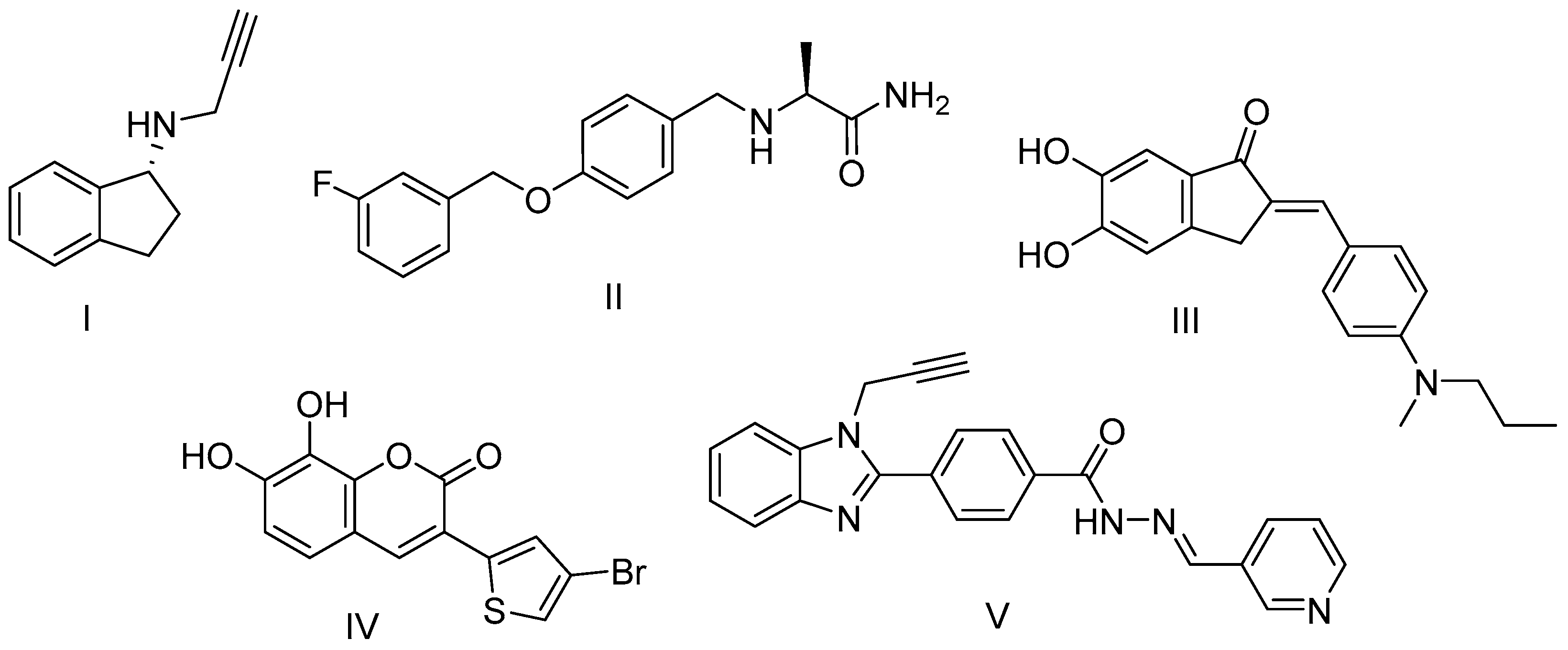
:1. Introduction
2. Materials and Methods
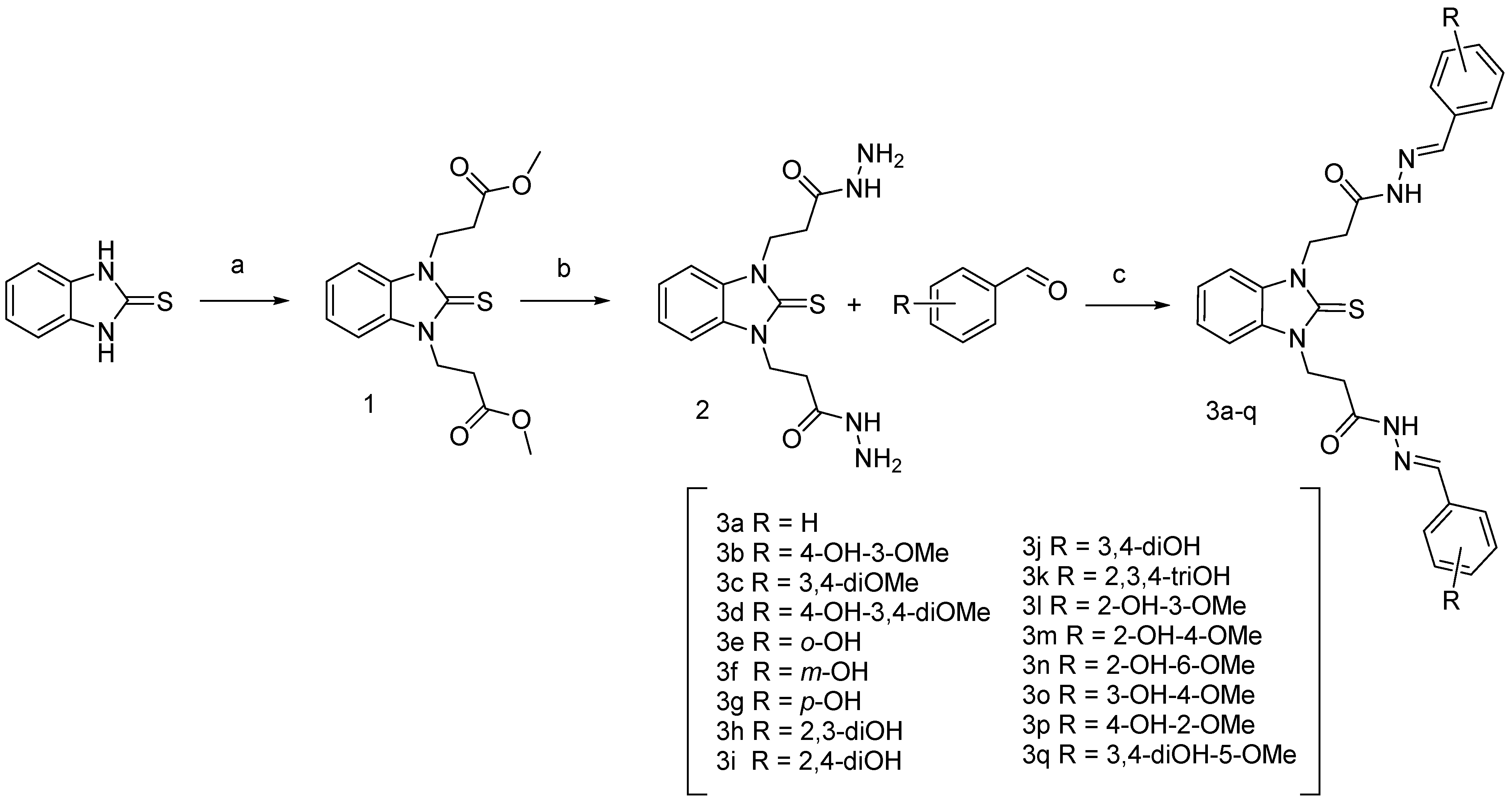
2.1. Chemistry
2.2. General Procedure for the Synthesis of Compounds 3–19
2.2.1. 3,3′-(2-Thioxo-1H-benzo[d]imidazole-1,3(2H)-diyl)bis(N′-benzylidene)propanehydrazide) (3a)
2.2.2. 3,3′-(2-Thioxo-1H-benzo[d]imidazole-1,3(2H)-diyl)bis(N′-(4-hydroxy-3-methoxybenzylidene)propanehydrazide) (3b)
2.2.3. 3,3′-(2-Thioxo-1H-benzo[d]imidazole-1,3(2H)-diyl)bis(N′-(3,4 dimethoxybenzylidene)propanehydrazide) (3c)
2.2.4. 3,3′-(2-Thioxo-1H-benzo[d]imidazole-1,3(2H)-diyl)bis(N′-(4-hydroxy-3,5-dimethoxybenzylidene)propanehydrazide) (3d)
2.2.5. 3,3′-(2-Thioxo-1H-benzo[d]imidazole-1,3(2H)-diyl)bis(N′-(2-hydroxybenzylidene)propanehydrazide) (3e)
2.2.6. 3,3′-(2-Thioxo-1H-benzo[d]imidazole-1,3(2H)-diyl)bis(N′-(3-hydroxybenzylidene)propanehydrazide) (3f)
2.2.7. 3,3′-(2-Thioxo-1H-benzo[d]imidazole-1,3(2H)-diyl)bis(N′-(4-hydroxybenzylidene)propanehydrazide) (3g)
2.2.8. 3,3′-(2-Thioxo-1H-benzo[d]imidazole-1,3(2H)-diyl)bis(N′-(2,3-dihydroxybenzylidene)propanehydrazide) (3h)
2.2.9. 3,3′-(2-Thioxo-1H-benzo[d]imidazole-1,3(2H)-diyl)bis(N′-(2,4-dihydroxybenzylidene)propanehydrazide) (3i)
2.2.10. 3,3′-(2-Thioxo-1H-benzo[d]imidazole-1,3(2H)-diyl)bis(N′-(3,4-dihydroxybenzylidene)propanehydrazide) (3j)
2.2.11. 3,3′-(2-Thioxo-1H-benzo[d]imidazole-1,3(2H)-diyl)bis(N′-(2,3,4-trihydroxybenzylidene)propanehydrazide) (3k)
2.2.12. 3,3′-(2-Thioxo-1H-benzo[d]imidazole-1,3(2H)-diyl)bis(N′-(2-hydroxy-3-methoxybenzylidene)propanehydrazide) (3l)
2.2.13. 3,3′-(2-Thioxo-1H-benzo[d]imidazole-1,3(2H)-diyl)bis(N′-(2-hydroxy-4-methoxybenzylidene)propanehydrazide) (3m)
2.2.14. 3,3′-(2-Thioxo-1H-benzo[d]imidazole-1,3(2H)-diyl)bis(N′-(2-hydroxy-6-methoxybenzylidene)propanehydrazide) (3n)
2.2.15. 3,3′-(2-Thioxo-1H-benzo[d]imidazole-1,3(2H)-diyl)bis(N′-(3-hydroxy-4-methoxybenzylidene)propanehydrazide) (3o)
2.2.16. 3,3′-(2-Thioxo-1H-benzo[d]imidazole-1,3(2H)-diyl)bis(N′-(4-hydroxy-2-methoxybenzylidene)propanehydrazide) (3p)
2.2.17. 3,3′-(2-Thioxo-1H-benzo[d]imidazole-1,3(2H)-diyl)bis(N′-(3,4-hydroxy-5-methoxybenzylidene)propanehydrazide) (3q)
2.3. Cell Culture and Cultivation
2.4. MTT-Dye Reduction Assay
2.5. H2O2-Induced Oxidative Stress Model In Vitro
2.6. Measurement of Monoamine Oxidase B Enzyme Activity
2.7. Animals
2.8. Isolation and Incubation of Synaptosomes
2.9. Synaptosomes Viability Assay
2.10. Glutathione (GHS) Assay in Isolated Synaptosomes
2.11. Model of 6-Hydroxy Dopamine-Induced Neurotoxicity in Isolated Rat Synaptosomes
2.12. Statistical Analysis
2.13. Iron Induced Oxidative Damage
2.14. Superoxide Scavenging Properties
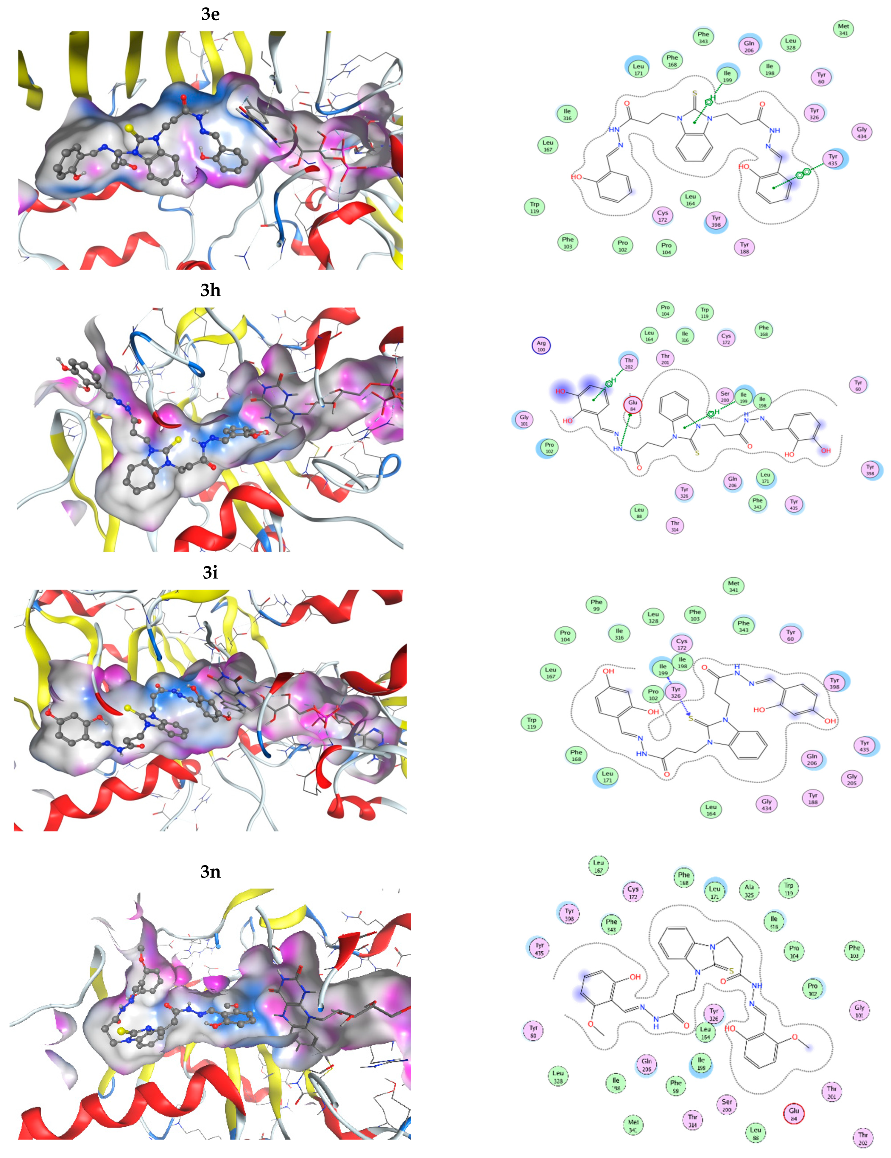
2.15. Molecular Docking Study of the Interactions with MAO-B
3. Results and Discussion
3.1. Synthesis of the Target Compounds
3.2. Neurotoxicity Profile and Neuroprotection Evaluation
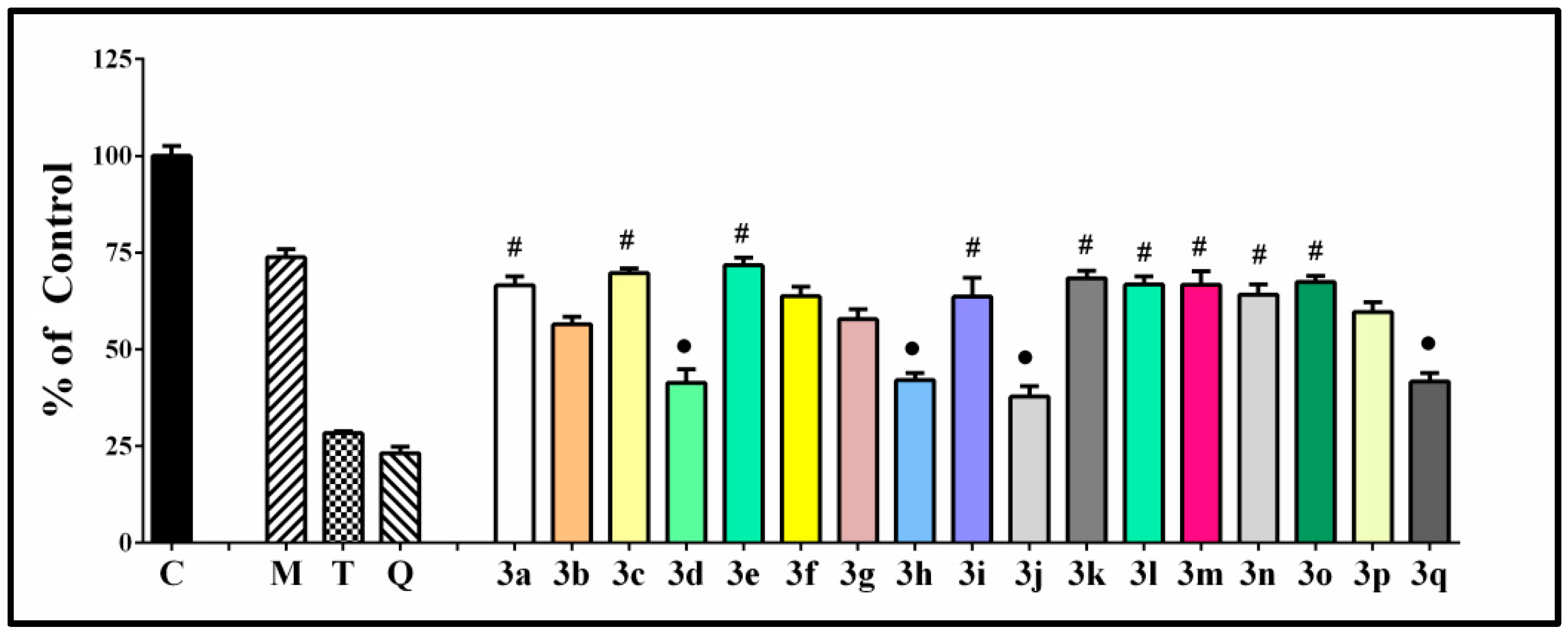
3.2.1. In Vitro Toxicity Evaluation on SH-SY5Y Cells
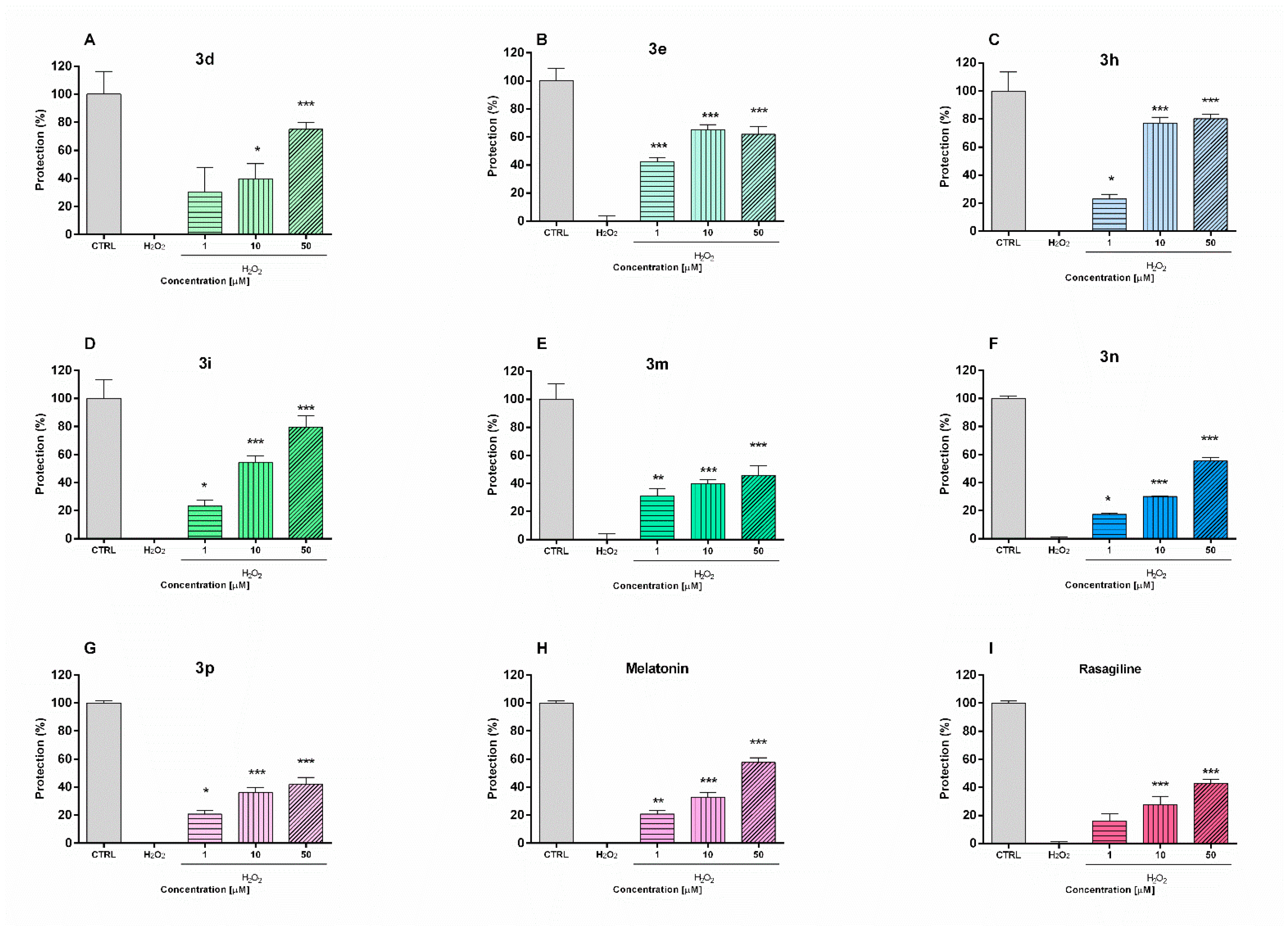
3.2.2. Neuroprotective Effects in H2O2-Induced Oxidative Stress on SH-SY5Y Cells In Vitro
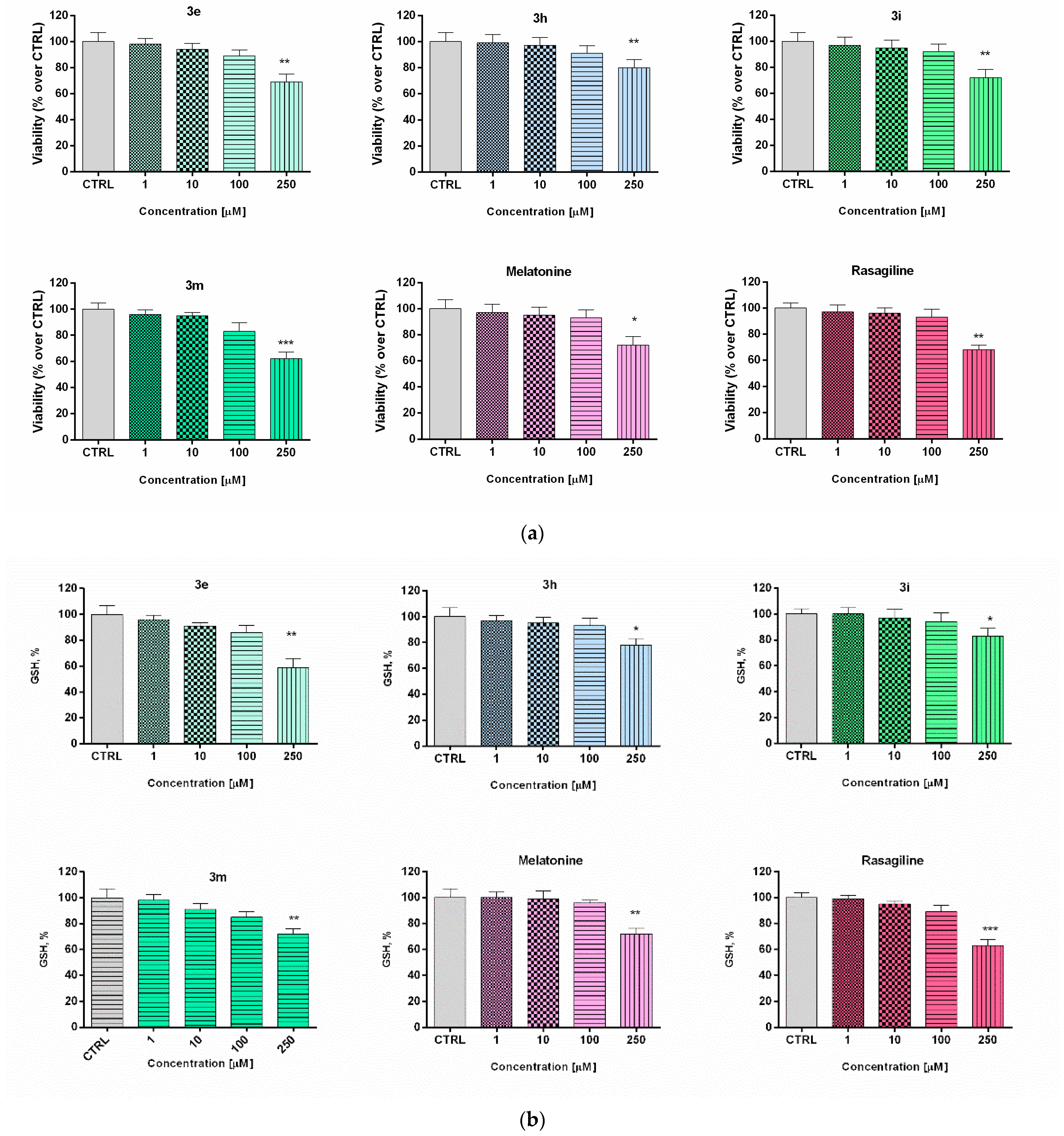
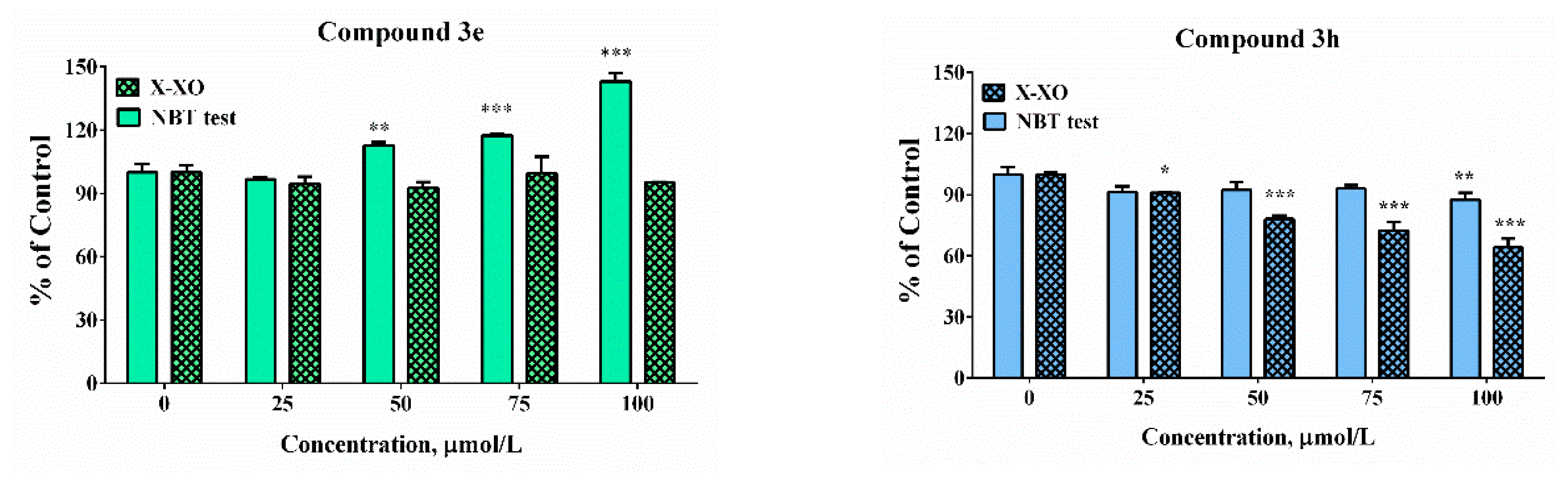
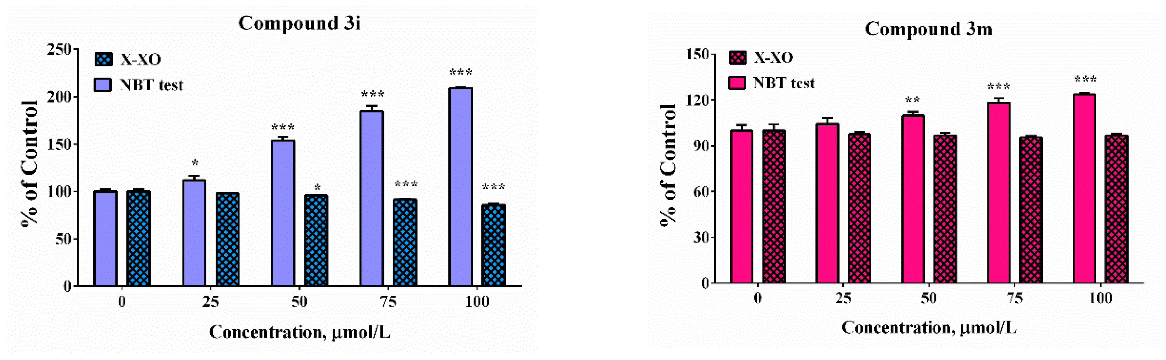
3.2.3. Toxicity Evaluation in Isolated Rat Brain Synaptosomes
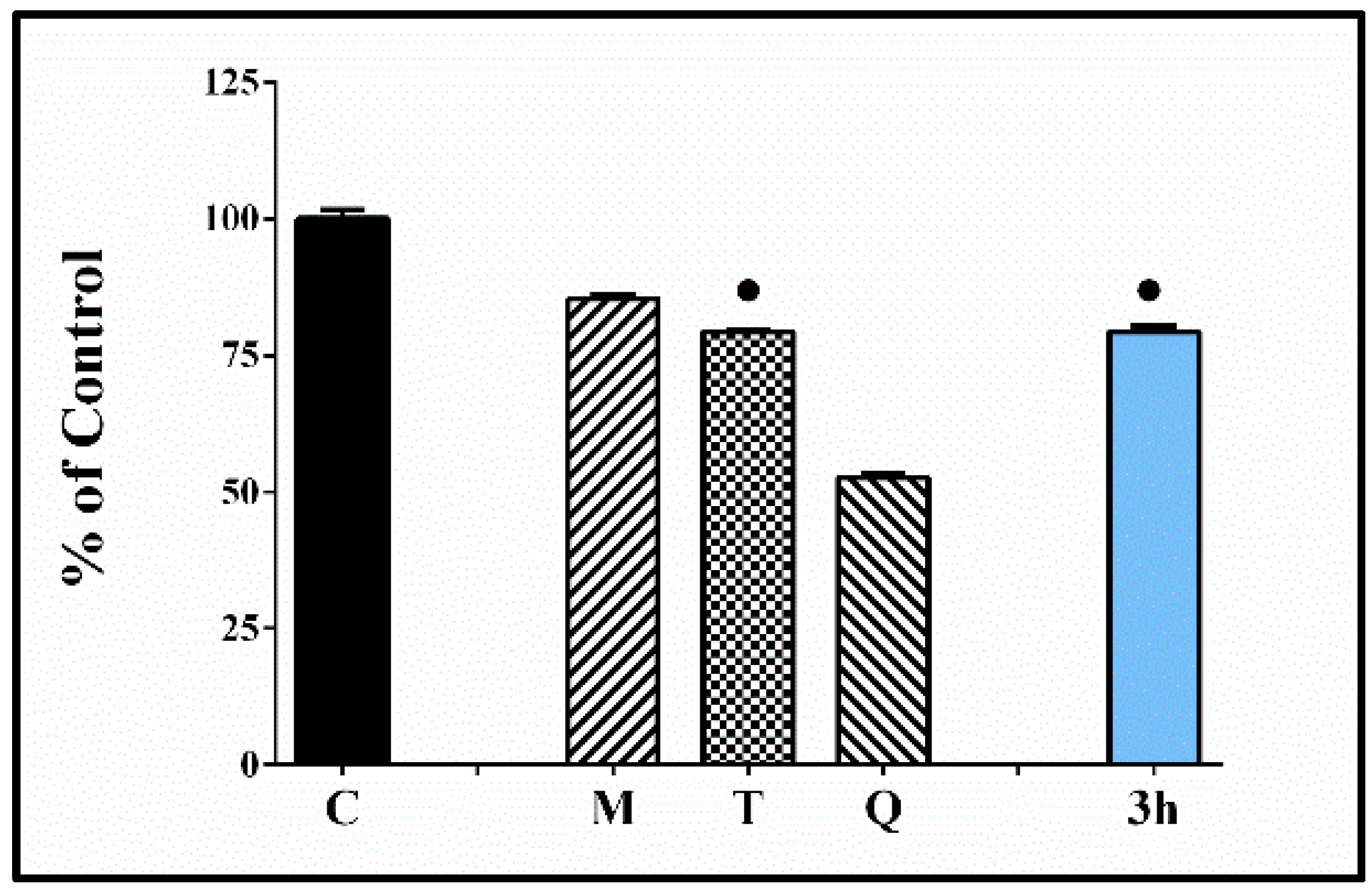
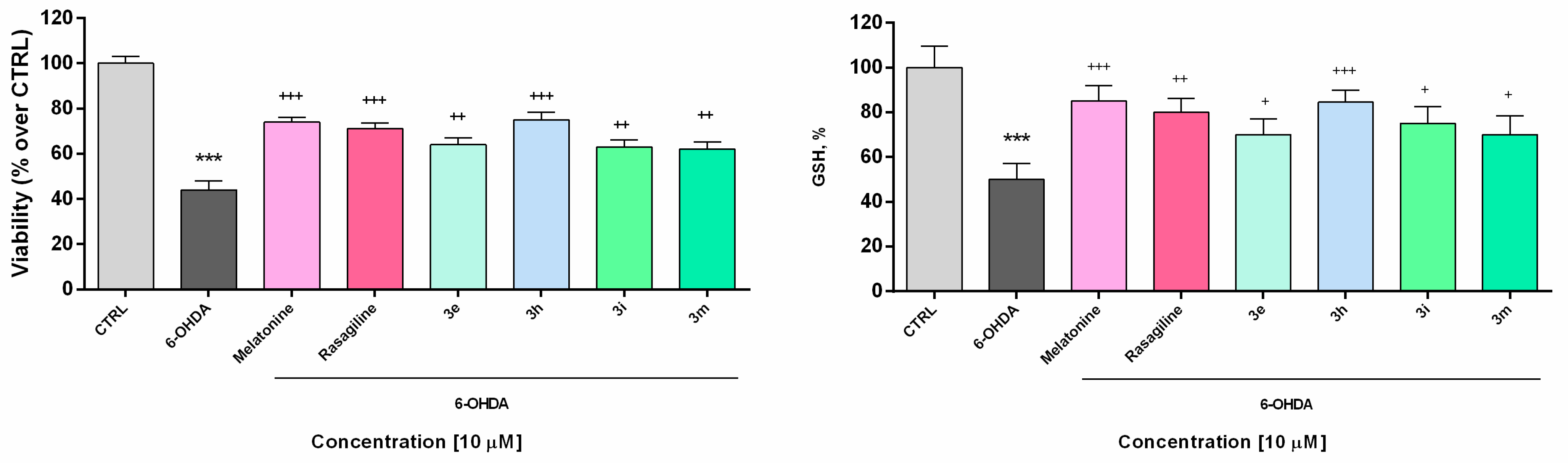
3.2.4. Neuroprotective Effects in a Model of 6-OHDA-Induced Neurotoxicity in Rat Brain Synaptosomes
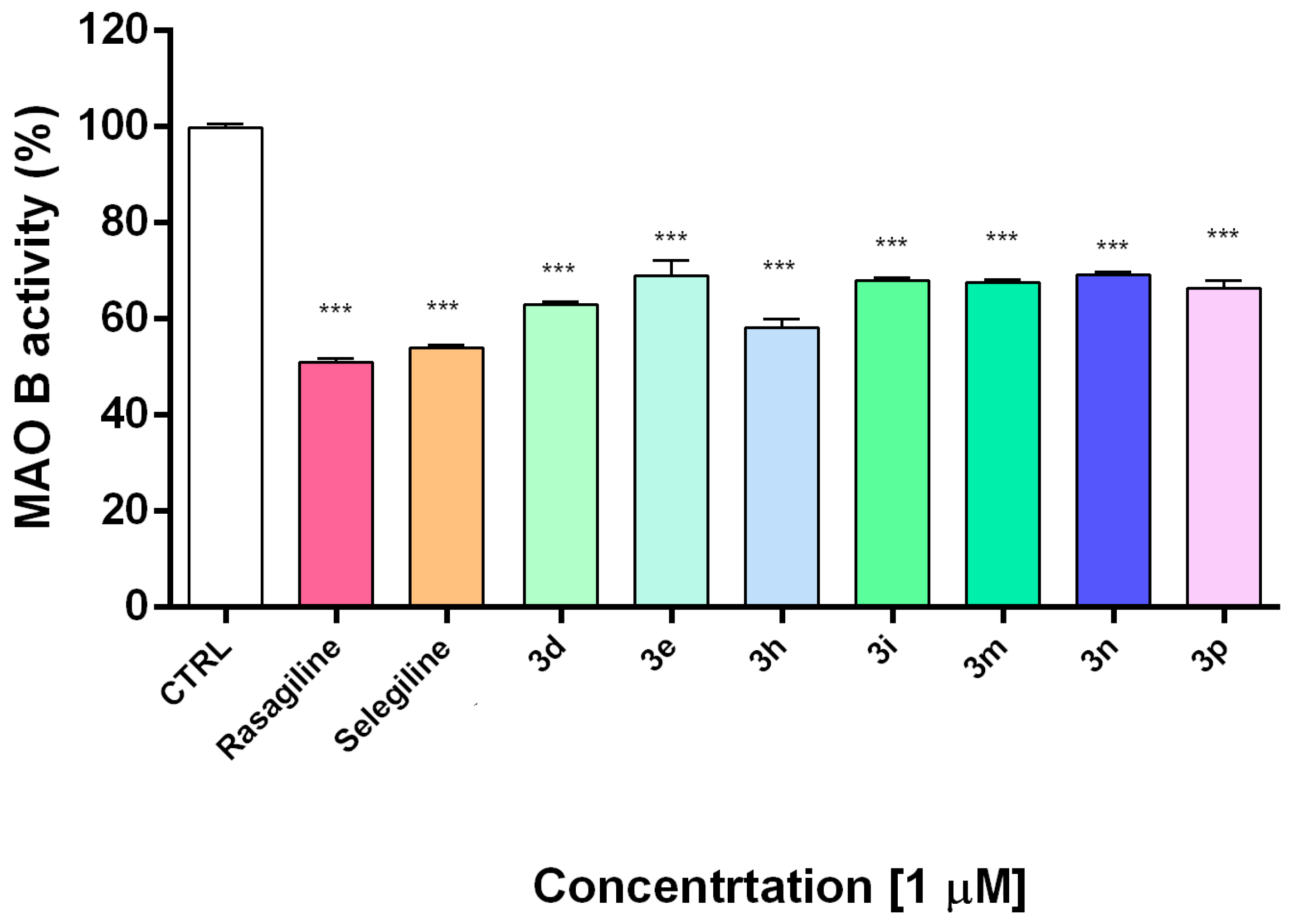
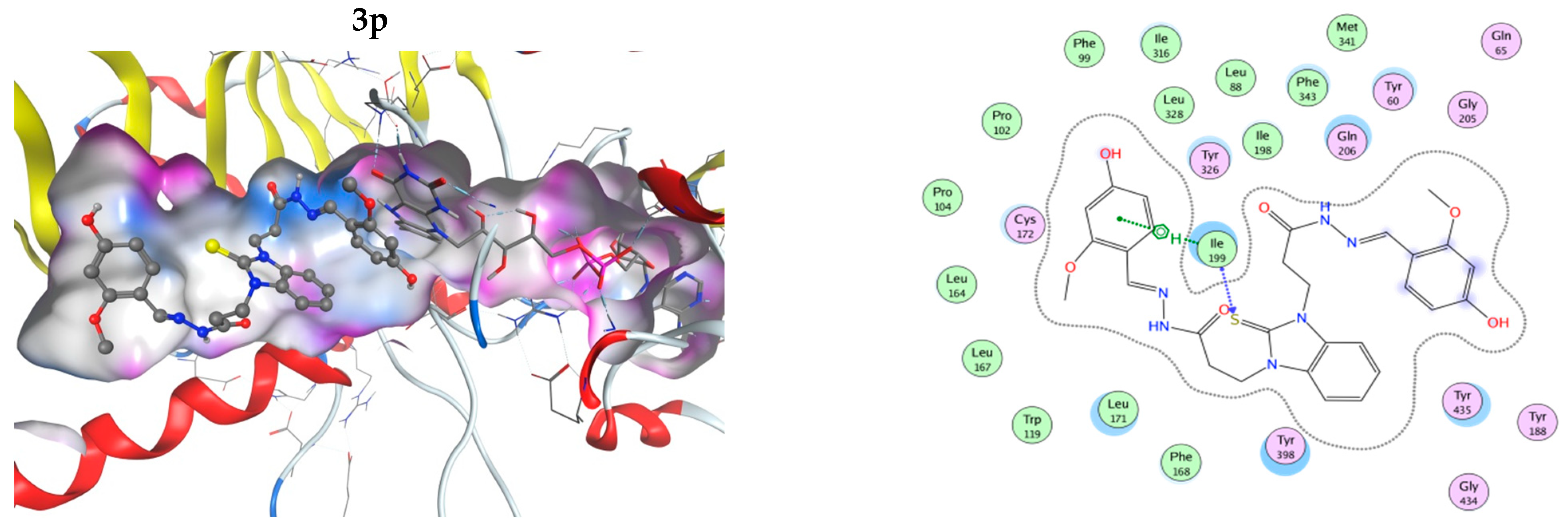
3.3. Inhibitory Activity towards hMAOB and Modeling of the Interactions by Molecular Docking
3.4. Iron-Induced Oxidative Damage of Lecithin
3.5. Scavenging Capability against Superoxide Radicals and Effect on Fe Induced Degradation of Deoxyribose
4. Conclusions
Supplementary Materials
Author Contributions
Funding
Institutional Review Board Statement
Informed Consent Statement
Data Availability Statement
Acknowledgments
Conflicts of Interest
References
- Dorsey, E.R.; Sherer, T.; Okun, M.S.; Bloem, B.R. The Emerging Evidence of the Parkinson Pandemic. J. Parkinson’s Dis. 2018, 8, S3–S8. [Google Scholar] [CrossRef] [Green Version]
- Terao, Y.; Fukuda, H.; Ugawa, Y.; Hikosaka, O. New perspectives on the pathophysiology of Parkinson’s disease as assessed by saccade performance: A clinical review. Clin. Neurophysiol. 2013, 124, 1491–1506. [Google Scholar] [CrossRef]
- Giguère, N.; Burke, N.S.; Trudeau, L. On Cell Loss and Selective Vulnerability of Neuronal Populations in Parkinson’s Disease. Front. Neurol. 2018, 9, 455. [Google Scholar] [CrossRef] [PubMed]
- Kouli, A.; Torsney, K.M.; Kuan, W.L. Parkinson’s Disease: Etiology, Neuropathology, and Pathogenesis; Stoker, T.B., Greenland, J.C., Eds.; Codon Publications: Brisbane, Australia, 2018; Chapter 1; p. 3. [Google Scholar] [CrossRef]
- Rizek, P.; Kumar, N.; Jog, M.S. An update on the diagnosis and treatment of Parkinson disease. CMAJ 2016, 188, 1157–1165. [Google Scholar] [CrossRef] [PubMed] [Green Version]
- Puspita, L.; Chung, S.Y.; Shim, J.-W. Oxidative stress and cellular pathologies in Parkinson’s disease. Mol. Brain 2017, 10, 53. [Google Scholar] [CrossRef] [Green Version]
- Nakabeppu, Y.; Tsuchimoto, D.; Yamaguchi, H.; Sakumi, K. Oxidative damage in nucleic acids and Parkinson’s disease. J. Neurosci. Res. 2007, 85, 919–934. [Google Scholar] [CrossRef] [PubMed]
- Xicoy, H.; Wieringa, B.; Martens, G.J.M. The Role of Lipids in Parkinson’s Disease. Cells 2019, 8, 27. [Google Scholar] [CrossRef] [Green Version]
- Trist, B.G.; Hare, D.J.; Double, K.L. Oxidative stress in the aging substantia nigra and the etiology of Parkinson’s disease. Aging Cell 2019, 18, e13031. [Google Scholar] [CrossRef] [Green Version]
- Flockhart, D.A. Dietary restrictions and drug interactions with monoamine oxidase inhibitors: An update. J. Clin. Psychiatry 2012, 73, 17–24. [Google Scholar] [CrossRef]
- Guo, J.; Zhao, X.; Li, Y.; Li, G.; Liu, X. Damage to dopaminergic neurons by oxidative stress in Parkinson’s disease. Int. J. Mol. Med. 2018, 41, 1817–1825. [Google Scholar] [CrossRef] [Green Version]
- Zeevalk, G.D.; Razmpour, R.; Bernard, L.P. Glutathione and Parkinson’s disease: Is this the elephant in the room? Biomed. Pharmacother. 2008, 62, 236–249. [Google Scholar] [CrossRef] [PubMed]
- Chamoli, M.; Chinta, S.J.; Andersen, J.K. An inducible MAO-B mouse model of Parkinson’s disease: A tool towards better understanding basic disease mechanisms and developing novel therapeutics. J. Neural Transm. 2018, 125, 1651–1658. [Google Scholar] [CrossRef] [PubMed]
- Robakis, D.; Fahn, S. Defining the Role of the Monoamine Oxidase-B Inhibitors for Parkinson’s Disease. CNS Drugs 2015, 29, 433–441. [Google Scholar] [CrossRef]
- Elmabruk, A.; Das, B.; Yedlapudi, D.; Xu, L.; Antonio, T.; Reith, M.E.A.; Dutta, A.K. Design, synthesis, and pharmacological characterization of carbazole based dopamine agonists as potential symptomatic and neuroprotective therapeutic agents for Parkinson’s disease. ACS Chem. Neurosci. 2019, 10, 396–411. [Google Scholar] [CrossRef] [PubMed]
- Nayak, L.; Henchcliffe, C. Rasagiline in treatment of Parkinson’s disease. Neuropsychiatr. Dis. Treat. 2008, 4, 23–32. [Google Scholar] [PubMed]
- Solla, P.; Ercoli, T.; Masala, C.; Orofino, G.; Fadda, L.; Corda, D.G.; Zarbo, I.R.; Meloni, M.; Sechi, E.; Bagella, C.F.; et al. Rasagiline Withdrawal Syndrome in Parkinson’s Disease. Brain Sci. 2022, 12, 219. [Google Scholar] [CrossRef] [PubMed]
- Blair, H.A.; Dhillon, S. Safinamide: A Review in Parkinson’s Disease. CNS Drugs 2017, 31, 169–176. [Google Scholar] [CrossRef]
- Huang, L.; Lu, C.; Sun, Y.; Mao, F.; Luo, Z.; Su, T.; Jiang, H.; Shan, W.; Li, X. Multitarget-Directed Benzylideneindanone Derivatives: Anti-β-Amyloid (Aβ) Aggregation, Antioxidant, Metal Chelation, and Monoamine Oxidase B (MAO-B) Inhibition Properties against Alzheimer’s Disease. Med. Chem. 2012, 55, 8483–8492. [Google Scholar] [CrossRef]
- Can, Ö.D.; Osmaniye, D.; Demir Özkay, Ü.; Sağlık, B.N.; Levent, S.; Ilgın, S.; Baysal, M.; Özkay, Y.; Kaplancıklı, Z.A. MAO enzymes inhibitory activity of new benzimidazole derivatives including hydrazone and propargyl side chains. Eur. J. Med. Chem. 2017, 5, 92–106. [Google Scholar] [CrossRef]
- Rodríguez-Enríquez, F.; Viña, D.; Uriarte, E.; Fontenla, J.A.; Matos, M.J. Discovery and optimization of 3-thiophenylcoumarins as novel agents against Parkinson’s disease: Synthesis, in vitro and in vivo studies. Bioorg. Chem. 2020, 101, 103986. [Google Scholar] [CrossRef]
- Márquez-Navarro, A.; Nogueda-Torres, B.; Hernández-Campos, A.; Soria-Arteche, O.; Castillo, R.; Rodríguez-Morales, S.; Yépez-Mulia, L.; Hernández-Luis, F. Anthelmintic activity of benzimidazole derivatives against Toxocara canis second-stage larvae and Hymenolepis nana adults. Acta Trop. 2009, 109, 232–235. [Google Scholar] [CrossRef] [PubMed]
- Hwu, J.R.; Singha, R.; Hong, S.C.; Chang, Y.H.; Das, A.R.; Vliegen, I.; Neyts, J. Synthesis of new benzimidazole–coumarin conjugates as anti-hepatitis C virus agents. Antivir. Res. 2008, 77, 157–162. [Google Scholar] [CrossRef]
- Ganji, L.V.; Agrawal, P.N. Design, Synthesis and Antiinflammatory Evaluation of 5(6)-(un)-substituted-1H-Benzimidazol-2-ylthioacetylpiperazine Derivatives. Ind. J. Phrm. Sci. 2020, 82, 21–31. [Google Scholar] [CrossRef] [Green Version]
- Shingalapur, R.V.; Hosamani, K.M.; Keri, R.S.; Hugar, M.H. Derivatives of benzimidazole pharmacophore: Synthesis, anticonvulsant, antidiabetic and DNA cleavage studies. Eur. J. Med. Chem. 2010, 45, 1753–1759. [Google Scholar] [CrossRef] [PubMed]
- Mohammed, S.O.; El Ashry, S.H.E.; Khalid, A.; Amer, M.R.; Metwaly, A.M.; Eissa, I.H.; Elkaeed, E.B.; Elshobaky, A.; Hafez, E.E. Expression, Purification, and Comparative Inhibition of Helicobacter pylori Urease by Regio-Selectively Alkylated Benzimidazole 2-Thione Derivatives. Molecules 2022, 27, 865. [Google Scholar] [CrossRef]
- Evranos-Aksöz, B.; Yabanoğlu Çiftçi, S.; Uçar, G.; Yelekçi, K.; Ertan, R. Synthesis of some novel hydrazone and 2-pyrazoline derivatives: Monoamine oxidase inhibitory activities and docking studies. Bioorg. Med. Chem. Lett. 2014, 24, 3278–3284. [Google Scholar] [CrossRef] [PubMed]
- Tripathi, R.K.; Ayyannan, S.R. Design, synthesis, and evaluation of 2-amino-6-nitrobenzothiazole-derived hydrazones as MAO inhibitors: Role of the methylene spacer group. ChemMedChem 2016, 11, 1551–1567. [Google Scholar] [CrossRef]
- Can, N.Ö.; Osmaniye, D.; Levent, S.; Sağlık, B.N.; İnci, B.; Ilgın, S.; Özkay, Y.; Kaplancıklı, Z.A. Synthesis of New Hydrazone Derivatives for MAO Enzymes Inhibitory Activity. Molecules 2017, 22, 1381. [Google Scholar] [CrossRef]
- Puskullu, M.O.; Shirinzadeh, H.; Nenni, M.; Gurer-Orhan, H.; Suzen, S. Synthesis and evaluation of antioxidant activity of new quinoline-2-carbaldehyde hydrazone derivatives: Bioisosteric melatonin analogues. J. Enzym. Inhib. Med. Chem. 2016, 31, 121–125. [Google Scholar] [CrossRef] [Green Version]
- Anastassova, N.; Yancheva, D.; Hristova-Avakumova, N.; Hadjimitova, V.; Traykov, T.; Aluani, D.; Tzankova, V.; Kondeva-Burdina, M. New benzimidazole-aldehyde hybrids as neuroprotectors with hypochlorite and super oxide radical scavenging activity. Pharmacol. Rep. 2020, 72, 846–856. [Google Scholar] [CrossRef]
- Anastassova, N.; Aluani, D.; Kostadinov, A.; Rangelov, M.; Todorova, N.; Hristova-Avakumova, N.; Argirova, M.; Lumov, N.; Kondeva-Burdina, M.; Tzankova, V.; et al. Evaluation of the combined activity of benzimidazole arylhydrazones as new anti-Parkinsonian agents: Monoamine oxidase-B inhibition, neuroprotection and oxidative stress modulation. Neural Regen. Res. 2021, 16, 2299–2309. [Google Scholar] [CrossRef] [PubMed]
- Anastassova, N.O.; Yancheva, D.Y.; Mavrova, A.T.; Kondeva-Burdina, M.S.; Tzankova, V.I.; Hristova-Avakumova, N.G.; Hadjimitova, V.A. Design, synthesis, antioxidant properties and mechanism of action of new N,N′-disubstituted benzimidazole-2-thione hydrazone derivatives. J. Mol. Struct. 2018, 1165, 162–176. [Google Scholar] [CrossRef]
- Mosmann, T. Rapid colorimetric assay for cellular growth and survival: Application to proliferation and cytotoxicity assays. J. Immunol. Methods 1983, 65, 55–63. [Google Scholar] [CrossRef]
- Watson, N.; Diamandis, T.; Gonzales-Portillo, C.; Reyes, S.; Borlongan, C.V. Melatonin as an antioxidant for stroke neuroprotection. Cell Transplant. 2016, 25, 883–891. [Google Scholar] [CrossRef] [PubMed] [Green Version]
- Salamon, A.; Zádori, D.; Szpisjak, L. Neuroprotection in Parkinson’s disease: Facts and hopes. J. Neural Transm. 2020, 127, 821–829. [Google Scholar] [CrossRef] [Green Version]
- Bautista-Aguilera, O.M.; Esteban, G.; Bolea, I.; Nikolic, K.; Agbaba, D.; Moraleda, I.; Iriepa, I.; Samadi, A.; Soriano, E.; Unzeta, M.; et al. Design, synthesis, pharmacological evaluation, QSAR analysis, molecular modeling and ADMET of novel donepezil-indolyl hybrids as multipotent cholinesterase/monoamine oxidase inhibitors for the potential treatment of Alzheimer’s disease. Eur. J. Med. 2014, 75, 82–95. [Google Scholar] [CrossRef]
- Kasabova-Angelova, A.; Kondeva-Burdina, M.; Mitkov, J.; Georgieva, M.; Tzankova, V.; Zlatkov, A. Neuroprotective and MAOB inhibitory effects of a series of caffeine-8-thioglycoloc acid amides. Braz. J. Pharm. Sci. 2020, 56, e18255. [Google Scholar] [CrossRef]
- Taupin, P.; Zini, S.; Cesselin, F.; Ben-Ari, Y.; Roisin, M.P. Subcellular fractionation on Percoll gradient of mossy fiber synaptosomes: Morphological and biochemical characterization in control degranulated rat hippocampus. J. Neurochem. 1994, 62, 1586–1595. [Google Scholar] [CrossRef] [PubMed]
- Lowry, O.H.; Rosebrough, N.J.; Farr, A.L.; Randall, R.J. Protein measurement with the Folin phenol reagent. J. Biol. Chem. 1951, 193, 265–275. [Google Scholar] [CrossRef]
- Mungarro-Menchaca, X.; Ferrera, P.; Morán, J.; Arias, C. Beta-Amyloid peptide induces ultrastructural changes in synaptosomes and potentiates mitochondrial dysfunction in the presence of ryanodine. J. Neurosci. Res. 2002, 68, 89–96. [Google Scholar] [CrossRef]
- Robyt, J.F.; Ackerman, R.J.; Chittenden, C.G. Reaction of protein disulfide groups with Ellman’s reagent: A case study of the number of sulfhydryl and disulfide groups in Aspergillus oryzae-amylase, papain, and lysozyme. Arch. Biochem. Biophys. 1971, 147, 262–269. [Google Scholar] [CrossRef]
- Stokes, A.H.; Freeman, W.M.; Mitchell, S.G.; Burnette, T.A.; Hellmann, G.M.; Vrana, K.E. Induction of GADD45 and GADD153 in Neuroblastoma Cells by Dopamine-Induced Toxicity. Neurotoxicology 2002, 23, 675–684. [Google Scholar] [CrossRef]
- Aasakawa, T.; Matsushita, S. Coloring conditions of thiobarbituric acid test for detecting lipid hydroperoxides. Lipids 1980, 15, 137–140. [Google Scholar] [CrossRef]
- Wang, G.; Wang, T. Oxidative stability of egg and soy lecithin as affected by transition metal ions and pH in emulsion. J. Agric. Food Chem. 2008, 56, 11424–114231. [Google Scholar] [CrossRef] [Green Version]
- Sadowska-Bartosz, I.; Galiniak, S.; Bartosz, G. Modification of the deoxyribose test to detect strong iron binding. Acta Biochim. Pol. 2017, 64, 195–198. [Google Scholar] [CrossRef]
- Shintani, H. Determination of xanthine oxidase. Pharm. Anal. Acta 2013, 7. [Google Scholar] [CrossRef] [Green Version]
- Liu, R.; Fu, S.; Zhan, H.; Lucia, L.A. General Spectroscopic Protocol to Obtain the Concentration of the Superoxide Anion Radicals. Ind. Eng. Chem. Res. 2009, 48, 9331–9334. [Google Scholar] [CrossRef]
- Molecular Operating Environment (MOE); Chemical Computing Group Inc.: Montreal, QC, Canada, 2016.
- Binda, C.; Aldeco, M.; Mattevi, A.; Edmondson, D.E. Interactions of monoamine oxidases with the antiepileptic drug zonisamide: Specificity of inhibition and structure of the human monoamine oxidase B complex. J. Med. Chem. 2011, 54, 909–912. [Google Scholar] [CrossRef] [Green Version]
- Anastassova, N.; Mavrova, A.; Yancheva, D.; Kondeva-Burdina, M.; Tzankova, V.; Stoyanov, S. Hepatotoxicity and antioxidant activity of some new N,N′-disubstituted benzimidazole-2-thiones, radical scavenging mechanism and structure-activity relationship. Arab. J. Chem. 2017, 11, 353–369. [Google Scholar] [CrossRef]
- Kovalevich, J.; Langford, D. Considerations for the use of SH-SY5Y neuroblastoma cells in neurobiology. Methods Mol. Biol. 2013, 1078, 9–21. [Google Scholar] [CrossRef] [Green Version]
- Heusinkveld, H.J.; Westerink, R.H. Comparison of different in vitro cell models for the assessment of pesticide-induced dopaminergic neurotoxicity. Toxicol. In Vitro 2017, 45, 81–88. [Google Scholar] [CrossRef] [PubMed]
- Kitamura, Y.; Kosaka, T.; Kakimura, J.I.; Matsuoka, Y.; Kohno, Y.; Nomura, Y.; Taniguchi, T. Protective effects of the antiparkinsonian drugs talipexole and pramipexole against 1-methyl-4-phenylpyridinium-induced apoptotic death in human neuroblastoma SH-SY5Y cells. Mol. Pharmacol. 1988, 54, 1046–1054. [Google Scholar] [CrossRef]
- Abudayyak, M.; Guzel, E.; Özhan, G. Nickel oxide nanoparticles are highly toxic to SH-SY5Y neuronal cells. Neurochem. Int. 2017, 108, 7–14. [Google Scholar] [CrossRef] [PubMed]
- Devi, S.L.; Kannappan, S.; Anuradha, C.V. Evaluation of in vitro antioxidant activity of Indian bay leaf, Cinnamomum tamala (Buch.-Ham.) T. Nees & Eberm using rat brain synaptosomes as model system. Indian J. Exp. Biol. 2007, 45, 778–784. [Google Scholar]
- Student, A.K.; Edwards, D.J. Subcellular localization of types A and B monoxidase in rat brain. Biochem. Pharmacol. 1977, 26, 2337–2342. [Google Scholar] [CrossRef]
- Blum, D.; Torch, S.; Lambeng, N.; Nissou, M.F.; Benabid, A.L.; Sadoul, R.; Verna, J.M. Molecular pathways involved in the neurotoxicity of 6-OHDA, dopamine and MPTP: Contribution to the apoptotic theory in Parkinson’s disease. Prog. Neurobiol. 2007, 65, 135–172. [Google Scholar] [CrossRef]
- Tiffany-Castiglioni, E.; Saneto, R.P.; Proctor, P.H.; Perez-Polo, J.R. Participation of active oxygen species in 6-hydroxydopamine toxicity to a human neuroblastoma cell line. Biochem. Pharmacol. 1982, 31, 181–188. [Google Scholar] [CrossRef]
- Zeng, X.S.; Geng, W.S.; Jia, J.J. Neurotoxin-Induced Animal Models of Parkinson Disease: Pathogenic Mechanism and Assessment. ASN Neuro 2018, 10, 1759091418777438. [Google Scholar] [CrossRef]
- Glinka, Y.Y.; Youdim, M.B. Inhibition of mitochondrial complexes I and IV by 6-hydroxydopamine. Eur. J. Pharmacol. 1995, 16, 329–332. [Google Scholar] [CrossRef]
- Edmondson, D.E.; De Colibus, L.; Binda, C.; Li, M.; Mattevi, A. New insights into the structures and functions of human monoamine oxidases A and B. J. Neural Transm. 2007, 114, 703–705. [Google Scholar] [CrossRef]
- Carradori, S.; Silvestri, R. New Frontiers in Selective Human MAO-B Inhibitors. J. Med. Chem. 2015, 10, 6717–6732. [Google Scholar] [CrossRef] [PubMed]
- Binda, C.; Hubálek, F.; Li, M.; Herzig, Y.; Sterling, J.; Edmondson, D.E.; Mattevi, A. Crystal structures of monoamine oxidase B in complex with four inhibitors of the N-propargylaminoindan class. J. Med. Chem. 2004, 25, 1767–1774. [Google Scholar] [CrossRef] [PubMed]
- Bozkurt, A.A.; Mustafa, G.; Tarık, A. Syringaldehyde exerts neuroprotective effect on cerebral ischemia injury in rats through anti-oxidative and anti-apoptotic properties. Neural Regen. Res. 2014, 9, 1884–1890. [Google Scholar] [CrossRef]
- De Farias, C.C.; Maes, M.; Bonifácio, K.L.; Bortolasci, C.C.; de Souza Nogueira, A.; Brinholi, F.F.; Matsumoto, A.K.; do Nascimento, M.A.; de Melo, L.B.; Nixdorf, S.L.; et al. Highly specific changes in antioxidant levels and lipid peroxidation in Parkinson’s disease and its progression: Disease and staging biomarkers and new drug targets. Neurosci. Lett. 2016, 617, 66–71. [Google Scholar] [CrossRef]
- Gurer-Orhan, H.; Karaaslan, C.; Ozcan, S.; Firuzi, O.; Tavakkoli, M.; Saso, L. Novel indole-based melatonin analogues: Evaluation of antioxidant activity and protective effect against amyloid β-induced damage. Bioorg. Med. Chem. 2016, 24, 1658–1664. [Google Scholar] [CrossRef]
- Sharma, A.; Kaur, P.; Kumar, B.; Prabhakar, S.; Gill, K.D. Plasma lipid peroxidation and antioxidant status of Parkinson’s disease patients in the Indian population. Parkinsonism Relat. Disord. 2008, 14, 52–57. [Google Scholar] [CrossRef]
- Aruoma, O.I. Deoxyribose assay for detecting hydroxyl radicals. In Oxygen Radicals in Biological Systems, Pt C; Siels, H., Abelson, J., Melvin, S., Eds.; Academic Press Inc.: San Diego, CA, USA, 1994; Volume 233, pp. 57–66. [Google Scholar]
- Chobot, V. Simultaneous detection of pro- and antioxidative effects in the variants of the deoxyribose degradation assay. J. Agric. Food. Chem. 2010, 58, 2088–2094. [Google Scholar] [CrossRef]


| Compound | IC50 (µM) | 95% Confidence Intervals |
|---|---|---|
| 3a | 103.25 | 98.73 to 110.12 |
| 3b | 101.24 | 90.26 to 120.23 |
| 3c | 112.32 | 101.3 to 121.13 |
| 3d | 268.23 | 250.98 to 283.43 |
| 3e | 235.2 | 215.78 to 240.11 |
| 3f | 77.74 | 70.01 to 83.21 |
| 3g | 77.79 | 72.03 to 85.45 |
| 3h | 295.90 | 291.31 to 302.54 |
| 3i | 301.11 | 298.23 to 306.24 |
| 3j | 187.20 | 178.23 to 194.23 |
| 3k | 65.30 | 59.84 to 81.70 |
| 3l | 199.51 | 187.42 to 204.54 |
| 3m | 211.21 | 203.45 to 220.21 |
| 3n | 269.40 | 258. 34 to 275.45 |
| 3o | 73.61 | 68.63 to 79.28 |
| 3p | 253.50 | 240.26 to 261.34 |
| 3q | 77.71 | 70.72 to 88.26 |
| Melatonin | >500 | |
| Rasagiline HCl | >500 |
Publisher’s Note: MDPI stays neutral with regard to jurisdictional claims in published maps and institutional affiliations. |
© 2022 by the authors. Licensee MDPI, Basel, Switzerland. This article is an open access article distributed under the terms and conditions of the Creative Commons Attribution (CC BY) license (https://creativecommons.org/licenses/by/4.0/).
Share and Cite
Anastassova, N.; Aluani, D.; Hristova-Avakumova, N.; Tzankova, V.; Kondeva-Burdina, M.; Rangelov, M.; Todorova, N.; Yancheva, D. Study on the Neuroprotective, Radical-Scavenging and MAO-B Inhibiting Properties of New Benzimidazole Arylhydrazones as Potential Multi-Target Drugs for the Treatment of Parkinson’s Disease. Antioxidants 2022, 11, 884. https://doi.org/10.3390/antiox11050884
Anastassova N, Aluani D, Hristova-Avakumova N, Tzankova V, Kondeva-Burdina M, Rangelov M, Todorova N, Yancheva D. Study on the Neuroprotective, Radical-Scavenging and MAO-B Inhibiting Properties of New Benzimidazole Arylhydrazones as Potential Multi-Target Drugs for the Treatment of Parkinson’s Disease. Antioxidants. 2022; 11(5):884. https://doi.org/10.3390/antiox11050884
Chicago/Turabian StyleAnastassova, Neda, Denitsa Aluani, Nadya Hristova-Avakumova, Virginia Tzankova, Magdalena Kondeva-Burdina, Miroslav Rangelov, Nadezhda Todorova, and Denitsa Yancheva. 2022. "Study on the Neuroprotective, Radical-Scavenging and MAO-B Inhibiting Properties of New Benzimidazole Arylhydrazones as Potential Multi-Target Drugs for the Treatment of Parkinson’s Disease" Antioxidants 11, no. 5: 884. https://doi.org/10.3390/antiox11050884
APA StyleAnastassova, N., Aluani, D., Hristova-Avakumova, N., Tzankova, V., Kondeva-Burdina, M., Rangelov, M., Todorova, N., & Yancheva, D. (2022). Study on the Neuroprotective, Radical-Scavenging and MAO-B Inhibiting Properties of New Benzimidazole Arylhydrazones as Potential Multi-Target Drugs for the Treatment of Parkinson’s Disease. Antioxidants, 11(5), 884. https://doi.org/10.3390/antiox11050884