Article

Mycosynthesis of Silica Nanoparticles Using Aspergillus niger: Control of Alternaria solani Causing Early Blight Disease, Induction of Innate Immunity and Reducing of Oxidative Stress in Eggplant

by
Marzough A. Albalawi
1,
Amer M. Abdelaziz
2,*,
Mohamed S. Attia
2,*,
Ebrahim Saied
2,
Hussein H. Elganzory
3,* and
Amr H. Hashem
2,*
1
Department of Chemistry, Alwajh College, University of Tabuk, Tabuk 71491, Saudi Arabia
2
Botany and Microbiology Department, Faculty of Science, Al-Azhar University, Nasr City, Cairo 11884, Egypt
3
Department of Chemistry, College of Science, Qassim University, Buraidah 51452, Saudi Arabia
*
Authors to whom correspondence should be addressed.
Antioxidants 2022, 11(12), 2323; https://doi.org/10.3390/antiox11122323
Submission received: 1 October 2022 / Revised: 13 November 2022 / Accepted: 16 November 2022 / Published: 24 November 2022

Abstract

:
The threats to the life and production of crops are exacerbated by climate change and the misuse of chemical pesticides. This study was designed to evaluate the effectiveness of biosynthesized silica nanoparticles (SiO2-NPs) as an alternative to pesticides against early blight disease of eggplant. Antifungal activity, disease index, photosynthetic pigments, osmolytes, oxidative stress, antioxidant enzymes activities were tested for potential tolerance of eggplant infected with Alternaria solani. Silica nanoparticles were successfully biosynthesized using Aspergillus niger through green and ecofriendly method. Results revealed that SiO2-NPs exhibited promising antifungal activity against A. solani where MIC was 62.5 µg/mL, and inhibition growth at concentration 1000 µg/mL recorded 87.8%. The disease Index (DI) as a result of infection with A. solani reached 82.5%, and as a result, a severe decrease in stem and root length and number of leaves occurred, which led to a sharp decrease in the photosynthetic pigments. However, contents of free proline, total phenol and antioxidant enzymes activity were increased in infected plants. On the other hand, the treatment with SiO2-NPs 100 ppm led to a great reduction in the disease Index (DI) by 25% and a high protection rate by 69.69%. A clear improvement in growth characteristics and a high content of chlorophyll and total carotenoids was also observed in the plants as a result of treatment with silica nanoparticles in (healthy and infected) plants. Interestingly, the noticeable rise in the content of infected and healthy plants of proline and phenols and an increase in the activity of super oxide dismutase (SOD) and polyphenol oxidase (PPO). It could be suggested that foliar application of SiO2-NPs especially 100 ppm could be commercially used as antifungal and strong inducer of plant physiological immunity against early blight disease.

1. Introduction

The risk of plant diseases is exacerbated by the increase in the severity of climate fluctuations, which increases the risks of plant production. The eggplant is one of the most important Egyptian vegetables that is exposed to many biological stresses such as fungal diseases, nematodes and bacteria [1,2]. Soil-born fungi including Fusarium and Alternaria caused highly noticeable destructive effects on plants in organic and conventional agriculture [3,4,5]. Early blight fungal disease in eggplant that caused by Alternaria solani is one of the most important vegetative diseases, which results in a weakness in the quantity and quality of production [6]. As the disease causes the leaves to fall and dry, the fruits become exposed to direct sunlight and the infection of the fruits leads to rotting, as well as to their unsuitability for marketing and storage [7,8]. Symptoms appear on adult leaves in the form of circular or polygonal spots defined with clear edges and within which are characteristic typical circles [9]. All plants possess a defense system capable of resisting pathogens. The plant’s synthetic or chemical immunity can be induced by bio stimulants [10,11,12]. Mineral nutrients such as silicon or its derivatives are considered one of the most important factors for inducing plant resistance to their direct entry in strengthening the internal structures of plant tissues [13,14]. Silica is important for plant health in general, and the use of silica can be applied at any stage of the plant life cycle [15]. Silica helps in strengthening plants by establishment cell walls from the inside and these results in stronger branches that are resistant to breakage during carry flowers and fruits [16]. Silica helps plants reduce plant shock during pathogen exposure and incomplete infection stages [17]. Silica induces plant immunity against pathogens and insects, with regular use silica accumulating in plant cell walls, thus making it difficult for the pathogen to penetrate the plant cells [18]. It also controls the rate of transpiration in plants. As the plant increases silicon levels, it increases nutrient uptake and distribution, and increases the concentration of chlorophyll and carboxylase in leaves, thus the sick plant recovers from disease [19]. Silicon also improves the efficiency of photosynthesis, and also increases the endurance of leaves and the xylem, the high rates of transpiration caused by the disease, by increasing the deposition of silicon other silicon-containing substances in the cell walls of roots, leaves, twigs, and the main stem [20]. In crops, after absorbing silicon, the hardness of the cell walls improved, the cortex layer increased, thus improving the defense capacity against insects, fungi, especially blight, rot, and bacterial diseases and reducing the incidence of red and white spiders [21,22,23]. The fabrication of nanomaterials is one of nanotechnology’s most challenging and rapidly expanding sectors [24]. Nanoparticle synthesis can be accomplished through physical, chemical, or biological methods [25,26,27,28,29]. The employment of biological mass, such as bacteria, fungus, yeast, plant extract or biomass, and algal extract or biomass, is an alternative to conventional methods for the ecofriendly, healthier, faster, and less expensive synthesis of nanoparticles [30,31,32,33]. Fungi also are able to solubilize silica by producing organic acids and different complexing agents which then helps in the release of soluble silica. (i) Production of Exopolysaccharides, Hydrogen Sulphide and Siderophores. Siderophores which are chelators with a small molecular weight having a high affinity for divalent and trivalent metals. Siderophores are also thought to have a role in silicate solubilization in acidic media [34,35]. (ii) Enzyme-based mechanism. Role of enzymes in silica extraction largely lacks experimental proof. However, there are few studies which indicate that enzymes are involved in this process. Piela and co-workers proposed enzyme-based mechanism for fungus-mediated bioleaching of silica nanoparticles. Under stressed circumstances, when fungal cells come in contact with the substrate, they produce specific proteins and enzymes which interact with the silica structure of biomass leading to the formation of an enzyme-silicic acid complex. This complex is further degraded by the hydrolytic enzymes secreted by fungal cells, leading to the release of siliceous groups present in the substrate in form of silica nanoparticles [36]. Generally, filamentous fungi have played a vital role in the industrial production of biological products and in the fermentation industry due to their ability to secrete proteins and enzymes, high growth rates, ease of handling in large-scale production, and low-cost requirements for production in comparison to other microorganisms [37]. The output of the filamentous fungi cultivation is a high-quality biomass (high protein and fat levels) that can be used as an alternative added to the main diet instead of more expensive sources such as soybean and fish [38]. SiO2-NPs have a variety of effects on plants and are excellent in reducing agricultural pests. Silica nanoparticles have additional uses as nanopesticides, nanoherbicides, and nanofertilizers. They might also be used in plants to transport molecules like proteins and nucleotides [39]. SiO2-NPs are gaining a lot of interest in the agriculture sector due to their enormous surface area and small size. Herein, this study aimed to biosynthesize silica nanoparticles using Aspergillus niger through an ecofriendly method. Furthermore, to apply SiO2-NPs for biocontrol of Alternaria solani causing early blight disease.

2. Experimental Procedure

2.1. Chemical Used

All the chemicals, reagents, Potato dextrose agar (PDA) and Potato dextrose broth (PDB) media, acetone, sulfosalicylic acid, ninhydrin acid, glacial acetic acid, ethanol, Folin’s reagent, toluene, sodium carbonate, and sodium silicate were purchased from Sigma-Aldrich. Sodium silicate was used as a precursor. All the biological syntheses in this study were achieved using distilled water.

2.2. Fungi Mediated Biosynthesis of SiO2-NPs

Silica nanoparticles were mycosynthesized using a biomass filtrate of A. niger AH1, which was previously isolated, morphologically and genetically identified, and deposited in a gene bank with accession number MW680847 [33]. Two disks (1.0 cm) of A. niger were used to inoculate in 100 mL of PDB medium at pH 5. The mixture was then cultured for seven days at 28 ± 2 °C, and shake at 150 rpm. To obtain the extracellular components of fungal strains, after fermentation, the biomass was separated by filtration and then washed with distilled water to remove any media components. Then, approximately 15 g of fungal strain was resuspended in 100 mL distilled water for 48 h in a dark and shaking condition [40]. After filtration for the previous mixture, the biomass filtrate (supernatant) was extracted and employed in the subsequent step to create SiO2-NPs in the following process: 99 mL of filtrate was mixed with one ml 4 M of sodium silicate solution to prepare mixture soln. of 40 mM concentration, and followed by incubation at 25 °C overnight. The resulting turbid-white precipitate of silica nanoparticles was collected, cleaned up by washing it three times with distilled water, and then dried for four hours at 150 °C. The biomass filtrate without any metal precursor, as well as sodium silicate solution, were run as controls for the experiment.

2.3. Characterization of Biosynthesized Silica Nanoparticles

UV-Vis spectroscopy (JENWAY 6305 Spectrophotometer) at a wavelength of 250–500 nm was used to observe the color change brought on by the synthesis of NPs. By adopting the drop coating technique, the morphological properties (size and shape) of biosynthesized SiO2-NPs were examined by TEM (JEM-1230, Japan, Akishima, Tokyo 196-8558). Applying FT-IR spectroscopy (Agilent system Cary 660 FT-IR model), the functional groups’ contributions to the reducing, capping, and stabilization of SiO2-NPs were evaluated. The samples were analyzed for FT-IR spectra in the range of 400–4000 cm−1 [41]. Furthermore, the crystallinity of biosynthesized SiO2-NPs was investigated using X-ray diffractometer X′ Pert Pro (Philips, Eindhoven, the Netherlands). The 2θ values were in the range of 4°–80°. The corresponding voltage and current were 40 kV and 30 mA, respectively. The average size of SiO2-NPs synthesized by fungal metabolites was calculated using the Debye–Scherrer Equation (1) [42]:
D = (Kλ/β Cos θ)
where D is the mean particle size, where K is the Scherrer constant, λ is wave length of the X-ray beam used (1.54, 184 Å), β is the Full width at half maximum (FWHM) of the peak and θ is the Bragg angle, respectively. The particle size distribution of the SiO2-NPs was evaluated using dynamic light scattering (DLS) measurements, which gives the hydrodynamic diameter of particles [43]. This technique was also used (Malvern Instruments Ltd., Worcestershire, UK) and carried out at the Tabbin institute for metallurgical studies, Cairo, Egypt.

2.4. Source of the Alternaria solani

Alternaria solani was brought from the plant pathology lab of botany and microbiology Dep., Faculty of science, Al-Azhar University Cairo, Egypt. The fungus was confirmed by Koch’s postulate and the suspensions of conidial were prepared and spore density was counted by a hemocytometer and adjusted to 106 spores/mL [6].

2.5. In Vitro Antifungal Activity

Antifungal activity of biosynthesized silica nanoparticles was carried out using well diffusion method. A. solani was inoculated on PDA broth medium, and then incubated at 28 ± 2 °C for 3–5 days. Fungal inoculum of A. solani was spread on the surface of PDA plates. Then, wells with 8 mm diameter were made using sterile cork-borer on each agar plate (90 mm). The wells were filled with 100 µL of different concentrations of SiO2-NPs (7.81–1000 µg/mL) individually with three replicates. The culture plates were incubated at 25 °C for 7 days and the zones of inhibition were observed and measured. Radial growth of A. solani was evaluated at different concentrations of SiO2-NPs (7.81, 15.62, 31.25, 62.5, 125, 250, 500 and 1000 µg/mL). Inhibition percentage of pathogen growth was calculated using the following Equation (2):
Inhibition   of   pathogen   growth = Control   growth treatment   growth control   growth × 100

2.6. In Vivo Experiment

Identical and uniform 3-week-old eggplant seedlings were selected from the Agricultural Research Center (ARC), Giza, Egypt. One plant/pot (30 cm in diameter containing a mixture of sand and clay (1:3 W/W) with a total of 7 kg was planted in the Research Garden, Faculty of Science, Al-Azhar University, Cairo, Egypt. The pots were distributed with 10 replicates for each treatment. For five days, the plants were irrigated routinely. Then, before and after flowering, a one-handed pressure sprayer was used to spray treatments to the plants three times (20 mL per plant once every week (50 ppm (50 mg SiO2 NPs/one litter distilled water), 100 (100 mg SiO2 NPs/one litter distilled water) ppm of SiO2 NPs or sodium silicate 100 ppm (100 mg sodium silicate/one litter distilled water) separately). The aqueous solution pH was 5. The treatments were arranged as follows: T1-Healthy control, T2-Infected control, T3-healthy plants treated with SiO2-NPs (50 ppm), T4-healthy plants treated SiO2-NPs (100 ppm), T5-healthy plants treated with sodium silicate (100 ppm), T6-infected plants treated with SiO2-NPs (50 ppm), T7-infected plants treated with SiO2-NPs (100 ppm), T8-infected plants treated with sodium silicate (100 ppm). One week after transplanting the plants, an artificial infection was made with the pathogenic fungus and plants were sprayed with treatments after the week of infection (SiO2-NPs and sodium silicate) to assess plant resistance, record disease symptoms, as well as take samples for biochemical tests from plant samples 45 days after sowing, and the disease was examined.
Disease symptoms were recorded 45 days after transplanting, and disease severity and protection ratio were calculated, as described in Attia et al. [44] using five score classes: 0 (no symptoms), 1 (slight yellow of leaves), 2 (moderate yellow plant), 3 (wilted plant and browning of vascular bands), and 4 (plants completely destroyed). (PDI) was calculated by the equation PDI = (1n1+ 2n2 + 3n3 + 4n4) × 100/4nt where n1–n4 are the number of plants in each class and Nt is the total number of plants. Percent protection was calculated by: Protection% = A − B/A × 100% where A = PDI in infected control plants B = PDI in infected-treated plants.

2.7. Measurement of Growth Traits

The samples were collected for different growth traits (shoot length, root length and number of leaves) and biochemical analysis after four weeks from transplanting.

2.8. Measurement of Photosynthetic Pigments

Measurement of photosynthetic pigments was carried out using the established method Vernon and Selly, [45] to assess the presence of chlorophyll a (Chl a), chlorophyll b (Chl b) and carotenoids in fresh leaves. Photosynthetic pigments were extracted from fresh leaves (1 g) using 100 mL of 80% acetone, then the color was determined spectrophotometrically at 665, 649, and 470 nm after the extract was filtered.

2.9. Determination of the Content of Proline

The proline content of fresh plant leaves was measured according to Bates et al. [46]. The dried shoots (1 g) were digested by 20 mL (3%) of sulfosalicylic acid. In a boiling water bath, 2 mL of the plant filtrate was mixed with 2 mL of ninhydrin acid and glacial acetic acid for one hour. The mixture was placed in an ice bath to stop the reaction. A total of 4 mL of toluene was added to the mixture, then the absorbance determined at 520 nm.

2.10. Determination of Total Phenol Contents

The total dry phenol content was measured using the procedure of Dai et al. [47]. One g of dried eggplant shoots was extracted in 5–10 mL of 80% ethanol for 24 h. the remaining residue was extracted three times 5–10 mL of 80% ethanol. The extract was then filled to a capacity of 50 mL with 80% ethanol, and then 0.5 mL of the extract was mixed well with 0.5 mL of Folin’s reagent and shaken for 3 min. A total of 3 mL of distilled water and 1 mL of saturated sodium carbonate solution were added and mixed. The color was detected at 725 nm after 1 h.

2.11. Antioxidant Enzyme Activity Assay

Superoxide dismutase (SOD) activity was evaluated according to that method described by Bergmeyer [48]. PPO activity was calculated by the procedure used by Lavid et al. [49]. SOD and PPO activities were evaluated in fresh eggplant leaves. A total of 1 g of the fresh plant was homogenized with 10 mL of phosphate buffer pH 6.8 (0.1 M), then centrifuged at 2 °C for 20 min at 20,000× g rpm. For SOD activity 3.6 mL of distilled water, 0.1 mL of enzyme, 5.5 mL of 50 mM phosphate buffer (pH 7.8) and 0.8 mL of 3 mM pyrogallol (dissolved in 10 mM HCl) were used. The rate of pyrogallol reduction was measured at 325 nm with UV-spectrophotometer (Jenway, Missouri City, TX, USA). For PPO activity assayed by using 125 µmol of phosphate buffer (pH 6.8), 100 µmol pyrogallol, and 0.1 mL of enzyme extract. After 5 min at 25 °C, the reaction was stopped by adding 1 mL 5% H2SO4. The developed color was determined at 430 nm.

2.12. Statistical Analysis

Analyses were conducted using one-way variance (ANOVA). The statistically significant differences between treatments was assessed with a p-value of 0.05 or lower, and the LSD test was used with CoStat (CoHort, Monterey, CA, USA) [50].

3. Results and Discussion

3.1. Biosynthesis of SiO2-NPs Using Biomass Filtrate of A. niger

A. niger strain was employed in this work as a biocatalyst for the ecofriendly production of silica nanoparticles. After being combined with a precursor and the fungal biomass filtrate, the color was changed to turbid-white as a result of the silicananoparticle synthesis, which was then calcinated at 150 °C to form SiO2-NPs powder. Biocatalyst exhibiting desired metabolic activity is the key step of process evaluation and decides on its final outcome. In case of presented research, biocatalysts belonging to Fusarium genus seemed to be ideal candidates for nano-silica biogeneration process. Among them, a number of species are plant pathogens, causing diseases of arable crops, i.e., corn, for which covers have been used in described experiments as a substrate for biotransformation. It was reasonable to assume that fungal cells can exhibit proper metabolic capacity leading to Silica-nanoparticle synthesis, in particular, since the genus Fusarium is known from its high activity towards production of silica nanoparticles from rice husk ash aqueous anionic complexes SiF62−, sand and aqueous solutions of potassium silicofluoride [36]. The activities of proteins, enzymes, and carbohydrates produced by fungal metabolites may be responsible for conversion of sodium silicate to SiO2-NPs [51]. Kannan et al. [52] successfully fabricated SiO2-NPs by using Fusarium oxysporum and potassium silicofluoride as a precursor. Moreover, the filtrate containing the extracellular proteins secreted by the fungus F. oxysporum was reacted with 10−3 M K2SiF6 under ambient conditions for 24 h as reported by [53] for biogenic of silica nanoparticles. The results of our manuscript are agreeing with the result reported by Zamani, Jafari, Mousavi and Darezereshki [51] which uses the similar material sodium silicate to biosynthesize silica nanoparticles by using of Saccharomyces cervisiae. Moreover, he studied the effect of pH on sodium silicate without biomass filtrate and study the effect of pH on biomass filtrate with precursor (sodium silicate). He performed in the pH range between 6 and 11 so the effect of pH alteration in the absence of the yeast should be obtained. So, a test is designed that the pH increases in presence of the precursor while it is on the stirrer. It is observed that the pH increases from 6.51 to 10.5 has no effect on the solution and not any sediment was observed. Therefore, it can be concluded that the increase in pH does not have an effect on the production of silica nanoparticles in the absence of other parameters. This test results show that the yeast is reacted with the precursor and the pH has been affected by this reaction. A process of pH effect repeated with the yeast culture media. It is observed that also the presence of the culture medium did not yield nanoparticle production, and as a result, yeast as the precursor is the most important factor in the production of silica nanoparticles. As is clear, nanosilica production will not occur in the absence of precursor and it is observed with consideration of pH and medium effect that the presence of medium is the crucial part in the synthesis process too. This test was studied to select the best pH for biosynthesis in the range between 4–11 on the precursor only and the precursor with biomass filtrate of Aspergillus sp., but only the best conditions are written in the manuscript.
Na 2 SiO 3 +   H 2 O Yeast   Extract SiO 2 + 2 NaOH
Additionally, a study was conducted by Singh, et al. [54] in which they produced silica nanospheres of the size ranging from 5 to 250 nm using liquid bacterial cultures of Actinobacter sp. using potassium fluorosilicate (K2SiF6) as silica precursor. They reported that synthesis of silica nanoparticles was mediated by oxidase and reductase enzymes produced by the bacteria. Addition of precursor molecules to Actinobacter sp. leads to ionic stress and in a way to reduce the stress these species synthesize hydrolyzing enzymes to convert the metal precursor into nanocomposites.

3.2. Characterization of Biosynthesized SiO2-NPs

3.2.1. UV-Vis Spectroscopy

At a wavelength between 250 and 500 nm, UV-Vis spectroscopy was used to monitor the color change. The generated shape, size, and distribution of silica nanoparticles are largely affected by surface plasmon resonance (SPR). The biosynthesized SiO2-NPs had a single SPR band that was detected at a wavelength of 295 nm, as illustrated in Figure 1. According to Biradar et al. [55], silica nanoparticle production is indicated at absorption in the UV region at 297 nm. Similarly, Kannan et al. [52] reported that SiO2-NPs were successfully made utilizing Fusarium oxysporum and the silica surface plasmon band was found to form at about 280 nm. El-Gazzar et al. [56] found that the creation of SiO2-NPs occurred at around 336 nm of absorption, when exposed to a UV-visible spectral scan and corresponds to surface Plasmon resonance. A wide SiO2-NPs absorption band between 270 and 380 nm was observed by Ghosh et al. [57]. This indicated that cuminaldehyde had been successfully loaded into the MSNs.

3.2.2. Fourier Transform Infrared (FT-IR) Spectroscopy

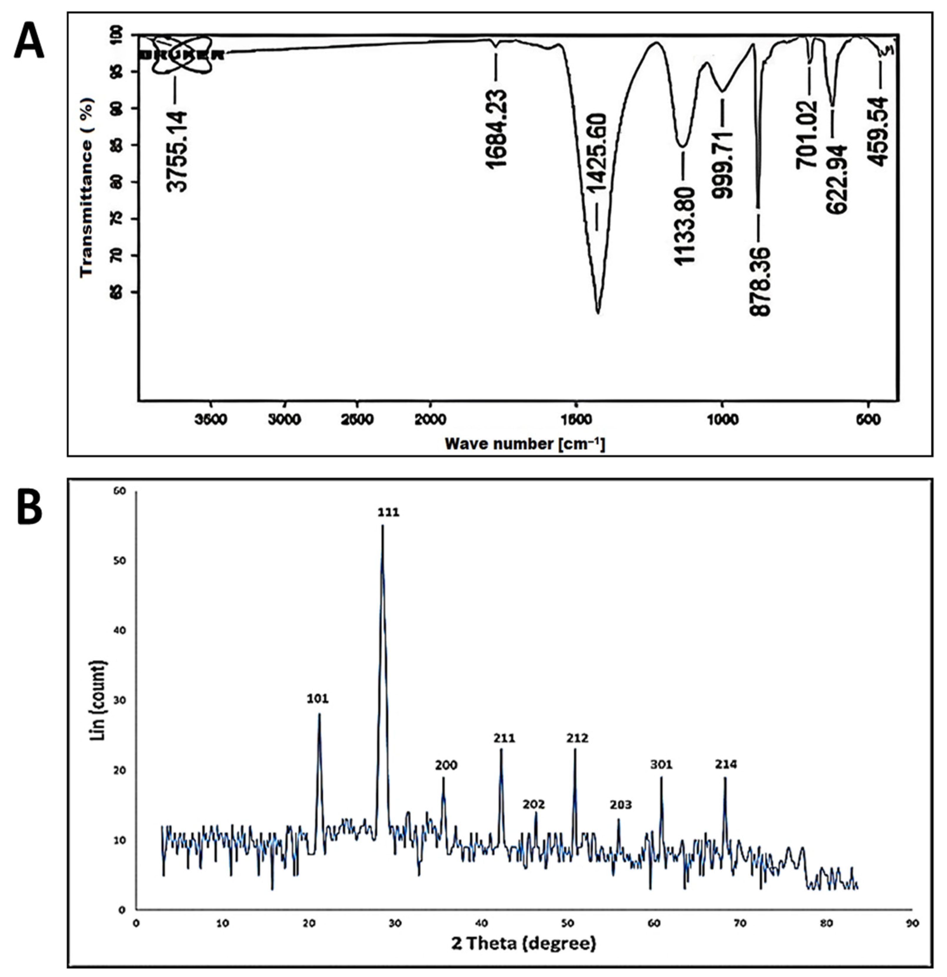
Figure 2A shows the FT-IR spectra of the biosynthesized silica nanoparticles, which are analyzed in the range between 400 to 4000 cm−1. The absorption peaks centered at 3755, 1684, 1425, 1133, 999, 878, 701, 622 and 459 cm−1. The band observed at 3755 is assigned to O–H stretching vibrations and the bending vibration of C–OH [58]. The peak observed at 1684 cm−1 was attributed to the O–H stretching band of the surface of silanol groups [59]. The strong band observed at 1425 cm−1 represents the stretching mode of CO of carboxylate salt and the adsorbed CO2 and CO32- at the surface of SiO2-NPs [60]. The adsorption of these new functional groups on the SiO2-NPs surface plays a significant role in catalytic reactions [61]. The absorption peak at 1133 cm−1 is assigned to the Si-O-Si asymmetric stretching vibration, and the peaks at 999 cm−1 are attributed to the asymmetric bending and stretching vibration of Si-OH, respectively. On the other hand, the symmetric stretching vibrations of Si-O-Si appear at 878 cm−1 [55,62]. The observed band at 459–701 cm−1 indicates the successful formation of SiO2 [63].

3.2.3. X-ray Diffraction (XRD) Pattern

The crystal structure of the as-formed SiO2-NPs was analyzed by XRD technique. The XRD analyzed silica nanoparticle spectra are depicted in Figure 2B. The results show that the SiO2-NPs have intensities at different 2θ, indicating that the silica nanoparticle structure is crystalline. The XRD pattern showed the typical Bragg reflections at 2ϴ equal to 21.2°, 28.6°, 35.6°, 42.3°, 46.36°, 50.91°, 55.94°, 60.92°, and 68.28°, which match the XRD pattern of the indices (101), (111), (200), (211), (202), (212), (203), (301) and (214), respectively. From the XRD pattern, it is confirmed that the material formed has a crystal structure. The XRD pattern observed was in accordance with the synthesis of silica nanoparticle produced by [61]. The Debye–Scherrer equation estimates that SiO2-NPs had an average crystallite size of 77 nm, where Peak Position (2θ) equals 55.94 o and FWHM (2θ) equals 0.1217.

3.2.4. Transmission Electron Microscopy (TEM)

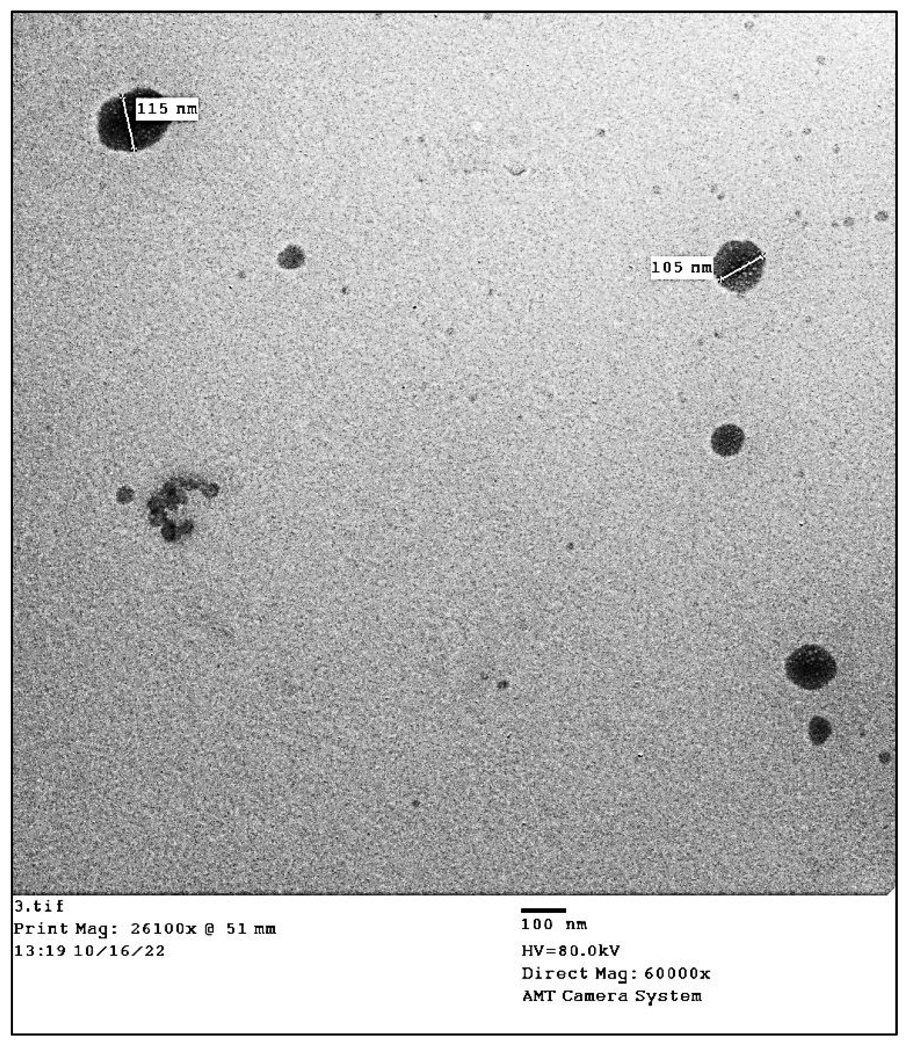
According to Wang and Lu, [64], the activity of NPs is often associated with their shape and size, with activity progressively increasing as size decreases. Therefore, determining the size and shape of the biosynthesized SiO2-NPs is important. The effectiveness of the metabolites present in the fungal biomass filtrate in stabilizing the shape of silica nanoparticles is seen in Figure 3. According to the data analysis, the spherical shapes are created with diameters of between 75 and 115 nm. Vetchinkina et al. [65] reported that when A. bisporus humus fungi were grown with Na2SiO3, the biogenic nanoparticles had a diameter of 30–100 nm and with A. arvensis, the particle diameter was up to 250 nm. Additionally, Saccharomyces cervisiae was utilized by Zamani et al. [51] for the successful biosynthesis of spherical silica nanoparticles with a diameter size range of 40 to 70 nm. Moreover, Zielonka et al. [51] demonstrated that an A. parasiticus strain was able to transform the amorphous silica conglomerates into structured nanoparticles (NPs) in the process of RH biotransformation, which produces silica NPs with sizes ranging from 3 to 400 nm. The presence of biomolecules and other metabolites in biomass filtrate, which are utilized in the bio-reduction and bio-capping of produced nanoparticles, may be the cause of the variation in nano-size and nano-shape. The largest challenges that require urgent and extended studies are thought to be the behaviors of metabolites like proteins and enzymes in creating and modifying certain NPs shapes like spherical, cubic, hexagonal, nano-flowering, rectangular, and rods [40].

3.2.5. Particle Size Distribution DLS

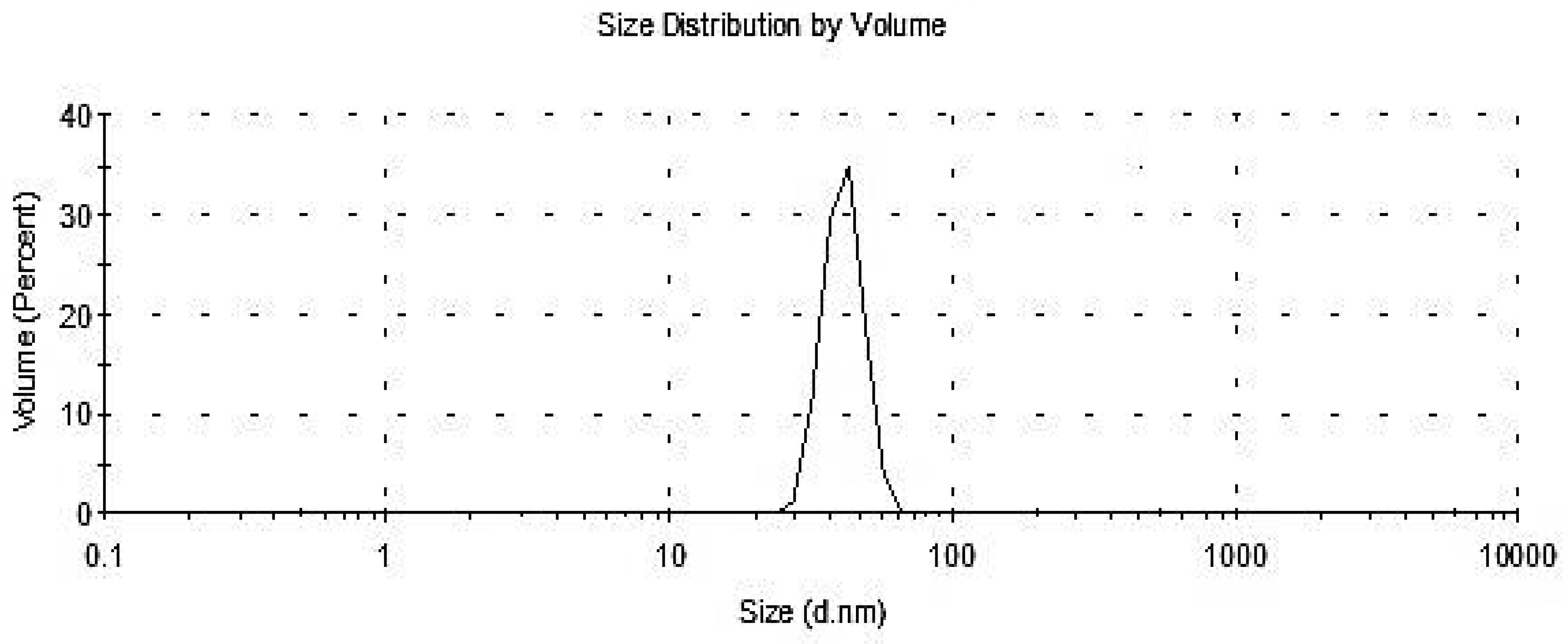
Particle size distribution was analyzed by DLS and the histogram showed an average particle size (based on intensity distribution) of about 70.1 nm with a volume of 33% (Figure 4). According to El-Gazzar et al. [56], particle size distribution analyzed by DLS showed an average particle size (based on intensity distribution) of about 89 nm. Moreover, Foroutan et al. [61] reported that silica nanoparticles have a particle morphology with an average particle size of about 30–40 nm. The DLS analysis offers additional details regarding the homogeneity of particles in the colloidal solution (PDI) [66]. The solution expands when the PDI value goes up over 1.0. In this investigation, the biosynthesized SiO2-NPs colloidal solution exhibited a PDI value of 0.03867, indicating that the solution was homogeneous.

3.3. In Vitro Antifungal Activity of Silica Nanoparticles against A. solani

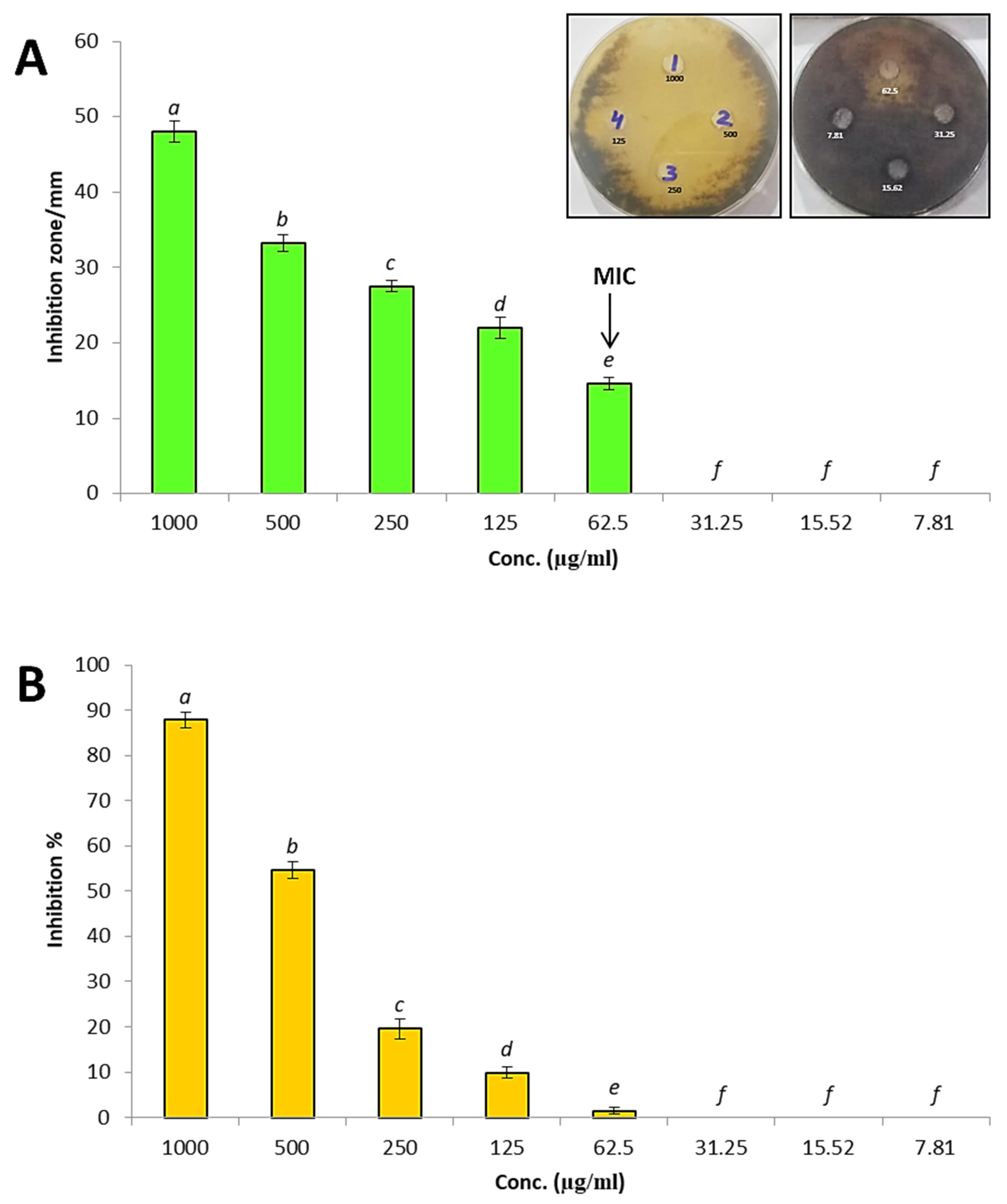
Metal nanoparticles are wildly used for biocontrol of A. solani causing early blight disease such as zinc, selenium, silver, copper nanoparticles [67,68,69,70]. In this study silica nanoparticles were used for biocontrol of A. solani causing early blight disease. Antifungal activity of SiO2-NPs was evaluated against A. solani using the agar well diffusion method, as shown in Figure 5A. Results revealed that biosynthesized SiO2-NPs exhibited promising antifungal activity against A. solani where inhibition zone 48 mm at concentration 1000 µg/mL. Also, different concentrations of silica nanoparticles were tested against A. solani, where results illustrated that inhibition zones were 33.2, 27.5, 22 and 14.6 mm at concentrations 500, 250, 125 and 62.5 µg/mL, respectively. On the other hand, other low concentrations 31.25, 15.62 and 7.81 µg/mL did not give any inhibition toward A. solani. Therefore, MIC of SiO2-NPs against A. solani was 62.5 µg/mL where this concentration exhibited minimum inhibition on growth of A. solani. Furthermore, the linear growth method was performed to determine inhibition percentages of SiO2 NPs at different concentrations toward A. solani as illustrated in Figure 5B. Results showed that inhibition percentages of SiO2-NPs at concentrations 1000, 500, 250, 125 and 62.5 µg/mL were 87.8%, 54.6%, 19.5%, 9.85% and 1.5% respectively. Meanwhile, concentrations of 31.25, 15.62 and 7.81 µg/mL did not give inhibition. Derbalah, et al. [71] fabricated silica nanoparticles to evaluate their antifungal activity against A. solani, and showed promising antifungal activity against A. solani. Ahamad and Siddiqui [72] studied effect of SiO2-NPs against A. dauci where results revealed that SiO2-NPs exhibited promising antifungal activity toward A. dauci and reduced effect of the fungus on carrot. Abdelrhim, et al. [73] reported that the silica nanoparticles significantly reduced mycelial radial growth of Rhizoctonia solani, and also induced innate immune response in wheat. Khan and Siddiqui [74] used SiO2-NPs as seed priming for controlling of R. solani, where results illustrated that SiO2-NPs exhibited antifungal activity against R. solani, and this led to enhance growth, chlorophyll and defense enzymes.

3.4. Disease Index

As shown in the Table 1, Alternaria infection caused a high DI, reaching 82.5%. The results of this study agree with several studies on Alternaria [6,44,75]. Reducing the severity of the disease and increasing the protection% considers the most important indications of the occurrence of plant resistance [76]. Biosynthesized nanoparticles are considered a magic solution to overcome the plant biotic stresses, especially fungal plant diseases, because they are very highly efficiency as bio fertilizers and have antifungal properties [77,78,79]. Silica helps plants reduce plant shock during pathogen exposure and incomplete infection stages [17]. The results of the current study showed that treatment with SiO2-NPs caused a decrease in the DI% and an increase in protection% compared to the untreated plants. In this regard, application of SiO2-NPs (either 50 or 100 ppm) on infected plants, showed a higher decrease in the disease symptoms and PDI and thus gives a high percentage of protection compared with untreated infected plants. Treatment of infected plants with SiO2-NPs (50 ppm) and (100 ppm) reduced DI% to 23% and 25% and high protection by 69.99% and 57.57%, respectively and next came silicon that recorded DI% 60% and protection 27.27%. Scientific reports have proven that silicon reduces the toxic effect on plants resulting from various stresses, as it works to strengthen cell walls and structural barriers, preventing the penetration of pathogens or impeding the movement of the pathogen inside the plant [80,81].

3.5. Measurement of Growth Traits

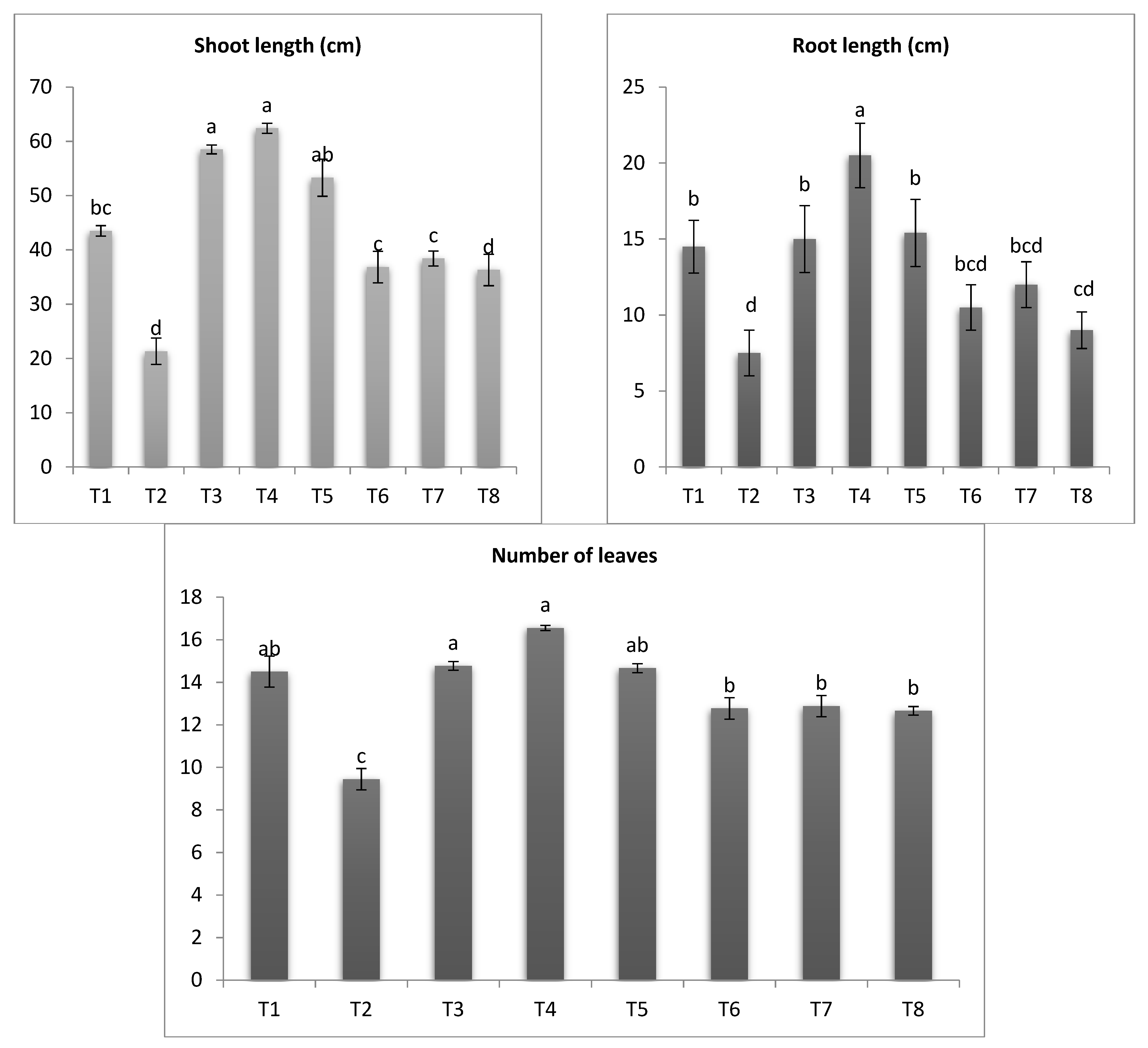
Heavily recently studies demonstrated that fungal plant diseases caused significant changes in vegetative growth [82,83,84]. Results in Figure 6 shows A. solani affected greatly reduced eggplant growth traits; shoot length, root length and number of leaves by 50.96%, 48.27% and 34.89%. Our results are consistent with [44,85,86]; they concluded that Alternaria infection causes a significant decrease in vegetative traits of many crops. In this respect, this severe deficiency can be explained by dangerous disturbances within the cell metabolism as a result of oxidative blasts during the A. solani infection process [85,87]. On the other hand, application of SiO2-NPs resulted to enhancement of both healthy and infected plants compared to control. Likewise, treatment infected plants with SiO2-NPs (100 ppm) and (50 ppm), showed a significant increase in shoot length, root length and number of leaves and next came Si, respectively. The highest increase in shoot length, root length and number of leaves was recorded in SiO2-NPs 100 ppm treated plants. Interestingly, the application of SiO2-NPs on healthy plants showed a significant improvement in the vegetative growth characteristics as shown in the Figure 6. Many previous studies support these results [73,87,88]. Many recently studies proved that the plant resistance against biotic as well as abiotic stresses can be enhanced and stimulated by application of safe organisms and their metabolites through different mechanisms [88,89,90,91]. The results can be explained by the fact that fertilizing with silicon increases the efficiency of phosphorous uptake, which is reflected in an increase in lipid substances in plant cells, which increases the efficiency of roots to absorb. In addition, silicon supports the hardness of cell walls, which prevents the penetration of pathogenic fungi and reduces stress [39,92,93].
(T1-Healthy control, T2-Infected control, T3-healthy plants treated with SiO2-NPs (50 ppm), T4-healthy plants treated SiO2-NPs (100 ppm), T5-healthy plants treated with sodium silicate (100 ppm), T6-infected plants treated with SiO2-NPs (50 ppm), T7-infected plants treated with SiO2-NPs (100 ppm), T8- infected plants treated with sodium silicate (100 ppm)).

3.6. Photosynthetic Pigments

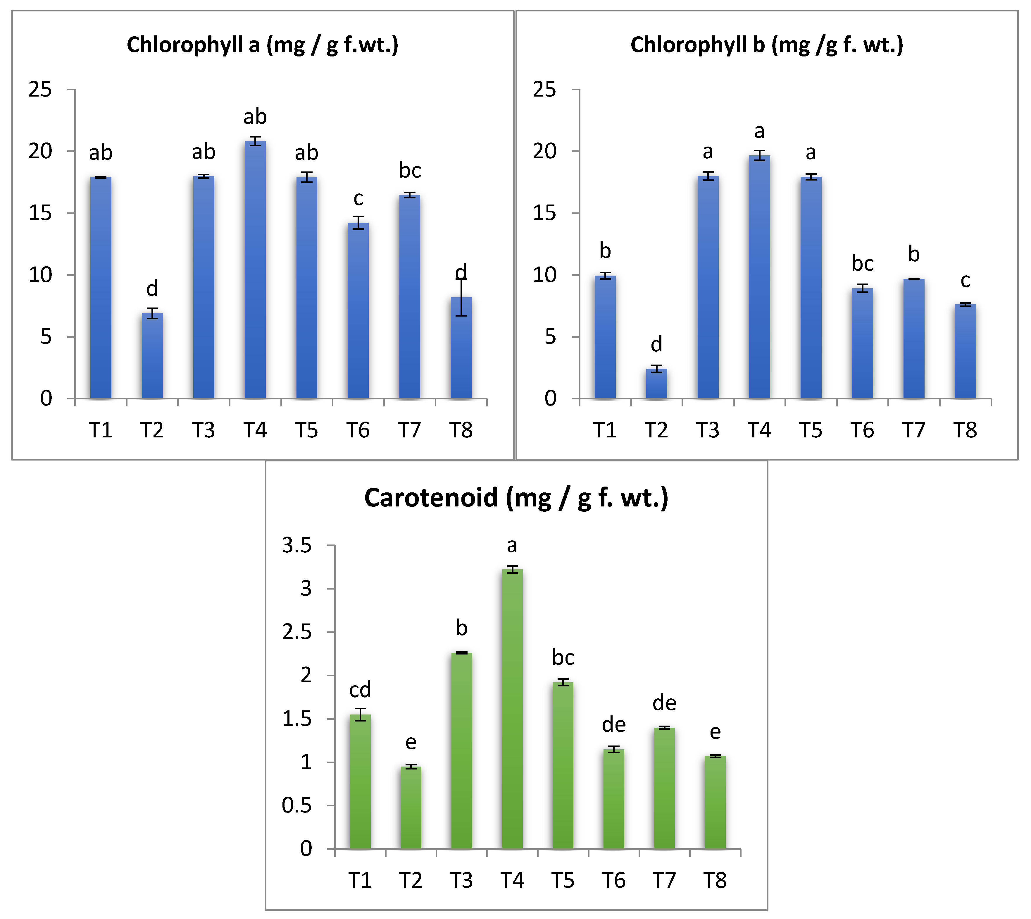
As shown in Figure 7, a sharp decrease in chlorophyll (a, b) and carotenoid was observed due to infection with Alternaria fungus compared to uninfected plants. These results are in agreement with several studies [6,46,87,94]. This acute deficiency in the components of chlorophyll and carotenoids may be due to the plant’s failure to capture light due to a deficiency in the leaf area responsible for light capture and photosynthesis, leaves and plant height, or it may also be due to oxidative explosion and increased activity of chlorophyll-degrading enzymes and chlorophyllase under the conditions of injury [6,81,88]. However, Alternaria-infected plants treated with silica nanoparticles (50 and 100 ppm) or Si showed a marked increase when compared with untreated infected plants. Concerning the effect SiO2-NPs and SiO2 on the infected plants with A. solani, it was found that SiO2-NPs 100 and 50 ppm indicated a greatly significant rise in the contents of Chl a and Chl b. The uninfected eggplant seedlings treated with SiO2-NPs or SiO2 presented a major increase in chlorophyll pigments a and b. Also, the obtained results illustrated that in A. solani infection plants, the content of carotenoids was increased in response to the treatment with SiO2-NPs. This increase could be attributed to improved number of leaves, stomata conductance, transpiration rate and/or cell size and number [95,96,97]. This improvement in the level of photosynthetic pigments may be explained by the fact that SiO2-NPs have been described as plant immune-stimulants [94,98]. It may trigger NADPH oxidase activity, thereby stimulating the production of H2O2. Thus, SiO2-NPs could stimulate reactive oxygen species (ROS) in plants. Silicon also increases the productivity of photosynthesis, and also rises the strength of leaves and the xylem, the high rates of transpiration caused by the disease by increasing the deposition of silicon in the cell walls of roots, leaves, twigs, and the main stem [20].

4. Oxidative Stress Markers

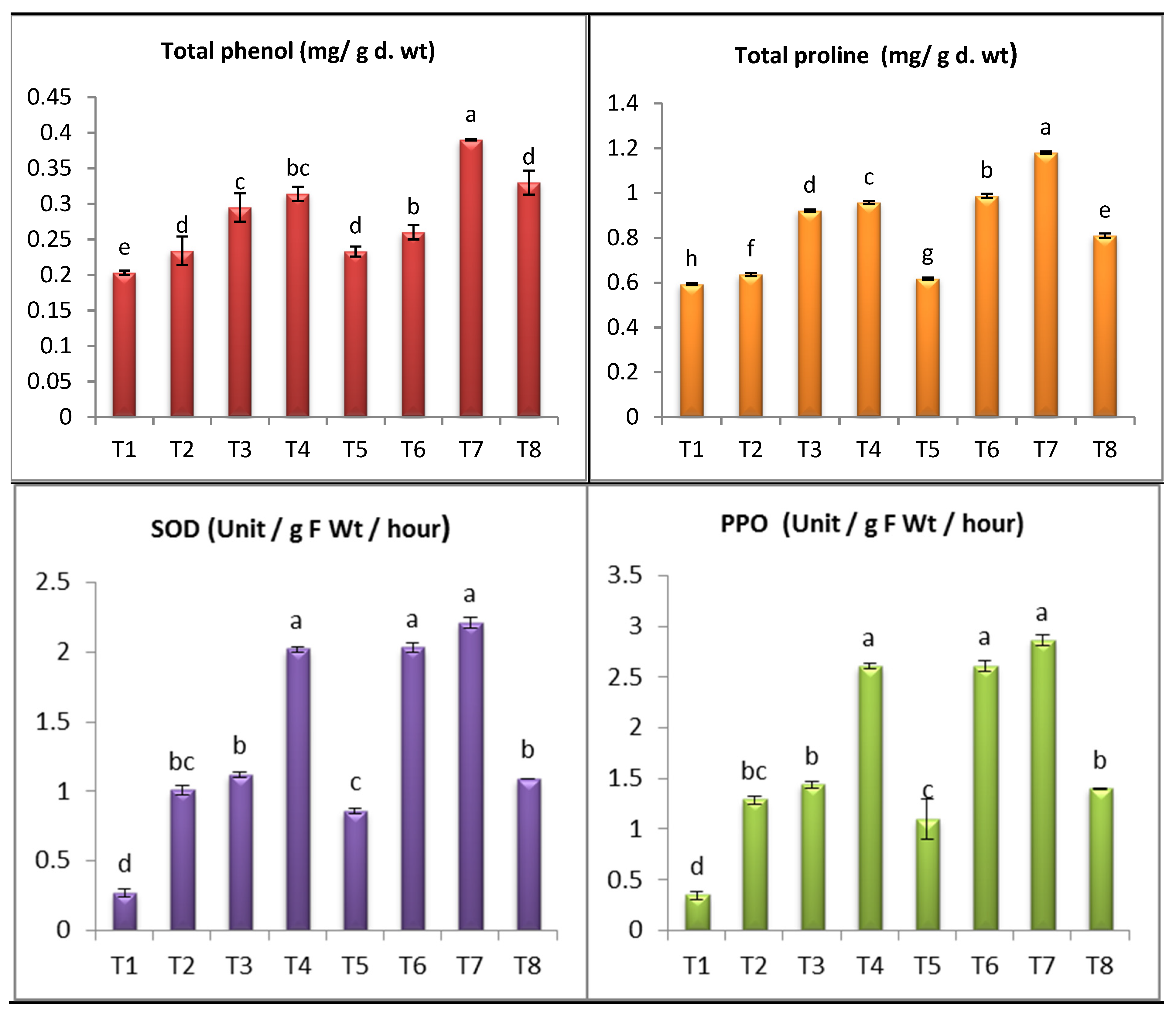
Alternaria infected eggplants showed significant increases in contents of free proline, total phenols by 7.25%, 15.27%, respectively, relative to uninfected control plants (Figure 8). Our results are in agreement with other studies [4,6,99,100]. The increase of proline and phenols acts as one of the imperative biological activities as it works a critical role in capturing free radicals, keeping cells from oxidation, and providing plant cells with energy [101,102]. Moreover, application of SiO2-NPs or SiO2 on infected plants resulted in significant increase in content of proline by 85.53%, 55.18%, and 27% at SiO2-NPs 100 ppm, 50 ppm and SiO2. Also, contents of phenols increased by 66.66%, 41.02% and 11.11% with SiO2-NPs 100 and 50 ppm, respectively, when compared untreated infected plants. The highest increase in contents of proline, total phenols was recorded in SiO2-NPs 50 ppm treated plants. Proline accumulation in eggplants shoots prevents the photosynthetic process throughout inhibiting injury of photosynthetic pigments caused by ROS [45,100,101]. Phenols perform an chief role in capturing free radicals, which decreases oxidative stress in cells [103]. The increase of phenolic compounds is an adaptive approach against disease [104]. Through our results, a significant increase in the content of proline and phenol was observed due to the addition of silica nanoparticles, especially concentration (100 ppm), which confirms the role of SiO2-NPs in inducing plant immunity, as the accumulation of proline in plants prevents damage to photosynthetic pigments by capturing free radicals that cause damage and failure in the photosynthesis process [105,106]. In these results, a clear and positive link to the use of SiO2-NPs in terms of improving vegetative growth characteristics, increasing the level of photosynthetic pigments, and raising the content of proline and phenol in infected plants cells, which supports the role of silicon in strengthening plant immunity against stress, which was confirmed by many previous studies [14,107,108].
To achieve a stronger sign on resistance, mean activities of superoxide dismutase and polyphenol oxidase of the tested eggplants were established in this experiment. SOD and PPO activities were greater in the Alternaria–infected plants and treated with (SiO2-NPs 100, 50 ppm and SiO2). The activity of SOD and PPO were increased in Alternaria-infected eggplants compared to healthy plants. The activity of SOD and PPO was boosted in infected plants comparing to healthy control plants (Figure 8). Application of (SiO2-NPs 100 ppm, SiO2-NPs 50 ppm and sodium silicate 100 ppm) increased the activity of SOD by (643.46%, 301.17% and 218.86%) and PPO by (312.39%, 642.03% and 215.64%) respectively, over healthy controsl. Moreover, application of (SiO2-NPs 100 ppm and SiO2-NPs 50 ppm) increased the activity of SOD by (119.46% and 101.58%) and PPO by (122% and 103%) respectively, over infected plants (Figure 8). Meanwhile, sodium silicate 100 ppm was the least effective and increased (SOD and PPO) activity by (7.24% and 9.01%) (Figure 8). Under non-infected conditions, application of SiO2-NPs 100 ppm, SiO2-NPs 50 ppm and sodium silicate 100 ppm boosted the activities of SOD and PPO in treated plants as compared with control. The study carried out by Kiadaliri et al. [109] reported that antioxidant enzymes SOD and PPO provide a large number of defensive enzymes associated with infection. These enzymes act as initial steps in increasing plant resistance to various stresses as well as the formation of phenolic compounds. The plant presented different mechanisms to cope with infection stress as they increase the activity of certain antioxidant enzymes to keep ROS at the lower level in the cell [110]. Increasing the activity of antioxidant enzymes (SOD, PPO) as a result of treatment with SiO2-NPs on infected plants provides strong evidence of induction of resistance against the disease. Antioxidant enzymes mitigate the oxidative blast caused by injury by capturing free radicals from cells [111].

5. Conclusions

In the current study, SiO2-NPs were successfully mycosynthesized using A. niger, and fully characterized using Uv-Vis, FTIR, XRD, TEM and DLS techniques. Mycosynthesized SiO2-NPs exhibited promising antifungal activity toward A. solani. The application of SiO2-NPs at different concentrations (50 and 100 ppm) greatly offers the possibility of early blight recovery in eggplant. For more, mycosynthesized SiO2-NPs can be applied as a plant growth-promoting agent instead of chemical synthesized fertilizers in healthy and infected eggplant. Additionally, SiO2-NPs showed the highest increase in contents of proline and phenols. It is worth mentioned that the prepared SiO2-NPs can modify plant antioxidant defense system by inducing overexpress oxidative stress in plants. So, the usage of the tested SiO2-NPs can increase not only the anti-fungal activity, but also the systematic immune response of treated plants. Therefore, mycosynthesized SiO2-NPs can be applied as a confident and safe alternative bio fungicide against Alternaria solani causing early blight disease.

Author Contributions

Conceptualization, A.M.A., M.S.A., E.S. and A.H.H.; methodology, M.A.A., A.M.A., M.S.A., E.S., H.H.E. and A.H.H.; software A.M.A., M.S.A., E.S. and A.H.H.; validation, A.M.A., M.S.A., E.S. and A.H.H.; resources, A.M.A., M.S.A., E.S. and A.H.H.; data curation, A.M.A., M.S.A., E.S. and A.H.H.; writing—original draft preparation, A.M.A., M.S.A., E.S. and A.H.H.; writing—review and editing, M.A.A., A.M.A., M.S.A., E.S., H.H.E. and A.H.H.; All authors have read and agreed to the published version of the manuscript.

Funding

This research received no external funding.

Institutional Review Board Statement

Not applicable.

Informed Consent Statement

Not applicable.

Data Availability Statement

Data are contained within the article.

Acknowledgments

The authors would like to thank Botany and Microbiology Department, Faculty of Science, Al-Azhar University for promoting this research.

Conflicts of Interest

The authors declare no conflict of interest.

References

  1. Abdelaziz, A.M.; Salem, S.S.; Khalil, A.; El-Wakil, D.A.; Fouda, H.M.; Hashem, A.H. Potential of biosynthesized zinc oxide nanoparticles to control Fusarium wilt disease in eggplant (Solanum melongena) and promote plant growth. BioMetals 2022, 35, 601–616. [Google Scholar] [CrossRef]
  2. Attia, M.S.; El-Sayyad, G.S.; Abd Elkodous, M.; Khalil, W.F.; Nofel, M.M.; Abdelaziz, A.M.; Farghali, A.A.; El-Batal, A.I.; El Rouby, W.M. Chitosan and EDTA conjugated graphene oxide antinematodes in Eggplant: Toward improving plant immune response. Int. J. Biol. Macromol. 2021, 179, 333–344. [Google Scholar] [CrossRef]
  3. Khalil, A.M.; Ahmed, A.F.; Mahmoud, E.E.; Abdelaziz, A.M. Influence of organic farming system on microbial biomass and fungal communities of agricultural soil. Afr. J. Mycol. Biotechnol. 2015, 20, 23–40. [Google Scholar]
  4. Attia, M.S.; Abdelaziz, A.M.; Al-Askar, A.A.; Arishi, A.A.; Abdelhakim, A.M.; Hashem, A.H. Plant growth-promoting fungi as biocontrol tool against fusarium wilt disease of tomato plant. J. Fungi 2022, 8, 775. [Google Scholar] [CrossRef]
  5. Abd Alhakim, A.; Hashem, A.; Abdelaziz, A.M.; Attia, M.S. Impact of plant growth promoting fungi on biochemical defense performance of Tomato under Fusarial infection. Egypt. J. Chem. 2022. [Google Scholar] [CrossRef]
  6. Attia, M.S.; Hashem, A.H.; Badawy, A.A.; Abdelaziz, A.M. Biocontrol of early blight disease of eggplant using endophytic Aspergillus terreus: Improving plant immunological, physiological and antifungal activities. Bot. Stud. 2022, 63, 26. [Google Scholar] [CrossRef] [PubMed]
  7. Tsedaley, B. Review on early blight (Alternaria spp.) of potato disease and its management options. J. Biol. Agric. Healthc. 2014, 4, 191–199. [Google Scholar]
  8. Gupta, S.K.; Thind, T. Disease Problems in Vegetable Production; Scientific Publishers: Singapore, 2018. [Google Scholar]
  9. Opoku, B.A. Department of Crop and Soil Sciences; Cornell University Press: Ithacab, NY, USA, 2012. [Google Scholar]
  10. Rachidi, F.; Benhima, R.; Kasmi, Y.; Sbabou, L.; Arroussi, H.E. Evaluation of microalgae polysaccharides as biostimulants of tomato plant defense using metabolomics and biochemical approaches. Sci. Rep. 2021, 11, 930. [Google Scholar] [CrossRef]
  11. Attia, M.S.; Younis, A.M.; Ahmed, A.F.; Elaziz, A. Comprehensive management for wilt disease caused by Fusarium oxysporum in tomato plant. Int. J. Innov. Sci. Eng. Technol. 2016, 4, 2348–7968. [Google Scholar]
  12. Farrag, A.; Attia, M.S.; Younis, A.; Abd Elaziz, A. Potential impacts of elicitors to improve tomato plant disease resistance. Al Azhar. Bull. Sci. 2017, 9, 311–321. [Google Scholar]
  13. Sarwar, N.; Malhi, S.S.; Zia, M.H.; Naeem, A.; Bibi, S.; Farid, G. Role of mineral nutrition in minimizing cadmium accumulation by plants. J. Sci. Food Agric. 2010, 90, 925–937. [Google Scholar] [CrossRef] [PubMed]
  14. Souri, Z.; Khanna, K.; Karimi, N.; Ahmad, P. Silicon and plants: Current knowledge and future prospects. J. Plant Growth Regul. 2021, 40, 906–925. [Google Scholar] [CrossRef]
  15. Gogos, A.; Knauer, K.; Bucheli, T.D. Nanomaterials in plant protection and fertilization: Current state, foreseen applications, and research priorities. J. Agric. Food Chem. 2012, 60, 9781–9792. [Google Scholar] [CrossRef]
  16. Gall, H.L.; Philippe, F.; Domon, J.-M.; Gillet, F.; Pelloux, J.; Rayon, C. Cell wall metabolism in response to abiotic stress. Plants 2015, 4, 112–166. [Google Scholar] [CrossRef]
  17. El-Shetehy, M.; Moradi, A.; Maceroni, M.; Reinhardt, D.; Petri-Fink, A.; Rothen-Rutishauser, B.; Mauch, F.; Schwab, F. Silica nanoparticles enhance disease resistance in Arabidopsis plants. Nat. Nanotechnol. 2021, 16, 344–353. [Google Scholar] [CrossRef]
  18. Islam, W.; Tayyab, M.; Khalil, F.; Hua, Z.; Huang, Z.; Chen, H.Y. Silicon-mediated plant defense against pathogens and insect pests. Pestic. Biochem. Physiol. 2020, 168, 104641. [Google Scholar] [CrossRef] [PubMed]
  19. Ahire, M.; Mundada, P.; Nikam, T.; Bapat, V.; Penna, S. Multifaceted roles of silicon in mitigating environmental stresses in plants. Plant Physiol. Biochem. 2021, 169, 291–310. [Google Scholar] [CrossRef] [PubMed]
  20. Zhang, Y.; Yu, S.; Gong, H.-J.; Zhao, H.-L.; Li, H.-L.; Hu, Y.-H.; Wang, Y.-C. Beneficial effects of silicon on photosynthesis of tomato seedlings under water stress. J. Integr. Agric. 2018, 17, 2151–2159. [Google Scholar] [CrossRef] [Green Version]
  21. Bakhat, H.F.; Bibi, N.; Zia, Z.; Abbas, S.; Hammad, H.M.; Fahad, S.; Ashraf, M.R.; Shah, G.M.; Rabbani, F.; Saeed, S. Silicon mitigates biotic stresses in crop plants: A review. Crop Prot. 2018, 104, 21–34. [Google Scholar] [CrossRef]
  22. Vasanthi, N.; Saleena, L.M.; Raj, S.A. Silicon in crop production and crop protection-A review. Agric. Rev. 2014, 35, 14–23. [Google Scholar] [CrossRef]
  23. Babu, T. Calibration and Categorization of Plant Available Silicon in Louisiana Soils; Louisiana State University and Agricultural and Mechanical College: Baton Rouge, LA, USA, 2015. [Google Scholar]
  24. Hashem, A.H.; Abdelaziz, A.M.; Attia, M.S.; Salem, S.S. Selenium and Nano-Selenium-Mediated Biotic Stress Tolerance in Plants. In Selenium and Nano-Selenium in Environmental Stress Management and Crop Quality Improvement; Hossain, M.A., Ahammed, G.J., Kolbert, Z., El-Ramady, H., Islam, M.T., Schiavon, M., Eds.; Springer International Publishing: Cham, Switzerland, 2022; pp. 209–226. [Google Scholar] [CrossRef]
  25. Saied, E.; Hashem, A.H.; Ali, O.M.; Selim, S.; Almuhayawi, M.S.; Elbahnasawy, M.A. Photocatalytic and Antimicrobial Activities of Biosynthesized Silver Nanoparticles Using Cytobacillus firmus. Life 2022, 12, 1331. [Google Scholar] [CrossRef] [PubMed]
  26. Saied, E.; Salem, S.S.; Al-Askar, A.A.; Elkady, F.M.; Arishi, A.A.; Hashem, A.H. Mycosynthesis of Hematite (α-Fe2O3) Nanoparticles Using Aspergillus niger and Their Antimicrobial and Photocatalytic Activities. Bioengineering 2022, 9, 397. [Google Scholar] [CrossRef]
  27. Hashem, A.H.; Selim, T.A.; Alruhaili, M.H.; Selim, S.; Alkhalifah, D.H.M.; Al Jaouni, S.K.; Salem, S.S. Unveiling Antimicrobial and Insecticidal Activities of Biosynthesized Selenium Nanoparticles Using Prickly Pear Peel Waste. J. Funct. Biomater. 2022, 13, 112. [Google Scholar] [CrossRef]
  28. Salem, S.S.; Hashem, A.H.; Sallam, A.-A.M.; Doghish, A.S.; Al-Askar, A.A.; Arishi, A.A.; Shehabeldine, A.M. Synthesis of Silver Nanocomposite Based on Carboxymethyl Cellulose: Antibacterial, Antifungal and Anticancer Activities. Polymers 2022, 14, 3352. [Google Scholar] [CrossRef]
  29. Shehabeldine, A.M.; Salem, S.S.; Ali, O.M.; Abd-Elsalam, K.A.; Elkady, F.M.; Hashem, A.H. Multifunctional Silver Nanoparticles Based on Chitosan: Antibacterial, Antibiofilm, Antifungal, Antioxidant, and Wound-Healing Activities. J. Fungi 2022, 8, 612. [Google Scholar] [CrossRef]
  30. El-Naggar, M.E.; Hasanin, M.; Hashem, A.H. Eco-Friendly Synthesis of Superhydrophobic Antimicrobial Film Based on Cellulose Acetate/Polycaprolactone Loaded with the Green Biosynthesized Copper Nanoparticles for Food Packaging Application. J. Polym. Environ. 2022, 30, 1820–1832. [Google Scholar] [CrossRef]
  31. Salem, S.S.; Ali, O.M.; Reyad, A.M.; Abd-Elsalam, K.A.; Hashem, A.H. Pseudomonas indica-Mediated Silver Nanoparticles: Antifungal and Antioxidant Biogenic Tool for Suppressing Mucormycosis Fungi. J. Fungi 2022, 8, 126. [Google Scholar] [CrossRef] [PubMed]
  32. Lashin, I.; Hasanin, M.; Hassan, S.A.M.; Hashem, A.H. Green biosynthesis of zinc and selenium oxide nanoparticles using callus extract of Ziziphus spina-christi: Characterization, antimicrobial, and antioxidant activity. Biomass Convers. Biorefinery 2021. [Google Scholar] [CrossRef]
  33. Hasanin, M.; Hashem, A.H.; Lashin, I.; Hassan, S.A.M. In vitro improvement and rooting of banana plantlets using antifungal nanocomposite based on myco-synthesized copper oxide nanoparticles and starch. Biomass Convers. Biorefinery 2021. [Google Scholar] [CrossRef]
  34. Vasanthi, N.; Saleena, L.M.; Raj, S.A. Silica Solubilization Potential of Certain Bacterial Species in the Presence of Different Silicate Minerals. Silicon 2018, 10, 267–275. [Google Scholar] [CrossRef]
  35. Raturi, G.; Sharma, Y.; Rana, V.; Thakral, V.; Myaka, B.; Salvi, P.; Singh, M.; Dhar, H.; Deshmukh, R. Exploration of silicate solubilizing bacteria for sustainable agriculture and silicon biogeochemical cycle. Plant Physiol. Biochem. PPB 2021, 166, 827–838. [Google Scholar] [CrossRef] [PubMed]
  36. Pieła, A.; Żymańczyk-Duda, E.; Brzezińska-Rodak, M.; Duda, M.; Grzesiak, J.; Saeid, A.; Mironiuk, M.; Klimek-Ochab, M. Biogenic synthesis of silica nanoparticles from corn cobs husks. Dependence of the productivity on the method of raw material processing. Bioorganic Chem. 2020, 99, 103773. [Google Scholar] [CrossRef] [PubMed]
  37. Tarshan, M. Digestibility of Fungi-Based Protein Products Fed to Broiler Chickens; Swedish University of Agricultural Sciences Library: Lomma, Sweden, 2017. [Google Scholar]
  38. Mahboubi, A.; Ferreira, J.A.; Taherzadeh, M.J.; Lennartsson, P.R. Value-added products from dairy waste using edible fungi. Waste Manag. 2016, 59, 518–525. [Google Scholar] [CrossRef] [PubMed]
  39. Mathur, P.; Roy, S. Nanosilica facilitates silica uptake, growth and stress tolerance in plants. Plant Physiol. Biochem. 2020, 157, 114–127. [Google Scholar] [CrossRef] [PubMed]
  40. Fouda, A.; Hassan, S.E.-D.; Saied, E.; Hamza, M.F. Photocatalytic degradation of real textile and tannery effluent using biosynthesized magnesium oxide nanoparticles (MgO-NPs), heavy metal adsorption, phytotoxicity, and antimicrobial activity. J. Environ. Chem. Eng. 2021, 9, 105346. [Google Scholar] [CrossRef]
  41. Alhazmi, H.A. FT-IR spectroscopy for the identification of binding sites and measurements of the binding interactions of important metal ions with bovine serum albumin. Sci. Pharm. 2019, 87, 5. [Google Scholar] [CrossRef] [Green Version]
  42. Goswami, P.; Mathur, J. Application of agro-waste-mediated silica nanoparticles to sustainable agriculture. Bioresour. Bioprocess. 2022, 9, 9. [Google Scholar] [CrossRef]
  43. Babick, F. Dynamic light scattering (DLS). In Characterization of Nanoparticles; Elsevier: Amsterdam, The Netherlands, 2020; pp. 137–172. [Google Scholar]
  44. Attia, M.S.; El-Sayyad, G.S.; Abd Elkodous, M.; El-Batal, A.I. The effective antagonistic potential of plant growth-promoting rhizobacteria against Alternaria solani-causing early blight disease in tomato plant. Sci. Hortic. 2020, 266, 109289. [Google Scholar] [CrossRef]
  45. Vernon, L.P.; Seely, G.R. The Chlorophylls; Academic Press: Cambridge, MA, USA, 2014. [Google Scholar]
  46. Bates, L.S.; Waldren, R.P.; Teare, I. Rapid determination of free proline for water-stress studies. Plant Soil 1973, 39, 205–207. [Google Scholar] [CrossRef]
  47. Dai, G.; Andary, C.; Cosson-Mondolot, L.; Boubals, D. Polyphenols and resistance of grapevines to downy mildew. Int. Symp. Nat. Phenols Plant Resist. 1993, 381, 763–766. [Google Scholar] [CrossRef]
  48. Bergmeyer, H. Determination with glucose oxidase and peroxidase. Methods Enzym. Anal. 1974, 1205–1215. [Google Scholar]
  49. Lavid, N.; Schwartz, A.; Yarden, O.; Tel-Or, E. The involvement of polyphenols and peroxidase activities in heavy-metal accumulation by epidermal glands of the waterlily (Nymphaeaceae). Planta 2001, 212, 323–331. [Google Scholar] [CrossRef]
  50. Snedecor, G.W. Comparisons of two samples. In Statistical Methods; Snedecor, G.W., Cochran, W.G., Eds.; Iowa State University Press: Ames, IA, USA, 1980. [Google Scholar]
  51. Zamani, H.; Jafari, A.; Mousavi, S.M.; Darezereshki, E. Biosynthesis of silica nanoparticle using Saccharomyces cervisiae and its application on enhanced oil recovery. J. Pet. Sci. Eng. 2020, 190, 107002. [Google Scholar] [CrossRef]
  52. Kannan, M.; Uma Sangareswari, K.; Suganya, P.; Ganesan, R.; Rajarathinam, K. Biobased approach for the synthesis, characterization, optimization and application of silica nanoparticles by fungus Fusarium oxysporum. Pharmaceut. Biol. Eval 2015, 2, 223–233. [Google Scholar]
  53. Bansal, V.; Rautaray, D.; Bharde, A.; Ahire, K.; Sanyal, A.; Ahmad, A.; Sastry, M. Fungus-mediated biosynthesis of silica and titania particles. J. Mater. Chem. 2005, 15, 2583–2589. [Google Scholar] [CrossRef]
  54. Singh, S.; Bhatta, U.M.; Satyam, P.; Dhawan, A.; Sastry, M.; Prasad, B. Bacterial synthesis of silicon/silica nanocomposites. J. Mater. Chem. 2008, 18, 2601–2606. [Google Scholar] [CrossRef]
  55. Biradar, A.I.; Sarvalkar, P.D.; Teli, S.B.; Pawar, C.; Patil, P.; Prasad, N.R. Photocatalytic degradation of dyes using one-step synthesized silica nanoparticles. Mater. Today Proc. 2021, 43, 2832–2838. [Google Scholar] [CrossRef]
  56. El-Gazzar, N.; Almanaa, T.N.; Reda, R.M.; El Gaafary, M.; Rashwan, A.; Mahsoub, F. Assessment the using of silica nanoparticles (SiO2NPs) biosynthesized from rice husks by Trichoderma harzianum MF780864 as water lead adsorbent for immune status of Nile tilapia (Oreochromis niloticus). Saudi J. Biol. Sci. 2021, 28, 5119–5130. [Google Scholar] [CrossRef] [PubMed]
  57. Ghosh, S.; Kundu, M.; Dutta, S.; Mahalanobish, S.; Ghosh, N.; Das, J.; Sil, P.C. Enhancement of anti-neoplastic effects of cuminaldehyde against breast cancer via mesoporous silica nanoparticle based targeted drug delivery system. Life Sci. 2022, 298, 120525. [Google Scholar] [CrossRef]
  58. Olga, M.; Jana, M.; Anna, M.; Irena, K.; Jan, M.; Alena, Č. Antimicrobial properties and applications of metal nanoparticles biosynthesized by green methods. Biotechnol. Adv. 2022, 58, 107905. [Google Scholar]
  59. Lai, H.; Liu, Y.; Huang, G.; Chen, Y.; Song, Y.; Ma, Y.; Yue, P. Fabrication and antibacterial evaluation of peppermint oil-loaded composite microcapsules by chitosan-decorated silica nanoparticles stabilized Pickering emulsion templating. Int. J. Biol. Macromol. 2021, 183, 2314–2325. [Google Scholar] [CrossRef]
  60. Nieto-Ortega, B.; Bürgi, T. Vibrational properties of thiolate-protected gold nanoclusters. Acc. Chem. Res. 2018, 51, 2811–2819. [Google Scholar] [CrossRef]
  61. Foroutan, R.; Mohammadi, R.; Peighambardoust, S.J.; Jalali, S.; Ramavandi, B. Application of nano-silica particles generated from offshore white sandstone for cadmium ions elimination from aqueous media. Environ. Technol. Innov. 2020, 19, 101031. [Google Scholar] [CrossRef]
  62. Agusnar, H.; Sugita, P.; Nainggolan, I. Preparation sodium silicate from rice husk to synthesize silica nanoparticles by sol-gel method for adsorption water in analysis of methamphetamine. South Afr. J. Chem. Eng. 2022, 40, 80–86. [Google Scholar]
  63. Mohanraj, R.; Gnanamangai, B.M.; Poornima, S.; Oviyaa, V.; Ramesh, K.; Vijayalakshmi, G.; Nithya, M.; Karthik, N.; Ponmurugan, P.; Robinson, J.P. Decolourisation efficiency of immobilized silica nanoparticles synthesized by actinomycetes. Mater. Today Proc. 2020, 48, 129–135. [Google Scholar] [CrossRef]
  64. Wang, H.; Lu, J. A review on particle size effect in metal-catalyzed heterogeneous reactions. Chin. J. Chem. 2020, 38, 1422–1444. [Google Scholar] [CrossRef]
  65. Vetchinkina, E.; Loshchinina, E.; Kupryashina, M.; Burov, A.; Nikitina, V. Shape and size diversity of gold, silver, selenium, and silica nanoparticles prepared by green synthesis using fungi and bacteria. Ind. Eng. Chem. Res. 2019, 58, 17207–17218. [Google Scholar] [CrossRef]
  66. Stetefeld, J.; McKenna, S.A.; Patel, T.R. Dynamic light scattering: A practical guide and applications in biomedical sciences. Biophys. Rev. 2016, 8, 409–427. [Google Scholar] [CrossRef] [PubMed] [Green Version]
  67. Singh, A.; Gaurav, S.S.; Shukla, G.; Rani, P. Assessment of mycogenic zinc nano-fungicides against pathogenic early blight (Alternaria solani) of potato (Solanum tuberosum L.). Mater. Today Proc. 2022, 49, 3528–3537. [Google Scholar] [CrossRef]
  68. Lahuf, A.A.; Abdullah, K.M.; Mohammadali, M.T. Assessment of the nanosized particles of ZnO and MgO and some cultivars in control of Alternaria solani causing tomato early blight. Ecol. Environ. Conserv. 2020, 26, 89–95. [Google Scholar]
  69. Quiterio-Gutiérrez, T.; Ortega-Ortiz, H. The Application of Selenium and Copper Nanoparticles Modifies the Biochemical Responses of Tomato Plants under Stress by Alternaria solani. Int. J. Mol. Sci. 2019, 20, 1950. [Google Scholar] [CrossRef] [Green Version]
  70. Ismail, A.-W.A.; Sidkey, N.M.; Arafa, R.A.; Fathy, R.M.; El-Batal, A.I. Evaluation of in vitro antifungal activity of silver and selenium nanoparticles against Alternaria solani caused early blight disease on potato. Br. Biotechnol. J. 2016, 12, 1. [Google Scholar] [CrossRef]
  71. Derbalah, A.; Shenashen, M.; Hamza, A.; Mohamed, A.; El Safty, S. Antifungal activity of fabricated mesoporous silica nanoparticles against early blight of tomato. Egypt. J. Basic Appl. Sci. 2018, 5, 145–150. [Google Scholar] [CrossRef] [Green Version]
  72. Ahamad, L.; Siddiqui, Z.A. Effects of silicon dioxide, zinc oxide and titanium dioxide nanoparticles on Meloidogyne incognita, Alternaria dauci and Rhizoctonia solani disease complex of carrot. Exp. Parasitol. 2021, 230, 108176. [Google Scholar] [CrossRef]
  73. Abdelrhim, A.S.; Mazrou, Y.S.; Nehela, Y.; Atallah, O.O.; El-Ashmony, R.M.; Dawood, M.F. Silicon Dioxide Nanoparticles Induce Innate Immune Responses and Activate Antioxidant Machinery in Wheat Against Rhizoctonia solani. Plants 2021, 10, 2758. [Google Scholar] [CrossRef]
  74. Khan, M.R.; Siddiqui, Z.A. Use of silicon dioxide nanoparticles for the management of Meloidogyne incognita, Pectobacterium betavasculorum and Rhizoctonia solani disease complex of beetroot (Beta vulgaris L.). Sci. Hortic. 2020, 265, 109211. [Google Scholar] [CrossRef]
  75. Matrood, A.A.; Rhouma, A. Evaluation of the efficiency of Paecilomyces lilacinus and Trichoderma harzianum as biological control agents against Alternaria solani causing early blight disease of eggplant. Pak. J. Phytopathol. 2021, 33, 171–176. [Google Scholar] [CrossRef]
  76. Barzman, M.; Bàrberi, P.; Birch, A.N.E.; Boonekamp, P.; Dachbrodt-Saaydeh, S.; Graf, B.; Hommel, B.; Jensen, J.E.; Kiss, J.; Kudsk, P. Eight principles of integrated pest management. Agron. Sustain. Dev. 2015, 35, 1199–1215. [Google Scholar] [CrossRef] [Green Version]
  77. Hashem, A.H.; Abdelaziz, A.M.; Askar, A.A.; Fouda, H.M.; Khalil, A.M.; Abd-Elsalam, K.A.; Khaleil, M.M. Bacillus megaterium-mediated synthesis of selenium nanoparticles and their antifungal activity against Rhizoctonia solani in Faba Bean Plants. J. Fungi 2021, 7, 195. [Google Scholar] [CrossRef] [PubMed]
  78. Abdelaziz, A.M.; Dacrory, S.; Hashem, A.H.; Attia, M.S.; Hasanin, M.; Fouda, H.M.; Kamel, S.; ElSaied, H. Protective role of zinc oxide nanoparticles based hydrogel against wilt disease of pepper plant. Biocatal. Agric. Biotechnol. 2021, 35, 102083. [Google Scholar] [CrossRef]
  79. Elbasuney, S.; El-Sayyad, G.S.; Attia, M.S.; Abdelaziz, A.M. Ferric oxide colloid: Towards green nano-fertilizer for tomato plant with enhanced vegetative growth and immune response against fusarium wilt disease. J. Inorg. Organomet. Polym. Mater. 2022, 32, 4270–4283. [Google Scholar] [CrossRef] [PubMed]
  80. Luyckx, M.; Hausman, J.-F.; Lutts, S.; Guerriero, G. Silicon and plants: Current knowledge and technological perspectives. Front. Plant Sci. 2017, 8, 411. [Google Scholar] [CrossRef] [PubMed] [Green Version]
  81. Mandlik, R.; Thakral, V.; Raturi, G.; Shinde, S.; Nikolić, M.; Tripathi, D.K.; Sonah, H.; Deshmukh, R. Significance of silicon uptake, transport, and deposition in plants. J. Exp. Bot. 2020, 71, 6703–6718. [Google Scholar] [CrossRef]
  82. Aldinary, A.M.; Abdelaziz, A.M.; Farrag, A.A.; Attia, M.S. Biocontrol of tomato Fusarium wilt disease by a new Moringa endophytic Aspergillus isolates. Mater. Today Proc. 2021. [Google Scholar] [CrossRef]
  83. Abdelaziz, A.M.; El-Wakil, D.A.; Attia, M.S.; Ali, O.M.; AbdElgawad, H.; Hashem, A.H. Inhibition of Aspergillus flavus Growth and Aflatoxin Production in Zea mays L. Using Endophytic Aspergillus fumigatus. J. Fungi 2022, 8, 482. [Google Scholar] [CrossRef]
  84. Abdelaziz, A.M.; Attia, M.S.; Salem, M.S.; Refaay, D.A.; Alhoqail, W.A.; Senousy, H.H. Cyanobacteria-mediated immune responses in pepper plants against fusarium wilt. Plants 2022, 11, 2049. [Google Scholar] [CrossRef]
  85. Attia, M.S.; Sharaf, A.; Zayed, A.S. Protective action of some bio-pesticides against early blight disease caused by Alternaria solani in tomato plant. JISET Int. J. Innov. Sci. Eng. Tech 2017, 4, 67–94. [Google Scholar]
  86. Alhaithloul, H.A.S.; Attia, M.S.; Abdein, M.A. Dramatic biochemical and anatomical changes in eggplant due to infection with Alternaria solani causing early blight disease. Int. J. Bot. Stud. 2019, 4, 55–60. [Google Scholar]
  87. Kruse, C.; Jost, R.; Lipschis, M.; Kopp, B.; Hartmann, M.; Hell, R. Sulfur-enhanced defence: Effects of sulfur metabolism, nitrogen supply, and pathogen lifestyle. Plant Biol. 2007, 9, 608–619. [Google Scholar] [CrossRef]
  88. Khalil, A.; Abdelaziz, A.; Khaleil, M.; Hashem, A. Fungal endophytes from leaves of Avicennia marina growing in semi-arid environment as a promising source for bioactive compounds. Lett. Appl. Microbiol. 2021, 72, 263–274. [Google Scholar] [CrossRef]
  89. Badawy, A.A.; Alotaibi, M.O.; Abdelaziz, A.M.; Osman, M.S.; Khalil, A.M.; Saleh, A.M.; Mohammed, A.E.; Hashem, A.H. Enhancement of seawater stress tolerance in barley by the endophytic fungus Aspergillus ochraceus. Metabolites 2021, 11, 428. [Google Scholar] [CrossRef]
  90. Sharaf, M.H.; Abdelaziz, A.M.; Kalaba, M.H.; Radwan, A.A.; Hashem, A.H. Antimicrobial, antioxidant, cytotoxic activities and phytochemical analysis of fungal endophytes isolated from ocimum basilicum. Appl. Biochem. Biotechnol. 2022, 194, 1271–1289. [Google Scholar] [CrossRef] [PubMed]
  91. Khattab, A.M.; Abo-Taleb, H.A.; Abdelaziz, A.M.; El-Tabakh, M.A.; El-Feky, M.M.; Abu-Elghait, M. Daphnia magna and Gammarus pulex, novel promising agents for biomedical and agricultural applications. Sci. Rep. 2022, 12, 13690. [Google Scholar] [CrossRef]
  92. Sahebi, M.; Hanafi, M.M.; Siti Nor Akmar, A.; Rafii, M.Y.; Azizi, P.; Tengoua, F.; Nurul Mayzaitul Azwa, J.; Shabanimofrad, M. Importance of silicon and mechanisms of biosilica formation in plants. BioMed Res. Int. 2015, 2015, 396010. [Google Scholar] [CrossRef]
  93. Puppe, D.; Sommer, M. Experiments, uptake mechanisms, and functioning of silicon foliar fertilization—A review focusing on maize, rice, and wheat. Adv. Agron. 2018, 152, 1–49. [Google Scholar]
  94. Zahedi, S.M.; Moharrami, F.; Sarikhani, S.; Padervand, M. Selenium and silica nanostructure-based recovery of strawberry plants subjected to drought stress. Sci. Rep. 2020, 10, 17672. [Google Scholar] [CrossRef] [PubMed]
  95. Gowayed, M.; Al-Zahrani, H.S.; Metwali, E.M. Improving the salinity tolerance in potato (Solanum tuberosum) by exogenous application of silicon dioxide nanoparticles. Int. J. Agric. Biol. 2017, 19, 183–194. [Google Scholar]
  96. Ali, S.; Mehmood, A.; Khan, N. Uptake, translocation, and consequences of nanomaterials on plant growth and stress adaptation. J. Nanomater. 2021, 2021, 6677616. [Google Scholar] [CrossRef]
  97. González-García, Y.; González-Moscoso, M.; Hernández-Hernández, H.; Méndez-López, A.; Juárez-Maldonado, A. Induction of Stress Tolerance in Crops by Applying Nanomaterials. In Nanotechnology in Plant Growth Promotion and Protection: Recent Advances and Impacts; Wiley Publishing: New York, NY, USA, 2021; pp. 129–169. [Google Scholar]
  98. Li, S.; Liu, J.; Wang, Y.; Gao, Y.; Zhang, Z.; Xu, J.; Xing, G. Comparative physiological and metabolomic analyses revealed that foliar spraying with zinc oxide and silica nanoparticles modulates metabolite profiles in cucumber (Cucumis sativus L.). Food Energy Secur. 2021, 10, e269. [Google Scholar] [CrossRef]
  99. Attia, M.S.; El-Naggar, H.A.; Abdel-Daim, M.M.; El-Sayyad, G.S. The potential impact of Octopus cyanea extracts to improve eggplant resistance against Fusarium-wilt disease: In vivo and in vitro studies. Environ. Sci. Pollut. Res. 2021, 28, 35854–35869. [Google Scholar] [CrossRef]
  100. Mohamed, A.H. Reactions of Starch Potato to Alternaria Leaf Spot; Leibniz Universität Hannover: Hannover, Germany, 2018. [Google Scholar]
  101. Sachdev, S.; Ansari, S.A.; Ansari, M.I.; Fujita, M.; Hasanuzzaman, M. Abiotic stress and reactive oxygen species: Generation, signaling, and defense mechanisms. Antioxidants 2021, 10, 277. [Google Scholar] [CrossRef] [PubMed]
  102. Ahmad, F.; Kamal, A. Oxidative Stress Alleviation by Modulation of the Antioxidant System under Environmental Stressors. In Organic Solutes, Oxidative Stress, and Antioxidant Enzymes Under Abiotic Stressors; CRC Press: Boca Raton, FL, USA, 2021; pp. 191–212. [Google Scholar]
  103. Sanati, M.; Afshari, A.R.; Kesharwani, P.; Sukhorukov, V.N.; Sahebkar, A. Recent trends in the application of nanoparticles in cancer therapy: The involvement of oxidative stress. J. Control. Release 2022, 348, 287–304. [Google Scholar] [CrossRef]
  104. Hassanpour, H.; Ahmadi, N.; Hekmati, M.; Ghanbarzadeh, M. Effect of SiO2 nanoparticles on phytochemical and anatomical alterations in Anthemis gilanica. Iran. J. Plant Physiol. 2020, 10, 3223–3231. [Google Scholar]
  105. Khan, M.I.R.; Ashfaque, F.; Chhillar, H.; Irfan, M.; Khan, N.A. The intricacy of silicon, plant growth regulators and other signaling molecules for abiotic stress tolerance: An entrancing crosstalk between stress alleviators. Plant Physiol. Biochem. 2021, 162, 36–47. [Google Scholar] [CrossRef]
  106. Ahmad, Z.; Tahseen, S.; Wasi, A.; Ganie, I.B.; Shahzad, A.; Emamverdian, A.; Ramakrishnan, M.; Ding, Y. Nanotechnological Interventions in Agriculture. Nanomaterials 2022, 12, 2667. [Google Scholar] [CrossRef]
  107. El-Saadony, M.T.; Desoky, E.-S.M.; Saad, A.M.; Eid, R.S.; Selem, E.; Elrys, A.S. Biological silicon nanoparticles improve Phaseolus vulgaris L. yield and minimize its contaminant contents on a heavy metals-contaminated saline soil. J. Environ. Sci. 2021, 106, 1–14. [Google Scholar] [CrossRef]
  108. Ghorbanpour, M.; Mohammadi, H.; Kariman, K. Nanosilicon-based recovery of barley (Hordeum vulgare) plants subjected to drought stress. Environ. Sci. Nano 2020, 7, 443–461. [Google Scholar] [CrossRef]
  109. Kiadaliri, M.; Firouzbakhsh, F.; Deldar, H. Effects of feeding with red algae (Laurencia caspica) hydroalcoholic extract on antioxidant defense, immune responses, and immune gene expression of kidney in rainbow trout (Oncorhynchus mykiss) infected with Aeromonas hydrophila. Aquaculture 2020, 526, 735361. [Google Scholar] [CrossRef]
  110. Sharma, P.; Jha, A.B.; Dubey, R.S.; Pessarakli, M. Reactive oxygen species, oxidative damage, and antioxidative defense mechanism in plants under stressful conditions. J. Bot. 2012, 2012, 217037. [Google Scholar] [CrossRef] [Green Version]
  111. Babizhayev, M.A.; Strokov, I.A.; Nosikov, V.V.; Savel’yeva, E.L.; Sitnikov, V.F.; Yegorov, Y.E.; Lankin, V.Z. The role of oxidative stress in diabetic neuropathy: Generation of free radical species in the glycation reaction and gene polymorphisms encoding antioxidant enzymes to genetic susceptibility to diabetic neuropathy in population of type I diabetic patients. Cell Biochem. Biophys. 2015, 71, 1425–1443. [Google Scholar] [CrossRef]
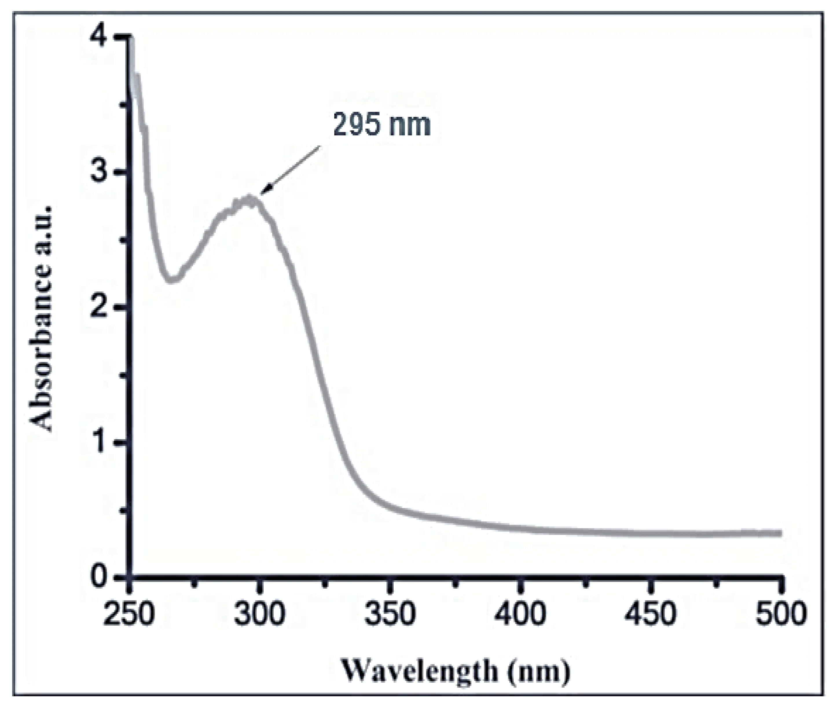
Figure 1. UV–vis spectrum of silica nanoparticles.
Antioxidants 11 02323 g001
Figure 2. FT-IR spectra (A) and XRD diffractogram of SiO2-NPs (B).
Antioxidants 11 02323 g002
Figure 3. TEM image for SiO2-NPs showing spherical shapes.
Antioxidants 11 02323 g003
Figure 4. DLS analysis of biosynthesized SiO2-NPs colloidal solution.
Antioxidants 11 02323 g004
Figure 5. Antifungal activity of SiO2-NPs at different concentrations against A. solani (A,B): (A) Inhibition zones. (B) Inhibition percentages. (Data represent mean ± SD, n = 3). (a > b > c > d > e > f revealed to significance power). Numbers 1,2,3,4 in the upper petri dish mean 1000, 500, 250 and 125 µg/ml.
Antioxidants 11 02323 g005
Figure 6. Effect of SiO2-NPs and sodium silicate on morphological characters of S. melongena. (Data represent mean ± SD, n = 3). (a–d revealed to significance letters).
Antioxidants 11 02323 g006
Figure 7. Effect of silica nanoparticles and SiO2 on photosynthetic pigments of S. melongena. (Data represent mean ± SD, n = 3). (a–e revealed to significance letters).
Antioxidants 11 02323 g007
Figure 8. Effect of SiO2-NPs and SiO2 on oxidative stress markers of S. melongena. (a–h revealed to significance letters).
Antioxidants 11 02323 g008
Table 1. Effect of SiO2-NPs and SiO2 on A. solani early blight disease index of S. melongena.
TreatmentDisease Symptoms ClassesDI (%)Protection (%)
01234
Control Infected 0015482.50
Infected + SiO2-NPs 50 ppm404203557.57
Infected + SiO2-NPs 100 ppm513102569.69
Infected + sodium silicate 100 ppm211336027.27
Publisher’s Note: MDPI stays neutral with regard to jurisdictional claims in published maps and institutional affiliations.