Abstract
Reactive oxygen species (ROS) are normal products of a number of biochemical reactions and are important signaling molecules. However, at the same time, they are toxic to cells and have to be strictly regulated by their antioxidant systems. The etiology and pathogenesis of many diseases are associated with increased ROS levels, and many external stress factors directly or indirectly cause oxidative stress in cells. Within this context, the overexpression of genes encoding the proteins in antioxidant systems seems to have become a viable approach to decrease the oxidative stress caused by pathological conditions and to increase cellular stress resistance. However, such manipulations unavoidably lead to side effects, the most dangerous of which is an increased probability of healthy tissue malignization or increased tumor aggression. The aims of the present review were to collect and systematize the results of studies devoted to the effects resulting from the overexpression of antioxidant system genes on stress resistance and carcinogenesis in vitro and in vivo. In most cases, the overexpression of these genes was shown to increase cell and organism resistances to factors that induce oxidative and genotoxic stress but to also have different effects on cancer initiation and promotion. The last fact greatly limits perspectives of such manipulations in practice. The overexpression of GPX3 and SOD3 encoding secreted proteins seems to be the “safest” among the genes that can increase cell resistance to oxidative stress. High efficiency and safety potential can also be found for SOD2 overexpression in combinations with GPX1 or CAT and for similar combinations that lead to no significant changes in H2O2 levels. Accumulation, systematization, and the integral analysis of data on antioxidant gene overexpression effects can help to develop approaches for practical uses in biomedical and agricultural areas. Additionally, a number of factors such as genetic and functional context, cell and tissue type, differences in the function of transcripts of one and the same gene, regulatory interactions, and additional functions should be taken into account.
1. Introduction
Reactive oxygen species (ROS) participate in the regulation of physiological processes, including gene expression regulation, the activation of stress-response signaling cascades, and apoptosis (for a review, see [1]). Mitochondria, which are the main source of ROS in the cell, use them as signal molecules [2]. At submicromolar concentrations, ROS induce the proliferative activity of cells, working as mitogens. It is known [3] that mitochondria can activate cellular immune response by changing their membrane potential and by directly interacting with Toll-like receptors as well as by participating in signaling that regulates innate immune responses. However, ROS can cause macromolecule damage [1], and therefore, their levels are strictly regulated by antioxidant systems [4].
Oxidative stress is a disbalance between ROS and reactive nitrogen species (RNS) on the one hand and antioxidant defense on the other. Among the causes of oxidative stress are the imperfect Red-Ox regulation of normal physiological processes, primarily anaerobic respiration, and oxidative phosphorylation in mitochondria [5], as well as decreased antioxidant protein levels due to pathological conditions or senescence. Oxidative stress can be the result of chemical and physical factors with direct ROS and RNS generation activity or can be achieved indirectly by acting through endocrine disruption [6,7,8]. Causality works both ways when it comes to the relationship between oxidative stress and inflammation. Through various signaling cascades, chronic oxidative stress can activate pro-inflammatory cytokines [9], and the inflammation itself, in turn, leads to the production of additional amounts of ROS [10]. The emerging vicious circle underlies many pathologies, including the initiation of carcinogenesis [9,11].
Oxidative stress is an important component in the etiology and pathogenesis of oncology [5,12], cardiovascular (ischemia-reperfusion) diseases [13], neurodegenerative diseases (Alzheimer’s, Parkinson’s diseases, etc.) [12,14,15], epilepsy [16], Huntington’s disease [17,18], diabetes mellitus [19], and multiple organ dysfunction syndrome [20]. In addition to that, oxidative stress is associated with mechanisms of psoriasis development [21,22,23,24] and with skin fibrosis in systemic scleroderma [25]. Oxidative stress, which is primarily caused by mitochondrial dysfunction, plays one of the leading roles in the processes of aging, both in tissues and in the whole organism [5,26,27]. All that makes our knowledge of the possibilities and consequences of cell antioxidant systems regulation very important for the development of strategies and approaches for reducing the negative effects of oxidative stress as well as for prophylaxis and the treatment of a wide variety of diseases. The main strategies can be classified into pharmacological, nutritiological, genetic, and ecological [28,29,30,31]. The latter is more concerned with preventing the formation of ROS where possible [28]. Naturally, the most straightforward strategies are built on pharmacological interventions, predominantly with various antioxidants. Studies on the topic include assessments of the effects of curcumin and curcuminoids [32,33], plant-derived polyphenols [34,35], flavonoids [36], boswellic acids [37], coenzyme Q10 [38], quercetin [39] N-acetylcysteine [40], monoclonal antibodies to cytokines (infliximab, tocilizumab [41]), and many other compounds and pharmaceuticals. The efficacy and success rates of pharmacological strategies are, as one can conclude, limited, with four possible reasons suggested: (1) vitamin antioxidants based upon in vitro studies may be inefficient scavengers in vivo; (2) detrimental ROS may be compartmentalized at sites within the cell that are inaccessible by the administered antioxidant; (3) antioxidants may possess toxicities that mask their advantageous effect; and (4) heterogeneity of the groups under study [42]. The use of nutritional approaches to treat and prevent diseases and to leveling unhealthy environment adverse effects comes from eating foods rich in antioxidants or food additives/drugs based on such foods [43,44]. However, systematic or meta-analysis results suggest that eating antioxidants is more often harmless, but there is insufficient evidence on the positive effects of this for disease treatment or for the improvement of life quality [43,45,46], or the benefits are only revealed under a strictly defined set of measures or under a strictly defined disease stage with the observance of dosages [47,48,49,50]. Genetic approaches involve the regulation of the cell’s own antioxidant system. The results obtained when discovering the targets for such regulation are reviewed below.
In addition, the rational control of antioxidant system activity is required for the regulation of organisms and for normal cells’ resistance to stressful and adverse conditions. It is also required to regulate the resistances of malignant cells and tissues to chemotherapeutics and irradiation. Previously, for example, gene therapies with constructs that transiently overexpress antioxidant genes were used to elucidate approaches for protecting normal tissues during tumor radiotherapy [51]. Taking into account that almost all intracellular systems, including antioxidant ones, consist of more than one protein, approaches utilizing transcription regulation of antioxidant genes in functionally rational combinations seem to be the most promising ones [52]. These approaches have become viable because of our vast knowledge of the functions of both individual-proteins and whole-protein subsystems and because of development of CRISPRa and CRISPRi technologies for the regulation of gene expression in cells. At the same time, one must take into account potential side effects. For example, the overexpression of a transcription factor gene NFE2L2 (also known as Nrf2) can lead to the induction of skin anomalies in mice [53], and the overexpression of SOD1 without the simultaneous overexpression of H2O2-utilizing enzymes genes lead to the faster loss of function in retina cones. GPX4 overexpression decreased these negative effects of SOD1 overexpression [54]. However, the most important finding was that the overexpression of particular antioxidant genes could increase or decrease the probability of malignization as well as differently affect processes in already-developed tumor tissues.
The aim of this review was to systematize the results of gain of function studies of antioxidant systems genes and to search for candidate genes and their combinations for the safe and effective treatment and prevention of oxidative stress with different genesis. Special attention was devoted to the results of experimental studies covering the effects resulting from the overexpression of particular antioxidant genes and their combinations on the stress resistance of cells, the probability of tumor induction, and tumor progression. We believe that systematization would be an important intermediate point for further studies for the manipulation and control of oxidative stress resistance in cells and organisms.
2. Loss of Function Studies of Antioxidant Genes
Obviously, knockout or knockdown genes, products of which are components of primary life-support systems ([55] for review), result in the decreased viability and stress resistance of cells and organisms and an increased probability of malignization in the majority of cases.
For example, the knockdown or knockout of antioxidant defense genes decreased cell resistance to pro-oxidants (SOD2: [56]; SOD1, TRX1: [57]; PRXD2: [58]; PRDX1: [59]; CAT: [60]; PRDX2: [61]), alkylating agents (PRDX1: [62]; PRDX3: [63]; PRDX2: [64]), and ionizing radiation (PRDX1: [65]; PRDX4: [66]; PRDX1, PRDX5: [67]; GSL2: [68]; PRDX2: [61,64]). At the scale of the whole organism, PRDX1 knockdown in mice led to decreased lifespan, the development of hemolytic anemia, and malignant neoplasms (lymphomas, sarcomas, and carcinomas) [59]. The suppression of NFE2L2 significantly affected the development of endothelial dysfunction and decreased microvascular bed density in normotensive rats under oxidative stress caused by NaCl [69]. Decreased SOD1 activity elevated the neurotoxic potential of quinolonic and kainic acids in the corpus striatum of transgenic mice [70].
However, there are opposite examples of the effects of antioxidant gene knockout, knockdown, or suppression. TRXR1 knockdown in A549 cells decreased their resistance to 1-chloro-2,4-dinitrobenzene or menadione, but at the same time, it significantly increased their resistance to cisplatin. This effect can be explained by TrxR1 protein derivatized with cisplatin being able to induce cell death [71].
The results of a number of loss of function studies were very important for deciphering the functions of many antioxidant genes and of the mechanisms of antioxidant systems. Presently, the elements of the reaction cascade that provides Red/Ox homeostasis are well-studied. These data are required for the development of new approaches to overcome tumor resistance to chemo- and radiotherapy as well as to decrease side effects in normal tissues and whole organisms [1,51]. However, much less is known about such effects on the cellular level and at the organizational levels above it that are induced by the increased gene expression of antioxidant proteins, both individually and in various combinations. Although such interventions also interfere with a fine-tuned cell balance, the effects produced are significantly diverse, and not only bring new fundamental knowledge, but also have potential importance in applied contexts. Therefore, the systematic review we present here is focused on the available results of gain of function studies of antioxidant defense genes.
3. Effects of Antioxidant Defense Gene Overexpression on Stress Resistance
The main aim of this review was to systemize and sum up the available studies covering the effects of antioxidant defense gene overexpression on cell resistance to oxidative stress. We analyzed 166 such works [20,51,56,58,62,65,66,71,72,73,74,75,76,77,78,79,80,81,82,83,84,85,86,87,88,89,90,91,92,93,94,95,96,97,98,99,100,101,102,103,104,105,106,107,108,109,110,111,112,113,114,115,116,117,118,119,120,121,122,123,124,125,126,127,128,129,130,131,132,133,134,135,136,137,138,139,140,141,142,143,144,145,146,147,148,149,150,151,152,153,154,155,156,157,158,159,160,161,162,163,164,165,166,167,168,169,170,171,172,173,174,175,176,177,178,179,180,181,182,183,184,185,186,187,188,189,190,191,192,193,194,195,196,197,198,199,200,201,202,203,204,205,206,207,208,209,210,211,212,213,214,215,216,217,218,219,220,221,222,223,224,225,226,227] published during the period covering 1988–2022. Table 1 contains extremely condensed information on the effects of overexpression of the following antioxidant genes (with direct and indirect antioxidant action): SOD1, SOD2, SOD3 (EC-SOD), CAT, MTI, MTII, ALDH3A1, TRX1, TRX2, GCLC, GCLM, GPX1, TXN1, NQO1, PRDX1, PRDX2, PRDX3, PRDX4, PRDX5, PRDX6, TXD1, SLC31A1, SLC7A11, GPX2, GPX3, GPX4, GSTP1, SRXN1, TRXR1, PON1, PON2, and PON3. More detailed information about the analyzed studies is presented in the Supplementary Materials (Table S1). Resistance in vivo or in vitro was assessed to determine the oxidative stress caused by various irradiation types (UV, X-rays, and gamma), chemical compounds (hydrogen peroxide, paraquat, etc.), and other factors (ischemia, sepsis, surgical interventions, hypoxia, and endotoxemia). In the majority of published studies that we analyzed, the overexpression of antioxidant defense genes in vitro and in vivo was achieved by introducing an additional copy of the gene or a transgene. CRISPRa technology, which became available in 2013 [228], despite its obvious advantages [52], was registered in one work [177].

Table 1.
Influence of antioxidant gene overexpression on resistance to oxidative and genotoxic stress factors in vitro and in vivo.
Cases of spontaneous gene overexpression in tumors resistant to particular therapy types were also reviewed, but only in such cases where the abolition of overexpression of a particular gene led to the abolition of the changed phenotype.
In most of the studies analyzed, the overexpression of antioxidant defense genes resulted in increased resistance to oxidative stress induced by different factors, both in vitro and in vivo. For example, the increased survival of cells was found in NK-92 and NK-92MI exposed to H2O2 (PRDX1, [151]). Mice overexpressing TRX1 survived better after adriamycin treatment [219], and mice overexpressing PON1 or PON3 had less liver damage after treatment with carbon tetrachloride [135]. Decreased apoptosis levels were observed for PRDX6 in vitro in SCOV-3 cell lines treated with cisplatin [169], for PRDX5 in human tendon fibroblasts treated with H2O2 [161], and in HT29 cells treated with shikonin [166], for TRX1 in WEHI7.2 lymphoma cells treated with H2O2 [220], in in vivo studies on transgenic mice that were treated with methamphetamine [214], and for SRXN1 in retinal ganglion cells (RGC) of mice treated with high glucose concentrations [229]. Among the positive effects of overexpression, for PRDX5, researchers have shown decreased DNA damage in hamster cell cultures CHO-K1 exposed to H2O2 and tert-butylhydroperoxide (tBHP) [162] as well as for TRX1 in the mitochondria of transgenic mice Trx1-Tg in vivo in sepsis-induced myocardial dysfunction and sham surgery [216,217]. Increased expression of TRX1 in mice in vivo [217] and ex vivo [218] decreased the negative consequences of ischemia for the cardiovascular system. Using oxygen–glucose deprivation/reoxygenation in in vitro experiments showed that the ability of PON2 to enhance glucose transport and to suppress oxidative stress and apoptosis is potentially a good set of properties to prevent cerebral ischemia-reperfusion injury [143].
In some cases, the authors noted that increased expression of the antioxidant defense genes in fact decreases cell resistance to oxidative stress; however, such studies are scarce. For example, decreased resistance to oxidative stress was shown in the brain neurons of mice overexpressing SOD1 when exposed to menadione [182], in human cells overexpressing NFE2L2 that were treated with cisplatin [126,129], and in human cells overexpressing GPX1 when exposed to radiation [100]. No changes in stress resistance were found in human cells TK6 overexpressins SOD1 when treated with gamma-irradiation [181]; in normal human keratinocytes overexpressing SOD1 when irradiated with UV [73]; in brain neurons of mice overexpressing SOD1 treated with H2O2 [182]; in Chinese hamster ovary cells overexpressing MTII when treated with gamma-rays, bleomycin, MMS, and N-hydroxyethyl-N-chloroethylnitrosourea [122]; in mouse C127 cells overexpressing MTII that had been exposed to 5-fluorouracil and vincristine [123]; in Chinese hamster cells V79 overexpressing MTI when exposed to alkylating agents [121]; in vivo in D. melanogaster overexpressing SOD1 when the flies were exposed to paraquat [185]; and in SOD2-overexpressing ones exposed to 100% O2 [199].
The number of studies that have shown the overexpression of various antioxidant defense genes increasing the oxidative stress resistance on the cell and organism levels gives ground to careful optimism when assessing the future feasibility of gene expression regulation in these genes for therapeutic purposes. However, the in vitro effects that are considered beneficial at that level might be dangerous or even harmful at higher levels of structural and functional organization. Similarly, relatively positive effects in in vivo models that have been observed to be beneficial at the endpoints selected by researchers might cause negative side effects in the long term according to other parameters. Taking this into account, an extra task was to assess the effects of the increased expression of the antioxidant defense genes on the probabilities of malignization in normal cells and on the growth and development of tumor cells.
4. Effects of the Antioxidant Defense Genes Overexpression on Carcinogenesis
Logically, the overexpression of antioxidant genes, as said above, has predominantly cytoprotective effects and must decrease the frequency of spontaneous and induced mutations and therefore must decrease the probability of malignization. On the other hand, in already malignantly transformed cells, it can stimulate faster proliferation, decrease apoptosis and permanent cell cycle arrest, and increase tumor resistance to therapeutic interventions. Previously, the authors of a study on PRDX6 came to similar conclusions [230]. Another review devoted to glutathione peroxidases came to similar conclusions [231], with the authors stating that all GPXs prevent cancer initiation in normal cells by removing hydroperoxides, but that in already malignantly transformed cells, they have various effects, including tumor promotion. In a limited set of studies on SOD3 overexpression, a completely opposite picture can be found. The increased expression of SOD3 led to the immortalization of mouse embryonic fibroblasts and carcinogenesis despite the fact that, in the developed tumors, this gene’s expression is usually suppressed [232]. Additionally, there is direct evidence that SOD3 overexpression suppresses growth and aggression in tumor cells both in vitro and in vivo [233,234]. Limited samples of focused studies of the overexpression of other genes also support and disprove the proposed simplified idea (see Figure 1 and Table S2). Additionally, we often talk about one and the same gene. For instance, increased SOD2 expression has often been observed in tumor tissues and is frequently correlated with tumor stage and metastasis activity [235,236]. More than that, some studies have demonstrated that SOD2 overexpression in tumors promotes growth and metastasis [237,238] and increases resistance to anoikis [239]. However, there are many works that present evidence for the opposite. Cancer cells from the MCF-7, U118, and MIA PaCa-2 lines found to overexpress SOD2 when injected into nude mice showed significantly lesser and slower tumor development and decreased mortality when compared with the same cell lines without SOD2 overexpression [240,241,242]. An even more pronounced effect of decreased tumor growth and increased animal survival was achieved in MIA PaCa-2 cells when SOD2 and GPX1 were overexpressed simultaneously [243]. The overexpression of SOD2 with the lentiviruses that deliver this gene with powerful promotors, in human cell cultures and in xenografts, and in surrounding tissues in mice simultaneously led to the decreased resistance and proliferation of cancer cells and provided protection against radiation damage for normal cells at the same time [206]. In other types of experiments, increased expression of SOD2 also suppressed the development and metastasis of tumors. In contrast with the results of the study mentioned above, in which increased levels of H2O2 associated with SOD2 overexpression stimulated the migration and invasion of HT-1080 cells [238], the same effects were achieved by suppressing SOD2 expression in ovarian cancer cells resulting from increased superoxide–radical concentrations [244,245]. In other words, in different experimental systems, SOD2 overexpression can either stimulate tumor growth and metastasis or suppress them. It seems that an important role belongs to the ratios of expression of superoxide dismutases with expression of CAT and/or GPXs, the protein products of which have hydrogen peroxide as a substrate [236]. As a consequence, H2O2 and superoxide radicals probably are among the key signal molecules involved in the regulation of such processes [238,244,245,246,247,248].
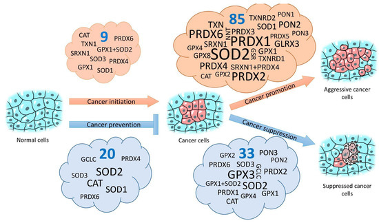
Figure 1.
Effects of the overexpression of antioxidant defense genes on the probability of normal cells/tissues malignization and on the aggressiveness of already developed cancer cells/tumors. The font size is proportional to the number of studies that showed the effect of the overexpression of that gene. Numbers in blue show total numbers of studies in each group.
The avoidance of H2O2 overproduction is likely to be the cause of no promotion of cancer cells when there is simultaneous overexpression of SOD2 and CAT both in vitro [247] and in vivo [246] and of tumor-preventive [249] and tumor-suppressive [243] effects in the case of the simultaneous overexpression of SOD2 and GPX1. This is another argument for higher efficacy antioxidant gene overexpression in functionally viable combinations in contrast with the overexpression of individual genes.
A graphical representation of the effects of the overexpression of antioxidant genes on the induction, prevention, promotion, or suppression of tumor processes (based on an analysis of 130 studies) [58,63,66,87,88,89,90,103,104,105,114,117,159,173,174,175,192,201,210,230,232,233,234,238,239,240,241,242,243,244,245,246,247,248,249,250,251,252,253,254,255,256,257,258,259,260,261,262,263,264,265,266,267,268,269,270,271,272,273,274,275,276,277,278,279,280,281,282,283,284,285,286,287,288,289,290,291,292,293,294,295,296,297,298,299,300,301,302,303,304,305,306,307,308,309,310,311,312,313,314,315,316,317,318,319,320,321,322,323,324,325,326,327,328,329,330,331,332,333,334,335,336,337,338,339,340,341,342,343] is given in Figure 1. More detailed information and references to actual studies are provided in Table S2.
5. Problems and Perspectives of Applied Regulation of Cell Stress Resistance by Overexpressing the Antioxidant Defense Genes
Based on the above, the overexpression of antioxidant defense genes has practical potential for the therapeutic regulation of oxidative stress and for increasing cellular stress resistance. On the other hand, studies on side effects of such overexpression, primarily its cancerogenic potential, give very contradictory results, and we have to first elucidate which genes and in which context overexpression can occur without incurring unjustified risks.
For example, SOD2 overexpression, the consequences of which are apparently the best-studied ones, seems to be counterproductive without the simultaneous overexpression of genes, products of which eliminate H2O2, such as CAT or GPXs. Among the last group the most applicable ones are GPX1 [243,249], the overexpression of which by itself has different effects on cancer initiation and development in different experimental systems (cancer initiation [249]; promotion [103,105,301]; and suppression [104,243]). The same is true for GPX4 [231,340]. Catalase looks like an obvious and even more viable partner for the simultaneous overexpression with SOD2. Firstly, for this gene combination, we already know the effect of increased cell resistance to gamma rays [82]. Secondly, it was reported that catalase prevents SOD2 overexpression-induced promotion of cancer cells in vitro [247] and in vivo [246]. Thirdly, the isolated overexpression of CAT in experiments led to the prevention of malignant transformation [87,318,319,320,321] or to the suppression of tumor cells growth [89,90] significantly more frequently than it did to cancer promotion [88]. It is possible that cytoplasmic superoxide dismutase (SOD1) is an even more “suitable” partner for CAT overexpression because for this combination, increased cell resistance to UV [73], paraquat, and H2O2 [83] were observed in vitro, and increased cell resistance to benzopyrene was observed in vivo [75]. However, data on effects of the simultaneous expression of CAT and SOD1 on cancer promotion are not available.
A special attention is given to one other member of the glutathione peroxidase family sGPX3. Its overexpression increased cell resistance to ascorbate [109] and cisplatin [110]. Despite the fact that these findings were made in tumor cells that spontaneously overexpressed GPX3 and that high GPX3 expression was associated with poorer overall survival in patients with ovarian cancer and with increased tumor stage [109], all direct manipulations with GPX3 overexpression conversely indicate the potential safety of such manipulation from the perspective of carcinogenesis. The suppression of proliferation, migration, and invasion in vitro [326,327,332,333] as well as of tumorigenicity in vivo [328,329,330,331] were shown in models overexpressing GPX3. Additionally, several other studies have provided evidence that in tumor tissues and cancer cell lines, GPX3 expression is suppressed or completely blocked due to promoter hypermethylation or gene deletion [329,331,344,345,346]. Thus, GPX3 functions as a tumor suppressor in an overwhelming majority of studies, and its overexpression, isolated or in combination with other components of the antioxidant system, is very promising for the suppression of endogenous or exogenous oxidative stress.
GPX3 encodes a secreted protein; therefore, SOD3, which encodes extracellular superoxide dismutase, could be a potentially effective functional partner for it. We know that transgenic mice that overexpress SOD3 have increased resistance to rib cage irradiation with 4-MV photons [208], to focal cerebral ischemia [209], to 12-O-tetradecanoylphorbol-13-acetate (TPA) exposure [210], and to lipopolysaccharide-induced endotoxemia [20]. At the same time, SOD3 overexpression decreases dimethylbenzanthracene/TPA-induced tumor formation [210] and suppresses cancer cells’ growth in vitro [233,234] and in vivo [234]. However, increased expression of SOD3 in mouse embryonic fibroblasts, can induce carcinogenesis [232].
All examples of genes and gene combinations that have been discussed above are by no means an exhaustive list of potentially effective and safe targets for therapeutic overexpression. Seeming contradictions in the effects resulting from the overexpression of some antioxidant genes on stress resistance and carcinogenesis were, apparently, caused by a number of non-obvious factors. In some cases, different effects of overexpression of the same gene could have risen from functional differences between the isoforms of product protein created by alternative splicing of the same gene. For example, the activity of invasion and metastasis of colon cancer cells increased when a splice variant b of the GLRX3 gene (Txl-2b) was expressed, decreased when GLRX3c was expressed, and did not change during GLRX3a expression [294]. The activities of different isoforms of glutaminase, which is involved in the glutathione-dependent antioxidant defense system of the cell, could either promote tumor progression or suppress it through certain pathways involving miRNAs [347]. Besides different isoforms of proteins transcribed from one gene, non-coding circular RNAs (circRNAs) that have regulatory functions can be transcribed. For example, circRNA transcribed from the SOD2 gene plays an important role in hepatocellular carcinoma progression [348]. Therefore, different functional properties of different transcripts of the gene must be taken into account when choosing cDNA for the overexpression of the gene by a classic method via the introduction of an additional copy of the gene or when designing guide RNAs for promoters of genes that have several alternative transcription initiation sites (such as SOD2) for CRISPRa technology application. However, these features of genes are discussed far less than the effects of the simultaneous overexpression of functional gene combinations are.
Nevertheless, the selection of only one splice variant of the gene is not supposed to be a perfect decision. Dysregulation of the gene’s expression because of the introduction of cDNA under a synthetic promoter or another gene’s promoter can cause contradicting effects, as, obviously, the degree of overexpression, its changes with time, and the presence or absence of a transcriptional response to various signals would affect the phenotypic manifestation of the gene. Preserving the chromosome context and, to some degree, the natural regulation when overexpressing a cell’s own genes with technologies, such as CRISPRa, could provide better results. In addition to that, CRISPRa allows the simultaneous overexpression of combinations of a number of genes. Presently, there are very few studies in which the researchers have overexpressed a cells’ own antioxidant genes.
Even the stage of tumor development may determine contradictions in the effects of antioxidant defense gene overexpression, apart from those related to the functional properties of different transcripts of the same gene and such obvious factors as the genetic context and the tissue (if it is a local gene therapy). Thus, SOD2 overexpression in mice with benign thyroid tumors resulted in an increased tumor burden. In contrast, in mice with aggressive follicular thyroid cancer, the overexpression of SOD2 reduced tumor proliferation and improved mortality rates [259].
Knowledge of the cytoprotective potential of even those genes whose overexpression has a pronounced pro-oncogenic effect can be of high applied value. For example, the overexpression of PON2 in most experimental systems led to cancer promotion [313,314,315,316]. The overexpression of PON2 is found in many tumors and often correlates with tumor aggressiveness and poor prognosis [315,316,349]. At the same time, a unique set of properties of the product of this gene, including the ability to activate glucose transport [314,316] and to suppress oxidative stress and apoptosis [350], makes its temporary or local activation a promising manipulation to reduce the damage caused by ischemia-reperfusion [143].
6. Conclusions
The complexity and ambiguity of the effects of antioxidant defense gene overexpression also resulted from the multifunctionality of product proteins. In addition to their main function of ROS and organic radical detoxification, they are frequent participants in signal cascades and are regulators of other protein activities [90,104,194], including those that occur due to changes in ROS levels as signaling molecules [1,2,6]. Thus, the overexpression of individual genes and their combinations that encode the effectors of the antioxidant system seems to be more promising than attempts to regulate the transcription factors affecting them, such as NFE2L2. Furthermore, common approaches based on weak effects on multiple functionally complementary targets, for example, when using plant extracts [351,352,353] in the therapy of various diseases, seem to be more practically applicable than those with strong effects on a single target.
Supplementary Materials
The following supporting information can be downloaded at: https://www.mdpi.com/article/10.3390/antiox11122316/s1.
Author Contributions
Conceptualization, I.O.V., E.S.B. and M.M.T.; methodology, I.O.V.; validation, I.O.V. and E.S.B.; formal analysis, M.M.T., E.E.R., Z.B.I., A.A.C. and A.V.R.; investigation, M.M.T., E.E.R., Z.B.I., A.A.C. and A.V.R.; data curation, M.M.T. and I.O.V.; writing—original draft preparation, A.V.R., E.S.B. and I.O.V.; writing—review and editing, E.S.B., M.M.T., A.A.C., A.V.R. and I.O.V.; visualization, I.O.V.; supervision, I.O.V.; funding acquisition, I.O.V. All authors have read and agreed to the published version of the manuscript.
Funding
The study was funded by a Russian Science Foundation Grant No. 22-24-01042, https://rscf.ru/project/22-24-01042/, accessed on 15 December 2021.
Conflicts of Interest
The authors declare no conflict of interest.
References
- Cui, Q.; Wang, J.-Q.; Assaraf, Y.G.; Ren, L.; Gupta, P.; Wei, L.; Ashby, C.R.; Yang, D.-H.; Chen, Z.-S. Modulating ROS to Overcome Multidrug Resistance in Cancer. Drug Resist. Update 2018, 41, 1–25. [Google Scholar] [CrossRef]
- Kasai, S.; Shimizu, S.; Tatara, Y.; Mimura, J.; Itoh, K. Regulation of Nrf2 by Mitochondrial Reactive Oxygen Species in Physiology and Pathology. Biomolecules 2020, 10, 320. [Google Scholar] [CrossRef] [PubMed]
- Banoth, B.; Cassel, S.L. Mitochondria in Innate Immune Signaling. Transl. Res. 2018, 202, 52–68. [Google Scholar] [CrossRef]
- Poljsak, B.; Šuput, D.; Milisav, I. Achieving the Balance between ROS and Antioxidants: When to Use the Synthetic Antioxidants. Oxidative Med. Cell. Longev. 2013, 2013, 956792. [Google Scholar] [CrossRef] [PubMed]
- Kudryavtseva, A.V.; Krasnov, G.S.; Dmitriev, A.A.; Alekseev, B.Y.; Kardymon, O.L.; Sadritdinova, A.F.; Fedorova, M.S.; Pokrovsky, A.V.; Melnikova, N.V.; Kaprin, A.D.; et al. Mitochondrial Dysfunction and Oxidative Stress in Aging and Cancer. Oncotarget 2016, 7, 44879–44905. [Google Scholar] [CrossRef]
- Księżakowska-Łakoma, K.; Żyła, M.; Wilczyński, J.R. Mitochondrial Dysfunction in Cancer. Menopausal Rev. 2014, 2, 136–144. [Google Scholar] [CrossRef]
- Chaiswing, L.; St. Clair, W.H.; St. Clair, D.K. Redox Paradox: A Novel Approach to Therapeutics-Resistant Cancer. Antioxid. Redox Signal. 2018, 29, 1237–1272. [Google Scholar] [CrossRef] [PubMed]
- Quagliariello, V.; Coppola, C.; Mita, D.G.; Piscopo, G.; Iaffaioli, R.V.; Botti, G.; Maurea, N. Low Doses of Bisphenol A Have Pro-Inflammatory and pro-Oxidant Effects, Stimulate Lipid Peroxidation and Increase the Cardiotoxicity of Doxorubicin in Cardiomyoblasts. Environ. Toxicol. Pharmacol. 2019, 69, 1–8. [Google Scholar] [CrossRef]
- Reuter, S.; Gupta, S.C.; Chaturvedi, M.M.; Aggarwal, B.B. Oxidative Stress, Inflammation, and Cancer: How Are They Linked? Free. Radic. Biol. Med. 2010, 49, 1603–1616. [Google Scholar] [CrossRef]
- Vaziri, N.D. Causal Link between Oxidative Stress, Inflammation, and Hypertension. Iran. J. Kidney Dis. 2008, 2, 1–10. [Google Scholar]
- Federico, A.; Morgillo, F.; Tuccillo, C.; Ciardiello, F.; Loguercio, C. Chronic Inflammation and Oxidative Stress in Human Carcinogenesis. Int. J. Cancer 2007, 121, 2381–2386. [Google Scholar] [CrossRef]
- Poprac, P.; Jomova, K.; Simunkova, M.; Kollar, V.; Rhodes, C.J.; Valko, M. Targeting Free Radicals in Oxidative Stress-Related Human Diseases. Trends Pharmacol. Sci. 2017, 38, 592–607. [Google Scholar] [CrossRef]
- Marczin, N.; El-Habashi, N.; Hoare, G.S.; Bundy, R.E.; Yacoub, M. Antioxidants in Myocardial Ischemia–reperfusion Injury: Therapeutic Potential and Basic Mechanisms. Arch. Biochem. Biophys. 2003, 420, 222–236. [Google Scholar] [CrossRef]
- Chen, Z.; Zhong, C. Oxidative Stress in Alzheimer’s Disease. Neurosci. Bull. 2014, 30, 271–281. [Google Scholar] [CrossRef]
- Dhapola, R.; Sarma, P.; Medhi, B.; Prakash, A.; Reddy, D.H. Recent Advances in Molecular Pathways and Therapeutic Implications Targeting Mitochondrial Dysfunction for Alzheimer’s Disease. Mol. Neurobiol. 2022, 59, 535–555. [Google Scholar] [CrossRef]
- Yang, N.; Guan, Q.-W.; Chen, F.-H.; Xia, Q.-X.; Yin, X.-X.; Zhou, H.-H.; Mao, X.-Y. Antioxidants Targeting Mitochondrial Oxidative Stress: Promising Neuroprotectants for Epilepsy. Oxidative Med. Cell. Longev. 2020, 2020, 6687185. [Google Scholar] [CrossRef] [PubMed]
- Zheng, J.; Winderickx, J.; Franssens, V.; Liu, B. A Mitochondria-Associated Oxidative Stress Perspective on Huntington’s Disease. Front. Mol. Neurosci. 2018, 11, 329. [Google Scholar] [CrossRef] [PubMed]
- Tang, Q.; Liu, H.; Shi, X.-J.; Cheng, Y. Blood Oxidative Stress Marker Aberrations in Patients with Huntington’s Disease: A Meta-Analysis Study. Oxidative Med. Cell. Longev. 2020, 2020, 9187195. [Google Scholar] [CrossRef] [PubMed]
- Asmat, U.; Abad, K.; Ismail, K. Diabetes Mellitus and Oxidative Stress—A Concise Review. Saudi Pharm. J. 2016, 24, 547–553. [Google Scholar] [CrossRef] [PubMed]
- Call, J.A.; Donet, J.; Martin, K.S.; Sharma, A.K.; Chen, X.; Zhang, J.; Cai, J.; Galarreta, C.A.; Okutsu, M.; Du, Z.; et al. Muscle-Derived Extracellular Superoxide Dismutase Inhibits Endothelial Activation and Protects against Multiple Organ Dysfunction Syndrome in Mice. Free Radic. Biol. Med. 2017, 113, 212–223. [Google Scholar] [CrossRef]
- Xu, F.; Xu, J.; Xiong, X.; Deng, Y. Salidroside Inhibits MAPK, NF-ΚB, and STAT3 Pathways in Psoriasis-Associated Oxidative Stress via SIRT1 Activation. Redox Rep. 2019, 24, 70–74. [Google Scholar] [CrossRef] [PubMed]
- Kharaeva, Z.; Gostova, E.; De Luca, C.; Raskovic, D.; Korkina, L. Clinical and Biochemical Effects of Coenzyme Q10, Vitamin E, and Selenium Supplementation to Psoriasis Patients. Nutrition 2009, 25, 295–302. [Google Scholar] [CrossRef]
- Baek, J.-O.; Byamba, D.; Wu, W.H.; Kim, T.-G.; Lee, M.-G. Assessment of an Imiquimod-Induced Psoriatic Mouse Model in Relation to Oxidative Stress. Arch. Dermatol. Res. 2012, 304, 699–706. [Google Scholar] [CrossRef]
- Gabr, S.A.; Al-Ghadir, A.H. Role of Cellular Oxidative Stress and Cytochrome c in the Pathogenesis of Psoriasis. Arch. Dermatol. Res. 2012, 304, 451–457. [Google Scholar] [CrossRef]
- Baral, H.; Sekiguchi, A.; Uchiyama, A.; Nisaa Amalia, S.; Yamazaki, S.; Inoue, Y.; Yokoyama, Y.; Ogino, S.; Torii, R.; Hosoi, M.; et al. Inhibition of Skin Fibrosis in Systemic Sclerosis by Botulinum Toxin B via the Suppression of Oxidative Stress. J. Dermatol. 2021, 48, 1052–1061. [Google Scholar] [CrossRef] [PubMed]
- Finkel, T.; Holbrook, N.J. Oxidants, Oxidative Stress and the Biology of Ageing. Nature 2000, 408, 239–247. [Google Scholar] [CrossRef]
- Schriner, S.E.; Linford, N.J. Extension of Mouse Lifespan by Overexpression of Catalase. Age 2006, 28, 209–218. [Google Scholar] [CrossRef]
- Poljsak, B. Strategies for Reducing or Preventing the Generation of Oxidative Stress. Oxidative Med. Cell. Longev. 2011, 2011, 194586. [Google Scholar] [CrossRef]
- García-Sánchez, A.; Miranda-Díaz, A.G.; Cardona-Muñoz, E.G. The Role of Oxidative Stress in Physiopathology and Pharmacological Treatment with Pro- and Antioxidant Properties in Chronic Diseases. Oxidative Med. Cell. Longev. 2020, 2020, 2082145. [Google Scholar] [CrossRef] [PubMed]
- Hamilton, C.A.; Miller, W.H.; Al-Benna, S.; Julia BROSNAN, M.; Drummond, R.D.; McBRIDE, M.W.; Dominiczak, A.F. Strategies to Reduce Oxidative Stress in Cardiovascular Disease. Clin. Sci. 2004, 106, 219–234. [Google Scholar] [CrossRef] [PubMed]
- Münzel, T.; Daiber, A. Environmental Stressors and Their Impact on Health and Disease with Focus on Oxidative Stress. Antioxid. Redox Signal. 2018, 28, 735–740. [Google Scholar] [CrossRef]
- Gupta, S.C.; Patchva, S.; Aggarwal, B.B. Therapeutic Roles of Curcumin: Lessons Learned from Clinical Trials. AAPS J. 2013, 15, 195–218. [Google Scholar] [CrossRef]
- Yucel, C.; Quagliariello, V.; Iaffaioli, R.V.; Ferrari, G.; Donsì, F. Submicron Complex Lipid Carriers for Curcumin Delivery to Intestinal Epithelial Cells: Effect of Different Emulsifiers on Bioaccessibility and Cell Uptake. Int. J. Pharm. 2015, 494, 357–369. [Google Scholar] [CrossRef]
- Elejalde, E.; Villarán, M.C.; Alonso, R.M. Grape Polyphenols Supplementation for Exercise-Induced Oxidative Stress. J. Int. Soc. Sport. Nutr. 2021, 18, 3. [Google Scholar] [CrossRef]
- Joseph, S.V.; Edirisinghe, I.; Burton-Freeman, B.M. Fruit Polyphenols: A Review of Anti-Inflammatory Effects in Humans. Crit. Rev. Food Sci. Nutr. 2016, 56, 419–444. [Google Scholar] [CrossRef]
- Li, G.; Ding, K.; Qiao, Y.; Zhang, L.; Zheng, L.; Pan, T.; Zhang, L. Flavonoids Regulate Inflammation and Oxidative Stress in Cancer. Molecules 2020, 25, 5628. [Google Scholar] [CrossRef] [PubMed]
- Efferth, T.; Oesch, F. Anti-Inflammatory and Anti-Cancer Activities of Frankincense: Targets, Treatments and Toxicities. Semin. Cancer Biol. 2022, 80, 39–57. [Google Scholar] [CrossRef] [PubMed]
- Gutierrez-Mariscal, F.M.; Arenas-de Larriva, A.P.; Limia-Perez, L.; Romero-Cabrera, J.L.; Yubero-Serrano, E.M.; López-Miranda, J. Coenzyme Q10 Supplementation for the Reduction of Oxidative Stress: Clinical Implications in the Treatment of Chronic Diseases. Int. J. Mol. Sci. 2020, 21, 7870. [Google Scholar] [CrossRef]
- Anand David, A.; Arulmoli, R.; Parasuraman, S. Overviews of Biological Importance of Quercetin: A Bioactive Flavonoid. Phcog. Rev. 2016, 10, 84. [Google Scholar] [CrossRef] [PubMed]
- Tardiolo, G.; Bramanti, P.; Mazzon, E. Overview on the Effects of N-Acetylcysteine in Neurodegenerative Diseases. Molecules 2018, 23, 3305. [Google Scholar] [CrossRef]
- Costa, N.T.; Iriyoda, T.M.V.; Alfieri, D.F.; Simão, A.N.C.; Dichi, I. Influence of Disease-Modifying Antirheumatic Drugs on Oxidative and Nitrosative Stress in Patients with Rheumatoid Arthritis. Inflammopharmacology 2018, 26, 1151–1164. [Google Scholar] [CrossRef] [PubMed]
- Patterson, C.; Madamanchi, N.R.; Runge, M.S. The Oxidative Paradox: Another Piece in the Puzzle. Circ. Res. 2000, 87, 1074–1076. [Google Scholar] [CrossRef]
- Li, N.; Wu, X.; Zhuang, W.; Xia, L.; Chen, Y.; Wu, C.; Rao, Z.; Du, L.; Zhao, R.; Yi, M.; et al. Tomato and Lycopene and Multiple Health Outcomes: Umbrella Review. Food Chem. 2021, 343, 128396. [Google Scholar] [CrossRef] [PubMed]
- Suhett, L.G.; de Miranda Monteiro Santos, R.; Silveira, B.K.S.; Leal, A.C.G.; de Brito, A.D.M.; de Novaes, J.F.; Lucia, C.M.D. Effects of Curcumin Supplementation on Sport and Physical Exercise: A Systematic Review. Crit. Rev. Food Sci. Nutr. 2021, 61, 946–958. [Google Scholar] [CrossRef] [PubMed]
- Parks, N.E.; Jackson-Tarlton, C.S.; Vacchi, L.; Merdad, R.; Johnston, B.C. Dietary Interventions for Multiple Sclerosis-Related Outcomes. Cochrane Database Syst. Rev. 2020, 2020, CD004192. [Google Scholar] [CrossRef]
- Ramdas, W.D. The Relation between Dietary Intake and Glaucoma: A Systematic Review. Acta Ophthalmol. 2018, 96, 550–556. [Google Scholar] [CrossRef]
- Jenkins, D.J.A.; Kitts, D.; Giovannucci, E.L.; Sahye-Pudaruth, S.; Paquette, M.; Blanco Mejia, S.; Patel, D.; Kavanagh, M.; Tsirakis, T.; Kendall, C.W.C.; et al. Selenium, Antioxidants, Cardiovascular Disease, and All-Cause Mortality: A Systematic Review and Meta-Analysis of Randomized Controlled Trials. Am. J. Clin. Nutr. 2020, 112, 1642–1652. [Google Scholar] [CrossRef] [PubMed]
- Jenkins, D.J.A.; Spence, J.D.; Giovannucci, E.L.; Kim, Y.; Josse, R.; Vieth, R.; Blanco Mejia, S.; Viguiliouk, E.; Nishi, S.; Sahye-Pudaruth, S.; et al. Supplemental Vitamins and Minerals for CVD Prevention and Treatment. J. Am. Coll. Cardiol. 2018, 71, 2570–2584. [Google Scholar] [CrossRef]
- Yang, J.; Fernández-Galilea, M.; Martínez-Fernández, L.; González-Muniesa, P.; Pérez-Chávez, A.; Martínez, J.A.; Moreno-Aliaga, M.J. Oxidative Stress and Non-Alcoholic Fatty Liver Disease: Effects of Omega-3 Fatty Acid Supplementation. Nutrients 2019, 11, 872. [Google Scholar] [CrossRef]
- Aune, D.; Keum, N.; Giovannucci, E.; Fadnes, L.T.; Boffetta, P.; Greenwood, D.C.; Tonstad, S.; Vatten, L.J.; Riboli, E.; Norat, T. Dietary Intake and Blood Concentrations of Antioxidants and the Risk of Cardiovascular Disease, Total Cancer, and All-Cause Mortality: A Systematic Review and Dose-Response Meta-Analysis of Prospective Studies. Am. J. Clin. Nutr. 2018, 108, 1069–1091. [Google Scholar] [CrossRef]
- Greenberger, J.S.; Epperly, M.W.; Gretton, J.; Jefferson, M.; Nie, S.; Bernarding, M.; Kagan, V.; Guo, H.L. Radioprotective Gene Therapy. Curr. Gene Ther. 2003, 3, 183–195. [Google Scholar] [CrossRef]
- Velegzhaninov, I.; Ievlev, V.; Pylina, Y.; Shadrin, D.; Vakhrusheva, O. Programming of Cell Resistance to Genotoxic and Oxidative Stress. Biomedicines 2018, 6, 5. [Google Scholar] [CrossRef]
- Schäfer, M.; Farwanah, H.; Willrodt, A.; Huebner, A.J.; Sandhoff, K.; Roop, D.; Hohl, D.; Bloch, W.; Werner, S. Nrf2 Links Epidermal Barrier Function with Antioxidant Defense. EMBO Mol. Med. 2012, 4, 364–379. [Google Scholar] [CrossRef] [PubMed]
- Usui, S.; Oveson, B.C.; Iwase, T.; Lu, L.; Lee, S.Y.; Jo, Y.-J.; Wu, Z.; Choi, E.-Y.; Samulski, R.J.; Campochiaro, P.A. Overexpression of SOD in Retina: Need for Increase in H2O2-Detoxifying Enzyme in Same Cellular Compartment. Free Radic. Biol. Med. 2011, 51, 1347–1354. [Google Scholar] [CrossRef]
- Snezhkina, A.V.; Kudryavtseva, A.V.; Kardymon, O.L.; Savvateeva, M.V.; Melnikova, N.V.; Krasnov, G.S.; Dmitriev, A.A. ROS Generation and Antioxidant Defense Systems in Normal and Malignant Cells. Oxidative Med. Cell. Longev. 2019, 2019, 6175804. [Google Scholar] [CrossRef] [PubMed]
- Fukui, M.; Zhu, B.T. Mitochondrial Superoxide Dismutase SOD2, but Not Cytosolic SOD1, Plays a Critical Role in Protection against Glutamate-Induced Oxidative Stress and Cell Death in HT22 Neuronal Cells. Free Radic. Biol. Med. 2010, 48, 821–830. [Google Scholar] [CrossRef] [PubMed]
- Sharma, V.; Joseph, C.; Ghosh, S.; Agarwal, A.; Mishra, M.K.; Sen, E. Kaempferol Induces Apoptosis in Glioblastoma Cells through Oxidative Stress. Mol. Cancer Ther. 2007, 6, 2544–2553. [Google Scholar] [CrossRef]
- Lu, W.; Fu, Z.; Wang, H.; Feng, J.; Wei, J.; Guo, J. Peroxiredoxin 2 Is Upregulated in Colorectal Cancer and Contributes to Colorectal Cancer Cells’ Survival by Protecting Cells from Oxidative Stress. Mol. Cell Biochem. 2014, 387, 261–270. [Google Scholar] [CrossRef]
- Neumann, C.A.; Krause, D.S.; Carman, C.V.; Das, S.; Dubey, D.P.; Abraham, J.L.; Bronson, R.T.; Fujiwara, Y.; Orkin, S.H.; Van Etten, R.A. Essential Role for the Peroxiredoxin Prdx1 in Erythrocyte Antioxidant Defence and Tumour Suppression. Nature 2003, 424, 561–565. [Google Scholar] [CrossRef]
- Song, L.-L.; Tu, Y.-Y.; Xia, L.; Wang, W.-W.; Wei, W.; Ma, C.-M.; Wen, D.-H.; Lei, H.; Xu, H.-Z.; Wu, Y.-L. Targeting Catalase but Not Peroxiredoxins Enhances Arsenic Trioxide-Induced Apoptosis in K562 Cells. PLoS ONE 2014, 9, e104985. [Google Scholar] [CrossRef]
- Smith-Pearson, P.S.; Kooshki, M.; Spitz, D.R.; Poole, L.B.; Zhao, W.; Robbins, M.E. Decreasing Peroxiredoxin II Expression Decreases Glutathione, Alters Cell Cycle Distribution, and Sensitizes Glioma Cells to Ionizing Radiation and H2O2. Free Radic. Biol. Med. 2008, 45, 1178–1189. [Google Scholar] [CrossRef]
- Poschmann, G.; Grzendowski, M.; Stefanski, A.; Bruns, E.; Meyer, H.E.; Stühler, K. Redox Proteomics Reveal Stress Responsive Proteins Linking Peroxiredoxin-1 Status in Glioma to Chemosensitivity and Oxidative Stress. Biochim. Biophys. Acta (BBA) Proteins Proteom. 2015, 1854, 624–631. [Google Scholar] [CrossRef] [PubMed]
- Duan, J.; Lang, Y.; Song, C.; Xiong, J.; Wang, Y.; Yan, Y. SiRNA Targeting of PRDX3 Enhances Cisplatin-Induced Apoptosis in Ovarian Cancer Cells through the Suppression of the NF-ΚB Signaling Pathway. Mol. Med. Rep. 2013, 7, 1688–1694. [Google Scholar] [CrossRef] [PubMed]
- Cerda, M.B.; Lloyd, R.; Batalla, M.; Giannoni, F.; Casal, M.; Policastro, L. Silencing Peroxiredoxin-2 Sensitizes Human Colorectal Cancer Cells to Ionizing Radiation and Oxaliplatin. Cancer Lett. 2017, 388, 312–319. [Google Scholar] [CrossRef]
- Chen, M.-F.; Keng, P.C.; Shau, H.; Wu, C.-T.; Hu, Y.-C.; Liao, S.-K.; Chen, W.-C. Inhibition of Lung Tumor Growth and Augmentation of Radiosensitivity by Decreasing Peroxiredoxin I Expression. Int. J. Radiat. Oncol. Biol. Phys. 2006, 64, 581–591. [Google Scholar] [CrossRef] [PubMed]
- Kim, T.H.; Song, J.; Alcantara Llaguno, S.R.; Murnan, E.; Liyanarachchi, S.; Palanichamy, K.; Yi, J.-Y.; Viapiano, M.S.; Nakano, I.; Yoon, S.O.; et al. Suppression of Peroxiredoxin 4 in Glioblastoma Cells Increases Apoptosis and Reduces Tumor Growth. PLoS ONE 2012, 7, e42818. [Google Scholar] [CrossRef] [PubMed]
- Gao, M.; Jia, X.; Wu, Q.; Cheng, Y.; Chen, F.; Zhang, J. Silencing Prx1 and/or Prx5 Sensitizes Human Esophageal Cancer Cells to Ionizing Radiation and Increases Apoptosis via Intracellular ROS Accumulation. Acta Pharm. Sin. 2011, 32, 528–536. [Google Scholar] [CrossRef]
- Xiang, L.; Xie, G.; Liu, C.; Zhou, J.; Chen, J.; Yu, S.; Li, J.; Pang, X.; Shi, H.; Liang, H. Knock-down of Glutaminase 2 Expression Decreases Glutathione, NADH, and Sensitizes Cervical Cancer to Ionizing Radiation. Biochim. Biophys. Acta (BBA) Mol. Cell Res. 2013, 1833, 2996–3005. [Google Scholar] [CrossRef]
- Priestley, J.R.C.; Kautenburg, K.E.; Casati, M.C.; Endres, B.T.; Geurts, A.M.; Lombard, J.H. The NRF2 Knockout Rat: A New Animal Model to Study Endothelial Dysfunction, Oxidant Stress, and Microvascular Rarefaction. Am. J. Physiol. Heart Circ. Physiol. 2016, 310, H478–H487. [Google Scholar] [CrossRef]
- Schwartz, P.J.; Reaume, A.; Scott, R.; Coyle, J.T. Effects of Over- and under-Expression of Cu,Zn-Superoxide Dismutase on the Toxicity of Glutamate Analogs in Transgenic Mouse Striatum. Brain Res. 1998, 789, 32–39. [Google Scholar] [CrossRef]
- Eriksson, S.E.; Prast-Nielsen, S.; Flaberg, E.; Szekely, L.; Arnér, E.S.J. High Levels of Thioredoxin Reductase 1 Modulate Drug-Specific Cytotoxic Efficacy. Free Radic. Biol. Med. 2009, 47, 1661–1671. [Google Scholar] [CrossRef]
- Voulgaridou, G.-P.; Kiziridou, M.; Mantso, T.; Chlichlia, K.; Galanis, A.; Koukourakis, M.I.; Franco, R.; Panayiotidis, M.I.; Pappa, A. Aldehyde Dehydrogenase 3A1 Promotes Multi-Modality Resistance and Alters Gene Expression Profile in Human Breast Adenocarcinoma MCF-7 Cells. Int. J. Biochem. Cell Biol. 2016, 77, 120–128. [Google Scholar] [CrossRef] [PubMed]
- Rezvani, H.R.; Mazurier, F.; Cario-André, M.; Pain, C.; Ged, C.; Taïeb, A.; de Verneuil, H. Protective Effects of Catalase Overexpression on UVB-Induced Apoptosis in Normal Human Keratinocytes. J. Biol. Chem. 2006, 281, 17999–18007. [Google Scholar] [CrossRef]
- Yang, F.; Yang, H.; Ramesh, A.; Goodwin, J.S.; Okoro, E.U.; Guo, Z. Overexpression of Catalase Enhances Benzo(a)Pyrene Detoxification in Endothelial Microsomes. PLoS ONE 2016, 11, e162561. [Google Scholar] [CrossRef]
- Yang, H.; Zhou, L.; Wang, Z.; Roberts, L.J.; Lin, X.; Zhao, Y.; Guo, Z. Overexpression of Antioxidant Enzymes in ApoE-Deficient Mice Suppresses Benzo(a)Pyrene-Accelerated Atherosclerosis. Atherosclerosis 2009, 207, 51–58. [Google Scholar] [CrossRef]
- Wang, Z.; Yang, H.; Ramesh, A.; Roberts, L.J.; Zhou, L.; Lin, X.; Zhao, Y.; Guo, Z. Overexpression of Cu/Zn-Superoxide Dismutase and/or Catalase Accelerates Benzo(a)Pyrene Detoxification by Upregulation of the Aryl Hydrocarbon Receptor in Mouse Endothelial Cells. Free Radic. Biol. Med. 2009, 47, 1221–1229. [Google Scholar] [CrossRef]
- Sun, J.; Tower, J. FLP Recombinase-Mediated Induction of Cu/Zn-Superoxide Dismutase Transgene Expression Can Extend the Life Span of Adult Drosophila Melanogaster Flies. Mol. Cell Biol. 1999, 19, 216–228. [Google Scholar] [CrossRef]
- Kang, Y.J.; Chen, Y.; Epstein, P.N. Suppression of Doxorubicin Cardiotoxicity by Overexpression of Catalase in the Heart of Transgenic Mice. J. Biol. Chem. 1996, 271, 12610–12616. [Google Scholar] [CrossRef]
- Liao, A.C.; Craver, B.M.; Tseng, B.P.; Tran, K.K.; Parihar, V.K.; Acharya, M.M.; Limoli, C.L. Mitochondrial-Targeted Human Catalase Affords Neuroprotection from Proton Irradiation. Radiat. Res. 2013, 180, 1–6. [Google Scholar] [CrossRef]
- Parihar, V.K.; Allen, B.D.; Tran, K.K.; Chmielewski, N.N.; Craver, B.M.; Martirosian, V.; Morganti, J.M.; Rosi, S.; Vlkolinsky, R.; Acharya, M.M.; et al. Targeted Overexpression of Mitochondrial Catalase Prevents Radiation-Induced Cognitive Dysfunction. Antioxid. Redox Signal. 2015, 22, 78–91. [Google Scholar] [CrossRef]
- Narasimhan, M.; Riar, A.K.; Rathinam, M.L.; Vedpathak, D.; Henderson, G.; Mahimainathan, L. Hydrogen Peroxide Responsive MiR153 Targets Nrf2/ARE Cytoprotection in Paraquat Induced Dopaminergic Neurotoxicity. Toxicol. Lett. 2014, 228, 179–191. [Google Scholar] [CrossRef] [PubMed]
- Epperly, M.W.; Melendez, J.A.; Zhang, X.; Nie, S.; Pearce, L.; Peterson, J.; Franicola, D.; Dixon, T.; Greenberger, B.A.; Komanduri, P.; et al. Mitochondrial Targeting of a Catalase Transgene Product by Plasmid Liposomes Increases Radioresistance In Vitro and In Vivo. Radiat. Res. 2009, 171, 588–595. [Google Scholar] [CrossRef] [PubMed]
- Mele, J.; Van Remmen, H.; Vijg, J.; Richardson, A. Characterization of Transgenic Mice That Overexpress Both Copper Zinc Superoxide Dismutase and Catalase. Antioxid. Redox Signal. 2006, 8, 628–638. [Google Scholar] [CrossRef] [PubMed]
- Xiao, D.; Powolny, A.A.; Singh, S.V. Benzyl Isothiocyanate Targets Mitochondrial Respiratory Chain to Trigger Reactive Oxygen Species-Dependent Apoptosis in Human Breast Cancer Cells. J. Biol. Chem. 2008, 283, 30151–30163. [Google Scholar] [CrossRef]
- Dasgupta, J.; Subbaram, S.; Connor, K.M.; Rodriguez, A.M.; Tirosh, O.; Beckman, J.S.; Jourd’Heuil, D.; Melendez, J.A. Manganese Superoxide Dismutase Protects from TNF-Alpha-Induced Apoptosis by Increasing the Steady-State Production of H2O2. Antioxid. Redox Signal. 2006, 8, 1295–1305. [Google Scholar] [CrossRef] [PubMed]
- Tome, M.E.; Baker, A.F.; Powis, G.; Payne, C.M.; Briehl, M.M. Catalase-Overexpressing Thymocytes Are Resistant to Glucocorticoid-Induced Apoptosis and Exhibit Increased Net Tumor Growth1. Cancer Res. 2001, 61, 2766–2773. [Google Scholar]
- Son, Y.-O.; Pratheeshkumar, P.; Wang, L.; Wang, X.; Fan, J.; Kim, D.-H.; Lee, J.-Y.; Zhang, Z.; Lee, J.-C.; Shi, X. Reactive Oxygen Species Mediate Cr(VI)-Induced Carcinogenesis through PI3K/AKT-Dependent Activation of GSK-3β/β-Catenin Signaling. Toxicol. Appl. Pharm. 2013, 271, 239–248. [Google Scholar] [CrossRef]
- Flor, S.; Oliva, C.R.; Ali, M.Y.; Coleman, K.L.; Greenlee, J.D.; Jones, K.A.; Monga, V.; Griguer, C.E. Catalase Overexpression Drives an Aggressive Phenotype in Glioblastoma. Antioxidants 2021, 10, 1988. [Google Scholar] [CrossRef]
- Finch, J.S.; Tome, M.E.; Kwei, K.A.; Bowden, G.T. Catalase Reverses Tumorigenicity in a Malignant Cell Line by an Epidermal Growth Factor Receptor Pathway. Free Radic. Biol. Med. 2006, 40, 863–875. [Google Scholar] [CrossRef]
- Glorieux, C.; Dejeans, N.; Sid, B.; Beck, R.; Calderon, P.B.; Verrax, J. Catalase Overexpression in Mammary Cancer Cells Leads to a Less Aggressive Phenotype and an Altered Response to Chemotherapy. Biochem. Pharmacol. 2011, 82, 1384–1390. [Google Scholar] [CrossRef]
- Herpers, B.L.; Schrama, L.H.; Kaal, E.C.; Joosten, E.A.; Dop Bär, P.R. Microinjection of Catalase CDNA Prevents Hydrogen Peroxide-Induced Motoneuron Death. Neuroreport 1999, 10, 2647–2650. [Google Scholar] [CrossRef] [PubMed]
- Moskalev, A.; Shaposhnikov, M.; Proshkina, E.; Belyi, A.; Fedintsev, A.; Zhikrivetskaya, S.; Guvatova, Z.; Sadritdinova, A.; Snezhkina, A.; Krasnov, G.; et al. The Influence of Pro-Longevity Gene Gclc Overexpression on the Age-Dependent Changes in Drosophila Transcriptome and Biological Functions. BMC Genom. 2016, 17, 273–289. [Google Scholar] [CrossRef]
- Cortes-Wanstreet, M.M.; Giedzinski, E.; Limoli, C.L.; Luderer, U. Overexpression of Glutamate–cysteine Ligase Protects Human COV434 Granulosa Tumour Cells against Oxidative and γ-Radiation-Induced Cell Death. Mutagenesis 2009, 24, 211–224. [Google Scholar] [CrossRef] [PubMed]
- Fan, Y.; Wu, D.; Jin, L.; Yin, Z. Human Glutamylcysteine Synthetase Protects HEK293 Cells against UV-Induced Cell Death through Inhibition of c-Jun NH2-Terminal Kinase. Cell Biol. Int. 2005, 29, 695–702. [Google Scholar] [CrossRef] [PubMed]
- Fiorillo, M.; Sotgia, F.; Sisci, D.; Cappello, A.R.; Lisanti, M.P. Mitochondrial “Power” Drives Tamoxifen Resistance: NQO1 and GCLC Are New Therapeutic Targets in Breast Cancer. Oncotarget 2017, 8, 20309–20327. [Google Scholar] [CrossRef]
- Baliga, M.S.; Wang, H.; Zhuo, P.; Schwartz, J.L.; Diamond, A.M. Selenium and GPx-1 Overexpression Protect Mammalian Cells against UV-Induced DNA Damage. Biol. Trace Elem. Res. 2007, 115, 227–241. [Google Scholar] [CrossRef] [PubMed]
- Ren, Z.; Chen, C.; Fan, Y.; Chen, C.; He, H.; Wang, X.; Zhang, Z.; Zuo, Z.; Peng, G.; Hu, Y.; et al. Toxicity of DON on GPx1-Overexpressed or Knockdown Porcine Splenic Lymphocytes In Vitro and Protective Effects of Sodium Selenite. Oxidative Med. Cell. Longev. 2019, 2019, 5769752. [Google Scholar] [CrossRef]
- Weisbrot-Lefkowitz, M.; Reuhl, K.; Perry, B.; Chan, P.H.; Inouye, M.; Mirochnitchenko, O. Overexpression of Human Glutathione Peroxidase Protects Transgenic Mice against Focal Cerebral Ischemia/Reperfusion Damage. Mol. Brain Res. 1998, 53, 333–338. [Google Scholar] [CrossRef]
- Chen, B.; Shen, Z.; Wu, D.; Xie, X.; Xu, X.; Lv, L.; Dai, H.; Chen, J.; Gan, X. Glutathione Peroxidase 1 Promotes NSCLC Resistance to Cisplatin via ROS-Induced Activation of PI3K/AKT Pathway. BioMed Res. Int. 2019, 2019, 7640547. [Google Scholar] [CrossRef]
- Yang, W.; Shen, Y.; Wei, J.; Liu, F. MicroRNA-153/Nrf-2/GPx1 Pathway Regulates Radiosensitivity and Stemness of Glioma Stem Cells via Reactive Oxygen Species. Oncotarget 2015, 6, 22006–22027. [Google Scholar] [CrossRef]
- Mai, H.N.; Chung, Y.H.; Shin, E.-J.; Kim, D.-J.; Sharma, N.; Lee, Y.J.; Jeong, J.H.; Nah, S.-Y.; Jang, C.-G.; Kim, H.-C. Glutathione Peroxidase-1 Overexpressing Transgenic Mice Are Protected from Cocaine-Induced Drug Dependence. Neurochem. Int. 2019, 124, 264–273. [Google Scholar] [CrossRef] [PubMed]
- Shin, E.-J.; Hwang, Y.G.; Pham, D.T.; Lee, J.W.; Lee, Y.J.; Pyo, D.; Jeong, J.H.; Lei, X.G.; Kim, H.-C. Glutathione Peroxidase-1 Overexpressing Transgenic Mice Are Protected from Neurotoxicity Induced by Microcystin-Leucine-Arginine. Environ. Toxicol. 2018, 33, 1019–1028. [Google Scholar] [CrossRef] [PubMed]
- Gan, X.; Chen, B.; Shen, Z.; Liu, Y.; Li, H.; Xie, X.; Xu, X.; Li, H.; Huang, Z.; Chen, J. High GPX1 Expression Promotes Esophageal Squamous Cell Carcinoma Invasion, Migration, Proliferation and Cisplatin-Resistance but Can Be Reduced by Vitamin D. Int. J. Clin. Exp. Med. 2014, 7, 2530–2540. [Google Scholar] [CrossRef] [PubMed]
- Meng, Q.; Shi, S.; Liang, C.; Liang, D.; Hua, J.; Zhang, B.; Xu, J.; Yu, X. Abrogation of Glutathione Peroxidase-1 Drives EMT and Chemoresistance in Pancreatic Cancer by Activating ROS-Mediated Akt/GSK3β/Snail Signaling. Oncogene 2018, 37, 5843–5857. [Google Scholar] [CrossRef]
- Huang, Z.; Liu, Y.; Huang, Z.; Li, H.; Gan, X.; Shen, Z. 1,25-Dihydroxyvitamin D3 Alleviates Salivary Adenoid Cystic Carcinoma Progression by Suppressing GPX1 Expression through the NF-ΚB Pathway. Int. J. Oncol. 2016, 48, 1271–1279. [Google Scholar] [CrossRef]
- Gouazé, V.; Mirault, M.E.; Carpentier, S.; Salvayre, R.; Levade, T.; Andrieu-Abadie, N. Glutathione Peroxidase-1 Overexpression Prevents Ceramide Production and Partially Inhibits Apoptosis in Doxorubicin-Treated Human Breast Carcinoma Cells. Mol. Pharm. 2001, 60, 488–496. [Google Scholar]
- Faucher, K.; Rabinovitch-Chable, H.; Barrière, G.; Cook-Moreau, J.; Rigaud, M. Overexpression of Cytosolic Glutathione Peroxidase (GPX1) Delays Endothelial Cell Growth and Increases Resistance to Toxic Challenges. Biochimie 2003, 85, 611–617. [Google Scholar] [CrossRef]
- Du, H.; Chen, B.; Jiao, N.-L.; Liu, Y.-H.; Sun, S.-Y.; Zhang, Y.-W. Elevated Glutathione Peroxidase 2 Expression Promotes Cisplatin Resistance in Lung Adenocarcinoma. Oxidative Med. Cell. Longev. 2020, 2020, 7370157. [Google Scholar] [CrossRef]
- Worley, B.L.; Kim, Y.S.; Mardini, J.; Zaman, R.; Leon, K.E.; Vallur, P.G.; Nduwumwami, A.; Warrick, J.I.; Timmins, P.F.; Kesterson, J.P.; et al. GPx3 Supports Ovarian Cancer Progression by Manipulating the Extracellular Redox Environment. Redox Biol. 2018, 25, 101051. [Google Scholar] [CrossRef]
- Saga, Y.; Ohwada, M.; Suzuki, M.; Konno, R.; Kigawa, J.; Ueno, S.; Mano, H. Glutathione Peroxidase 3 Is a Candidate Mechanism of Anticancer Drug Resistance of Ovarian Clear Cell Adenocarcinoma. Oncol. Rep. 2008, 20, 1299–1303. [Google Scholar] [CrossRef]
- Kanno, S.-I.; Tomizawa, A.; Yomogida, S.; Hara, A. Glutathione Peroxidase 3 Is a Protective Factor against Acetaminophen-Induced Hepatotoxicity in Vivo and in Vitro. Int. J. Mol. Med. 2017, 40, 748–754. [Google Scholar] [CrossRef] [PubMed]
- Theodossiou, T.A.; Olsen, C.E.; Jonsson, M.; Kubin, A.; Hothersall, J.S.; Berg, K. The Diverse Roles of Glutathione-Associated Cell Resistance against Hypericin Photodynamic Therapy. Redox Biol. 2017, 12, 191–197. [Google Scholar] [CrossRef] [PubMed]
- Kinowaki, Y.; Kurata, M.; Ishibashi, S.; Ikeda, M.; Tatsuzawa, A.; Yamamoto, M.; Miura, O.; Kitagawa, M.; Yamamoto, K. Glutathione Peroxidase 4 Overexpression Inhibits ROS-Induced Cell Death in Diffuse Large B-Cell Lymphoma. Lab. Investig. 2018, 98, 609–619. [Google Scholar] [CrossRef]
- Peng, G.; Tang, Z.; Xiang, Y.; Chen, W. Glutathione Peroxidase 4 Maintains a Stemness Phenotype, Oxidative Homeostasis and Regulates Biological Processes in Panc-1 Cancer Stem-like Cells. Oncol. Rep. 2019, 41, 1264–1274. [Google Scholar] [CrossRef]
- Imai, H.; Sumi, D.; Sakamoto, H.; Hanamoto, A.; Arai, M.; Chiba, N.; Nakagawa, Y. Overexpression of Phospholipid Hydroperoxide Glutathione Peroxidase Suppressed Cell Death Due to Oxidative Damage in Rat Basophile Leukemia Cells (RBL-2H3). Biochem. Biophys. Res. Commun. 1996, 222, 432–438. [Google Scholar] [CrossRef]
- Nomura, K.; Imai, H.; Koumura, T.; Arai, M.; Nakagawa, Y. Mitochondrial Phospholipid Hydroperoxide Glutathione Peroxidase Suppresses Apoptosis Mediated by a Mitochondrial Death Pathway. J. Biol. Chem. 1999, 274, 29294–29302. [Google Scholar] [CrossRef] [PubMed]
- Zhu, Z.; Du, S.; Du, Y.; Ren, J.; Ying, G.; Yan, Z. Glutathione Reductase Mediates Drug Resistance in Glioblastoma Cells by Regulating Redox Homeostasis. J. Neurochem. 2018, 144, 93–104. [Google Scholar] [CrossRef]
- Kim, S.-J.; Jung, H.-J.; Hyun, D.-H.; Park, E.-H.; Kim, Y.-M.; Lim, C.-J. Glutathione Reductase Plays an Anti-Apoptotic Role against Oxidative Stress in Human Hepatoma Cells. Biochimie 2010, 92, 927–932. [Google Scholar] [CrossRef]
- Banmeyer, I.; Marchand, C.; Clippe, A.; Knoops, B. Human Mitochondrial Peroxiredoxin 5 Protects from Mitochondrial DNA Damages Induced by Hydrogen Peroxide. FEBS Lett. 2005, 579, 2327–2333. [Google Scholar] [CrossRef]
- Schwarz, M.A.; Lazo, J.S.; Yalowich, J.C.; Reynolds, I.; Kagan, V.E.; Tyurin, V.; Kim, Y.M.; Watkins, S.C.; Pitt, B.R. Cytoplasmic Metallothionein Overexpression Protects NIH 3T3 Cells from Tert-Butyl Hydroperoxide Toxicity. J. Biol. Chem. 1994, 269, 15238–15243. [Google Scholar] [CrossRef]
- Goncharova, E.I.; Rossman, T.G. A Role for Metallothionein and Zinc in Spontaneous Mutagenesis. Cancer Res. 1994, 54, 5318–5323. [Google Scholar] [PubMed]
- Kaina, B.; Lohrer, H.; Karin, M.; Herrlich, P. Overexpressed Human Metallothionein IIA Gene Protects Chinese Hamster Ovary Cells from Killing by Alkylating Agents. Proc. Natl. Acad. Sci. USA 1990, 87, 2710–2714. [Google Scholar] [CrossRef]
- Kelley, S.L.; Basu, A.; Teicher, B.A.; Hacker, M.P.; Hamer, D.H.; Lazo, J.S. Overexpression of Metallothionein Confers Resistance to Anticancer Drugs. Science 1988, 241, 1813–1815. [Google Scholar] [CrossRef] [PubMed]
- Chen, H.; Carlson, E.C.; Pellet, L.; Moritz, J.T.; Epstein, P.N. Overexpression of Metallothionein in Pancreatic Beta-Cells Reduces Streptozotocin-Induced DNA Damage and Diabetes. Diabetes 2001, 50, 2040–2046. [Google Scholar] [CrossRef] [PubMed]
- Xian, D.; Xiong, X.; Xu, J.; Xian, L.; Lei, Q.; Song, J.; Zhong, J. Nrf2 Overexpression for the Protective Effect of Skin-Derived Precursors against UV-Induced Damage: Evidence from a Three-Dimensional Skin Model. Oxidative Med. Cell. Longev. 2019, 2019, 7021428. [Google Scholar] [CrossRef]
- Lister, A.; Nedjadi, T.; Kitteringham, N.R.; Campbell, F.; Costello, E.; Lloyd, B.; Copple, I.M.; Williams, S.; Owen, A.; Neoptolemos, J.P.; et al. Nrf2 Is Overexpressed in Pancreatic Cancer: Implications for Cell Proliferation and Therapy. Mol. Cancer 2011, 10, 37. [Google Scholar] [CrossRef]
- Zou, X.; Feng, Z.; Li, Y.; Wang, Y.; Wertz, K.; Weber, P.; Fu, Y.; Liu, J. Stimulation of GSH Synthesis to Prevent Oxidative Stress-Induced Apoptosis by Hydroxytyrosol in Human Retinal Pigment Epithelial Cells: Activation of Nrf2 and JNK-P62/SQSTM1 Pathways. J. Nutr. Biochem. 2012, 23, 994–1006. [Google Scholar] [CrossRef]
- Syu, J.-P.; Chi, J.-T.; Kung, H.-N. Nrf2 Is the Key to Chemotherapy Resistance in MCF7 Breast Cancer Cells under Hypoxia. Oncotarget 2016, 7, 14659–14672. [Google Scholar] [CrossRef]
- Cai, L.; Jin, X.; Zhang, J.; Li, L.; Zhao, J. Metformin Suppresses Nrf2-Mediated Chemoresistance in Hepatocellular Carcinoma Cells by Increasing Glycolysis. Aging 2020, 12, 17582–17600. [Google Scholar] [CrossRef]
- Samatiwat, P.; Prawan, A.; Senggunprai, L.; Kukongviriyapan, U.; Kukongviriyapan, V. Nrf2 Inhibition Sensitizes Cholangiocarcinoma Cells to Cytotoxic and Antiproliferative Activities of Chemotherapeutic Agents. Tumor Biol. 2016, 37, 11495–11507. [Google Scholar] [CrossRef]
- Kim, S.K.; Yang, J.W.; Kim, M.R.; Roh, S.H.; Kim, H.G.; Lee, K.Y.; Jeong, H.G.; Kang, K.W. Increased Expression of Nrf2/ARE-Dependent Anti-Oxidant Proteins in Tamoxifen-Resistant Breast Cancer Cells. Free Radic. Biol. Med. 2008, 45, 537–546. [Google Scholar] [CrossRef] [PubMed]
- Ju, H.-Q.; Gocho, T.; Aguilar, M.; Wu, M.; Zhuang, Z.-N.; Fu, J.; Yanaga, K.; Huang, P.; Chiao, P.J. Mechanisms of Overcoming Intrinsic Resistance to Gemcitabine in Pancreatic Ductal Adenocarcinoma through the Redox Modulation. Mol. Cancer Ther. 2015, 14, 788–798. [Google Scholar] [CrossRef]
- Aldonza, M.B.D.; Son, Y.S.; Sung, H.-J.; Ahn, J.M.; Choi, Y.-J.; Kim, Y.-I.; Cho, S.; Cho, J.-Y. Paraoxonase-1 (PON1) Induces Metastatic Potential and Apoptosis Escape via Its Antioxidative Function in Lung Cancer Cells. Oncotarget 2017, 8, 42817–42835. [Google Scholar] [CrossRef]
- Charles-Schoeman, C.; Wang, J.; Shahbazian, A.; Lee, Y.Y.; Wang, X.; Grijalva, V.; Brahn, E.; Shih, D.M.; Devarajan, A.; Montano, C.; et al. Suppression of Inflammatory Arthritis in Human Serum Paraoxonase 1 Transgenic Mice. Sci. Rep. 2020, 10, 16848. [Google Scholar] [CrossRef] [PubMed]
- Peng, W.; Zhang, C.; Lv, H.; Zhu, J.; Zang, Y.; Pang, X.; Zhang, J.; Qin, J. Comparative Evaluation of the Protective Potentials of Human Paraoxonase 1 and 3 against CCl4-Induced Liver Injury. Toxicol. Lett. 2010, 193, 159–166. [Google Scholar] [CrossRef] [PubMed]
- Burillo, E.; Tarin, C.; Torres-Fonseca, M.-M.; Fernandez-García, C.-E.; Martinez-Pinna, R.; Martinez-Lopez, D.; Llamas-Granda, P.; Camafeita, E.; Lopez, J.A.; Vega de Ceniga, M.; et al. Paraoxonase-1 Overexpression Prevents Experimental Abdominal Aortic Aneurysm Progression. Clin. Sci. 2016, 130, 1027–1038. [Google Scholar] [CrossRef]
- Zhang, C.; Peng, W.; Jiang, X.; Chen, B.; Zhu, J.; Zang, Y.; Zhang, J.; Zhu, T.; Qin, J. Transgene Expression of Human PON1 Q in Mice Protected the Liver against CCl4-Induced Injury. J. Gene Med. 2008, 10, 94–100. [Google Scholar] [CrossRef]
- Rozenberg, O.; Shiner, M.; Aviram, M.; Hayek, T. Paraoxonase 1 (PON1) Attenuates Diabetes Development in Mice through Its Antioxidative Properties. Free Radic. Biol. Med. 2008, 44, 1951–1959. [Google Scholar] [CrossRef]
- Krüger, M.; Pabst, A.M.; Al-Nawas, B.; Horke, S.; Moergel, M. Paraoxonase-2 (PON2) Protects Oral Squamous Cell Cancer Cells against Irradiation-Induced Apoptosis. J. Cancer Res. Clin. Oncol. 2015, 141, 1757–1766. [Google Scholar] [CrossRef]
- Bacchetti, T.; Sartini, D.; Pozzi, V.; Cacciamani, T.; Ferretti, G.; Emanuelli, M. Exploring the Role of Paraoxonase-2 in Bladder Cancer: Analyses Performed on Tissue Samples, Urines and Cell Culturess. Oncotarget 2017, 8, 28785–28795. [Google Scholar] [CrossRef]
- Campagna, R.; Bacchetti, T.; Salvolini, E.; Pozzi, V.; Molinelli, E.; Brisigotti, V.; Sartini, D.; Campanati, A.; Ferretti, G.; Offidani, A.; et al. Paraoxonase-2 Silencing Enhances Sensitivity of A375 Melanoma Cells to Treatment with Cisplatin. Antioxidants 2020, 9, 1238. [Google Scholar] [CrossRef]
- Hui, P.-Y.; Chen, Y.-H.; Qin, J.; Jiang, X.-H. PON2 Blockade Overcomes Dexamethasone Resistance in Acute Lymphoblastic Leukemia. Hematology 2022, 27, 32–42. [Google Scholar] [CrossRef]
- Bai, J.; Jia, P.; Zhang, Y.; Wang, K.; Wu, G. Paraoxonase 2 Protects against Oxygen-Glucose Deprivation/Reoxygenation-Induced Neuronal Injury by Enhancing Nrf2 Activation via GSK-3β Modulation. Hum. Exp. Toxicol. 2021, 40, 1342–1354. [Google Scholar] [CrossRef] [PubMed]
- Ravi, R.; Subramaniam Rajesh, B. Paraoxonase 2 Protects against the CML Mediated Mitochondrial Dysfunction through Modulating JNK Pathway in Human Retinal Cells. Biochim. Biophys. Acta (BBA) Gen. Subj. 2022, 1866, 130043. [Google Scholar] [CrossRef] [PubMed]
- Horke, S.; Witte, I.; Altenhöfer, S.; Wilgenbus, P.; Goldeck, M.; Förstermann, U.; Xiao, J.; Kramer, G.L.; Haines, D.C.; Chowdhary, P.K.; et al. Paraoxonase 2 Is Down-Regulated by the Pseudomonas aeruginosa Quorumsensing Signal N-(3-Oxododecanoyl)-L-Homoserine Lactone and Attenuates Oxidative Stress Induced by Pyocyanin. Biochem. J. 2010, 426, 73–83. [Google Scholar] [CrossRef] [PubMed]
- Sulaiman, D.; Li, J.; Devarajan, A.; Cunningham, C.M.; Li, M.; Fishbein, G.A.; Fogelman, A.M.; Eghbali, M.; Reddy, S.T. Paraoxonase 2 Protects against Acute Myocardial Ischemia-Reperfusion Injury by Modulating Mitochondrial Function and Oxidative Stress via the PI3K/Akt/GSK-3β RISK Pathway. J. Mol. Cell. Cardiol. 2019, 129, 154–164. [Google Scholar] [CrossRef] [PubMed]
- Ng, C.J.; Hama, S.Y.; Bourquard, N.; Navab, M.; Reddy, S.T. Adenovirus Mediated Expression of Human Paraoxonase 2 Protects against the Development of Atherosclerosis in Apolipoprotein E-Deficient Mice. Mol. Genet. Metab. 2006, 89, 368–373. [Google Scholar] [CrossRef]
- Horke, S.; Witte, I.; Wilgenbus, P.; Krüger, M.; Strand, D.; Förstermann, U. Paraoxonase-2 Reduces Oxidative Stress in Vascular Cells and Decreases Endoplasmic Reticulum Stress–Induced Caspase Activation. Circulation 2007, 115, 2055–2064. [Google Scholar] [CrossRef]
- Peng, W.; Jiang, X.; Haiqin, L.; Zhang, C.; Zhu, J.; Zhang, J.; Zang, Y.; Qin, J. Protective Effects of Transgene Expressed Human PON3 against CCl4-Induced Subacute Liver Injury in Mice. Biomed. Pharmacother. 2009, 63, 592–598. [Google Scholar] [CrossRef]
- Myers, C.R. Enhanced Targeting of Mitochondrial Peroxide Defense by the Combined Use of Thiosemicarbazones and Inhibitors of Thioredoxin Reductase. Free Radic. Biol. Med. 2016, 91, 81–92. [Google Scholar] [CrossRef]
- Klopotowska, M.; Bajor, M.; Graczyk-Jarzynka, A.; Kraft, A.; Pilch, Z.; Zhylko, A.; Firczuk, M.; Baranowska, I.; Lazniewski, M.; Plewczynski, D.; et al. PRDX-1 Supports the Survival and Antitumor Activity of Primary and CAR-Modified NK Cells under Oxidative Stress. Cancer Immunol. Res 2022, 10, 228–244. [Google Scholar] [CrossRef]
- Kim, Y.-J.; Lee, W.-S.; Ip, C.; Chae, H.-Z.; Park, E.-M.; Park, Y.-M. Prx1 Suppresses Radiation-Induced c-Jun NH2-Terminal Kinase Signaling in Lung Cancer Cells through Interaction with the Glutathione S-Transferase Pi/c-Jun NH2-Terminal Kinase Complex. Cancer Res. 2006, 66, 7136–7142. [Google Scholar] [CrossRef]
- Hu, X.; Weng, Z.; Chu, C.T.; Zhang, L.; Cao, G.; Gao, Y.; Signore, A.; Zhu, J.; Hastings, T.; Greenamyre, J.T.; et al. Peroxiredoxin-2 Protects against 6-Hydroxydopamine-Induced Dopaminergic Neurodegeneration via Attenuation of the Apoptosis Signal-Regulating Kinase (ASK1) Signaling Cascade. J. Neurosci. 2011, 31, 247–261. [Google Scholar] [CrossRef] [PubMed]
- McDonald, C.; Muhlbauer, J.; Perlmutter, G.; Taparra, K.; Phelan, S.A. Peroxiredoxin Proteins Protect MCF-7 Breast Cancer Cells from Doxorubicin-Induced Toxicity. Int. J. Oncol. 2014, 45, 219–226. [Google Scholar] [CrossRef]
- Gao, T.; Che, X.-X.; Wang, R.; Xiao, C.-S.; Jia, Y.-P. Protective Effect of Overexpression of PrxII on H2O2-Induced Cardiomyocyte Injury. Eur. Rev. Med. Pharm. Sci. 2020, 24, 9055–9062. [Google Scholar] [CrossRef]
- Wang, T.; Tamae, D.; LeBon, T.; Shively, J.E.; Yen, Y.; Li, J.J. The Role of Peroxiredoxin II in Radiation-Resistant MCF-7 Breast Cancer Cells. Cancer Res. 2005, 65, 10338–10346. [Google Scholar] [CrossRef]
- Wang, Y.-G.; Li, L.; Liu, C.-H.; Hong, S.; Zhang, M.-J. Peroxiredoxin 3 Is Resistant to Oxidation-Induced Apoptosis of Hep-3b Cells. Clin. Transl. Oncol. 2014, 16, 561–566. [Google Scholar] [CrossRef]
- Vivas-Mejía, P.E.; Ozpolat, B.; Chen, X.; Lopez-Berestein, G. Downregulation of the C-MYC Target Gene, Peroxiredoxin III, Contributes to Arsenic Trioxide-Induced Apoptosis in Acute Promyelocytic Leukemia. Int. J. Cancer 2009, 125, 264–275. [Google Scholar] [CrossRef]
- Zheng, J.; Guo, X.; Nakamura, Y.; Zhou, X.; Yamaguchi, R.; Zhang, J.; Ishigaki, Y.; Uramoto, H.; Yamada, S. Overexpression of PRDX4 Modulates Tumor Microenvironment and Promotes Urethane-Induced Lung Tumorigenesis. Oxidative Med. Cell. Longev. 2020, 2020, 8262730. [Google Scholar] [CrossRef]
- Park, J.J.; Chang, H.W.; Jeong, E.-J.; Roh, J.-L.; Choi, S.-H.; Jeon, S.-Y.; Ko, G.H.; Kim, S.Y. Peroxiredoxin IV Protects Cells from Radiation-Induced Apoptosis in Head-and-Neck Squamous Cell Carcinoma. Int. J. Radiat. Oncol. Biol. Phys. 2009, 73, 1196–1202. [Google Scholar] [CrossRef]
- Yuan, J.; Murrell, G.A.C.; Trickett, A.; Landtmeters, M.; Knoops, B.; Wang, M.-X. Overexpression of Antioxidant Enzyme Peroxiredoxin 5 Protects Human Tendon Cells against Apoptosis and Loss of Cellular Function during Oxidative Stress. Biochim. Biophys. Acta 2004, 1693, 37–45. [Google Scholar] [CrossRef]
- Banmeyer, I.; Marchand, C.; Verhaeghe, C.; Vucic, B.; Rees, J.-F.; Knoops, B. Overexpression of Human Peroxiredoxin 5 in Subcellular Compartments of Chinese Hamster Ovary Cells: Effects on Cytotoxicity and DNA Damage Caused by Peroxides. Free Radic. Biol. Med. 2004, 36, 65–77. [Google Scholar] [CrossRef] [PubMed]
- De Simoni, S.; Linard, D.; Hermans, E.; Knoops, B.; Goemaere, J. Mitochondrial Peroxiredoxin-5 as Potential Modulator of Mitochondria-ER Crosstalk in MPP+-Induced Cell Death. J. Neurochem. 2013, 125, 473–485. [Google Scholar] [CrossRef] [PubMed]
- Kropotov, A.; Gogvadze, V.; Shupliakov, O.; Tomilin, N.; Serikov, V.B.; Tomilin, N.V.; Zhivotovsky, B. Peroxiredoxin V Is Essential for Protection against Apoptosis in Human Lung Carcinoma Cells. Exp. Cell Res. 2006, 312, 2806–2815. [Google Scholar] [CrossRef]
- Avila, P.C.; Kropotov, A.V.; Krutilina, R.; Krasnodembskay, A.; Tomilin, N.V.; Serikov, V.B. Peroxiredoxin V Contributes to Antioxidant Defense of Lung Epithelial Cells. Lung 2008, 186, 103–114. [Google Scholar] [CrossRef] [PubMed]
- Chandimali, N.; Sun, H.-N.; Kong, L.-Z.; Zhen, X.; Liu, R.; Kwon, T.; Lee, D.-S. Shikonin-Induced Apoptosis of Colon Cancer Cells Is Reduced by Peroxiredoxin V Expression. Anticancer Res. 2019, 39, 6115–6123. [Google Scholar] [CrossRef]
- Jin, Y.-Z.; Sun, H.-N.; Liu, Y.; Lee, D.-H.; Kim, J.-S.; Kim, S.-U.; Jiao, B.-Y.; Han, Y.-H.; Jin, M.-H.; Shen, G.-N.; et al. Peroxiredoxin V Inhibits Emodin-Induced Gastric Cancer Cell Apoptosis via the ROS/Bcl2 Pathway. In Vivo 2019, 33, 1183–1192. [Google Scholar] [CrossRef] [PubMed]
- Liu, Y.; Kwon, T.; Kim, J.-S.; Chandimali, N.; Jin, Y.-H.; Gong, Y.-X.; Xie, D.-P.; Han, Y.-H.; Jin, M.-H.; Shen, G.-N.; et al. Peroxiredoxin V Reduces β-Lapachone-Induced Apoptosis of Colon Cancer Cells. Anticancer Res. 2019, 39, 3677–3686. [Google Scholar] [CrossRef]
- Pak, J.H.; Choi, W.H.; Lee, H.M.; Joo, W.-D.; Kim, J.-H.; Kim, Y.-T.; Kim, Y.-M.; Nam, J.-H. Peroxiredoxin 6 Overexpression Attenuates Cisplatin-Induced Apoptosis in Human Ovarian Cancer Cells. Cancer Investig. 2011, 29, 21–28. [Google Scholar] [CrossRef]
- Fang, Z.; Liu, T.; Liu, X.; Lu, Y.; Sun, Y.; Xiao, R.; Fan, R.; Liu, L. PRDX6 Promotes Proliferation and Induces Chemo-Resistance via Peroxidase Activity in Toledo Diffuse Large B-Cell Lymphoma Cells. Transl. Cancer Res. 2019, 8, 1772–1781. [Google Scholar] [CrossRef]
- Tulsawani, R.; Kelly, L.S.; Fatma, N.; Chhunchha, B.; Kubo, E.; Kumar, A.; Singh, D.P. Neuroprotective Effect of Peroxiredoxin 6 against Hypoxia-Induced Retinal Ganglion Cell Damage. BMC Neurosci. 2010, 11, 125. [Google Scholar] [CrossRef] [PubMed]
- Kümin, A.; Huber, C.; Rülicke, T.; Wolf, E.; Werner, S. Peroxiredoxin 6 Is a Potent Cytoprotective Enzyme in the Epidermis. Am. J. Pathol. 2006, 169, 1194–1205. [Google Scholar] [CrossRef] [PubMed]
- Xu, X.; Lu, D.; Zhuang, R.; Wei, X.; Xie, H.; Wang, C.; Zhu, Y.; Wang, J.; Zhong, C.; Zhang, X.; et al. The Phospholipase A2 Activity of Peroxiredoxin 6 Promotes Cancer Cell Death Induced by Tumor Necrosis Factor Alpha in Hepatocellular Carcinoma. Mol. Carcinog. 2016, 55, 1299–1308. [Google Scholar] [CrossRef] [PubMed]
- Yun, H.-M.; Park, K.-R.; Park, M.H.; Kim, D.H.; Jo, M.R.; Kim, J.Y.; Kim, E.-C.; Yoon, D.Y.; Han, S.B.; Hong, J.T. PRDX6 Promotes Tumor Development via the JAK2/STAT3 Pathway in a Urethane-Induced Lung Tumor Model. Free Radic. Biol. Med. 2015, 80, 136–144. [Google Scholar] [CrossRef]
- He, Y.; Xu, W.; Xiao, Y.; Pan, L.; Chen, G.; Tang, Y.; Zhou, J.; Wu, J.; Zhu, W.; Zhang, S.; et al. Overexpression of Peroxiredoxin 6 (PRDX6) Promotes the Aggressive Phenotypes of Esophageal Squamous Cell Carcinoma. J. Cancer 2018, 9, 3939–3949. [Google Scholar] [CrossRef]
- Gan, Y.; Ji, X.; Hu, X.; Luo, Y.; Zhang, L.; Li, P.; Liu, X.; Yan, F.; Vosler, P.; Gao, Y.; et al. Transgenic Overexpression of Peroxiredoxin-2 Attenuates Ischemic Neuronal Injury via Suppression of a Redox-Sensitive pro-Death Signaling Pathway. Antioxid. Redox Signal. 2012, 17, 719–732. [Google Scholar] [CrossRef]
- Reczek, C.R.; Birsoy, K.; Kong, H.; Martínez-Reyes, I.; Wang, T.; Gao, P.; Sabatini, D.M.; Chandel, N.S. A CRISPR Screen Identifies a Pathway Required for Paraquat-Induced Cell Death. Nat. Chem. Biol. 2017, 13, 1274–1279. [Google Scholar] [CrossRef]
- Zhang, P.; Wang, W.; Wei, Z.; Xu, L.; Yang, X.; Du, Y. XCT Expression Modulates Cisplatin Resistance in Tca8113 Tongue Carcinoma Cells. Oncol. Lett. 2016, 12, 307–314. [Google Scholar] [CrossRef]
- Polewski, M.D.; Reveron-Thornton, R.F.; Cherryholmes, G.A.; Marinov, G.K.; Aboody, K.S. SLC7A11 Overexpression in Glioblastoma Is Associated with Increased Cancer Stem Cell-Like Properties. Stem. Cells Dev. 2017, 26, 1236–1246. [Google Scholar] [CrossRef]
- Veldwijk, M.R.; Trah, J.; Wang, M.; Maier, P.; Fruehauf, S.; Zeller, W.J.; Herskind, C.; Wenz, F. Overexpression of Manganese Superoxide Dismutase Does Not Increase Clonogenic Cell Survival despite Effect on Apoptosis in Irradiated Lymphoblastoid Cells. Radiat. Res. 2011, 176, 725–731. [Google Scholar] [CrossRef]
- Veldwijk, M.R.; Herskind, C.; Sellner, L.; Radujkovic, A.; Laufs, S.; Fruehauf, S.; Zeller, W.J.; Wenz, F. Normal-Tissue Radioprotection by Overexpression of the Copper-Zinc and Manganese Superoxide Dismutase Genes. Strahlenther. Onkol. 2009, 185, 517–523. [Google Scholar] [CrossRef] [PubMed]
- Ying, W.; Anderson, C.M.; Chen, Y.; Stein, B.A.; Fahlman, C.S.; Copin, J.C.; Chan, P.H.; Swanson, R.A. Differing Effects of Copper, Zinc Superoxide Dismutase Overexpression on Neurotoxicity Elicited by Nitric Oxide, Reactive Oxygen Species, and Excitotoxins. J. Cereb. Blood Flow Metab. 2000, 20, 359–368. [Google Scholar] [CrossRef]
- Gao, Z.; Sarsour, E.H.; Kalen, A.L.; Li, L.; Kumar, M.G.; Goswami, P.C. Late ROS Accumulation and Radiosensitivity in SOD1-Overexpressing Human Glioma Cells. Free Radic. Biol. Med. 2008, 45, 1501–1509. [Google Scholar] [CrossRef] [PubMed]
- Parkes, T.L.; Elia, A.J.; Dickinson, D.; Hilliker, A.J.; Phillips, J.P.; Boulianne, G.L. Extension of Drosophila Lifespan by Overexpression of Human SOD1 in Motorneurons. Nat. Genet. 1998, 19, 171–174. [Google Scholar] [CrossRef] [PubMed]
- Orr, W.C.; Sohal, R.S. Effects of Cu-Zn Superoxide Dismutase Overexpression of Life Span and Resistance to Oxidative Stress in Transgenic Drosophila Melanogaster. Arch. Biochem. Biophys. 1993, 301, 34–40. [Google Scholar] [CrossRef] [PubMed]
- Hour, T.-C.; Lai, Y.-L.; Kuan, C.-I.; Chou, C.-K.; Wang, J.-M.; Tu, H.-Y.; Hu, H.-T.; Lin, C.-S.; Wu, W.-J.; Pu, Y.-S.; et al. Transcriptional Up-Regulation of SOD1 by CEBPD: A Potential Target for Cisplatin Resistant Human Urothelial Carcinoma Cells. Biochem. Pharm. 2010, 80, 325–334. [Google Scholar] [CrossRef]
- Brown, D.P.G.; Chin-Sinex, H.; Nie, B.; Mendonca, M.S.; Wang, M. Targeting Superoxide Dismutase 1 to Overcome Cisplatin Resistance in Human Ovarian Cancer. Cancer Chemother. Pharm. 2009, 63, 723–730. [Google Scholar] [CrossRef] [PubMed]
- Ding, W.-Q.; Vaught, J.L.; Yamauchi, H.; Lind, S.E. Differential Sensitivity of Cancer Cells to Docosahexaenoic Acid-Induced Cytotoxicity: The Potential Importance of down-Regulation of Superoxide Dismutase 1 Expression. Mol. Cancer Ther. 2004, 3, 1109–1117. [Google Scholar] [CrossRef] [PubMed]
- Chen, Y.; Chan, P.H.; Swanson, R.A. Astrocytes Overexpressing Cu,Zn Superoxide Dismutase Have Increased Resistance to Oxidative Injury. Glia 2001, 33, 343–347. [Google Scholar] [CrossRef]
- Li, J.-J.; Oberley, L.W. Overexpression of Manganese-Containing Superoxide Dismutase Confers Resistance to the Cytotoxicity of Tumor Necrosis Factor α and/or Hyperthermia1. Cancer Res. 1997, 57, 1991–1998. [Google Scholar]
- Salem, K.; McCormick, M.L.; Wendlandt, E.; Zhan, F.; Goel, A. Copper–zinc Superoxide Dismutase-Mediated Redox Regulation of Bortezomib Resistance in Multiple Myeloma. Redox Biol. 2014, 4, 23–33. [Google Scholar] [CrossRef] [PubMed]
- Li, S.; Fu, L.; Tian, T.; Deng, L.; Li, H.; Xia, W.; Gong, Q. Disrupting SOD1 Activity Inhibits Cell Growth and Enhances Lipid Accumulation in Nasopharyngeal Carcinoma. Cell Commun. Signal. 2018, 16, 28. [Google Scholar] [CrossRef] [PubMed]
- Veldwijk, M.R.; Herskind, C.; Laufs, S.; Zeller, W.J.; Fruehauf, S.; Wenz, F. Recombinant Adeno-Associated Virus 2-Mediated Transfer of the Human Superoxide-Dismutase Gene Does Not Confer Radioresistance on HeLa Cervical Carcinoma Cells. Radiother. Oncol. 2004, 72, 341–350. [Google Scholar] [CrossRef] [PubMed]
- Chen, P.-M.; Cheng, Y.-W.; Wu, T.-C.; Chen, C.-Y.; Lee, H. MnSOD Overexpression Confers Cisplatin Resistance in Lung Adenocarcinoma via the NF-ΚB/Snail/Bcl-2 Pathway. Free Radic. Biol. Med. 2015, 79, 127–137. [Google Scholar] [CrossRef] [PubMed]
- Fisher, C.J.; Goswami, P.C. Mitochondria-Targeted Antioxidant Enzyme Activity Regulates Radioresistance in Human Pancreatic Cancer Cells. Cancer Biol. Ther. 2008, 7, 1271–1279. [Google Scholar] [CrossRef] [PubMed]
- Guo, G.; Yan-Sanders, Y.; Lyn-Cook, B.D.; Wang, T.; Tamae, D.; Ogi, J.; Khaletskiy, A.; Li, Z.; Weydert, C.; Longmate, J.A.; et al. Manganese Superoxide Dismutase-Mediated Gene Expression in Radiation-Induced Adaptive Responses. Mol. Cell Biol. 2003, 23, 2362–2378. [Google Scholar] [CrossRef]
- Motoori, S.; Majima, H.J.; Ebara, M.; Kato, H.; Hirai, F.; Kakinuma, S.; Yamaguchi, C.; Ozawa, T.; Nagano, T.; Tsujii, H.; et al. Overexpression of Mitochondrial Manganese Superoxide Dismutase Protects against Radiation-Induced Cell Death in the Human Hepatocellular Carcinoma Cell Line HLE. Cancer Res. 2001, 61, 5382–5388. [Google Scholar] [PubMed]
- Hur, G.-C.; Cho, S.J.; Kim, C.-H.; Kim, M.K.; Bae, S.I.; Nam, S.Y.; Park, J.-W.; Kim, W.H.; Lee, B.L. Manganese Superoxide Dismutase Expression Correlates with Chemosensitivity in Human Gastric Cancer Cell Lines. Clin. Cancer Res. 2003, 9, 5768–5775. [Google Scholar]
- Mockett, R.J.; Orr, W.C.; Rahmandar, J.J.; Benes, J.J.; Radyuk, S.N.; Klichko, V.I.; Sohal, R.S. Overexpression of Mn-Containing Superoxide Dismutase in Transgenic Drosophila Melanogaster. Arch. Biochem. Biophys. 1999, 371, 260–269. [Google Scholar] [CrossRef]
- Kumar, A.P.; Loo, S.Y.; Shin, S.W.; Tan, T.Z.; Eng, C.B.; Singh, R.; Putti, T.C.; Ong, C.W.; Salto-Tellez, M.; Goh, B.C.; et al. Manganese Superoxide Dismutase Is a Promising Target for Enhancing Chemosensitivity of Basal-like Breast Carcinoma. Antioxid. Redox Signal. 2014, 20, 2326–2346. [Google Scholar] [CrossRef]
- Zuo, J.; Zhao, M.; Liu, B.; Han, X.; Li, Y.; Wang, W.; Zhang, Q.; Lv, P.; Xing, L.; Shen, H.; et al. TNF-α-mediated Upregulation of SOD-2 Contributes to Cell Proliferation and Cisplatin Resistance in Esophageal Squamous Cell Carcinoma. Oncol. Rep. 2019, 42, 1497–1506. [Google Scholar] [CrossRef] [PubMed]
- Zhou, J.; Du, Y. Acquisition of Resistance of Pancreatic Cancer Cells to 2-Methoxyestradiol Is Associated with the Upregulation of Manganese Superoxide Dismutase. Mol. Cancer Res. 2012, 10, 768–777. [Google Scholar] [CrossRef] [PubMed]
- Hosoki, A.; Yonekura, S.-I.; Zhao, Q.-L.; Wei, Z.-L.; Takasaki, I.; Tabuchi, Y.; Wang, L.-L.; Hasuike, S.; Nomura, T.; Tachibana, A.; et al. Mitochondria-Targeted Superoxide Dismutase (SOD2) Regulates Radiation Resistance and Radiation Stress Response in HeLa Cells. J. Radiat. Res. 2012, 53, 58–71. [Google Scholar] [CrossRef]
- Reddy, V.N.; Kasahara, E.; Hiraoka, M.; Lin, L.-R.; Ho, Y.-S. Effects of Variation in Superoxide Dismutases (SOD) on Oxidative Stress and Apoptosis in Lens Epithelium. Exp. Eye Res. 2004, 79, 859–868. [Google Scholar] [CrossRef]
- Epperly, M.W.; Sikora, C.A.; DeFilippi, S.J.; Gretton, J.A.; Zhan, Q.; Kufe, D.W.; Greenberger, J.S. Manganese Superoxide Dismutase (SOD2) Inhibits Radiation-Induced Apoptosis by Stabilization of the Mitochondrial Membrane. Radiat. Res. 2002, 157, 568–577. [Google Scholar] [CrossRef]
- Zhang, Z.; Lang, J.; Cao, Z.; Li, R.; Wang, X.; Wang, W. Radiation-Induced SOD2 Overexpression Sensitizes Colorectal Cancer to Radiation While Protecting Normal Tissue. Oncotarget 2017, 8, 7791–7800. [Google Scholar] [CrossRef]
- Kahlos, K.; Anttila, S.; Asikainen, T.; Kinnula, K.; Raivio, K.O.; Mattson, K.; Linnainmaa, K.; Kinnula, V.L. Manganese Superoxide Dismutase in Healthy Human Pleural Mesothelium and in Malignant Pleural Mesothelioma. Am. J. Respir. Cell Mol. Biol. 1998, 18, 570–580. [Google Scholar] [CrossRef][Green Version]
- Rabbani, Z.N.; Anscher, M.S.; Folz, R.J.; Archer, E.; Huang, H.; Chen, L.; Golson, M.L.; Samulski, T.S.; Dewhirst, M.W.; Vujaskovic, Z. Overexpression of Extracellular Superoxide Dismutase Reduces Acute Radiation Induced Lung Toxicity. BMC Cancer 2005, 5, 59. [Google Scholar] [CrossRef]
- Sheng, H.; Bart, R.; Oury, T.; Pearlstein, R.; Crapo, J.; Warner, D. Mice Overexpressing Extracellular Superoxide Dismutase Have Increased Resistance to Focal Cerebral Ischemia. Neuroscience 1999, 88, 185–191. [Google Scholar] [CrossRef]
- Kim, S.-H.; Kim, M.-O.; Gao, P.; Youm, C.A.; Park, H.; Lee, T.S.; Kim, K.-S.; Suh, J.-G.; Lee, H.; Park, B.-J.; et al. Overexpression of Extracellular Superoxide Dismutase (EC-SOD) in Mouse Skin Plays a Protective Role in DMBA/TPA-Induced Tumor Formation. Oncol. Res. 2005, 15, 333–341. [Google Scholar] [CrossRef]
- Yang, R.; Wei, L.; Fu, Q.-Q.; Wang, H.; You, H.; Yu, H.-R. SOD3 Ameliorates H2O2-Induced Oxidative Damage in SH-SY5Y Cells by Inhibiting the Mitochondrial Pathway. Neurochem. Res. 2016, 41, 1818–1830. [Google Scholar] [CrossRef] [PubMed]
- Umeda-Kameyama, Y.; Tsuda, M.; Ohkura, C.; Matsuo, T.; Namba, Y.; Ohuchi, Y.; Aigaki, T. Thioredoxin Suppresses Parkin-Associated Endothelin Receptor-like Receptor-Induced Neurotoxicity and Extends Longevity in Drosophila. J. Biol. Chem. 2007, 282, 11180–11187. [Google Scholar] [CrossRef] [PubMed]
- Das, K.C.; Muniyappa, H.; Kundumani-Sridharan, V.; Subramani, J. Thioredoxin Decreases Anthracycline Cardiotoxicity, But Sensitizes Cancer Cell Apoptosis. Cardiovasc. Toxicol. 2021, 21, 142–151. [Google Scholar] [CrossRef] [PubMed]
- Yang, L.; Guo, N.; Fan, W.; Ni, C.; Huang, M.; Bai, L.; Zhang, L.; Zhang, X.; Wen, Y.; Li, Y.; et al. Thioredoxin-1 Blocks Methamphetamine-Induced Injury in Brain through Inhibiting Endoplasmic Reticulum and Mitochondria-Mediated Apoptosis in Mice. NeuroToxicology 2020, 78, 163–169. [Google Scholar] [CrossRef] [PubMed]
- Bai, L.; Yan, F.; Deng, R.; Gu, R.; Zhang, X.; Bai, J. Thioredoxin-1 Rescues MPP+/MPTP-Induced Ferroptosis by Increasing Glutathione Peroxidase 4. Mol. Neurobiol. 2021, 58, 3187–3197. [Google Scholar] [CrossRef]
- Sánchez-Villamil, J.P.; D’Annunzio, V.; Finocchietto, P.; Holod, S.; Rebagliati, I.; Pérez, H.; Peralta, J.G.; Gelpi, R.J.; Poderoso, J.J.; Carreras, M.C. Cardiac-Specific Overexpression of Thioredoxin 1 Attenuates Mitochondrial and Myocardial Dysfunction in Septic Mice. Int. J. Biochem. Cell Biol. 2016, 81, 323–334. [Google Scholar] [CrossRef]
- Perez, V.; D’Annunzio, V.; Valdez, L.B.; Zaobornyj, T.; Bombicino, S.; Mazo, T.; Carbajosa, N.L.; Gironacci, M.M.; Boveris, A.; Sadoshima, J.; et al. Thioredoxin-1 Attenuates Ventricular and Mitochondrial Postischemic Dysfunction in the Stunned Myocardium of Transgenic Mice. Antioxid. Redox Signal. 2016, 25, 78–88. [Google Scholar] [CrossRef]
- D’Annunzio, V.; Perez, V.; Mazo, T.; Muñoz, M.C.; Dominici, F.P.; Carreras, M.C.; Poderoso, J.J.; Sadoshima, J.; Gelpi, R.J. Loss of Myocardial Protection against Myocardial Infarction in Middle-Aged Transgenic Mice Overexpressing Cardiac Thioredoxin-1. Oncotarget 2016, 7, 11889–11898. [Google Scholar] [CrossRef]
- Shioji, K.; Kishimoto, C.; Nakamura, H.; Masutani, H.; Yuan, Z.; Oka, S.; Yodoi, J. Overexpression of Thioredoxin-1 in Transgenic Mice Attenuates Adriamycin-Induced Cardiotoxicity. Circulation 2002, 106, 1403–1409. [Google Scholar] [CrossRef]
- Berggren, M.I.; Husbeck, B.; Samulitis, B.; Baker, A.F.; Gallegos, A.; Powis, G. Thioredoxin Peroxidase-1 (Peroxiredoxin-1) Is Increased in Thioredoxin-1 Transfected Cells and Results in Enhanced Protection against Apoptosis Caused by Hydrogen Peroxide but Not by Other Agents Including Dexamethasone, Etoposide, and Doxorubicin. Arch. Biochem. Biophys. 2001, 392, 103–109. [Google Scholar] [CrossRef]
- Huang, M.; Kong, L.; Yang, L.; Li, X.; Zhou, X.; Li, Y.; Bai, J. The Role of Thioredoxin-1 in Resisting Methamphetamine-Induced Rewarding Effect. Behav. Brain Res. 2018, 337, 280–286. [Google Scholar] [CrossRef] [PubMed]
- Park, W. Upregulation of Thioredoxin and Its Reductase Attenuates Arsenic Trioxide-induced Growth Suppression in Human Pulmonary Artery Smooth Muscle Cells by Reducing Oxidative Stress. Oncol. Rep. 2019, 43, 358–367. [Google Scholar] [CrossRef] [PubMed]
- Tian, C.; Gao, P.; Zheng, Y.; Yue, W.; Wang, X.; Jin, H.; Chen, Q. Redox Status of Thioredoxin-1 (TRX1) Determines the Sensitivity of Human Liver Carcinoma Cells (HepG2) to Arsenic Trioxide-Induced Cell Death. Cell Res. 2008, 18, 458–471. [Google Scholar] [CrossRef] [PubMed]
- Park, W.H. Upregulated Thioredoxin and Its Reductase Prevent H2O2-Induced Growth Inhibition and Death in Human Pulmonary Artery Smooth Muscle Cells. Toxicol. In Vitro 2019, 61, 104590. [Google Scholar] [CrossRef]
- Zhou, F.; Gomi, M.; Fujimoto, M.; Hayase, M.; Marumo, T.; Masutani, H.; Yodoi, J.; Hashimoto, N.; Nozaki, K.; Takagi, Y. Attenuation of Neuronal Degeneration in Thioredoxin-1 Overexpressing Mice after Mild Focal Ischemia. Brain Res. 2009, 1272, 62–70. [Google Scholar] [CrossRef] [PubMed]
- Ni, P.; Xu, W.; Zhang, Y.; Chen, Q.; Li, A.; Wang, S.; Xu, S.; Zhou, J. TXNL1 Induces Apoptosis in Cisplatin Resistant Human Gastric Cancer Cell Lines. Curr. Cancer Drug Targets 2015, 14, 850–859. [Google Scholar] [CrossRef]
- Zhang, Y.; Chen, F.; Tai, G.; Wang, J.; Shang, J.; Zhang, B.; Wang, P.; Huang, B.; Du, J.; Yu, J.; et al. TIGAR Knockdown Radiosensitizes TrxR1-Overexpressing Glioma in Vitro and in Vivo via Inhibiting Trx1 Nuclear Transport. Sci. Rep. 2017, 7, srep42928. [Google Scholar] [CrossRef]
- Cheng, A.W.; Wang, H.; Yang, H.; Shi, L.; Katz, Y.; Theunissen, T.W.; Rangarajan, S.; Shivalila, C.S.; Dadon, D.B.; Jaenisch, R. Multiplexed Activation of Endogenous Genes by CRISPR-on, an RNA-Guided Transcriptional Activator System. Cell Res. 2013, 23, 1163–1171. [Google Scholar] [CrossRef]
- Zhu, F.; Shao, J.; Tian, Y.; Xu, Z. Sulfiredoxin-1 Protects Retinal Ganglion Cells from High Glucose-Induced Oxidative Stress and Inflammatory Injury by Potentiating Nrf2 Signaling via the Akt/GSK-3β Pathway. Int. Immunopharmacol. 2021, 101, 108221. [Google Scholar] [CrossRef]
- Rolfs, F.; Huber, M.; Gruber, F.; Böhm, F.; Pfister, H.J.; Bochkov, V.N.; Tschachler, E.; Dummer, R.; Hohl, D.; Schäfer, M.; et al. Dual Role of the Antioxidant Enzyme Peroxiredoxin 6 in Skin Carcinogenesis. Cancer Res. 2013, 73, 3460–3469. [Google Scholar] [CrossRef]
- Brigelius-Flohé, R.; Kipp, A. Glutathione Peroxidases in Different Stages of Carcinogenesis. Biochim. Biophys. Acta (BBA) Gen. Subj. 2009, 1790, 1555–1568. [Google Scholar] [CrossRef] [PubMed]
- Castellone, M.D.; Langella, A.; Cantara, S.; Laurila, J.P.; Laatikainen, L.E.; Bellelli, R.; Pacini, F.; Salvatore, M.; Laukkanen, M.O. Extracellular Superoxide Dismutase Induces Mouse Embryonic Fibroblast Proliferative Burst, Growth Arrest, Immortalization, and Consequent In Vivo Tumorigenesis. Antioxid. Redox Signal. 2014, 21, 1460–1474. [Google Scholar] [CrossRef] [PubMed]
- Teoh, M.L.T.; Fitzgerald, M.P.; Oberley, L.W.; Domann, F.E. Overexpression of Extracellular Superoxide Dismutase Attenuates Heparanase Expression and Inhibits Breast Carcinoma Cell Growth and Invasion. Cancer Res. 2009, 69, 6355–6363. [Google Scholar] [CrossRef] [PubMed]
- Jeon, Y.-J.; Yoo, H.; Kim, B.H.; Lee, Y.S.; Jeon, B.; Kim, S.-S.; Kim, T.-Y. IFNγ-Mediated Inhibition of Cell Proliferation through Increased PKCδ-Induced Overexpression of EC-SOD. BMB Rep. 2012, 45, 659–664. [Google Scholar] [CrossRef]
- Li, J.; Liu, Y.; Liu, Q. Expression of superoxide dismutase 2 in breast cancer and its clinical significance. Nan Fang Yi Ke Da Xue Xue Bao 2020, 40, 1103–1111. [Google Scholar] [CrossRef]
- Miar, A.; Hevia, D.; Muñoz-Cimadevilla, H.; Astudillo, A.; Velasco, J.; Sainz, R.M.; Mayo, J.C. Manganese Superoxide Dismutase (SOD2/MnSOD)/Catalase and SOD2/GPx1 Ratios as Biomarkers for Tumor Progression and Metastasis in Prostate, Colon, and Lung Cancer. Free Radic. Biol. Med. 2015, 85, 45–55. [Google Scholar] [CrossRef]
- Song, Y.-H.; Wang, J.; Chen, Y.-J.; Li, X.; Jiang, X.; Cao, W.-H. MicroRNA-509-5p Functions as an Antioncogene in Breast Cancer via Targeting SOD2. Eur. Rev. Med. Pharm. Sci. 2017, 21, 3617–3625. [Google Scholar] [CrossRef]
- Connor, K.M.; Hempel, N.; Nelson, K.K.; Dabiri, G.; Gamarra, A.; Belarmino, J.; Van De Water, L.; Mian, B.M.; Melendez, J.A. Manganese Superoxide Dismutase Enhances the Invasive and Migratory Activity of Tumor Cells. Cancer Res. 2007, 67, 10260–10267. [Google Scholar] [CrossRef]
- Kamarajugadda, S.; Cai, Q.; Chen, H.; Nayak, S.; Zhu, J.; He, M.; Jin, Y.; Zhang, Y.; Ai, L.; Martin, S.S.; et al. Manganese Superoxide Dismutase Promotes Anoikis Resistance and Tumor Metastasis. Cell Death Dis. 2013, 4, e504. [Google Scholar] [CrossRef]
- Li, J.-J.; Oberley, L.W.; St Clair, D.K.; Ridnour, L.A.; Oberley, T.D. Phenotypic Changes Induced in Human Breast Cancer Cells by Overexpression of Manganese-Containing Superoxide Dismutase. Oncogene 1995, 10, 1989–2000. [Google Scholar]
- Zhong, W.; Oberley, L.W.; Oberley, T.D.; Clair, D.K.S. Suppression of the Malignant Phenotype of Human Glioma Cells by Overexpression of Manganese Superoxide Dismutase. Oncogene 1997, 14, 481–490. [Google Scholar] [CrossRef] [PubMed]
- Weydert, C.; Roling, B.; Liu, J.; Hinkhouse, M.M.; Ritchie, J.M.; Oberley, L.W.; Cullen, J.J. Suppression of the Malignant Phenotype in Human Pancreatic Cancer Cells by the Overexpression of Manganese Superoxide Dismutase. Mol. Cancer Ther. 2003, 2, 361–369. [Google Scholar] [PubMed]
- Liu, J.; Hinkhouse, M.M.; Sun, W.; Weydert, C.J.; Ritchie, J.M.; Oberley, L.W.; Cullen, J.J. Redox Regulation of Pancreatic Cancer Cell Growth: Role of Glutathione Peroxidase in the Suppression of the Malignant Phenotype. Hum. Gene Ther. 2004, 15, 239–250. [Google Scholar] [CrossRef]
- Hu, Y.; Rosen, D.G.; Zhou, Y.; Feng, L.; Yang, G.; Liu, J.; Huang, P. Mitochondrial Manganese-Superoxide Dismutase Expression in Ovarian Cancer. J. Biol. Chem. 2005, 280, 39485–39492. [Google Scholar] [CrossRef] [PubMed]
- Hemachandra, L.P.M.P.; Shin, D.-H.; Dier, U.; Iuliano, J.N.; Engelberth, S.A.; Uusitalo, L.M.; Murphy, S.K.; Hempel, N. Mitochondrial Superoxide Dismutase Has a Pro-Tumorigenic Role in Ovarian Clear Cell Carcinoma. Cancer Res. 2015, 75, 4973–4984. [Google Scholar] [CrossRef]
- Nelson, K.K.; Ranganathan, A.C.; Mansouri, J.; Rodriguez, A.M.; Providence, K.M.; Rutter, J.L.; Pumiglia, K.; Bennett, J.A.; Melendez, J.A. Elevated Sod2 Activity Augments Matrix Metalloproteinase Expression: Evidence for the Involvement of Endogenous Hydrogen Peroxide in Regulating Metastasis. Clin. Cancer Res. 2003, 9, 424–432. [Google Scholar]
- Li, F.; Wang, H.; Huang, C.; Lin, J.; Zhu, G.; Hu, R.; Feng, H. Hydrogen Peroxide Contributes to the Manganese Superoxide Dismutase Promotion of Migration and Invasion in Glioma Cells. Free Radic. Res. 2011, 45, 1154–1161. [Google Scholar] [CrossRef]
- Kattan, Z.; Minig, V.; Leroy, P.; Dauça, M.; Becuwe, P. Role of Manganese Superoxide Dismutase on Growth and Invasive Properties of Human Estrogen-Independent Breast Cancer Cells. Breast Cancer Res. Treat. 2008, 108, 203–215. [Google Scholar] [CrossRef]
- Lu, Y.P.; Lou, Y.R.; Yen, P.; Newmark, H.L.; Mirochnitchenko, O.I.; Inouye, M.; Huang, M.T. Enhanced Skin Carcinogenesis in Transgenic Mice with High Expression of Glutathione Peroxidase or Both Glutathione Peroxidase and Superoxide Dismutase. Cancer Res. 1997, 57, 1468–1474. [Google Scholar]
- Liao, X.; Huang, C.; Zhang, D.; Wang, J.; Li, J.; Jin, H.; Huang, C. Mitochondrial Catalase Induces Cells Transformation through Nucleolin-Dependent Cox-2 MRNA Stabilization. Free Radic. Biol. Med. 2017, 113, 478–486. [Google Scholar] [CrossRef]
- Mishra, M.; Jiang, H.; Chawsheen, H.A.; Gerard, M.; Toledano, M.B.; Wei, Q. Nrf2-Activated Expression of Sulfiredoxin Contributes to Urethane-Induced Lung Tumorigenesis. Cancer Lett. 2018, 432, 216–226. [Google Scholar] [CrossRef] [PubMed]
- Gomez, M.L.; Shah, N.; Kenny, T.C.; Jenkins, E.C.; Germain, D. SOD1 Is Essential for Oncogene-Driven Mammary Tumor Formation but Dispensable for Normal Development and Proliferation. Oncogene 2019, 38, 5751–5765. [Google Scholar] [CrossRef] [PubMed]
- Flores, L.C.; Roman, M.G.; Cunningham, G.M.; Cheng, C.; Dube, S.; Allen, C.; Van Remmen, H.; Hubbard, G.B.; Saunders, T.L.; Ikeno, Y. Continuous Overexpression of Thioredoxin 1 Enhances Cancer Development and Does Not Extend Maximum Lifespan in Male C57BL/6 Mice. Pathobiol. Aging Age-Relat. Dis. 2018, 8, 1533754. [Google Scholar] [CrossRef] [PubMed]
- Yi, L.; Shen, H.; Zhao, M.; Shao, P.; Liu, C.; Cui, J.; Wang, J.; Wang, C.; Guo, N.; Kang, L.; et al. Inflammation-Mediated SOD-2 Upregulation Contributes to Epithelial-Mesenchymal Transition and Migration of Tumor Cells in Aflatoxin G1-Induced Lung Adenocarcinoma. Sci. Rep. 2017, 7, 7953. [Google Scholar] [CrossRef]
- Liu, Z.; Li, S.; Cai, Y.; Wang, A.; He, Q.; Zheng, C.; Zhao, T.; Ding, X.; Zhou, X. Manganese Superoxide Dismutase Induces Migration and Invasion of Tongue Squamous Cell Carcinoma via H2O2-Dependent Snail Signaling. Free Radic. Biol. Med. 2012, 53, 44–50. [Google Scholar] [CrossRef]
- Chang, B.; Yang, H.; Jiao, Y.; Wang, K.; Liu, Z.; Wu, P.; Li, S.; Wang, A. SOD2 Deregulation Enhances Migration, Invasion and Has Poor Prognosis in Salivary Adenoid Cystic Carcinoma. Sci. Rep. 2016, 6, 25918. [Google Scholar] [CrossRef]
- Zhang, Y.; Zhang, Y.; Wang, S.; Li, Q.; Cao, B.; Huang, B.; Wang, T.; Guo, R.; Liu, N. SP1-Induced LncRNA ZFPM2 Antisense RNA 1 (ZFPM2-AS1) Aggravates Glioma Progression via the MiR-515-5p/Superoxide Dismutase 2 (SOD2) Axis. Bioengineered 2021, 12, 2299–2310. [Google Scholar] [CrossRef]
- Dai, J.; Zhang, S.; Sun, H.; Wu, Y.; Yan, M. LncRNA MAFG-AS1 Affects the Tumorigenesis of Breast Cancer Cells via the MiR-574-5p/SOD2 Axis. Biochem. Biophys. Res. Commun. 2021, 560, 119–125. [Google Scholar] [CrossRef]
- Ashtekar, A.; Huk, D.; Magner, A.; La Perle, K.M.D.; Boucai, L.; Kirschner, L.S. Alterations in Sod2-Induced Oxidative Stress Affect Endocrine Cancer Progression. J. Clin. Endocrinol. Metab. 2018, 103, 4135–4145. [Google Scholar] [CrossRef]
- Fu, A.; Ma, S.; Wei, N.; Tan, B.X.X.; Tan, E.Y.; Luo, K.Q. High Expression of MnSOD Promotes Survival of Circulating Breast Cancer Cells and Increases Their Resistance to Doxorubicin. Oncotarget 2016, 7, 50239–50257. [Google Scholar] [CrossRef]
- Jiang, H.; Wu, L.; Mishra, M.; Chawsheen, H.A.; Wei, Q. Expression of Peroxiredoxin 1 and 4 Promotes Human Lung Cancer Malignancy. Am. J. Cancer Res. 2014, 4, 445–460. [Google Scholar] [PubMed]
- Niu, W.; Zhang, M.; Chen, H.; Wang, C.; Shi, N.; Jing, X.; Ge, L.; Chen, T.; Tang, X. Peroxiredoxin 1 Promotes Invasion and Migration by Regulating Epithelial-to-Mesenchymal Transition during Oral Carcinogenesis. Oncotarget 2016, 7, 47042–47051. [Google Scholar] [CrossRef] [PubMed]
- Song, C.; Xiong, G.; Yang, S.; Wei, X.; Ye, X.; Huang, W.; Zhang, R. PRDX1 Stimulates Non-Small-Cell Lung Carcinoma to Proliferate via the Wnt/β-Catenin Signaling. Panminerva Med. 2020. [Google Scholar] [CrossRef] [PubMed]
- Gong, F.; Hou, G.; Liu, H.; Zhang, M. Peroxiredoxin 1 Promotes Tumorigenesis through Regulating the Activity of MTOR/P70S6K Pathway in Esophageal Squamous Cell Carcinoma. Med. Oncol. 2015, 32, 25. [Google Scholar] [CrossRef] [PubMed]
- Aguilar-Melero, P.; Prieto-Álamo, M.-J.; Jurado, J.; Holmgren, A.; Pueyo, C. Proteomics in HepG2 Hepatocarcinoma Cells with Stably Silenced Expression of PRDX1. J. Proteom. 2013, 79, 161–171. [Google Scholar] [CrossRef]
- Taniuchi, K.; Furihata, M.; Hanazaki, K.; Iwasaki, S.; Tanaka, K.; Shimizu, T.; Saito, M.; Saibara, T. Peroxiredoxin 1 Promotes Pancreatic Cancer Cell Invasion by Modulating P38 MAPK Activity. Pancreas 2015, 44, 331–340. [Google Scholar] [CrossRef]
- Ha, B.; Kim, E.-K.; Kim, J.-H.; Lee, H.N.; Lee, K.O.; Lee, S.Y.; Jang, H.H. Human Peroxiredoxin 1 Modulates TGF-Β1-Induced Epithelial–mesenchymal Transition through Its Peroxidase Activity. Biochem. Biophys. Res. Commun. 2012, 421, 33–37. [Google Scholar] [CrossRef]
- Riddell, J.R.; Bshara, W.; Moser, M.T.; Spernyak, J.A.; Foster, B.A.; Gollnick, S.O. Peroxiredoxin 1 Controls Prostate Cancer Growth through Toll-Like Receptor 4–Dependent Regulation of Tumor Vasculature. Cancer Res. 2011, 71, 1637–1646. [Google Scholar] [CrossRef]
- Li, H.-X.; Sun, X.-Y.; Yang, S.-M.; Wang, Q.; Wang, Z.-Y. Peroxiredoxin 1 Promoted Tumor Metastasis and Angiogenesis in Colorectal Cancer. Pathol. Res. Pract. 2018, 214, 655–660. [Google Scholar] [CrossRef]
- Cai, A.-L.; Zeng, W.; Cai, W.-L.; Liu, J.-L.; Zheng, X.-W.; Liu, Y.; Yang, X.-C.; Long, Y.; Li, J. Peroxiredoxin-1 Promotes Cell Proliferation and Metastasis through Enhancing Akt/MTOR in Human Osteosarcoma Cells. Oncotarget 2018, 9, 8290–8302. [Google Scholar] [CrossRef]
- Chen, Q.; Li, J.; Yang, X.; Ma, J.; Gong, F.; Liu, Y. Prdx1 Promotes the Loss of Primary Cilia in Esophageal Squamous Cell Carcinoma. BMC Cancer 2020, 20, 372. [Google Scholar] [CrossRef] [PubMed]
- Song, Y.; Liu, H.; Cui, C.; Peng, X.; Wang, C.; Tian, X.; Li, W. Silencing of Peroxiredoxin 1 Inhibits the Proliferation of Esophageal Cancer Cells and Promotes Apoptosis by Inhibiting the Activity of the PI3K/AKT Pathway. Cancer Manag. Res. 2019, 11, 10883–10890. [Google Scholar] [CrossRef] [PubMed]
- Ho, J.-N.; Lee, S.B.; Lee, S.-S.; Yoon, S.H.; Kang, G.Y.; Hwang, S.-G.; Um, H.-D. Phospholipase A2 Activity of Peroxiredoxin 6 Promotes Invasion and Metastasis of Lung Cancer Cells. Mol. Cancer Ther. 2010, 9, 825–832. [Google Scholar] [CrossRef]
- Lee, S.B.; Ho, J.-N.; Yoon, S.H.; Kang, G.Y.; Hwang, S.-G.; Um, H.-D. Peroxiredoxin 6 Promotes Lung Cancer Cell Invasion by Inducing Urokinase-Type Plasminogen Activator via P38 Kinase, Phosphoinositide 3-Kinase, and Akt. Mol. Cells 2009, 28, 583–588. [Google Scholar] [CrossRef]
- Schmitt, A.; Schmitz, W.; Hufnagel, A.; Schartl, M.; Meierjohann, S. Peroxiredoxin 6 Triggers Melanoma Cell Growth by Increasing Arachidonic Acid-Dependent Lipid Signalling. Biochem. J. 2015, 471, 267–279. [Google Scholar] [CrossRef]
- Chang, X.-Z.; Li, D.-Q.; Hou, Y.-F.; Wu, J.; Lu, J.-S.; Di, G.-H.; Jin, W.; Ou, Z.-L.; Shen, Z.-Z.; Shao, Z.-M. Identification of the Functional Role of Peroxiredoxin 6 in the Progression of Breast Cancer. Breast Cancer Res. 2007, 9, R76. [Google Scholar] [CrossRef]
- Yun, H.-M.; Park, K.-R.; Lee, H.P.; Lee, D.H.; Jo, M.; Shin, D.H.; Yoon, D.-Y.; Han, S.B.; Hong, J.T. PRDX6 Promotes Lung Tumor Progression via Its GPx and IPLA2 Activities. Free Radic. Biol. Med. 2014, 69, 367–376. [Google Scholar] [CrossRef]
- Mu, R.; Li, Y.; Xing, J.; Li, Y.; Lin, R.; Ye, S.; Zhang, Y.; Mu, H.; Guo, X.; An, L. Effect of Lentivirus-Mediated Peroxiredoxins 6 Gene Silencing on the Phenotype of Human Gastric Cancer BGC-823 Cells. J. Cancer Res. Ther. 2022, 18, 411–417. [Google Scholar] [CrossRef]
- Jing, X.; Du, L.; Niu, A.; Wang, Y.; Wang, Y.; Wang, C. Silencing of PRDX2 Inhibits the Proliferation and Invasion of Non-Small Cell Lung Cancer Cells. BioMed Res. Int. 2020, 2020, 1276328. [Google Scholar] [CrossRef]
- Lu, W.; Fu, Z.; Wang, H.; Feng, J.; Wei, J.; Guo, J. Peroxiredoxin 2 Knockdown by RNA Interference Inhibits the Growth of Colorectal Cancer Cells by Downregulating Wnt/β-Catenin Signaling. Cancer Lett. 2014, 343, 190–199. [Google Scholar] [CrossRef]
- Chen, Y.; Yang, S.; Zhou, H.; Su, D. PRDX2 Promotes the Proliferation and Metastasis of Non-Small Cell Lung Cancer In Vitro and In Vivo. BioMed Res. Int. 2020, 2020, 8359860. [Google Scholar] [CrossRef] [PubMed]
- Lv, Z.; Wei, J.; You, W.; Wang, R.; Shang, J.; Xiong, Y.; Yang, H.; Yang, X.; Fu, Z. Disruption of the C-Myc/MiR-200b-3p/PRDX2 Regulatory Loop Enhances Tumor Metastasis and Chemotherapeutic Resistance in Colorectal Cancer. J. Transl. Med. 2017, 15, 257. [Google Scholar] [CrossRef]
- Park, Y.-H.; Kim, S.-U.; Kwon, T.-H.; Kim, J.-M.; Song, I.-S.; Shin, H.-J.; Lee, B.-K.; Bang, D.-H.; Lee, S.-J.; Lee, D.-S.; et al. Peroxiredoxin II Promotes Hepatic Tumorigenesis through Cooperation with Ras/Forkhead Box M1 Signaling Pathway. Oncogene 2016, 35, 3503–3513. [Google Scholar] [CrossRef] [PubMed]
- Kwon, T.; Kyung Rho, J.; Cheol Lee, J.; Park, Y.-H.; Shin, H.-J.; Cho, S.; Kang, Y.-K.; Kim, B.-Y.; Yoon, D.-Y.; Yu, D.-Y. An Important Role for Peroxiredoxin II in Survival of A549 Lung Cancer Cells Resistant to Gefitinib. Exp. Mol. Med. 2015, 47, e165. [Google Scholar] [CrossRef] [PubMed]
- Peng, L.; Xiong, Y.; Wang, R.; Xiang, L.; Zhou, H.; Fu, Z. The Critical Role of Peroxiredoxin-2 in Colon Cancer Stem Cells. Aging 2021, 13, 11170–11187. [Google Scholar] [CrossRef]
- Wang, W.; Shen, X.-B.; Huang, D.-B.; Jia, W.; Liu, W.-B.; He, Y.-F. Peroxiredoxin 4 Suppresses Anoikis and Augments Growth and Metastasis of Hepatocellular Carcinoma Cells through the β-Catenin/ID2 Pathway. Cell Oncol. 2019, 42, 769–781. [Google Scholar] [CrossRef] [PubMed]
- Wei, Q.; Jiang, H.; Xiao, Z.; Baker, A.; Young, M.R.; Veenstra, T.D.; Colburn, N.H. Sulfiredoxin-Peroxiredoxin IV Axis Promotes Human Lung Cancer Progression through Modulation of Specific Phosphokinase Signaling. Proc. Natl. Acad. Sci. USA 2011, 108, 7004–7009. [Google Scholar] [CrossRef]
- Chang, K.-P.; Yu, J.-S.; Chien, K.-Y.; Lee, C.-W.; Liang, Y.; Liao, C.-T.; Yen, T.-C.; Lee, L.-Y.; Huang, L.-L.; Liu, S.-C.; et al. Identification of PRDX4 and P4HA2 as Metastasis-Associated Proteins in Oral Cavity Squamous Cell Carcinoma by Comparative Tissue Proteomics of Microdissected Specimens Using ITRAQ Technology. J. Proteome Res. 2011, 10, 4935–4947. [Google Scholar] [CrossRef]
- Bhatia, M.; McGrath, K.L.; Di Trapani, G.; Charoentong, P.; Shah, F.; King, M.M.; Clarke, F.M.; Tonissen, K.F. The Thioredoxin System in Breast Cancer Cell Invasion and Migration. Redox Biol. 2016, 8, 68–78. [Google Scholar] [CrossRef]
- Raninga, P.V.; Di Trapani, G.; Vuckovic, S.; Bhatia, M.; Tonissen, K.F. Inhibition of Thioredoxin 1 Leads to Apoptosis in Drug-Resistant Multiple Myeloma. Oncotarget 2015, 6, 15410–15424. [Google Scholar] [CrossRef]
- Cao, M.-Q.; You, A.-B.; Cui, W.; Zhang, S.; Guo, Z.-G.; Chen, L.; Zhu, X.-D.; Zhang, W.; Zhu, X.-L.; Guo, H.; et al. Cross Talk between Oxidative Stress and Hypoxia via Thioredoxin and HIF-2α Drives Metastasis of Hepatocellular Carcinoma. FASEB J. 2020, 34, 5892–5905. [Google Scholar] [CrossRef] [PubMed]
- Shang, W.; Xie, Z.; Lu, F.; Fang, D.; Tang, T.; Bi, R.; Chen, L.; Jiang, L. Increased Thioredoxin-1 Expression Promotes Cancer Progression and Predicts Poor Prognosis in Patients with Gastric Cancer. Oxidative Med. Cell. Longev. 2019, 2019, 9291683. [Google Scholar] [CrossRef] [PubMed]
- He, F.; Wei, L.; Luo, W.; Liao, Z.; Li, B.; Zhou, X.; Xiao, X.; You, J.; Chen, Y.; Zheng, S.; et al. Glutaredoxin 3 Promotes Nasopharyngeal Carcinoma Growth and Metastasis via EGFR/Akt Pathway and Independent of ROS. Oncotarget 2016, 7, 37000–37012. [Google Scholar] [CrossRef] [PubMed]
- Lu, Y.; Zhao, X.; Li, K.; Luo, G.; Nie, Y.; Shi, Y.; Zhou, Y.; Ren, G.; Feng, B.; Liu, Z.; et al. Thioredoxin-like Protein 2 Is Overexpressed in Colon Cancer and Promotes Cancer Cell Metastasis by Interaction with Ran. Antioxid. Redox Signal. 2013, 19, 899–911. [Google Scholar] [CrossRef] [PubMed]
- Li, B.; Chen, M.; Lu, M.; Xin-Xiang, J.; Meng-Xiong, P.; Jun-Wu, M. Glutaredoxin 3 Promotes Migration and Invasion via the Notch Signalling Pathway in Oral Squamous Cell Carcinoma. Free Radic. Res. 2018, 52, 390–401. [Google Scholar] [CrossRef]
- Qu, Y.; Wang, J.; Ray, P.S.; Guo, H.; Huang, J.; Shin-Sim, M.; Bukoye, B.A.; Liu, B.; Lee, A.V.; Lin, X.; et al. Thioredoxin-like 2 Regulates Human Cancer Cell Growth and Metastasis via Redox Homeostasis and NF-ΚB Signaling. J. Clin. Investig. 2011, 121, 212–225. [Google Scholar] [CrossRef]
- Somwar, R.; Erdjument-Bromage, H.; Larsson, E.; Shum, D.; Lockwood, W.W.; Yang, G.; Sander, C.; Ouerfelli, O.; Tempst, P.J.; Djaballah, H.; et al. Superoxide Dismutase 1 (SOD1) Is a Target for a Small Molecule Identified in a Screen for Inhibitors of the Growth of Lung Adenocarcinoma Cell Lines. Proc. Natl. Acad. Sci. USA 2011, 108, 16375–16380. [Google Scholar] [CrossRef]
- Wang, X.; Zhang, H.; Sapio, R.; Yang, J.; Wong, J.; Zhang, X.; Guo, J.Y.; Pine, S.; Van Remmen, H.; Li, H.; et al. SOD1 Regulates Ribosome Biogenesis in KRAS Mutant Non-Small Cell Lung Cancer. Nat. Commun. 2021, 12, 2259. [Google Scholar] [CrossRef]
- Song, I.-S.; Jeong, Y.J.; Jeong, S.H.; Heo, H.J.; Kim, H.K.; Bae, K.B.; Park, Y.-H.; Kim, S.U.; Kim, J.-M.; Kim, N.; et al. FOXM1-Induced PRX3 Regulates Stemness and Survival of Colon Cancer Cells via Maintenance of Mitochondrial Function. Gastroenterology 2015, 149, 1006–1016.e9. [Google Scholar] [CrossRef]
- Liu, Z.; Hu, Y.; Liang, H.; Sun, Z.; Feng, S.; Deng, H. Silencing PRDX3 Inhibits Growth and Promotes Invasion and Extracellular Matrix Degradation in Hepatocellular Carcinoma Cells. J. Proteome Res. 2016, 15, 1506–1514. [Google Scholar] [CrossRef]
- Cheng, Y.; Xu, T.; Li, S.; Ruan, H. GPX1, a Biomarker for the Diagnosis and Prognosis of Kidney Cancer, Promotes the Progression of Kidney Cancer. Aging 2019, 11, 12165–12176. [Google Scholar] [CrossRef] [PubMed]
- Lv, X.; Yu, H.; Zhang, Q.; Huang, Y.; Hong, X.; Yu, T.; Lan, H.; Mei, C.; Zhang, W.; Luo, H.; et al. SRXN1 Stimulates Hepatocellular Carcinoma Tumorigenesis and Metastasis through Modulating ROS/P65/BTG2 Signalling. J. Cell. Mol. Med. 2020, 24, 10714–10729. [Google Scholar] [CrossRef] [PubMed]
- Jiang, H.; Wu, L.; Chen, J.; Mishra, M.; Chawsheen, H.A.; Zhu, H.; Wei, Q. Sulfiredoxin Promotes Colorectal Cancer Cell Invasion and Metastasis through a Novel Mechanism of Enhancing EGFR Signaling. Mol. Cancer Res. 2015, 13, 1554–1566. [Google Scholar] [CrossRef] [PubMed]
- Lee, D.; Xu, I.M.-J.; Chiu, D.K.-C.; Leibold, J.; Tse, A.P.-W.; Bao, M.H.-R.; Yuen, V.W.-H.; Chan, C.Y.-K.; Lai, R.K.-H.; Chin, D.W.-C.; et al. Induction of Oxidative Stress Through Inhibition of Thioredoxin Reductase 1 Is an Effective Therapeutic Approach for Hepatocellular Carcinoma. Hepatology 2019, 69, 1768–1786. [Google Scholar] [CrossRef]
- Bu, L.; Li, W.; Ming, Z.; Shi, J.; Fang, P.; Yang, S. Inhibition of TrxR2 Suppressed NSCLC Cell Proliferation, Metabolism and Induced Cell Apoptosis through Decreasing Antioxidant Activity. Life Sci. 2017, 178, 35–41. [Google Scholar] [CrossRef]
- Bu, L.; Tian, Y.; Wen, H.; Jia, W.; Yang, S. MiR-195-5p Exerts Tumor-Suppressive Functions in Human Lung Cancer Cells through Targeting TrxR2. Acta Biochim. Biophys. Sin. 2021, 53, 189–200. [Google Scholar] [CrossRef]
- Chen, H.; Xu, L.; Shan, Z.; Chen, S.; Hu, H. GPX8 Is Transcriptionally Regulated by FOXC1 and Promotes the Growth of Gastric Cancer Cells through Activating the Wnt Signaling Pathway. Cancer Cell Int. 2020, 20, 346. [Google Scholar] [CrossRef]
- Yin, X.; Zhang, P.; Xia, N.; Wu, S.; Liu, B.; Weng, L.; Shang, M. GPx8 Regulates Apoptosis and Autophagy in Esophageal Squamous Cell Carcinoma through the IRE1/JNK Pathway. Cell. Signal. 2022, 93, 110307. [Google Scholar] [CrossRef]
- Li, S.; Zhuang, Z.; Wu, T.; Lin, J.-C.; Liu, Z.-X.; Zhou, L.-F.; Dai, T.; Lu, L.; Ju, H.-Q. Nicotinamide Nucleotide Transhydrogenase-Mediated Redox Homeostasis Promotes Tumor Growth and Metastasis in Gastric Cancer. Redox Biol. 2018, 18, 246–255. [Google Scholar] [CrossRef]
- Kim, B.; Kim, Y.S.; Ahn, H.-M.; Lee, H.J.; Jung, M.K.; Jeong, H.Y.; Choi, D.K.; Lee, J.H.; Lee, S.-R.; Kim, J.M.; et al. Peroxiredoxin 5 Overexpression Enhances Tumorigenicity and Correlates with Poor Prognosis in Gastric Cancer. Int. J. Oncol. 2017, 51, 298–306. [Google Scholar] [CrossRef]
- Naiki, T.; Naiki-Ito, A.; Asamoto, M.; Kawai, N.; Tozawa, K.; Etani, T.; Sato, S.; Suzuki, S.; Shirai, T.; Kohri, K.; et al. GPX2 Overexpression Is Involved in Cell Proliferation and Prognosis of Castration-Resistant Prostate Cancer. Carcinogenesis 2014, 35, 1962–1967. [Google Scholar] [CrossRef] [PubMed]
- Zhu, L.; Shen, Y.; Sun, W. Paraoxonase 3 Promotes Cell Proliferation and Metastasis by PI3K/Akt in Oral Squamous Cell Carcinoma. Biomed. Pharmacother. 2017, 85, 712–717. [Google Scholar] [CrossRef] [PubMed]
- Fumarola, S.; Cecati, M.; Sartini, D.; Ferretti, G.; Milanese, G.; Galosi, A.B.; Pozzi, V.; Campagna, R.; Morresi, C.; Emanuelli, M.; et al. Bladder Cancer Chemosensitivity Is Affected by Paraoxonase-2 Expression. Antioxidants 2020, 9, 175. [Google Scholar] [CrossRef]
- Nagarajan, A.; Dogra, S.K.; Sun, L.; Gandotra, N.; Ho, T.; Cai, G.; Cline, G.; Kumar, P.; Cowles, R.A.; Wajapeyee, N. Paraoxonase 2 Facilitates Pancreatic Cancer Growth and Metastasis by Stimulating GLUT1-Mediated Glucose Transport. Mol. Cell 2017, 67, 685–701. [Google Scholar] [CrossRef] [PubMed]
- Wang, X.; Xu, G.; Zhang, J.; Wang, S.; Ji, M.; Mo, L.; Zhu, M.; Li, J.; Zhou, G.; Lu, J.; et al. The Clinical and Prognostic Significance of Paraoxonase-2 in Gastric Cancer Patients: Immunohistochemical Analysis. Hum. Cell 2019, 32, 487–494. [Google Scholar] [CrossRef]
- Pan, L.; Hong, C.; Chan, L.N.; Xiao, G.; Malvi, P.; Robinson, M.E.; Geng, H.; Reddy, S.T.; Lee, J.; Khairnar, V.; et al. PON2 Subverts Metabolic Gatekeeper Functions in B Cells to Promote Leukemogenesis. Proc. Natl. Acad. Sci. USA 2021, 118, e2016553118. [Google Scholar] [CrossRef] [PubMed]
- Zhao, Y.; Xue, Y.; Oberley, T.D.; Kiningham, K.K.; Lin, S.M.; Yen, H.C.; Majima, H.; Hines, J.; St Clair, D. Overexpression of Manganese Superoxide Dismutase Suppresses Tumor Formation by Modulation of Activator Protein-1 Signaling in a Multistage Skin Carcinogenesis Model. Cancer Res. 2001, 61, 6082–6088. [Google Scholar]
- Zhang, Z.; Pratheeshkumar, P.; Budhraja, A.; Son, Y.-O.; Kim, D.; Shi, X. Role of Reactive Oxygen Species in Arsenic-Induced Transformation of Human Lung Bronchial Epithelial (BEAS-2B) Cells. Biochem. Biophys. Res. Commun. 2015, 456, 643–648. [Google Scholar] [CrossRef]
- Son, Y.-O.; Wang, L.; Poyil, P.; Budhraja, A.; Hitron, J.A.; Zhang, Z.; Lee, J.-C.; Shi, X. Cadmium Induces Carcinogenesis in BEAS-2B Cells through ROS-Dependent Activation of PI3K/AKT/GSK-3β/β-Catenin Signaling. Toxicol. Appl. Pharm. 2012, 264, 153–160. [Google Scholar] [CrossRef]
- Pratheeshkumar, P.; Son, Y.-O.; Divya, S.P.; Roy, R.V.; Hitron, J.A.; Wang, L.; Kim, D.; Dai, J.; Asha, P.; Zhang, Z.; et al. Luteolin Inhibits Cr(VI)-Induced Malignant Cell Transformation of Human Lung Epithelial Cells by Targeting ROS Mediated Multiple Cell Signaling Pathways. Toxicol. Appl. Pharm. 2014, 281, 230–241. [Google Scholar] [CrossRef]
- Wang, X.; Son, Y.-O.; Chang, Q.; Sun, L.; Hitron, J.A.; Budhraja, A.; Zhang, Z.; Ke, Z.; Chen, F.; Luo, J.; et al. NADPH Oxidase Activation Is Required in Reactive Oxygen Species Generation and Cell Transformation Induced by Hexavalent Chromium. Toxicol. Sci. 2011, 123, 399–410. [Google Scholar] [CrossRef] [PubMed]
- St. Clair, D.K.; Steven Wan, X.; Oberley, T.D.; Muse, K.E.; St. Clair, W.H. Suppression of Radiation-Induced Neoplastic Transformation by Overexpression of Mitochondrial Superoxide Dismutase. Mol. Carcinog. 1992, 6, 238–242. [Google Scholar] [CrossRef] [PubMed]
- Nakamura, Y.; Gindhart, T.D.; Winterstein, D.; Tomita, I.; Seed, J.L.; Colburn, N.H. Early Superoxide Dismutase-Sensitive Event Promotes Neoplastic Transformation in Mouse Epidermal JB6 Cells. Carcinogenesis 1988, 9, 203–207. [Google Scholar] [CrossRef]
- Jia, X.; Guan, B.; Liao, J.; Hu, X.; Fan, Y.; Li, J.; Zhao, H.; Huang, Q.; Ma, Z.; Zhu, X.; et al. Down-Regulation of GCLC Is Involved in Microcystin-LR-Induced Malignant Transformation of Human Liver Cells. Toxicology 2019, 421, 49–58. [Google Scholar] [CrossRef]
- Guo, X.; Noguchi, H.; Ishii, N.; Homma, T.; Hamada, T.; Hiraki, T.; Zhang, J.; Matsuo, K.; Yokoyama, S.; Ishibashi, H.; et al. The Association of Peroxiredoxin 4 with the Initiation and Progression of Hepatocellular Carcinoma. Antioxid. Redox Signal. 2019, 30, 1271–1284. [Google Scholar] [CrossRef]
- Yi, Z.; Jiang, L.; Zhao, L.; Zhou, M.; Ni, Y.; Yang, Y.; Yang, H.; Yang, L.; Zhang, Q.; Kuang, Y.; et al. Glutathione Peroxidase 3 (GPX3) Suppresses the Growth of Melanoma Cells through Reactive Oxygen Species (ROS)-Dependent Stabilization of Hypoxia-Inducible Factor 1-α and 2-α. J. Cell. Biochem. 2019, 120, 19124–19136. [Google Scholar] [CrossRef]
- An, B.C.; Choi, Y.-D.; Oh, I.-J.; Kim, J.H.; Park, J.-I.; Lee, S.-W. GPx3-Mediated Redox Signaling Arrests the Cell Cycle and Acts as a Tumor Suppressor in Lung Cancer Cell Lines. PLoS ONE 2018, 13, e0204170. [Google Scholar] [CrossRef]
- Qi, X.; Ng, K.T.P.; Lian, Q.Z.; Liu, X.B.; Li, C.X.; Geng, W.; Ling, C.C.; Ma, Y.Y.; Yeung, W.H.; Tu, W.W.; et al. Clinical Significance and Therapeutic Value of Glutathione Peroxidase 3 (GPx3) in Hepatocellular Carcinoma. Oncotarget 2014, 5, 11103–11120. [Google Scholar] [CrossRef]
- Yu, Y.P.; Yu, G.; Tseng, G.; Cieply, K.; Nelson, J.; Defrances, M.; Zarnegar, R.; Michalopoulos, G.; Luo, J.-H. Glutathione Peroxidase 3, Deleted or Methylated in Prostate Cancer, Suppresses Prostate Cancer Growth and Metastasis. Cancer Res. 2007, 67, 8043–8050. [Google Scholar] [CrossRef]
- Cai, M.; Sikong, Y.; Wang, Q.; Zhu, S.; Pang, F.; Cui, X. Gpx3 Prevents Migration and Invasion in Gastric Cancer by Targeting NFκB/Wnt5a/JNK Signaling. Int. J. Clin. Exp. Pathol. 2019, 12, 1194–1203. [Google Scholar]
- Zhu, X.; Wang, J.; Li, L.; Deng, L.; Wang, J.; Liu, L.; Zeng, R.; Wang, Q.; Zheng, Y. GPX3 Suppresses Tumor Migration and Invasion via the FAK/AKT Pathway in Esophageal Squamous Cell Carcinoma. Am. J. Transl. Res. 2018, 10, 1908–1920. [Google Scholar] [PubMed]
- Peng, D.-F.; Hu, T.-L.; Schneider, B.G.; Chen, Z.; Xu, Z.-K.; El-Rifai, W. Silencing of Glutathione Peroxidase 3 through DNA Hypermethylation Is Associated with Lymph Node Metastasis in Gastric Carcinomas. PLoS ONE 2012, 7, e46214. [Google Scholar] [CrossRef] [PubMed]
- Lou, W.; Ding, B.; Wang, S.; Fu, P. Overexpression of GPX3, a Potential Biomarker for Diagnosis and Prognosis of Breast Cancer, Inhibits Progression of Breast Cancer Cells in Vitro. Cancer Cell. Int. 2020, 20, 378. [Google Scholar] [CrossRef]
- Hong, S.H.; Min, C.; Jun, Y.; Lee, D.J.; Kim, S.H.; Park, J.H.; Cheong, J.H.; Park, Y.J.; Kim, S.-Y.; Lee, S.; et al. Silencing of Peroxiredoxin II by Promoter Methylation Is Necessary for the Survival and Migration of Gastric Cancer Cells. Exp. Mol. Med. 2018, 50, e443. [Google Scholar] [CrossRef]
- Feng, J.; Fu, Z.; Guo, J.; Lu, W.; Wen, K.; Chen, W.; Wang, H.; Wei, J.; Zhang, S. Overexpression of Peroxiredoxin 2 Inhibits TGF-Β1-Induced Epithelial-Mesenchymal Transition and Cell Migration in Colorectal Cancer. Mol. Med. Rep. 2014, 10, 867–873. [Google Scholar] [CrossRef] [PubMed][Green Version]
- Xiao, H.; Yang, T.; Yan, L.; Feng, J.; Huang, B.; Jiang, Y. PRDX1 Is a Tumor Suppressor for Nasopharyngeal Carcinoma by Inhibiting PI3K/AKT/TRAF1 Signaling. Onco Targets Ther. 2020, 13, 9123–9133. [Google Scholar] [CrossRef]
- Wang, Y.; Liu, M.; Yang, P.; Peng, H. Peroxiredoxin 1 (PRDX1) Suppresses Progressions and Metastasis of Osteosarcoma and Fibrosarcoma of Bone. Med. Sci. Monit. 2018, 24, 4113–4120. [Google Scholar] [CrossRef]
- Mougiakakos, D.; Okita, R.; Ando, T.; Dürr, C.; Gadiot, J.; Ichikawa, J.; Zeiser, R.; Blank, C.; Johansson, C.C.; Kiessling, R. High Expression of GCLC Is Associated with Malignant Melanoma of Low Oxidative Phenotype and Predicts a Better Prognosis. J. Mol. Med. 2012, 90, 935–944. [Google Scholar] [CrossRef]
- Ren, Z.; Liang, H.; Galbo, P.M.; Dharmaratne, M.; Kulkarni, A.S.; Fard, A.T.; Aoun, M.L.; Martinez-Lopez, N.; Suyama, K.; Benard, O.; et al. Redox Signaling by Glutathione Peroxidase 2 Links Vascular Modulation to Metabolic Plasticity of Breast Cancer. Proc. Natl. Acad. Sci. USA 2022, 119, e2107266119. [Google Scholar] [CrossRef]
- Heirman, I.; Ginneberge, D.; Brigelius-Flohé, R.; Hendrickx, N.; Agostinis, P.; Brouckaert, P.; Rottiers, P.; Grooten, J. Blocking Tumor Cell Eicosanoid Synthesis by GPx4 Impedes Tumor Growth and Malignancy. Free Radic. Biol. Med. 2006, 40, 285–294. [Google Scholar] [CrossRef]
- Devarajan, A.; Su, F.; Grijalva, V.; Yalamanchi, M.; Yalamanchi, A.; Gao, F.; Trost, H.; Nwokedi, J.; Farias-Eisner, G.; Farias-Eisner, R.; et al. Paraoxonase 2 Overexpression Inhibits Tumor Development in a Mouse Model of Ovarian Cancer. Cell Death Dis. 2018, 9, 392. [Google Scholar] [CrossRef] [PubMed]
- Cai, J.; Yuan, S.-X.; Yang, F.; Tao, Q.-F.; Yang, Y.; Xu, Q.-G.; Wang, Z.-G.; Yu, J.; Lin, K.-Y.; Wang, Z.-Y.; et al. Paraoxonase 3 Inhibits Cell Proliferation and Serves as a Prognostic Predictor in Hepatocellular Carcinoma. Oncotarget 2016, 7, 70045–70057. [Google Scholar] [CrossRef]
- Huang, D.; Wang, Y.; He, Y.; Wang, G.; Wang, W.; Han, X.; Sun, Y.; Lin, L.; Shan, B.; Shen, G.; et al. Paraoxonase 3 Is Involved in the Multi-Drug Resistance of Esophageal Cancer. Cancer Cell Int. 2018, 18, 168. [Google Scholar] [CrossRef] [PubMed]
- Chen, B.; Rao, X.; House, M.G.; Nephew, K.P.; Cullen, K.J.; Guo, Z. GPx3 Promoter Hypermethylation Is a Frequent Event in Human Cancer and Is Associated with Tumorigenesis and Chemotherapy Response. Cancer Lett. 2011, 309, 37–45. [Google Scholar] [CrossRef]
- Peng, D.F.; Razvi, M.; Chen, H.; Washington, K.; Roessner, A.; Schneider-Stock, R.; El-Rifai, W. DNA Hypermethylation Regulates the Expression of Members of the Mu-Class Glutathione S-Transferases and Glutathione Peroxidases in Barrett’s Adenocarcinoma. Gut 2009, 58, 5–15. [Google Scholar] [CrossRef]
- Zhang, X.; Yang, J.-J.; Kim, Y.S.; Kim, K.-Y.; Ahn, W.S.; Yang, S. An 8-Gene Signature, Including Methylated and down-Regulated Glutathione Peroxidase 3, of Gastric Cancer. Int. J. Oncol. 2010, 36, 405–414. [Google Scholar]
- de los Santos-Jiménez, J.; Campos-Sandoval, J.A.; Márquez-Torres, C.; Urbano-Polo, N.; Brøndegaard, D.; Martín-Rufián, M.; Lobo, C.; Peñalver, A.; Gómez-García, M.C.; Martín-Campos, J.; et al. Glutaminase Isoforms Expression Switches MicroRNA Levels and Oxidative Status in Glioblastoma Cells. J. Biomed. Sci. 2021, 28, 14. [Google Scholar] [CrossRef]
- Zhao, Z.; Song, J.; Tang, B.; Fang, S.; Zhang, D.; Zheng, L.; Wu, F.; Gao, Y.; Chen, C.; Hu, X.; et al. CircSOD2 Induced Epigenetic Alteration Drives Hepatocellular Carcinoma Progression through Activating JAK2/STAT3 Signaling Pathway. J. Exp. Clin. Cancer Res. 2020, 39, 259. [Google Scholar] [CrossRef]
- Bacchetti, T.; Salvolini, E.; Pompei, V.; Campagna, R.; Molinelli, E.; Brisigotti, V.; Togni, L.; Lucarini, G.; Sartini, D.; Campanati, A.; et al. Paraoxonase-2: A Potential Biomarker for Skin Cancer Aggressiveness. Eur. J. Clin. Investig. 2021, 51, e13452. [Google Scholar] [CrossRef]
- Bacchetti, T.; Ferretti, G.; Sahebkar, A. The Role of Paraoxonase in Cancer. Semin. Cancer Biol. 2019, 56, 72–86. [Google Scholar] [CrossRef]
- Gandhi, G.R.; Leão, G.C.d.S.; Calisto, V.K.d.S.; Vasconcelos, A.B.S.; Almeida, M.L.D.; Quintans, J.d.S.S.; Barreto, E.; Narain, N.; Júnior, L.J.Q.; Gurgel, R.Q. Modulation of Interleukin Expression by Medicinal Plants and Their Secondary Metabolites: A Systematic Review on Anti-Asthmatic and Immunopharmacological Mechanisms. Phytomedicine 2020, 70, 153229. [Google Scholar] [CrossRef] [PubMed]
- Sitarek, P.; Merecz-Sadowska, A.; Kowalczyk, T.; Wieczfinska, J.; Zajdel, R.; Śliwiński, T. Potential Synergistic Action of Bioactive Compounds from Plant Extracts against Skin Infecting Microorganisms. Int. J. Mol. Sci. 2020, 21, 5105. [Google Scholar] [CrossRef] [PubMed]
- Hasanpour, M.; Iranshahy, M.; Iranshahi, M. The Application of Metabolomics in Investigating Anti-Diabetic Activity of Medicinal Plants. Biomed. Pharmacother. 2020, 128, 110263. [Google Scholar] [CrossRef] [PubMed]

Publisher’s Note: MDPI stays neutral with regard to jurisdictional claims in published maps and institutional affiliations. |
© 2022 by the authors. Licensee MDPI, Basel, Switzerland. This article is an open access article distributed under the terms and conditions of the Creative Commons Attribution (CC BY) license (https://creativecommons.org/licenses/by/4.0/).