Article

Analysis of the Antioxidant Composition of Low Molecular Weight Metabolites from the Agarolytic Bacterium Alteromonas macleodii QZ9-9: Possibilities for High-Added Value Utilization of Macroalgae

1
State Key Laboratory of Marine Resource Utilization in the South China Sea, Hainan University, Haikou 570228, China
2
Hainan Provincial Key Laboratory for Tropical Hydrobiology and Biotechnology, Hainan University, Haikou 570228, China
3
College of Marine Sciences, Hainan University, Haikou 570228, China
4
Laboratory of Development and Utilization of Marine Microbial Resource, Hainan University, Haikou 570228, China
*
Authors to whom correspondence should be addressed.
Antioxidants 2022, 11(10), 1977; https://doi.org/10.3390/antiox11101977
Submission received: 10 September 2022 / Revised: 29 September 2022 / Accepted: 1 October 2022 / Published: 3 October 2022

Abstract

:
Agar accounts for ~60% of the dry weight of some red macroalgae, and the breakdown of this kind of polysaccharide releases high-value compounds; therefore, the resource utilization of agar is of great significance to improve the added value of these macroalgae. Herein, Alteromonas macleodii QZ9-9 isolated from tropical Gracilaria hainanensis in Hainan Island was characterized as an agarolytic bacterium, which displayed a high agar-degrading activity. The highest diameters of the degradation zones of the A. macleodii QZ9-9 and its extracellular-agarase (12.16 U/mL) were 41.46 mm and 22.89 mm, respectively, and the first-order degradation rate constants of those were 0.02 h−1 and 0.77 U−1, respectively. Importantly, the fermentation products of A. macleodii QZ9-9 exhibited antioxidant activity, and the peak of DPPH scavenging activity of 50 h fermentation products of this strain was up to 50.79% in the reaction for 1 h; the DPPH scavenging activity of low molecule metabolites (≤3 kDa) in particular was up to ~85.85%. A total of 766 metabolites were detected in the low molecule metabolites by metabolomics. The peptide-like metabolites, such as prolyl–histidine, isoleucyl–histidine, isoleucyl–proline and arginyl–proline, and the antioxidant maculosin were found in the top 20 metabolites with relatively high abundance. Additionally, the antioxidant activity of maculosin was further verified in this work. We concluded that the low molecule metabolites of A. macleodii QZ9-9 with relatively high antioxidant activity are interesting candidates for preparing desirable non-toxic antioxidants, thereby facilitating the high value-added utilization of macroalgae in the fields of cosmetic, food preservation, and pharmaceutical industries.

1. Introduction

Macroalgae are commonly divided into three subclassifies, including green algae (Chlorophyta), brown algae (Phaeophyta), and red algae (Rhodophyta), and found in abundance along the coast in intertidal zones, sublittoral-to-littoral zones, and lighted rock or other hard substrata [1]. Agar, as a main heterogeneous hydrocolloid polysaccharide compound of red algae (such as Gelidium, Gracilaria, and Ceramium), mainly consists of agarose and agaropectin and is commonly employed as a food additive and gelling agent for bacterial culture with a huge production of 10,600 tons per year [2,3]. However, the potential highly valuable bioactive of the agar of these macroalgae species has not given full play to its function related to the properties of high viscosity, low water solubility, and difficulty in being absorbed.
Reportedly, numerous agarolytic bacteria could metabolize agar by the agarase secretion [3], which are ubiquitous in marine environments, such as Vibrio, Pseudomonas, Pseudoalteromonas, Streptomyces, Alteromonas, Microbulbifer, etc. [4,5]. More importantly, the agarases produced by agarolytic bacteria can convert agar into high-value chemicals such as ethanol, butanol, and various oligosaccharides [6]. Of the high-value chemicals, the oligosaccharides are reported as prebiotics with the advantageous properties of anti-inflammatory, antibacterial, whitening effect, immune modulation, and antitumor and antioxidant activities [7,8,9,10,11,12,13,14]. Therefore, the high-value utilization of macroalgae by degrading its agar has become an existing research hotspot.
Currently, the main methods of agar degradation are chemical and bio-degradation, whereas the research in the preparation of high-value compounds from macroalgae agar has been devoted toward bio-degradation because of its high specificity, mild conditions, and no secondary pollution [15]. However, it is worth noting that most reports about agarolytic bacteria solely focus on their agarase and the enzymatic hydrolysis products of this species, while few works have comprehensively investigated the metabolites of these agarolytic bacteria. Agarolytic bacteria have many points of concern, for instance, several agarolytic bacteria of Vibrio-like species have been regarded as nitrogen fixers for anaerobic growth using agar as the sole source of carbon and energy [14,16], and the agarolytic extreme thermophile Caldicellulosiruptor saccharolyticus displayed the capacity of producing hydrogen by degradation of natural polysaccharides agar [17].
Alteromonas macleodii QZ9-9 (CGMCC No. 24568), identified as an agarolytic bacterium, was isolated from tropical G. hainanensis, and the low molecule fermentation products (≤30 kDa) of this strain exhibited relatively high antioxidant activity. In this work, the agar-degrading capacity of A. macleodii QZ9-9 and analysis of the antioxidant components in the metabolites of this agarolytic bacterium were investigated, thereby facilitating the high value-added utilization of macroalgae in the future.

2. Materials and Methods

2.1. Screening and Identification of Agarolytic Bacteria

The fresh G. hainanensis samples were collected from the coast of Hainan Island, China, ground, and centrifuged at 8000 rpm for 10 min in turn to obtain the bacterial mixture. Subsequently, the bacterial mixture was used to enrich the agarolytic bacteria in an agar-enriched medium (agar as the sole source of carbon and energy; w/v: containing 0.12% agar, 3% NaCl, 0.5% (NH4)2SO4, 0.2% K2HPO4, 0.1% MgSO4·7H2O and 0.01% FeSO4·7H2O, pH 7.5). The incubation was carried out at 30 °C in a shaking incubator at 180 rpm for 48 h. After several subcultures, the enrichment culture was serially diluted and spread onto mineral salts agar plates (w/v: containing 0.2% agar, 3% NaCl, 0.5% (NH4)2SO4, 0.2% K2HPO4, 0.1% MgSO4·7H2O and 0.01% FeSO4·7H2O, pH 7.5) and incubated at 30 °C for 48 h. The individual colonies with obvious degradation zones were subcultured on fresh plates to isolate pure agarolytic bacteria.
The strain QZ9-9 with efficient agar-degrading capacity was isolated and characterized as an agarolytic bacterium, and its genomic DNA was extracted using the bacterial genomic DNA extraction kit (TIANGEN Biotech Co., Ltd., Beijing, China). The 16S rRNA gene was amplified from the genomic DNA by PCR using the primers 27F (5′-AGAGTTTGATCCTGGCTCAG-3′) and 1492R (5′-TAGGCTACCTTGTTACGACTT-3′), and the amplified fragment of 1382 bp was obtained. PCR products were sequenced, and 16S rRNA homological gene sequences were searched by the BLAST program in the GenBank database (https://blast.ncbi.nlm.nih.gov/Blast.cgi?CMD=Web&PAGETYPE=BLASTHome; accessed on 1 January 2022). The phylogenetic tree was built with the neighbor-joining method using Mega 11.0 software to identify the strain. Additionally, the genome of A. macleodii QZ9-9 was deposited in the NCBI database under the accession number CP098772.1.
Additionally, the ultrastructure of A. macleodii QZ9-9 was observed by scanning electron microscopy (SEM). Ten microliters of culture broth was spread-plated on sterile glass coverslips, then dried by natural air. The coverslips were immersed in a liquid containing 2.5% (v/v) glutaraldehyde for fixation. All fixed coverslips were gradually dehydrated by ethanol (ranging from 50 to 100% with a gradient of 10%) for 10 min, then dried by natural air. In turn, the dehydrated coverslips were sputter-coated with gold and viewed by SEM (Verios G4 UC, Thermo Scientific, Waltham, MA, USA).

2.2. Detection of Agar-Degrading Activity

The agar-degrading activity of agarolytic bacterium A. macleodii QZ9-9 was assessed by the disc diffusion assay. Briefly, 1.5 μL aliquots of a broth culture of QZ9-9 bacterial droplets were plated on the surface of mineral salts agar plates, which were subsequently incubated in a constant temperature incubator (30 °C), and the diameter of the degradation zone was observed every 12 h. Additionally, the agar-degrading activity of crude agarase was also assessed by the disc diffusion assay. Briefly, the strain QZ9-9 was cultivated in an agar-enriched medium at 30 °C under shaking of 180 rpm for 32 h. The fermentation without bacterial cells was used to prepare the crude agarase. The crude enzyme was serially diluted to different enzyme activities and added into Oxford cups (7.8 mm outer diameter × 6 mm inner diameter × 10 mm height) that were previously placed on the mineral salt agar plates. After incubation for 15 h, the diameters of the degradation zones were measured. All the degradation circles were colored with Lugol’s iodine solution (10% (w/v) KI, 5% (w/v) I of 100 mL ddH2O).

2.3. Enzyme Activity Assay

The standard assay of lyase activity was commonly determined by measuring the amount of released reducing sugar equivalent using the 3,5-dinitrosalicylic acid (DNS) method [9,13,14,18,19,20,21,22,23,24]. In this work, the agarase activity was evaluated by the DNS method with some slight modifications. Briefly, the reaction mixture of 1 mL PBS buffer (pH 7.5) containing 0.2% (w/v) agar and 1 mL crude enzyme solution was incubated at 40 °C for 30 min. In turn, 3 mL DNS was added to the experimental and control mixtures to terminate the reactions, and 1 mL enzyme was added to the control group. The mixture was boiled for 5 min at 100 °C. After cooling to room temperature, the absorbance of the mixture was measured at 540 nm. One unit of agarase activity was defined as the amount of enzyme that released 1 μg of reducing sugar (measured as glucose) from agar in 1 min under the assay condition.

2.4. The Curves of Growth and Agarase Production

Strain QZ9-9 was, respectively, inoculated with 2% inoculum for 72 h in a non-agar carbon source medium (w/v: containing 0.5% tryptone, 0.1% yeast extract, 100 mL seawater, pH 7.5) and a mineral salt medium (w/v: containing 0.2% agar, 3% NaCl, 0.5% (NH4)2SO4, 0.2% K2HPO4, 0.1% MgSO4·7H2O, and 0.01% FeSO4·7H2O, pH 7.5), with agar as the only carbon source. The curves of growth and agarase production of the strain were completely investigated at 600 nm.

2.5. Antioxidant Activity Assay

The strain QZ9-9 was aerobically cultured in a non-agar carbon source medium for 20 h at 30 °C with shaking of 180 rpm. Subsequently, 1 mL culture broth was added to 50 mL of the fresh medium to subculture at 30 °C for 6 h. In turn, 10 mL of the above solution was added to 500 mL agar-enriched medium and cultured at 30 °C with shaking of 180 rpm. The samples from different periods of culture broth without bacterial cells were intercepted into the metabolites with different molecular weights by Amicon Ultra Tube (Millipore Sigma, Burlington, MA, USA). The antioxidant activities of ultrafiltrates of different molecular weights were measured at 517 nm with an interval of 1 min using the microplate tester.
The method of antioxidant determination was as follows:
Antioxidant activity was measured by α,α-diphenyl-β-picrylhydrazyl (DPPH) scavenging activity. The DPPH quenching ability was measured by the method proposed by Jiao et al. [25] with slight modifications. Briefly, 50 μL fresh DPPH solution (0.1 mM anhydrous ethanol) was mixed with a 150 μL sample. The mixture reacted in darkness at 25 °C for 1 h, and the absorbance of the reaction solution was measured at 517 nm. Anhydrous ethanol was used as a negative control. The equation of scavenging radical was calculated as follows:
Scavenging Rate (%) = (A0A1)/A0 × 100
where A0 is the absorbance of the control, and A1 is the absorbance of the sample.

2.6. Sample Preparation for Metabolomics of Antioxidant Fermentation

The strain QZ9-9 exhibiting antioxidant properties was aerobically cultured for 50 h in the agar-enriched medium at 30 °C at 180 rpm. After centrifugation, the fermentation products were separated into supernatants with different molecular weight segments by Amicon Ultra Tube (Millipore Sigma, USA). The DPPH scavenging activity of the sample with molecular weight ≤3 kDa (10 mg/mL) was up to 40.88%, so the low molecule metabolites (≤3 kDa) were used in subsequent experiments. The samples with six replicates were then transferred to a new glass vial for metabolic analysis in random order. The quality control samples were prepared by mixing aliquots (20 μL) of each sample and periodically analyzed to monitor the stability of the instrument.

2.7. Metabolomics Analysis by UHPLC–QE/MS

The prepared samples were collected for the non-targeted metabolomics analysis using ultra-high-performance liquid chromatography coupled with Q Exactive Orbitrap mass spectrometry (UHPLC-QE/MS; Vanquish Horizon system; Thermo Scientific, Bremen, Germany). ACQUITY UPLC HSS T3 column (2.1 mm × 100 mm × 1.8 m, Waters, Milford, MA, USA) was used to separate metabolites. The chromatography parameters were as follows: flow rate—0.4–0.6 mL/min; sample injection volume—2 μL; gradient mobile phase—water containing 5% acetonitrile and 0.1% formic acid as solvent A, 47.5% acetonitrile, and 47.5% isopropanol containing 0.1% formic acid as solvent B; column temperature—40 °C. The gradient mobile phase program applied was as follows: t = 0 min, 100% A, 0.4 mL/min; t = 3.5 min, 75.5% A, 0.4 mL/min; t = 5 min, 35% A, 0.4 mL/min; t = 5.5 min, 0% A, 0.4 mL/min; t = 7.4 min, 0% A, 0.6 mL/min; t = 7.6 min, 48.5% A, 0.6 mL/min; t = 7.8 min, 100% A, 0.5 mL/min; t = 9 min, 100% A, 0.4 mL/min; t = 10 min, 100% A, 0.4 mL/min. The mass spectrometry parameters were performed in ESI (+): scan type—70–1050 (m/z 70–1000); sheath gas flow rate—50 arb; auxiliary gas flow rate—13 arb; heater temperature—425 °C; capillary temperature—325 °C; spray voltage—3.5 kV (+); S-Lens RF level—55; resolution—60,000 Full MS and 7500 MS2; MS/MS collision energies—20, 40 and 60 eV.

2.8. Maculosin Specific Antioxidant Activity

According to the results of the composition analysis of the molecular weight ≤3 kDa, the maculosin was selected to vitrify the antioxidant activity by the method of the DPPH free radical scavenging experiment (the details are presented in the Section 2.5), which was purchased from Shanghai Apeptide Co., Ltd., Shanghai, China. The vitrified concentration of maculosin ranged from 0 μg/mL to 750 μg/mL, and Vitamin C was set as a positive control.

2.9. Statistical Analysis

Metabolite data from UHPLC–QE/MS were imported into Progenesis QI software (Waters Corporation, Milford, MA, USA) for nonlinear retention time correction, peak filtration, extraction, and matching. The metabolite molecules and metabolic pathways were further identified by the following databases: HMDB (http://www.hmdb.ca/ (accessed on 9 September 2022)), METLIN (http://metlin.scripps.edu/ (accessed on 9 September 2022)), iPath 3.0 (http://pathways.embl.de (accessed on 9 September 2022)), and KEGG pathway (http://www.genome.jp/kegg/ (accessed on 9 September 2022)). The statistical analyses of other experimental results were performed by SPSS version 17.0 for Windows (SPSS Inc., Chicago, IL, USA), with p < 0.05 considered statistically significant differences through one-way analysis of variance (ANOVA) with Dunnett’s post-test.

3. Results

3.1. Characterization and Identification of Agarolytic Bacteria

The strain QZ9-9 from G. hainanensis was successfully screened and isolated by the agar-enriched medium with agar as the sole source of carbon and energy for the strain growth and showed a strong agar-degrading capacity according to the results of the disc diffusion assay (Figure 1A,B). The individual colony on the agar plate was circular, entirely convex, smooth, yellowish-white, and with neat edges. The diameter of the hydrolytic zone was approximately 4 mm after incubation for two days at 30 °C (Figure 1A). Strain QZ9-9 was identified as an aerobic Gram-negative bacterium (data not shown), and the ultrastructure of strain QZ9-9 showed that this strain was short rod-shaped and approximately 1.26 μm in length with no flagella (Figure 1C). Meanwhile, according to the whole genome (CP098772.1) and 16S rRNA, strain QZ9-9 was classified into the branch of Alteromonas macleodii and named “Alteromonas macleodii QZ9-9” (Figure 1D).

3.2. The Curves of Growth and Agarase Production of A. macleodii QZ9-9

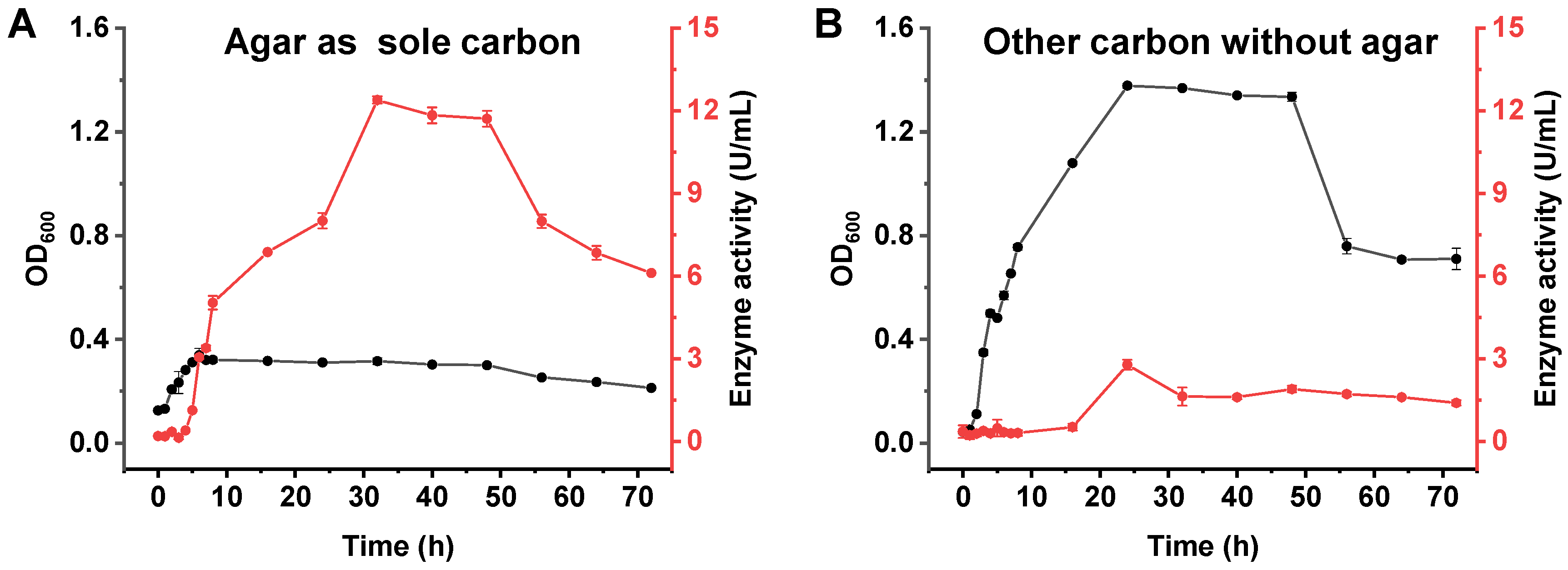
The curves of growth and agarase production of A. macleodii QZ9-9 were investigated in an agar-enriched medium with agar as the sole carbon source and agar-deficient medium (non-agar carbon source medium), where yeast extract instead of agar was used as the carbon source. As shown in Figure 2, A. macleodii QZ9-9 grew better in a non-agar carbon source medium than that in an agar-enriched medium, indicating that the non-agar carbon source medium contained more nutrients than the agar-enriched medium that no other ingredients except agar as carbon source and ions for the growth of strain QZ9-9. It is worth noting that the enzyme-producing capacity of strain QZ9-9 in an agar-enriched medium was better than that in a non-agar carbon source medium (Figure 2), suggesting that the agar must make a great contribution to the agarase production of agarolytic bacteria. Additionally, when strain QZ9-9 was cultured in an agar-enriched medium, the peak enzyme production (about 12.39 U/mL) occurred at 32 h (Figure 2A). However, no matter which medium was used for culturing agarolytic bacterial A. macleodii QZ9-9, the growth and enzyme production of this strain showed a short delay period, indicating that A. macleodii QZ9-9 had strong environmental adaptability (Figure 2; the red parts).

3.3. Analysis of the Agar-Degrading Activity of A. macleodii QZ9-9

The agar-degrading capacities of A. macleodii QZ9-9 and agarase secreted by this strain were completely investigated with the disc diffusion assay.
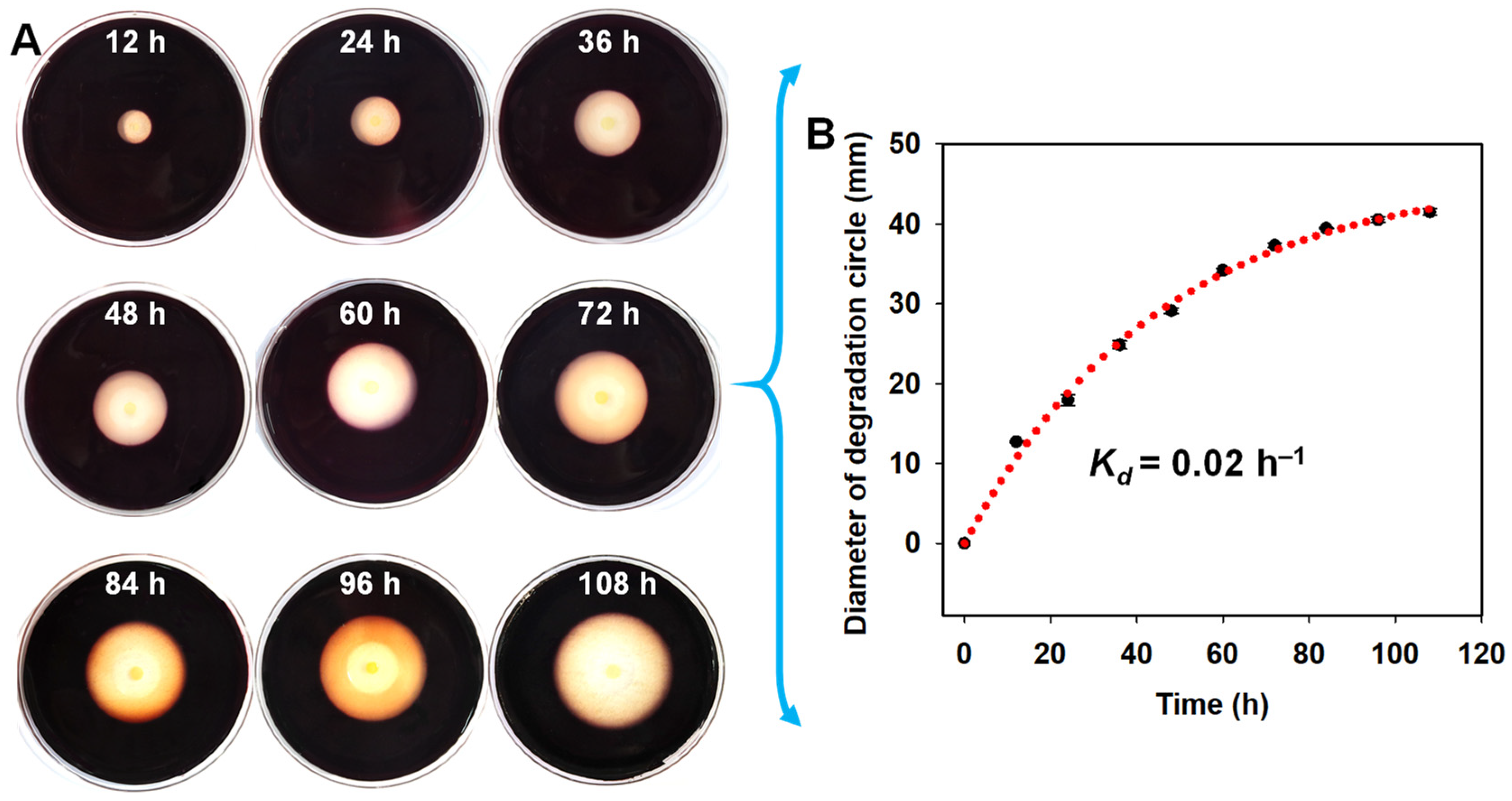
As shown in Figure 3, the diameter of the degradation zone generated by the agarolytic bacterium kept increasing with the increase in culture time (Figure 3A), and the first-order degradation rate constant was about 0.02 h−1 (Figure 3B). The maximum degradation diameter of 41.46 mm of the strain QZ9-9 occurred at 108 h. Additionally, the degradation growth rate of strain QZ9-9 was maintained at a relatively high level in the period from 0 h to 60 h, whereas that tended to be gentle after 60 h (Figure 3B).
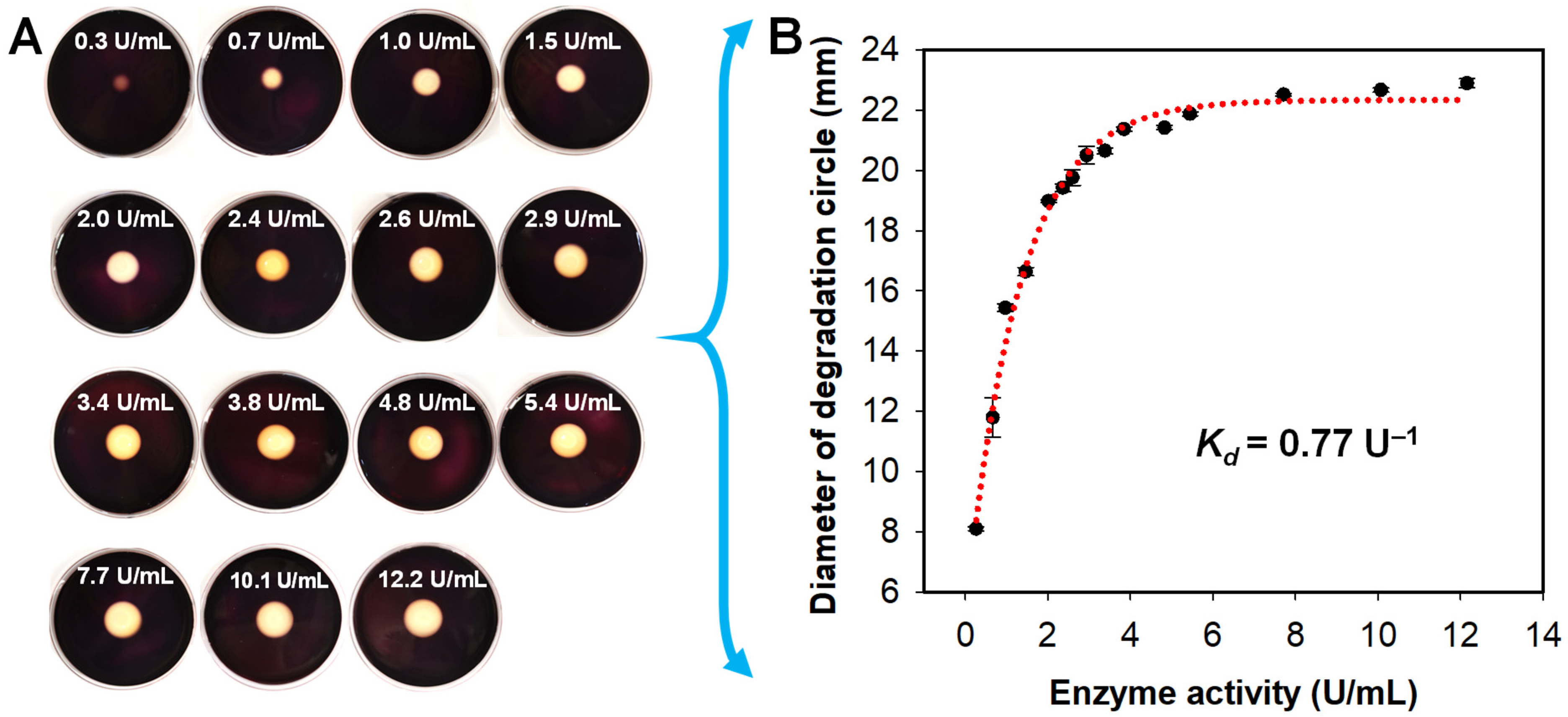
As shown in Figure 4, the diameter of the degradation zone generated by agarase increased with the increase in enzyme activity. The results showed that the agar-degrading ability of agarase was considerable (Figure 4A), and the first-order degradation rate constant was about 0.77 U−1 (Figure 4B). At the same time, the degradation growth rate of agarase was maintained at a high level at the enzyme activity ranging from 0 to 3.84 U/mL, while that tended to be gentle when the agarase was higher than 3.84 U/mL. Meanwhile, the maximum degradation diameter of 22.89 mm of agarase activity was about 12.2 U/mL.

3.4. Analysis of the Antioxidant Activity of Fermentation Products of A. macleodii QZ9-9

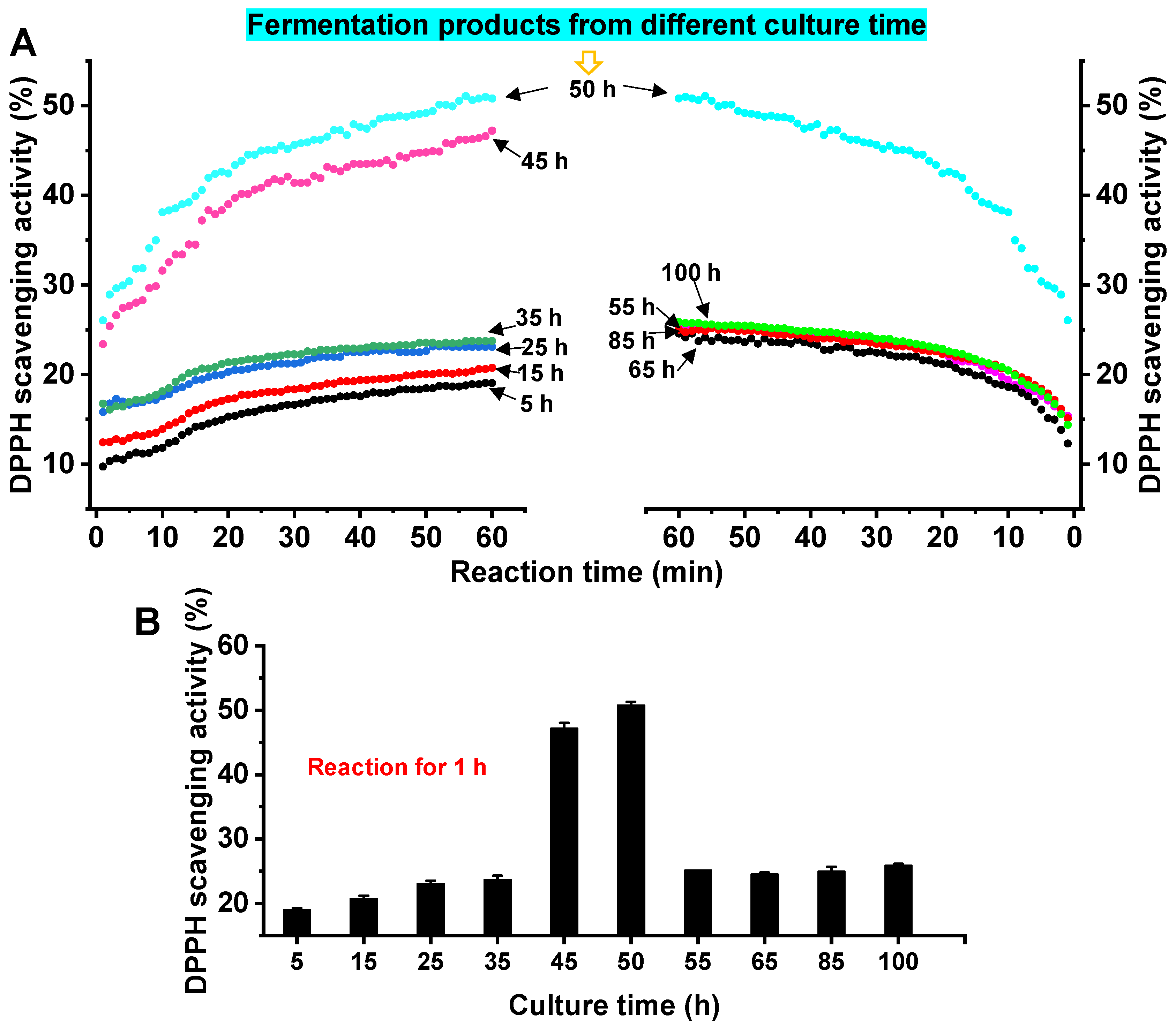
After a lot of preliminary explorations of the function of A. macleodii QZ9-9, we found that the agarolytic bacterial fermentation products without cells displayed a strong antioxidant property. Therefore, the antioxidant properties of the A. macleodii QZ9-9 fermentation products were characterized by the method of the DPPH free radical scavenging experiment. The kinetics of antioxidant properties of fermentation products (10 mg/mL) from different culture periods are regularly changed. As shown in Figure 5, the DPPH scavenging activity of fermentation products from each fermentation period increased with the increase in reaction time, and the scavenging rate increased rapidly in the period of 0 min to 23 min, indicating that the fermentation products can quickly react with DPPH. Additionally, the DPPH scavenging activity of fermentation products increased with the time increase during the fermentation period of 5 h to 50 h, and the scavenging activity reached the highest when the fermentation time was 50 h (Figure 5A). However, when the fermentation time exceeded 50 h, the DPPH clearance rate decreased sharply during the fermentation period of 50 h to 65 h, then tended to be flat (Figure 5B). We reason that the occurrence of this phenomenon depended on the characteristics of the growth and enzyme production of agarolytic bacterial strain A. macleodii QZ9-9. A. macleodii QZ9-9 had been growing and continuously producing agarase leading to more productions of antioxidant substances including metabolites and degradants during the culture of 50 h, whereas the growth and the enzyme production capacities of this strain gradually decreased leading to the decline and decomposition of the production of bioactive substances (particularly antioxidant products) after culture of 50 h. Additionally, as shown in Figure 5C, the fermentation broths of strain QZ9-9 from the culture period of 40 h to 50 h exhibited high DPPH scavenging activity, and the scavenging rate of DPPH reached the maximum of 50.79% at 50 h.
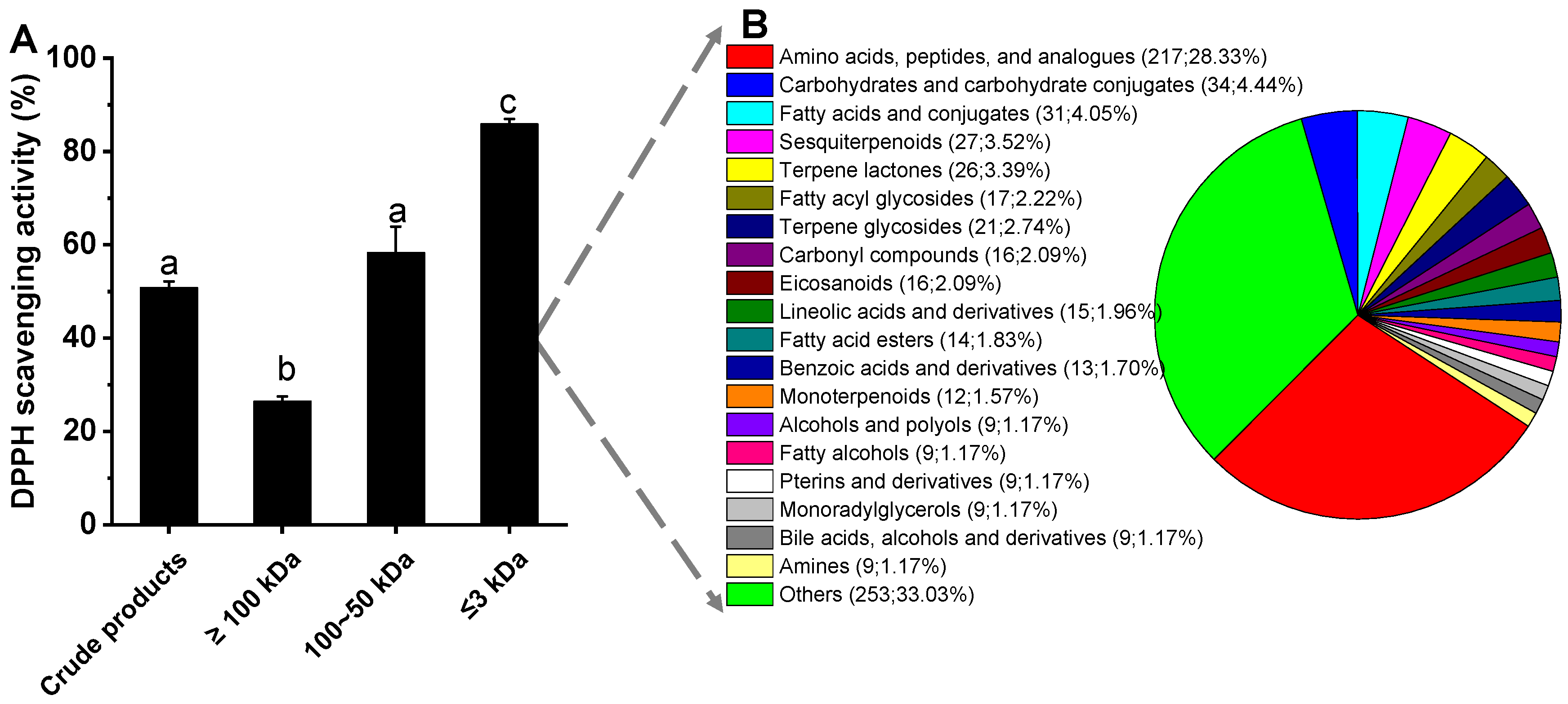
According to the above results, the fermentation products (without bacterial cells) of strain QZ9-9 from the culture broth of 50 h were used for further analysis of antioxidant composition. As shown in Figure 6A, the fermentation broth with a molecular weight of less than 3 kDa showed relatively high antioxidant activity, and its antioxidant activity increased with the increase in its concentration. Therefore, we reason that there are certain amounts and types of antioxidants in the low molecule metabolites (≤3 kDa) of A. macleodii QZ9-9 after incubation for 50 h.

3.5. Overview of the Composition of the Low-Molecule Metabolites

A comprehensive understanding of the reasons why low molecular weight (≤3 kDa) fermentation broth of A. macleodii QZ9-9 exhibited relatively high antioxidant activity will lay a foundation for the subsequent separation and utilization of bioactive substances from this strain. In this section, through the method of metabolomics, we completely investigated the global composition of the low molecular weight fermentation products of A. macleodii QZ9-9, which exhibited relatively high DPPH scavenging activity. A total of 766 metabolites in positive ion mode were detected in the low molecular weight (≤3 kDa) fermentation broth (Figure 6B; the detail of metabolomics is shown in Supplementary Materials), and there were 219 metabolites annotated in KEGG and 555 metabolites annotated in the database of HMDB. All the low molecular weight (≤3 kDa) were divided into chemical categories as 217 amino acids/peptides/analogs, 34 carbohydrates/carbohydrate conjugates, 31 fatty acids/conjugates, 27 sesquiterpenoids, 26 terpene lactones, 17 fatty acyl glycosides, 21 terpene glycosides, 16 carbonyl compounds, 16 eicosanoids, 15 lineolic acids/derivatives, 14 fatty acid esters, 13 benzoic acids/derivatives, 12 monoterpenoids, 9 alcohols/polyols, 9 fatty alcohols, 9 pterins/derivatives, 9 monoradylglycerols, 9 bile acids/alcohols/derivatives, and 9 amines. The metabolites of the low molecular weight fermentation products of A. macleodii QZ9-9 with the relative abundances of the top 20, such as 54.96% meso–pristane, 2.68% prolyl–histidine, 1.58% C16 sphinganine hexadecasphinganine, 1.20% glycerol, 1.12% 6-hydroxyoctadecanoic acid, and 0.95% maculosin, are listed in Table 1.

3.6. Maculosin Specific Antioxidant Activity

Due to the relatively high content of maculosin, which was reported to have antioxidant properties in the low molecular weight (≤3 kDa) of A. macleodii QZ9-9, the DPPH scavenging activity of maculosin was verified in this work. If the substance possesses antioxidant properties, it can fade the purple color of the DPPH solution. As shown in Figure 7, the DPPH scavenging activity of maculosin increased with the increase in its concentration, while its antioxidant activity is still at an over low level compared to that of Vitamin C; for instance, the DPPH scavenging activities of 7.5 μg/mL maculosin and 7.5 μg/mL Vitamin C were 38.22% and 74.44%, respectively (Figure 7B).

4. Discussion

Marine bacteria have been regarded as valuable sources for novel bioactivity metabolites with better biocatalytic properties, e.g., high salinity, high pressure, hyperthermostability, barophilicity, alkali resistance, and low temperature, which can serve various biotechnological applications, such as pharmaceutical, food, and cosmetics [14,18,22,23,24,26,27,28]. Agar in the macroalgae accounted for ~60% of dry weight in its cell wall [29], and the bacteria with the capacity of metabolizing agar as their only carbon and energy source are recognized as agarolytic bacteria. Additionally, unique enzymes and metabolites with unpreceded applications in industry and medicine have been derived from agarolytic bacteria [3,30,31].
In the present work, an agarolytic bacterium strain QZ9-9 was successfully isolated from tropical G. hainanensis in Hainan Island, China, which was classified into the species of Alteromonas macleodii (Gene Bank No.: CP098772.1). A. macleodii QZ9-9 grew well in either high nutrient medium or only carbon source medium, whereas the agarase secreted by the strain QZ9-9 must be inducible by agar. The production process of the substrate (agar)-induced agarase of A. macleodii QZ9-9 is similar to that of many other induced enzymes, such as dextranase induced by dextran [9,18], β-galactosidase induced by lactose or galactose [32], α-amylase induced by amylum [33], and agarase induced by agar [14,34,35].
A. macleodii QZ9-9 displayed a particularly strong ability to degrade agar. With the increase in culture time, the diameter of the degradation zone generated by this strain also increased, and the maximum diameter of the degradation zone reached 41.46 mm when the culture time was 108 h, and the first-order degradation rate constant was about 0.02 h1. As expected, the degradation capacity of the crude agarase of A. macleodii QZ9-9 also increased with the increase in enzyme activity. When the enzyme activity reached 12.16 U/mL, the maximum diameter of the degradation zone was up to about 22.89 mm, and the first-order degradation rate constant was about 0.77 U−1. Only a few species belonging to the Alteromonas-like Gammaproteobacteria are reported to be agarolytic or weakly agarolytic, such as Pseudoalteromonas sp. [36,37], Shewanella sp. [38,39], and Glaciecola sp. [40], whereas our results indicated that A. macleodii QZ9-9 is a good candidate for agarolytic bacteria with a strong agar-degrading capacity.
In particular, the fermentation broth of A. macleodii QZ9-9 was found to possess high antioxidant activity, and the peak of DPPH scavenging activity of its fermentation products after culture for 50 h was up to 50.79% in the reaction for 1 h. Furthermore, the low molecule metabolites (≤3 kDa) of A. macleodii QZ9-9 after incubation for 50 h displayed the highest DPPH scavenging activity of 85.85%, which contained a total of 766 metabolites. Among them, most metabolites (217 metabolites) were identified as amino acids/peptides/analogs. More importantly, the peptide-like metabolites, such as prolyl–histidine, isoleucyl–histidine, isoleucyl–proline, and arginyl–proline, were found in the top 20 metabolites with high abundance.
Reportedly, antioxidants are compounds that stop the oxidation process by inhibiting free radical reactions, and the antioxidants protect against the damaging effects of metabolism in biological systems, such as neutralizing excess free radicals in the bio-body. There are many kinds of antioxidant substances in the ocean, mainly including polysaccharides, terpenes, polyphenolic compounds, enzymatic antioxidants, amino acids, and peptides. Of the antioxidants, peptides consisting of 2–20 amino acid residues were frequently reported due to their antioxidant activities, such as glutathione, carnosine, cyclo (His-Pro), and human tripeptide GHK (glycyl-l-histidyl-Llysine) [41], and the antioxidant activity of antioxidant peptides are mainly related to the arrangement of amino acid and the structure of the peptide. Additionally, the study on the antibacterial, anticancer, anti-inflammatory, and other biological activities of antioxidant peptides is also of high research interest [42]. The marine sources of antioxidant peptides are varied, such as algae, sponges, bryozoans, lichens, microalgae, bacteria, etc., which are all natural sources of antioxidants [43]; for instance, Tripathi et al. [44] found that short peptides in Kocuria Marina CDMP 10 extract were effective in reducing DPPH free radicals during antioxidant molecular production screening of marine microbial resources in the Bay of Bengal, India. Sonani et al. [45] isolated three water-soluble phycobilin proteins: phycoerythrin, phycocyanin, and allophycocyanin from the cyanobacteria Lyngbya sp. A09DM, which showed strong antioxidant and free radical scavenging activities and could be used to prolong the life span of Caenorhabditis elegans. Abuine et al. [46] found that short peptides purified from fish skin hydrolysates, in addition to exhibiting antioxidant activity, also displayed hypoglycemic, antihypertensive, antibacterial, and anti-aging activities. In this work, we reason that the low molecule metabolites (≤3 kDa) of A. macleodii QZ9-9 containing high peptide contents may be a strong candidate to screen and prepare antioxidant peptides due to its relatively high antioxidant activity.
Additionally, we focused on a maculosin (C14H16N2O3) metabolite in the low molecule metabolites (≤3 kDa) of A. macleodii QZ9-9 due to its relatively high abundance. Maculosin was first reported in 1988 as a host-specific phytotoxin for spotted knapweed produced by Alternaria alternata [47]. After that, it was successively isolated from Schizophyllum commune [48], Pseudomonas sp. (ABS-36) [49], Streptomyces sp. ZZ446 [50], Bacillus pumilus AMK1 [51], and Streptomyces sp. KTM18 [52]. In terms of the bioactivity of maculosin, there were only a few reports on the properties of the bioactive compound; for instance, the maculosin isolated from Streptomyces sp. ZZ446 showed good antibacterial activity against methicillin-resistant Staphylococcus aureus, Escherichia coli, and Candida albicans [50]; Karanam et al. [51] reported that the maculosin played an anticancer role by inducing apoptosis. In terms of antioxidant activity, the maculosin isolated from Streptomyces sp. KTM18 displayed antioxidant activity [52]. Given this, the antioxidant activity of maculosin was furtherly confirmed by the method of DPPH scavenging activity in this work. We found that although the DPPH scavenging activity of the maculosin was weaker than that of Vitamin C, the obvious scavenging ability for DPPH was visible after treatment of maculosin, and the purple of DPPH solution gradually faded to lavender with the increase in maculosin concentration. When the concentration of maculosin increased to 750 μg/mL, the DPPH scavenging activity was up to about 56.89%, indicating that maculosin has a certain antioxidant effect. In addition, maculosin was reported to be a non-toxic substance to a certain extent [51,52]. Therefore, we reason that the maculosin-enriched low molecule metabolites (≤3 kDa) of A. macleodii QZ9-9 with relatively high antioxidant activity must be the strong candidates for preparing antioxidant maculosin.

5. Conclusions

In summary, the agarolytic bacterium-A. macleodii QZ9-9 isolated from tropical G. hainanensis in Hainan Island was a strong candidate for utilization of macroalgae resources by degrading the agar that is the main component of macroalgae. In particular, the low molecule metabolites (≤3 kDa) of A. macleodii QZ9-9 with relatively high antioxidant activity are strong candidates for preparing non-toxic antioxidants such as antioxidant peptides and maculosin. Therefore, we reason that the agarolytic bacterium-A. macleodii QZ9-9 and its metabolites/degradants are possibilities for high-added value utilization of macroalgae in the fields of cosmetics, food preservation, and the pharmaceutical industry.

Supplementary Materials

The following supporting information can be downloaded at: https://www.mdpi.com/article/10.3390/antiox11101977/s1.

Author Contributions

Conceptualization, W.R. and Z.X.; methodology, data curation, formal analysis, and writing—original draft, W.R. and X.W.; investigation, X.W., Z.F. and C.L.; supervision, X.C., H.L., X.Z., A.H., Y.Z., W.R. and Z.X.; writing—review and editing, X.W., W.R. and Z.X.; funding acquisition, W.R. and Z.X. All authors have read and agreed to the published version of the manuscript.

Funding

This research was funded by the Hainan Province Science and Technology Special Fund (ZDYF2022XDNY216), Hainan Provincial Natural Science Foundation of China (321QN176), National Key Research and Development Project of China (2019YFD0901304 and 2020YFD0901104), and Scientific Research Foundation of Hainan University (KYQD(ZR)21005, KYQD(ZR)1967, KYQD(ZR)1819 and KYQD(ZR)20060).

Institutional Review Board Statement

Not applicable.

Informed Consent Statement

Not applicable.

Data Availability Statement

The data presented in this study are available in the article and Supplementary Materials.

Conflicts of Interest

The authors declare no conflict of interest.

References

  1. Ross, A.B.; Jones, J.M.; Kubacki, M.L.; Bridgeman, T. Classification of macroalgae as fuel and its thermochemical behaviour. Bioresour. Technol. 2008, 99, 6494–6504. [Google Scholar] [CrossRef]
  2. Chi, W.-J.; Seo, J.W.; Hong, S.-K. Characterization of Two Thermostable β-agarases from a Newly Isolated Marine Agarolytic Bacterium, Vibrio sp. S1. Biotechnol. Bioprocess Eng. 2019, 24, 799–809. [Google Scholar] [CrossRef]
  3. Khalifa, A.; Aldayel, M. Isolation and characterisation of the agarolytic bacterium Pseudoalteromonas ruthenica. Open Life Sci. 2019, 14, 588–594. [Google Scholar] [CrossRef] [PubMed]
  4. Geng, Y.; Wang, K.; Chen, D.; Huang, X.; He, M.; Yin, Z. Stenotrophomonas maltophilia, an emerging opportunist pathogen for cultured channel catfish, Ictalurus punctatus, in China. Aquaculture 2010, 308, 132–135. [Google Scholar] [CrossRef]
  5. Schroeder, D.C.; Jaffer, M.A.; Coyne, V.E. Investigation of the role of a β (1–4) agarase produced by Pseudoalteromonas gracilis B9 in eliciting disease symptoms in the red alga Gracilaria gracilis. Microbiology 2003, 149 Pt 10, 2919–2929. [Google Scholar] [CrossRef] [PubMed] [Green Version]
  6. Chi, W.-J.; Chang, Y.-K.; Hong, S.-K. Agar degradation by microorganisms and agar-degrading enzymes. Appl. Microbiol. Biotechnol. 2012, 94, 917–930. [Google Scholar] [CrossRef] [PubMed]
  7. Yun, E.J.; Yu, S.; Kim, K.H. Current knowledge on agarolytic enzymes and the industrial potential of agar-derived sugars. Appl. Microbiol. Biotechnol. 2017, 101, 5581–5589. [Google Scholar] [CrossRef] [PubMed]
  8. Wang, J.; Jiang, X.; Mou, H.; Guan, H. Anti-oxidation of agar oligosaccharides produced by agarase from a marine bacterium. J. Appl. Phycol. 2004, 16, 333–340. [Google Scholar] [CrossRef]
  9. Ren, W.; Cai, R.; Yan, W.; Lyu, M.; Fang, Y.; Wang, S. Purification and characterization of a biofilm-degradable dextranase from a marine bacterium. Mar. Drugs 2018, 16, 51. [Google Scholar] [CrossRef] [Green Version]
  10. Kim, J.H.; Yun, E.J.; Yu, S.; Kim, K.H.; Kang, N.J. Different Levels of Skin Whitening Activity among 3,6-Anhydro-l-galactose, Agarooligosaccharides, and Neoagarooligosaccharides. Mar. Drugs 2017, 15, 321. [Google Scholar] [CrossRef]
  11. Mussatto, S.I.; Mancilha, I.M. Non-digestible oligosaccharides: A review. Carbohydr. Polym. 2007, 68, 587–597. [Google Scholar] [CrossRef]
  12. Bhattarai, Y.; Kashyap, P.C. Agaro-oligosaccharides: A new frontier in the fight against colon cancer? Am. J. Physiol.-Gastrointest. Liver Physiol. 2016, 310, G335–G336. [Google Scholar] [CrossRef] [PubMed] [Green Version]
  13. Ren, W.; Wang, S.; Lü, M.; Wang, X.; Fang, Y.; Jiao, Y.; Hu, J. Optimization of four types of antimicrobial agents to increase the inhibitory ability of marine Arthrobacter oxydans KQ11 dextranase mouthwash. Chin. J. Oceanol. Limnol. 2016, 34, 354–366. [Google Scholar] [CrossRef]
  14. Ren, W.; Li, P.; Wang, X.; Che, Y.; Long, H.; Zhang, X.; Cai, X.; Huang, A.; Zeng, Y.; Xie, Z. Cross-habitat distribution pattern of Bacillus communities and their capacities of producing industrial hydrolytic enzymes in Paracel Islands: Habitat-dependent differential contributions of the environment. J. Environ. Manag. 2022, 323, 116252. [Google Scholar] [CrossRef]
  15. Jin, M.; Liu, H.; Hou, Y.; Chan, Z.; Di, W.; Li, L.; Zeng, R. Preparation, characterization and alcoholic liver injury protective effects of algal oligosaccharides from Gracilaria lemaneiformis. Food Res. Int. 2017, 100, 186–195. [Google Scholar] [CrossRef] [PubMed]
  16. Shieh, W.Y.; Simidu, U.; Maruyama, Y. Nitrogen Fixation by Marine Agar-degrading Bacteria. Microbiology 1988, 134, 1821–1825. [Google Scholar] [CrossRef] [Green Version]
  17. Ivanova, G.; Rákhely, G.; Kovács, K.L. Hydrogen production from biopolymers by Caldicellulosiruptor saccharolyticus and stabilization of the system by immobilization. Int. J. Hydrogen Energy 2008, 33, 6953–6961. [Google Scholar] [CrossRef]
  18. Ren, W.; Liu, L.; Gu, L.; Yan, W.; Feng, Y.L.; Dong, D.; Wang, S.; Lyu, M.; Wang, C. Crystal structure of GH49 dextranase from Arthrobacter oxidans KQ11: Identification of catalytic base and improvement of thermostability using semirational design based on B-factors. J. Agric. Food Chem. 2019, 67, 4355–4366. [Google Scholar] [CrossRef]
  19. Miller, G.L. Use of Dinitrosalicylic Acid Reagent for Determination of Reducing Sugar. Anal. Chem. 1959, 31, 426–428. [Google Scholar] [CrossRef]
  20. Wang, X.; Lu, M.; Wang, S.; Fang, Y.; Wang, D.; Ren, W.; Zhao, G. The atmospheric and room-temperature plasma (ARTP) method on the dextranase activity and structure. Int. J. Biol. Macromol. 2014, 70, 284–291. [Google Scholar] [CrossRef] [PubMed]
  21. Liu, H.; Ren, W.; Ly, M.; Li, H.; Wang, S. Characterization of an Alkaline GH49 Dextranase from Marine Bacterium Arthrobacter oxydans KQ11 and Its Application in the Preparation of Isomalto-Oligosaccharide. Mar. Drugs 2019, 17, 479. [Google Scholar] [CrossRef] [Green Version]
  22. Ren, W.; Ding, Y.; Gu, L.; Yan, W.; Wang, C.; Lyu, M.; Wang, C.; Wang, S. Characterization and mechanism of the effects of Mg–Fe layered double hydroxide nanoparticles on a marine bacterium: New insights from genomic and transcriptional analyses. Biotechnol. Biofuels 2019, 12, 196. [Google Scholar] [CrossRef]
  23. Ren, W.; Xu, X.; Long, H.; Zhang, X.; Cai, X.; Huang, A.; Xie, Z. Tropical Cellulolytic Bacteria: Potential Utilization of Sugarcane Bagasse as Low-Cost Carbon Source in Aquaculture. Front. Microbiol. 2021, 12, 745853. [Google Scholar] [CrossRef] [PubMed]
  24. Wei, Y.; Bu, J.; Long, H.; Zhang, X.; Cai, X.; Huang, A.; Ren, W.; Xie, Z. Community structure of protease-producing bacteria cultivated from aquaculture systems: Potential impact of a tropical environment. Front. Microbiol. 2021, 12, 638129. [Google Scholar] [CrossRef] [PubMed]
  25. Jiao, L.; Li, B.; Wang, M.; Liu, Z.; Zhang, X.; Liu, S. Antioxidant activities of the oligosaccharides from the roots, flowers and leaves of Panax ginseng CA Meyer. Carbohydr. Polym. 2014, 106, 293–298. [Google Scholar] [CrossRef]
  26. Wei, Y.; Wang, F.; Gao, J.; Huang, Y.; Ren, W.; Sheng, H. Culture-dependent and culture-independent characterization of bacterial community diversity in different types of sandy lands: The case of Minqin County, China. BMC Microbiol. 2021, 21, 87. [Google Scholar] [CrossRef] [PubMed]
  27. Wei, Y.; Long, Z.; Ren, M. Microbial community and functional prediction during the processing of salt production in a 1000-year-old marine solar saltern of South China. Sci. Total Environ. 2022, 819, 152014. [Google Scholar] [CrossRef]
  28. Gu, L.; Yan, W.; Wu, H.; Fan, S.; Ren, W.; Wang, S.; Lyu, M.; Liu, J. Selection of DNAzymes for Sensing Aquatic Bacteria: Vibrio Anguillarum. Anal. Chem. 2019, 91, 7887–7893. [Google Scholar] [CrossRef]
  29. De Jesus Raposo, M.F.; De Morais, A.M.B.; De Morais, R.M.S.C. Marine Polysaccharides from Algae with Potential Biomedical Applications. Mar. Drugs 2015, 13, 2967–3028. [Google Scholar] [CrossRef] [PubMed] [Green Version]
  30. Sakai, M.; Hosoda, A.; Ogura, K.; Ikenaga, M. The Growth of Steroidobacter agariperforans sp nov., a Novel Agar-Degrading Bacterium Isolated from Soil, is Enhanced by the Diffusible Metabolites Produced by Bacteria Belonging to Rhizobiales. Microbes Environ. 2014, 29, 89–95. [Google Scholar] [CrossRef] [PubMed]
  31. Pathiraja, D.; Christiansen, L.; Park, B.; Schultz-Johansen, M.; Bang, G.; Stougaard, P.; Choi, I.-G.; Stabb, E.V. A Novel Auxiliary Agarolytic Pathway Expands Metabolic Versatility in the Agar-Degrading Marine Bacterium Colwellia echini A3T. Appl. Environ. Microbiol. 2021, 87, e00230-21. [Google Scholar] [CrossRef] [PubMed]
  32. de Albuquerque, T.L.; de Sousa, M.; Silva, N.C.G.E.; Neto, C.A.C.G.; Gonçalves, L.R.B.; Fernandez-Lafuente, R.; Rocha, M.V.P. β-Galactosidase from Kluyveromyces lactis: Characterization, production, immobilization and applications—A review. Int. J. Biol. Macromol. 2021, 191, 881–898. [Google Scholar] [CrossRef]
  33. Spiridon, I.; Popescu, M.C.; Bodârlău, R.; Vasile, C. Enzymatic degradation of some nanocomposites of poly(vinyl alcohol) with starch. Polym. Degrad. Stab. 2008, 93, 1884–1890. [Google Scholar] [CrossRef]
  34. Feng, Z.; Peng, L.; Chen, M.; Li, M. Rhodococcus sp. Q5, a novel agarolytic bacterium isolated from printing and dyeing wastewater. Folia Microbiol. 2012, 57, 379–386. [Google Scholar] [CrossRef] [PubMed]
  35. Lakshmikanth, M.; Manohar, S.; Souche, Y.; Lalitha, J. Extracellular β-agarase LSL-1 producing neoagarobiose from a newly isolated agar-liquefying soil bacterium, Acinetobacter sp., AG LSL-1. World J. Microbiol. Biotechnol. 2006, 22, 1087–1094. [Google Scholar] [CrossRef]
  36. Chi, W.-J.; Lee, C.-R.; Dugerjonjuu, S.; Park, J.-S.; Kang, D.-K.; Hong, S.-K. Biochemical characterization of a novel iron-dependent GH16 β-agarase, AgaH92, from an agarolytic bacterium Pseudoalteromonas sp H9. FEMS Microbiol. Lett. 2015, 362, fnv035. [Google Scholar] [CrossRef] [PubMed] [Green Version]
  37. Wang, Y.; Liu, T.; Guo, S.; Zhang, P.; Sun, P.; Chen, M.; Ming, H. Characterization and overexpression of a glycosyl hydrolase family 16 β-agarase Aga0917 from Pseudoalteromonas fuliginea YTW1-15-1. J. Gen. Appl. Microbiol. 2018, 64, 276–283. [Google Scholar] [CrossRef] [Green Version]
  38. Wang, W.; Wang, J.; Yan, R.; Zeng, R.; Zuo, Y.; Wang, D.; Qu, W. Expression and Characterization of a Novel Cold-Adapted and Stable β-Agarase Gene agaW1540 from the Deep-Sea Bacterium Shewanella sp. WPAGA9. Mar. Drugs 2021, 19, 431. [Google Scholar] [CrossRef]
  39. Skerratt, J.H.; Bowman, J.P.; Nichols, P.D. Shewanella olleyana sp. nov., a marine species isolated from a temperate estuary which produces high levels of polyunsaturated fatty acids. Int. J. Syst. Evol. Microbiol. 2002, 52 Pt 6, 2101–2106. [Google Scholar] [CrossRef] [PubMed] [Green Version]
  40. Teramoto, M.; Nishijima, M. Agaribacter marinus gen. nov., sp nova, an agar-degrading bacterium from surface seawater. Int. J. Syst. Evol. Microbiol. 2014, 64, 2416–2423. [Google Scholar] [CrossRef]
  41. Wong, F.-C.; Xiao, J.; Wang, S.; Ee, K.-Y.; Chai, T.-T. Advances on the antioxidant peptides from edible plant sources. Trends Food Sci. Technol. 2020, 99, 44–57. [Google Scholar] [CrossRef]
  42. Cunha, S.A.; Pintado, M.E. Bioactive peptides derived from marine sources: Biological and functional properties. Trends Food Sci. Technol. 2022, 119, 348–370. [Google Scholar] [CrossRef]
  43. Vladkova, T.; Georgieva, N.; Staneva, A.; Gospodinova, D. Recent Progress in Antioxidant Active Substances from Marine Biota. Antioxidants 2022, 11, 439. [Google Scholar] [CrossRef]
  44. Tripathi, V.C.; Horam, S.; Singh, A.; Lata, M.; Reddy, T.J.; Arockiaraj, J.; Pasupuleti, M. The discovery of antioxidants in marine microorganisms and their protective effects on the hepatic cells from chemical-induced oxidative stress. Free Radic. Res. 2020, 54, 150–161. [Google Scholar] [CrossRef] [PubMed]
  45. Sonani, R.R.; Singh, N.K.; Kumar, J.; Thakar, D.; Madamwar, D. Concurrent purification and antioxidant activity of phycobiliproteins from Lyngbya sp. A09DM: An antioxidant and anti-aging potential of phycoerythrin in Caenorhabditis elegans. Process Biochem. 2014, 49, 1757–1766. [Google Scholar] [CrossRef]
  46. Abuine, R.; Rathnayake, A.U.; Byun, H.-G. Biological activity of peptides purified from fish skin hydrolysates. Fish. Aquat. Sci. 2019, 22, 10. [Google Scholar] [CrossRef] [Green Version]
  47. Cardellina, J.; Strobel, G. Maculosin, a host-specific phytotoxin for spotted knapweed from Alternaria alternata. Proc. Natl. Acad. Sci. USA 1988, 85, 8008–8011. [Google Scholar] [CrossRef] [Green Version]
  48. Biasetto, C.; Somensi, A.; Figueiro, F.; Moraes, L.; Silva, G.; Young, M.C.M.; Bolzani, V.; Araujo, A. Diketopiperazines and arylethylamides produced by Schizophyllum commune, an endophytic fungus in Alchornea glandulosa. Eclética Química J. 2019, 44, 36–42. [Google Scholar] [CrossRef]
  49. Ahil, S.B.; Hira, K.; Shaik, A.B.; Pal, P.P.; Kulkarni, O.P.; Araya, H.; Fujimoto, Y. L-Proline-based-cyclic dipeptides from Pseudomonas sp. (ABS-36) inhibit pro-inflammatory cytokines and alleviate crystal-induced renal injury in mice. Int. Immunopharmacol. 2019, 73, 395–404. [Google Scholar] [CrossRef]
  50. Chen, S.; Zhang, D.; Chen, M.; Zhang, Z.; Lian, X.-Y. A rare diketopiperazine glycoside from marine-sourced Streptomyces sp. ZZ446. Nat. Prod. Res. 2020, 34, 1046–1050. [Google Scholar] [CrossRef] [PubMed]
  51. Karanam, G.; Arumugam, M.K. Reactive oxygen species generation and mitochondrial dysfunction for the initiation of apoptotic cell death in human hepatocellular carcinoma HepG2 cells by a cyclic dipeptide Cyclo(-Pro-Tyr). Mol. Biol. Rep. 2020, 47, 3347–3359. [Google Scholar] [CrossRef]
  52. Paudel, B.; Maharjan, R.; Rajbhandari, P.; Aryal, N.; Aziz, S.; Bhattarai, K.; Baral, B.; Malla, R.; Bhattarai, H.D. Maculosin, a non-toxic antioxidant compound isolated from Streptomyces sp. KTM18. Pharm. Biol. 2021, 59, 933–936. [Google Scholar] [CrossRef]
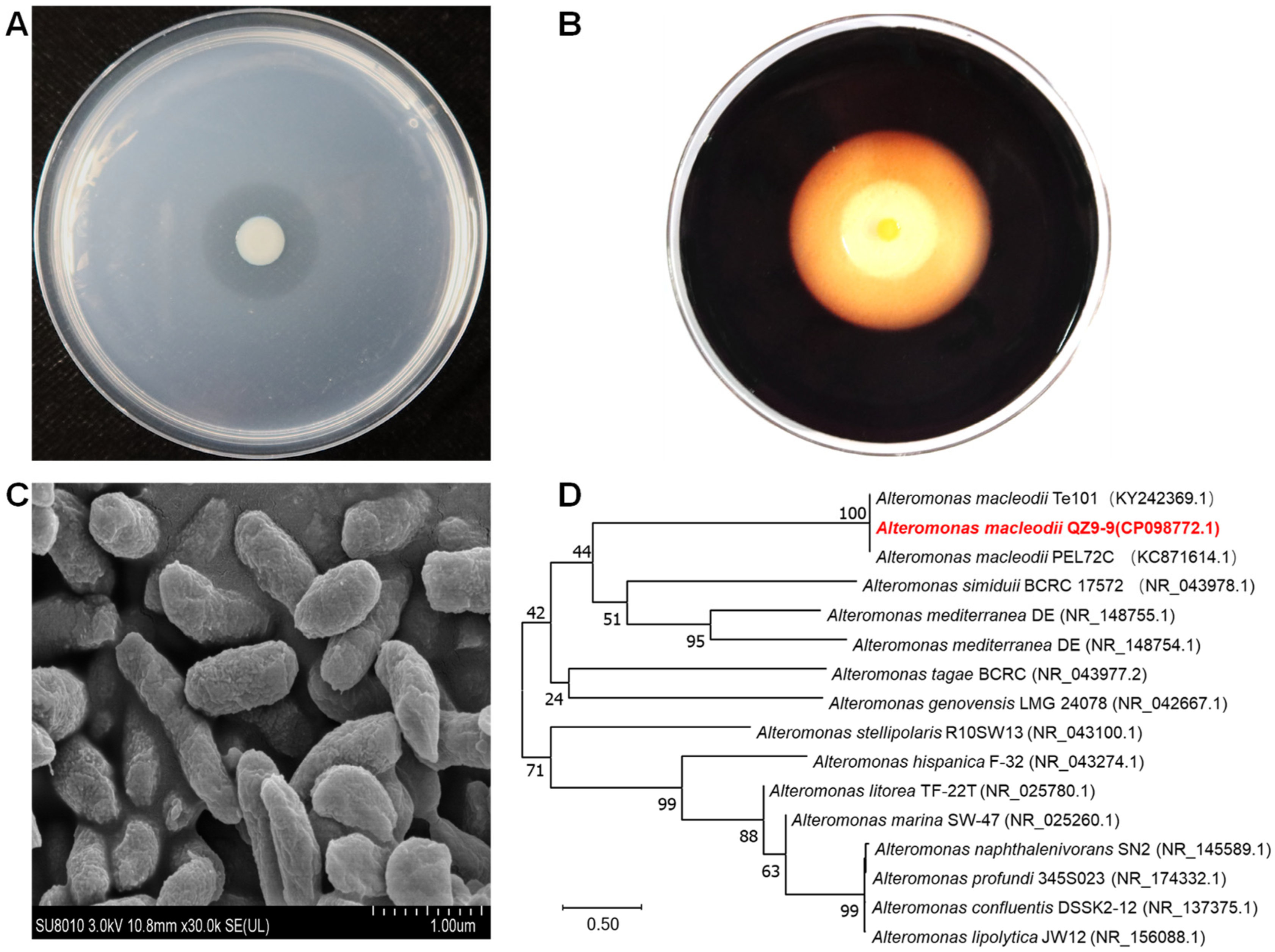
Figure 1. The morphology of agarolytic bacterial A. macleodii QZ9-9. (A) The colony of strain QZ9-9 on mineral salts agar plate after static incubation for 48 h, and a clear transparent hydrolysis zone surrounds this colony. (B) Hydrolysis zone colored with Lugol’s iodine solution after static incubation for 96 h on mineral salts agar plate. (C) Close-up of the electron microscopy of strain QZ9-9. (D) Phylogenetic tree of A. macleodii QZ9-9 (marked by red color) prepared using Mega 11.0 software. Note: the code in each parenthesis represents the sequence accession number in GenBank, and the number at each branch point is the percentage supported by bootstrap.
Antioxidants 11 01977 g001
Figure 2. The curves of growth and agarase production of A. macleodii QZ9-9 in agar-enriched medium (w/v: containing 0.2% agar, 3% NaCl, 0.5% (NH4)2SO4, 0.2% K2HPO4, 0.1% MgSO4·7H2O and 0.01% FeSO4·7H2O, pH 7.5) with agar as the sole carbon source (A) and agar-deficient medium (w/v: containing 0.5% tryptone, 0.1% yeast extract, 100 mL seawater, pH 7.5) that other carbon instead of agar as the carbon source (B).
Antioxidants 11 01977 g002
Figure 3. The agar-degrading capacity of A. macleodii QZ9-9. (A) Characterization of agar-degrading activity of A. macleodii QZ9-9 on the mineral salts agar plates (stained with Lugol’s iodine solution) at different degradation periods. (B) Kinetics of agar degradation by A. macleodii QZ9-9. Note: The data were fitted according to the first-order rate equation Yt = Y0 + a(1 − ekx), where Yt and Y0 are the degradation fraction at a given culture time t and 0 min, respectively; a is the fitting coefficients; and k is the observed rate constant.
Antioxidants 11 01977 g003
Figure 4. The agar-degrading capacities of agarase secreted by A. macleodii QZ9-9. (A) Characterization of agar-degrading activity of crude agarase with different enzyme activities on the mineral salts agar plates (stained with Lugol’s iodine solution). (B) Kinetics of agar degradation by agarase. Note: The data were fitted according to the first-order rate equation YEA = Y0 + a(1 − ekx), where YEA and Y0 are the degradation fraction at a given enzyme activity EA and 0 U/mL, respectively; a is the fitting coefficients; and k is the observed rate constant.
Antioxidants 11 01977 g004
Figure 5. Kinetics of DPPH scavenging activity of fermentation products from two culture periods during the reaction time of 1 h, (A) the period of 0 h to 50 h, and (B) the period of 50 h to 100 h. (C) DPPH scavenging activity of fermentation products during the culture of strain QZ9-9 after 1 h reaction. Note: The reaction time refers to the time consumed by the combination reaction between fermentation products and DPPH solution.
Antioxidants 11 01977 g005
Figure 6. (A) The DPPH scavenging activity of fermentation products with different molecular weights. (B) Comparison of the relative abundance of metabolites in the low molecular weight (≤3 kDa) products. The different superscript letters indicate a significant difference at p < 0.05.
Antioxidants 11 01977 g006
Figure 7. Confirmation of DPPH scavenging activity of maculosin by visual observation (A) and quantification (B). Vitamin C is a positive control.
Antioxidants 11 01977 g007
Table 1. The metabolites of the low molecular weight fermentation products of A. macleodii QZ9-9 with the relative abundances of the top 20.
No.Metabolitem/zRetention Time (min)FormulaKEGG IDLibrary IDArea (Counts)Relative Abundance (%)
1Meso–Pristane332.3310.87C19H40-HMDB00344975.39 × 10954.96%
2Prolyl–Histidine252.002.91C11H16N4O3-HMDB00290192.63 × 1082.68%
3Hexadecasphinganine273.2710.20C16H35NO2C13915LMSP010400011.54 × 1081.58%
4Glycerol115.041.01C3H8O3C00116HMDB0000131;
56-81-5
1.17 × 1081.20%
56-Hydroxyoctadecanoic acid318.3010.21C18H36O3-HMDB01121931.10 × 1081.12%
6Maculosin261.125.90C14H16N2O3C10605-9.28 × 1070.95%
7Benzalkonium304.3010.49C21H37N--7.91 × 1070.81%
8MG(0:0/16:0/0:0)353.2712.680C19H38O4-HMDB0011533;
LMGL01010025
5.28 × 1070.54%
9(2xi,6xi)-7-Methyl-3-methylene-1,2,6,7-octanetetrol227.139.63C10H20O4-HMDB0033217;
LMFA05000554
4.71 × 1070.48%
1010,20-Dihydroxyeicosanoic acid362.3310.22C20H40O4-HMDB00319234.04 × 1070.41%
11(+/−)-Flurbiprofen245.104.43C15H13FO2--3.69 × 1070.38%
122-Isopropyl-3,5-dimethoxy-6-methylpyrazine197.136.26C10H16N2O2-HMDB00297413.54 × 1070.36%
13Acrylamide181.0416.30C3H5NOC01659HMDB00042963.41 × 1070.35%
14Isoleucyl–Histidine251.153.94C12H20N4O3-HMDB00289092.90 × 1070.30%
154-(3,7-dimethylocta-2,6-dien-1-yl)benzene-1,2,3,5-tetrol301.1411.45C16H22O4--2.88 × 1070.29%
16Isoleucyl–proline211.147.34C11H20N2O3-HMDB00111742.80 × 1070.29%
17Arginyl–Proline254.163.73C11H21N5O3-HMDB00287172.62 × 1070.27%
181-(4-hydroxy-3-methoxyphenyl)-7-(3-hydroxyphenyl)heptane-3,5-dione365.1411.38C20H22O5--2.45 × 1070.25%
192-Deoxyglucose187.061.04C6H12O5-HMDB00624772.30 × 1070.23%
20Taurocholic acid538.2811.42C26H45NO7SC0512281-24-3;
LMST05040001;
HMDB0000036
2.28 × 1070.23%
Publisher’s Note: MDPI stays neutral with regard to jurisdictional claims in published maps and institutional affiliations.