Article

Auditory Stimulation Rescues Cognitive Deficit in Fmr1-KO Mice
by
Mohamed Ouardouz
1,*,
Amanda E. Hernan
1,2,
J. Matthew Mahoney
3 and
Rodney C. Scott
1,2,4,5
1
Nemours Children’s Hospital, 1600 Rockland Road, Wilmington, DE 19803, USA
2
Department of Psychological and Brain Science, College of Art and Science, University of Delaware, 210 South College Street, Newark, DE 19716, USA
3
Service of Computational Science, The Jackson Laboratory, 600 Main Street, Bar Harbor, ME 04609, USA
4
Sidney Kimmel College of Medicine, Thomas Jefferson University, 1035 Walnut Street, Philadelphia, PA 19107, USA
5
Great Ormond Street Hospital NHS Trust, Great Ormond Street, London WC1N 3JH, UK
*
Author to whom correspondence should be addressed.
Brain Sci. 2026, 16(4), 380; https://doi.org/10.3390/brainsci16040380
Submission received: 23 January 2026 / Revised: 25 March 2026 / Accepted: 26 March 2026 / Published: 30 March 2026
(This article belongs to the Special Issue Rethinking Neurodevelopmental Disorders: Beyond One-Size-Fits-All)

Abstract

Background/Objectives: Fragile X Syndrome (FXS) is a neurodevelopmental disorder caused by a triplet repeat expansion in the Fmr1 gene leading to the loss of Fragile X Messenger Ribonucleoprotein (Fmr1 protein). The loss of Fmr1 protein modulates many cell biological processes and leads to the emergence of intellectual disability and autism. FXS is modeled in Fmr1-KO mice that display features consistent with human FXS, including hypersensitivity, cognitive and learning deficits, hyperactivity and audiogenic seizures. Here, we investigated the effect of auditory stimulation during a range of developmental stages on recognition memory and sociability deficits in Fmr1-KO mice. Methods: Fmr1-KO mice were subjected to auditory stimulation for 2 min three times a day at one-hour intervals for 5 days at the nursing, juvenile and adult stages. The animals were tested for social interaction and novel object recognition at 2 to 3 months old. Results: During auditory stimulation, the wild running phenotype was observed in the Fmr1-KO juvenile animals and two animals at the nursing stage experienced status epilepticus and died. Fmr1-KO animals showed social deficits compared to both the control and animals exposed to auditory stimulation at the juvenile stage. In the novel object recognition task, auditory stimulation was more effective at the nursing and juvenile stages. Conclusions: These data show that auditory stimulation may be an effective way to restore cognitive and social deficits in FXS.

1. Introduction

Fragile X Syndrome (FXS) is the most common inherited cause of intellectual disability after Down Syndrome and the leading genetic contributor to autism spectrum disorder (ASD) [1]. FXS is caused by CGG trinucleotide repeat expansion exceeding 200 repeats in the Fmr1 gene, leading to hypermethylation and functional silencing [2,3]. The resulting loss of Fragile X Messenger Ribonucleoprotein (FmrP), an RNA-binding protein that regulates the translation of a broad repertoire of synaptic proteins and coordinates multiple cell biological processes [4,5,6,7], leads to widespread synaptic dysfunction that underlies the core features of the syndrome.
The Fmr1-Knockout (KO) mouse is the most extensively characterized preclinical model of FXS [1], recapitulating a wide spectrum of the disorder’s hallmarks. These include physical symptoms (macro-orchidism [8]), cognitive deficits [9,10,11], behavioral impairments including changes in social interaction [12,13], repetitive behaviors [14,15], anxiety [12,16] and hyperactivity [17,18,19]. At the physiological level, Fmr1-ko mice exhibit dendritic spine dysmorphology [20,21,22,23,24,25] and heightened seizure susceptibility [26,27,28]. Despite considerable progress in identifying the pathophysiological mechanisms underlying these abnormal phenotypes, pharmacological interventions targeting these pathways, including mGluR5 antagonists and GABA receptors modulators, have largely failed to demonstrate clinical efficacy in human trials [29,30,31], highlighting the urgent need for alternative therapeutic strategies. Sensory stimulation has emerged as a promising non-pharmacological and non-invasive approach for treating neurological disease. In mouse models of Alzheimer’s disease, 40 Hz auditory stimulation reduces amyloid and tau buildup and improves cognition and memory [32,33,34,35,36,37,38]. In autism spectrum disorder and ADHD, random noise stimulation has been shown to enhance attention, social skills and emotional regulation and to reduce hyperactivity [39]. Directly relevant to FXS, gamma-frequency electrical stimulation of the medial septum/diagonal band of Broca in Fmr1-ko mice rescued social interaction, recognition memory, spatial learning and fear memory as well as normalizing basal synaptic transmission short- and long-term plasticity [40]. These findings suggest that restoring gamma-frequency neural activity may be a viable strategy for ameliorating the cognitive and behavioral deficits of FXS.
Auditory stimulation represents a particularly appealing delivery mechanism for gamma entrainment as it is non-invasive and readily translatable. In Fmr1-ko mice, auditory stimulation increases hippocampal gamma power [41], raising the question of whether this form of neural entrainment can produce functional cognitive benefits in this model. However, a critical complication is that high decibel auditory stimulation (~120 dB) reliably induces audiogenic seizures (AGS) in Fmr1-ko mice, with juvenile animals (P21-60) showing markedly greater susceptibility than adults [26,42,43]. This age-dependent seizure vulnerability mirrors the childhood-onset epilepsy that typically remits during adolescence in FXS patients [44,45]. While the cognitive consequences of AGS in Fmr1-ko mice have not, to our knowledge, been directly examined, early-life seizures induced by kainic acid or flurothyl in this model significantly disrupt cognitive development and social communication, with the severity and pattern of impairment varying by behavioral domain and seizure frequency [46,47,48,49,50]. These observations underscore the importance of designing auditory stimulation protocols that achieve therapeutic neural entrainment while minimizing seizure risk.
The present study was therefore designed to evaluate an auditory stimulation protocol optimized for both safety and efficacy in Fmr1-ko mice. We employed multifrequency white noise delivered at a moderate intensity (80 dB) to reduce AGS incidence while preserving the potential for therapeutic gamma entrainment. Social interaction and novel object were selected as primary outcome measures, given their well-established impairment in this model. We report that five days of repeated auditory stimulation was sufficient to rescue both social interaction and recognition memory in Fmr1-ko mice, providing proof-of-principle support for auditory stimulation as a non-invasive therapeutic strategy for FXS.

2. Materials and Methods

Wild-type and Fmr1-KO mice were obtained from the Jackson Laboratories (Bar Harbor, ME, USA). All animal work was approved by the Nemours Foundation Institutional Animal Care and Use Committee in accordance with the National Institutes of Health Guide for the Care and Use of Laboratory Animals. All animals were housed in the AAALAC-accredited Nemours Children’s Hospital Life Science Center. The animals were kept in a 12 h/12 h light/dark cycle with ad libitum access to water and food.

2.1. Auditory Stimulation

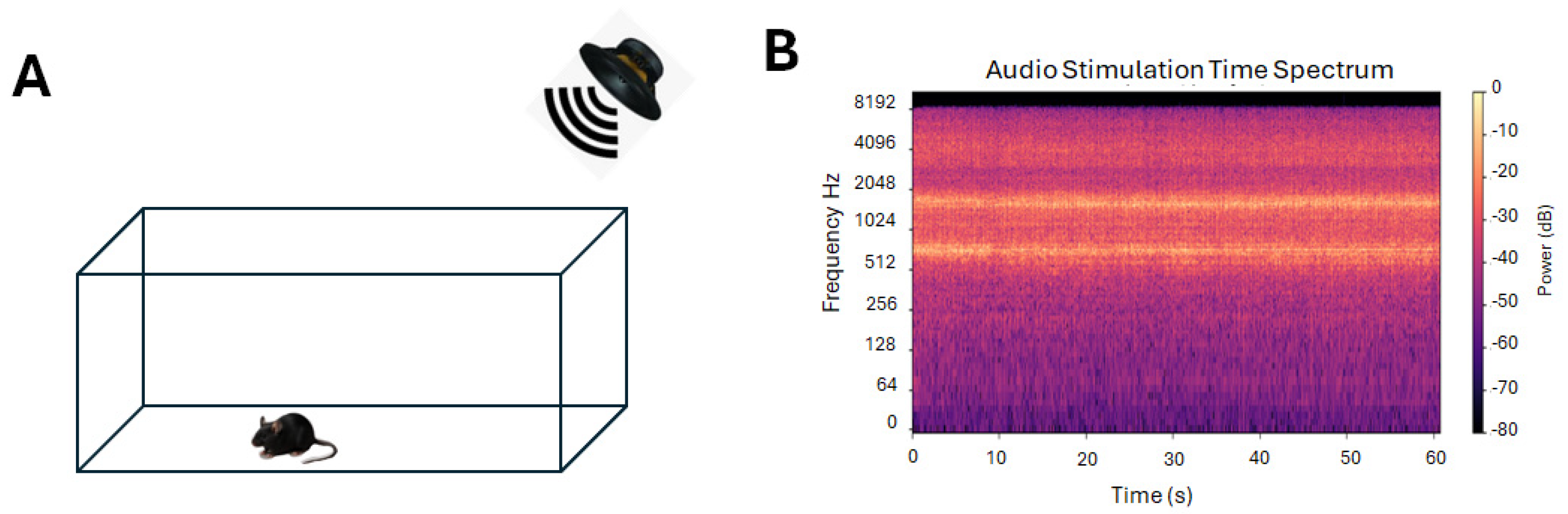
Littermate Fmr1-KO mice were separated into two groups. One group was subjected to auditory stimulation daily for 2 min 3 times a day at one-hour intervals for 5 days. The other group was not stimulated. Different age groups were subject to stimulation as follows: stage 1 (nursing: newborn to 21 days), stage 2 (juveniles: 22 days to 6 weeks), and stage 3 (adults: older than 6 weeks). The auditory stimulation consisted of multifrequency white noise stimulation at medium decibels (80 dB). Spectral analysis of the sound showed a Spectral Centroid (mean) of 2887.63 Hz, Spectral Rolloff (mean) of 5216.01 Hz, Spectral Bandwidth (mean) of 1980.82 Hz, and Dominant Frequency of 718.12 Hz. A Skullcandy Ounce speaker (Audio power 5 Watts, Skullcandy Inc., Park City, UT, USA) positioned 10 cm from the animal box was used to play the white noise (Figure 1). Spectral analysis of the white noise was done using Python 3.13 code (see Supplementary Material).
The mouse response to auditory stimulation was categorized as follows: no response (stage 0), wild running (stage 1) not considered as seizure (ref), tonic seizure (stage 2), clonic seizure (stage 3) and death (stage 4).

2.2. Behavioral Analysis

Social interaction test: A three-chamber paradigm separated by doors was used (Home made). A central empty chamber communicated through doors to two lateral chambers with a cage where a mouse can fit. After a habituation to the apparatus with no other animal for 10 min, the test mouse was put back in the home cage for 10 min before putting it back in the apparatus. During the first session, a mouse was placed in one lateral chamber and an object in the other one. The test mouse freely explored and interacted with the other mouse or the object for 10 min and was then put back in its cage for 1 h. During the second session, the object was replaced by a new animal. The test mouse freely explored and interacted with the new and familiar mouse. The exploration time and the discrimination index were calculated and compared between groups.
Recognition memory was assessed using the novel object recognition (NOR) test. After 10 min of habituation in an open field (cube or cylinder, Home made) as reported by previous work in Fmr1-ko mice [51,52,53], the animal was put back in its cage for 10 min. Two similar objects (glass rectangular cuboid or cylinder form) were placed in opposite sides of the arena, and the mouse was allowed to freely explore the objects for 10 min and then put back in its cage. After 1 h, one of the objects was replaced by a new one and the mouse was put back in the arena and allowed to explore for 10 min. The position of the objects differed between animals. Experiments were videotaped for later analysis. The exploration time (head of the animal toward the object at less than one cm distance, sniffing the object) and the discrimination index were calculated and compared between groups.

2.3. Data and Statistical Analysis

All data are presented as mean ± sem and compared using a sample Student’s t-test, ANOVA, using JASP software 0.19.3 version (University of Amsterdam and others). The sample t-test was used to determine whether the discrimination index differed from zero, and the ANOVA was used to determine whether there were differences between groups.

3. Results

3.1. Effect of Auditory Stimulation

Fmr1-KO mice from each litter were randomly separated into two groups; one received auditory stimulation, and the other one did not. Fmr1-KO mice were subject to 5 days of auditory stimulation for 2 min three times a day and were monitored for audiogenic seizures (AGS). Auditory stimulation induced death after wild running and tonic–clonic seizure in 3 animals out of 21 at development stage 1, after wild running in 6 animals out 11 at development stage 2, and no effect was observed in 10 animals at development stage 3. Wild running is not considered seizure [54,55]. Only 3 animals out of 41 animals tested at nursing stage 1 developed seizure and died.

3.2. Auditory Stimulation Restores Social Discrimination in Fmr1-KO Mice

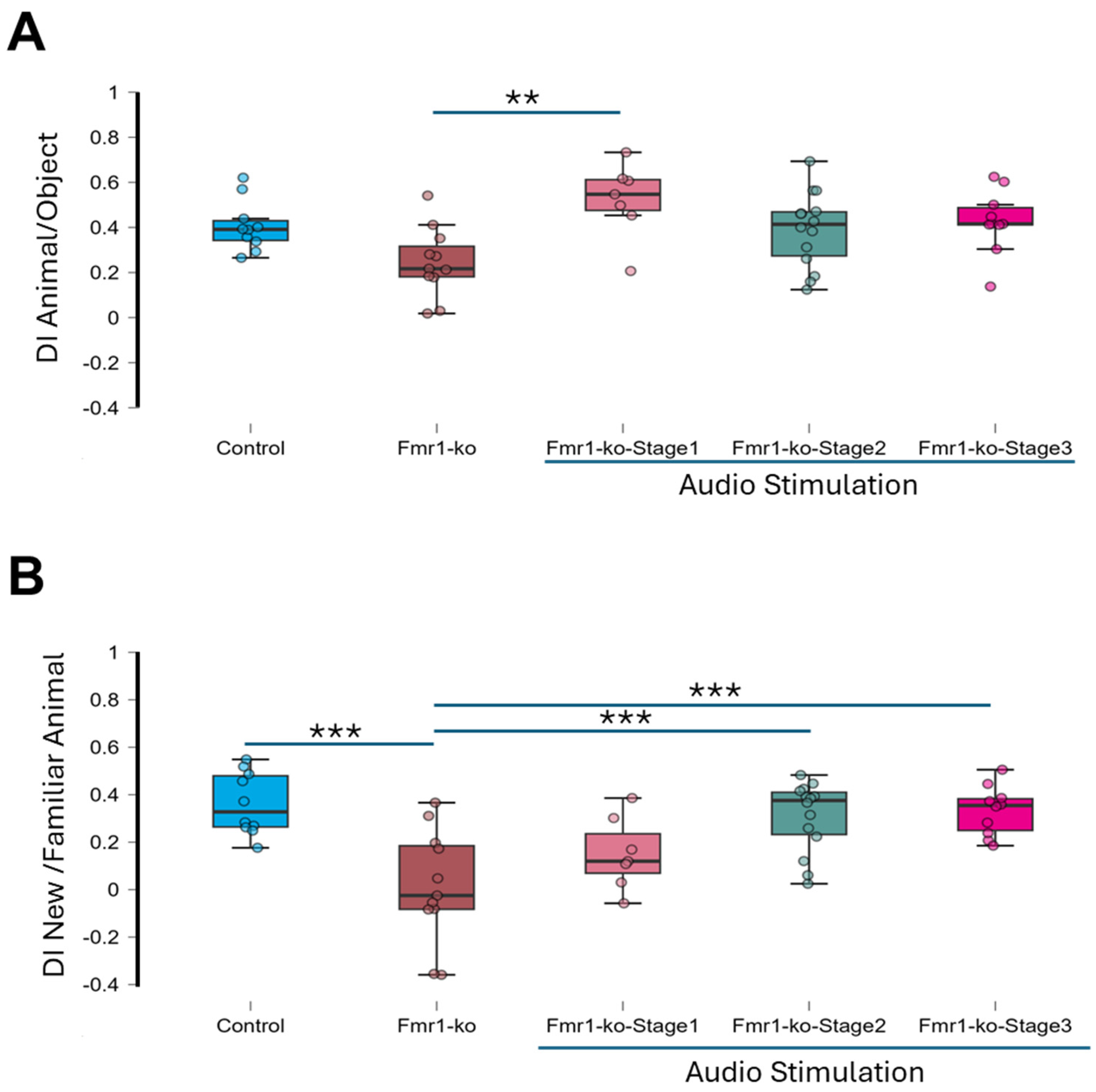
A three-chamber apparatus was used to assess social interaction. Animals were divided into six groups: control group (n = 10), unstimulated Fmr1-KO group (n = 11), and Fmr1-KO mice exposed to auditory stimulation at different developmental stages, namely Fmr1-KO-Stage1 group (n = 7), Fmr1-KO-Stage2 group (n = 14) and Fmr1-KO-Stage3 group (n = 10). A mouse was put in the apparatus for a 10 min habituation period and then returned to its home cage. After one hour, animals were reintroduced to the apparatus, now containing a novel mouse in one lateral chamber and an object in the other lateral chamber for 10 min (Figure 2A).
All groups explored the unfamiliar mouse more than the object, but to varying degrees, as follows: control (discrimination index (DI) = 0.41 ± 0.04, p < 0.001), Fmr1-KO (DI = 0.25 ± 0.05, p < 0.001), Fmr1-KO-Stage1 (DI = 0.52 ± 0.06, p < 0.001), Fmr1-KO-Stage2 (DI = 0.39 ± 0.04, p < 0.001) and Fmr1-KO-Stage3 (DI = 0.43 ± 0.04, p < 0.001). A one-way ANOVA revealed significant group differences (F = 4.14, p = 0.006), with post hoc tests showing that only Fmr1-KO-Stage1 mice performed significantly better than Fmr1-KO mice (p < 0.01, Table 1).
One hour later, to assess memory for social novelty, the object was replaced with an unfamiliar mouse (new animal). Regarding the control group (Control, DI = 0.36 ± 0.04, p < 0.001) and Fmr1-ko-Stim group, Fmr1-ko-Stage1 (DI = 0.15 ± 0.06, p < 0.05), Fmr1-ko-Stage2 (DI = 0.31 ± 0.04, p < 0.001) and Fmr1-ko-Stage3 (DI = 0.33 ± 0.03, p < 0.001) mice showed a strong preference for the new mouse while Fmr1-ko mice failed to discriminate between the two mice (DI = 0.01 ± 0.07; p = 0.87, Figure 2B). Group differences were significant (ANOVA F = 8.71, p < 0.001), with post hoc tests confirming impaired performance in Fmr1-ko mice compared to the control, Fmr1-KO-Stage-2 and Fmr1-ko-Stage3 mice but not the Fmr1-ko-Stage1 mice (Table 2). No sex differences were observed (ANOVA test, F = 0.006, p = 0.94). In the Fmr1-ko-Stage2 group, 6 mice out of 14 experienced wide running during auditory stimulation. To test whether wild running affects social interaction, we compared the discrimination index of Fmr1-ko-Stage2 mice that experienced wild running (wild running group, n = 6) and the one that did not (no response group, n = 8). The discrimination index was not different between the wild running group and no response group (animal/object: wild running DI of 0.44 ± 0.07, no-response DI of 0.35 ± 0.05, p = 0.27 t-test; new/familiar animal: wild running DI of 0.31 ± 0.06, no-response DI of 0.30 ± 0.06, p = 0.85 t-test).
These results indicate that auditory stimulation restores social discrimination in Fmr1-KO mice and that the effect persists for more than two weeks; behavioral testing was conducted in 2–3-month-old animals, whereas the auditory stimulation sessions occurred earlier in development, ruling out any stressful effect of the stimulation.

3.3. Auditory Stimulation Rescues Recognition Memory in Fmr1-KO Mice

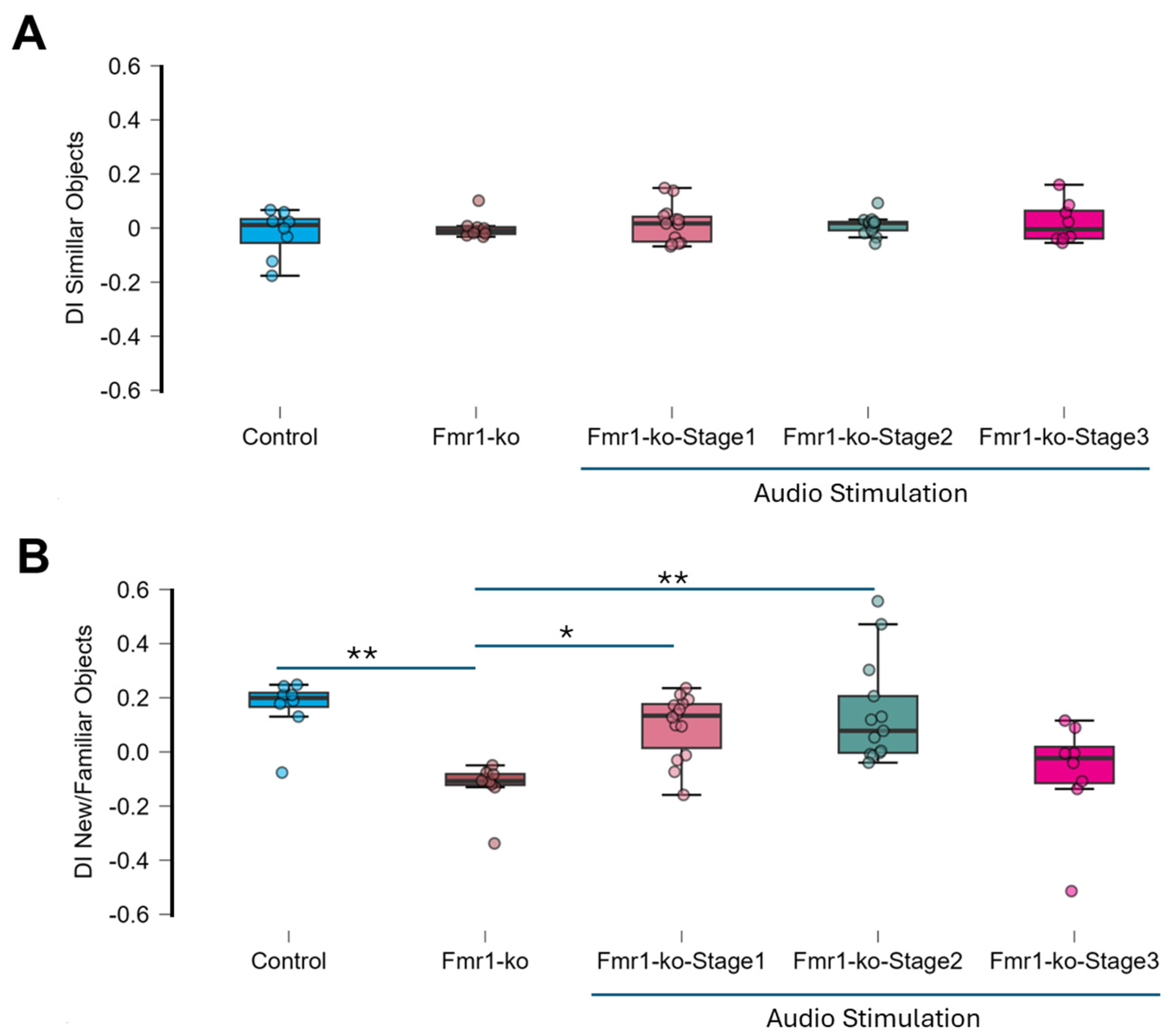
We used the novel object recognition (NOR) task in five groups: wild-type controls, Fmr1-KO mice without stimulation (Fmr1-KO), and Fmr1-KO mice receiving auditory stimulation at different development stages (Fmr1-KO-Stage1 group, Fmr1-KO-Stage2 group, and Fmr1-KO-Stage3 group). During the training phase, all groups explored the two identical objects without preference (sample t-test, p > 0.05, Figure 3A, Table 3). The ANOVA test revealed no significant difference in the discrimination index between groups (F = 0.54, p = 0.707). There is no sex difference (ANOVA test, F = 0.54, p = 0.707).
In the test phase, conducted one hour later, one of the objects was replaced by a novel object (Figure 2B). The control group (discrimination index 0.17 ± 0.04, p = 0.003) and Fmr1-KO-Stage1 (discrimination index 0.10 ± 0.03, p = 0.01) and Fmr1-KO-Stage2 (discrimination index 0.14 ± 0.05, p = 0.02) mice showed a significant preference for the novel object, indicating intact recognition memory. The Fmr1-KO-Stage3 mice (discrimination index −0.076 ± 0.07, p = 0.31) did not discriminate between the two objects, and the Fmr1-KO mice (discrimination index—0.13 ± 0.03, p = 0.005) explored the familiar object more than the new object. The ANOVA test revealed a significant difference between groups (F = 6.94, p < 0.001), with post hoc pairwise comparisons shown in Table 4. The control (p < 0.01), Fmr1-ko-Stage1 (p < 0.05) and Fmr1-ko-Stage2 (p < 0.01) mice are statistically different than the Fmr1-ko mice.
Overall, we show here that auditory stimulation in Fmr1-KO animals leads to long-lasting rescue of recognition memory when done at the nursing or juvenile development stage.

4. Discussion

Cognitive impairment in neurodevelopmental disorder profoundly affects memory, executive function and attention among other domains, reducing the quality of life of patients and their families and caregivers. Despite advances in experimental research, translation to clinical therapeutics has remained challenging. Sensory stimulation has emerged as a promising non-invasive therapeutic strategy for neurological disease [32,33,34,35,36,37,38]. The present work extends this therapeutic framework by demonstrating that auditory stimulation can rescue cognitive impairment in a mouse model of FXS.
Fmr1-KO mice recapitulate many core features of FXS and represent a well-validated preclinical model for testing therapeutic interventions [1]. Here, auditory stimulation delivered at distinct developmental stages effectively rescued social interaction and recognition memory. Notably, these benefits persisted for at least two weeks or more beyond the last stimulation session, as cognitive assessments were conducted at 2–3 months of age while stimulation was given at the early development stage. This enduring effect suggests that auditory stimulation may induce long-lasting neuroplastic changes rather than simply producing transient functional improvements. However, safety considerations must be carefully addressed. During the nursing stage, 3 out of 21 animals experienced tonic–clonic seizures triggered by auditory stimulation, raising important concerns about the use of high-intensity stimulation at the early development stage. Reducing stimulus intensity may mitigate the risk and warrants systematic investigation. Notably, adult Fmr1-ko mice showed reduced sensitivity to high-decibel stimulation (120 dB) and did not develop AGS [26], suggesting a developmental window during which auditory stimulation exerts its greatest—but also potentially most hazardous—effects. In juvenile animals, stimulation at 80 dB successfully rescued both social interaction and recognition memory, though six out of eleven animals experienced wild running during auditory stimulation. While wild running is not classified as seizure [54,55], its occurrence nonetheless supports the recommendation to use the lowest effective stimulus intensity.
The central mechanism underlying the therapeutic potential of auditory stimulation is likely neural entrainment—the synchronization of brain oscillations by auditory stimulation [35,56,57]. Disruptions in oscillatory activity, particularly in the gamma frequency band (30–80 Hz), have been documented in Fmr1-ko mice [58,59,60,61], Alzheimer’s disease [56,62], Schizophrenia [63,64] and autism spectrum disorder [65,66]. Auditory stimulation at 40 Hz (gamma band) has demonstrated efficacy in Alzheimer’s disease models by reducing pathological burden and improving cognition [38]. In Fmr1-ko mice, gamma electrical stimulation of the medial septum/diagonal band of Broca was effective in rescuing cognitive deficit and normalized basal synaptic transmission and short- and long-term plasticity [40]. Beyond oscillatory entrainment, auditory stimulation may also engage neuromodulatory systems like cholinergic and dopaminergic pathways, thereby enhancing attention, learning and motor performance [67,68].
The observation that wild running did not negatively impact cognitive outcomes—and was in fact associated with improvement—is intriguing, though the deaths of three animals from tonic–clonic seizures underscores that the therapeutic window must be defined more precisely. These findings collectively indicate that while auditory stimulation holds genuine therapeutic promise, further work is needed to establish stimulation parameters that reliably produce cognitive benefits without adverse effects. As such, the present study should be considered a proof-of-principle demonstration, and careful caution is warranted before any extrapolation to human populations.
The broader therapeutic relevance of auditory stimulation is supported by findings across multiple neurological conditions. In stroke rehabilitation, music-supported therapy promotes cortical reorganization, probably by reinforcing sensorimotor integration and experience-dependent plasticity [69]. In epilepsy, exposure to music has been associated with reduction in seizure frequency [70,71]. Similarly, in autism spectrum disorder, music exposure was beneficial in social communication and emotional processing [72]. Together, these findings suggest that auditory stimulation engages shared neural mechanisms that extend across diagnostic boundaries.

5. Conclusions

Auditory stimulation rescued cognitive impairment in mouse models of FXS, with benefits persisting well beyond the stimulation period when administered during the juvenile development stage. These results support the hypothesis that auditory stimulation can restore disrupted neural dynamics and improve functional outcomes in FXS. Elucidating the precise mechanisms underlying these effects, and optimizing stimulation parameters for safety and efficacy, represents a critical next step toward translating this approach into viable clinical intervention.

Supplementary Materials

The following supporting information can be downloaded at: https://www.mdpi.com/article/10.3390/brainsci16040380/s1, social interaction.csv; Python code audio analysis.txt; norsound.csv.

Author Contributions

Conceptualization: M.O. and R.C.S.; Methodology: M.O. and R.C.S.; Investigation: M.O.; Visualization: M.O., R.C.S., J.M.M. and A.E.H.; Funding Acquisition: R.C.S.; Project Administration: R.C.S.; Supervision: R.C.S.; Writing—Original Draft: M.O.; Writing—Review and Editing: R.C.S., J.M.M. and A.E.H. All authors have read and agreed to the published version of the manuscript.

Funding

This work received funding from the Swank Foundation, R21NS117112 (RS); start-up funds from the Nemours Foundation.

Institutional Review Board Statement

The animal study protocol was approved by the Nemours Foundation Institutional Animal Care and Use Committee in accordance with the National Institutes of Health Guide for the Care and Use of Laboratory Animals (protocol code RSP21-40780-002, approved on 15 December 2023.

Informed Consent Statement

Not applicable.

Data Availability Statement

The original contributions presented in this study are included in the article/Supplementary Materials. Further inquiries can be directed to the corresponding author.

Conflicts of Interest

The authors declare no conflicts of interest.

Abbreviations

AGSAudiogenic seizures
ADHDAttention-deficit/hyperactivity disorder
FXSFragile X Syndrome
Fmr1-KOFragile X Messenger Ribonucleoprotein 1 Knockout

References

  1. Hagerman, R.J.; Berry-Kravis, E.; Hazlett, H.C.; Bailey, D.B., Jr.; Moine, H.; Kooy, R.F.; Tassone, F.; Gantois, I.; Sonenberg, N.; Mandel, J.L.; et al. Fragile X syndrome. Nat. Rev. Dis. Primers 2017, 3, 17065. [Google Scholar] [CrossRef] [Scilit]
  2. Verkerk, A.J.; Pieretti, M.; Sutcliffe, J.S.; Fu, Y.H.; Kuhl, D.P.; Pizzuti, A.; Reiner, O.; Richards, S.; Victoria, M.F.; Zhang, F.P.; et al. Identification of a gene (FMR-1) containing a CGG repeat coincident with a breakpoint cluster region exhibiting length variation in fragile X syndrome. Cell 1991, 65, 905–914. [Google Scholar] [CrossRef] [Scilit] [PubMed]
  3. Pieretti, M.; Zhang, F.P.; Fu, Y.H.; Warren, S.T.; Oostra, B.A.; Caskey, C.T.; Nelson, D.L. Absence of expression of the FMR-1 gene in fragile X syndrome. Cell 1991, 66, 817–822. [Google Scholar] [CrossRef] [Scilit] [PubMed]
  4. Darnell, J.C.; Van Driesche, S.J.; Zhang, C.; Hung, K.Y.; Mele, A.; Fraser, C.E.; Stone, E.F.; Chen, C.; Fak, J.J.; Chi, S.W.; et al. FMRP stalls ribosomal translocation on mRNAs linked to synaptic function and autism. Cell 2011, 146, 247–261. [Google Scholar] [CrossRef] [Scilit]
  5. Ascano, M., Jr.; Mukherjee, N.; Bandaru, P.; Miller, J.B.; Nusbaum, J.D.; Corcoran, D.L.; Langlois, C.; Munschauer, M.; Dewell, S.; Hafner, M.; et al. FMRP targets distinct mRNA sequence elements to regulate protein expression. Nature 2012, 492, 382–386. [Google Scholar] [CrossRef] [Scilit] [PubMed]
  6. Brown, M.R.; Kronengold, J.; Gazula, V.R.; Chen, Y.; Strumbos, J.G.; Sigworth, F.J.; Navaratnam, D.; Kaczmarek, L.K. Fragile X mental retardation protein controls gating of the sodium-activated potassium channel Slack. Nat. Neurosci. 2010, 13, 819–821. [Google Scholar] [CrossRef] [Scilit]
  7. Bassell, G.J.; Warren, S.T. Fragile X syndrome: Loss of local mRNA regulation alters synaptic development and function. Neuron 2008, 60, 201–214. [Google Scholar] [CrossRef] [Scilit]
  8. Slegtenhorst-Eegdeman, K.E.; de Rooij, D.G.; Verhoef-Post, M.; van de Kant, H.J.; Bakker, C.E.; Oostra, B.A.; Grootegoed, J.A.; Themmen, A.P. Macroorchidism in FMR1 knockout mice is caused by increased Sertoli cell proliferation during testicular development. Endocrinology 1998, 139, 156–162. [Google Scholar] [CrossRef]
  9. D’Hooge, R.; Nagels, G.; Franck, F.; Bakker, C.E.; Reyniers, E.; Storm, K.; Kooy, R.F.; Oostra, B.A.; Willems, P.J.; De Deyn, P.P. Mildly impaired water maze performance in male Fmr1 knockout mice. Neuroscience 1997, 76, 367–376. [Google Scholar] [CrossRef] [Scilit]
  10. Dobkin, C.; Radu, G.; Ding, X.H.; Brown, W.T.; Nolin, S.L. Fragile X prenatal analyses show full mutation females at high risk for mosaic Turner syndrome: Fragile X leads to chromosome loss. Am. J. Med. Genet. A 2009, 149A, 2152–2157. [Google Scholar] [CrossRef] [Scilit]
  11. Ventura, S.; Duncan, S.; Ainge, J.A. Increased flexibility of CA3 memory representations following environmental enrichment. Curr. Biol. 2024, 34, 2011–2019.e7. [Google Scholar] [CrossRef] [Scilit]
  12. Spencer, C.M.; Alekseyenko, O.; Serysheva, E.; Yuva-Paylor, L.A.; Paylor, R. Altered anxiety-related and social behaviors in the Fmr1 knockout mouse model of fragile X syndrome. Genes Brain Behav. 2005, 4, 420–430. [Google Scholar] [CrossRef] [Scilit]
  13. Mines, M.A.; Yuskaitis, C.J.; King, M.K.; Beurel, E.; Jope, R.S. GSK3 influences social preference and anxiety-related behaviors during social interaction in a mouse model of fragile X syndrome and autism. PLoS ONE 2010, 5, e9706. [Google Scholar] [CrossRef] [Scilit]
  14. Thomas, A.M.; Bui, N.; Graham, D.; Perkins, J.R.; Yuva-Paylor, L.A.; Paylor, R. Genetic reduction of group 1 metabotropic glutamate receptors alters select behaviors in a mouse model for fragile X syndrome. Behav. Brain Res. 2011, 223, 310–321. [Google Scholar] [CrossRef] [Scilit]
  15. Kazdoba, T.M.; Leach, P.T.; Silverman, J.L.; Crawley, J.N. Modeling fragile X syndrome in the Fmr1 knockout mouse. Intractable Rare Dis. Res. 2014, 3, 118–133. [Google Scholar] [CrossRef] [Scilit]
  16. Qin, M.; Xia, Z.; Huang, T.; Smith, C.B. Effects of chronic immobilization stress on anxiety-like behavior and basolateral amygdala morphology in Fmr1 knockout mice. Neuroscience 2011, 194, 282–290. [Google Scholar] [CrossRef] [Scilit] [PubMed]
  17. Kim, L.; He, L.; Maaswinkel, H.; Zhu, L.; Sirotkin, H.; Weng, W. Anxiety, hyperactivity and stereotypy in a zebrafish model of fragile X syndrome and autism spectrum disorder. Prog. Neuro-Psychopharmacol. Biol. Psychiatry 2014, 55, 40–49. [Google Scholar] [CrossRef] [Scilit]
  18. Gauducheau, M.; Lemaire-Mayo, V.; D’Amato, F.R.; Oddi, D.; Crusio, W.E.; Pietropaolo, S. Age-specific autistic-like behaviors in heterozygous Fmr1-KO female mice. Autism Res. 2017, 10, 1067–1078. [Google Scholar] [CrossRef] [Scilit] [PubMed]
  19. Pietropaolo, S.; Guilleminot, A.; Martin, B.; D’Amato, F.R.; Crusio, W.E. Genetic-background modulation of core and variable autistic-like symptoms in Fmr1 knock-out mice. PLoS ONE 2011, 6, e17073. [Google Scholar] [CrossRef] [Scilit] [PubMed]
  20. Jawaid, S.; Kidd, G.J.; Wang, J.; Swetlik, C.; Dutta, R.; Trapp, B.D. Alterations in CA1 hippocampal synapses in a mouse model of fragile X syndrome. Glia 2018, 66, 789–800. [Google Scholar] [CrossRef] [Scilit]
  21. Testa-Silva, G.; Loebel, A.; Giugliano, M.; de Kock, C.P.; Mansvelder, H.D.; Meredith, R.M. Hyperconnectivity and slow synapses during early development of medial prefrontal cortex in a mouse model for mental retardation and autism. Cereb. Cortex 2012, 22, 1333–1342. [Google Scholar] [CrossRef] [Scilit]
  22. Comery, T.A.; Harris, J.B.; Willems, P.J.; Oostra, B.A.; Irwin, S.A.; Weiler, I.J.; Greenough, W.T. Abnormal dendritic spines in fragile X knockout mice: Maturation and pruning deficits. Proc. Natl. Acad. Sci. USA 1997, 94, 5401–5404. [Google Scholar] [CrossRef] [Scilit]
  23. McKinney, B.C.; Grossman, A.W.; Elisseou, N.M.; Greenough, W.T. Dendritic spine abnormalities in the occipital cortex of C57BL/6 Fmr1 knockout mice. Am. J. Med. Genet. Part B Neuropsychiatr. Genet. 2005, 136B, 98–102. [Google Scholar] [CrossRef] [Scilit]
  24. Cruz-Martin, A.; Crespo, M.; Portera-Cailliau, C. Delayed stabilization of dendritic spines in fragile X mice. J. Neurosci. 2010, 30, 7793–7803. [Google Scholar] [CrossRef] [Scilit]
  25. Pan, F.; Aldridge, G.M.; Greenough, W.T.; Gan, W.B. Dendritic spine instability and insensitivity to modulation by sensory experience in a mouse model of fragile X syndrome. Proc. Natl. Acad. Sci. USA 2010, 107, 17768–17773. [Google Scholar] [CrossRef] [Scilit]
  26. Musumeci, S.A.; Calabrese, G.; Bonaccorso, C.M.; D’Antoni, S.; Brouwer, J.R.; Bakker, C.E.; Elia, M.; Ferri, R.; Nelson, D.L.; Oostra, B.A.; et al. Audiogenic seizure susceptibility is reduced in fragile X knockout mice after introduction of Fmr1 transgenes. Exp. Neurol. 2007, 203, 233–240. [Google Scholar] [CrossRef] [Scilit]
  27. Rotschafer, S.; Razak, K. Altered auditory processing in a mouse model of fragile X syndrome. Brain Res. 2013, 1506, 12–24. [Google Scholar] [CrossRef] [Scilit] [PubMed]
  28. Ding, Q.; Sethna, F.; Wang, H. Behavioral analysis of male and female Fmr1 knockout mice on C57BL/6 background. Behav. Brain Res. 2014, 271, 72–78. [Google Scholar] [CrossRef] [Scilit]
  29. Protic, D.D.; Aishworiya, R.; Salcedo-Arellano, M.J.; Tang, S.J.; Milisavljevic, J.; Mitrovic, F.; Hagerman, R.J.; Budimirovic, D.B. Fragile X Syndrome: From Molecular Aspect to Clinical Treatment. Int. J. Mol. Sci. 2022, 23, 1935. [Google Scholar] [CrossRef] [Scilit] [PubMed]
  30. Protic, D.; Hagerman, R. State-of-the-art therapies for fragile X syndrome. Dev. Med. Child Neurol. 2024, 66, 863–871. [Google Scholar] [CrossRef] [Scilit] [PubMed]
  31. Romagnoli, A.; Di Marino, D. The Use of Peptides in the Treatment of Fragile X Syndrome: Challenges and Opportunities. Front. Psychiatry 2021, 12, 754485. [Google Scholar] [CrossRef] [Scilit]
  32. Zhang, X.; Lv, R.; Sun, Y.; Liu, T.C. The safety and effectiveness of 40 Hz gamma-tACS in Alzheimer’s disease: A meta-analysis. J. Alzheimer’s Dis. 2024, 102, 295–307. [Google Scholar] [CrossRef] [Scilit]
  33. Yang, Y.H.; Chang, H.W.; Chien, C.F.; Hour, T.C. Investigating the effects of 40 Hz sound stimulation on Alzheimer’s disease pathways: Modulation of amyloid-beta42 secretion, tau phosphorylation, phagocytosis, and autophagy. J. Alzheimer’s Dis. 2025, 106, 184–196. [Google Scholar] [CrossRef] [Scilit]
  34. Yang, Y.; Ondrejcak, T.; Hu, N.W.; Islam, S.; O’Rourke, E.; Reilly, R.B.; Cunningham, C.; Rowan, M.J.; Klyubin, I. Gamma-patterned sensory stimulation reverses synaptic plasticity deficits in rat models of early Alzheimer’s disease. Eur. J. Neurosci. 2023, 58, 3402–3411. [Google Scholar] [CrossRef] [Scilit]
  35. Chan, D.; de Weck, G.; Jackson, B.L.; Suk, H.J.; Milman, N.P.; Kitchener, E.; Avalos, V.S.F.; Quay, M.J.; Aoki, K.; Ruiz, E.; et al. Gamma sensory stimulation in mild Alzheimer’s dementia: An open-label extension study. Alzheimer’s Dement. 2025, 21, e70792. [Google Scholar] [CrossRef] [Scilit] [PubMed]
  36. Cimenser, A.; Hempel, E.; Travers, T.; Strozewski, N.; Martin, K.; Malchano, Z.; Hajos, M. Sensory-Evoked 40-Hz Gamma Oscillation Improves Sleep and Daily Living Activities in Alzheimer’s Disease Patients. Front. Syst. Neurosci. 2021, 15, 746859. [Google Scholar] [CrossRef] [Scilit]
  37. Iaccarino, H.F.; Singer, A.C.; Martorell, A.J.; Rudenko, A.; Gao, F.; Gillingham, T.Z.; Mathys, H.; Seo, J.; Kritskiy, O.; Abdurrob, F.; et al. Gamma frequency entrainment attenuates amyloid load and modifies microglia. Nature 2016, 540, 230–235. [Google Scholar] [CrossRef] [Scilit] [PubMed]
  38. Martorell, A.J.; Paulson, A.L.; Suk, H.J.; Abdurrob, F.; Drummond, G.T.; Guan, W.; Young, J.Z.; Kim, D.N.; Kritskiy, O.; Barker, S.J.; et al. Multi-sensory Gamma Stimulation Ameliorates Alzheimer’s-Associated Pathology and Improves Cognition. Cell 2019, 177, 256–271.e22. [Google Scholar] [CrossRef] [Scilit] [PubMed]
  39. Soderlund, G.B.W.; Hadjikhani, N.; Thorsson, M.; E-Said, S.; Claesdotter-Knutsson, E.; Gustafsson, P.; Johnels, J.A. Sensory white noise in clinical ADHD: Who benefits from noise, and who performs worse? Scand. J. Child Adolesc. Psychiatry Psychol. 2024, 12, 92–99. [Google Scholar] [CrossRef] [Scilit]
  40. Ouardouz, M.; Hernan, A.; Mahoney, M.; Scott, R. Stimulation of the Medial Septum Diagonal Band of Broca Rescues Learning and Memory Deficits in Fmr1 KO Mice. iScience 2026, 29, 115066. [Google Scholar] [CrossRef] [Scilit]
  41. Sinclair, D.; Featherstone, R.; Naschek, M.; Nam, J.; Du, A.; Wright, S.; Pance, K.; Melnychenko, O.; Weger, R.; Akuzawa, S.; et al. GABA-B Agonist Baclofen Normalizes Auditory-Evoked Neural Oscillations and Behavioral Deficits in the Fmr1 Knockout Mouse Model of Fragile X Syndrome. Eneuro 2017, 4, 1–13. [Google Scholar] [CrossRef] [Scilit]
  42. Contractor, A.; Klyachko, V.A.; Portera-Cailliau, C. Altered Neuronal and Circuit Excitability in Fragile X Syndrome. Neuron 2015, 87, 699–715. [Google Scholar] [CrossRef] [Scilit]
  43. Chen, L.; Toth, M. Fragile X mice develop sensory hyperreactivity to auditory stimuli. Neuroscience 2001, 103, 1043–1050. [Google Scholar] [CrossRef] [Scilit]
  44. Berry-Kravis, E.; Raspa, M.; Loggin-Hester, L.; Bishop, E.; Holiday, D.; Bailey, D.B. Seizures in fragile X syndrome: Characteristics and comorbid diagnoses. Am. J. Intellect. Dev. Disabil. 2010, 115, 461–472. [Google Scholar] [CrossRef] [Scilit]
  45. Qiu, L.F.; Hao, Y.H.; Li, Q.Z.; Xiong, Z.Q. Fragile X syndrome and epilepsy. Neurosci. Bull. 2008, 24, 338–344. [Google Scholar] [CrossRef] [Scilit]
  46. Hodges, S.L.; Reynolds, C.D.; Nolan, S.O.; Huebschman, J.L.; Okoh, J.T.; Binder, M.S.; Lugo, J.N. A single early-life seizure results in long-term behavioral changes in the adult Fmr1 knockout mouse. Epilepsy Res. 2019, 157, 106193. [Google Scholar] [CrossRef] [Scilit] [PubMed]
  47. Blandin, K.J.; Narvaiz, D.A.; Sullens, D.G.; Womble, P.D.; Hodges, S.L.; Binder, M.S.; Faust, A.; Nguyen, P.H.; Pranske, Z.J.; Lugo, J.N. A Two-Hit Approach Inducing Flurothyl Seizures in Fmr1 Knockout Mice Impacts Anxiety and Repetitive Behaviors. Brain Sci. 2024, 14, 892. [Google Scholar] [CrossRef] [Scilit] [PubMed]
  48. Smith, G.; Ahmed, N.; Arbuckle, E.; Lugo, J.N. Early-life status epilepticus induces long-term deficits in anxiety and spatial learning in mice. Int. J. Epilepsy 2017, 4, 36–45. [Google Scholar] [CrossRef] [Scilit]
  49. Lugo, J.N.; Swann, J.W.; Anderson, A.E. Early-life seizures result in deficits in social behavior and learning. Exp. Neurol. 2014, 256, 74–80. [Google Scholar] [CrossRef] [Scilit]
  50. Nguyen, P.H.; Narvaiz, D.A.; Womble, P.D.; Sullens, D.G.; Binder, M.S.; Hodges, S.L.; Kwok, E.; Lugo, J.N. Multiple Early-Life Seizures Alters Neonatal Communicative Behavior in Fmr1 Knockout Mice. Dev. Neurosci. 2022, 44, 478–486. [Google Scholar] [CrossRef] [Scilit] [PubMed]
  51. Borreca, A.; De Luca, M.; Ferrante, A.; Boussadia, Z.; Pignataro, A.; Martire, A.; Ammassari-Teule, M. Fmr1-KO mice failure to detect object novelty associates with a post-test decrease of structural and synaptic plasticity upstream of the hippocampus. Sci. Rep. 2023, 13, 755. [Google Scholar] [CrossRef] [Scilit] [PubMed]
  52. Costa, L.; Sardone, L.M.; Bonaccorso, C.M.; D’Antoni, S.; Spatuzza, M.; Gulisano, W.; Tropea, M.R.; Puzzo, D.; Leopoldo, M.; Lacivita, E.; et al. Activation of Serotonin 5-HT(7) Receptors Modulates Hippocampal Synaptic Plasticity by Stimulation of Adenylate Cyclases and Rescues Learning and Behavior in a Mouse Model of Fragile X Syndrome. Front. Mol. Neurosci. 2018, 11, 353. [Google Scholar] [CrossRef] [Scilit] [PubMed]
  53. Rosenheck, M.; Sheeler, C.; Sare, R.M.; Gurney, M.E.; Smith, C.B. Effects of chronic inhibition of phosphodiesterase-4D on behavior and regional rates of cerebral protein synthesis in a mouse model of fragile X syndrome. Neurobiol. Dis. 2021, 159, 105485. [Google Scholar] [CrossRef] [Scilit]
  54. Ross, K.C.; Coleman, J.R. Developmental and genetic audiogenic seizure models: Behavior and biological substrates. Neurosci. Biobehav. Rev. 2000, 24, 639–653. [Google Scholar] [CrossRef] [Scilit]
  55. Wenker, I.C.; Boscia, A.R.; Lewis, C.; Tariq, A.; Miralles, R.; Hanflink, J.C.; Saraf, P.; Patel, M.K. Forebrain epileptiform activity is not required for seizure-induced apnea in a mouse model of Scn8a epilepsy. Front. Neural Circuits 2022, 16, 1002013. [Google Scholar] [CrossRef] [Scilit]
  56. Traikapi, A.; Konstantinou, N. Gamma Oscillations in Alzheimer’s Disease and Their Potential Therapeutic Role. Front. Syst. Neurosci. 2021, 15, 782399. [Google Scholar] [CrossRef] [Scilit]
  57. Wu, L.; Wei, Y.; He, K.; Gao, Q. The Effects and Safety of Gamma Rhythm Stimulation on Cognitive Function in Alzheimer’s Disease: A Systematic Review and Meta-Analysis. Neurorehabilit. Neural Repair 2025, 39, 1046–1059. [Google Scholar] [CrossRef] [Scilit]
  58. Radwan, B.; Dvorak, D.; Fenton, A.A. Impaired cognitive discrimination and discoordination of coupled theta-gamma oscillations in Fmr1 knockout mice. Neurobiol. Dis. 2016, 88, 125–138. [Google Scholar] [CrossRef] [Scilit] [PubMed]
  59. Talbot, Z.N.; Sparks, F.T.; Dvorak, D.; Curran, B.M.; Alarcon, J.M.; Fenton, A.A. Normal CA1 Place Fields but Discoordinated Network Discharge in a Fmr1-Null Mouse Model of Fragile X Syndrome. Neuron 2018, 97, 684–697.e4. [Google Scholar] [CrossRef] [Scilit]
  60. Kozono, N.; Okamura, A.; Honda, S.; Matsumoto, M.; Mihara, T. Gamma power abnormalities in a Fmr1-targeted transgenic rat model of fragile X syndrome. Sci. Rep. 2020, 10, 18799. [Google Scholar] [CrossRef] [Scilit]
  61. Arbab, T.; Battaglia, F.P.; Pennartz, C.M.A.; Bosman, C.A. Abnormal hippocampal theta and gamma hypersynchrony produces network and spike timing disturbances in the Fmr1-KO mouse model of Fragile X syndrome. Neurobiol. Dis. 2018, 114, 65–73. [Google Scholar] [CrossRef] [Scilit]
  62. Traikapi, A.; Kalli, I.; Kyriakou, A.; Stylianou, E.; Symeou, R.T.; Kardama, A.; Christou, Y.P.; Phylactou, P.; Konstantinou, N. Episodic memory effects of gamma frequency precuneus transcranial magnetic stimulation in Alzheimer’s disease: A randomized multiple baseline study. J. Neuropsychol. 2023, 17, 279–301. [Google Scholar] [CrossRef] [Scilit] [PubMed]
  63. Leung, L.S.; Ma, J. Medial septum modulates hippocampal gamma activity and prepulse inhibition in an N-methyl-d-aspartate receptor antagonist model of schizophrenia. Schizophr. Res. 2018, 198, 36–44. [Google Scholar] [CrossRef] [Scilit]
  64. Amann, L.C.; Gandal, M.J.; Halene, T.B.; Ehrlichman, R.S.; White, S.L.; McCarren, H.S.; Siegel, S.J. Mouse behavioral endophenotypes for schizophrenia. Brain Res. Bull. 2010, 83, 147–161. [Google Scholar] [CrossRef] [Scilit]
  65. Kessler, K.; Seymour, R.A.; Rippon, G. Brain oscillations and connectivity in autism spectrum disorders (ASD): New approaches to methodology, measurement and modelling. Neurosci. Biobehav. Rev. 2016, 71, 601–620. [Google Scholar] [CrossRef] [Scilit]
  66. David, N.; Schneider, T.R.; Peiker, I.; Al-Jawahiri, R.; Engel, A.K.; Milne, E. Variability of cortical oscillation patterns: A possible endophenotype in autism spectrum disorders? Neurosci. Biobehav. Rev. 2016, 71, 590–600. [Google Scholar] [CrossRef] [Scilit]
  67. Hasselmo, M.E. The role of acetylcholine in learning and memory. Curr. Opin. Neurobiol. 2006, 16, 710–715. [Google Scholar] [CrossRef] [Scilit]
  68. Salimpoor, V.N.; Benovoy, M.; Larcher, K.; Dagher, A.; Zatorre, R.J. Anatomically distinct dopamine release during anticipation and experience of peak emotion to music. Nat. Neurosci. 2011, 14, 257–262. [Google Scholar] [CrossRef] [Scilit] [PubMed]
  69. Sarkamo, T.; Tervaniemi, M.; Laitinen, S.; Forsblom, A.; Soinila, S.; Mikkonen, M.; Autti, T.; Silvennoinen, H.M.; Erkkila, J.; Laine, M.; et al. Music listening enhances cognitive recovery and mood after middle cerebral artery stroke. Brain 2008, 131, 866–876. [Google Scholar] [CrossRef] [Scilit] [PubMed]
  70. Hughes, J.R. The Mozart effect. J. R. Soc. Med. 2001, 94, 316. [Google Scholar] [CrossRef] [Scilit]
  71. Sesso, G.; Sicca, F. Safe and sound: Meta-analyzing the Mozart effect on epilepsy. Clin. Neurophysiol. 2020, 131, 1610–1620. [Google Scholar] [CrossRef] [Scilit] [PubMed]
  72. Sharda, M.; Tuerk, C.; Chowdhury, R.; Jamey, K.; Foster, N.; Custo-Blanch, M.; Tan, M.; Nadig, A.; Hyde, K. Music improves social communication and auditory-motor connectivity in children with autism. Transl. Psychiatry 2018, 8, 231. [Google Scholar] [CrossRef] [Scilit] [PubMed]
Figure 1. (A) Schematic representation of the apparatus for auditory stimulation. (B) Time spectrum of the auditory stimulation.
Brainsci 16 00380 g001
Figure 2. Auditory stimulation effect on social interaction. (A) Animal/object: All animals explore the animal more than the object (sample t-test p < 0.001). The ANOVA test showed a significant difference in the discrimination index between the 5 groups (F = 4.14, p < 0.006). Post hoc analysis showed a significant difference only between Fmr1-ko and Fmr1-ko-Stage1 groups (p < 001). (B): Novel/familiar animal: Only Fmr1-ko mice did not discriminate between the novel and familiar animal object (DI = 0.01 ± 0.07, p = 0.868), while all other groups did, including the control (DI = 0.41 ± 0.06, p < 0.001), Fmr1-ko-Stage1 (DI = 0.51 ± 0.06, p < 0.001), Fmr1-ko-Stage2 (DI = 0.31 ± 0.04, p < 0.001) and Fmr1-ko-Stage3 groups (DI = 0.33 ± 0.03, p < 0.001). ANOVA test revealed significant group differences (F = 8.71, p < 0.001). Post hoc analysis: The control, Fmr1-ko-Stage2 and Fmr1-ko-Stage3 mice are statistically significantly different than the Fmr1-ko mice (p < 0.001). ** p < 0.01 and *** p < 0.001; ANOVA post hoc holm test.
Brainsci 16 00380 g002
Figure 3. Auditory stimulation effect on recognition memory. (A) Similar object: All animals explore the two identical objects without preference (sample t-test p > 0.05) and there is no significant difference in the discrimination index between the 5 groups (F = 0.54, p = 0.707). (B) Novel/familiar object: The Fmr1-ko mice explored the familiar object more than the novel object (DI = 0.13 ± 0.03, p < 0.01). Fmr1-ko-Stege3 mice did not discriminate between the novel and familiar objects (DI = −0.08 ± 0.07, p = 0.314), while all other groups explored the new object more than the familiar object (control DI = 0.17 ± 0.04, p < 0.001; Fmr1-KO-Stage1 DI = 0.10 ± 0.06, p = 0.01; and Fmr1-KO-Stage2 DI 0.14 ± 0.05, p = 0.02). The ANOVA test shows a significant difference between groups (F = 6.94, p < 0.001). Post hoc pairwise comparisons revealed that the control (p < 0.01), Fmr1-ko-Stage1 (p < 0.05) and Fmr1-ko-Stage2 (p < 0.01) mice are statistically different than the Fmr1-ko mice. * p < 0.05, ** p < 0.01; ANOVA post hoc holm test.
Brainsci 16 00380 g003
Table 1. Post Hoc Comparisons—D.I. Animal/Object.
Mean DifferenceSEMdftpholm
Control(Fmr1-ko)0.1610.065472.4680.138
(Fmr1-ko-Stage1)−0.1170.07447−1.5810.603
(Fmr1-ko-Stage2)0.0160.062470.2611.000
(Fmr1-ko-Stage3)−0.0210.06747−0.3081.000
(Fmr1-ko)(Fmr1-ko-Stage1)−0.2780.07247−3.8420.004
(Fmr1-ko-Stage2)−0.1450.06047−2.4080.140
(Fmr1-ko-Stage3)−0.1820.06547−2.7840.069
(Fmr1-ko-Stage1)(Fmr1-ko-Stage2)0.1330.069471.9160.368
(Fmr1-ko-Stage3)0.0960.074471.3010.799
(Fmr1-ko-Stage2)(Fmr1-ko-Stage3)−0.0370.06247−0.5941.000
Note. p-value adjusted for comparing a family of 10 estimates. df: degree of freedom.
Table 2. Post Hoc Comparisons—New/Familiar Animal.
Mean DifferenceSEMdftpholm
Control(Fmr1-ko)0.3500.071474.915<0.001
(Fmr1-ko-Stage1)0.2110.080472.6310.080
(Fmr1-ko-Stage2)0.0550.067470.8081.000
(Fmr1-ko-Stage3)0.0290.073470.4001.000
(Fmr1-ko)(Fmr1-ko-Stage1)−0.1390.07947−1.7600.340
(Fmr1-ko-Stage2)−0.2950.06647−4.500<0.001
(Fmr1-ko-Stage3)−0.3210.07147−4.506<0.001
(Fmr1-ko-Stage1)(Fmr1-ko-Stage2)−0.1570.07547−2.0780.216
(Fmr1-ko-Stage3)−0.1820.08047−2.2680.168
(Fmr1-ko-Stage2)(Fmr1-ko-Stage3)−0.0250.06747−0.3761.000
Note. p-value adjusted for comparing a family of 10 estimates. df: degree of freedom.
Table 3. One Sample t-Test—D.I. for Similar objects and D.I. New Object/Familiar Object.
tdfp
Control Similar Objects−0.64370.541
Fmr1-ko Similar Objects0.10070.923
Fmr1-ko-stage1 Similar Objects0.864130.403
Fmr1-ko-stage2 Similar Objects1.045120.316
Fmr1-ko-stage3 Similar Objects0.74570.481
C New Object/Familiar Object4.48570.003
Fmr1-ko New Object/Familiar Object−4.01370.005
Fmr1-ko-stage1 New Object/Familiar Object3.006130.010
Fmr1-ko-stage2 New Object/Familiar Object2.679120.020
Fmr1-ko-stage3 New Object/Familiar Object−1.08470.314
Note. For the Student’s t-test, the alternative hypothesis specifies that the mean is different from 0. df: degree of freedom.
Table 4. Post Hoc Comparisons—D.I. New Object/Familiar Object.
Mean DifferenceSEMdftpholm
Control(Fmr1-ko)0.2930.075463.9160.003
(Fmr1-ko-Stage1)0.0710.066461.0741.000
(Fmr1-ko-Stage2)0.0240.067460.3531.000
(Fmr1-ko-Stage3)0.2420.075463.2340.015
(Fmr1-ko)(Fmr1-ko-Stage1)−0.2220.06646−3.3440.013
(Fmr1-ko-Stage2)−0.2690.06746−4.0050.002
(Fmr1-ko-Stage3)−0.0510.07546−0.6821.000
(Fmr1-ko-Stage1)(Fmr1-ko-Stage2)−0.0480.05846−0.8251.000
(Fmr1-ko-Stage3)0.1710.066462.5740.067
(Fmr1-ko-Stage2)(Fmr1-ko-Stage3)0.2180.067463.2460.015
Note. p-value adjusted for comparing a family of 10 estimates. df: degree of freedom.
Disclaimer/Publisher’s Note: The statements, opinions and data contained in all publications are solely those of the individual author(s) and contributor(s) and not of MDPI and/or the editor(s). MDPI and/or the editor(s) disclaim responsibility for any injury to people or property resulting from any ideas, methods, instructions or products referred to in the content.