Article

A Cross-Sectional Study: Structural and Related Functional Connectivity Changes in the Brain: Stigmata of Adverse Parenting in Patients with Major Depressive Disorder?

1
Department of Psychiatry, Renmin Hospital of Wuhan University, Wuhan 430060, China
2
PET/CT/MRI and Molecular Imaging Center, Renmin Hospital of Wuhan University, Wuhan 430060, China
3
Taikang Center for Life and Medical Sciences, Wuhan University, Wuhan 430072, China
*
Authors to whom correspondence should be addressed.
Brain Sci. 2023, 13(4), 694; https://doi.org/10.3390/brainsci13040694
Submission received: 20 March 2023 / Revised: 17 April 2023 / Accepted: 18 April 2023 / Published: 21 April 2023
(This article belongs to the Section Psychiatric Diseases)

Abstract

:
Background: There is a high correlation between the risk of major depressive disorder (MDD) and adverse childhood experiences (ACEs) such as adverse parenting (AP). While there appears to be an association between ACEs and changes in brain structure and function, there have yet to be multimodal neuroimaging studies of associations between parenting style and brain developmental changes in MDD patients. To explore the effect of AP on brain structure and function. Methods: In this cross-sectional study, 125 MDD outpatients were included in the study and divided into the AP group and the optimal parenting (OP) group. Participants completed self-rating scales to assess depressive severity, symptoms, and their parents’ styles. They also completed magnetic resonance imaging within one week of filling out the instruments. The differences between groups of gender, educational level, and medications were analyzed using the chi-squared test and those of age, duration of illness, and scores on scales using the independent samples t-test. Differences in gray matter volume (GMV) and resting-state functional connectivity (RS-FC) were assessed between groups. Results: AP was associated with a significant increase in GMV in the right superior parietal lobule (SPL) and FC between the right SPL and the bilateral medial superior frontal cortex in MDD patients. Limitations: The cross-cultural characteristics of AP will result in the lack of generalizability of the findings. Conclusions: The results support the hypothesis that AP during childhood may imprint the brain and affect depressive symptoms in adulthood. Parents should pay attention to the parenting style and avoid a style that lacks warmth.

1. Introduction

Adverse childhood experiences (ACEs), intense stresses experienced by children, are implicated in subsequent psychological health [1,2,3]. Many children are exposed to ACEs, with about a quarter of individuals living in Europe and the United States having experienced at least one ACE [4]. The childhood experience is a critical determinant of health throughout life [5,6]. Studies have revealed that one ACE, adverse parenting (AP), is related to the risk of developing major depressive disorder (MDD) [7,8,9,10,11,12]. Gao et al. [13] explored the relationship between received parenting and the risk of MDD in 4567 Chinese women and found that lower levels of warmth and protectiveness and higher parental authoritarianism (mothers, fathers, or both) were significant risk factors for recurrent MDD. Long et al. [14] examined 1303 monozygotic twin pairs and found that parental authoritarianism may increase the risk of MDD, and Liu et al. [15] reported that lower levels of parental care were related to higher depression symptom scores.
However, the relationship between ACEs and depression is still debated. It has been hypothesized that ACEs can harm children both directly (e.g., physical abuse, emotional neglect) and indirectly (e.g., parental conflict and AP [6,16]), creating a stressful environment for development [6]. It has been postulated that these events remodel brain development and might leave traits detectable by neuroimaging [17]. This hypothesis seems plausible since childhood is a vulnerable period of neural development [18,19,20], and some data already support this. Childhood adversity was associated with amygdala and hippocampus volume differences [21], while Miguel et al. [20] detected an association between an early adverse environment and changes in brain volume and connectivity, particularly in the amygdala and hippocampus. Childhood maltreatment appears to have significant negative associations with right amygdala and anterior hippocampal volumes [22].
In addition to affecting brain structure, ACEs might also affect brain function and resting-state functional connectivity (RS-FC). RS-FC is an index that measures the degree of temporally synchrony of the blood oxygenation level-dependent signal among different brain regions. Yeo et al. organized a 7-network parcellation based on distinct characteristics of different cortical regions and data from 1000 subjects [23]. Despite the RS-FC raising some initial skepticism, it has withstood extensive experimental work over the years. RS-FC can not only explain some behavioral outputs, such as sustained attention, severity of psychiatric symptoms, and personality traits, but can also differentiate subjects with accuracy [24,25,26]. For example, childhood neglect is related to weaker amygdala RS-FC, with clusters detected within the left dorsal precuneus [27] and increased RS-FC between the salience network (SN) and the default mode network (DMN) [28]. Early-life stress is positively associated with the within-network connectivity of the ventral attention network (VAN), the dorsal attention network (DAN), and the between-network connectivity of the VAN-DAN [18]. Both ACEs and internalizing symptoms were related to a decreased anticorrelation between the DMN and the DAN [29], and positive parenting was associated with decreased coactivation of the superior parietal lobule (SPL), a component of the DAN, with the executive control network [29]. The DMN plays a vital role in cognitive and social functions, such as self-referential processes, and the FC within the DMN is associated with positive parenting traits [30,31]. The DAN and the VAN are specialized for disparate subprocesses of attention but tend to cooperate to maintain normal cognitive processes [32]. The RS-FC within and between the VAN and the DAN is related to early-life stress [18].
Of all the ACEs, parent–child bonding styles that make the child feel unloved when growing up may not be regarded as serious as other ACEs. Nevertheless, they are common in the population exposed to ACEs [33]. Despite their prevalence, there is a serious underrepresentation of studies on AP, perhaps because it is viewed as a “lesser” ACE. Only a few small, heterogeneous studies have explored the association between AP and brain alterations, and AP (such as overprotection) may be negatively correlated with the gray matter volume (GMV) of the hippocampus [34] and the dorsolateral prefrontal cortex (DLPFC) [35]. Similarly, while patients with MDD are commonly affected by ACEs, there have been no AP-relevant neuroimaging studies in this population, and there is a need for robust data on the impact of AP on the development of depression.
Here we examined the association between AP and multimodal neuroimaging features in patients with MDD participating in the Early Warning System and Comprehensive Intervention for Depression (ESCID) project [36,37]. We hypothesized that childhood AP was related to a decrease in GMV of the amygdala and hippocampus and changes inter- or intra-FC of some specific brain networks, such as the DMN, the DAN, and the VAN. Our major objectives were to explore possible structural and inter- or intra-network FC changes occurring in MDD patients exposed to AP. First, we measured the GMV within the whole brain and then compared it between the optimal parenting (OP) group and the AP group. We hypothesized that the differential brain region of GMV was the amygdala or hippocampus. Then we calculated the inter- and intra-FC of the DMN, the DAN, and the VAN. Our exploratory objectives were to examine relationships between neuroimaging changes and clinical and cognitive features. We estimated the relationship of the parental bonding instrument (PBI) scores with the values of regional GMV and FC to investigate the effect of parenting style on brain development.

2. Methods

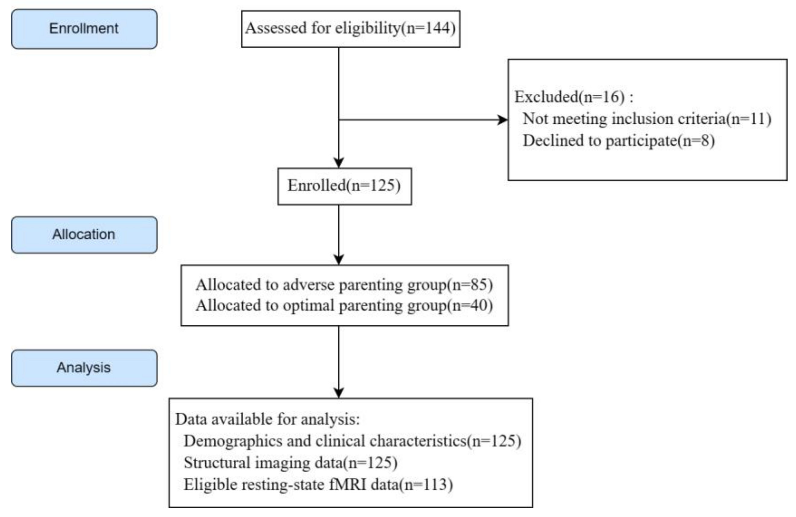
2.1. Study Design, Participants, and Inclusion and Exclusion Criteria

This observational study was undertaken at the Renmin Hospital of Wuhan University, following the principles of the Declaration of Helsinki [38]. The Ethics Committee of Renmin Hospital of Wuhan University, Wuhan, Hubei, China, approved the study protocol. Written informed consent was obtained from all participants. This report was written following the STROBE statement [39].
MDD patients were selected from the Early Warning System and Comprehensive Intervention for Depression (ESCID) project. We calculated the sample size using G*Power software (latest ver. 3.1.9.7; Heinrich-Heine-Universität Düsseldorf, Düsseldorf, Germany). Informed consents for 8 participants were withdrawn, and 11 participants did not meet the inclusion criteria. One hundred and twenty-five MDD outpatients were recruited between May 2019 and April 2022. The inclusion criteria were: (1) being aged 18–50 years; (2) meeting DSM-5 diagnostic criteria for MDD diagnosed by two experienced psychiatrists; and (3) having a junior high school education or higher. Exclusion criteria were: (1) mental health disorders other than MDD diagnosed according to the DSM-5; (2) a history of organic brain disease; (3) severe stupor or other symptoms that could interfere with the study; (4) transcranial magnetic stimulation (TMS) or electroconvulsive therapy (ECT) treatment within the prior six months; and (5) pregnancy (Figure 1).

2.2. Assessment Instruments and Grouping Criteria

Enrolled patients completed the following instruments: the PBI [40], the Hamilton rating scale for depression (HAMD-17) [41], the patient health questionnaire-9 (PHQ-9) [42], the Hamilton Anxiety Scale (HAMA) [43], the Snaith–Hamilton pleasure scale (SHAPS) [44], and the digit symbol substitution test (DSST) [45] on the ESCID website. Magnetic resonance imaging was then completed within one week of completing these instruments.
The grouping criteria for optimal parenting (OP) and AP were modified from the studies of Parker and Margaret [46,47], who regarded high care and low overprotection as OP and affectionless control (low care, high overprotection) as AP. Some studies have suggested that there are cultural differences in parenting between western countries and China [9,13]. Contrary to western countries, parental protectiveness is positively related to warmth and decreases the risk of MDD in China. The effect of protectiveness is considered a representation of parental warmth and lovingness rather than increasing the risk for major depression as it does in the West. Consequently, we chose the relatively representative items of warmth and authoritarianism as grouping items, so the AP group was defined as having low warmth and high authoritarianism and the OP group as having high warmth and low authoritarianism according to the mean parental warmth and authoritarianism scores.
The PBI was used as a measure of participants’ attitudes toward their parents’ parenting style and to generate the AP and OP groups (with OP as the control group). The PBI, which contains 16 items, was modified by Kendler [40] and assesses warmth, protectiveness, and authoritarianism in both mothers and fathers before their children are 16 years old. PBI scores are not influenced by depressive states [48]. In total, data from 125 patients who completed four PBI item scores, including parental warmth and authoritarianism, were subjected to hierarchical cluster analysis by the Ward method to generate two clusters. In addition to the PBI, the clinician-rated HAMD-17 and self-rated PHQ-9 were used to evaluate depressive symptoms, SHAPS for anhedonia, and DSST for cognitive function.

2.3. MRI Acquisition

MRI data were acquired at the PET Center of Renmin Hospital of Wuhan University using a 3T scanner (General Electric, Milwaukee, Brookfield, WI, USA). T1-weighted structural images were acquired with the following parameters: repetition time (TR) = 8.5 ms; echo time (TE) = 3.2 ms; inversion time = 450 ms; flip angle (FA) = 12°; visual field (FOV) = 256 mm × 256 mm; matrix size = 256 × 256; slice thickness = 1 mm; slice gap = 0 mm; and locs per slab = 180. Subjects were kept in a comfortable position to keep them quiet, calm, and moving as little as possible with closed eyes. Resting-state BOLD fMRI data was acquired by employing spin echoplanar imaging (EPI) sequences with the following parameters: 212 times; 32 slices; slice thickness = 3.0 mm; slice gap = 0 mm; interval = 1 mm; TR = 2000 ms; TE = 30 ms; FA = 90°; matrix size = 64 × 64; and FOV = 240 mm × 240 mm.

2.4. MRI Data Processing

The VBM8 toolbox (http://dbm.neuro.uni-jena.de/vbm8/, accessed on 2 April 2022) in Statistical Parametric Mapping 8 (SPM 8; https://www.fil.ion.ucl.ac.uk/spm/software/, accessed on 2 April 2022) was used to preprocess the structural imaging data. Preprocessing included data conversion, quality testing, segmentation and normalization, index extraction, quality re-testing, and smoothing. DICOM images were converted to the NIFTI format for processing. The brain was separated into gray matter, white matter, and cerebrospinal fluid after segmentation. Images were normalized with the diffeomorphic anatomical registration through the exponentiated lie (DARTEL) algorithm to the Montreal Neurological Institute (MNI) template. We then extracted the GMV of all subjects. All structural images were smoothed with an isotropic Gaussian kernel of 8 mm at full width at half maximum (FWHM).
The resting-state fMRI (rs-fMRI) data was preprocessed using the RESTplus v1.2 toolbox in SPM 12. After data conversion, the first 10 volumes were deleted to ensure steady state. In addition, slice timing was carried out for time-level correction. Spatial-level correction consisted of realignment and normalization. We excluded subjects with excessive head movement (>3 mm or >3°) according to the realignment parameters. Six participants were excluded from each of the AP and OP groups, and 113 participants were included in the subsequent analysis. All functional images were smoothed with a 6 mm FWHM Gaussian filter. Then, we performed detrending, nuisance covariate regression, and filtering (0.01–0.08 Hz). All operations were conducted on the MATLAB R2013b platform (MathWorks, Sherborn, MA, USA).

2.5. Voxel-Wise FC Analyses

Wake Forest University’s (WFU) PickAtlas 3.0.5b [49] was used to obtain region of interest (ROI) templates in the MNI space. FC analysis was completed using RESTplus v1.2 after data preprocessing. Brain regions in which GMV differed significantly between the AP group and OP group were selected as seed points (ROI 1) in the voxel-wise FC analysis. To increase the robustness of the FC results, we performed a sensitivity analysis as follows: a sphere with a radius of 6 mm centered on the coordinates of ROI 1 was selected as the region of interest (ROI 2) using WFU PickAtlas 3.0.5b. The average time series of all the voxels in ROIs 1 and 2 was extracted and calculated. Then, voxel-wise Pearson’s correlation coefficients were calculated on the time series between ROI 1/2 and DMN [23] and then converted to Z values by Fisher’s equation to generate subject-specific maps [50]. Thus, the Z value representation of the functional connections between ROIs and the DMN were obtained. We used the same approach to analyze the intra-network FCs.

2.6. Statistical Analyses

Differences in sex and education levels between the AP and OP groups were assessed by chi-squared analysis. The independent samples t-test was applied to explore between-group differences in age, duration of illness, and PBI, PHQ-9, and HAMD-17 scores. Pearson’s correlation coefficients were used to determine correlations between GMVs and PBI item scores. A p-value of <0.05 (two-tailed) was deemed statistically significant. All analyses were conducted in IBM SPSS Statistics v25.0 (IBM Statistics, Armonk, NY, USA). GMV and FC analyses were executed in SPM 8 and SPM 12 separately using the general linear model. To control for confounding effects of age, gender, level of education, duration of illness, and head motion parameter, these factors were included in the calculation as covariates. The Gaussian random field (GRF) correction (voxel p-value < 0.001, cluster p-value < 0.05, one-tailed) was performed to correct for multiple comparisons using the RESTplus v1.2 toolbox [51,52].

3. Results

3.1. Clinical Characteristics

After hierarchical clustering, 85 patients were assigned to the AP group and 40 to the OP group. As expected, there were statistically significant differences in the four PBI items between the two groups (p < 0.05). There were no significant differences in age, gender, education level, medications, HAMD-17, PHQ-9, HAMA, SHAPS, DSST, or duration of illness between the two groups (Table 1).

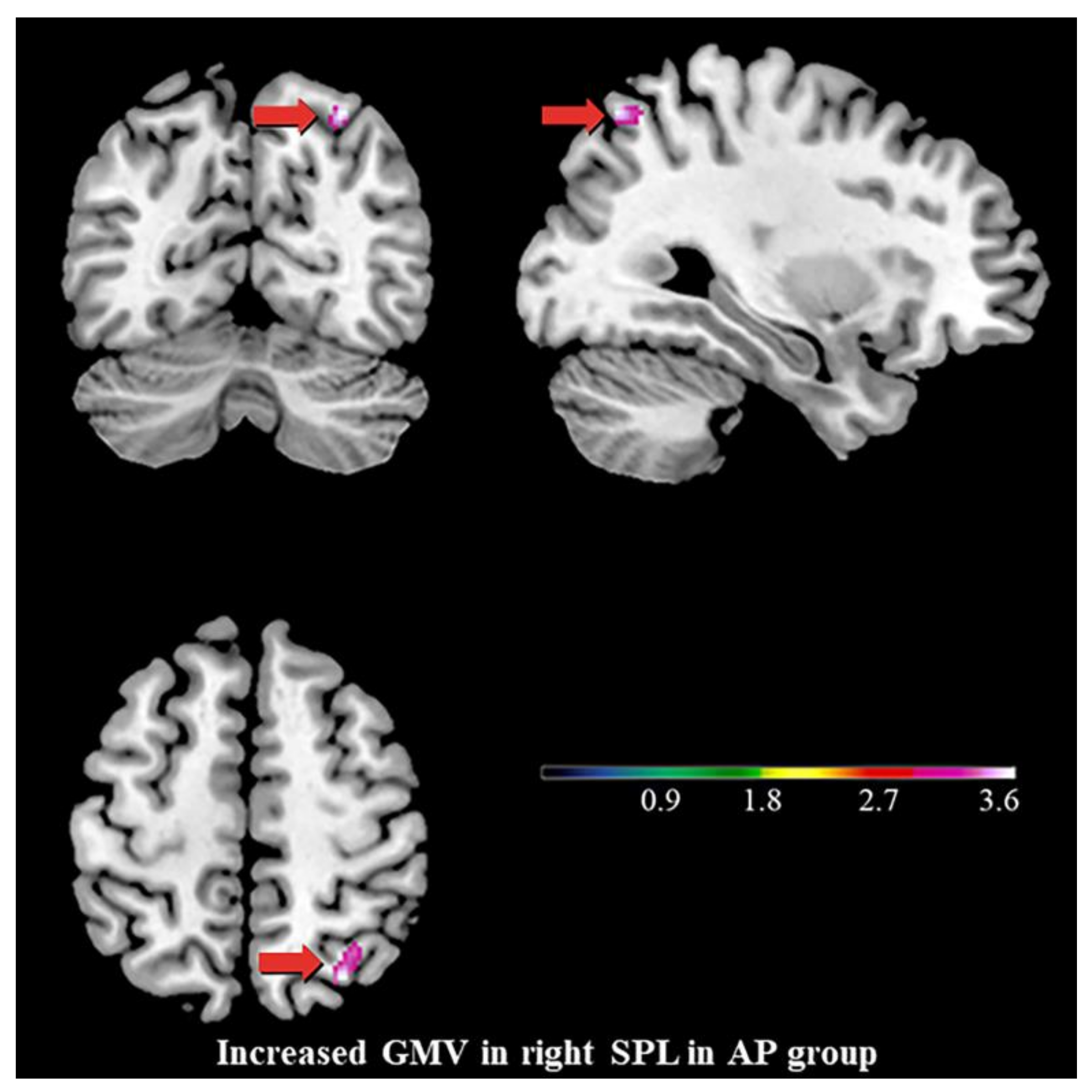
3.2. Differences in GMV

Compared with the OP group, the AP group showed increased GMV in a cluster that had maximally intense voxels at MNI coordinates x = 30, y = −67.5, z = 55.5 (74 voxels, T = 3.892, p < 0.001, GRF correction) in the right SPL (Figure 2).

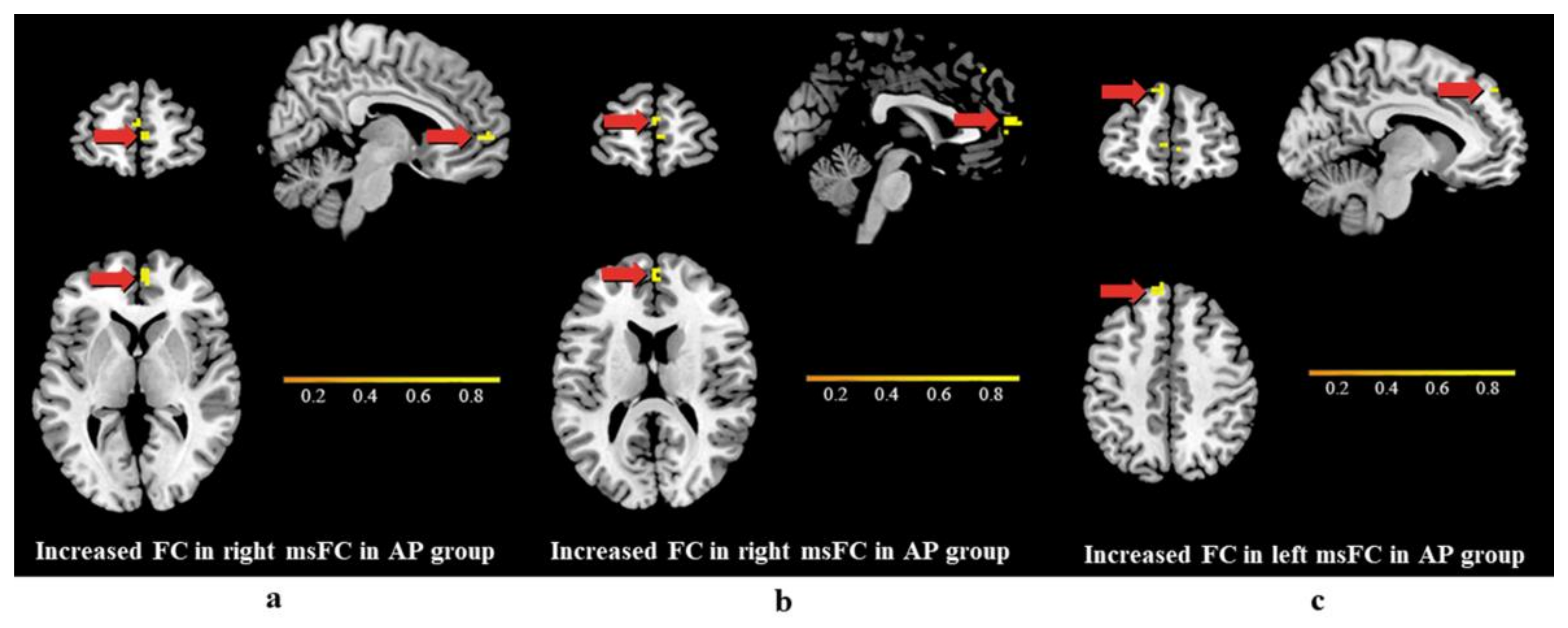
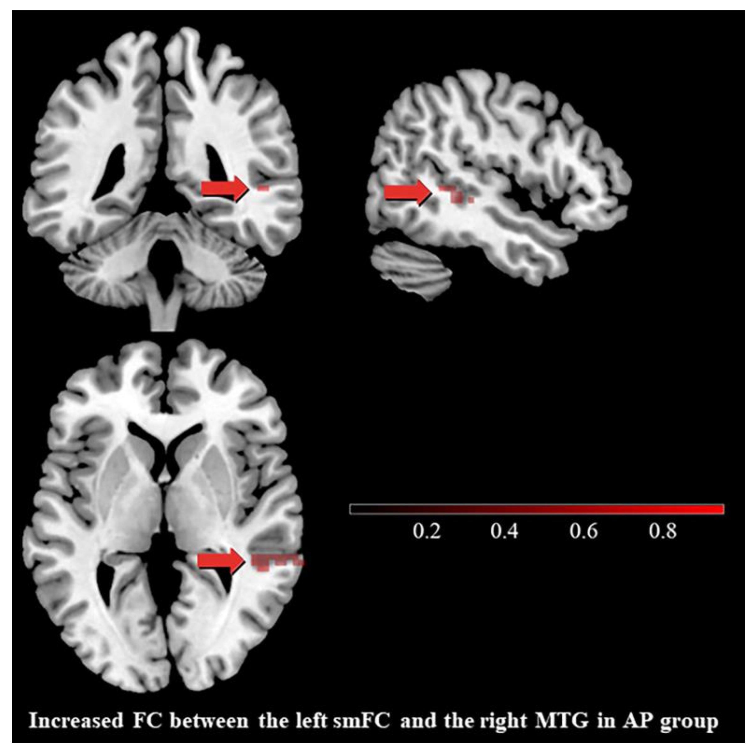
3.3. Differences in Functional Connectivity

The differential brain region of the GMV analysis between two groups is the right SPL. Although the SPL is not the predicted region of structural change, it is a part of DAN. We hypothesized that childhood AP is related to changes in the inter-FC of the DAN and the DMN. Consequently, we calculated the FC between the SPL and the DMN. In the DMN, voxel-wise FC between the right SPL and the right smFC (Cluster 1), the right smFC (Cluster 2), and the left smFC (Cluster 3) were significantly different between the AP group and the OP group (Table 2). Maximally intense voxels were at MNI coordinates x = 6, y = 57, z = 3 (13 voxels, T = 4.762, p < 0.001 GRF correction) for the first cluster (Figure 3a), the second cluster (Figure 3b) had maximally intense voxels at MNI coordinates x = 0, y = 60, z = 15 (13 voxels, T = 4.092, p < 0.001 GRF correction), while the third cluster (Figure 3c) had maximally intense voxels at MNI coordinates x = −9, y = 51, z = 45 (19 voxels, T = 4.069, p < 0.001 GRF correction). Using a second analytical approach to set the ROI and test robustness, the results were similar (see Figure S1 of the Supplementary Material).
As our hypothesis mentioned, AP may relate to changes in the inter- or intra-FC of some specific brain networks, such as the DMN and the DAN. To further investigate the effect of parenting style on the FC of the DAN and the DMN, we analyzed intra-network connectivity within the DMN, the VAN, and the DAN. No cluster survived the intra-DAN and intra-VAN FCs. Compared with the OP group, the intra-DMN FC showed an increase in a cluster with maximally intense voxels at MNI coordinates x = 48, y = −42, z = 3 (43 voxels, T = 5.509, p < 0.001, GRF correction) in the right middle temporal gyrus (MTG) (Figure 4).

3.4. Exploratory Data Analyses

As shown in Figure 5, the GMV of the ROI was significantly negatively correlated with maternal warmth scores (r = −0.329, p < 0.001) and positively correlated with maternal authoritarianism scores (r = 0.196, p = 0.037) and paternal authoritarianism scores (r = 0.195, p = 0.038).
To further explore the relationship between changes in FC and specific parenting styles, we performed a Pearson correlation. We found the FC signal between the right SPL region and the smFC was negatively correlated with parental warmth scores (r = −0.368~−0.207, p < 0.05). The FC signal between the left smFC and the right MTG is negatively correlated with maternal warmth scores (r = −0.504, p < 0.001) and paternal warmth scores (r = −0.219, p < 0.05), while positively correlated with maternal authoritarianism scores (r = 0.251, p < 0.001). (See Section S3.1 of the Supplementary Material).
We additionally calculated correlations between PBI item scores and depressive symptoms such as anhedonia, sleep disturbances, psychomotor retardation, and anxiety/somatization. There was a positive correlation between paternal authoritarianism and anxiety/somatization scores but no significant results in the other symptoms (see Supplementary Material). Although AP and brain structure were significantly correlated, the interactions between AP, GMV, and depressive symptoms are also worth exploring in future analyses.

4. Discussion

Here, we report differential brain structure features as detected by rs-fMRI mainly in the right SPL and associated functional alternations in the bilateral medial superior frontal cortex (smFC) and right MTG in MDD patients experiencing AP or OP. These imaging traits were further associated with AP characteristics.

4.1. AP in MDD

There are profound cross-cultural differences in parenting styles [53] and a notable difference between Eastern and Western cultures [54]. The values and characteristics of parenting are specific to Eastern cultures, in particular differences in how cultures evaluate parental care. The most representative is overprotectiveness. High overprotectiveness is believed to be a component of affectionless control in western populations, where people attach great importance to individuality and privacy [55,56,57]. In Eastern cultures, people tend to foster collectivism, regard the family as a single unit, and think highly of the maintenance of emotional bonds between family members [58,59]. Therefore, Eastern cultures consider parental over-interference or protection as caring and protective [13,58,59]. Given the controversy over high overprotectiveness and the cross-cultural consistency of warmth, we treated warmth rather than high overprotectiveness as a feature of OP. Furthermore, to acquire a relatively reliable and culturally specific grouping, we defined the AP and OP groups using hierarchical cluster analysis, as previously described [54,58,59]. The results of our grouping are similar to those of Hiroko et al. [60], with 32% of MDD patients evaluated classified as OP in our sample.

4.2. Increased GMV of the Right SPL, the Stigmata of Childhood AP?

We found that markedly increased GMV in the right SPL was associated with AP. Previous studies have suggested that AP experiences during childhood and adolescence generate chronic hypothalamus-pituitary-adrenal (HPA) axis hyperactivity, resulting in abnormal neurodevelopment in the normal (non-depressed) population; for example, reduced GMV in the hippocampus [61,62]. Exposure to AP could activate the release of corticotropin-releasing hormone (CRH) and induce increased production of glucocorticoids from the HPA axis and higher levels of proinflammatory cytokines in the brain [63,64]. The glucocorticoids and proinflammatory cytokines are related to brain abnormalities in depression [65]. A study of 50 healthy young adults suggested that lower parental care and higher parental overprotection scores, namely AP, correlated with reductions in the GMV of the DLPFC [35]. Interestingly, our study in MDD patients showed a positive association between the GMV of the right SPL and AP. This finding allows us to formulate another valuable hypothesis for comprehending how early adversity modifies brain structure. AP may affect the normal procedure of synaptic pruning during childhood, resulting in increased GMV of the right SPL. Pruning is a part of brain maturation, which occurs during a sensitive period in late childhood and early adolescence [66,67,68,69,70]. The role of pruning is to strengthen the most frequently used connections and remove infrequently used connections to make the brain circuits more efficient [71]. Pruning is a critical feature of brain maturation, benefiting reasoning processes [72]. There are plenty of reports that synaptic pruning induces decreases in parietal GMV and density before adulthood [73,74,75,76]. The thinning of the superior parietal cortex is associated with age-related improvements on a working memory task, thought to result from selective pruning of inefficient synaptic connections [77,78]. It has previously been observed that thinner parietal cortices were associated with improved verbal learning and memory, visuospatial functioning, and spatial planning and problem solving [79]. Sowell et al. consistently found that longitudinal cortical thickness development was negatively related to vocabulary knowledge in the left dorsal frontal and parietal lobes [80].
Early-life adversity is known to alter the trajectories of structural and functional brain maturation by impairing synaptic pruning [72,81,82]. Abnormalities in neuroendocrine and neuroinflammation cellular and molecular mechanisms caused by early-life adversity may be related to disruption of pruning [83,84,85]. Our finding may reflect that we conducted the study in patients with MDD, and indeed, our result is similar to that reported by Akemi et al., who performed their study in patients with higher depressive levels and found that exposure to parental verbal abuse was associated with increased GMV in the superior temporal gyrus [86]. Hence, exposure to AP may interfere with neurodevelopment by impairing pruning. This is consistent with our findings: as expected, there was a significant negative correlation between the ROI GMV and the mother’s warmth scores and a positive correlation between both parents’ authoritarianism scores. We therefore presume that the worse the parenting style, the larger the GMV. However, the potential mechanism of AP affecting brain development requires more and further study to illuminate.

4.3. Differences in Functional Connectivity

Brain structure and function are closely related, and structure serves as the foundation for function. In addition to the effects on brain structure, there were also changes in the FCs between the right SPL and bilateral smFC in patients exposed to AP. The SPL, which is part of the attention and executive control networks, coordinates attention under competing conditions and voluntary attention [87] and also plays a key role in attention shifting [88,89,90]. The smFC, including the supplementary motor area (SMA) and presupplementary motor area (preSMA), is related to movement and cognitive control [91]. Increased neural activity in the SMA and precuneus is closely associated with the execution of attentional shift between object features, and these regions may play a critical role in the cognitive process of nonspatial attentional shift [92]. Attention-shifting dysfunction is also a feature of people exposed to AP. Elena et al. showed that greater maternal negative behavior could be associated with a decreased ability to disengage attention from negative stimuli [93]. Parental depressive symptoms, a more negative family atmosphere, and maternal authoritarianism predicted children’s inattentive symptoms [93]. The dysfunctional FC between the SPL and smFC also prompts us to consider attention shifting. Attention, as a complex cognitive function, is closely related to the DAN [94] and DMN [95], especially the FC between the DAN and DMN [96,97,98]. Two attentional states have been proposed: one that relies on the DMN and is relatively effortless and ideal for sustaining attention (such as when resting or watching a movie), and another that relies on the DAN, which is effortful and more suited to selective or executive attention (such as when performing a task) [99]. Christiane and colleagues reported that a stronger FC between the DAN and the DMN was associated with weaker attention switching traits [100]. Similarly, we found that childhood AP was related to an increased FC between the right SPL (part of the DAN) and bilateral smFC (part of the DMN). Alterations in DMN activity have been consistently reported in MDD [101,102]. The intra-DMN FC increased in the AP group. The relationship between ACEs and the intra-DMN FC is consistent with previous findings. For example, Keila et al. [103] emphasized that the family environment may be associated with differences in intrinsic DMN FC, with an unharmonious family environment associated with infants exhibiting stronger connectivity between two core DMN regions [104]. The DMN may be a crucial marker of the effects of ACEs and early-life stress closely related to emotional development and subsequent psychological health.

4.4. Limitations

Our study has some limitations. First, the cross-cultural characteristics of AP [13] will affect the generalizability of the findings, and we acknowledge the difficulties in comparing Eastern and Western countries. Hence, a suitable instrument to enhance comparability and promote cross-cultural polycentric research is needed to address this limitation. Second, we failed to find a statistically significant difference in DLPFC [35] and hippocampus [34] characteristics previously reported for general populations exposed to AP. This may be because MDD patients have pre-existing changes that mask the effects of AP or because our sample size was insufficient to detect differences in these brain regions. Third, due to the cross-sectional nature of the study and limitations of attention testing, we cannot confirm a causal relationship between AP, attention shifting, and our findings. Nevertheless, our study is hypothesis-generating, namely, that specific alterations in brain structure and function may be stigmata of AP in patients with MDD. Fourth, as a retrospective study, we did not further explore possible peripheral biomarkers associated with changes in brain imaging. For instance, Kosuke et al. [34] detected a significant association between parental overprotection scores, cortisol responses, and hippocampal GMV. Fifth, it is unethical not to give timely medication to patients with MDD, and there may be selection bias if only unmedicated patients are included in cohort studies. For these reasons, we did not apply an exclusion criterion with respect to the patients’ medications. Instead, we analyzed medication status between the AP and OP groups, and no significant differences were found.

5. Conclusions

Here we found that AP, as a low warmth and high authoritarianism parenting style during childhood, is related to significant enlargements in the right SPL and increased FC between the right SPL and the bilateral smFC in MDD patients. A more well-designed longitudinal study with a larger sample from different cultural backgrounds is essential to verify the effect of AP. The findings further emphasize the importance of optimal and appropriate parenting and arouse the attention of society and families to the parenting style. Our study generates an intriguing hypothesis that brain changes may be the imprint of the childhood AP that affects certain depressive symptoms.

Supplementary Materials

The following supporting information can be downloaded at: https://www.mdpi.com/article/10.3390/brainsci13040694/s1, Figure S1: FC between the ROI 2 and the left smFC also increased in AP group (22 voxels, T = 4.242, p < 0.001, GRF correction). Figure S2: FC signals between the right SPL region and Cluster 1 were negatively correlated with parental warmth scores. Table S1: Pearson correlation between PBI item scores and FC signals. Table S2: Correlation between item scores of PBI and total score of the SHAPS and item scores of the HAMD-17.

Author Contributions

Conceptualization, L.K.; Data curation, W.W., S.X., M.C., Q.G., E.Z. and M.L.; Writing—original draft, Z.N.; Writing—review and editing, X.X.; Visualization, W.W.; Supervision, X.X., L.K., L.Y., H.W., L.B. and Z.L.; Funding acquisition, Z.L. All authors have read and agreed to the published version of the manuscript.

Funding

This work was supported by grants from the National Natural Science Foundation of China (grant number U21A20364) and the National Key R&D Program of China (grant number 2018YFC1314600). Fund provided by Zhongchun Liu.

Institutional Review Board Statement

This observational study was undertaken at the Renmin Hospital of Wuhan University following the principles of the Declaration of Helsinki. The Ethics Committee of Renmin Hospital of Wuhan University, Wuhan, Hubei, China, approved the study protocol.

Informed Consent Statement

Written informed consent was obtained from all participants.

Data Availability Statement

The data that support the findings of this study are available from the corresponding author upon reasonable request.

Conflicts of Interest

The authors declare no conflict of interest.

References

  1. Dagnino, P.; Ugarte, M.J.; Morales, F.; Gonzalez, S.; Saralegui, D.; Ehrenthal, J.C. Risk Factors for Adult Depression: Adverse Childhood Experiences and Personality Functioning. Front. Psychol. 2020, 11, 594698. [Google Scholar] [CrossRef]
  2. Korotana, L.M.; Dobson, K.S.; Pusch, D.; Josephson, T. A review of primary care interventions to improve health outcomes in adult survivors of adverse childhood experiences. Clin. Psychol. Rev. 2016, 46, 59–90. [Google Scholar] [CrossRef]
  3. Lovallo, W.R.; Farag, N.H.; Sorocco, K.H.; Cohoon, A.J.; Vincent, A.S. Lifetime adversity leads to blunted stress axis reactivity: Studies from the Oklahoma Family Health Patterns Project. Biol. Psychiatry 2012, 71, 344–349. [Google Scholar] [CrossRef] [PubMed]
  4. Bellis, M.A.; Hughes, K.; Ford, K.; Ramos Rodriguez, G.; Sethi, D.; Passmore, J. Life course health consequences and associated annual costs of adverse childhood experiences across Europe and North America: A systematic review and meta-analysis. Lancet Public Health 2019, 4, e517–e528. [Google Scholar] [CrossRef]
  5. Felitti, V.J.; Anda, R.F.; Nordenberg, D.; Williamson, D.F.; Spitz, A.M.; Edwards, V.; Koss, M.P.; Marks, J.S. Relationship of childhood abuse and household dysfunction to many of the leading causes of death in adults. The Adverse Childhood Experiences (ACE) Study. Am. J. Prev. Med. 1998, 14, 245–258. [Google Scholar] [CrossRef] [PubMed]
  6. Hughes, K.; Bellis, M.A.; Hardcastle, K.A.; Sethi, D.; Butchart, A.; Mikton, C.; Jones, L.; Dunne, M.P. The effect of multiple adverse childhood experiences on health: A systematic review and meta-analysis. Lancet Public Health 2017, 2, e356–e366. [Google Scholar] [CrossRef]
  7. Parker, G. Parental characteristics in relation to depressive disorders. Br. J. Psychiatry 1979, 134, 138–147. [Google Scholar] [CrossRef]
  8. Duggan, C.; Sham, P.; Minne, C.; Lee, A.; Murray, R. Quality of parenting and vulnerability to depression: Results from a family study. Psychol. Med. 1998, 28, 185–191. [Google Scholar] [CrossRef] [PubMed]
  9. Kendler, K.S.; Myers, J.; Prescott, C.A. Parenting and adult mood, anxiety and substance use disorders in female twins: An epidemiological, multi-informant, retrospective study. Psychol. Med. 2000, 30, 281–294. [Google Scholar] [CrossRef]
  10. Perris, C.; Arrindell, W.A.; Perris, H.; Eisemann, M.; van der Ende, J.; von Knorring, L. Perceived depriving parental rearing and depression. Br. J. Psychiatry 1986, 148, 170–175. [Google Scholar] [CrossRef]
  11. Lancaster, G.; Rollinson, L.; Hill, J. The measurement of a major childhood risk for depression: Comparison of the Parental Bonding Instrument (PBI) ‘Parental Care’ and the Childhood Experience of Care and Abuse (CECA) ‘Parental Neglect’. J. Affect. Disord. 2007, 101, 263–267. [Google Scholar] [CrossRef]
  12. Oakley-Browne, M.A.; Joyce, P.R.; Wells, J.E.; Bushnell, J.A.; Hornblow, A.R. Adverse parenting and other childhood experience as risk factors for depression in women aged 18–44 years. J. Affect. Disord. 1995, 34, 13–23. [Google Scholar] [CrossRef] [PubMed]
  13. Gao, J.; Li, Y.; Cai, Y.; Chen, J.; Shen, Y.; Ni, S.; Wei, Y.; Qiu, Y.; Zhu, X.; Liu, Y.; et al. Perceived parenting and risk for major depression in Chinese women. Psychol. Med. 2012, 42, 921–930. [Google Scholar] [CrossRef] [PubMed]
  14. Long, E.C.; Aggen, S.H.; Gardner, C.; Kendler, K.S. Differential parenting and risk for psychopathology: A monozygotic twin difference approach. Soc. Psychiatry Psychiatr. Epidemiol. 2015, 50, 1569–1576. [Google Scholar] [CrossRef] [PubMed]
  15. Liu, Y.L. Parent-child interaction and children’s depression: The relationships between parent-child interaction and children’s depressive symptoms in Taiwan. J. Adolesc. 2003, 26, 447–457. [Google Scholar] [CrossRef]
  16. Anno, K.; Shibata, M.; Ninomiya, T.; Iwaki, R.; Kawata, H.; Sawamoto, R.; Kubo, C.; Kiyohara, Y.; Sudo, N.; Hosoi, M. Paternal and maternal bonding styles in childhood are associated with the prevalence of chronic pain in a general adult population: The Hisayama Study. BMC Psychiatry 2015, 15, 181. [Google Scholar] [CrossRef]
  17. McCrory, E.; De Brito, S.A.; Viding, E. Research review: The neurobiology and genetics of maltreatment and adversity. J. Child Psychol. Psychiatry 2010, 51, 1079–1095. [Google Scholar] [CrossRef]
  18. Mao, Y.; Xiao, H.; Ding, C.; Qiu, J. The role of attention in the relationship between early life stress and depression. Sci. Rep. 2020, 10, 6154. [Google Scholar] [CrossRef]
  19. Gilbert, R.; Widom, C.S.; Browne, K.; Fergusson, D.; Webb, E.; Janson, S. Burden and consequences of child maltreatment in high-income countries. Lancet 2009, 373, 68–81. [Google Scholar] [CrossRef]
  20. Miguel, P.M.; Pereira, L.O.; Silveira, P.P.; Meaney, M.J. Early environmental influences on the development of children’s brain structure and function. Dev. Med. Child Neurol. 2019, 61, 1127–1133. [Google Scholar] [CrossRef]
  21. McLaughlin, K.A.; Weissman, D.; Bitran, D. Childhood Adversity and Neural Development: A Systematic Review. Annu. Rev. Dev. Psychol. 2019, 1, 277–312. [Google Scholar] [CrossRef]
  22. Aghamohammadi-Sereshki, A.; Coupland, N.J.; Silverstone, P.H.; Huang, Y.; Hegadoren, K.M.; Carter, R.; Seres, P.; Malykhin, N.V. Effects of childhood adversity on the volumes of the amygdala subnuclei and hippocampal subfields in individuals with major depressive disorder. J. Psychiatry Neurosci. 2021, 46, E186–E195. [Google Scholar] [CrossRef] [PubMed]
  23. Yeo, B.T.; Krienen, F.M.; Sepulcre, J.; Sabuncu, M.R.; Lashkari, D.; Hollinshead, M.; Roffman, J.L.; Smoller, J.W.; Zollei, L.; Polimeni, J.R.; et al. The organization of the human cerebral cortex estimated by intrinsic functional connectivity. J. Neurophysiol. 2011, 106, 1125–1165. [Google Scholar] [CrossRef]
  24. Finn, E.S.; Shen, X.; Scheinost, D.; Rosenberg, M.D.; Huang, J.; Chun, M.M.; Papademetris, X.; Constable, R.T. Functional connectome fingerprinting: Identifying individuals using patterns of brain connectivity. Nat. Neurosci. 2015, 18, 1664–1671. [Google Scholar] [CrossRef] [PubMed]
  25. Rosenberg, M.D.; Finn, E.S.; Scheinost, D.; Papademetris, X.; Shen, X.; Constable, R.T.; Chun, M.M. A neuromarker of sustained attention from whole-brain functional connectivity. Nat. Neurosci. 2016, 19, 165–171. [Google Scholar] [CrossRef] [PubMed]
  26. Hsu, W.T.; Rosenberg, M.D.; Scheinost, D.; Constable, R.T.; Chun, M.M. Resting-state functional connectivity predicts neuroticism and extraversion in novel individuals. Soc. Cogn. Affect. Neurosci. 2018, 13, 224–232. [Google Scholar] [CrossRef]
  27. Cheng, T.W.; Mills, K.L.; Miranda Dominguez, O.; Zeithamova, D.; Perrone, A.; Sturgeon, D.; Feldstein Ewing, S.W.; Fisher, P.A.; Pfeifer, J.H.; Fair, D.A.; et al. Characterizing the impact of adversity, abuse, and neglect on adolescent amygdala resting-state functional connectivity. Dev. Cogn. Neurosci. 2021, 47, 100894. [Google Scholar] [CrossRef]
  28. Fadel, E.; Boeker, H.; Gaertner, M.; Richter, A.; Kleim, B.; Seifritz, E.; Grimm, S.; Wade-Bohleber, L.M. Differential Alterations in Resting State Functional Connectivity Associated with Depressive Symptoms and Early Life Adversity. Brain Sci. 2021, 11, 591. [Google Scholar] [CrossRef]
  29. Albertina, E.A.; Barch, D.M.; Karcher, N.R. Internalizing Symptoms and Adverse Childhood Experiences Associated with Functional Connectivity in a Middle Childhood Sample. Biol. Psychiatry Cogn. Neurosci. Neuroimaging 2022. [CrossRef]
  30. Andrews-Hanna, J.R. The brain’s default network and its adaptive role in internal mentation. Neuroscientist 2012, 18, 251–270. [Google Scholar] [CrossRef]
  31. Davis, K.; Hirsch, E.; Gee, D.; Andover, M.; Roy, A.K. Mediating role of the default mode network on parental acceptance/warmth and psychopathology in youth. Brain Imaging Behav. 2022, 16, 2229–2238. [Google Scholar] [CrossRef]
  32. Vossel, S.; Geng, J.J.; Fink, G.R. Dorsal and ventral attention systems: Distinct neural circuits but collaborative roles. Neuroscientist 2014, 20, 150–159. [Google Scholar] [CrossRef]
  33. Houtepen, L.C.; Heron, J.; Suderman, M.J.; Tilling, K.; Howe, L.D. Adverse childhood experiences in the children of the Avon Longitudinal Study of Parents and Children (ALSPAC). Wellcome Open Res. 2018, 3, 106. [Google Scholar] [CrossRef]
  34. Narita, K.; Fujihara, K.; Takei, Y.; Suda, M.; Aoyama, Y.; Uehara, T.; Majima, T.; Kosaka, H.; Amanuma, M.; Fukuda, M.; et al. Associations among parenting experiences during childhood and adolescence, hypothalamus-pituitary-adrenal axis hypoactivity, and hippocampal gray matter volume reduction in young adults. Hum. Brain Mapp. 2012, 33, 2211–2223. [Google Scholar] [CrossRef]
  35. Narita, K.; Takei, Y.; Suda, M.; Aoyama, Y.; Uehara, T.; Kosaka, H.; Amanuma, M.; Fukuda, M.; Mikuni, M. Relationship of parental bonding styles with gray matter volume of dorsolateral prefrontal cortex in young adults. Prog. Neuropsychopharmacol. Biol. Psychiatry 2010, 34, 624–631. [Google Scholar] [CrossRef] [PubMed]
  36. Ma, S.; Kang, L.; Guo, X.; Liu, H.; Yao, L.; Bai, H.; Chen, C.; Hu, M.; Du, L.; Du, H.; et al. Discrepancies between self-rated depression and observed depression severity: The effects of personality and dysfunctional attitudes. Gen. Hosp. Psychiatry 2021, 70, 25–30. [Google Scholar] [CrossRef] [PubMed]
  37. Kang, L.; Li, R.; Liu, H.; Ma, S.; Sun, S.; Zhang, N.; Yao, L.; Wang, Y.; Zong, X.; Ai, C.; et al. Nonsuicidal self-injury in undergraduate students with major depressive disorder: The role of psychosocial factors. J. Affect. Disord. 2021, 290, 102–108. [Google Scholar] [CrossRef] [PubMed]
  38. World Medical, A. World Medical Association Declaration of Helsinki: Ethical principles for medical research involving human subjects. JAMA 2013, 310, 2191–2194. [Google Scholar] [CrossRef]
  39. Von Elm, E.; Altman, D.G.; Egger, M.; Pocock, S.J.; Gotzsche, P.C.; Vandenbroucke, J.P.; Initiative, S. The Strengthening the Reporting of Observational Studies in Epidemiology (STROBE) statement: Guidelines for reporting observational studies. PLoS Med. 2007, 4, e296. [Google Scholar] [CrossRef]
  40. Kendler, K.S. Parenting: A genetic-epidemiologic perspective. Am. J. Psychiatry 1996, 153, 11–20. [Google Scholar] [CrossRef]
  41. Trajkovic, G.; Starcevic, V.; Latas, M.; Lestarevic, M.; Ille, T.; Bukumiric, Z.; Marinkovic, J. Reliability of the Hamilton Rating Scale for Depression: A meta-analysis over a period of 49 years. Psychiatry Res. 2011, 189, 1–9. [Google Scholar] [CrossRef]
  42. Spitzer, R.L.; Kroenke, K.; Williams, J.B. Validation and utility of a self-report version of PRIME-MD: The PHQ primary care study. Primary Care Evaluation of Mental Disorders. Patient Health Questionnaire. JAMA 1999, 282, 1737–1744. [Google Scholar] [CrossRef]
  43. Maier, W.; Buller, R.; Philipp, M.; Heuser, I. The Hamilton Anxiety Scale: Reliability, validity and sensitivity to change in anxiety and depressive disorders. J. Affect. Disord. 1988, 14, 61–68. [Google Scholar] [CrossRef] [PubMed]
  44. Snaith, R.P.; Hamilton, M.; Morley, S.; Humayan, A.; Hargreaves, D.; Trigwell, P. A scale for the assessment of hedonic tone the Snaith-Hamilton Pleasure Scale. Br. J. Psychiatry 1995, 167, 99–103. [Google Scholar] [CrossRef] [PubMed]
  45. Proust-Lima, C.; Amieva, H.; Dartigues, J.F.; Jacqmin-Gadda, H. Sensitivity of four psychometric tests to measure cognitive changes in brain aging-population-based studies. Am. J. Epidemiol. 2007, 165, 344–350. [Google Scholar] [CrossRef] [PubMed]
  46. Parker, G. Parental representations of patients with anxiety neurosis. Acta Psychiatr. Scand. 1981, 63, 33–36. [Google Scholar] [CrossRef]
  47. Plantes, M.M.; Prusoff, B.A.; Brennan, J.; Parker, G. Parental representations of depressed outpatients from a U.S.A. sample. J. Affect. Disord. 1988, 15, 149–155. [Google Scholar] [CrossRef]
  48. Parker, G. Parental reports of depressives. An investigation of several explanations. J. Affect. Disord. 1981, 3, 131–140. [Google Scholar] [CrossRef]
  49. Maldjian, J.A.; Laurienti, P.J.; Kraft, R.A.; Burdette, J.H. An automated method for neuroanatomic and cytoarchitectonic atlas-based interrogation of fMRI data sets. Neuroimage 2003, 19, 1233–1239. [Google Scholar] [CrossRef]
  50. Liu, Y.; Yu, C.; Liang, M.; Li, J.; Tian, L.; Zhou, Y.; Qin, W.; Li, K.; Jiang, T. Whole brain functional connectivity in the early blind. Brain 2007, 130, 2085–2096. [Google Scholar] [CrossRef]
  51. Cheng, H.; Zhang, Z.; Zhang, B.; Zhang, W.; Wang, J.; Ni, W.; Miao, Y.; Liu, J.; Bi, Y. Enhancement of Impaired Olfactory Neural Activation and Cognitive Capacity by Liraglutide, but Not Dapagliflozin or Acarbose, in Patients with Type 2 Diabetes: A 16-Week Randomized Parallel Comparative Study. Diabetes Care 2022, 45, 1201–1210. [Google Scholar] [CrossRef] [PubMed]
  52. Nichols, T.E. Multiple testing corrections, nonparametric methods, and random field theory. Neuroimage 2012, 62, 811–815. [Google Scholar] [CrossRef]
  53. Kiefner-Burmeister, A.; Hinman, N. The Role of General Parenting Style in Child Diet and Obesity Risk. Curr. Nutr. Rep. 2020, 9, 14–30. [Google Scholar] [CrossRef] [PubMed]
  54. Liu, J.; Li, L.; Fang, F. Psychometric properties of the Chinese version of the Parental Bonding Instrument. Int. J. Nurs. Stud. 2011, 48, 582–589. [Google Scholar] [CrossRef] [PubMed]
  55. Amianto, F.; Martini, M.; Olandese, F.; Davico, C.; Abbate-Daga, G.; Fassino, S.; Vitiello, B. Affectionless control: A parenting style associated with obesity and binge eating disorder in adulthood. Eur. Eat. Disord. Rev. 2021, 29, 178–192. [Google Scholar] [CrossRef]
  56. Goschin, S.; Briggs, J.; Blanco-Lutzen, S.; Cohen, L.J.; Galynker, I. Parental affectionless control and suicidality. J. Affect. Disord. 2013, 151, 1–6. [Google Scholar] [CrossRef]
  57. Otani, K.; Suzuki, A.; Matsumoto, Y.; Sadahiro, R.; Enokido, M. Affectionless control by the same-sex parents increases dysfunctional attitudes about achievement. Compr. Psychiatry 2014, 55, 1411–1414. [Google Scholar] [CrossRef]
  58. Chao, R.K. Extending research on the consequences of parenting style for Chinese Americans and European Americans. Child Dev. 2001, 72, 1832–1843. [Google Scholar] [CrossRef]
  59. Rohner, R.P.; Pettengill, S.M. Perceived parental acceptance-rejection and parental control among Korean adolescents. Child Dev. 1985, 56, 524–528. [Google Scholar] [CrossRef]
  60. Handa, H.; Ito, A.; Tsuda, H.; Ohsawa, I.; Ogawa, T. Low level of parental bonding might be a risk factor among women with prolonged depression: A preliminary investigation. Psychiatry Clin. Neurosci. 2009, 63, 721–729. [Google Scholar] [CrossRef]
  61. Bremner, J.D.; Vythilingam, M.; Vermetten, E.; Southwick, S.M.; McGlashan, T.; Nazeer, A.; Khan, S.; Vaccarino, L.V.; Soufer, R.; Garg, P.K.; et al. MRI and PET study of deficits in hippocampal structure and function in women with childhood sexual abuse and posttraumatic stress disorder. Am. J. Psychiatry 2003, 160, 924–932. [Google Scholar] [CrossRef] [PubMed]
  62. Stein, M.B.; Koverola, C.; Hanna, C.; Torchia, M.G.; McClarty, B. Hippocampal volume in women victimized by childhood sexual abuse. Psychol. Med. 1997, 27, 951–959. [Google Scholar] [CrossRef] [PubMed]
  63. Haapakoski, R.; Mathieu, J.; Ebmeier, K.P.; Alenius, H.; Kivimaki, M. Cumulative meta-analysis of interleukins 6 and 1beta, tumour necrosis factor alpha and C-reactive protein in patients with major depressive disorder. Brain Behav. Immun. 2015, 49, 206–215. [Google Scholar] [CrossRef] [PubMed]
  64. Iob, E.; Lacey, R.; Steptoe, A. The long-term association of adverse childhood experiences with C-reactive protein and hair cortisol: Cumulative risk versus dimensions of adversity. Brain Behav. Immun. 2020, 87, 318–328. [Google Scholar] [CrossRef] [PubMed]
  65. Zunszain, P.A.; Anacker, C.; Cattaneo, A.; Carvalho, L.A.; Pariante, C.M. Glucocorticoids, cytokines and brain abnormalities in depression. Prog. Neuropsychopharmacol. Biol. Psychiatry 2011, 35, 722–729. [Google Scholar] [CrossRef]
  66. Milbocker, K.A.; Campbell, T.S.; Collins, N.; Kim, S.; Smith, I.F.; Roth, T.L.; Klintsova, A.Y. Glia-Driven Brain Circuit Refinement Is Altered by Early-Life Adversity: Behavioral Outcomes. Front. Behav. Neurosci. 2021, 15, 786234. [Google Scholar] [CrossRef]
  67. Emoto, K.; Hensch, T.K.; Yuzaki, M. “Scrap & build” functional circuits: Molecular and cellular basis of neural remodeling. Neurosci. Res. 2021, 167, 1–2. [Google Scholar] [CrossRef]
  68. Espinosa, J.S.; Stryker, M.P. Development and plasticity of the primary visual cortex. Neuron 2012, 75, 230–249. [Google Scholar] [CrossRef]
  69. Huh, C.Y.L.; Abdelaal, K.; Salinas, K.J.; Gu, D.; Zeitoun, J.; Figueroa Velez, D.X.; Peach, J.P.; Fowlkes, C.C.; Gandhi, S.P. Long-term Monocular Deprivation during Juvenile Critical Period Disrupts Binocular Integration in Mouse Visual Thalamus. J. Neurosci. 2020, 40, 585–604. [Google Scholar] [CrossRef]
  70. Vainchtein, I.D.; Chin, G.; Cho, F.S.; Kelley, K.W.; Miller, J.G.; Chien, E.C.; Liddelow, S.A.; Nguyen, P.T.; Nakao-Inoue, H.; Dorman, L.C.; et al. Astrocyte-derived interleukin-33 promotes microglial synapse engulfment and neural circuit development. Science 2018, 359, 1269–1273. [Google Scholar] [CrossRef]
  71. Blakemore, S.J.; Choudhury, S. Brain development during puberty: State of the science. Dev. Sci. 2006, 9, 11–14. [Google Scholar] [CrossRef]
  72. Luby, J.L.; Baram, T.Z.; Rogers, C.E.; Barch, D.M. Neurodevelopmental Optimization after Early-Life Adversity: Cross-Species Studies to Elucidate Sensitive Periods and Brain Mechanisms to Inform Early Intervention. Trends Neurosci. 2020, 43, 744–751. [Google Scholar] [CrossRef] [PubMed]
  73. Modrono, C.; Navarrete, G.; Nicolle, A.; Gonzalez-Mora, J.L.; Smith, K.W.; Marling, M.; Goel, V. Developmental grey matter changes in superior parietal cortex accompany improved transitive reasoning. Think. Reason. 2019, 25, 151–170. [Google Scholar] [CrossRef] [PubMed]
  74. Linkersdorfer, J.; Jurcoane, A.; Lindberg, S.; Kaiser, J.; Hasselhorn, M.; Fiebach, C.J.; Lonnemann, J. The association between gray matter volume and reading proficiency: A longitudinal study of beginning readers. J. Cogn. Neurosci. 2015, 27, 308–318. [Google Scholar] [CrossRef]
  75. Devous, M.D., Sr.; Altuna, D.; Furl, N.; Cooper, W.; Gabbert, G.; Ngai, W.T.; Chiu, S.; Scott, J.M., 3rd; Harris, T.S.; Payne, J.K.; et al. Maturation of speech and language functional neuroanatomy in pediatric normal controls. J. Speech Lang. Hear. Res. 2006, 49, 856–866. [Google Scholar] [CrossRef] [PubMed]
  76. Whitford, T.J.; Rennie, C.J.; Grieve, S.M.; Clark, C.R.; Gordon, E.; Williams, L.M. Brain maturation in adolescence: Concurrent changes in neuroanatomy and neurophysiology. Hum. Brain Mapp. 2007, 28, 228–237. [Google Scholar] [CrossRef]
  77. Dosenbach, N.U.; Nardos, B.; Cohen, A.L.; Fair, D.A.; Power, J.D.; Church, J.A.; Nelson, S.M.; Wig, G.S.; Vogel, A.C.; Lessov-Schlaggar, C.N.; et al. Prediction of individual brain maturity using fMRI. Science 2010, 329, 1358–1361. [Google Scholar] [CrossRef]
  78. Kharitonova, M.; Martin, R.E.; Gabrieli, J.D.; Sheridan, M.A. Cortical gray-matter thinning is associated with age-related improvements on executive function tasks. Dev. Cogn. Neurosci. 2013, 6, 61–71. [Google Scholar] [CrossRef]
  79. Squeglia, L.M.; Jacobus, J.; Sorg, S.F.; Jernigan, T.L.; Tapert, S.F. Early adolescent cortical thinning is related to better neuropsychological performance. J. Int. Neuropsychol. Soc. 2013, 19, 962–970. [Google Scholar] [CrossRef]
  80. Sowell, E.R.; Thompson, P.M.; Leonard, C.M.; Welcome, S.E.; Kan, E.; Toga, A.W. Longitudinal mapping of cortical thickness and brain growth in normal children. J. Neurosci. 2004, 24, 8223–8231. [Google Scholar] [CrossRef] [PubMed]
  81. Tooley, U.A.; Bassett, D.S.; Mackey, A.P. Environmental influences on the pace of brain development. Nat. Rev. Neurosci. 2021, 22, 372–384. [Google Scholar] [CrossRef] [PubMed]
  82. Bolton, J.L.; Short, A.K.; Othy, S.; Kooiker, C.L.; Shao, M.; Gunn, B.G.; Beck, J.; Bai, X.; Law, S.M.; Savage, J.C.; et al. Early stress-induced impaired microglial pruning of excitatory synapses on immature CRH-expressing neurons provokes aberrant adult stress responses. Cell Rep. 2022, 38, 110600. [Google Scholar] [CrossRef] [PubMed]
  83. Andersen, S.L. Neuroinflammation, Early-Life Adversity, and Brain Development. Harv. Rev. Psychiatry 2022, 30, 24–39. [Google Scholar] [CrossRef]
  84. Ivy, A.S.; Rex, C.S.; Chen, Y.; Dube, C.; Maras, P.M.; Grigoriadis, D.E.; Gall, C.M.; Lynch, G.; Baram, T.Z. Hippocampal dysfunction and cognitive impairments provoked by chronic early-life stress involve excessive activation of CRH receptors. J. Neurosci. 2010, 30, 13005–13015. [Google Scholar] [CrossRef]
  85. Malave, L.; van Dijk, M.T.; Anacker, C. Early life adversity shapes neural circuit function during sensitive postnatal developmental periods. Transl. Psychiatry 2022, 12, 306. [Google Scholar] [CrossRef] [PubMed]
  86. Tomoda, A.; Sheu, Y.S.; Rabi, K.; Suzuki, H.; Navalta, C.P.; Polcari, A.; Teicher, M.H. Exposure to parental verbal abuse is associated with increased gray matter volume in superior temporal gyrus. Neuroimage 2011, 54 (Suppl. S1), S280–S286. [Google Scholar] [CrossRef]
  87. Corbetta, M.; Shulman, G.L. Control of goal-directed and stimulus-driven attention in the brain. Nat. Rev. Neurosci. 2002, 3, 201–215. [Google Scholar] [CrossRef]
  88. Behrmann, M.; Geng, J.J.; Shomstein, S. Parietal cortex and attention. Curr. Opin. Neurobiol. 2004, 14, 212–217. [Google Scholar] [CrossRef]
  89. Caspari, N.; Arsenault, J.T.; Vandenberghe, R.; Vanduffel, W. Functional Similarity of Medial Superior Parietal Areas for Shift-Selective Attention Signals in Humans and Monkeys. Cereb. Cortex 2018, 28, 2085–2099. [Google Scholar] [CrossRef]
  90. Vandenberghe, R.; Gitelman, D.R.; Parrish, T.B.; Mesulam, M.M. Functional specificity of superior parietal mediation of spatial shifting. Neuroimage 2001, 14, 661–673. [Google Scholar] [CrossRef]
  91. Zhang, S.; Ide, J.S.; Li, C.S. Resting-state functional connectivity of the medial superior frontal cortex. Cereb. Cortex 2012, 22, 99–111. [Google Scholar] [CrossRef] [PubMed]
  92. Nagahama, Y.; Okada, T.; Katsumi, Y.; Hayashi, T.; Yamauchi, H.; Sawamoto, N.; Toma, K.; Nakamura, K.; Hanakawa, T.; Konishi, J.; et al. Transient neural activity in the medial superior frontal gyrus and precuneus time locked with attention shift between object features. Neuroimage 1999, 10, 193–199. [Google Scholar] [CrossRef]
  93. Pozzi, E.; Simmons, J.G.; Bousman, C.A.; Vijayakumar, N.; Bray, K.O.; Dandash, O.; Richmond, S.; Schwartz, O.; Seal, M.; Sheeber, L.; et al. The Influence of Maternal Parenting Style on the Neural Correlates of Emotion Processing in Children. J. Am. Acad Child Adolesc. Psychiatry 2020, 59, 274–282. [Google Scholar] [CrossRef]
  94. Machner, B.; Braun, L.; Imholz, J.; Koch, P.J.; Munte, T.F.; Helmchen, C.; Sprenger, A. Resting-State Functional Connectivity in the Dorsal Attention Network Relates to Behavioral Performance in Spatial Attention Tasks and May Show Task-Related Adaptation. Front. Hum. Neurosci. 2021, 15, 757128. [Google Scholar] [CrossRef] [PubMed]
  95. Castellanos, F.X.; Aoki, Y. Intrinsic Functional Connectivity in Attention-Deficit/Hyperactivity Disorder: A Science in Development. Biol. Psychiatry Cogn. Neurosci. Neuroimaging 2016, 1, 253–261. [Google Scholar] [CrossRef]
  96. Zhang, Z.; Luh, W.M.; Duan, W.; Zhou, G.D.; Weinschenk, G.; Anderson, A.K.; Dai, W. Longitudinal effects of meditation on brain resting-state functional connectivity. Sci. Rep. 2021, 11, 11361. [Google Scholar] [CrossRef]
  97. Lin, H.; Lin, Q.; Li, H.; Wang, M.; Chen, H.; Liang, Y.; Bu, X.; Wang, W.; Yi, Y.; Zhao, Y.; et al. Functional Connectivity of Attention-Related Networks in Drug-Naive Children with ADHD. J. Atten. Disord. 2021, 25, 377–388. [Google Scholar] [CrossRef]
  98. Froeliger, B.; Garland, E.L.; Kozink, R.V.; Modlin, L.A.; Chen, N.K.; McClernon, F.J.; Greeson, J.M.; Sobin, P. Meditation-State Functional Connectivity (msFC): Strengthening of the Dorsal Attention Network and Beyond. Evid.-Based Complement. Altern. Med. 2012, 2012, 680407. [Google Scholar] [CrossRef] [PubMed]
  99. Esterman, M.; Rosenberg, M.D.; Noonan, S.K. Intrinsic fluctuations in sustained attention and distractor processing. J. Neurosci. 2014, 34, 1724–1730. [Google Scholar] [CrossRef]
  100. Rohr, C.S.; Dimond, D.; Schuetze, M.; Cho, I.Y.K.; Lichtenstein-Vidne, L.; Okon-Singer, H.; Dewey, D.; Bray, S. Girls’ attentive traits associate with cerebellar to dorsal attention and default mode network connectivity. Neuropsychologia 2019, 127, 84–92. [Google Scholar] [CrossRef]
  101. Kaiser, R.H.; Andrews-Hanna, J.R.; Wager, T.D.; Pizzagalli, D.A. Large-Scale Network Dysfunction in Major Depressive Disorder: A Meta-analysis of Resting-State Functional Connectivity. JAMA Psychiatry 2015, 72, 603–611. [Google Scholar] [CrossRef] [PubMed]
  102. Zhong, X.; Pu, W.; Yao, S. Functional alterations of fronto-limbic circuit and default mode network systems in first-episode, drug-naive patients with major depressive disorder: A meta-analysis of resting-state fMRI data. J. Affect. Disord. 2016, 206, 280–286. [Google Scholar] [CrossRef]
  103. Rebello, K.; Moura, L.M.; Bueno, A.P.A.; Picon, F.A.; Pan, P.M.; Gadelha, A.; Miguel, E.C.; Bressan, R.A.; Rohde, L.A.; Sato, J.R. Associations between Family Functioning and Maternal Behavior on Default Mode Network Connectivity in School-Age Children. Int. J. Environ. Res. Public Health 2022, 19, 6055. [Google Scholar] [CrossRef] [PubMed]
  104. Graham, A.M.; Pfeifer, J.H.; Fisher, P.A.; Carpenter, S.; Fair, D.A. Early life stress is associated with default system integrity and emotionality during infancy. J. Child Psychol. Psychiatry 2015, 56, 1212–1222. [Google Scholar] [CrossRef] [PubMed]
Figure 1. Flowchart representing the procedure of study design and participants.
Brainsci 13 00694 g001
Figure 2. Significant difference in GMV between the AP and OP groups. This cluster contained 74 voxels, mainly in the right SPL, with a peak T value of 3.892. Significant at voxel p < 0.001, cluster p < 0.05, corrected by the GRF. The red arrow shows the corresponding brain region.
Brainsci 13 00694 g002
Figure 3. The images show increased FC from the right SPL (ROI 1) to the bilateral medial superior frontal cortex (smFC) in the AP group compared with the OP group at rest. In the DMN, voxel-wised FC between the right SPL and the right smFC were significantly different between the AP group and the OP group (a). In the DMN, voxel-wised FC between the right SPL and the right smFC increased in the AP group (b). In the DMN, voxel-wised FC between the right SPL and the left smFC also increased in the AP group (c). The red arrow shows the corresponding brain region. The right SPL was set as the seed because the GMV of this region was increased in the AP group (see Figure 2).
Brainsci 13 00694 g003
Figure 4. In the DMN, differences in FC between groups between the left smFC and the right MTG. The red arrow shows the corresponding brain region. The positive value (red) means OP < AP at a threshold of p < 0.001, GRF-corrected.
Brainsci 13 00694 g004
Figure 5. A negative correlation between the GMV of the ROI and maternal warmth scores (r = −0.329, p < 0.001) and a positive correlation between the GMV of the ROI and parental authoritarianism scores (for mother: r = 0.196, p = 0.037; for father: r = 0.195, p = 0.038) were observed. The GMV of the ROI is negatively correlated to maternal warmth scores (r = −0.329, p < 0.001) (a). The GMV of the ROI is positively correlated to maternal authoritarianism scores (r = 0.196, p = 0.037) (b). There is no significant correlation between the GMV of the ROI and paternal warmth (c). The GMV of the ROI is positively correlated to paternal authoritarianism scores (r = 0.195, p = 0.038) (d). The triangle represents the AP group and the circle represents the OP group. The density curves represent the distribution of the variables.
Brainsci 13 00694 g005
Table 1. Demographics and clinical characteristics.
Characteristics (Mean ± SD) Adverse ParentingOptimal Parentingχ2/tp
n (%)n (%)
GenderFemale67 (78.8%) 26 (65.0%)2.729 a 0.099 a
Male18 (21.2%)14 (35.0%)
Education levelHigh school or6 (7.1%)4 (10%)0.339 a 0.844 a
less
Undergraduate65 (76.4%)30 (75%)
Postgraduate or14 (16.5%) 6 (15%)
higher
MedicationsDrug-naïve50(58.8%)23(57.5%)0.020 a0.889 a
Age (years) 25.6 ± 6.224.7 ± 5.40.744 b 0.458 b
Duration of illness (years) 5.6 ± 4.34.8 ± 3.21.028 b 0.306 b
PHQ-9 15.92 ± 5.7315.25 ± 5.420.619 b 0.537 b
HAMD-1718.87 ± 7.2218.23 ± 6.390.484 b 0.630 b
HAMA17.39 ± 7.7515.75 ± 6.661.151 b0.252 b
SHAPS32.63 ± 7.4033.79 ± 5.85−0.854 b0.395 b
DSST (n = 60)61.30 ± 14.0861.75 ± 11.74−0.115 b0.909 b
Maternal warmth 7.09 ± 3.2714.80 ± 2.74−12.912 b <0.001 b
Maternal authoritarianism 7.28 ± 2.974.58 ± 2.674.896 b<0.001 b
Paternal warmth 6.27 ± 4.4611.4 ± 4.372−6.036 b <0.001 b
Paternal authoritarianism 6.54 ± 3.074.13 ± 2.374.394 b <0.001 b
a: Chi-square value, b: t value.
Table 2. Differences in the right SPL-DMN FC between the AP group and the OP group.
ClusterBrain AreaL/RVoxelMNI CoordinatesT Values (Peak)
xyz
Cluster 1medial superior frontal cortex R1365734.762
Cluster 2medial superior frontal cortex R13060154.092
Cluster 3medial superior frontal cortex L19−951454.069
Disclaimer/Publisher’s Note: The statements, opinions and data contained in all publications are solely those of the individual author(s) and contributor(s) and not of MDPI and/or the editor(s). MDPI and/or the editor(s) disclaim responsibility for any injury to people or property resulting from any ideas, methods, instructions or products referred to in the content.

