Communication

The TREM2 H157Y Variant Influences Microglial Phagocytosis, Polarization, and Inflammatory Cytokine Release

1
Department of Neurology, Nanjing First Hospital, China Pharmaceutical University, No.639 Longmian Road, Nanjing 211100, China
2
Department of Neurology, Nanjing First Hospital, Nanjing Medical University, No.68 Changle Road, Nanjing 210006, China
*
Author to whom correspondence should be addressed.
These authors contributed equally to this work.
Brain Sci. 2023, 13(4), 642; https://doi.org/10.3390/brainsci13040642
Submission received: 21 March 2023 / Revised: 4 April 2023 / Accepted: 7 April 2023 / Published: 9 April 2023
(This article belongs to the Section Neurodegenerative Diseases)

Abstract

:
Previously, we reported that H157Y, a rare coding variant on exon 3 of the triggering receptor expressed on myeloid cells 2 gene (TREM2), was associated with Alzheimer’s disease (AD) risk in a Han Chinese population. To date, how this variant increases AD risk has remained unclear. In this study, using CRISPR-Cas9-engineered BV2 microglia, we tried to investigate the influence of the Trem2 H157Y variant on AD-related microglial functions. For the first time, we revealed that the Trem2 H157Y variant inhibits microglial phagocytosis of amyloid-β, promotes M1-type polarization of microglia, and facilitates microglial release of inflammatory cytokines, including interleukin (IL)-1β, IL-6, and tumor necrosis factor-α. These findings provide new insights into the cellular mechanisms by which the TREM2 H157Y variant elevates the risk of AD.

1. Introduction

Currently, Alzheimer’s disease (AD) is the most common type of neurodegenerative disorder in the elderly population [1]. It is characterized by progressive decline in cognition, including memory, decision making, and linguistic functions [2]. The pathological features of AD include amyloid-β (Aβ) deposition, tau hyperphosphorylation, loss of neurons and synapses, and neuroinflammation [3]. However, the etiology and pathogenesis of AD remain elusive.
Accumulating evidence suggests that activation of microglia contributes to the pathogenesis of AD [4]. On the one hand, activated microglia participate in the phagocytosis of Aβ, thus preventing amyloid plaque formation [5]. On the other hand, long-term activation of microglia leads to release of inflammatory cytokines, which subsequently causes bystander neuronal and synaptic injuries [6]. Several lines of evidence have indicated that multiple important functions of microglia, including phagocytosis, polarization, and release of cytokines, are tightly regulated by several immune receptors, such as triggering receptor expressed on myeloid cells 2 (TREM2) [7,8,9,10,11]. TREM2 is located on human chromosome 6p21, a hot zone linked with AD susceptibility [12,13]. We previously revealed that H157Y, a coding variant on exon 3 of TREM2, was associated with AD risk in a Han Chinese population [14,15]. To date, how this variant increases AD risk has not been fully understood.
In this study, using CRISPR-Cas9-engineered BV2 microglia, we tried to investigate the influence of the Trem2 H157Y variant on AD-related microglial functions. For the first time, we revealed that the Trem2 H157Y variant inhibits microglial phagocytosis of Aβ, promotes M1-type polarization of microglia, and facilitates microglial release of inflammatory cytokines. These findings provide insights into the cellular mechanism by which the Trem2 H157Y variant elevates the risk of AD.

2. Methods

2.1. CRISPR-Cas9-Mediated Trem2 H157Y Variant Knock-In

BV2 mouse microglia with the Trem2 H157Y variant were generated using CRISPR-Cas9 technology. Based on the genomic sequence of mouse Trem2, gRNA was designed to target the regions near the variant site [16]. Cleavage efficiency was estimated by sequencing trace analysis with online tools [17,18]. A donor template containing the wild type (WT) C or variant T allele on gRNA recognition sites was designed. Trem2 was mutated by co-transfection of plasmids containing the gRNA and Cas9. The transfected BV2 microglia were plated in 96-well plates by limit dilution to generate isogenic single clones. The clones were selected from wells and screened by restriction endonuclease digestion and DNA sequencing.

2.2. BV2 Microglia Culture and LPS Stimulation

BV2 microglia were cultured in DMEM medium (Thermo Fisher Scientific, Inc., Waltham, MA, USA) containing 10% fetal bovine serum (Thermo Fisher Scientific, Inc., Waltham, MA, USA) and 1% penicillin-streptomycin solution (Thermo Fisher Scientific, Inc., Waltham, MA, USA). To induce microglial inflammatory response, BV2 microglia were stimulated with 100 ng/mL lipopolysaccharide (LPS) diluted in DMEM medium at 37 °C for 24 h, as described previously [19,20].

2.3. Aβ Phagocytosis and Degradation Assays

An in vitro assay was employed to evaluate microglial Aβ1–42 phagocytosis and degradation, as described previously [21,22]. Aβ1–42 (AnaSpec, Inc., Liege, Belgium) was dissolved in phosphate-buffered saline (PBS) at a concentration of 1 mM. To measure the ability for Aβ1–42 phagocytosis, BV2 microglia were incubated with 5 μM Aβ1–42 for 6 h. Cells were then washed using PBS and trypsinized to remove surface-bound Aβ1–42, followed by lysis. The amount of internalized Aβ1–42 was measured by enzyme-linked immunosorbent assay (ELISA; Thermo Fisher Scientific, Inc., Waltham, MA, USA). To assess the capacity for Aβ1–42 degradation, BV2 microglia were incubated with 5 μM Aβ1–42 for 6 h first (t = 6 h), washed with fresh medium, and maintained for an additional 6 h in serum- and Aβ1–42-free conditions (t = 12 h). Afterward, the cells were trypsinized and lysed for ELISA. The Aβ1–42 degradation index was expressed as the total amount of internalized Aβ1–42 (t = 6 h)/remaining Aβ1–42 (t = 12 h) ratio. A higher degradation index suggests a better capacity for degradation of Aβ1–42.

2.4. Western Blot Analysis

Western blot analysis was carried out as described previously [20]. Total protein was extracted from BV2 microglia. Equal amounts of protein were separated on sodium dodecyl sulfate polyacrylamide gels, transferred to polyvinylidene fluoride membranes, and blocked with 5% bull serum albumin for 1 h at 25 °C. Membranes were incubated overnight with an antibody against iNOS (1:800, Abcam, Inc., Boston, MA, USA), an antibody against ARG1 (1:800, Abcam, Inc., Boston, MA, USA), an antibody against CD206 (1:1200, Abcam, Inc., Boston, MA, USA), or an antibody against β-actin (1:1200, Cell Signaling Technology, Inc., Danvers, MA, USA), then washed and incubated with horseradish peroxidase-coupled secondary antibodies for 2 h at 25 °C. The protein bands were detected by chemiluminescence (BioRad, Inc., Hercules, CA, USA), and their optical density was measured using Quantity One software (BioRad, Inc., Hercules, CA, USA). Relative protein levels were normalized to β-actin.

2.5. ELISA

ELISA was carried out as described previously [20]. The culture medium of BV2 microglia was collected and centrifuged at 1000 × g for 15 min to remove cellular debris. The levels of the inflammatory cytokines, including interleukin (IL)-1β (Abcam, Inc., Boston, MA, USA), IL-6 (Abcam, Inc., Boston, MA, USA), and tumor necrosis factor (TNF)-α (Abcam, Inc., Boston, MA, USA), were detected by specific detection kits according to the manufacturer’s protocols.

2.6. Statistical Analysis

The statistical analysis was carried out using GraphPad Prism software (GraphPad Software, Inc., San Diego, CA, USA), as described previously [20]. One-way ANOVA followed by Tukey’s post hoc test was used to analyze differences among three groups. The Student’s t-test was employed for comparisons between two groups. Data are expressed as the means ± SDs. p < 0.05 was considered statistically significant.

3. Result

3.1. The Trem2 H157Y Variant Inhibits Microglial Phagocytosis of Aβ

Given that microglial phagocytosis and degradation of Aβ plays a crucial role in AD progression [5], we employed an in vitro assay to investigate the influence of the Trem2 H157Y variant on microglial Aβ phagocytosis and degradation. As revealed in Figure 1A, BV2 microglia with the Trem2 H157Y variant showed a 51.1% decrease in internalized Aβ1–42 levels when compared with WT Trem2 BV2 microglia. It is worth noting that no significant difference was observed in Aβ1–42 degradation indexes between BV2 microglia with the Trem2 H157Y variant and WT Trem2 BV2 microglia (Figure 1C).

3.2. The Trem2 H157Y Variant Promotes M1-Type Polarization of Microglia

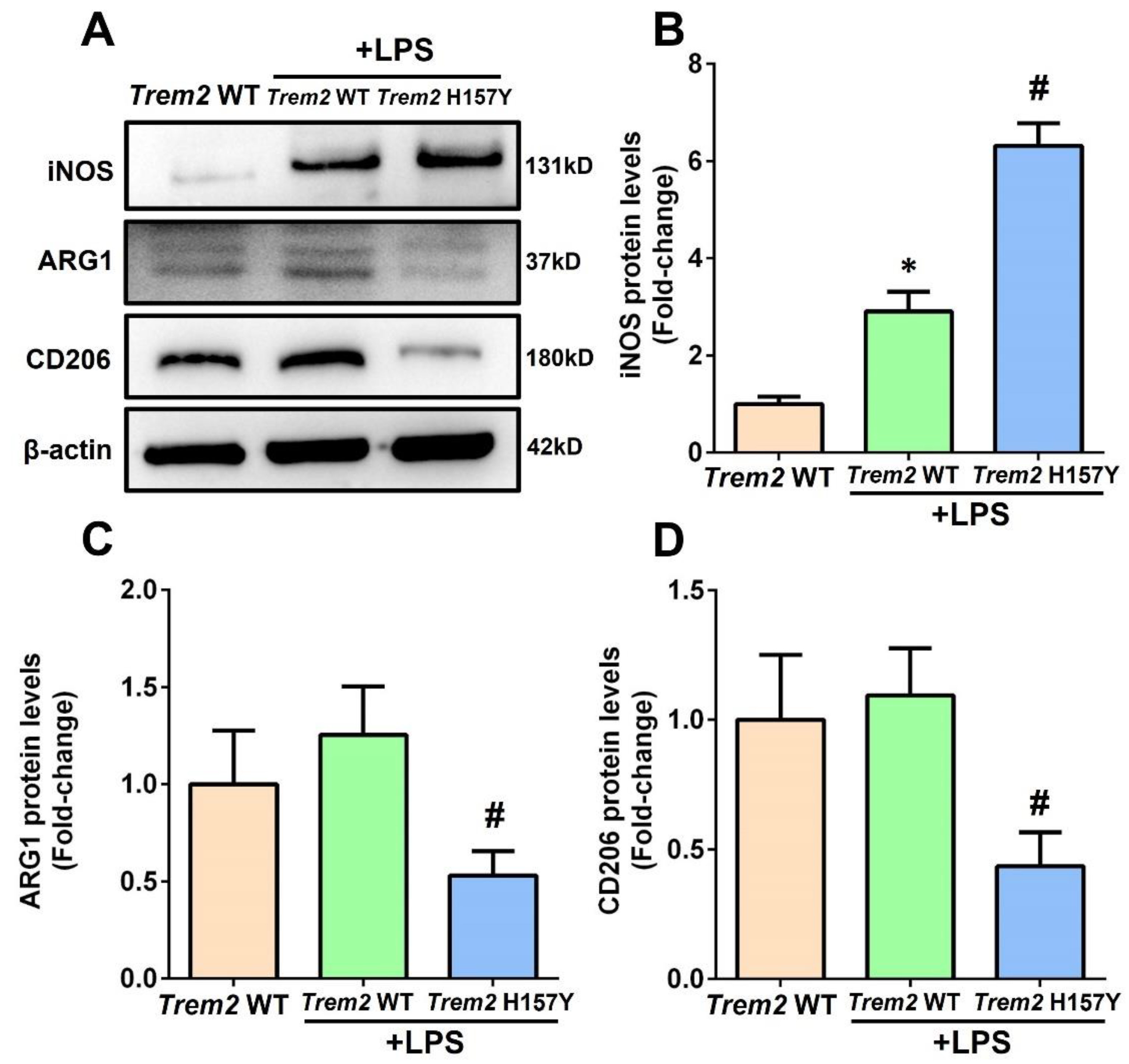
After stimulation, microglia showed either pro-inflammatory status (M1-type polarization) or anti-inflammatory status (M2-type polarization) [23]. To explore the influence of the Trem2 H157Y variant on microglial polarization, BV2 microglia were stimulated with 100 ng/mL LPS. As revealed in Figure 2A, B, LPS stimulation increased the levels of iNOS, a marker of M1-type polarization (p < 0.05) [24], in WT Trem2 BV2 microglia. Contrastingly, BV2 microglia with the Trem2 H157Y variant exhibited significantly higher levels of iNOS after LPS stimulation. As indicated in Figure 2A, C, D, LPS stimulation increased the protein levels of ARG1 and CD206, two markers of M2-type polarization [25], in WT Trem2 BV2 microglia, but these increments did not reach statistical significance (p > 0.05). In contrast, BV2 microglia with the Trem2 H157Y variant showed reduced protein levels of ARG1 and CD206 following LPS stimulation.

3.3. The Trem2 H157Y Variant Facilitates Microglial Release of Inflammatory Cytokines

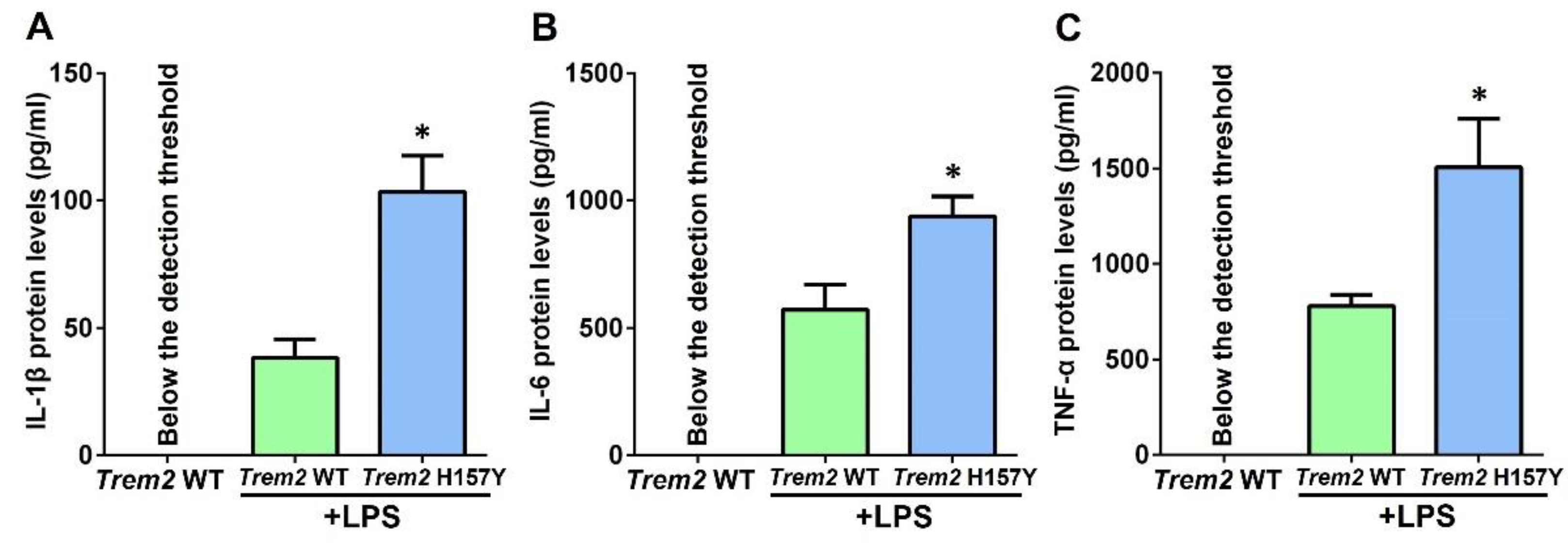
Afterward, we investigated the influence of the Trem2 H157Y variant on microglial inflammatory cytokine release. Stimulating WT Trem2 BV2 microglia with LPS led to release of IL-1β, IL-6, and TNF-α into the culture medium. As revealed in Figure 3A–C, BV2 microglia with the Trem2 H157Y variant released more inflammatory cytokines, including IL-1β, IL-6, and TNF-α, than WT Trem2 BV2 microglia after LPS stimulation.

4. Discussion

TREM2 is a newly identified susceptibility gene for AD [26]. In Caucasians, a rare coding variant, R47H, was reported to increase the risk of AD with an odds ratio of 4.7 [27,28]. Meanwhile, we identified that another rare coding variant H157Y substantially elevated AD risk in a Han Chinese population [14]. To date, how the TREM2 H157Y variant increases AD risk has not been fully understood. Human TREM2 encodes a 230-amino acid type I transmembrane receptor, which is exclusively expressed in microglia in the brain [29]. It contains an Ig-like V-type domain, a stalk region, a transmembrane domain, and a short cytoplasmic tail [30]. An increasing amount of evidence suggested that full-length TREM2 participated in AD progression via facilitated microglial phagocytosis of Aβ and suppressed M1-type polarization of microglia [22,31]. In this study, we have provided the first evidence that the Trem2 H157Y variant inhibits microglial Aβ phagocytosis. This finding was supported by a recent study from Schlepckow and colleagues showing that the TREM2 H157Y variant impaired phagocytosis of E.coli pHrodo particles in HEK293 cells [32]. Moreover, in the current study, we observed that the M1-type polarization marker iNOS was increased while the M2-type polarization markers ARG1 and CD206 were reduced in BV2 microglia with the Trem2 H157Y variant. To our knowledge, this is the first study reporting that the Trem2 H157Y variant induced microglial M1-type polarization. Under physiological conditions, a disintegrin and metalloproteinase (ADAM) 10/17 cleaves the full-length TREM2 protein at the H157-S158 site [33]. In HEK293 cells, the TREM2 H157Y variant was revealed to facilitate ADAM10/17 cleavage and thus reduce membrane-associated full-length TREM2 protein levels [32]. In view of the above evidence, the M1-type microglial polarization and impaired microglial Aβ phagocytosis caused by the H157Y variant might be ascribed to the reduction in full-length TREM2 protein levels on the surface of microglia.
Another interesting finding of this study was that the Trem2 H157Y variant promoted microglial release of inflammatory cytokines. The TREM2 H157Y variant was reported to enhance sTREM2 production via enhanced ADAM10/17 cleavage [33]. As a bioactive fragment of full-length TREM2 protein, sTREM2 was reported to promote microglial inflammatory cytokine release and subsequently induce neuroinflammation [34]. Therefore, the elevated inflammatory cytokine release caused by the Trem2 H157Y variant might be attributed to the enhanced sTREM2 production. However, our findings appeared to contradict those of a recent study by Qiao and colleagues [35], as they found that the Trem2 H157Y variant downregulated neuroinflammation-related genes in Trem2 H157Y knock-in 5xFAD transgenic mice. It should be noted that our experiments were conducted in a cellular inflammation model induced by LPS, whilst Qiao et al. employed an animal model of amyloid pathology to validate the function of the Trem2 H157Y variant [35]. The pathophysiological bases underlying these two models are quite different, which may account for these opposite observations.
This study also had some limitations. First, the impacts of the H157Y variant on the TREM2 downstream signaling pathway were not investigated in this study. In the future, the main components of the TREM2 downstream signaling pathway, such as TYROBP and β-catenin in microglia with the Trem2 H157Y variant, should be assessed [36,37,38]. Second, in this study, the functions of the H157Y variant were investigated using mouse BV2 microglial cells. Although TREM2 protein seemed to be conserved across mammals, our findings should be further confirmed using human microglia.

5. Conclusions

Summarily, in the current study, using CRISPR-Cas9-engineered BV2 microglia, we provide the first evidence that the Trem2 H157Y variant inhibits microglial phagocytosis of Aβ, promotes M1-type polarization of microglia, and facilitates microglial release of inflammatory cytokines. These findings provide new insights into the cellular mechanisms by which the TREM2 H157Y variant elevates the risk of AD.

Author Contributions

Funding acquisition: T.J. and Y.-D.Z.; Study design: T.J.; Original draft preparation: X.-X.F. and S.-Y.C.; Manuscript editing and revision: T.J.; Experiments: X.-X.F., S.-Y.C. and H.-W.L.; Data collection and analysis: Y.D. and R.D. All authors have read and agreed to the published version of the manuscript.

Funding

This study was supported by the National Science and Technology Innovation 2030—Major program of “Brain Science and Brain-Inspired Intelligence Research”, Ministry of Science and Technology of the People’s Republic of China (award to Y.-D.Z. (2021ZD0201807)), the National Natural Science Foundation of China (award to T.J. (81974156)), and the Natural Science Foundation of Jiangsu Province Department of Science and Technology (award to T.J. (BK20221175)).

Institutional Review Board Statement

Not applicable.

Data Availability Statement

The data that support the findings of this study are available from the corresponding author upon reasonable request.

Conflicts of Interest

The authors declare no conflict of interest.

References

  1. Scheltens, P.; Blennow, K.; Breteler, M.M.; de Strooper, B.; Frisoni, G.B.; Salloway, S.; Van der Flier, W.M. Alzheimer’s disease. Lancet 2016, 388, 505–517. [Google Scholar] [CrossRef] [PubMed]
  2. Jiang, T.; Yu, J.T.; Tian, Y.; Tan, L. Epidemiology and Etiology of Alzheimer’s disease: From Genetic to Non-Genetic Factors. Curr. Alzheimer Res. 2013, 10, 852–867. [Google Scholar] [CrossRef] [PubMed]
  3. Graham, W.V.; Bonito-Oliva, A.; Sakmar, T.P. Update on Alzheimer’s Disease Therapy and Prevention Strategies. Annu. Rev. Med. 2017, 68, 413–430. [Google Scholar] [CrossRef] [PubMed] [Green Version]
  4. Hansen, D.V.; Hanson, J.E.; Sheng, M. Microglia in Alzheimer’s disease. J. Cell Biol. 2018, 217, 459–472. [Google Scholar] [CrossRef]
  5. Tajbakhsh, A.; Read, M.; Barreto, G.E.; Avila-Rodriguez, M.; Gheibi-Hayat, S.M.; Sahebkar, A. Apoptotic neurons and amyloid-beta clearance by phagocytosis in Alzheimer’s disease: Pathological mechanisms and therapeutic outlooks. Eur. J. Pharmacol. 2021, 895, 173873. [Google Scholar] [CrossRef] [PubMed]
  6. Cai, Y.; Liu, J.; Wang, B.; Sun, M.; Yang, H. Microglia in the Neuroinflammatory Pathogenesis of Alzheimer’s Disease and Related Therapeutic Targets. Front. Immunol. 2022, 13, 856376. [Google Scholar] [CrossRef]
  7. Schoch, K.M.; Ezerskiy, L.A.; Morhaus, M.M.; Bannon, R.N.; Sauerbeck, A.D.; Shabsovich, M.; Jafar-Nejad, P.; Rigo, F.; Miller, T.M. Acute Trem2 reduction triggers increased microglial phagocytosis, slowing amyloid deposition in mice. Proc. Natl. Acad. Sci. USA. 2021, 118, e2100356118. [Google Scholar] [CrossRef]
  8. Liu, W.; Taso, O.; Wang, R.; Bayram, S.; Graham, A.C.; Garcia-Reitboeck, P.; Mallach, A.; Andrews, W.D.; Piers, T.M.; Botia, J.A.; et al. Trem2 promotes anti-inflammatory responses in microglia and is suppressed under pro-inflammatory conditions. Hum. Mol. Genet. 2020, 29, 3224–3248. [Google Scholar] [CrossRef] [PubMed]
  9. Qiu, Z.; Wang, H.; Qu, M.; Zhu, S.; Zhang, H.; Liao, Q.; Miao, C. Consecutive Injection of High-Dose Lipopolysaccharide Modulates Microglia Polarization via TREM2 to Alter Status of Septic Mice. Brain Sci. 2023, 13, 126. [Google Scholar] [CrossRef]
  10. Hu, Y.; Li, C.; Wang, X.; Chen, W.; Qian, Y.; Dai, X. TREM2, Driving the Microglial Polarization, Has a TLR4 Sensitivity Profile After Subarachnoid Hemorrhage. Front. Cell Dev. Biol. 2021, 9, 693342. [Google Scholar] [CrossRef] [PubMed]
  11. Li, Y.; Long, W.; Gao, M.; Jiao, F.; Chen, Z.; Liu, M.; Yu, L. TREM2 Regulates High Glucose-Induced Microglial Inflammation via the NLRP3 Signaling Pathway. Brain Sci. 2021, 11, 896. [Google Scholar] [CrossRef]
  12. Carmona, S.; Zahs, K.; Wu, E.; Dakin, K.; Bras, J.; Guerreiro, R. The role of TREM2 in Alzheimer’s disease and other neurodegenerative disorders. Lancet Neurol. 2018, 17, 721–730. [Google Scholar] [CrossRef]
  13. Cruchaga, C.; Kauwe, J.S.; Harari, O.; Jin, S.C.; Cai, Y.; Karch, C.M.; Benitez, B.A.; Jeng, A.T.; Skorupa, T.; Carrell, D.; et al. GWAS of cerebrospinal fluid tau levels identifies risk variants for Alzheimer’s disease. Neuron 2013, 78, 256–268. [Google Scholar] [CrossRef] [Green Version]
  14. Jiang, T.; Tan, L.; Chen, Q.; Tan, M.S.; Zhou, J.S.; Zhu, X.C.; Lu, H.; Wang, H.F.; Zhang, Y.D.; Yu, J.T. A rare coding variant in TREM2 increases risk for Alzheimer’s disease in Han Chinese. Neurobiol. Aging 2016, 42, 217.e1–217.e3. [Google Scholar] [CrossRef] [PubMed]
  15. Jiang, T.; Hou, J.K.; Gao, Q.; Yu, J.T.; Zhou, J.S.; Zhao, H.D.; Zhang, Y.D. TREM2 p.H157Y Variant and the Risk of Alzheimer’s Disease: A Meta-Analysis Involving 14,510 Subjects. Curr. Neurovascular Res. 2016, 13, 318–320. [Google Scholar] [CrossRef] [PubMed]
  16. Concordet, J.P.; Haeussler, M. CRISPOR: Intuitive guide selection for CRISPR/Cas9 genome editing experiments and screens. Nucleic Acids Res. 2018, 46, W242–W245. [Google Scholar] [CrossRef] [Green Version]
  17. Brinkman, E.K.; Kousholt, A.N.; Harmsen, T.; Leemans, C.; Chen, T.; Jonkers, J.; van Steensel, B. Easy quantification of template-directed CRISPR/Cas9 editing. Nucleic Acids Res. 2018, 46, e58. [Google Scholar] [CrossRef] [PubMed]
  18. Brinkman, E.K.; Chen, T.; Amendola, M.; van Steensel, B. Easy quantitative assessment of genome editing by sequence trace decomposition. Nucleic Acids Res. 2014, 42, e168. [Google Scholar] [CrossRef]
  19. Wang, C.; Chen, S.; Guo, H.; Jiang, H.; Liu, H.; Fu, H.; Wang, D. Forsythoside a mitigates Alzheimer’s-like pathology by inhibiting ferroptosis-mediated neuroinflammation via Nrf2/GPX4 axis activation. Int. J. Biol. Sci. 2022, 18, 2075. [Google Scholar] [CrossRef]
  20. Wang, S.Y.; Fu, X.X.; Duan, R.; Wei, B.; Cao, H.M.; Yan, E.; Chen, S.Y.; Zhang, Y.D.; Jiang, T. The Alzheimer’s disease-associated gene TREML2 modulates inflammation by regulating microglia polarization and NLRP3 inflammasome activation. Neural Regen. Res. 2023, 18, 434–438. [Google Scholar] [CrossRef]
  21. Jiang, T.; Zhang, Y.D.; Gao, Q.; Zhou, J.S.; Zhu, X.C.; Lu, H.; Shi, J.Q.; Tan, L.; Chen, Q.; Yu, J.T. TREM1 facilitates microglial phagocytosis of amyloid beta. Acta Neuropathol. 2016, 132, 667–683. [Google Scholar] [CrossRef]
  22. Jiang, T.; Tan, L.; Zhu, X.C.; Zhang, Q.Q.; Cao, L.; Tan, M.S.; Gu, L.Z.; Wang, H.F.; Ding, Z.Z.; Zhang, Y.D.; et al. Upregulation of TREM2 ameliorates neuropathology and rescues spatial cognitive impairment in a transgenic mouse model of Alzheimer’s disease. Neuropsychopharmacol. Off. Publ. Am. Coll. Neuropsychopharmacol. 2014, 39, 2949–2962. [Google Scholar] [CrossRef] [Green Version]
  23. Chauhan, P.; Sheng, W.S.; Hu, S.; Prasad, S.; Lokensgard, J.R. Differential Cytokine-Induced Responses of Polarized Microglia. Brain Sci. 2021, 11, 1482. [Google Scholar] [CrossRef] [PubMed]
  24. Collmann, F.M.; Pijnenburg, R.; Hamzei-Taj, S.; Minassian, A.; Folz-Donahue, K.; Kukat, C.; Aswendt, M.; Hoehn, M. Individual in vivo Profiles of Microglia Polarization After Stroke, Represented by the Genes iNOS and Ym1. Front. Immunol. 2019, 10, 1236. [Google Scholar] [CrossRef] [Green Version]
  25. Kobashi, S.; Terashima, T.; Katagi, M.; Nakae, Y.; Okano, J.; Suzuki, Y.; Urushitani, M.; Kojima, H. Transplantation of M2-Deviated Microglia Promotes Recovery of Motor Function after Spinal Cord Injury in Mice. Mol. Ther. J. Am. Soc. Gene Ther. 2020, 28, 254–265. [Google Scholar] [CrossRef] [PubMed]
  26. Ulland, T.K.; Colonna, M. TREM2—a key player in microglial biology and Alzheimer disease. Nat. Rev. Neurol. 2018, 14, 667–675. [Google Scholar] [CrossRef] [PubMed]
  27. Guerreiro, R.; Wojtas, A.; Bras, J.; Carrasquillo, M.; Rogaeva, E.; Majounie, E.; Cruchaga, C.; Sassi, C.; Kauwe, J.S.; Younkin, S.; et al. TREM2 variants in Alzheimer’s disease. N. Engl. J. Med. 2013, 368, 117–127. [Google Scholar] [CrossRef] [Green Version]
  28. Jonsson, T.; Stefansson, H.; Steinberg, S.; Jonsdottir, I.; Jonsson, P.V.; Snaedal, J.; Bjornsson, S.; Huttenlocher, J.; Levey, A.I.; Lah, J.J.; et al. Variant of TREM2 associated with the risk of Alzheimer’s disease. N. Engl. J. Med. 2013, 368, 107–116. [Google Scholar] [CrossRef] [Green Version]
  29. Jiang, T.; Yu, J.T.; Zhu, X.C.; Tan, L. TREM2 in Alzheimer’s disease. Mol. Neurobiol. 2013, 48, 180–185. [Google Scholar] [CrossRef]
  30. Colonna, M. The biology of TREM receptors. Nat. Rev. Immunol. 2023, 1–15. [Google Scholar] [CrossRef]
  31. Jiang, T.; Zhang, Y.D.; Chen, Q.; Gao, Q.; Zhu, X.C.; Zhou, J.S.; Shi, J.Q.; Lu, H.; Tan, L.; Yu, J.T. TREM2 modifies microglial phenotype and provides neuroprotection in P301S tau transgenic mice. Neuropharmacology 2016, 105, 196–206. [Google Scholar] [CrossRef] [PubMed]
  32. Schlepckow, K.; Kleinberger, G.; Fukumori, A.; Feederle, R.; Lichtenthaler, S.F.; Steiner, H.; Haass, C. An Alzheimer-associated TREM2 variant occurs at the ADAM cleavage site and affects shedding and phagocytic function. EMBO Mol. Med. 2017, 9, 1356–1365. [Google Scholar] [CrossRef] [PubMed]
  33. Thornton, P.; Sevalle, J.; Deery, M.J.; Fraser, G.; Zhou, Y.; Stahl, S.; Franssen, E.H.; Dodd, R.B.; Qamar, S.; Gomez Perez-Nievas, B.; et al. TREM2 shedding by cleavage at the H157-S158 bond is accelerated for the Alzheimer’s disease-associated H157Y variant. EMBO Mol. Med. 2017, 9, 1366–1378. [Google Scholar] [CrossRef]
  34. Zhong, L.; Chen, X.F.; Wang, T.; Wang, Z.; Liao, C.; Wang, Z.; Huang, R.; Wang, D.; Li, X.; Wu, L.; et al. Soluble TREM2 induces inflammatory responses and enhances microglial survival. J. Exp. Med. 2017, 214, 597–607. [Google Scholar] [CrossRef] [Green Version]
  35. Qiao, W.; Chen, Y.; Zhong, J.; Madden, B.J.; Charlesworth, C.M.; Martens, Y.A.; Liu, C.C.; Knight, J.; Ikezu, T.C.; Aishe, K.; et al. Trem2 H157Y increases soluble TREM2 production and reduces amyloid pathology. Mol. Neurodegener. 2023, 18, 8. [Google Scholar] [CrossRef] [PubMed]
  36. Zheng, H.; Jia, L.; Liu, C.C.; Rong, Z.; Zhong, L.; Yang, L.; Chen, X.F.; Fryer, J.D.; Wang, X.; Zhang, Y.W.; et al. TREM2 Promotes Microglial Survival by Activating Wnt/beta-Catenin Pathway. J. Neurosci. 2017, 37, 1772–1784. [Google Scholar] [CrossRef] [Green Version]
  37. Haure-Mirande, J.V.; Audrain, M.; Ehrlich, M.E.; Gandy, S. Microglial TYROBP/DAP12 in Alzheimer’s disease: Transduction of physiological and pathological signals across TREM2. Mol. Neurodegener. 2022, 17, 55. [Google Scholar] [CrossRef]
  38. Pons, V.; Levesque, P.; Plante, M.M.; Rivest, S. Conditional genetic deletion of CSF1 receptor in microglia ameliorates the physiopathology of Alzheimer’s disease. Alzheimer’s Res. Ther. 2021, 13, 8. [Google Scholar] [CrossRef] [PubMed]
Figure 1. The Trem2 H157Y variant inhibits microglial phagocytosis of Aβ. An in vitro assay was employed to evaluate microglial Aβ1–42 phagocytosis and degradation. (A) WT Trem2 BV2 microglia or BV2 microglia with the Trem2 H157Y variant were incubated with 5 μM Aβ1–42 for 6 h. The amount of internalized Aβ1–42 was measured by ELISA (t = 6 h). (B) Later, the Aβ1–42 left in the medium was washed out, and WT Trem2 BV2 microglia or BV2 microglia with the Trem2 H157Y variant were maintained for an additional 6 h to allow for Aβ1–42 degradation. The intracellular Aβ1–42 levels were assessed by ELISA again (t = 12 h). (C) The index of Aβ1–42 degradation was expressed as the ratio of the total amount of internalized Aβ1–42 (t = 6 h) to remaining Aβ1–42 (t = 12 h). A higher degradation index indicates a better capacity for degradation of Aβ1–42. Data were analyzed by one-way ANOVA followed by Tukey’s post hoc test. Columns represent means ± SDs (n = 3, performed in triplicates), * p < 0.05 versus WT Trem2 BV2 microglia.
Brainsci 13 00642 g001
Figure 2. The Trem2 H157Y variant promotes M1-type polarization of microglia. (A) Levels of iNOS, ARG1, and CD206 in WT Trem2 BV2 microglia and BV2 microglia with the Trem2 H157Y variant after LPS stimulation were detected by Western blot analysis. (BD) Quantitative analysis of iNOS, ARG1, and CD206 protein levels. Data were normalized to β-actin. Data were analyzed by one-way ANOVA followed by Tukey’s post hoc test. Columns represent means ± SDs (n = 3, performed in triplicates). * p < 0.05 versus WT Trem2 BV2 microglia without LPS stimulation; # p < 0.05 versus LPS-stimulated WT Trem2 BV2 microglia.
Brainsci 13 00642 g002
Figure 3. The Trem2 H157Y variant facilitates microglial release of inflammatory cytokines. (AC) Levels of the inflammatory cytokines IL-1β, IL-6, and TNF-α in WT Trem2 BV2 microglia and BV2 microglia with the Trem2 H157Y variant were detected by ELISA. Data were analyzed using the Student’s t-test. Columns represent means ± SDs (n = 3, performed in triplicates). * p < 0.05 versus LPS-stimulated WT Trem2 BV2 microglia.
Brainsci 13 00642 g003
Disclaimer/Publisher’s Note: The statements, opinions and data contained in all publications are solely those of the individual author(s) and contributor(s) and not of MDPI and/or the editor(s). MDPI and/or the editor(s) disclaim responsibility for any injury to people or property resulting from any ideas, methods, instructions or products referred to in the content.