
A Review on Motor Imagery with Transcranial Alternating Current Stimulation: Bridging Motor and Cognitive Welfare for Patient Rehabilitation


Abstract
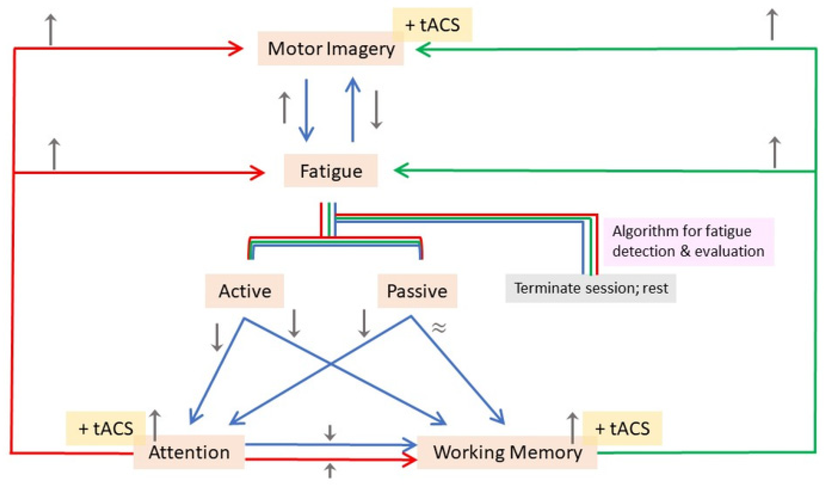
:1. Introduction
2. Review Selection and Criteria
3. Transcranial Direct Current Stimulation (tDCS) and Transcranial Alternating Current Stimulation (tACS)
3.1. Working Principles and Mechanisms
3.2. Implications on Neurophysiology
3.3. Potential Adverse Effects
3.4. Motor Imagery-Related tACS Studies
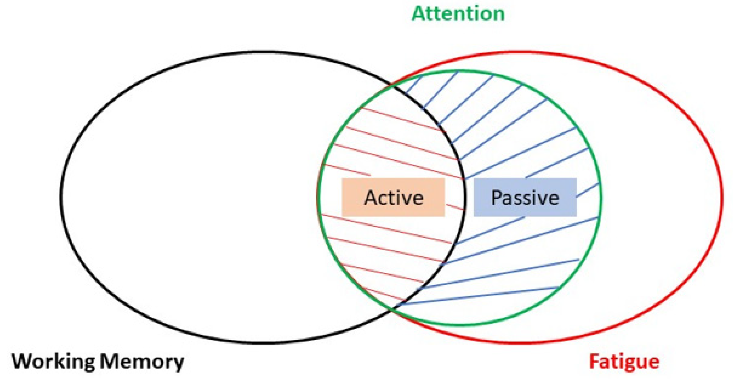
4. Cognition in Rehabilitation
4.1. Working Memory-Related tACS Studies

| Working Memory-Related Studies | ||||
|---|---|---|---|---|
| Study | Subjects and Design | Experimental Task | Stimulation Montage and Parameters | Outcome |
| Alekseichuk et al. (2016) [46] | 47 healthy adults (22 males, 25 females) | 2-back visual-spatial match-to-sample test | Active electrodes: central electrode over AF3 (10–10 system), the other 4 were equally spaced at 6 cm from AF3. 1 mA (peak-to-peak), 10 min (including 10 s ramp-up and ramp-down periods), 0.6 mA peak-to-baseline. Sham stimulation 1:30 s and then turned off. Sham stimulation 2:80 Hz at a lower intensity (0.2 mA peak-to-baseline). | θ and θ-γ frequency coupling improve working memory performance. High γ power (80 Hz γ bursts) coupled over the peak, but not the trough, of θ improves working memory. Optimal γ frequencies for improved performance are in the range of 80 to 100 Hz. |
| de Lara et al. (2018) [47] | 72 healthy adults (36 males, 36 females). Between-group design. | Paired-associative learning task using word-pairs | Active electrode at T7 and return electrodes at FPz and T8 (10–20 system). 1 mA (peak-to-baseline), 10 min (including a 10 s ramp-up and 10 s ramp-down). Sham stimulation: 10 s ramp-up, current delivered for 30 s, 10 s ramp-down. | γ bursts coupled to the troughs of θ-tACS resulted in behavioral impairment in memory performance. γ bursts coupled to the peaks of θ-tACS or superimposed over the whole θ-tACS waveform resulted in no significant behavioral effects. |
| Thompson et al. (2021) [48] | 51 healthy adults (21 males, 30 females). Within-subject design. | Visual retro-cue task | Active electrodes over P3 and P4 (10–20 system). 1.5 mA (20 s ramp-up and ramp-down 20 s), 20 min. Sham stimulation: 20 s. | Parietal γ-tACS resulted in significant improvement in recall precision only during invalid cue trials and only for high-baseline performers (assessed using a multi-level mixed model). |
| Tseng et al. (2016) [49] | 20 healthy adults (12 males, 8 females). Within-subject design. | Change detection task | Active electrodes at CP1 and T5 (10–20 system). 1.5 mA (peak-to-peak), 20 min (switched off in last 2 blocks). Sham stimulation: only for 30 s. | γ-tACS improved task performance only for shape-color binding trials in low-performers (based on d’ index from hit and false alarm rates). Improved performance lasted throughout the last 2 blocks (final 20 min offline session). |
| Lang et al. (2019) [51] | 59 healthy adults. Between-group design. | Visual associative memory task (designed in-house): Face and Scene Task (FAST) | Active electrodes at FP1, P2, P3, PO7, P10; anode at P10. 2 mA (peak-to-baseline), 10 min (30 s ramp-up and ramp-down). Sham stimulation: 30 s ramp-up to 2 mA, 30 s ramp-down to 0.06 mA for 9 min. | θ-tACS improved visual associative memory performance (based on correct hits and # errors). θ-tACS facilitated reduction in false memory and forgetting (based on # errors). Results suggest that tACS is more effective than tDCS. |
| Röhner et al. (2018) [52] | 30 healthy adults (15 males, 15 females). Within-subject design. | 2-back visual letter task | Active electrodes placed over F3 and P3 (10–20 system). 1 mA, 15 min (including a 15 s ramp-up and 15 s ramp-down). Sham stimulation: 1 min tDCS. | Offline effect of θ-tACS on RTs showed significant improvement in performance (not observed in anodal tDCS condition). |
| Abellaneda-Pérez et al. (2020) [54] | 44 healthy adults (22 males, 20 females). Between-group design. | Verbal n-back task | Active electrode over F3 and return electrode over FP2 (10–10 system). 2 mA (peak-to-peak), 20 min (15 s ramp-up and ramp-down). Sham stimulation: terminated after 30 s of delivery. | θ-tACS effects suggested to be driven by online brain activity changes during stimulation, but not post-stimulation. Enhanced brain activity was found in θ-tACS group in frontal, parietal and thalamic areas during lowest working memory load, and in right frontal areas during highest working memory load. |
| Meng et al. (2021) [55] | 20 healthy adults (8 males, 12 females). Within-subject design. | Visual associative memory task (designed in-house): Face and Scene Task (FAST), perceptual recognition test | Active electrode over P3 (10–20 system). 2 mA (peak-to-peak), 15 min (30 s ramp-up and 30 s ramp-down). Sham stimulation: 50 s. | θ-tACS impaired associative memory performance (based on d’ from hit and false alarm rates). No significant difference between θ-tACS and sham groups for perceptual recognition test. |
| Pahor and Jaušovec (2018) [56] | 72 healthy adults (females). Within-subject design. | Change detection tasks, n-back tasks | Pair combination of active electrodes placement over F3, F4, P3, P4 (10–20 system). 1.25 mA to 2 mA stepwise increment over 30 s, 15 min. Sham stimulation: 1 min. | θ-tACS modulates for significant changes in post-stim resting-state EEG amplitude relative to baseline. θ-tACS facilitated small improvements in performance only for certain n-back tasks. θ-tACS resulted in significant ERP amplitude and latency changes in n-back tasks compared to sham. |
| Nomura et al. (2019) [57] | 36 healthy adults (8 males, 28 females). Between-group design | Visual word recognition task | Active electrode over F3 (10–20 system). 750 uA (peak-to-baseline), 15 min, 100 cycles fade-in and fade-out. Sham stimulation: 10 s. | γ-tACS applied over the left prefrontal cortex enhances episodic memory (i.e., long term recall) response accuracy without affecting reaction time. |
4.2. Attention-Related tACS Studies
| Attention-Related Studies | ||||
|---|---|---|---|---|
| Study | Subjects and Design | Experimental Task | Stimulation Montage and Parameters | Outcome |
| Kasten et al. (2020) [61] | 20 healthy adults (10 males, 10 females). Crossover within-subject randomized design. | Spatial cue task | 2 pairs of electrodes at O1-P3 and O2-P4 (10–20 system). 1 mA (peak-to-baseline), ~8 min per block. | tACS modulates RTs only in endogenous attention. Right occipital α-tACS increases RTs in both valid and invalid cue trials. γ-tACS contralateral-to-cue modulated RTs more significantly than α-tACS ipsilateral-to-cue as compared to that for γ-tACS: observation only for invalid cue trials. |
| Schuhmann et al. (2019) [62] | 36 healthy adults (18 males, 18 females). Within-subject design. | Spatial cue task and Stimulus detection task | P3 (10–20 system). 1 mA (peak-to-peak), >40 min for all tasks. Sham stimulation: 100 cycles, ramped up and down immediately. | Larger leftward bias in RTs during endogenous attention task in tACS group. |
| Hopfinger et al. (2016) [63] | 23 healthy adults (9 males, 14 females). Within-subject design. | Spatial cue task | P6 and Cz (10–20 system). 1 mA (peak-to-baseline). Sham stimulation: 30 s (4 s ramp-up, maintained for 22 s, 4 s ramp-down). | Significantly slower RTs in invalid/uncued exogenous trials with α-tACS. Significantly faster RTs in invalid endogenous trials with γ-tACS. |
| Klírová et al. (2021) [65] | 20 healthy adults (10 males, 10 females). Crossover design. | Simon task, Stop Signal task, Conner’s Continuous Performance Test 3rd edition (CPT III), Stroop test | FCz and Pz. 1 mA (peak-to-baseline), 30 min (5 s ramp-up and 5 s ramp-down). Sham stimulation: 5 s ramp-up, 30 s, 29 min 30 s rest, 5 s ramp-down. | Only significant effect observed in the Stroop color-word test and Stroop interference scores, with better performance in the individualized stimulation group compared to that of the non-individualized group. However, neither group differed significantly in performance when compared to sham. |
| Lehr et al. (2019) [66] | 22 healthy adults (6 males, 16 females). Within-subject design. | Stroop color-word task | AF3, 4 return electrodes at F5, F2, Fp2, AF7 (10–10 system). 1 mA (peak-to-baseline), 20 min (including 10 s ramp-up and ramp-down). Sham stimulation: 30 s; at the beginning and end of the task. | θ-tACS reduced Stroop effect only in trials preceded by congruent trials. |
| Rostami et al. (2020) [67] | 13 healthy adults (7 males, 6 females). Within-subject design. | Rapid visual information processing (RVIP) task from CANTAB | Fpz (10–20 system). 1 mA (peak-to-peak), 20 min, (10 s ramp-up and down). Sham parameters: 30 s. | 6 Hz θ-tACS increased frontal-midline theta and resulted in significant changes in RVIP scores. Faster RTs for correct responses with 6 Hz θ-tACS. EEG power analysis showed changes in theta PSD in frontal, central, and temporal regions in right hemisphere. |
| Moliadze et al. (2019) [68] | 24 healthy adults (12 males, 12 females). Within-subject design. | Phonological task (words) | 1 electrode each located between F1, F5, FC3 (left) and F2, F6, FC4 (right). 1 mA, 20 min (15 s ramp-up and down). Sham stimulation: 15 s ramp-up, 30 s at 1 mA, 15 s ramp-down. | 10 Hz α-tACS significantly facilitates phonological word decision RTs. 10 Hz α-tACS significantly increases task-related theta power during phonological decisions. |
| Hutchinson et al. (2020) [71] | 71 healthy adults (34 males, 37 females). Between-group design. | Inattentional Blindness (IB) task developed by Pitts et al. (2012) | Oz and Cz. Current intensity customized to subject’s level of comfort or subjects reported phosphenes. Sham stimulation: a mild current of 30 s. | α-tACS group: 87% inattentionally blind; θ-tACS group: 45.8% inattentionally blind; Sham group: 50% inattentionally blind; α-tACS less perceptive of target stimulus than those with θ-tACS or sham applied. |
| van Schouwenburg et al. (2017) [72] | 37 healthy adults. Between-group design. | Spatial cue task | F4 and P4. 1 mA (peak-to-baseline), 5 min (15 s ramp up and down). Sham stimulation: immediate ramp down over 15 s. | Sham group showed significant attention bias (faster RTs to targets) in the right hemifield compared to the left hemifield. Sham group showed significant lateralization in frontoposterior alpha coherence that was not present in the α-tACS group. In α-tACS group, they found a relative increase in right hemispheric coherence (relative to the left) and an attentional shift towards the left hemifield. |
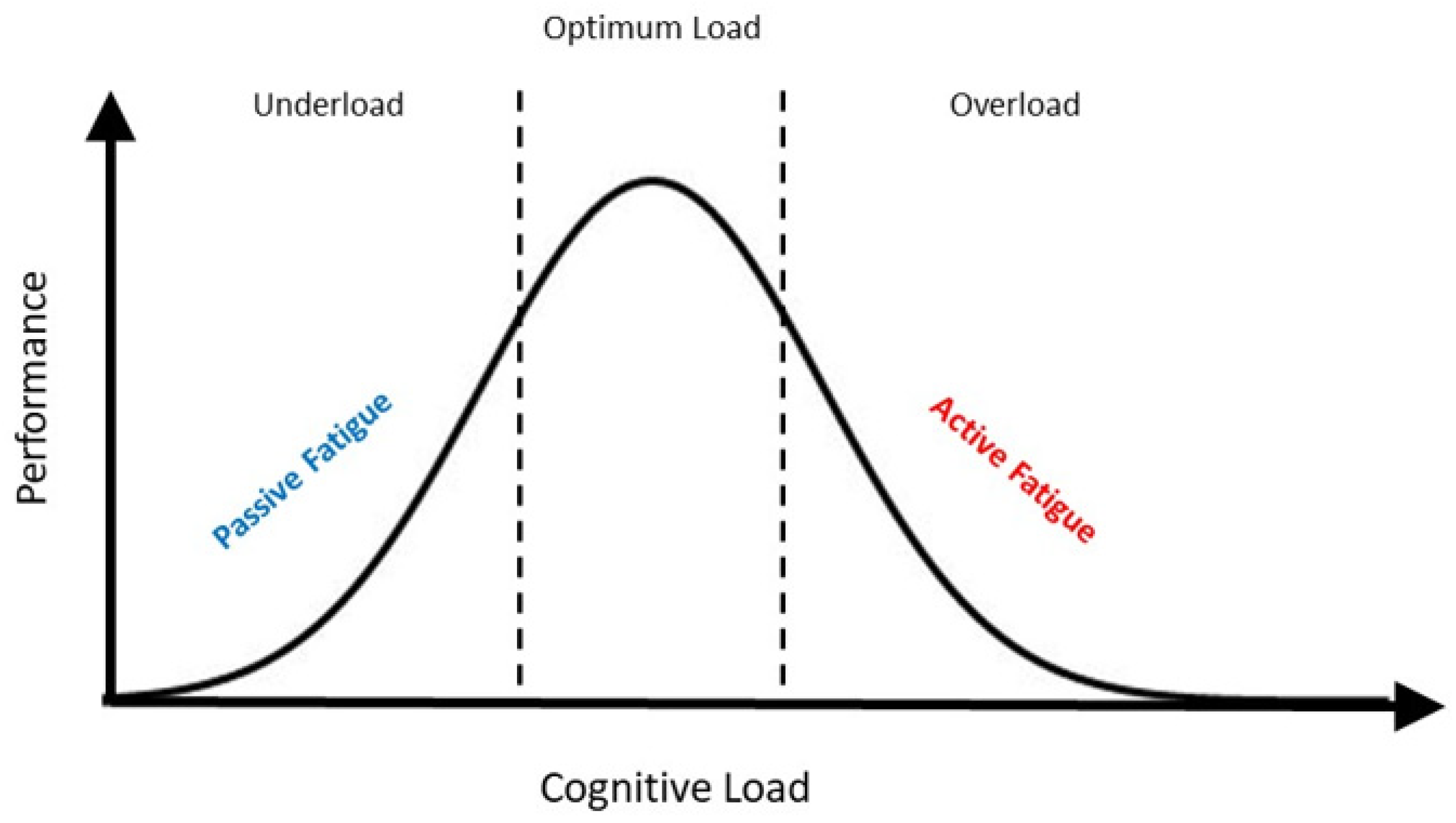
4.3. Fatigue-Related tACS Studies
| Fatigue-Related Studies (Non-tACS) | ||||
|---|---|---|---|---|
| Study | Subjects and Design | Experimental Task | Recording Montage | Outcome |
| Huang et al. (2016) [75] | 12 healthy adults (7 males, 5 females). | Virtual Reality-based highway driving: event-related lane-departure/deviation task | 32-channel EEG recording electrodes (10–20 system). | Increased theta activity in frontal midline and occipital areas. Average RTs of epochs with auditory warning maintained at 1.15 times the mean RT. Significantly slower RTs in trials in which α power exceeded warning threshold but was not given an auditory warning. Occipital EEG power spectra in θ and α bands decreased rapidly with warnings. |
| Foong et al. (2019) [78] | 29 healthy adults | Driving simulation task | EEG electrodes at FP1, FP2, TP9, TP10 (10–20 system). | Semi-supervised learning using labeled attentive data to predict and identify passive fatigue from unlabeled data. |
| Zhang et al. (2021) [82] | 48 healthy adults (24 males, 24 females). | Detection response task (DRT) in driving simulator | 64-channel EEG recording electrodes (10–20 system) | DRT performance declines at 40 min (based on RTs and response accuracy). α power was significantly higher in the automated driving group as opposed to the manual driving group; indicative of passive fatigue. |
5. Discussion
6. Conclusions
Author Contributions
Funding
Conflicts of Interest
References
- Sharma, N.; Pomeroy, V.M.; Baron, J.-C. Motor imagery. Stroke 2006, 37, 1941–1952. [Google Scholar] [CrossRef]
- Aflalo, T.; Kellis, S.; Klaes, C.; Lee, B.; Shi, Y.; Pejsa, K.; Shanfield, K.; Hayes-Jackson, S.; Aisen, M.; Heck, C.; et al. Decoding motor imagery from the posterior parietal cortex of a tetraplegic human. Science 2015, 348, 906–910. [Google Scholar] [CrossRef]
- Cui, H.; Andersen, R.A. Different Representations of Potential and Selected Motor Plans by Distinct Parietal Areas. J. Neurosci. 2011, 31, 18130–18136. [Google Scholar] [CrossRef]
- Hanakawa, T. Rostral premotor cortex as a gateway between motor and cognitive networks. Neurosci. Res. 2011, 70, 144–154. [Google Scholar] [CrossRef]
- Xu, L.; Zhang, H.; Hui, M.; Long, Z.; Jin, Z.; Liu, Y.; Yao, L. Motor execution and motor imagery: A comparison of functional connectivity patterns based on graph theory. Neuroscience 2014, 261, 184–194. [Google Scholar] [CrossRef]
- Kasahara, K.; DaSalla, C.S.; Honda, M.; Hanakawa, T. Neuroanatomical correlates of brain–computer interface performance. NeuroImage 2015, 110, 95–100. [Google Scholar] [CrossRef]
- Park, C.-H.; Chang, W.H.; Lee, M.; Kwon, G.H.; Kim, L.; Kim, S.T.; Kim, Y.-H. Which motor cortical region best predicts imagined movement? NeuroImage 2015, 113, 101–110. [Google Scholar] [CrossRef]
- Ang, K.K.; Guan, C.; Phua, K.S.; Wang, C.; Zhou, L.; Tang, K.Y.; Joseph, G.J.E.; Kuah, C.W.K.; Chua, K.S.G. Brain-computer interface-based robotic end effector system for wrist and hand rehabilitation: Results of a three-armed randomized controlled trial for chronic stroke. Front. Neuroeng. 2014, 7, 30. [Google Scholar] [CrossRef]
- Frolov, A.A.; Mokienko, O.; Lyukmanov, R.; Biryukova, E.; Kotov, S.; Turbina, L.; Nadareyshvily, G.; Bushkova, Y. Post-stroke Rehabilitation Training with a Motor-Imagery-Based Brain-Computer Interface (BCI)-Controlled Hand Exoskeleton: A Randomized Controlled Multicenter Trial. Front. Neurosci. 2017, 11, 400. [Google Scholar] [CrossRef]
- Johnson, S.H. Imagining the impossible. NeuroReport 2000, 11, 729–732. [Google Scholar] [CrossRef]
- Ang, K.K.; Guan, C. Brain–Computer Interface for Neurorehabilitation of Upper Limb After Stroke. Proc. IEEE 2015, 103, 944–953. [Google Scholar] [CrossRef]
- Ang, K.K.; Guan, C.; Phua, K.S.; Wang, C.; Zhao, L.; Teo, W.P.; Chen, C.; Ng, Y.S.; Chew, E. Facilitating effects of transcranial direct current stimulation on motor imagery brain-computer interface with robotic feedback for stroke rehabilitation. Arch. Phys. Med. Rehabil. 2015, 96, S79–S87. [Google Scholar] [CrossRef]
- Chew, E.; Teo, W.-P.; Tang, N.; Ang, K.K.; Ng, Y.S.; Zhou, J.H.; Teh, I.; Phua, K.S.; Zhao, L.; Guan, C. Using transcranial direct current stimulation to augment the effect of Motor Imagery-assisted brain-computer interface training in Chronic Stroke Patients—Cortical reorganization considerations. Front. Neurol. 2020, 11, 948. [Google Scholar] [CrossRef]
- Hu, M.; Cheng, H.-J.; Ji, F.; Chong, J.S.X.; Lu, Z.; Huang, W.; Ang, K.K.; Phua, K.S.; Chuang, K.-H.; Jiang, X.; et al. Brain Functional Changes in Stroke Following Rehabilitation Using Brain-Computer Interface-Assisted Motor Imagery With and Without tDCS: A Pilot Study. Front. Hum. Neurosci. 2021, 15, 692304. [Google Scholar] [CrossRef] [PubMed]
- Adams, A.G.; Schweitzer, D.; Molenberghs, P.; Henry, J.D. A meta-analytic review of social cognitive function following stroke. Neurosci. Biobehav. Rev. 2019, 102, 400–416. [Google Scholar] [CrossRef]
- Sensenbrenner, B.; Rouaud, O.; Graule-Petot, A.; Guillemin, S.; Piver, A.; Giroud, M.; Béjot, Y.; Jacquin-Piques, A. High Prevalence of Social Cognition Disorders and Mild Cognitive Impairment Long Term After Stroke. Alzheimer Dis. Assoc. Disord. 2019, 34, 72–78. [Google Scholar] [CrossRef]
- Kanellopoulos, D.; Wilkins, V.; Avari, J.; Oberlin, L.; Arader, L.; Chaplin, M.; Banerjee, S.; Alexopoulos, G.S. Dimensions of Poststroke Depression and Neuropsychological Deficits in Older Adults. Am. J. Geriatr. Psychiatry 2020, 28, 764–771. [Google Scholar] [CrossRef] [PubMed]
- Kenah, K.; Bernhardt, J.; Cumming, T.; Spratt, N.; Luker, J.; Janssen, H. Boredom in patients with acquired brain injuries during inpatient rehabilitation: A scoping review. Disabil. Rehabil. 2017, 40, 2713–2722. [Google Scholar] [CrossRef] [PubMed]
- Linnhoff, S.; Fiene, M.; Heinze, H.J.; Zaehle, T. Cognitive fatigue in multiple sclerosis: An objective approach to diagnosis and treatment by transcranial electrical stimulation. Brain Sci. 2019, 9, 100. [Google Scholar] [CrossRef] [PubMed]
- To, W.T.; James, E.; Ost, J.; Hart, J.; De Ridder, D.; Vanneste, S. Differential effects of bifrontal and occipital nerve stimulation on pain and fatigue using transcranial direct current stimulation in fibromyalgia patients. J. Neural Transm. 2017, 124, 799–808. [Google Scholar] [CrossRef]
- Zaehle, T. Frontal transcranial direct current stimulation as a potential treatment of Parkinson’s Disease-related Fatigue. Brain Sci. 2021, 11, 467. [Google Scholar] [CrossRef] [PubMed]
- Stagg, C.J.; Best, J.G.; Stephenson, M.C.; O’Shea, J.; Wylezinska, M.; Kincses, Z.T.; Morris, P.G.; Matthews, P.M.; Johansen-Berg, H. Polarity-Sensitive Modulation of Cortical Neurotransmitters by Transcranial Stimulation. J. Neurosci. 2009, 29, 5202–5206. [Google Scholar] [CrossRef] [PubMed]
- Liu, A.; Vöröslakos, M.; Kronberg, G.; Henin, S.; Krause, M.R.; Huang, Y.; Opitz, A.; Mehta, A.; Pack, C.C.; Krekelberg, B.; et al. Immediate neurophysiological effects of transcranial electrical stimulation. Nat. Commun. 2018, 9, 5092. [Google Scholar] [CrossRef] [PubMed]
- Cosgrove, K.P.; Mazure, C.M.; Staley, J.K. Evolving Knowledge of Sex Differences in Brain Structure, Function, and Chemistry. Biol. Psychiatry 2007, 62, 847–855. [Google Scholar] [CrossRef] [PubMed]
- Herculano-Houzel, S. The human brain in numbers: A linearly scaled-up primate brain. Front. Hum. Neurosci. 2009, 3, 31. [Google Scholar] [CrossRef] [PubMed]
- Buzsáki, G.; Kaila, K.; Raichle, M. Inhibition and Brain Work. Neuron 2007, 56, 771–783. [Google Scholar] [CrossRef] [PubMed]
- Dum, R.P.; Strick, P.L. Frontal Lobe Inputs to the Digit Representations of the Motor Areas on the Lateral Surface of the Hemisphere. J. Neurosci. 2005, 25, 1375–1386. [Google Scholar] [CrossRef]
- Cheng, H.-J.; Ng, K.K.; Qian, X.; Ji, F.; Lu, Z.K.; Teo, W.P.; Hong, X.; Nasrallah, F.A.; Ang, K.K.; Chuang, K.-H.; et al. Task-related brain functional network reconfigurations relate to motor recovery in chronic subcortical stroke. Sci. Rep. 2021, 11, 8442. [Google Scholar] [CrossRef]
- Hong, X.; Lu, Z.K.; Teh, I.; Nasrallah, F.A.; Teo, W.P.; Ang, K.K.; Phua, K.S.; Guan, C.; Chew, E.; Chuang, K.-H. Brain plasticity following MI-BCI training combined with tDCS in a randomized trial in chronic subcortical stroke subjects: A preliminary study. Sci. Rep. 2017, 7, 9222. [Google Scholar] [CrossRef]
- Xie, J.; Peng, M.; Lu, J.; Xiao, C.; Zong, X.; Wang, M.; Gao, D.; Qin, Y.; Liu, T. Enhancement of Event-Related Desynchronization in Motor Imagery Based on Transcranial Electrical Stimulation. Front. Hum. Neurosci. 2021, 15, 635351. [Google Scholar] [CrossRef]
- Pittenger, C.; Kandel, E.R. In search of general mechanisms for long-lasting plasticity: Aplysia the hippocampus. Philos. Trans. R. Soc. B 2003, 358, 757–763. [Google Scholar] [CrossRef] [PubMed]
- Yuste, R.; Bonhoeffer, T. Morphological Changes in Dendritic Spines Associated with Long-Term Synaptic Plasticity. Annu. Rev. Neurosci. 2001, 24, 1071–1089. [Google Scholar] [CrossRef] [PubMed]
- E Lisman, J.; Zhabotinsky, A.M. A Model of Synaptic Memory: A CaMKII/PP1 Switch that Potentiates Transmission by Organizing an AMPA Receptor Anchoring Assembly. Neuron 2001, 31, 191–201. [Google Scholar] [CrossRef] [PubMed]
- Kalweit, A.N.; Amanpour-Gharaei, B.; Colitti-Klausnitzer, J.; Manahan-Vaughan, D. Changes in neuronal oscillations accompany the loss of hippocampal LTP that occurs in an animal model of psychosis. Front. Behav. Neurosci. 2017, 11, 36. [Google Scholar] [CrossRef]
- Kar, K.; Ito, T.; Cole, M.W.; Krekelberg, B. Transcranial alternating current stimulation attenuates BOLD adaptation and increases functional connectivity. J. Neurophysiol. 2020, 123, 428–438. [Google Scholar] [CrossRef]
- Wang, L.; Zhang, J.; Zhang, Y.; Yan, R.; Liu, H.; Qiu, M. Conditional Granger Causality Analysis of Effective Connectivity during Motor Imagery and Motor Execution in Stroke Patients. BioMed Res. Int. 2016, 2016, 3870863. [Google Scholar] [CrossRef]
- Asamoah, B.; Khatoun, A.; Mc Laughlin, M. tACS motor system effects can be caused by transcutaneous stimulation of peripheral nerves. Nat. Commun. 2019, 10, 266. [Google Scholar] [CrossRef]
- Hu, K.; Wan, R.; Liu, Y.; Niu, M.; Guo, J.; Guo, F. Effects of transcranial alternating current stimulation on motor performance and motor learning for healthy individuals: A systematic review and meta-analysis. Front. Physiol. 2022, 13, 1064584. [Google Scholar] [CrossRef]
- Matsumoto, H.; Ugawa, Y. Adverse events of tDCS and tACS: A review. Clin. Neurophysiol. Pract. 2016, 2, 19–25. [Google Scholar] [CrossRef]
- Brinkman, L.; Stolk, A.; Marshall, T.R.; Esterer, S.; Sharp, P.; Dijkerman, H.C.; de Lange, F.P.; Toni, I. Independent Causal Contributions of Alpha- and Beta-Band Oscillations during Movement Selection. J. Neurosci. 2016, 36, 8726–8733. [Google Scholar] [CrossRef]
- Naros, G.; Gharabaghi, A. Physiological and behavioral effects of β-tACS on brain self-regulation in chronic stroke. Brain Stimul. 2016, 10, 251–259. [Google Scholar] [CrossRef]
- Zhang, L.; Chen, L.; Wang, Z.; Liu, X.; Ming, D. Enhancing Motor Imagery Performance by Antiphasic 10 Hz Transcranial Alternating Current Stimulation. IEEE Trans. Neural Syst. Rehabil. Eng. 2023, 31, 2747–2757. [Google Scholar] [CrossRef] [PubMed]
- Mane, R.; Chouhan, T.; Guan, C. BCI for stroke rehabilitation: Motor and beyond. J. Neural Eng. 2020, 17, 041001. [Google Scholar] [CrossRef]
- Zink, C.F.; Pagnoni, G.; Martin, M.E.; Dhamala, M.; Berns, G.S. Human Striatal Response to Salient Nonrewarding Stimuli. J. Neurosci. 2003, 23, 8092–8097. [Google Scholar] [CrossRef] [PubMed]
- Siegle, J.H.; Wilson, M.A. Enhancement of encoding and retrieval functions through theta phase-specific manipulation of hippocampus. eLife 2014, 3, e03061. [Google Scholar] [CrossRef]
- Alekseichuk, I.; Turi, Z.; Amador de Lara, G.; Antal, A.; Paulus, W. Spatial Working Memory in Humans Depends on Theta and High Gamma Synchronization in the Prefrontal Cortex. Curr. Biol. 2016, 26, 1513–1521. [Google Scholar] [CrossRef]
- de Lara, G.A.; Alekseichuk, I.; Turi, Z.; Lehr, A.; Antal, A.; Paulus, W. Perturbation of theta-gamma coupling at the temporal lobe hinders verbal declarative memory. Brain Stimul. 2018, 11, 509–517. [Google Scholar] [CrossRef] [PubMed]
- Thompson, L.; Khuc, J.; Saccani, M.S.; Zokaei, N.; Cappelletti, M. Gamma oscillations modulate working memory recall precision. Exp. Brain Res. 2021, 239, 2711–2724. [Google Scholar] [CrossRef]
- Tseng, P.; Chang, Y.-T.; Chang, C.-F.; Liang, W.-K.; Juan, C.-H. The critical role of phase difference in gamma oscillation within the temporoparietal network for binding visual working memory. Sci. Rep. 2016, 6, 32138. [Google Scholar] [CrossRef]
- Vosskuhl, J.; Huster, R.J.; Herrmann, C.S. Increase in short-term memory capacity induced by down-regulating individual theta frequency via transcranial alternating current stimulation. Front. Hum. Neurosci. 2015, 9, 257. [Google Scholar] [CrossRef] [PubMed]
- Lang, S.; Gan, L.S.; Alrazi, T.; Monchi, O. Theta band high definition transcranial alternating current stimulation, but not transcranial direct current stimulation, improves associative memory performance. Sci. Rep. 2019, 9, 8562. [Google Scholar] [CrossRef]
- Röhner, F.; Breitling, C.; Rufener, K.S.; Heinze, H.-J.; Hinrichs, H.; Krauel, K.; Sweeney-Reed, C.M. Modulation of Working Memory Using Transcranial Electrical Stimulation: A Direct Comparison Between TACS and TDCS. Front. Neurosci. 2018, 12, 761. [Google Scholar] [CrossRef]
- Sreeraj, V.S.; Shivakumar, V.; Sowmya, S.; Bose, A.; Nawani, H.; Narayanaswamy, J.C.; Venkatasubramanian, G. Online Theta Frequency Transcranial Alternating Current Stimulation for Cognitive Remediation in Schizophrenia. J. ECT 2018, 35, 139–143. [Google Scholar] [CrossRef]
- Abellaneda-Pérez, K.; Vaqué-Alcázar, L.; Perellón-Alfonso, R.; Bargalló, N.; Kuo, M.-F.; Pascual-Leone, A.; Nitsche, M.A.; Bartrés-Faz, D. Differential tDCS and tACS Effects on Working Memory-Related Neural Activity and Resting-State Connectivity. Front. Neurosci. 2020, 13, 1440. [Google Scholar] [CrossRef] [PubMed]
- Meng, A.; Kaiser, M.; de Graaf, T.A.; Dücker, F.; Sack, A.T.; De Weerd, P.; van de Ven, V. Transcranial alternating current stimulation at theta frequency to left parietal cortex impairs associative, but not perceptual, memory encoding. Neurobiol. Learn. Mem. 2021, 182, 107444. [Google Scholar] [CrossRef] [PubMed]
- Pahor, A.; Jaušovec, N. The Effects of Theta and Gamma tACS on Working Memory and Electrophysiology. Front. Hum. Neurosci. 2018, 11, 651. [Google Scholar] [CrossRef]
- Nomura, T.; Asao, A.; Kumasaka, A. Transcranial alternating current stimulation over the prefrontal cortex enhances episodic memory recognition. Exp. Brain Res. 2019, 237, 1709–1715. [Google Scholar] [CrossRef] [PubMed]
- Moran, J.; Desimone, R. Selective Attention Gates Visual Processing in the Extrastriate Cortex. Science 1985, 229, 782–784. [Google Scholar] [CrossRef]
- Desimone, R.; Duncan, J. Neural Mechanisms of Selective Visual Attention. Annu. Rev. Neurosci. 1995, 18, 193–222. [Google Scholar] [CrossRef]
- Zhang, Y.; Meyers, E.M.; Bichot, N.P.; Serre, T.; Poggio, T.A.; Desimone, R. Object decoding with attention in inferior temporal cortex. Proc. Natl. Acad. Sci. USA 2011, 108, 8850–8855. [Google Scholar] [CrossRef]
- Kasten, F.H.; Wendeln, T.; Stecher, H.I.; Herrmann, C.S. Hemisphere-specific, differential effects of lateralized, occipital–parietal α- versus γ-tacs on endogenous but not exogenous visual-spatial attention. Sci. Rep. 2020, 10, 12270. [Google Scholar] [CrossRef] [PubMed]
- Schuhmann, T.; Kemmerer, S.K.; Duecker, F.; De Graaf, T.A.; ten Oever, S.; De Weerd, P.; Sack, A.T. Left parietal tACS at alpha frequency induces a shift of visuospatial attention. PLoS ONE 2019, 14, e0217729. [Google Scholar] [CrossRef]
- Hopfinger, J.B.; Parsons, J.; Fröhlich, F. Differential effects of 10-Hz and 40-Hz transcranial alternating current stimulation (tACS) on endogenous versus exogenous attention. Cogn. Neurosci. 2016, 8, 102–111. [Google Scholar] [CrossRef] [PubMed]
- Fahimi, F.; Goh, W.B.; Lee, T.-S.; Guan, C. EEG predicts the attention level of elderly measured by RBANS. Int. J. Crowd Sci. 2018, 2, 272–282. [Google Scholar] [CrossRef]
- Klírová, M.; Voráčková v Horáček, J.; Mohr, P.; Jonáš, J.; Dudysová, D.U.; Kostýlková, L.; Fayette, D.; Krejčová, L.; Baumann, S.; Laskov, O.; et al. Modulating inhibitory control processes Using individualized high definition theta transcranial alternating current stimulation (HD θ-tACS) of the anterior cingulate and medial prefrontal cortex. Front. Syst. Neurosci. 2021, 15, 611507. [Google Scholar]
- Lehr, A.; Henneberg, N.; Nigam, T.; Paulus, W.; Antal, A. Modulation of Conflict Processing by Theta-Range tACS over the Dorsolateral Prefrontal Cortex. Neural Plast. 2019, 2019, 6747049. [Google Scholar] [CrossRef] [PubMed]
- Rostami, R.; Kazemi, R.; Mozaffarinejad, F.; Nasiri, Z.; Rostami, M.; Hadipour, A.L.; Sadeghihassanabadi, F. 6 Hz transcranial alternating current stimulation of mPFC improves sustained attention and modulates alpha phase synchronization and power in dorsal attention network. Cogn. Neurosci. 2020, 12, 1–13. [Google Scholar] [CrossRef]
- Moliadze, V.; Sierau, L.; Lyzhko, E.; Stenner, T.; Werchowski, M.; Siniatchkin, M.; Hartwigsen, G. After-effects of 10 Hz tACS over the prefrontal cortex on phonological word decisions. Brain Stimul. 2019, 12, 1464–1474. [Google Scholar] [CrossRef]
- Foxe, J.J.; Snyder, A.C. The Role of Alpha-Band Brain Oscillations as a Sensory Suppression Mechanism during Selective Attention. Front. Psychol. 2011, 2, 154. [Google Scholar] [CrossRef]
- Dallmer-Zerbe, I.; Popp, F.; Lam, A.P.; Philipsen, A.; Herrmann, C.S. Transcranial Alternating Current Stimulation (tACS) as a Tool to Modulate P300 Amplitude in Attention Deficit Hyperactivity Disorder (ADHD): Preliminary Findings. Brain Topogr. 2020, 33, 191–207. [Google Scholar] [CrossRef]
- Hutchinson, B.T.; Pammer, K.; Bandara, K. tACS Stimulation at Alpha Frequency Selectively Induces Inattentional Blindness. Brain Topogr. 2020, 33, 317–326. [Google Scholar] [CrossRef]
- van Schouwenburg, M.R.; Zanto, T.P.; Gazzaley, A. Spatial Attention and the Effects of Frontoparietal Alpha Band Stimulation. Front. Hum. Neurosci. 2017, 10, 658. [Google Scholar] [CrossRef] [PubMed]
- Lal, S.K.; Craig, A. Driver fatigue: Electroencephalography and psychological assessment. Psychophysiology 2002, 39, 313–321. [Google Scholar] [CrossRef] [PubMed]
- Huang, K.-C.; Huang, T.-Y.; Chuang, C.-H.; King, J.-T.; Wang, Y.-K.; Lin, C.-T.; Jung, T.-P. An EEG-Based Fatigue Detection and Mitigation System. Int. J. Neural Syst. 2016, 26, 1650018. [Google Scholar] [CrossRef] [PubMed]
- Jap, B.T.; Lal, S.; Fischer, P.; Bekiaris, E. Using EEG spectral components to assess algorithms for detecting fatigue. Expert Syst. Appl. 2009, 36, 2352–2359. [Google Scholar] [CrossRef]
- Zhao, C.; Zhao, M.; Liu, J.; Zheng, C. Electroencephalogram and electrocardiograph assessment of mental fatigue in a driving simulator. Accid. Anal. Prev. 2012, 45, 83–90. [Google Scholar] [CrossRef]
- Loffler, B.S.; Stecher, H.I.; Fudickar, S.; de Sordi, D.; Otto-Sobotka, F.; Hein, A.; Herrmann, C.S. Counteracting the Slowdown of Reaction Times in a Vigilance Experiment With 40-Hz Transcranial Alternating Current Stimulation. IEEE Trans. Neural Syst. Rehabil. Eng. 2018, 26, 2053–2061. [Google Scholar] [CrossRef]
- Foong, R.; Ang, K.K.; Zhang, Z.; Quek, C. An iterative cross-subject negative-unlabeled learning algorithm for quantifying passive fatigue. J. Neural Eng. 2019, 16, 056013. [Google Scholar] [CrossRef]
- Foong, R.; Ang, K.K.; Quek, C.; Guan, C.; Phua, K.S.; Kuah, C.W.K.; Deshmukh, V.A.; Yam, L.H.L.; Rajeswaran, D.K.; Tang, N.; et al. Assessment of the Efficacy of EEG-Based MI-BCI With Visual Feedback and EEG Correlates of Mental Fatigue for Upper-Limb Stroke Rehabilitation. IEEE Trans. Biomed. Eng. 2019, 67, 786–795. [Google Scholar] [CrossRef]
- Ayache, S.S.; Chalah, M.A. Transcranial direct current stimulation: A glimmer of hope for multiple sclerosis fatigue? J. Clin. Neurosci. 2018, 55, 10–12. [Google Scholar] [CrossRef]
- Chalah, M.A.; Grigorescu, C.; Padberg, F.; Kümpfel, T.; Palm, U.; Ayache, S.S. Bifrontal transcranial direct current stimulation modulates fatigue in multiple sclerosis: A randomized sham-controlled study. J. Neural Transm. 2020, 127, 953–961. [Google Scholar] [CrossRef] [PubMed]
- Zhang, Y.; Ma, J.; Zhang, C.; Chang, R. Electrophysiological frequency domain analysis of driver passive fatigue under automated driving conditions. Sci. Rep. 2021, 11, 20348. [Google Scholar] [CrossRef] [PubMed]
- Stecher, H.I.; Notbohm, A.; Kasten, F.H.; Herrmann, C.S. A Comparison of Closed Loop vs. Fixed Frequency tACS on Modulating Brain Oscillations and Visual Detection. Front. Hum. Neurosci. 2021, 15, 661432. [Google Scholar] [CrossRef] [PubMed]
- Mansour, S.; Giles, J.; Ang, K.K.; Nair, K.P.S.; Phua, K.S.; Arvaneh, M. Exploring the ability of stroke survivors in using the contralesional hemisphere to control a brain–computer interface. Sci. Rep. 2022, 12, 16223. [Google Scholar] [CrossRef]
- Zhang, S.; Ang, K.K.; Zheng, D.; Hui, Q.; Chen, X.; Li, Y.; Tang, N.; Chew, E.; Lim, R.Y.; Guan, C. Learning EEG Representations With Weighted Convolutional Siamese Network: A Large Multi-Session Post-Stroke Rehabilitation Study. IEEE Trans. Neural Syst. Rehabil. Eng. 2022, 30, 2824–2833. [Google Scholar] [CrossRef]
- Zhang, S.; Zheng, D.; Tang, N.; Chew, E.; Lim, R.Y.; Ang, K.K.; Guan, C. Online Adaptive CNN: A Session-to-session Transfer Learning Approach for Non-stationary EEG. In Proceedings of the 2022 IEEE Symposium Series on Computational Intelligence (SSCI), Singapore, 4–7 December 2022; pp. 164–170. [Google Scholar]

| Motor Imagery-Related Studies | ||||
|---|---|---|---|---|
| Study | Subjects and Design | Experimental Task | Recording Montage | Outcome |
| Xie et al. (2021) [30] | 15 male healthy adults. | Hand-grasping MI task. | 16-channel EEG recording electrodes (10–20 system). Anode at Cz. | Enhanced ERD of μ and β rhythms in left-hand MI task. Both average classification accuracy of tACS (88.19%) and tDCS (89.93%) groups improved significantly compared to pre-a nd sham groups. |
| Brinkman et al. (2016) [40] | 38 healthy adults (16 males, 22 females). Within-subject design. | MI task of grasping a tilted cylinder with either left or right hand | Stimulating electrodes over C3 and C4, reference at Pz (10–20 system). | A and β band oscillations have dissociable effects on movement selection. A band stimulation resulted in faster responses. |
| Naros and Gharabaghi (2016) [41] | 20 severely affected chronic stroke patients. Parallel group design. | Kinesthetic MI programmed into a Brain-robot interface (BRI) | 32-channel EEG recording with 1 stimulating electrode on contralesional brain region (10–20 system). | No sustained offline effects of β-tACS. No evidence of β-tACS facilitating motor skill acquisition or motor consolidation. β-tACS shown to stabilize intrinsic β-fluctuation to improve BRI performance. Stimulation paradigm did not influence MI-related β-ERD. |
| Zhang et al. (2023) [42] | 36 healthy adults. Randomized control design. | Hand-grasping MI task; Letter-writing MI task | Stimulating electrodes at P4 and F4, reference at Cz (10–10 system). | μ rhythm ERD and classification accuracy improved after anti-phase tACS. Anti-phase tACS caused ERD between frontoparietal network regions in letter-writing MI task. No beneficial effects of anti-phase tACS in the hand-grasping MI task. |
Disclaimer/Publisher’s Note: The statements, opinions and data contained in all publications are solely those of the individual author(s) and contributor(s) and not of MDPI and/or the editor(s). MDPI and/or the editor(s) disclaim responsibility for any injury to people or property resulting from any ideas, methods, instructions or products referred to in the content. |
© 2023 by the authors. Licensee MDPI, Basel, Switzerland. This article is an open access article distributed under the terms and conditions of the Creative Commons Attribution (CC BY) license (https://creativecommons.org/licenses/by/4.0/).
Share and Cite
Lim, R.Y.; Ang, K.K.; Chew, E.; Guan, C. A Review on Motor Imagery with Transcranial Alternating Current Stimulation: Bridging Motor and Cognitive Welfare for Patient Rehabilitation. Brain Sci. 2023, 13, 1584. https://doi.org/10.3390/brainsci13111584
Lim RY, Ang KK, Chew E, Guan C. A Review on Motor Imagery with Transcranial Alternating Current Stimulation: Bridging Motor and Cognitive Welfare for Patient Rehabilitation. Brain Sciences. 2023; 13(11):1584. https://doi.org/10.3390/brainsci13111584
Chicago/Turabian StyleLim, Rosary Yuting, Kai Keng Ang, Effie Chew, and Cuntai Guan. 2023. "A Review on Motor Imagery with Transcranial Alternating Current Stimulation: Bridging Motor and Cognitive Welfare for Patient Rehabilitation" Brain Sciences 13, no. 11: 1584. https://doi.org/10.3390/brainsci13111584
APA StyleLim, R. Y., Ang, K. K., Chew, E., & Guan, C. (2023). A Review on Motor Imagery with Transcranial Alternating Current Stimulation: Bridging Motor and Cognitive Welfare for Patient Rehabilitation. Brain Sciences, 13(11), 1584. https://doi.org/10.3390/brainsci13111584