Systematic Review

Neuromodulation Techniques in Children with Super-Refractory Status Epilepticus

by
Ioannis Stavropoulos
1,2,†,
Ho Lim Pak
3,†,
Gonzalo Alarcon
4,5,‡ and
Antonio Valentin
1,2,5,*,‡
1
Department of Clinical Neurophysiology, King’s College Hospital, London SE5 9RS, UK
2
Department of Basic and Clinical Neuroscience, Institute of Psychiatry, Psychology and Neuroscience, King’s College London, London SE5 8AB, UK
3
Faculty of Life Sciences and Medicine, King’s College London, London SE1 1UL, UK
4
Royal Manchester Children’s Hospital, Manchester M13 9WL, UK
5
Alder Hey Children’s Hospital, Liverpool L12 2AP, UK
*
Author to whom correspondence should be addressed.
These authors contributed equally to this work.
Brain Sci. 2023, 13(11), 1527; https://doi.org/10.3390/brainsci13111527
Submission received: 29 September 2023 / Revised: 18 October 2023 / Accepted: 24 October 2023 / Published: 30 October 2023

Abstract

:
Status epilepticus (SE) is a life-threatening condition and medical emergency which can have lifelong consequences, including neuronal death and alteration of neuronal networks, resulting in long-term neurologic and cognitive deficits in children. When standard pharmacological treatment for SE is not successful in controlling seizures, the condition evolves to refractory SE (rSE) and finally to super-refractory SE (srSE) if it exceeds 24 h despite using anaesthetics. In this systematic review, we present literature data on the potential uses of clinical neuromodulation techniques for the management of srSE in children, including electroconvulsive therapy, vagus nerve stimulation, and deep brain stimulation. The evaluation of these techniques is limited by the small number of published paediatric cases (n = 25, one with two techniques) in peer-reviewed articles (n = 18). Although neuromodulation strategies have not been tested through randomised, prospective controlled clinical trials, this review presents the existing data and the potential benefits of neuromodulation therapy, suggesting that these techniques, when available, could be considered at earlier stages within the course of srSE intending to prevent long-term neurologic complications. Clinical trials aiming to establish whether early intervention can prevent long-term sequelae are necessary in order to establish the potential clinical value of neuromodulation techniques for the treatment of srSE in children.

1. Introduction

Status epilepticus (SE) has been defined as “a condition characterized by an epileptic seizure that is sufficiently prolonged or repeated at sufficiently brief intervals to produce an unvarying and enduring epileptic condition” [1]. SE is a life-threatening condition and medical emergency, which has an incidence of 14.3–28.4 per 100,000 people per year, affecting all ages, particularly children and the elderly [2]. It is also a condition that, depending on the type and duration of seizures, can have lifelong consequences, including neuronal death and alteration of neuronal networks resulting in long-term neurological and cognitive deficits [1]. A duration longer than 5 min, beyond which, a long seizure or cluster of seizures (without returning to baseline) occurs, is considered as SE. It has been reported in animal models that durations longer than 30 min induce neuronal damage [3], and these timeframes may be variable depending on the type of SE.
Two different types of status epilepticus (SE) have been described: convulsive and nonconvulsive. Convulsive SE shows tonic–clonic, tonic, clonic, or myoclonic manifestations. [4,5,6,7]. Nonconvulsive SE (NCSE) is further classified as generalised, focal, and unknown, depending on the EEG findings. In cases of NCSE with coma and persisting behavioural or awareness changes, the EEG is the most accurate method for appropriate diagnosis [1,8,9].
SE is an emergency condition and guidelines for its management are well elaborated for the choice of first, second-, and third-line drugs of treatment. Benzodiazepines are suggested as the first line of treatment for SE [10]. NICE guidelines suggest intravenous lorazepam, but rectal diazepam or buccal midazolam can also be considered [10,11]. If the SE is not resolved, phenytoin, fosphenytoin, levetiracetam, lacosamide, or phenobarbital can be considered for sustained control. General anaesthesia with propofol, midazolam, or thiopental sodium can be tried in refractory cases of SE (rSE), and super-refractory SE (srSE) is established when it outlasts 24 h of anaesthesia [12,13]. srSE has high mortality and morbidity rates [12,14]. To date, there are no class I data to support recommendations for most antiepileptic drugs for established, refractory, and super-refractory SE.
Timings of SE treatment have been suggested by Shorvon and Ferlisi [12] and Trinka et al. [13], but without clear time recommendations for other types of treatments for rSE. Alternative non-pharmacological techniques have been suggested for rSE in children, including plasmapheresis, ketogenic diet, hypothermia, immunomodulation, and neuromodulation [15,16]. Plasmapheresis is a technique that has been found useful in generalised rSE, largely in adult populations [17]. In paediatric cohorts, only 7 out of 37 children appeared to achieve seizure control [18]. A ketogenic diet has been reported in small case series and larger reports (n = 8–17), showing electrographic seizure resolution within 7 days in 20–90% of patients [15]. A recent clinical trial with hypothermia in an adult population did not find efficacy in srSE compared with placebo [19].
Invasive and non-invasive neuromodulation techniques have been suggested as potential treatments capable of complementing standard pharmacological treatment [20] for srSE. A small number of case reports using neuromodulation techniques have shown some promising results controlling srSE when conventional treatment has failed in children and adults [21].

1.1. Non-Invasive Neuromodulation Techniques

Electroconvulsive therapy (ECT) with transcutaneous electrical stimulation of the brain cortex under EEG monitoring is considered a potential treatment for severe major depression and other mental disorders [22] and has also been suggested as a potential treatment for rSE [23]. The technique consists of several sessions of ECT with stimulation intensity and duration parameters, either based on the patient’s seizure threshold or a standard protocol [22,24]. Electrodes can be placed in several positions of the head, either bitemporal, right unilateral (left in left-handed), or bifrontal, depending on the clinical aims [25] (Figure 1A). A serious side effect of ECT is amnesia, retrograde, anterograde, or both, usually improving within 2 weeks [24,26], but a close monitoring of cognitive function is needed to prevent adverse cognitive effects [22]. Details on the technique, the parameters, and the possible side effects have been described in several previous publications [19,20,21,22,23].
Repetitive TMS (rTMS) has recently been considered a diagnostic and potential treatment tool for neurological and psychiatric disorders, including depression, epilepsy, and pain [27,28,29]. rTMS relies on the application of trains of magnetic pulses over the patient’s head, depolarising neurons in the target area [30], and can initially reduce seizures in patients with drug-resistant epilepsy. Transcranial direct current stimulation (tDCS) is a painless, non-invasive stimulation technique that uses polarity-specific electric current to modulate brain excitability and has been used in several conditions [31], including patients with mesial temporal lobe epilepsy [32]. rTMS and tDCS have been reported only for the treatment of srSE in adults and thus, details are not included in the present review.

1.2. Invasive Neuromodulation Techniques

Vagal nerve stimulation (VNS) is a NICE-approved procedure for children and adults suffering from drug-resistant epilepsy as an add-on to antiepileptic medication [11]. VNS equipment consists of a VNS pulse generator surgically implanted on the left subclavicular area, including a battery and a 43 cm lead wire with two platinum/iridium helical electrodes (Figure 1B). An external programming system is used to modify stimulation parameters [33,34]. A recent meta-analysis indicates that VNS interrupts srSE in 74% of patients, though the article raises concerns about reporting bias [35]. Reported VNS side effects include dyspnoea, dysphagia, and hoarseness due to vagus nerve damage; bradycardia/asystole during the implantation procedure; postsurgical infections; obstructive sleep apnoea; and tonsillar pain [34,36].
Deep brain stimulation (DBS) includes the implantation of multi-electrode bundles in the brain which are connected to a pulse generator to deliver electrical pulses to modulate the implanted region and functionally connected areas (Figure 1C). Deep brain stimulation is now a technique used worldwide, particularly for the treatment of movement disorders’, but also used in obsessive–compulsive disorder, depression, Tourette syndrome, headache, chronic pain, eating disorders, and epilepsy [37,38,39]. In patients with refractory epilepsy, DBS has been tried for different brain regions, particularly the anterior and the centromedian nucleus of the thalamus [40,41]. The SANTE trial studied the effects of anterior nucleus DBS in patients with focal seizures [41] while centromedian DBS has been mainly tried in patients with generalised epilepsy [42]. Potential DBS side effects include infection, skin erosion, lead migration or fracture, and malfunction of the DBS pulse generator [43]. DBS stimulation-related side effects depend on the area stimulated, the most common being paraesthesia related to DBS intensity [41,44,45].
During the last 4 years, another invasive neurostimulation/neuromodulation has also been described in srSE. This is the responsive neurostimulation (RNS) and consists of depth or subdural electrodes placed in or over one or two predetermined seizure foci. These are connected to a programmable device which is cranially implanted and can provide electrical stimulation in response to detected ictal electrocorticographic activity [46]. About 10 cases have been described and the results are positive [47,48,49,50], and likely more cases will be described soon, but the published peer-reviewed cases regard adults and thus further details were considered out of scope of this review for the paediatric population.
Although neuromodulation has potential in the management of epilepsy and srSE, there have been only a few studies in children showing the potential benefits of these techniques after conventional medical treatment for srSE has failed. This systematic review aims at evaluating the potential benefits of neuromodulation for srSE in the paediatric population.

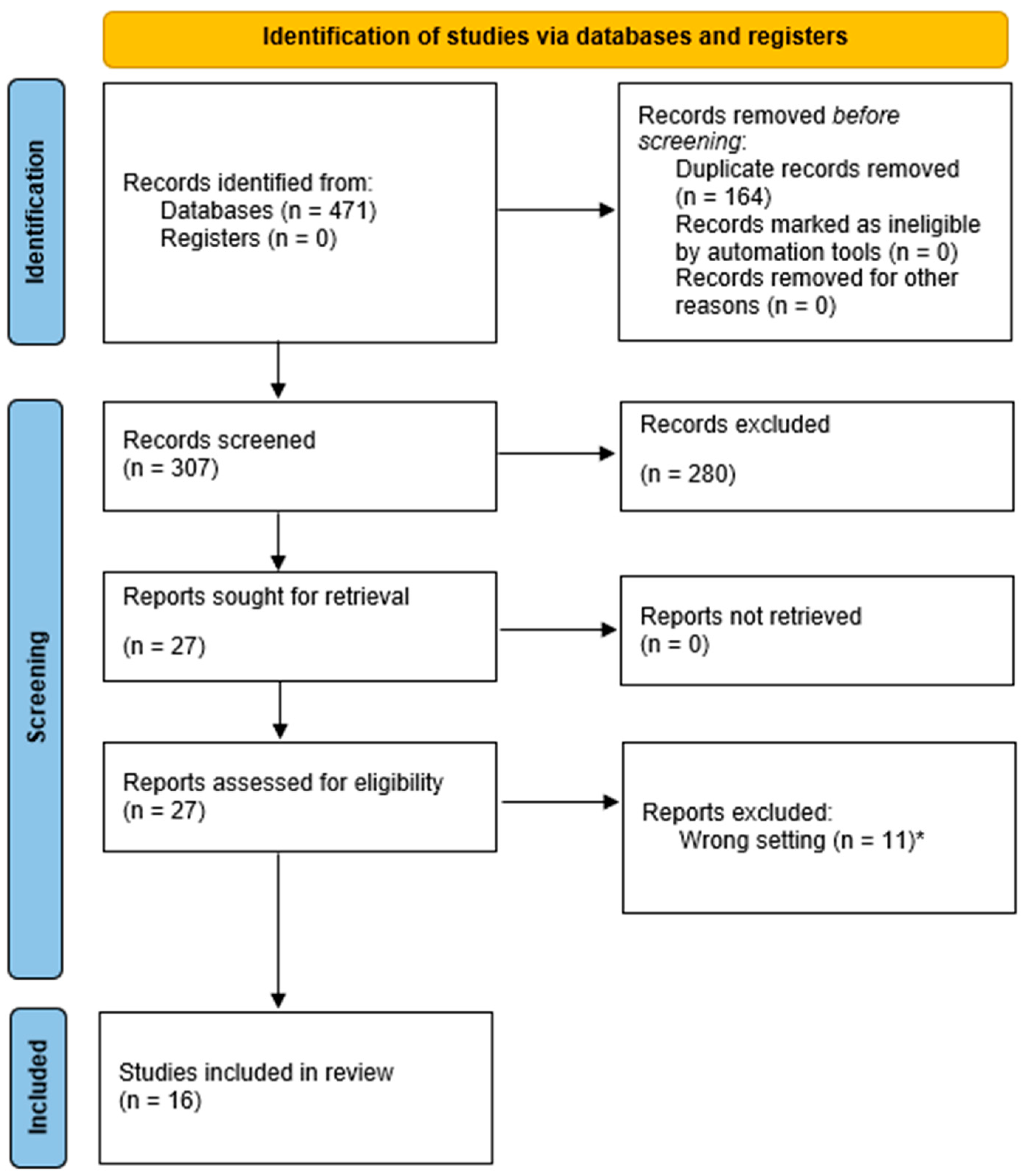
2. Materials and Methods

This systematic review was performed in line with the Preferred Reporting Items for Systematic Reviews and Meta-Analyses (PRISMA) Guidelines [51]. A comprehensive literature review was performed using PubMed and MEDLINE to find relevant articles published from 1946 to August 2022. The selection criteria included all relevant subject headings and freeform texts relating to neuromodulation techniques and paediatric status epilepticus. The following search strategy for PubMed was performed on 15 September 2021: (VNS OR vagal nerve stimulation OR vagus nerve stimulation) OR (transcranial magnetic stimulation OR TMS) OR (electroconvulsive therapy OR ECT) OR (deep brain stimulation OR DBS)) AND ((status epilepticus OR epilepsia partialis continua OR refractory status epilepticus OR rSE OR super-refractory status epilepticus OR srSE) OR (“Febrile infection-related epilepsy syndrome” OR “FIRES”) OR (“New-onset refractory status epilepticus” OR “NORSE”)). A similar search strategy was used for MEDLINE using subject headings and freeform text on the same day. All articles were imported for screening to Covidence (© Cochrane). Duplicates were automatically removed before initial screening by the software. Title and abstract screening and full-text assessment were performed independently by two reviewers (H.L.P. and A.V.). Articles in English referring to neuromodulation (VNS, TMS, ECT, or DBS) and status epilepticus lasting more than 24 h with anaesthesia in patients <18 years were included. Original articles, case studies, case reports, and letters to the editor were included while conference articles, literature reviews, and systematic reviews were excluded. Conflicts in screening and full-text assessment were resolved by three reviewers (H.L.P., A.V., and I.S.).
Studies should give an estimation of the number of days between SE onset and initiation of neuromodulation therapy, an estimation of the number of days from neuromodulation to any changes in patient condition, and the final results of the neuromodulation therapy. Neuromodulation therapy was regarded as successful when it led to cessation of both clinical and electrographic SE, and when the patient was stable enough to be transferred out of ICU. This data was then converted into graphs to illustrate the timeline for all relevant patients. Apart from the timeline details, data included demographics (patient’s age and gender), type of neuromodulation, and epilepsy before the onset of SE.

3. Results

The Prisma flowchart is presented in Figure 2. After excluding duplications and papers not fulfilling the inclusion criteria, 18 references were included for further analysis in the present review. The neuromodulation techniques reviewed were VNS (n = 15), DBS (n = 6), and ECT (n = 5). The mean age of the patients was 7.4 years, ranging from 0.5 to 17 years. Twelve out of twenty-five patients were female.
Basic demographics of patients are illustrated in Table 1 and Table 2. Different neuromodulation protocols for srSE in children have been described, with heterogeneous approaches for results description.

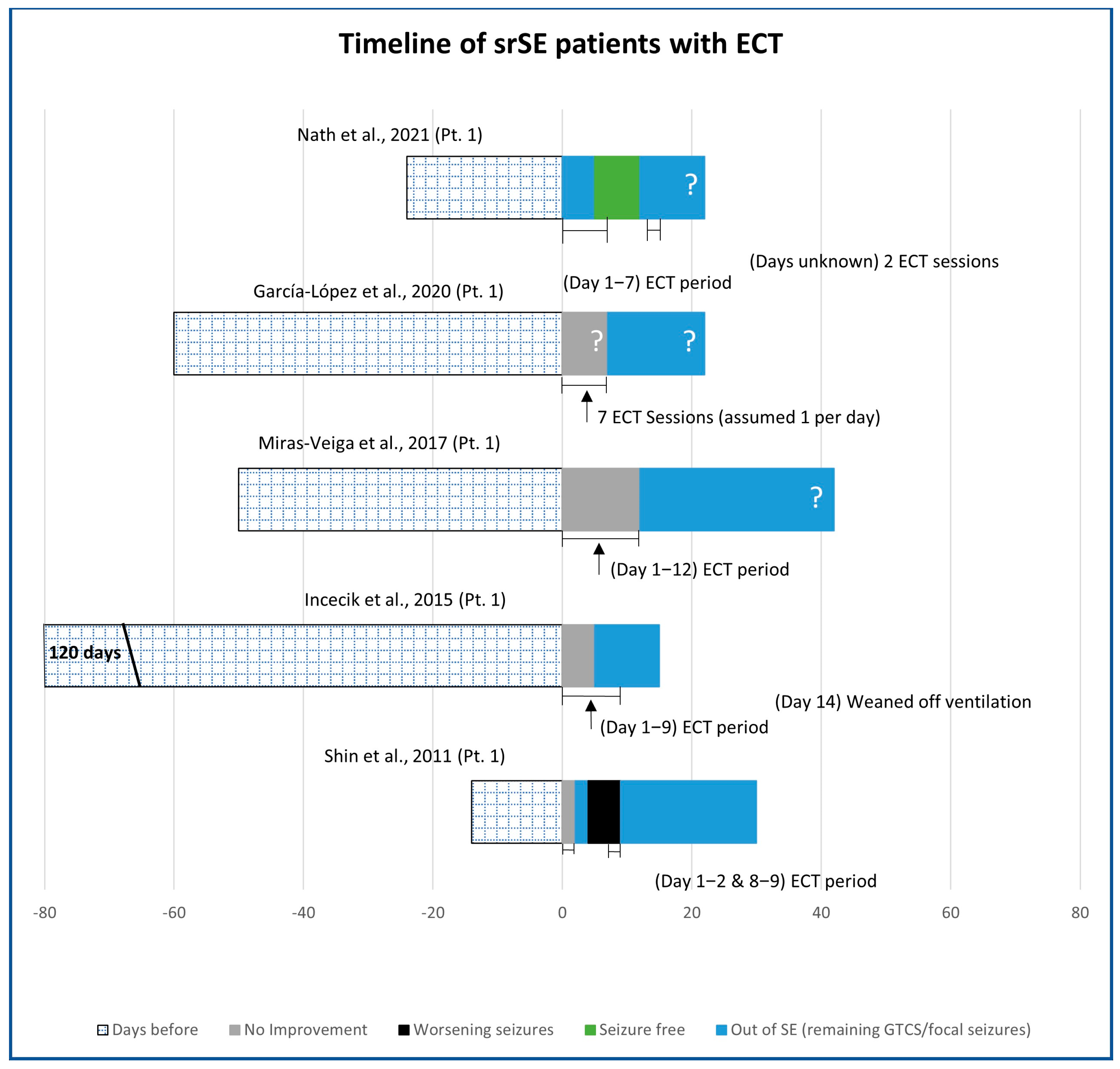
3.1. Electroconvulsive Technique (ECT)

ECT was used in five patients reported in five publications (Figure 3) [53,59,60,61,62]. srSE was focal in all patients, showing secondary generalisation in two. In three patients, srSE occurred de novo and in two was due to FIRES. MRI was not reported in one patient, showed bilateral polymicrogyria in one, and was initially normal in three (and later in the course of the srSE showed atrophy in one patient). ECT was applied between bitemporal electrodes in two patients and between frontotemporal electrodes in two patients, and electrode positions were not specified in one patient. The number and frequency of ECT sessions varied as seizure control was attempted. The number of days that each patient had been on srSE before ECT was started was 14, 24, 50, 60, and 120 days. Three patients had ECT initially on a number of consecutive days (7 or 12), one had five ECT sessions within 9 days whereas the fifth patient had ECT in two pairs of two consecutive days each, separated by 5 days. The number of days of ECT before srSE remission was 2, 5 (in two cases), 7, and 12 days. srSE stopped on the last day of ECT in three patients, while in two patients srSE stopped 4 days before the end of ECT was completed (after 29 and 125 days of SE). Therefore, ECT was considered to have contributed to stopping srSE in all five reported cases. Two patients remained out of the srSE without sequelae (one had learning difficulties beforehand), one had right temporal lobe surgery for focal cortical dysplasia, one developed severe epileptic encephalopathy, and one remained seizure-free but with severe motor dysfunction and cognitive decline.

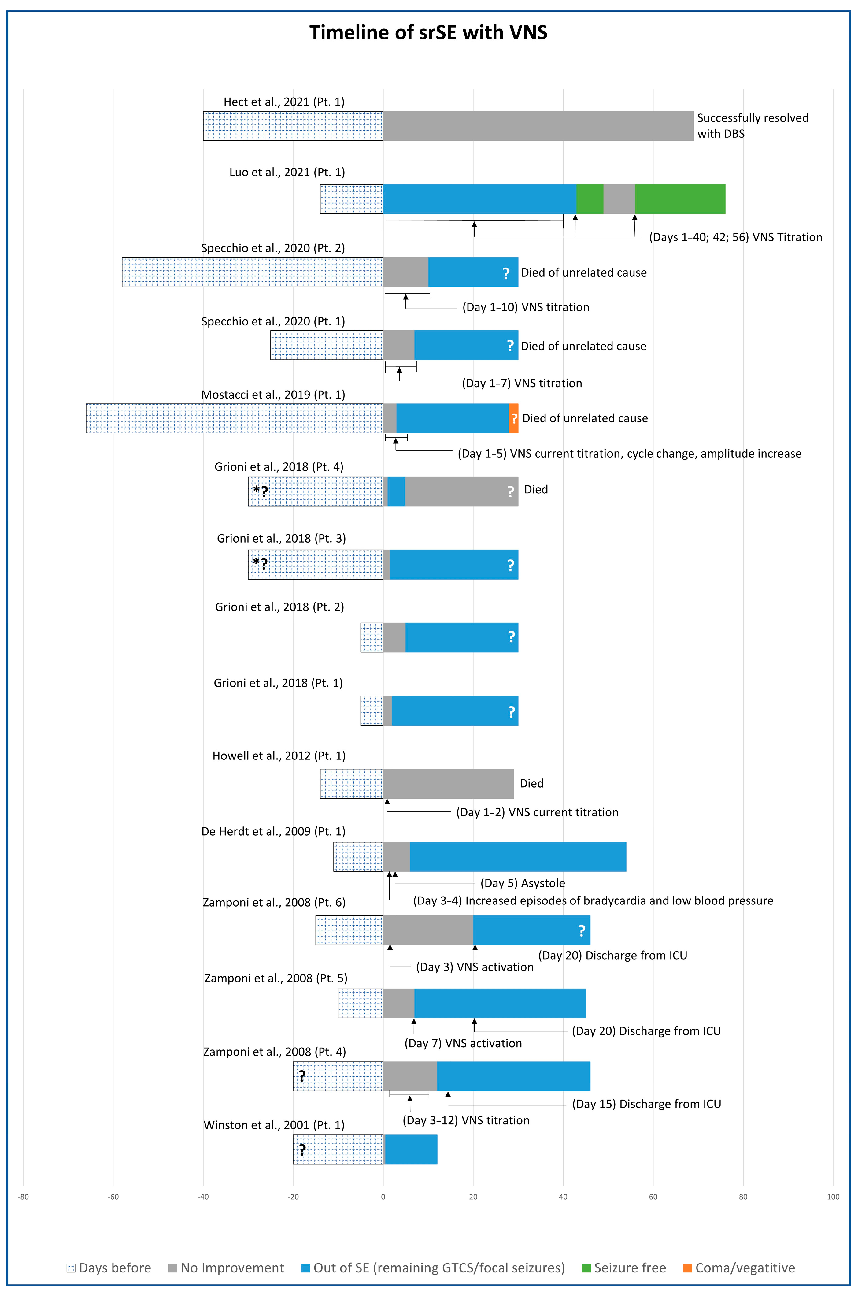
3.2. Vagus Nerve Stimulation (VNS)

Fifteen patients had VNS implantations for srSE in nine articles (Figure 4) [52,54,55,56,57,58,63,64,65]. In three patients, srSE occurred de novo due to FIRES. srSE was focal or secondarily generalised in 10 patients, myoclonic in one, spasms in two, primarily generalised (tonic, T-C, myoclonic, absence seizures) in one, and GTCS without further explanation in one [65]. Identified aetiologies among the focal srSE included malignant partial epilepsy of infancy in four patients (in one patient due to mother’s heroin abuse in pregnancy), neonatal venous thrombosis in one patient, FIRES in three, hemimegalencephaly in one, and one patient had bilateral frontal simplification of cortical gyri together with progressive diffuse cerebral atrophy. Spasms were due to non-ketotic hyperglycinemia in one patient and to microdeletion of 1q43q44 in another patient. Head MRI was normal in seven patients and showed cerebral atrophy/microcephaly in four patients, hemimegalencephaly in one, thalamic lesion/stroke in one, and there was no information about imaging in two patients. There was significant heterogeneity among the VNS parameters. The amplitude varied from 0.25 mA to 3 mA, usually with progressive increments during the course of the treatment until srSE improvement was achieved. The “on” time was 7 s in one case, 14 s in another case, and 30 s in the remaining patients. The “off” period was between 1.8 and 5 min.
Among the fifteen patients with VNS, the srSE was resolved in twelve. Two patients recovered without seizures, seven recovered from srSE remaining with seizures and learning difficulties due to underlying conditions, and three patients recovered but died of unrelated causes (dilated cardiomyopathy 5 months late, paediatric acute respiratory distress syndrome 2.5 years later, and tracheostomy-related late bleeding). Three patients continued with srSE after VNS implantation (two died and one was implanted with CMN DBS).

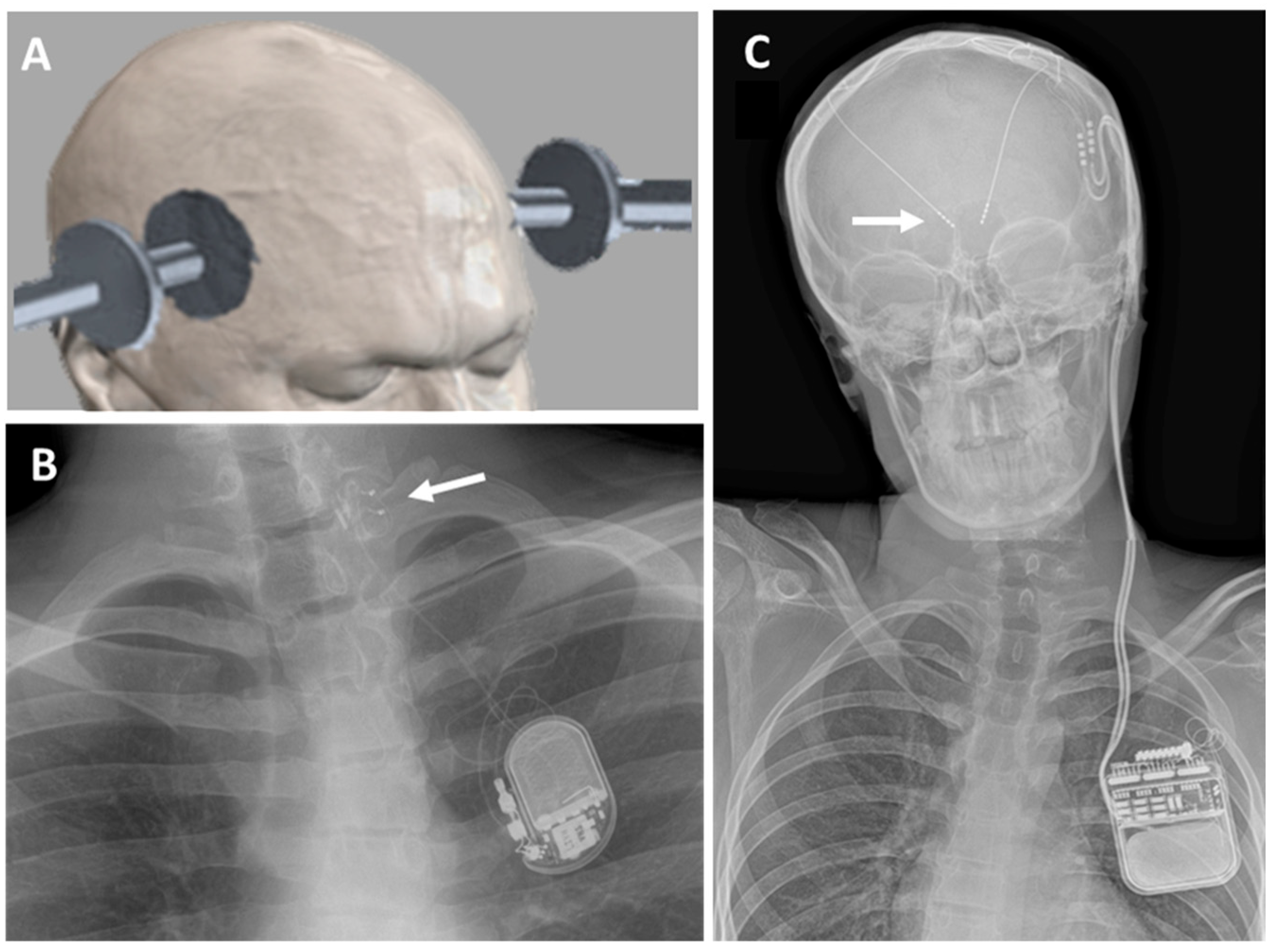
3.3. Deep Brain Stimulation (DBS)

DBS was performed on six patients reported in five articles (Figure 5) [52,66,67,68,69]. In four patients, srSE occurred de novo, due to FIRES in three cases. The centromedian nucleus was stimulated in five patients and the anterior nucleus in one. srSE was focal in five patients, requiring intubation and induced coma in one, and generalised tonic–clonic in one. Head MRI was reported in five patients and was normal in three, showed severe cytotoxic oedema in one, and another patient showed signal abnormalities in basal ganglia, external capsule, and cortex. Four patients had new onset SE without a previous history of seizures or epilepsy (de novo), including three patients where srSE followed febrile illness, suggesting FIRES. Four patients underwent trials of cessation of the stimulation after an initial improvement (stimulation period A) in order to demonstrate the stimulation effect. As SE returned, stimulation was then re-started (stimulation period B). The duration of stimulation period A was 15, 18, 22, and 67 days. After stimulation period B, patients were discharged with the stimulator on. The stimulation frequencies were 6 Hz (in three patients), 145 Hz, and 180 Hz. DBS had immediate effects on srSE in two patients. In the remaining four children, improvement occurred after 2, 4, 6, and 30 days of stimulation. SrSE resolved in all patients; one patient came back to her previous number of seizures, four children remained with seizures, and one child remained in a vegetative state.
In summary, 23 out of the 25 children treated with neuromodulation techniques recovered from srSE (Table 2). Among these children, twelve recovered without new medical conditions, four recovered with severe sequelae (epileptic encephalopathy or cognitive/motor decline), four developed new seizures after the srSE, and three died for unrelated reasons.

4. Discussion

Status epilepticus (SE) is a medical emergency with high mortality and morbidity rates [70,71,72]. The treatment protocols for the early management of SE are well standardised [12]. However, refractory and super-refractory SE (rSE and srSE) are often associated with significant and irreversible brain damage whose severity is related to SE duration and aetiology. A well-defined, effective, and fast-acting therapeutic protocol would be highly desirable to prevent potential longstanding neurologic complications. In this review, we present data showing that neuromodulation could be a potentially efficacious treatment option for shortening rSE and srSE duration in children when the routinely used 1st, 2nd, and 3rd line treatments have failed.
The features of status epilepticus in children can be slightly different from those in adults. As shown in the presented data, srSE in children is often caused by genetic/metabolic conditions, brain malformations, birth injuries, and febrile infection-related epilepsy syndrome (FIRES). The latter is a rare, life-threatening condition that presents with a non-specific febrile illness followed by refractory status epilepticus within 24 h to 2 weeks of the onset of the febrile illness in previously healthy children, with a mortality of up to 30% [73]. Its pathogenesis is unclear, but autoimmune mechanisms have been proposed [74], and a recent international consensus recommendation suggests that first-line immunological treatment should be started during the first 72 h [75].
There are no clinical trials performed to assess the efficacy of any of the neuromodulation techniques on srSE. The literature shows that three different neuromodulation techniques, one non-invasive (ECT) and two implantable devices (VNS and DBS), have been sporadically tried and could be beneficial in cases of rSE and srSE in children. However, the number of reported cases remains small, there is significant diversity regarding the cause of the srSE, and the mechanisms by which neuromodulation affects SE are not elucidated. DBS and ECT appear to have provided benefit in cases with the FIRES condition, while only one out of the three cases published with FIRES and VNS showed resolution of the srSE. All three techniques share unclear mechanisms of action. Animal studies suggest that ECT alters biological processes such as neuroplasticity and neurotransmitter function and might cause internalisation of NMDA receptors or other epigenetic effects [76,77].
Several studies suggest that VNS can modify norepinephrine and serotonin levels at the locus coeruleus and dorsal raphe nuclei [78]. It has also been suggested that VNS can cause changes in limbic structures’ functions modifying GABA and glutamate concentrations at nucleus tractus solitaries [79]. Similar mechanisms may be effective against srSE.
The main advantage of DBS is that the electrical stimulation can be applied locally to specific brain areas using implanted intracranial electrodes and that different stimulation parameters can be applied at the implanted region. In some published cases, electrode implantation probably induced a microlesion effect which was associated with major seizure improvement [39,80]. It has also been proposed that upon high-frequency stimulation (>60 Hz), inhibition of the stimulated area might be mediated by activation of GABAergic afferents or inactivation of voltage-gated currents [39,81]. Moreover, low-frequency stimulation (6 Hz) of the centromedian thalamic nucleus has recently been reported to be useful in reducing the severity and frequency of focal seizures in children and adults with srSE [66,67,82], probably via neuromodulation of cortical structures through the thalamocortical pathway [83].
At present, there is no consensus protocol for the use of ECT, VNS, and DBS in the management of rSE/srSE, and existing evidence is based on a limited number of reported patients. Not all neuromodulation techniques are clinically available in most centres, and they are only considered at late stages of rSE/srSE when standard treatment has failed. As brain damage caused by SE can start as early as 30 min from SE onset [3], the appropriate time for the application of neuromodulation for the treatment of srSE in children is a question of major importance.
Even though published paediatric cases show that neuromodulation was applied as a last-resort treatment, the results appear encouraging. Neuromodulation techniques were applied between 5 [56] and 120 days [59] after SE onset, and substantial brain damage may have already been present in most cases by that time. Regarding the use of non-invasive neuromodulation techniques, the use of ECT in children has given limited but promising results, suggesting that the non-invasive safer techniques could be considered earlier in the course of srSE. Despite the side effects and the invasive nature of VNS and DBS, such damage is unlikely to be induced by neuromodulation, as similar techniques are safe when used for the chronic treatment of epilepsy or other brain conditions [22,39,84,85]. Unfortunately, differences in the timing to apply neuromodulation techniques in different centres/cases do not allow reliable conclusions on the optimal timing for starting this treatment modality.
Regarding invasive procedures, the present series suggests that the effects of DBS and VNS can occur within the first week of treatment. Nonetheless, DBS was used in fewer children, and VNS appears to have been used in a higher number of patients with severe epilepsy (migrating epilepsy, severe cortical malformations, birth injury). DBS implantation led to improvement of srSE, with worsening in seizures when the DBS was turned off [66,67,69,82,86].
Even though the presented data look encouraging, clinical guidance cannot be based on published case reports due to the risk of significant bias [87]. For instance, it is common to find successful neuromodulation cases for srSE published as single case reports, but unsuccessful cases are usually published as part of a case series [56,88] or are not submitted for publication.
As suggested by Rossetti and Lowenstein [20], neuromodulation could be complementary to pharmacological treatment for the management of rSE. Non-invasive techniques such as ECT, tDCS, or rTMS could be considered as add-on treatments after the failure of standard treatment. If no improvement is noted, invasive techniques (DBS and VNS), when available, could be discussed and planned in a timely manner.

5. Conclusions

The evaluation of neuromodulation techniques for the treatment of srSE in children is limited by the small number of published cases and the variability of neuromodulation protocols used for the treatment of srSE. Although neuromodulation strategies have not been tested through randomised, prospective controlled clinical trials, this review presents the existing data and the potential benefits of neuromodulation therapy, suggesting that these techniques could be considered at earlier stages within the course of srSE intending to prevent long-term neurologic complications. Clinical trials aiming to establish whether early intervention can prevent long-term sequelae are necessary to establish the potential clinical value of neuromodulation techniques for the treatment of srSE in children.

Author Contributions

I.S., H.L.P., G.A. and A.V. wrote the manuscript. H.L.P. prepared the seizure graphs. All authors have read and agreed to the published version of the manuscript.

Funding

This research received no external funding.

Conflicts of Interest

AV has received an honorarium for lectures and consultancy from Medtronic Ltd. The rest of the authors declare that they have no competing interests.

References

  1. Trinka, E.; Cock, H.; Hesdorffer, D.; Rossetti, A.O.; Scheffer, I.E.; Shinnar, S.; Shorvon, S.; Lowenstein, D.H. A definition and classification of status epilepticus—Report of the ILAE Task Force on Classification of Status Epilepticus. Epilepsia 2015, 56, 1515–1523. [Google Scholar] [CrossRef] [PubMed]
  2. Sanchez, S.; Rincon, F. Status Epilepticus: Epidemiology and Public Health Needs. J. Clin. Med. 2016, 5, 71. [Google Scholar] [CrossRef] [PubMed]
  3. Lowenstein, D.H.; Bleck, T.; Macdonald, R.L. It’s time to revise the definition of status epilepticus. Epilepsia 1999, 40, 120–122. [Google Scholar] [CrossRef] [PubMed]
  4. Al-Mufti, F.; Claassen, J. Neurocritical care: Status epilepticus review. Crit. Care Clin. 2014, 30, 751–764. [Google Scholar] [CrossRef] [PubMed]
  5. Becker, L.L.; Gratopp, A.; Prager, C.; Elger, C.E.; Kaindl, A.M. Treatment of pediatric convulsive status epilepticus. Front. Neurol. 2023, 14, 1175370. [Google Scholar] [CrossRef] [PubMed]
  6. Cruickshank, M.; Imamura, M.; Counsell, C.; Aucott, L.; Manson, P.; Booth, C.; Scotland, G.; Brazzelli, M. Management of the first stage of convulsive status epilepticus in adults: A systematic review of current randomised evidence. J. Neurol. 2022, 269, 3420–3429. [Google Scholar] [CrossRef]
  7. Cornwall, C.D.; Krøigård, T.; Kristensen, J.S.S.; Callesen, H.E.; Beier, C.P. Outcomes and Treatment Approaches for Super-Refractory Status Epilepticus: A Systematic Review and Meta-Analysis. JAMA Neurol. 2023, 80, 959–968. [Google Scholar] [CrossRef] [PubMed]
  8. Bauer, G.; Trinka, E. Nonconvulsive status epilepticus and coma. Epilepsia 2010, 51, 177–190. [Google Scholar] [CrossRef]
  9. Trinka, E.; Brigo, F.; Shorvon, S. Recent advances in status epilepticus. Curr. Opin. Neurol. 2016, 29, 189–198. [Google Scholar] [CrossRef]
  10. Trinka, E.; Kälviäinen, R. 25 years of advances in the definition, classification and treatment of status epilepticus. Seizure 2017, 44, 65–73. [Google Scholar] [CrossRef]
  11. NICE. Epilepsies: Diagnosis and Management; National Institute for Health and Care Excellence (NICE): London, UK, 2019. [Google Scholar]
  12. Shorvon, S.; Ferlisi, M. The treatment of super-refractory status epilepticus: A critical review of available therapies and a clinical treatment protocol. Brain A J. Neurol. 2011, 134, 2802–2818. [Google Scholar] [CrossRef] [PubMed]
  13. Trinka, E.; Höfler, J.; Leitinger, M.; Brigo, F. Pharmacotherapy for Status Epilepticus. Drugs 2015, 75, 1499–1521. [Google Scholar] [CrossRef] [PubMed]
  14. Leitinger, M.; Trinka, E.; Giovannini, G.; Zimmermann, G.; Florea, C.; Rohracher, A.; Kalss, G.; Neuray, C.; Kreidenhuber, R.; Höfler, J.; et al. Epidemiology of status epilepticus in adults: A population-based study on incidence, causes, and outcomes. Epilepsia 2019, 60, 53–62. [Google Scholar] [CrossRef]
  15. Arya, R.; Rotenberg, A. Dietary, immunological, surgical, and other emerging treatments for pediatric refractory status epilepticus. Seizure 2019, 68, 89–96. [Google Scholar] [CrossRef] [PubMed]
  16. Vasquez, A.; Farias-Moeller, R.; Sanchez-Fernandez, I.; Abend, N.S.; Amengual-Gual, M.; Anderson, A.; Arya, R.; Brenton, J.N.; Carpenter, J.L.; Chapman, K.; et al. Super-Refractory Status Epilepticus in Children: A Retrospective Cohort Study. Pediatr. Crit. Care Med. 2021, 22, e613–e625. [Google Scholar] [CrossRef] [PubMed]
  17. Zeiler, F.A.; Matuszczak, M.; Teitelbaum, J.; Kazina, C.J.; Gillman, L.M. Plasmapheresis for refractory status epilepticus, part I: A scoping systematic review of the adult literature. Seizure 2016, 43, 14–22. [Google Scholar] [CrossRef] [PubMed]
  18. Zeiler, F.A.; Matuszczak, M.; Teitelbaum, J.; Kazina, C.J.; Gillman, L.M. Plasmapheresis for refractory status epilepticus Part II: A scoping systematic review of the pediatric literature. Seizure 2016, 43, 61–68. [Google Scholar] [CrossRef] [PubMed]
  19. Legriel, S.; Lemiale, V.; Schenck, M.; Chelly, J.; Laurent, V.; Daviaud, F.; Srairi, M.; Hamdi, A.; Geri, G.; Rossignol, T.; et al. Hypothermia for Neuroprotection in Convulsive Status Epilepticus. N. Engl. J. Med. 2016, 375, 2457–2467. [Google Scholar] [CrossRef]
  20. Rossetti, A.O.; Lowenstein, D.H. Management of refractory status epilepticus in adults: Still more questions than answers. Lancet Neurol. 2011, 10, 922–930. [Google Scholar] [CrossRef] [PubMed]
  21. San-Juan, D.; Davila-Rodriguez, D.O.; Jimenez, C.R.; Gonzalez, M.S.; Carranza, S.M.; Hernandez Mendoza, J.R.; Anschel, D.J. Neuromodulation techniques for status epilepticus: A review. Brain Stimul. 2019, 12, 835–844. [Google Scholar] [CrossRef]
  22. Weiner, R.D.; Reti, I.M. Key updates in the clinical application of electroconvulsive therapy. Int. Rev. Psychiatry 2017, 29, 54–62. [Google Scholar] [CrossRef] [PubMed]
  23. Zeiler, F.A.; Matuszczak, M.; Teitelbaum, J.; Gillman, L.M.; Kazina, C.J. Electroconvulsive therapy for refractory status epilepticus: A systematic review. Seizure 2016, 35, 23–32. [Google Scholar] [CrossRef] [PubMed]
  24. Semkovska, M.; McLoughlin, D.M. Objective cognitive performance associated with electroconvulsive therapy for depression: A systematic review and meta-analysis. Biol. Psychiatry 2010, 68, 568–577. [Google Scholar] [CrossRef] [PubMed]
  25. Kellner, C.H.; Tobias, K.G.; Wiegand, J. Electrode placement in electroconvulsive therapy (ECT): A review of the literature. J. ECT 2010, 26, 175–180. [Google Scholar] [CrossRef] [PubMed]
  26. Choi, J.; Lisanby, S.H.; Medalia, A.; Prudic, J. A conceptual introduction to cognitive remediation for memory deficits associated with right unilateral electroconvulsive therapy. J. ECT 2011, 27, 286–291. [Google Scholar] [CrossRef] [PubMed]
  27. Ferreira, N.R.; Junqueira, Y.N.; Correa, N.B.; Fonseca, E.O.; Brito, N.B.M.; Menezes, T.A.; Magini, M.; Fidalgo, T.K.S.; Ferreira, D.; de Lima, R.L.; et al. The efficacy of transcranial direct current stimulation and transcranial magnetic stimulation for chronic orofacial pain: A systematic review. PLoS ONE 2019, 14, e0221110. [Google Scholar] [CrossRef] [PubMed]
  28. Guo, Q.; Li, C.; Wang, J. Updated Review on the Clinical Use of Repetitive Transcranial Magnetic Stimulation in Psychiatric Disorders. Neurosci. Bull. 2017, 33, 747–756. [Google Scholar] [CrossRef]
  29. Zeiler, F.A.; Matuszczak, M.; Teitelbaum, J.; Gillman, L.M.; Kazina, C.J. Transcranial Magnetic Stimulation for Status Epilepticus. Epilepsy Res. Treat. 2015, 2015, 678074. [Google Scholar] [CrossRef]
  30. Kobayashi, M.; Pascual-Leone, A. Transcranial magnetic stimulation in neurology. Lancet Neurol. 2003, 2, 145–156. [Google Scholar] [CrossRef]
  31. Lefaucheur, J.P.; Antal, A.; Ayache, S.S.; Benninger, D.H.; Brunelin, J.; Cogiamanian, F.; Cotelli, M.; De Ridder, D.; Ferrucci, R.; Langguth, B.; et al. Evidence-based guidelines on the therapeutic use of transcranial direct current stimulation (tDCS). Clin. Neurophysiol. 2017, 128, 56–92. [Google Scholar] [CrossRef]
  32. San-Juan, D.; Espinoza Lopez, D.A.; Vazquez Gregorio, R.; Trenado, C.; Fernandez-Gonzalez Aragon, M.; Morales-Quezada, L.; Hernandez Ruiz, A.; Hernandez-Gonzalez, F.; Alcaraz-Guzman, A.; Anschel, D.J.; et al. Transcranial Direct Current Stimulation in Mesial Temporal Lobe Epilepsy and Hippocampal Sclerosis. Brain Stimul. 2017, 10, 28–35. [Google Scholar] [CrossRef] [PubMed]
  33. LivaNova. VNS Therapy® System Physician’s Manual. Available online: https://www.livanova.com/epilepsy-vnstherapy/en-us/hcp/product-manuals (accessed on 1 September 2023).
  34. Giordano, F.; Zicca, A.; Barba, C. Vagus nerve stimulation: Surgical technique of implantation and revision and related morbidity. Epilepsia 2017, 58 (Suppl. S1), 85–90. [Google Scholar] [CrossRef] [PubMed]
  35. Dibue-Adjei, M.; Brigo, F.; Yamamoto, T.; Vonck, K.; Trinka, E. Vagus nerve stimulation in refractory and super-refractory status epilepticus—A systematic review. Brain Stimul. 2019, 12, 1101–1110. [Google Scholar] [CrossRef]
  36. Trinka, E.; Brigo, F. Neurostimulation in the treatment of refractory and super-refractory status epilepticus. Epilepsy Behav. 2019, 101, 106551. [Google Scholar] [CrossRef]
  37. Aum, D.J.; Tierney, T.S. Deep brain stimulation: Foundations and future trends. Front. Biosci. 2018, 23, 162–182. [Google Scholar] [CrossRef]
  38. Laxpati, N.G.; Kasoff, W.S.; Gross, R.E. Deep brain stimulation for the treatment of epilepsy: Circuits, targets, and trials. Neurotherapeutics 2014, 11, 508–526. [Google Scholar] [CrossRef] [PubMed]
  39. Zangiabadi, N.; Ladino, L.D.; Sina, F.; Orozco-Hernández, J.P.; Carter, A.; Téllez-Zenteno, J.F. Deep Brain Stimulation and Drug-Resistant Epilepsy: A Review of the Literature. Front. Neurol. 2019, 10, 601. [Google Scholar] [CrossRef]
  40. Salanova, V. Deep brain stimulation for epilepsy. Epilepsy Behav. 2018, 88, 21–24. [Google Scholar] [CrossRef] [PubMed]
  41. Fisher, R.; Salanova, V.; Witt, T.; Worth, R.; Henry, T.; Gross, R.; Oommen, K.; Osorio, I.; Nazzaro, J.; Labar, D.; et al. Electrical stimulation of the anterior nucleus of thalamus for treatment of refractory epilepsy. Epilepsia 2010, 51, 899–908. [Google Scholar] [CrossRef]
  42. Fisher, R.S.; Uematsu, S.; Krauss, G.L.; Cysyk, B.J.; McPherson, R.; Lesser, R.P.; Gordon, B.; Schwerdt, P.; Rise, M. Placebo-controlled pilot study of centromedian thalamic stimulation in treatment of intractable seizures. Epilepsia 1992, 33, 841–851. [Google Scholar] [CrossRef]
  43. Jitkritsadakul, O.; Bhidayasiri, R.; Kalia, S.K.; Hodaie, M.; Lozano, A.M.; Fasano, A. Systematic review of hardware-related complications of Deep Brain Stimulation: Do new indications pose an increased risk? Brain Stimul. 2017, 10, 967–976. [Google Scholar] [CrossRef] [PubMed]
  44. Picillo, M.; Lozano, A.M.; Kou, N.; Munhoz, R.P.; Fasano, A. Programming Deep Brain Stimulation for Tremor and Dystonia: The Toronto Western Hospital Algorithms. Brain Stimul. 2016, 9, 438–452. [Google Scholar] [CrossRef]
  45. Picillo, M.; Lozano, A.M.; Kou, N.; Puppi Munhoz, R.; Fasano, A. Programming Deep Brain Stimulation for Parkinson’s Disease: The Toronto Western Hospital Algorithms. Brain Stimul. 2016, 9, 425–437. [Google Scholar] [CrossRef]
  46. Morrell, M.J. Responsive cortical stimulation for the treatment of medically intractable partial epilepsy. Neurology 2011, 77, 1295–1304. [Google Scholar] [CrossRef] [PubMed]
  47. Ernst, L.D.; Raslan, A.M.; Wabulya, A.; Shin, H.W.; Cash, S.S.; Yang, J.C.; Sagi, V.; King-Stephens, D.; Damisah, E.C.; Ramos, A.; et al. Responsive neurostimulation as a treatment for super-refractory focal status epilepticus: A systematic review and case series. J. Neurosurg. 2023, 9, 1–9. [Google Scholar] [CrossRef] [PubMed]
  48. Ernst, L.D.; Krause, K.L.; Kellogg, M.A.; Raslan, A.M.; Spencer, D.C. Novel Use of Responsive Neurostimulation (RNS System) in the Treatment of Super Refractory Status Epilepticus. J. Clin. Neurophysiol. 2019, 36, 242–245. [Google Scholar] [CrossRef] [PubMed]
  49. Yang, J.C.; Harid, N.M.; Nascimento, F.A.; Kokkinos, V.; Shaughnessy, A.; Lam, A.D.; Westover, M.B.; Leslie-Mazwi, T.M.; Hochberg, L.R.; Rosenthal, E.S.; et al. Responsive neurostimulation for focal motor status epilepticus. Ann. Clin. Transl. Neurol. 2021, 8, 1353–1361. [Google Scholar] [CrossRef]
  50. Ryvlin, P.; Jehi, L.E. Neuromodulation for Refractory Epilepsy. Epilepsy Curr. 2022, 22, 11–17. [Google Scholar] [CrossRef]
  51. Page, M.J.; McKenzie, J.E.; Bossuyt, P.M.; Boutron, I.; Hoffmann, T.C.; Mulrow, C.D.; Shamseer, L.; Tetzlaff, J.M.; Akl, E.A.; Brennan, S.E.; et al. The PRISMA 2020 statement: An updated guideline for reporting systematic reviews. BMJ 2021, 372, n71. [Google Scholar] [CrossRef] [PubMed]
  52. Hect, J.L.; Fernandez, L.D.; Welch, W.P.; Abel, T.J. Deep brain stimulation of the centromedian thalamic nucleus for the treatment of FIRES. Epilepsia Open 2022, 7, 187–193. [Google Scholar] [CrossRef]
  53. García-López, B.; Gómez-Menéndez, A.I.; Vázquez-Sánchez, F.; Pérez-Cabo, E.; Isidro-Mesas, F.; Zabalegui-Pérez, A.; Muñoz-Siscart, I.; Lloria-Gil, M.C.; Soto-Cámara, R.; González-Bernal, J.J.; et al. Electroconvulsive Therapy in Super Refractory Status Epilepticus: Case Series with a Defined Protocol. Int. J. Environ. Res. Public Health 2020, 17, 4023. [Google Scholar] [CrossRef] [PubMed]
  54. Winston, K.R.; Levisohn, P.; Miller, B.R.; Freeman, J. Vagal nerve stimulation for status epilepticus. Pediatr. Neurosurg. 2001, 34, 190–192. [Google Scholar] [CrossRef] [PubMed]
  55. Zamponi, N.; Rychlicki, F.; Corpaci, L.; Cesaroni, E.; Trignani, R. Vagus nerve stimulation (VNS) is effective in treating catastrophic 1 epilepsy in very young children. Neurosurg. Rev. 2008, 31, 291–297. [Google Scholar] [CrossRef] [PubMed]
  56. Grioni, D.; Landi, A.; Fiori, L.; Sganzerla, E.P. Does emergent implantation of a vagal nerve stimulator stop refractory status epilepticus in children? Seizure 2018, 61, 94–97. [Google Scholar] [CrossRef] [PubMed]
  57. Mostacci, B.; Bisulli, F.; Muccioli, L.; Minardi, I.; Bandini, M.; Licchetta, L.; Zucchelli, M.; Leta, C.; Michelucci, R.; Zanello, M.; et al. Super refractory status epilepticus in Lafora disease interrupted by vagus nerve stimulation: A case report. Brain Stimul. 2019, 12, 1605–1607. [Google Scholar] [CrossRef] [PubMed]
  58. Specchio, N.; Ferretti, A.; Pietrafusa, N.; Trivisano, M.; Calabrese, C.; Carfì Pavia, G.; De Benedictis, A.; Marras, C.E.; de Palma, L.; Vigevano, F. Refractory Status Epilepticus in Genetic Epilepsy-Is Vagus Nerve Stimulation an Option? Front. Neurol. 2020, 11, 443. [Google Scholar] [CrossRef] [PubMed]
  59. Incecik, F.; Horoz, O.O.; Herguner, O.M.; Yildizdas, D.; Altunbasak, S. Electroconvulsive therapy for refractory status epilepticus in a child: A case report. Ann. Indian Acad. Neurol. 2015, 18, 364–365. [Google Scholar] [CrossRef] [PubMed]
  60. Miras Veiga, A.; Moreno, D.C.; Menendez, A.I.; Siscart, I.M.; Fernandez, M.D.; Sanchez, E.G.; Gonzalez, M.G.; Saez, F.G. Effectiveness of Electroconvulsive Therapy for Refractory Status Epilepticus in Febrile Infection-Related Epilepsy Syndrome. Neuropediatrics 2017, 48, 45–48. [Google Scholar] [CrossRef] [PubMed]
  61. Shin, H.W.; O’Donovan, C.A.; Boggs, J.G.; Grefe, A.; Harper, A.; Bell, W.L.; McCall, W.V.; Rosenquist, P. Successful ECT treatment for medically refractory nonconvulsive status epilepticus in pediatric patient. Seizure 2011, 20, 433–436. [Google Scholar] [CrossRef]
  62. Nath, M.; Shah, Y.D.; Theroux, L.M.; Petrides, G.; Karkare, S.; Sanghani, S.N.; Kothare, S.V. A Role for Electroconvulsive Therapy in the Management of New Onset Refractory Status Epilepticus (NORSE) in a Young Child. Neurol. India 2021, 69, 1374–1379. [Google Scholar] [CrossRef]
  63. De Herdt, V.; Waterschoot, L.; Vonck, K.; Dermaut, B.; Verhelst, H.; Van Coster, R.; De Jaeger, A.; Van Roost, D.; Boon, P. Vagus nerve stimulation for refractory status epilepticus. Eur. J. Paediatr. Neurol. 2009, 13, 286–289. [Google Scholar] [CrossRef] [PubMed]
  64. Howell, K.B.; Katanyuwong, K.; Mackay, M.T.; Bailey, C.A.; Scheffer, I.E.; Freeman, J.L.; Berkovic, S.F.; Harvey, A.S. Long-term follow-up of febrile infection-related epilepsy syndrome. Epilepsia 2012, 53, 101–110. [Google Scholar] [CrossRef] [PubMed]
  65. Luo, T.; Wang, Y.; Lu, G.; Zhou, Y.; Wang, Y. Vagus nerve stimulation for super-refractory status epilepticus in febrile infection-related epilepsy syndrome: A pediatric case report and literature review. Child’S Nerv. Syst. 2022, 38, 1401–1404. [Google Scholar] [CrossRef] [PubMed]
  66. Sa, M.; Singh, R.; Pujar, S.; D’Arco, F.; Desai, N.; Eltze, C.; Hughes, E.; Al Obaidi, M.; Eleftheriou, D.; Tisdall, M.; et al. Centromedian thalamic nuclei deep brain stimulation and Anakinra treatment for FIRES—Two different outcomes. Eur. J. Paediatr. Neurol. 2019, 23, 749–754. [Google Scholar] [CrossRef] [PubMed]
  67. Stavropoulos, I.; Selway, R.; Hasegawa, H.; Hughes, E.; Rittey, C.; Jiménez-Jiménez, D.; Valentin, A. Low frequency centromedian thalamic nuclei deep brain stimulation for the treatment of super refractory status epilepticus: A case report and a review of the literature. Brain Stimul. 2021, 14, 226–229. [Google Scholar] [CrossRef] [PubMed]
  68. Lee, C.Y.; Lim, S.N.; Wu, T.; Lee, S.T. Successful Treatment of Refractory Status Epilepticus Using Anterior Thalamic Nuclei Deep Brain Stimulation. World Neurosurg. 2017, 99, 14–18. [Google Scholar] [CrossRef]
  69. Lehtimaki, K.; Langsjo, J.W.; Ollikainen, J.; Heinonen, H.; Mottonen, T.; Tahtinen, T.; Haapasalo, J.; Tenhunen, J.; Katisko, J.; Ohman, J.; et al. Successful management of super-refractory status epilepticus with thalamic deep brain stimulation. Ann. Neurol. 2017, 81, 142–146. [Google Scholar] [CrossRef]
  70. Hill, C.E.; Parikh, A.O.; Ellis, C.; Myers, J.S.; Litt, B. Timing is everything: Where status epilepticus treatment fails. Ann. Neurol. 2017, 82, 155–165. [Google Scholar] [CrossRef]
  71. Betjemann, J.P.; Lowenstein, D.H. Status epilepticus in adults. Lancet Neurol. 2015, 14, 615–624. [Google Scholar] [CrossRef]
  72. Claassen, J.; Lokin, J.K.; Fitzsimmons, B.F.; Mendelsohn, F.A.; Mayer, S.A. Predictors of functional disability and mortality after status epilepticus. Neurology 2002, 58, 139–142. [Google Scholar] [CrossRef]
  73. Farias-Moeller, R.; Bartolini, L.; Staso, K.; Schreiber, J.M.; Carpenter, J.L. Early ictal and interictal patterns in FIRES: The sparks before the blaze. Epilepsia 2017, 58, 1340–1348. [Google Scholar] [CrossRef]
  74. van Baalen, A.; Vezzani, A.; Häusler, M.; Kluger, G. Febrile infection–related epilepsy syndrome: Clinical review and hypotheses of epileptogenesis. Neuropediatrics 2017, 48, 005–018. [Google Scholar]
  75. Wickström, R.; Taraschenko, O.; Dilena, R.; Payne, E.T.; Specchio, N.; Nabbout, R.; Koh, S.; Gaspard, N.; Hirsch, L.J.; The International NORSE Consensus Group. International consensus recommendations for management of new onset refractory status epilepticus (NORSE) including febrile infection-related epilepsy syndrome (FIRES): Summary and clinical tools. Epilepsia 2022, 63, 2827–2839. [Google Scholar] [CrossRef]
  76. Singh, A.; Kar, S.K. How Electroconvulsive Therapy Works? Understanding the Neurobiological Mechanisms. Clin. Psychopharmacol. Neurosci. 2017, 15, 210–221. [Google Scholar] [CrossRef]
  77. Jang, S.S.; Royston, S.E.; Lee, G.; Wang, S.; Chung, H.J. Seizure-Induced Regulations of Amyloid-beta, STEP61, and STEP61 Substrates Involved in Hippocampal Synaptic Plasticity. Neural Plast. 2016, 2016, 2123748. [Google Scholar] [CrossRef]
  78. Krahl, S.E.; Clark, K.B. Vagus nerve stimulation for epilepsy: A review of central mechanisms. Surg. Neurol. Int. 2012, 3, S255–S259. [Google Scholar] [CrossRef]
  79. George, M.S.; Nahas, Z.; Bohning, D.E.; Mu, Q.; Andrew Kozel, F.; Borckhardt, J.; Denslow, S. Mechanisms of action of vagus nerve stimulation (VNS). Clin. Neurosci. Res. 2004, 4, 71–79. [Google Scholar] [CrossRef]
  80. Ashkan, K.; Rogers, P.; Bergman, H.; Ughratdar, I. Insights into the mechanisms of deep brain stimulation. Nat. Reviews. Neurol. 2017, 13, 548–554. [Google Scholar] [CrossRef] [PubMed]
  81. Chiken, S.; Nambu, A. Disrupting neuronal transmission: Mechanism of DBS? Front. Syst. Neurosci. 2014, 8, 33. [Google Scholar] [CrossRef] [PubMed]
  82. Valentin, A.; Nguyen, H.Q.; Skupenova, A.M.; Agirre-Arrizubieta, Z.; Jewell, S.; Mullatti, N.; Moran, N.F.; Richardson, M.P.; Selway, R.P.; Alarcon, G. Centromedian thalamic nuclei deep brain stimulation in refractory status epilepticus. Brain Stimul. 2012, 5, 594–598. [Google Scholar] [CrossRef]
  83. Martín-López, D.; Jiménez-Jiménez, D.; Cabañés-Martínez, L.; Selway, R.P.; Valentín, A.; Alarcón, G. The role of thalamus versus cortex in epilepsy: Evidence from human ictal centromedian recordings in patients assessed for deep brain stimulation. Int. J. Neural Syst. 2017, 27, 1750010. [Google Scholar] [CrossRef]
  84. Cooper, Y.A.; Pianka, S.T.; Alotaibi, N.M.; Babayan, D.; Salavati, B.; Weil, A.G.; Ibrahim, G.M.; Wang, A.C.; Fallah, A. Repetitive transcranial magnetic stimulation for the treatment of drug-resistant epilepsy: A systematic review and individual participant data meta-analysis of real-world evidence. Epilepsia Open 2018, 3, 55–65. [Google Scholar] [CrossRef] [PubMed]
  85. Perez-Carbonell, L.; Faulkner, H.; Higgins, S.; Koutroumanidis, M.; Leschziner, G. Vagus nerve stimulation for drug-resistant epilepsy. Pract. Neurol. 2019, 20, 189–198. [Google Scholar] [CrossRef] [PubMed]
  86. Yuan, L.; Zhang, S.; Liang, S.; Liu, N.; Yu, X.; Liang, S. Deep brain stimulation of the anterior nucleus of the thalamus in a patient with super-refractory convulsive status epilepticus. Epileptic Disord. Int. Epilepsy J. Videotape 2019, 21, 379–384. [Google Scholar] [CrossRef]
  87. Nissen, T.; Wynn, R. The clinical case report: A review of its merits and limitations. BMC Res. Notes 2014, 7, 264. [Google Scholar] [CrossRef]
  88. Liu, A.; Pang, T.; Herman, S.; Pascual-Leone, A.; Rotenberg, A. Transcranial magnetic stimulation for refractory focal status epilepticus in the intensive care unit. Seizure 2013, 22, 893–896. [Google Scholar] [CrossRef]
Figure 1. Invasive and non-invasive neuromodulation techniques tried in children with srSE. (A) Electrodes and typical positions for non-invasive electroconvulsive therapy (ECT); (B) X-ray showing vagus nerve stimulation (VNS) in a 14 y/o child. The white arrow indicates the position of the stimulating contacts in the vagus nerve; (C) X-ray showing deep brain stimulation (DBS) in a 12 y/o child. The white arrow indicates the DBS position in the centromedian thalamic nucleus in the brain.
Brainsci 13 01527 g001
Figure 2. PRISMA 2020 flow diagram for new systematic reviews. * 6 studies reported epilepsia partialis continua, 1 study reported asleep electrographic status epilepticus, 1 study reported Rasmussen’s encephalitis, and 3 studies where neuromodulation did not directly treat the status epilepticus.
Brainsci 13 01527 g002
Figure 3. Timeline of srSE patients treated with ECT. (? = the time period is estimated but not clearly stated) [53,59,60,61,62].
Brainsci 13 01527 g003
Figure 4. Timeline of srSE patients treated with VNS (* about 30 days but not clearly stated; ? = the time period is estimated but not clearly stated) [52,54,55,56,57,58,63,64,65].
Brainsci 13 01527 g004
Figure 5. Timeline of srSE patients treated with DBS [52,66,67,68,69].
Brainsci 13 01527 g005
Table 1. Basic demographic including age, gender, and type of neuromodulation. * One patient [52] was reported in both VNS and DBS as the patient underwent both neuromodulation techniques. VNS did not offer benefits, and DBS caused resolution of super-refractory status epilepticus.
Number of PatientsMaleFemaleStimulation Age (Years)De NovoPost Febrile
ECT5236.8 (3–−16)32
VNS15 *875.73 (0.5–16)33
DBS6 *3312.5 (5–17)43
All2513127.4 (0.5–17)97
Table 2. Outcomes following neuromodulation. Stim = stimulation; SE = status epilepticus. * One patient [53] was removed from calculation of “Duration of stimulation before SE resolution” as it was not adequately reported. ** Four patients ([54]; [pt 4] [55]; [pt 3 and pt 4] [56]) were removed from calculation of “Duration of SE before stimulation” as it was not adequately reported. *** 3 patients died of unrelated causes after SE resolved ([57]; [pt 1 and pt 2] [58]).
SE Duration before Stim (Days)Stim Duration before SE Resolution (Days)Recovered from SESevere Sequelae Post-SEDied during SE
ECT53.63 (14–120)
N = 5
4.75 (0–12) *
N = 4
530
VNS23.9 (5–66)
N = 11 **
6.2 (0–20)
N = 12
1202 ***
DBS49 (27–86)
N = 6
7.7 (0–29)
N = 6
610
All37.5 (5–120)
N = 21
6.3 (0–29) *
N = 22
2342
Disclaimer/Publisher’s Note: The statements, opinions and data contained in all publications are solely those of the individual author(s) and contributor(s) and not of MDPI and/or the editor(s). MDPI and/or the editor(s) disclaim responsibility for any injury to people or property resulting from any ideas, methods, instructions or products referred to in the content.

