Case Report

The Role of Folate Deficiency as a Potential Risk Factor for Nontraumatic Anterior Spinal Artery Syndrome in an Adolescent Girl

1
Department of Pediatrics, Tri-Service General Hospital, National Defense Medical Center, Taipei 11490, Taiwan
2
Department of General Medicine, Tri-Service General Hospital, National Defense Medical Center, Taipei 11490, Taiwan
3
Department of Molecular and Cellular Biology, University of California-Davis, Davis, CA 95616, USA
4
Department of Radiology, Tri-Service General Hospital, National Defense Medical Center, Taipei 11490, Taiwan
5
Center for Precision Medicine and Genomics, Tri-Service General Hospital, National Defense Medical Center, Taipei 11490, Taiwan
*
Author to whom correspondence should be addressed.
Brain Sci. 2022, 12(11), 1470; https://doi.org/10.3390/brainsci12111470
Submission received: 5 October 2022 / Revised: 26 October 2022 / Accepted: 28 October 2022 / Published: 29 October 2022
(This article belongs to the Topic Diagnosis and Management of Acute Ischemic Stroke)

Abstract

:
Nontraumatic anterior spinal artery syndrome (ASAS) is an extremely rare clinical condition in pediatric populations with a mostly unknown underlying etiology. Here we discuss the case of a previously healthy 14-year-old girl presenting with sudden onset acute flaccid paralysis to the emergency department. A spinal STIR/DWI MRI revealed hyperintensities extending from cervical vertebrae C3-6, consistent with the diagnosis of ASAS. In order to determine any precipitating causes of ASAS, we also extensively investigated established potential risk factors for ASAS in our patient and noticed that she had a marked folate deficiency requiring folic acid supplementation to prevent future episodes of ASAS as well as to repair the patient’s injured spinal cord. Interestingly, the patient did not display elevated levels of homocysteine nor did she possess the three pathogenic MTHFR mutations characteristic of ASAS. Although her folate deficiency did not cause responsive hyperhomocysteinemia, and she did not have pathogenic MTHFR mutations that impair the function of methylenetetrahydrofolate reductase in folate cycle, we suggest that isolated folate deficiency may play a role in adolescent cases of ASAS that, once identified, would require prompt identification and early intervention to improve the prognosis of these patients.

1. Introduction

Spinal cord infarction (SCI) is rare, accounting for approximately 0.3% to 1% of ischemic strokes and constituting only 1% to 2% of all neurological vascular emergencies [1]. Anterior spinal artery syndrome (ASAS) is an exceptionally rare acute ischemic spinal cord infarction, commonly presenting with an acute and painful myelopathy, resulting from the occlusion or hypoperfusion of the anterior spinal artery’s (ASA) blood supply, which consequentially impedes the circulation of the anterior two-thirds of the spinal cord [2]. The typical neurological symptoms of ASAS include acute loss of pain/temperature sensation and motor function below the affected levels of the ischemic spinal cord, which is often accompanied by neurogenic bowel/bladder dysfunction. ASAS is extremely rare in children, given the abundant collateral blood supply within their young spinal cords. Notably, spinal cord injuries without radiographic abnormality (SCIWORA), also known as ‘pediatric syndrome of traumatic myelopathy without demonstrable vertebral injury’, is one of the important differential diagnoses in ASAS. SCIWORA is mostly caused by hyperextension or flexion injuries due to characteristic anatomic differences of the more deformable and more elastic spine in children. These flexion–extension injuries, especially at the cervical spinal cord, may induce a transient occlusion of the vertebral arteries or the ASA with the result of an SCI [3]. While some cases of ASAS could be attributed to a vertebral fracture [4], spondylosis [2,5,6], and/or some minor trauma [4] before ASAS onset, a large proportion of cases may not have a definite cause. Although a few studies have tried to investigate the etiology or risk factors of ASAS, the identification of nontraumatic ASAS etiology in children remains a challenging topic needing further investigation since a large population of these patients remain idiopathic even after an extensive workup [7]. Here, we present one adolescent case of ASAS where we discovered, during our serial workup, that she had profound folate deficiency. We have also provided a focused review of the role of folate deficiency in ischemic stroke and recruited similar cases from external references for a summarized comparison and discussion.

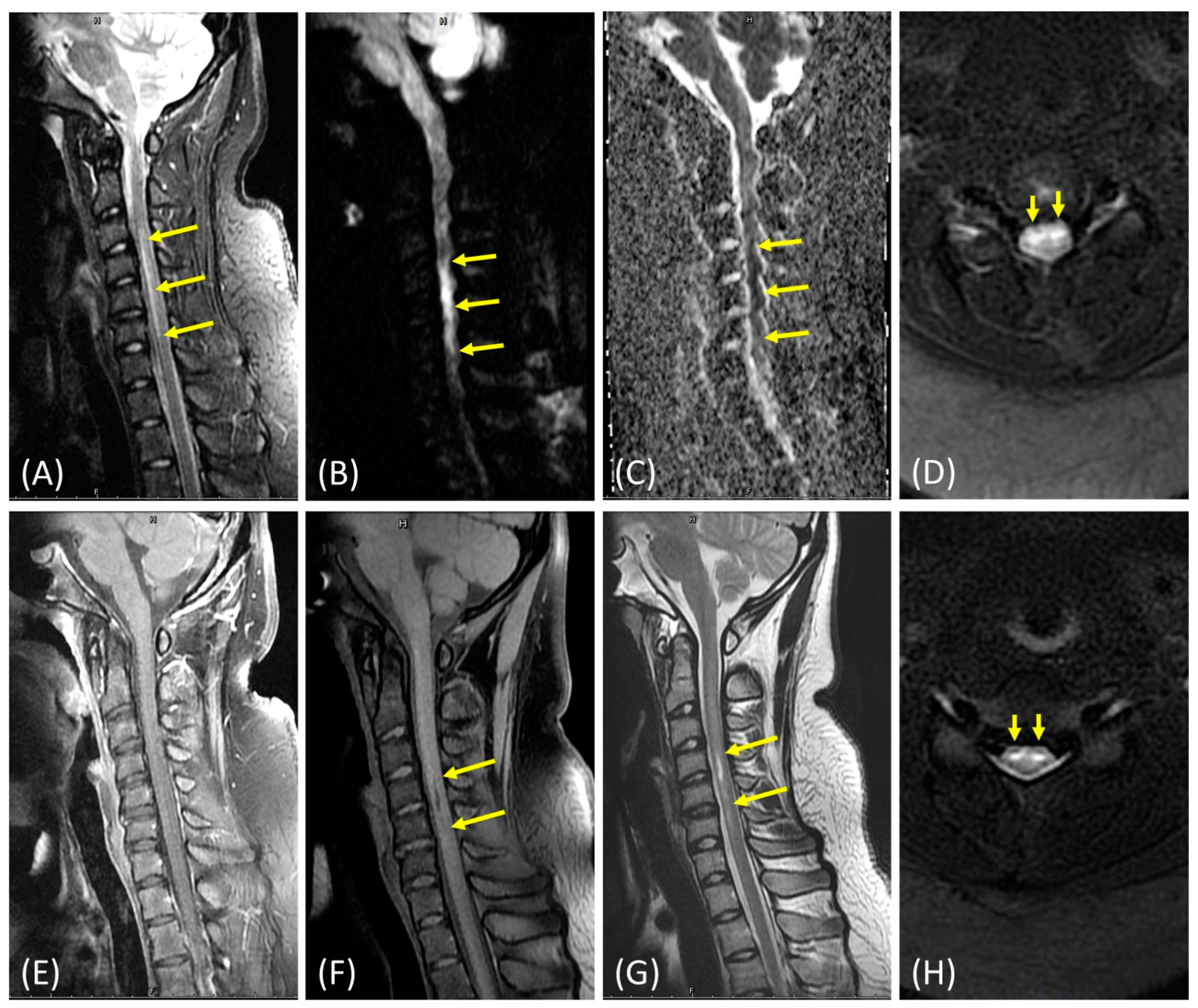
2. Case Presentation

A morbidly obese but relatively healthy and energetic 14-year-old right-handed female presented to the emergency department with a sudden onset of bilateral numbness and weakness in her arms, which spread downward into her legs. Before the onset of this episode, the patient was in a stable sitting position. She was alert, oriented, and verbally fluent. We inquired about any trauma history, even minor or trivial injury or posture (hyperextension or flexion), but she could not recall any relevant events that can cause this condition. Her physical examination showed intact cranial nerve function with relatively stable vitals. However, an unequal muscle weakness was observed in the patient’s four limbs, especially in the upper right and lower left extremities, which displayed muscle strength scores (Medical Research Council Scale, MRC scale) of 2/5 and 1/5, respectively. The patient’s sensory exam demonstrated that she had diminished responses to pain, temperature, and crude touch. Yet, proprioception and responses to vibration and fine touch were unaffected. An increased patellar reflex in the lower extremities, alongside a positive Babinski reflex on the right side, were also noted. However, no organic lesions in the patient’s cerebrum were detected by emergent contrast-enhanced brain computed tomography (CT) with CT angiography/venography reconstruction. While in a state of acute flaccid paralysis, the patient was admitted to the intensive care unit for further evaluation and management.
Soon after admission, a lumbar puncture was performed on the patient to harvest cerebral spinal fluid (CSF) to rule out the presence of infection and/or inflammation in her central nervous system. The patient’s biochemistry, cell counts, Gram staining, FilmArray® (Salt Lake City, UT, USA) meningitis/encephalitis (M/E) panel, and CSF culture were all negative. Despite an overall unremarkable cerebral CT and magnetic resonance image (MRI), hyperintensities extending from the cervical vertebrae C3-6 (Figure 1) were still observed in the short tau inversion recovery/diffusion weighted imaging (STIR/DWI) MRI, which is consistent with the possible ASAS present in the patient [8,9]. A young stroke panel was also used to extensively survey for hematological conditions (protein C, protein S, anti-thrombin, PT/aPTT/INR, and D-dimer), autoimmune diseases [10] (anti-ds DNA, anti-Cardiolipin IgG/IgM, ANA titer/pattern, anti-β2 glycoprotein, C3, C4, LA(DRVVT), anti-AQP4 IgG, and anti-MOG IgG), metabolic statuses (T3, T4, free T4, TSH, anti-TPO, HbA1C, plasma Lyso-Gb3, HDL, LDL, triglyceride, uric acid, total protein, vitamin B12, folate, and homocysteine), and infection issues (HIV Ag/Ab and RPR). Except for low serum folate levels (1.7 ng/mL, reference range: 4.8–37.3) and mildly increased LDL cholesterol levels (114 mg/dL, reference range: <100), no other abnormalities were identified in the patient. The patient received a five-day course of high-dose intravenous methylprednisolone to ameliorate the spinal cord edema, aspirin for the anticoagulation [11], and folic acid supplementation for the marked folate deficiency (Table 1). When folate levels were rechecked two weeks later, they were restored to normal levels (12.2 ng/mL). We also tested the patient’s genome for the presence of three pathogenic variants of the methylenetetrahydrofolate reductase (MTHFR) gene (NM_005957.5: c.1969A>G, p.Ter657Arg (rs768434408); c.1304_1305del, p.Phe435fs (rs4846051); c.470C>T, p.Arg157Gln (rs121434295)), but none were detected.
On the other hand, her spinal MRI revealed mild herniated intervertebral disc (HIVD), but no bone marrow edema, no obvious structural abnormality at the spine, and the vertebra was found and the chance of fibrocartilaginous embolism was low, so we did not perform spinal digital subtraction angiography (DSA) and magnetic resonance angiogram (MRA) in this case. Although the possibility of minor trauma was low in the initial evaluation, no advanced images to investigate the offending pathology to the ASA could be a potential factor that limits the value of this study. Additionally, a 96-day follow-up spinal MRI revealed a myelomalacia extending from cervical vertebrae C4–C5 from a previous infarction (Figure 1). A 20-week laboratory workup also indicated that the patient’s folate levels dropped back down to 4.2 ng/mL. Thus, the patient was again supplemented with folic acid beginning at her 24-week follow-up appointment. The patient’s initial modified Rankin Scale (mRS), a measure of neurologic disability, of 5/6 gradually improved to 1/6 by her final outpatient visit. Additionally, the patient’s self-evaluation score improved from 20/100 to 92/100 within 6 months of treatment. She can go to school and handle her daily life without assistance. Furthermore, she is able to play volleyball as usual even though she felt a bit clumsy and presented mildly impaired coordination. She once mentioned that she started to feel unbearable pain over her limbs, so we added pregabalin for pain management. Now, she does not rely on this medicine anymore. At present, we only prescribe a daily low-dose folic acid supplement, vitamin B-complex, and low-dose aspirin [11] and will recheck the folate level soon in the follow-up clinic (Table 1).

3. Discussion

Folate, or vitamin B9, is a water-soluble vitamin found in many foods whose metabolic derivatives are essential for the biosynthesis of purines and pyrimidines and protein metabolism [12]. Folate additionally plays a key role in the one-carbon metabolic pathway as it is necessary for the breakdown of homocysteine, an amino acid that can induce neurotoxicity when present at systematically elevated amounts [13]. Since hyperhomocysteinemia is an emerging comorbidity in ischemic stroke, folate intake and serum folate concentration are therefore hypothesized to be associated with cardiovascular disease risk through the modulation of homocysteine concentration [14]. Folate deficiency and/or MTHFR gene polymorphisms lead to hyperhomocysteinemia, which is a well-established risk factor for ischemic stroke in adult populations [14] as well as nontraumatic spinal cord infarction in children [15]. Thus, reductions in both folate and folate-associated nutrient (e.g., vitamins B6 and B12) levels within patient populations negatively impact the performance of the folate cycle, which subsequently impairs homocysteine metabolism [13].
Since methylenetetrahydrofolate reductase is the key enzyme for the biosynthesis of the active form of folate, we evaluated and selected three established pathogenic MTHFR gene variants, via ClinVar (https://www.ncbi.nlm.nih.gov/clinvar/ (accessed on 19 April 2022)), found in the Taiwan biobank database (https://taiwanview.twbiobank.org.tw/index (accessed on 19 April 2022)), which were verified by Sanger sequencing. However, no MTHFR gene variants were found to be applicable for this case (MTHFR gene (NM_005957.5: c.1969A>G, p.Ter657Arg (rs768434408); c.1304_1305del, p.Phe435fs (rs4846051); c.470C>T, p.Arg157Gln (rs121434295)). Our patient did not display elevated levels of homocysteine, nor did she possess three known pathogenic MTHFR mutations, and it was clear that the patient had a significant and isolated folate deficiency which can be treated via the supplementation of folic acid in order to prevent the onset of a new ASAS episode as well as to allow for the reparation of the patient’s injured spinal cord [4].
Next, we compared this case with similar reports involving the diagnosis of nontraumatic ASAS in adolescent populations (Table 2). We initially referred to similar cases fitting Zalewski’s criteria for spontaneous spinal cord infarction [16] and then excluded those with alternative acute myelopathy diagnoses, including those with intramedullary (e.g., inflammation, infection/post-infection, vascular anomalies, neoplasm, and toxin) and extramedullary causes (e.g., hematoma, abscess, compression, or spondylosis), and periprocedural acute myelopathy (e.g., aortic surgery). Finally, we recruited another 15 articles and an additional 27 cases with ages ranging from 10 to 19 years old (mean ± SD: 14.36 ± 1.82). A total of 28 cases are presented in Table 2, including 8 males (29%) and 20 females (71%). Some underlying medical conditions present within this population include obesity, asthma, early puberty, previously received spinal surgery, sickle-cell anemia, Down syndrome, and back pain. However, 19 cases did not have any underlying medical conditions of significance. Neurologically, most of the examined cases had sensory impairment (23/28, 82%), while all had various degrees of motor impairment. Additionally, 27 and 13 cases presented with bladder (27/28, 96%) and bowel dysfunction (13/28, 46%), respectively. Radiologic studies revealed lesions ranging from the lower medulla down to the conus medullaris, with the exception of two cases which did not receive the exam, and one which showed unspecified findings. Some potential etiologies include low serum folate levels, MTHFR gene variance, hemoglobinopathy, prothrombin gene variance, protein S deficiency, and fibrocartilaginous embolism (FCE), while 18 had unknown etiology. Later follow-up outcomes (mRS) revealed two cases of mortality; however, a single outcome was not available, with the remaining showing various degrees of neurologic improvement.
Overall, female cases outnumbered males in our pooled data, and more than half of the cases examined did not have any detectable underlying diseases or identified etiologies. Furthermore, the clinical outcome varied greatly among these patients in the long-term follow-up. Nedeltchev et al. proposed that severe initial impairment (American Spinal Injury Association grades A and B) and being female were unfavorable predictors in adult cohorts, but this may not apply to pediatric cases of acute spinal cord ischemia syndrome [29]. Hence, nontraumatic ASAS is still a challenging issue and a critical topic in the clinical milieu. More case studies and extensive workups are still needed for further discovery of potential etiologies and risk factors in pediatric populations.

4. Conclusions

The causes of nontraumatic ASAS remain mostly unknown, especially in pediatric populations. It is noteworthy that SCIWORA is one of the important differential diagnoses in ASAS. Since SCIWORA is mostly caused by hyperextension or flexion injuries of the pediatric spine, carefully evaluating the minor trauma and injuries is the first step in the approach of acute flaccid paralysis. After ruling out the traumatic etiology, extensive medical workups are necessary to explore the underlying pathomechanism of nontraumatic ASAS. Although more than half of the cases in our pooled data did not have underlying diseases or identified etiology, some still indicated potential predisposing factors, such as our case demonstrated here. Therefore, we suggest that folate deficiency is a potential risk factor in the adolescent ASAS population that needs to be medically identified and intervened upon early to improve the prognosis of these patients.

Author Contributions

Conceptualization, C.-C.H. and C.-F.H.; investigation, C.-C.H. and C.-F.H.; writing—original draft preparation, C.-C.H. and Y.-Y.Y.; writing—review and editing, G.W.G.L.; image interpretation, Y.-P.L.; primer design for three pathogenic variants of MTHFR gene as well as the interpretations of Sanger’s sequencing data, K.-S.H.; supervision and finalization of this manuscript, C.-F.H. All authors have read and agreed to the published version of the manuscript.

Funding

This research was funded by Tri-Service General Hospital, grant number TSGH-D-11039 (H.C.-F.).

Institutional Review Board Statement

The study was conducted according to the guidelines of the Declaration of Helsinki and approved by the Institutional Review Board of Tri-Service General Hospital (No. H20210923).

Informed Consent Statement

The written informed consent was given from the patient and her mother as the legal representative. The consent was for publication of the patient’s personal and medical information including any associated images and lab data.

Data Availability Statement

All the data are contained within the article.

Acknowledgments

The authors thank Elvis Quiroz for English editing.

Conflicts of Interest

The authors declare no conflict of interest.

References

  1. Xing, W.; Zhang, W.; Ma, G.; Ma, G.; He, J. Long-segment spinal cord infarction complicated with multiple cerebral infarctions: A case report. BMC Neurol. 2022, 22, 362. [Google Scholar] [CrossRef] [PubMed]
  2. Peng, T.; Zhang, Z.F. Anterior Spinal Artery Syndrome in a Patient with Cervical Spondylosis Demonstrated by CT An-giography. Orthop Surg 2019, 11, 1220–1223. [Google Scholar] [CrossRef] [Green Version]
  3. Ergun, A.; Oder, W. Pediatric care report of spinal cord injury without radiographic abnormality (SCIWORA): Case report and literature review. Spinal Cord 2003, 41, 249–253. [Google Scholar] [CrossRef] [PubMed] [Green Version]
  4. Nance, J.R.; Golomb, M.R. Ischemic Spinal Cord Infarction in Children Without Vertebral Fracture. Pediatr. Neurol. 2007, 36, 209–216. [Google Scholar] [CrossRef] [PubMed] [Green Version]
  5. Tykocki, T.; Poniatowski, Ł.A.; Czyz, M.; Wynne-Jones, G. Oblique corpectomy in the cervical spine. Spinal Cord 2018, 56, 426–435. [Google Scholar] [CrossRef] [PubMed] [Green Version]
  6. Kubaszewski, Ł.; Wojdasiewicz, P.; Rożek, M.; Słowińska, I.E.; Romanowska-Prochnicka, K.; Słowiński, R.; Poniatowski, Ł.A.; Gasik, R. Syndromes with chronic non-bacterial osteomyelitis in the spine. Reumatologia 2015, 53, 328–336. [Google Scholar] [CrossRef] [Green Version]
  7. Spencer, S.P.; Brock, T.D.; Matthews, R.R.; Stevens, W.K. Three Unique Presentations of Atraumatic Spinal Cord Infarction in the Pediatric Emergency Department. Pediatr. Emerg. Care 2014, 30, 354–357. [Google Scholar] [CrossRef]
  8. Lee, M.J.; Aronberg, R.; Manganaro, M.S.; Ibrahim, M.; Parmar, H.A. Diagnostic Approach to Intrinsic Abnormality of Spinal Cord Signal Intensity. RadioGraphics 2019, 39, 1824–1839. [Google Scholar] [CrossRef]
  9. Beslow, L.A.; Ichord, R.N.; Zimmerman, R.A.; Smith, S.E.; Licht, D.J. Role of Diffusion MRI in Diagnosis of Spinal Cord Infarction in Children. Neuropediatrics 2008, 39, 188–191. [Google Scholar] [CrossRef]
  10. Hsu, J.L.; Cheng, M.-Y.; Liao, M.-F.; Hsu, H.-C.; Weng, Y.-C.; Chang, K.-H.; Chang, H.-S.; Kuo, H.-C.; Huang, C.-C.; Lyu, R.-K.; et al. A comparison between spinal cord infarction and neuromyelitis optica spectrum disorders: Clinical and MRI studies. Sci. Rep. 2019, 9, 7435. [Google Scholar] [CrossRef]
  11. Boucher, A.A.; Taylor, J.M.; Luchtman-Jones, L. Aspirin in childhood acute ischemic stroke: The evidence for treatment and efficacy testing. Pediatr. Blood Cancer 2019, 66, e27665. [Google Scholar] [CrossRef] [PubMed]
  12. Golja, M.V.; Šmid, A.; Kuželički, N.K.; Trontelj, J.; Geršak, K.; Mlinarič-Raščan, I. Folate Insufficiency Due to MTHFR Deficiency Is Bypassed by 5-Methyltetrahydrofolate. J. Clin. Med. 2020, 9, 2836. [Google Scholar] [CrossRef]
  13. Poddar, R. Hyperhomocysteinemia is an emerging comorbidity in ischemic stroke. Exp. Neurol. 2021, 336, 113541. [Google Scholar] [CrossRef] [PubMed]
  14. Weng, L.-C.; Yeh, W.-T.; Bai, C.-H.; Chen, H.-J.; Chuang, S.-Y.; Chang, H.-Y.; Lin, B.-F.; Chen, K.-J.; Pan, W.-H. Is Ischemic Stroke Risk Related to Folate Status or Other Nutrients Correlated With Folate Intake? Stroke 2008, 39, 3152–3158. [Google Scholar] [CrossRef] [PubMed] [Green Version]
  15. Bar, C.; Cheuret, E.; Bessou, P.; Pedespan, J.-M. Childhood idiopathic spinal cord infarction: Description of 7 cases and review of the literature. Brain Dev. 2017, 39, 818–827. [Google Scholar] [CrossRef]
  16. Zalewski, N.L.; Rabinstein, A.A.; Krecke, K.N.; Brown, R.D.; Wijdicks, E.F.M.; Weinshenker, B.G.; Kaufmann, T.J.; Morris, J.M.; Aksamit, A.J.; Bartleson, J.D.; et al. Characteristics of Spontaneous Spinal Cord Infarction and Proposed Diagnostic Criteria. JAMA Neurol. 2019, 76, 56–63. [Google Scholar] [CrossRef] [PubMed] [Green Version]
  17. Nedeltchev, K.; Loher, T.J.; Stepper, F.; Arnold, M.; Schroth, G.; Mattle, H.P.; Sturzenegger, M. Long-Term Outcome of Acute Spinal Cord Ischemia Syndrome. Stroke 2004, 35, 560–565. [Google Scholar] [CrossRef] [Green Version]
  18. Sawada, D.; Ito, A.; Shiohama, T.; Tsukada, H.; Fujii, K. Spontaneous spinal cord infarction in a 10-year-old Japanese girl. Pediatr. Int. 2022, 64, e14909. [Google Scholar] [CrossRef]
  19. Seo, Z.W.; Huh, S.; Ko, H.-Y. Non-traumatic spinal cord infarction of the conus medullaris in a child: A case report. Spinal Cord Ser. Cases 2021, 7, 59. [Google Scholar] [CrossRef]
  20. Stettler, S.; El-Koussy, M.; Ritter, B.; Boltshauser, E.; Jeannet, P.-Y.; Kolditz, P.; Meyer-Heim, A.; Steinlin, M. Non-traumatic spinal cord ischaemia in childhood-clinical manifestation, neuroimaging and outcome. Eur. J. Paediatr. Neurol. 2013, 17, 176–184. [Google Scholar] [CrossRef]
  21. Márquez, J.C.; Granados, A.M.; Castillo, M. MRI of cervical spinal cord infarction in a patient with sickle cell disease. Clin. Imaging 2012, 36, 595–598. [Google Scholar] [CrossRef] [PubMed]
  22. Sohal, A.S.; Sundaram, M.; Mallewa, M.; Tawil, M.; Kneen, R. Anterior Spinal Artery Syndrome in a Girl With Down Syndrome: Case Report and Literature Review. J. Spinal Cord Med. 2009, 32, 349–353. [Google Scholar] [CrossRef] [PubMed] [Green Version]
  23. Hakimi, K.N.; Massagli, T.L. Anterior Spinal Artery Syndrome in Two Children With Genetic Thrombotic Disorders. J. Spinal Cord Med. 2005, 28, 69–73. [Google Scholar] [CrossRef]
  24. Ramelli, G.P.; Wyttenbach, R.; Giovanni, O.S.; Von Der Weid, N.; Ozdoba, C. Anterior Spinal Artery Syndrome in an Adolescent With Protein S Deficiency. J. Child Neurol. 2001, 16, 134–135. [Google Scholar] [CrossRef]
  25. Wilmshurst, J.; Walker, M.; E Pohl, K.R. Rapid onset transverse myelitis in adolescence: Implications for pathogenesis and prognosis. Arch. Dis. Child. 1999, 80, 137–142. [Google Scholar] [CrossRef] [Green Version]
  26. Yousef, O.M.; Appenzeller, P.; Kornfeld, M. Fibrocartilagenous Embolism: An Unusual Cause of Spinal Cord Infarction. Am. J. Forensic Med. Pathol. 1998, 19, 395–399. [Google Scholar] [CrossRef] [PubMed]
  27. Tosi, L.; Rigoli, G.; Beltramello, A. Fibrocartilaginous embolism of the spinal cord: A clinical and pathogenetic reconsidera-tion. J. Neurol. Neurosurg. Psychiatry 1996, 60, 55–60. [Google Scholar] [CrossRef] [Green Version]
  28. Toro, G.; Roman, G.C.; Navarro-Roman, L.; Cantillo, J.; Serrano, B.; Vergara, I. Natural History of Spinal Cord Infarction Caused by Nucleus Pulposus Embolism. Spine 1994, 19, 360–366. [Google Scholar] [CrossRef]
  29. Vandertop, W.P.; Elderson, A.; van Gijn, J.; Valk, J. Anterior spinal artery syndrome. AJNR Am. J. Neuroradiol. 1991, 12, 505–506. [Google Scholar]
Figure 1. MRI reveals a hyperintensity in the patient’s cervical spinal cord between cervical vertebrae C3-6 on sagittal STIR images (A), which are associated with restricted diffusion abnormalities in the patient’s sagittal DWI (B) and apparent diffusion coefficient (ADC) images (C). Symmetrical elevated intramedullary signals in the anterior horns of the patient’s spinal cord (i.e. the “owl-eyes” sign for spinal cord infarction) were observed in the axial STIR images. Edema was also present at the same spinal cord level (D). No internal enhancements were detected in the sagittal T1-weighted contrast-enhanced images, which is inconsistent with the presence of infection and/or inflammation (E). A three-month follow-up MRI revealed a hypointensity between cervical vertebrae C4-5 in the patient’s sagittal T1-weighted fat-saturated (T1FS) images (F) and a hyperintensity in the sagittal T2-weighted images (G) due to myelomalacia. Axial STIR images (H) show a T2-hyperintensity in the anterior grey columns, in which the owl-eyes characteristic sign is observed as well. Arrows indicate the presence of abnormalities.
Brainsci 12 01470 g001
Table 1. Case presentations of disease severity/score, clinical course, evaluations, prescriptions, and locations (Gantt chart).
CategoryModified RSS.E.BH: 1.62 mEvaluationsPrescriptionsLocation
Time Table01234560-100BW
(kg)
BMI
(kg/m2)
Laboratory StudyImage/Electrophysiologic StudyPulse TherapyABFLP.IP.OPIWHS
Week 1 2010038.1D1:Young stroke panel
(B12, folate, HC)
D1:Lumbar puncture
D1: Brain CTA/CTV
D2: Brain/Spine MRI, NCV/EMG, echocardiography
D2~D6TIDTIDQDQDQD
Week 2 5096.236.66B12, folate, HC TIDTID1/2 QDQDQD
* Week 3 6293.535.63 D21: Carotid artery sonography BIDTID1/4 QD QD
Week 4 7187.433.3 QDTID QD
Week 5 7589.934.26 D29: SSEP U/L limbs QDTID QD
* Week 6 8489.434.06 QDTID QD
* Week 9 NANANA QDTID QD
Week 10 8886.933.11 QDBID
Week 16 NANANA D96: Spine MRI follow-up QDBID QD
Week 20 909335.44B12, folate, HCD120: NCV/EMG follow-up QDQD QD
Week 24 9290.534.48 QDQD1/4 QD
RS = Rankin Scale; S.E. = self-evaluation score; Pulse therapy = methylprednisolone 1000 mg/day; A = aspirin (100 mg), B = B1 (50 mg), B6 (50 mg), B12 (500 ug) complex; F = folic acid (5 mg), L = Lipitor (10 mg); P.I. = piracetam IV form (1000 mg); P.O. = piracetam oral form (1200 mg); P = pregabalin (75 mg); I = ICU; W = ward; H = home; S = school; HC = homocysteine; CTA/CTV = computed tomography arteriography/venography; MRI = magnetic resonance imaging; NCV/EMG = nerve conduction velocity/electromyography; SSEP = somatosensory evoked potential; U/L = upper/lower; NA = not available. * Week 3: D21, start rehabilitation program; * Week 6: transfer to other hospital for further rehabilitation; * Week 9: discharge from hospital and sent back to home and school. Gray degree of mRS: the darker implies the severer of the neurologic disability. Gray degree of location: the lighter implies that she can live a more normal life. TID: three times a day, BID: two times a day, QD: once daily. D1: day 1, D21: day 21, D29: day 29, D96: day 96, D120: day 120.
Table 2. Comparison of cases with the diagnosis of nontraumatic ASAS in adolescence.
Study/
Year
No.
case
Age/
Gender
Underlying diseasesClinical ManifestationsRadiological lesionsPotential
Etiology
Outcome at the latest FU
(mRS)
Sensory
impairment
Motor
impairment
Bladder/Bowel dysfunction
Our Case 2022114Y/FObesity
BMI:38.1kg/m2
Dysesthesia, temp., painParesis: RU, LL+/+C3─C6Low serum folate level1/6
Sawada
[17] 2022
110Y/FNilTemp., painParaparesis+/−C7─T2Unknown1/6
Seo
[18] 2021
112Y/FNilNilParaparesis+/+T12─CMUnknown1/6
Bar
[15] 2017
115Y/FAsthmaNilPlegia: RL
Paresis: LL
+/+T11─L1Unknown3/6
214Y/FNilTemp., painParaplegia+/−T7─T11Unknown1/6
313Y/FNilAll modalitiesParaplegia+/+T8─T12Unknown3/6
413Y/FNilNilParaplegia+/+CMUnknown3/6
514Y/FEarly pubertyTemp., pain, LTPlegia: LL
Paresis: RL
+/−CMUnknown2/6
Spencer
[7] 2014
112Y/FNilAll modalitiesParaplegia+/+T1─CMUnknown3/6
214Y/MNilTemp., painParaplegia+/−T7─T12Unknown1/6
Stettler
[19] 2013
113Y/FS/P spinal surgeryDysesthesia, temp., painPlegia: RL
Paresis: LL
+/+T7─T9MTHFR, Ho, c.677C>T1/6
213Y/FNilDysesthesia, temp., painPlegia: RL
Paresis: LL
+/+T3─T5Unknown1/6
Márquez
[20] 2012
119Y/MSickle cell diseaseNilQuadriparesis+/−C2─C7Hemoglobi-nopathy2/6
Sohal
[21] 2009
116Y/FDown syndromeNot applicable1Paraplegia+/−T5─T12Unknown2/6
Nance
[4] 2007
114Y/FNilDysesthesia, painQuadriplegia+/+Low medulla,
C1─C7, T3
Unknown5/6
217Y/MBack pain, palpitationsAll modalitiesParaparesis−/−C2, T5─T9MTHFR,
CH, c.677C>T and 1298A>C
2/6
Hakimi
[22] 2005
115Y/MNilPain, LTParaplegia+/+Midthoracic level─CMProthrombin variant, He3/6
212Y/FS/P spinal surgeryTemp., pain, PCParaplegia+/+Not performed2Protein S deficiency3/6
Ramelli
[23] 2001
115Y/MNilTemp., painParaplegia+/+T5─T6Protein S deficiency3/6
Wilmshur st
[24] 1999
114Y/FNilAll modalitiesParaplegia+/−T9─CMUnknown4/6
215Y/FNilTemp., pain, PCParaplegia+/−Anterior+Right posterior cordUnknown3/6
314Y/MNilDysesthesia, temp., painParaplegia+/−Anterior distal thoracic cordUnknown3/6
416Y/MNilPainParaparesis+/−T3─T6Unknown3/6
515Y/FNilTemp., painParaplegia+/+T9─CMUnknown4/6
Yousef
[25] 1998
114Y/FObesity
BMI:33.6kg/m2
Pain, LTParaplegia+/−UnspecifiedFCE6/63
Tosi
[26] 1996
116Y/FNilTemp., pain, LT, PCParaplegia+/−T11-L1FCENo description
Toro
[27] 1994
116Y/FNilAll modalitiesParaplegia+/−Not performedFCE6/64
Vandertop
[28] 1991
117Y/MNilDysesthesia, temp., painQuadriparesis+/−C1-T3Unknown2/6
Abbreviations: Y: years old, F: female, M: male, Temp.: temperature, LT: light touch, PC: proprioception, C: cervical, T: thoracic, L: lumbar, CM: conus medullaris, S/P: status post, Ho: homozygous, He: heterozygous, CH: compound heterozygous, FCE: fibrocartilaginous embolism. Notes: 1 Not applicable: accurate sensory examination of the lower limbs was difficult due to learning difficulties and distress present in this patient, however, sensation to light touch, vibration, and joint position in the lower limbs appeared to be intact. 2 Not performed: due to extensive metallic hardware after corrective posterior spinal fusion. 3 The patient left the hospital against medical advice on the third day of hospitalization. She died 7 days later from a massive pulmonary thromboembolus proven by autopsy. 4 The patient died unexpectedly as a result of bronchial aspiration 6 weeks after disease onset.
Publisher’s Note: MDPI stays neutral with regard to jurisdictional claims in published maps and institutional affiliations.

