Article

Assessment of the Severity of Intermediate Coronary Artery Stenosis Using the Systemic Inflammatory Response Index

1
Department of Cardiology, School of Medicine, Kastamonu University, Kastamonu 37150, Turkey
2
Department of Cardiology, Ankara City Hospital, Ankara 06200, Turkey
*
Author to whom correspondence should be addressed.
Diagnostics 2025, 15(2), 162; https://doi.org/10.3390/diagnostics15020162
Submission received: 4 November 2024 / Revised: 9 January 2025 / Accepted: 11 January 2025 / Published: 13 January 2025
(This article belongs to the Special Issue New Progress in Diagnosis and Management of Cardiovascular Diseases)

Abstract

:
Background: Fractional Flow Reserve (FFR) is a method that enables the hemodynamic assessment of coronary artery stenosis. The Systemic Inflammatory Response Index (SIRI) is a new marker calculated by multiplying the neutrophil-to-lymphocyte ratio (NLR) with the monocyte count. It is indicative of the presence and severity of coronary artery disease. This study evaluates the relationship between the functional significance of FFR measurements and the SIRI in intermediate coronary stenosis. Methods: A total of 294 patients with 50–70% stenosis in their coronary arteries based on quantitative measurement following angiography who underwent FFR measurement were included in the study before the FFR procedure. Total and differential leukocyte counts and routine biochemical tests were performed. Results: A total of 37% of the patients were found to have a positive FFR, while 63% had a negative FFR. Significant differences were observed in the neutrophil count, monocyte count, Systemic Inflammation Response Index (SIRI), total cholesterol, and amount of adenosine used between the groups (p < 0.05). A SIRI value of 1.16 was 77% sensitive and 55% specific for FFR positivity. Multivariate logistic regression analysis identified the SIRI as an independent predictor of FFR positivity. Conclusions: Our study has demonstrated that high values of the SIRI may serve as a new biomarker for predicting FFR positivity.

1. Introduction

Ischemic heart diseases remain the primary cause of mortality and disability-adjusted life-year loss [1]. Today, coronary artery disease is the most important cause of mortality and morbidity in the world and our country. At the beginning of the 21st century, 50% of all deaths in developing countries were due to cardiovascular diseases (CVD), while this rate was 25% in developed countries. According to current knowledge, atherosclerosis is a multifactorial process in which chronic inflammation plays a role at every stage in its progression, from the beginning to the later period, and in which many risk factors contribute to the developing inflammation. Atherosclerosis is a systemic arterial disease that affects vessels of all sizes, primarily large and medium-sized elastic arteries. Atherosclerosis is a process that occurs in the form of intimal smooth muscle cell accumulation and proliferation; the infiltration of monocytes, macrophages, and T lymphocytes; a connective tissue matrix rich in collagen, elastin fibers, fibronectin, and proteoglycans; and lipid storage, especially in the form of free cholesterol or cholesterol esters within the cells and in the surrounding connective tissue.
Many methods have been used to diagnose coronary artery disease from the past to the present. Coronary angiography is the fundamental method used in the detection and intervention of coronary artery diseases [2]. As a result of coronary angiography, a qualitative evaluation of coronary lesions and stenosis is made. Qualitative assessment of coronary stenosis post-coronary angiography is only sometimes reliable. Moreover, an anatomically significant lesion may not be hemodynamically significant. Fractional Flow Reserve (FFR) is a method that enables the hemodynamic assessment of coronary artery stenoses. It is particularly beneficial in the hemodynamic evaluation of intermediate stenoses between 50 and 60% [3].
Studies have identified that endothelial dysfunction, oxidative stress, platelet activation, and inflammation play crucial roles in the development and progression of atherosclerosis [4,5]. Studies have shown that laboratory levels of the neutrophil–lymphocyte ratio (NLR) and platelet–lymphocyte ratio (PLR) indicate inflammation. They can provide insights into the severity of coronary artery disease. In addition, the NLR has been found helpful in predicting in-hospital mortality in patients presenting with acute decompensation of atrial fibrillation [6]. Recently, the Systemic Inflammation Response Index (SIRI, calculated by multiplying the NLR with the monocyte count), a newly introduced marker, has been shown to indicate the presence and severity of coronary artery disease [7]. The hemogram result, a simple test, can inform us about whether one has coronary artery disease. This study evaluates the relationship between the functional significance of FFR measurements and the SIRI in intermediate coronary stenoses.

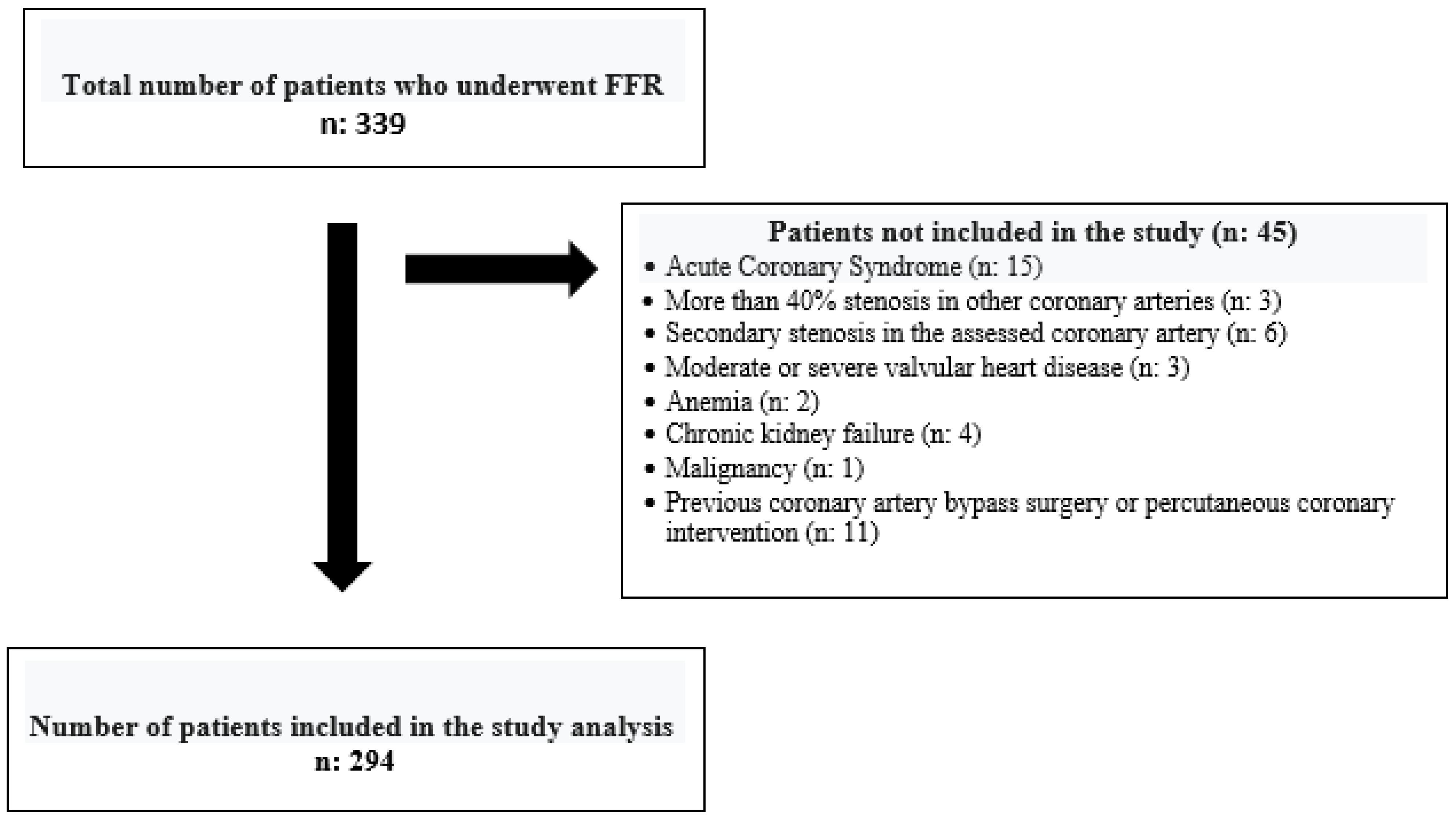
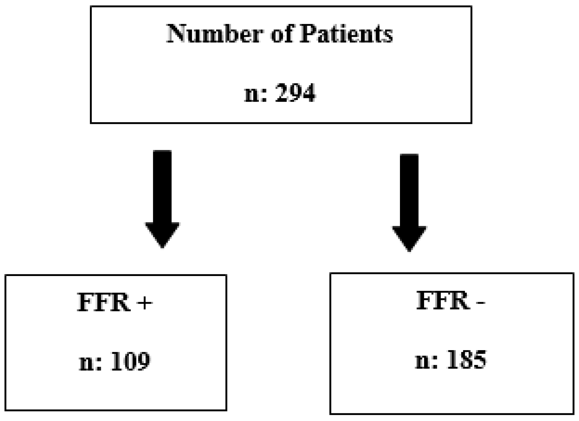
2. Materials and Methods

This retrospective study reviewed records from two centers between 1 January 2012 and 31 December 2022. A total of 339 patients who had 50–70% stenosis in their coronary arteries based on quantitative measurement following angiography and who underwent FFR measurement were included in the study. The patients were included in the survey, respectively, regardless of age, gender, and ethnicity. The patients considered for the study were those with stable coronary artery disease undergoing coronary angiography. Patients with acute coronary syndrome, moderate or severe valvular heart disease, significant arrhythmia, hemodynamic instability, secondary stenosis in the assessed coronary artery, more than 40% stenosis in other coronary arteries, previous coronary artery bypass surgery or percutaneous coronary intervention, acute–chronic or inflammatory bowel disease, anemia, chronic kidney failure, and malignancy were excluded from the study (Table 1). When patients were separated from the study according to the exclusion criteria, the remaining 294 patients were included. (Figure 1). All the patients meet the inclusion criteria.
Hospital records were utilized to ascertain patients’ angiographic information, clinical data, and demographic characteristics. Blood samples, including lipid profiles, serum creatinine values, and complete blood counts, were taken upon hospital admission. Hemogram tests of both FFR (+) and FFR (−) groups were measured automatically in the XN 1000 (Sysmex, Kobe, Japan). SIRI values were calculated by multiplying the NLR with the monocyte count.
For coronary narrowing ranging from 50% to 70%, a decision for Fractional Flow Reserve (FFR) measurement was made at the cardiologist’s discretion. After administering 5000 units of intracoronary heparin, the coronary artery was evaluated using a non-side-hole guiding catheter. Following calibration, a 0.014-inch pressure-monitoring guide wire (Prime Wire, Volcano, San Diego, CA, USA) was positioned distal to the lesion. Before FFR measurement, a 200 mcg intracoronary bolus of nitroglycerin was administered. Distal intracoronary measurements were taken initially and after achieving hyperemia with incrementally increased doses of adenosine. The ratio of the mean intracoronary pressure at peak hyperemia to the mean aortic pressure determined the FFR value.
An FFR value ≤ 0.80 was considered functionally significant. Patients with FFR values > 0.80 were classified as Group 1, and those with FFR values ≤ 0.80 as Group 2.
The study protocol was approved by the Kastamonu University Education and Research Hospital Ethics Committee and was performed in accordance with the Declaration of Helsinki. All patients provided written informed consent to participate in the study.

3. Results

The study included 294 patients, comprising 79 women (27%) and 215 men (73%). A total of 37% of the patients were found to have a positive FFR, while 63% had a negative FFR (Figure 2). The essential clinical and demographic characteristics, summarized in Table 2, showed no significant differences in age and gender between the groups with significant and non-significant FFRs (p > 0.05). Additionally, no significant differences were observed in the fasting blood sugar levels, creatinine levels, presence of diabetes and hypertension, and smoking habits between the two groups (p > 0.05).
Significant differences were observed in the neutrophil count, monocyte count, Systemic Inflammation Response Index (SIRI), total cholesterol, and amount of adenosine used between the groups (p < 0.05). The group with a non-significant FFR showed a lower neutrophil count (4.62 [3.69–5.68] vs. 5.32 [3.80–6.50]; p: 0.003), monocyte count (0.56 [0.5–0.7] vs. 0.65 [0.56–0.78]; p < 0.001), SIRI (1.10 [0.87–1.54] vs. 1.56 [1.18–2.13]; p < 0.001), and total cholesterol (185 [151–214] vs. 204 [162–234]; p = 0.008), while the amount of adenosine used was less (250 [150–300] vs. 200 [104–250]; p: 0.003) in the group with a significant FFR (Table 3).
The ROC analysis revealed an area under the curve (AUC) of 0.686 CI (0.622–0.750) (p < 0.001). A SIRI value of 1.25 was 71% sensitive and 61% specific for FFR positivity (Figure 3).
Multivariate logistic regression analysis identified the SIRI as an independent predictor of FFR positivity. (Odds ratio: 2.805; 95% CI [1.877–4.192]; p < 0.001) (Table 4).

4. Discussion

This study found that coronary narrowing was deemed functionally significant after Fractional Flow Reserve (FFR) measurements were independently associated with the Systemic Inflammation Response Index (SIRI) levels.
Oxidative stress and inflammation are fundamental mechanisms in the development and progression of atherosclerosis [4,5,8]. In coronary artery disease (CAD), standard inflammatory markers, such as white blood cells (WBCs) and C-reactive protein (CRP), have been linked with high cardiovascular risk [9], the severity of coronary artery disease [10], the instability of atherosclerotic plaque [11], and mortality related to CAD [12]. Chronic low-grade inflammation is believed to play a critical role in the progression of coronary artery disease [13]. Leukocytes and their subtypes (neutrophils, lymphocytes, and monocytes) can be measured in the total blood cell count, offering an economical and accessible method to assess inflammatory processes involved in the pathogenesis of CAD [14]. Monocytes, a specific subtype of lymphocytes, contribute to the initiation and progression of atherosclerosis by releasing pro-inflammatory cytokines, free oxygen radicals, and proteolytic enzymes [15]. They adhere to the endothelium, transform into macrophages, and become foam cells by absorbing lipid particles, activating cytokines and free oxygen radicals [16]. Studies have identified monocyte count as an independent short- and long-term marker of cardiovascular disease-related mortality, irrespective of other risk factors [17,18]. Neutrophils, the most common leukocyte subtype, exacerbate vascular wall inflammation by inducing small muscle cell apoptosis [19,20]. High neutrophil counts are positively associated with plaque rupture risk [20] and increase the risk of thrombosis in the microcirculation [21]. Conversely, lymphocytes inhibit the progression of atherosclerosis [22]. Lymphopenia is positively associated with Major Adverse Cardiac Events (MACE) [23] and is linked with an increased prevalence of heart failure [24] and poor prognosis in patients with acute coronary syndrome (ACS) [25]. Platelets influence atherosclerosis in two ways: their adhesion to the vessel wall facilitates plaque development [26], while their activation triggers inflammation and clot formation [27].
Recently, some parameters obtained by dividing white blood cell counts have been revealed due to their success in showing cardiovascular risk, myocardial infarcts, and the prevalence of atherosclerosis due to the results that can be easily calculated from hemogram results [28,29].
The monocyte–lymphocyte ratio (MLR), neutrophil–lymphocyte ratio (NLR), and platelet–lymphocyte ratio (PLR) serve as biomarkers reflecting a variety of immune pathways and cell functions. By integrating the effects of two distinct cell types that interact, their predictive value for cardiovascular disease (CVD) and mortality is enhanced synergistically [30,31,32].
The monocyte–lymphocyte ratio (MLR) is a parameter obtained by dividing monocytes by lymphocytes. In a study by Hua Yang et al., a higher baseline monocyte–lymphocyte ratio (MLR) was linked to an increased risk of death among US adults. MLR proved to be a strong independent predictor of overall mortality and cardiovascular mortality in the general population [33]. Similarly, research by Fan et al. found a significant association between an elevated MLR and a greater risk of mortality in the six months following intervention in patients with acute myocardial infarction (AMI) who underwent percutaneous coronary intervention (PCI) [34].
Another marker is the neutrophil–lymphocyte ratio (NLR), calculated by dividing the number of neutrophils by the number of lymphocytes. The NLR is the most researched white blood cell ratio related to cardiovascular diseases. Nunez et al. found that patients diagnosed with ST-elevation myocardial infarction (STEMI) had the highest neutrophil–lymphocyte ratio (NLR) within 12 to 24 h after being admitted to the hospital [35]. Various studies have shown a link between an elevated NLR and an increased occurrence of no-reflow following percutaneous coronary intervention (PCI) in STEMI patients. Furthermore, long-term follow-up of non-ST elevation myocardial infarction (NSTEMI) patients with slow coronary flow seen on angiography and a high NLR (greater than 3.88) was independently associated with a higher rate of recurrent myocardial infarction. A significant relationship was also observed between the NLR and myocardial injury, as reflected by elevated CK-MB levels. The NLR also demonstrated a negative correlation with cardiac contractility. Thus, a high NLR serves as a strong indicator of the extent of myocardial damage, suggesting that the level of inflammation is associated with compromised contractile function in the heart [36].
Another marker introduced is the platelet–lymphocyte ratio (PLR), obtained by dividing the platelet count by the lymphocyte count. As a cost-effective and widely accessible biomarker, the platelet–lymphocyte ratio (PLR) incorporates both inflammatory and thrombotic pathways, potentially offering more excellent prognostic value than individual platelet or lymphocyte counts. This makes it a promising tool for predicting acute coronary syndrome (ACS) [37,38]. Ye et al. found that a higher PLR was linked to worse clinical outcomes in patients with acute heart failure (HF), suggesting it could serve as a novel marker in the management of acute HF [39]. Additionally, a recent study by Turcato et al. demonstrated that PLR was independently associated with a threefold increase in the 30-day mortality risk following emergency department admission for acute decompensated HF [40].
As far as we know, our study is the first clinical investigation to demonstrate the relationship between clinically significant narrowing detected using Fractional Flow Reserve (FFR) measurements and the Systemic Inflammation Response Index (SIRI). Previous studies have explored the relationship between the Systemic inflammation Index (SII) and the extent of coronary artery disease. For instance, in their study involving 400 patients undergoing coronary angiography, Liu and colleagues investigated the relationship between the Gensini score, indicating the severity of coronary artery disease, and the SII. They concluded that the SII is an independent risk factor for diagnosing and determining the severity of coronary artery disease [7]. Similarly, a study by Candemir et al. involving 669 patients found a positive correlation between coronary artery severity measured using the SYNTAX score and the SII [41]. Additionally, Erdoğan et al. discovered a positive correlation between the SII and the likelihood of detecting functionally significant narrowing in the coronary artery using FFR [42].
One of the critical differences between the SIRI and SII is that the SIRI incorporates the monocyte count, whereas the SII includes the platelet count. This could contribute significantly to the results, as monocytes are the primary cells responsible for atherosclerotic plaque formation. Previous studies in patients with acute coronary syndrome have observed increased inflammatory markers. The SII has been found to predict the occurrence of Major Adverse Cardiac Events (MACE) in acute coronary syndrome patients more accurately than classical risk factors [43]. These findings are supported by Li et al., who discovered the association of lymphocyte-based inflammatory markers with MACE and reported the advantage of the SIRI over other inflammatory markers in this context [44]. In another observational study spanning ten years with 85,000 participants, a correlation was found between a high SIRI and increased incidence of ACS in patients under 60. In contrast, no correlation was found with the SII [45].
The limitations of our study include a small patient cohort and relatively broad exclusion criteria, which encompassed active neoplastic processes or paraneoplastic syndromes, diagnosed active viral or bacterial infections, chronic kidney disease (stages III-V), an elevated erythrocyte sedimentation rate, and serum CRP levels. Furthermore, this study’s retrospective, cross-sectional, and observational nature limits the ability to analyze causal relationships among variables. It diminishes the confidence in attributing low-grade inflammation directly to heart disease. Due to the retrospective character of the study, other inflammatory parameters such as the ESR, fibrinogen, quantitative CRP, homocysteine, and uric acid could not be obtained in all patients, so the effects of these parameters on the NLR and SIRI could not be studied. Since the height and weight measurements of all patients included in the study were not recorded in the system, a BMI calculation could not be made. Therefore, the obesity paradox could not be ruled out. Additionally, the assessment of coronary artery disease severity was conducted using coronary angiography without accounting for the stabilizing effects of coronary calcifications. Lastly, the influence of hypolipidemic medications could not be considered, even though they may affect low-grade inflammation, as all patients received comparable doses of statins.

5. Conclusions

Given that the SIRI, a novel inflammatory biomarker, shows promise as a clinical tool for assessing coronary artery disease and its potential complications, further in-depth and well-designed studies on larger patient groups are necessary.

Author Contributions

Conceptualization, S.A.; Methodology, S.A., A.K. and A.Y.; Software, S.A. and A.K.; Validation, S.A. and A.K.; Formal analysis, S.A.; Investigation, S.A. and A.K.; Resources, S.A.; Data curation, S.A. and A.K.; Writing—original draft, S.A.; Writing—review and editing, S.A.; Visualization, S.A. and A.Y.; Supervision, S.A. and A.Y.; Project administration, S.A. and A.Y.; Funding acquisition, S.A. All authors have read and agreed to the published version of the manuscript.

Funding

This research received no external funding.

Institutional Review Board Statement

The study was conducted in accordance with the Declaration of Helsinki and approved by the Kastamonu University Education and Research Hospital Ethics Committee (No: 2022-KAEK-40/; date: 20 April 2022).

Informed Consent Statement

Informed consent was obtained from all subjects involved in the study.

Data Availability Statement

The original contributions presented in the study are included in the article; further inquiries can be directed to the corresponding author.

Conflicts of Interest

The authors declare no conflicts of interest.

References

  1. Vedanthan, R.; Seligman, B.; Fuster, V. Global perspective on acute coronary syndrome: A burden on the young and poor. Circ. Res. 2014, 114, 1959–1975. [Google Scholar] [CrossRef] [PubMed]
  2. Task Force Members; Montalescot, G.; Sechtem, U.; Achenbach, S.; Andreotti, F.; Arden, C.; Budaj, A.; Bugiardini, R.; Crea, F.; Cuisset, T.; et al. 2013 ESC guidelines on the management of stable coronary artery disease: The task force on the management of stable coronary artery disease of the European Society of Cardiology. Eur. Heart J. 2013, 34, 2949–3003. [Google Scholar] [PubMed]
  3. Tobis, J.; Azarbal, B.; Slavin, L. Assessment of intermediate severity coronary lesions in the catheterization laboratory. J. Am. Coll. Cardiol. 2007, 49, 839–848. [Google Scholar] [CrossRef] [PubMed]
  4. Hansson, G.K. Inflammation, atherosclerosis, and coronary artery disease. N. Engl. J. Med. 2005, 352, 1685–1695. [Google Scholar] [CrossRef]
  5. Hilgendorf, I.; Swirski, F.K.; Robbins, C.S. Monocyte fate in atherosclerosis. Arterioscler. Thromb. Vasc. Biol. 2015, 35, 272–279. [Google Scholar] [CrossRef] [PubMed]
  6. Kundnani, N.R.; Sharma, A.; Lighezan, D.F.; Georgescu, D.; Morariu, S.I.; Nisulescu, D.D.; Bita, R.G.; Rosca, C.I. Use of Neutrophil-to-Lymphocyte Ratio to Predict In-Hospital Mortality in Patients Admitted with Acute Decompensation of Atrial Fibrillation. J. Clin. Med. 2024, 13, 4719. [Google Scholar] [CrossRef] [PubMed]
  7. Liu, Y.; Ye, T.; Chen, L.; Jin, T.; Sheng, Y.; Wu, G.; Zong, G. The systemic immune-inflammation index predicts coronary stenosis severity in coronary heart disease patients. Coron. Artery Dis. 2021, 32, 715–720. [Google Scholar] [CrossRef] [PubMed]
  8. Canpolat, U.; Çetin, E.H.; Cetin, S.; Aydin, S.; Akboga, M.K.; Yayla, C.; Turak, O.; Aras, D.; Aydogdu, S. The association of monocyte-to-HDL cholesterol ratio with slow coronary flow is linked to systemic inflammation. Clin. Appl. Thromb. Hemost. 2016, 22, 476–482. [Google Scholar] [CrossRef]
  9. Möckel, M.; Danne, O.; Müller, R.; Vollert, J.O.; Müller, C.; Lueders, C.; Störk, T.; Frei, U.; Koenig, W.; Dietz, R.; et al. Development of an optimized multimarker strategy for early risk assessment of patients with acute coronary syndromes. Clin. Chim. Acta 2008, 393, 103–109. [Google Scholar] [CrossRef]
  10. Hatmi, Z.N.; Saeid, A.K.; Broumand, M.A. Multiple inflammatory prognostic factors in acute coronary syndromes: A prospective inception cohort study. Acta Med. Iran. 2010, 48, 51–57. [Google Scholar]
  11. Blaschke, F.; Bruemmer, D.; Yin, F.; Takata, Y.; Wang, W.; Fishbein, M.C.; Okura, T.; Higaki, J.; Graf, K.; Fleck, E.; et al. C-reactive protein induces apoptosis in human coronary vascular smooth muscle cells. Circulation 2004, 110, 579–587. [Google Scholar] [CrossRef]
  12. de Lemos, J.A.; Morrow, D.A.; Blazing, M.A.; Jarolim, P.; Wiviott, S.D.; Sabatine, M.S.; Califf, R.M.; Braunwald, E. Serial measurement of monocyte chemoattractant protein-1 after acute coronary syndromes: A to Z trial results. J. Am. Coll. Cardiol. 2007, 50, 2117–2124. [Google Scholar] [CrossRef]
  13. Kaptoge, S.; Di Angelantonio, E.; Pennells, L.; Pennells, L.; Wood, A.M.; White, I.R.; Gao, P.; Walker, M.; Thompson, A.; Sarwar, N.; et al. C-reactive protein, fibrinogen, and cardiovascular disease prediction. N. Engl. J. Med. 2012, 367, 1310–1320. [Google Scholar]
  14. Shah, A.D.; Denaxas, S.; Nicholas, O.; Hingorani, A.D.; Hemingway, H. Low eosinophil and low lymphocyte counts and the incidence of 12 cardiovascular diseases: A CALIBER cohort study. Open Heart 2016, 3, e000477. [Google Scholar] [CrossRef] [PubMed]
  15. Gratchev, A.; Sobenin, I.; Orekhov, A.; Kzhyshkowska, J. Monocytes as a diagnostic marker of cardiovascular diseases. Immunobiology 2012, 217, 476–482. [Google Scholar] [CrossRef] [PubMed]
  16. Wu, M.-Y.; Li, C.-J.; Hou, M.-F.; Chu, P.-Y. New Insights into the Role of Inflammation in the Pathogenesis of Atherosclerosis. Int. J. Mol. Sci. 2017, 18, 2034. [Google Scholar] [CrossRef] [PubMed]
  17. Berg, K.E.; Ljungcrantz, I.; Andersson, L.; Bryngelsson, C.; Hedblad, B.; Fredrikson, G.N.; Nilsson, J.; Björkbacka, H. Elevated CD14++CD16− monocytes predict cardiovascular events. Circ. Cardiovasc. Genet. 2012, 5, 122–131. [Google Scholar] [CrossRef]
  18. Rogacev, K.S.; Seiler, S.; Zawada, A.M.; Reichart, B.; Herath, E.; Roth, D.; Ulrich, C.; Fliser, D.; Heine, G.H. CD14++CD16+ monocytes and cardiovascular outcome in patients with chronic kidney disease. Eur. Heart J. 2011, 32, 84–92. [Google Scholar] [CrossRef]
  19. Shah, A.D.; Denaxas, S.; Nicholas, O.; Hingorani, A.D.; Hemingway, H. Neutrophil counts and initial presentation of 12 cardiovascular diseases: A CALIBER cohort study. J. Am. Coll. Cardiol. 2017, 69, 1160–1169. [Google Scholar] [CrossRef] [PubMed]
  20. Fernandez-Ruiz, I. Neutrophil-driven SMC death destabilizes atherosclerotic plaques. Nat. Rev. Cardiol. 2019, 16, 455. [Google Scholar] [CrossRef] [PubMed]
  21. Sheridan, F.M.; Cole, P.G.; Ramage, D. Leukocyte adhesion to the coronary microvasculature during ischemia and reperfusion in an in vivo canine model. Circulation 1996, 93, 1784–1787. [Google Scholar] [CrossRef] [PubMed]
  22. Núñez, J.; Miñana, G.; Bodí, V.; Núñez, E.; Sanchis, J.; Husser, O.; Llàcer, A. Low lymphocyte count and cardiovascular diseases. Curr. Med. Chem. 2011, 18, 3226–3233. [Google Scholar] [CrossRef]
  23. Ommen, S.R.; Gibbons, R.J.; Hodge, D.O.; Thomson, S.P. The usefulness of the lymphocyte concentration as a prognostic marker in coronary artery disease. Am. J. Cardiol. 1997, 79, 812–814. [Google Scholar] [CrossRef] [PubMed]
  24. Levy, W.C.; Mozaffarian, D.; Linker, D.T.; Sutradhar, S.C.; Anker, S.D.; Cropp, A.B.; Anand, I.; Maggioni, A.; Burton, P.; Sullivan, M.D.; et al. The Seattle Heart Failure Model: Prediction of survival in heart failure. Circulation 2006, 113, 1424–1433. [Google Scholar] [CrossRef] [PubMed]
  25. Núñez, J.; Núñez, E.; Bodí, V.; Sanchis, J.; Mainar, L.; Miñana, G.; Fácila, L.; Bertomeu, V.; Merlos, P.; Darmofal, H.; et al. Low lymphocyte count in the acute ST-segment elevation myocardial infarction phase predicts long-term recurrent myocardial infarction. Coron. Artery Dis. 2010, 21, 1–7. [Google Scholar] [CrossRef]
  26. Massberg, S.; Brand, K.; Grüner, S.; Page, S.; Müller, E.; Müller, I.; Bergmeier, W.; Richter, T.; Lorenz, M.; Konrad, I.; et al. A critical role of platelet adhesion in the initiation of atherosclerotic lesion formation. J. Exp. Med. 2002, 196, 887–896. [Google Scholar] [CrossRef]
  27. Gawaz, M.; Langer, H.; May, A.E. Platelets in inflammation and atherogenesis. J. Clin. Investig. 2005, 115, 3378–3384. [Google Scholar] [CrossRef] [PubMed]
  28. Kim, J.H.; Lim, S.; Park, K.S.; Jang, H.C.; Choi, S.H. Total, and differential WBC counts are related to coronary artery atherosclerosis and increase Koreans’ cardiovascular risk. PLoS ONE 2017, 12, e0180332. [Google Scholar] [CrossRef]
  29. Wu, T.-H.; Chien, K.-L.; Lin, H.-J.; Hsu, H.-C.; Su, T.-C.; Chen, M.-F.; Lee, Y.-T. Total white blood cell count or neutrophil count predict ischemic stroke events among adult Taiwanese: Report from a community-based cohort study. BMC Neurol. 2013, 13, 7. [Google Scholar] [CrossRef]
  30. Fan, Z.; Ji, H.; Li, Y.; Jian, X.; Li, L.; Liu, T. Relationship between monocyte-to-lymphocyte ratio and coronary plaque vulnerability in patients with stable angina. Biomark. Med. 2017, 11, 979–990. [Google Scholar] [CrossRef] [PubMed]
  31. Ateş, A.H.; Aytemir, K.; Koçyiğit, D.; Yalcin, M.U.; Gürses, K.M.; Yorgun, H.; Canpolat, U.; Hazırolan, T.; Özer, N. Association of Neutrophil-to-Lymphocyte Ratio with the Severity and Morphology of Coronary Atherosclerotic Plaques Detected by Multidetector Computerized Tomography. Acta Cardiol. Sin. 2016, 32, 676–683. [Google Scholar] [CrossRef] [PubMed]
  32. Balta, S.; Ozturk, C. The platelet-lymphocyte ratio: A simple, inexpensive and rapid prognostic marker for cardiovascular events. Platelets 2015, 26, 680–681. [Google Scholar] [CrossRef]
  33. Hua, Y.; Sun, J.Y.; Lou, Y.X.; Sun, W.; Kong, X.Q. Monocyte-to-lymphocyte ratio predicts mortality and cardiovascular mortality in the general population. Int. J. Cardiol. 2023, 379, 118–126. [Google Scholar] [CrossRef] [PubMed]
  34. Fan, Z.; Li, Y.; Ji, H.; Jian, X. Prognostic utility of the combination of monocyte-to-lymphocyte ratio and neutrophil-to-lymphocyte ratio in patients with NSTEMI after primary percutaneous coronary intervention: A retrospective cohort study. BMJ Open 2018, 8, e023459. [Google Scholar] [CrossRef] [PubMed]
  35. Nunez, J.; Nunez, E.; Bodi, V.; Sanchis, J.; Minana, G.; Mainar, L.; Santas, E.; Merlos, P.; Rumiz, E.; Darmofal, H.; et al. The usefulness of the neutrophil to lymphocyte ratio in predicting long-term mortality in ST-segment elevation myocardial infarction. Am. J. Cardiol. 2008, 101, 747–752. [Google Scholar] [CrossRef] [PubMed]
  36. Chen, C.; Cong, B.L.; Wang, M.; Abdullah, M.; Wang, X.L.; Zhang, Y.H.; Xu, S.J.; Cui, L. Neutrophil to lymphocyte ratio as a predictor of myocardial damage and cardiac dysfunction in acute coronary syndrome patients. Integr. Med. Res. 2018, 7, 192–199. [Google Scholar] [CrossRef] [PubMed]
  37. William, H.A.; Harianto, J.C.; Cipta, H. Platelet-to-Lymphocyte Ratio at Admission as a Predictor of In-Hospital and Long-Term Outcomes in Patients with ST-Segment Elevation Myocardial Infarction Undergoing Primary Percutaneous Coronary Intervention: A Systematic Review and Meta-Analysis. Cardiol. Res. 2021, 12, 109–116. [Google Scholar] [CrossRef] [PubMed]
  38. Luke, K.; Purwanto, B.; Herawati, L.; Al-Farabi, M.J.; Oktaviono, Y.H. Predictive Value of Hematologic Indices in Diagnosing Acute Coronary Syndrome. Open Access Maced. J. Med. Sci. 2019, 7, 2428–2433. [Google Scholar] [CrossRef] [PubMed]
  39. Ye, G.-L.; Chen, Q.; Chen, X.; Yin, T.-T.; Meng, Q.-H.; Liu, Y.-C.; Wei, H.-Q.; Zhou, Q.-H. The prognostic role of platelet-to-lymphocyte ratio in patients with acute heart failure: A cohort study. Sci. Rep. 2019, 9, 10639. [Google Scholar] [CrossRef]
  40. Turcato, G.; Sanchis-Gomar, F.; Cervellin, G.; Zorzi, E.; Sivero, V.; Salvagno, G.L.; Tenci, A.; Lippi, G. Evaluation of Neutrophil-lymphocyte and platelet-lymphocyte ratios as predictors of 30-day mortality in patients hospitalized for an episode of acute decompensated heart failure. J. Med. Biochem. 2019, 38, 452–460. [Google Scholar] [CrossRef] [PubMed]
  41. Candemir, M.; Kiziltunç, E.; Nurkoç, S.; Şahinarslan, A. Relationship Between Systemic Immune-Inflammation Index (SII) and the Severity of Stable Coronary Artery Disease. Angiology 2021, 72, 575–581. [Google Scholar] [CrossRef] [PubMed]
  42. Erdoğan, M.; A Erdöl, M.; Öztürk, S.; Durmaz, T. The systemic immune-inflammation index is a novel marker to predict functionally significant coronary artery stenosis. Biomark. Med. 2020, 14, 1553–1561. [Google Scholar] [CrossRef] [PubMed]
  43. Yang, Y.-L.; Wu, C.-H.; Hsu, P.-F.; Chen, S.-C.; Huang, S.-S.; Chan, W.L.; Lin, S.-J.; Chou, C.-Y.; Chen, J.-W.; Pan, J.-P.; et al. Systemic immune-inflammation index (SII) predicted clinical outcomes in coronary artery disease patients. Eur. J. Clin. Investig. 2020, 50, e13230. [Google Scholar] [CrossRef] [PubMed]
  44. Li, Q.; Ma, X.; Shao, Q.; Yang, Z.; Wang, Y.; Gao, F.; Zhou, Y.; Yang, L.; Wang, Z. Prognostic Impact of Multiple Lymphocyte-Based Inflammatory Indices in Acute Coronary Syndrome Patients. Front. Cardiovasc. Med. 2022, 9, 811790. [Google Scholar] [CrossRef] [PubMed]
  45. Jin, Z.; Wu, Q.; Chen, S.; Gao, J.; Li, X.; Zhang, X.; Zhou, Y.; He, D.; Cheng, Z.; Zhu, Y.; et al. The Associations of Two Novel Inflammation Indexes, SII, and SIRI with the Risks for Cardiovascular Diseases and All-Cause Mortality: A Ten-Year Follow-Up Study in 85,154 Individuals. J. Inflamm. Res. 2021, 14, 131–140. [Google Scholar] [CrossRef]
Figure 1. Number of patients included and excluded in the study.
Diagnostics 15 00162 g001
Figure 2. The total number of patients included in the study and grouping according to the FFR results.
Diagnostics 15 00162 g002
Figure 3. Receiver operating characteristic curve of Systemic Inflammatory Response Index (SIRI) for predicting FFR positivity.
Diagnostics 15 00162 g003
Table 1. Study exclusion criteria.
  • Patients with acute coronary syndrome
  • Moderate or severe valvular heart disease
  • Significant arrhythmia
  • Hemodynamic instability
  • Secondary stenosis in the assessed coronary artery
  • More than 40% stenosis in other coronary arteries
  • Previous coronary artery bypass surgery or percutaneous coronary intervention
  • Acute–chronic or inflammatory bowel disease
  • Anemia
  • Chronic kidney failure
  • Malignancy
Table 2. The baseline characteristics of the study population according to the FFR significance.
FFR Non-SignificantFFR Significantp Value
Age61 (54–70)59 (51–66)0.054
Male gender128 (70%)87 (79%)0.079
DM257 (31%)26 (24%)0.227
HT72 (39%)40 (37%)0.709
Smoking94 (51%)48 (44%)0.277
Categorical variables are expressed as numbers and percentages; numerical variables are expressed as the standard deviation or median (min–max). Abbreviations: DM2: Type 2 Diabetes Mellitus; HT: hypertension.
Table 3. The baseline biochemical characteristics of the study population according to the FFR significance.
FFR Non-SignificantFFR Significantp Value
Glucose108 (94–151)107 (91–135)0.719
Urea34 (27–40)33 (26–39)0.635
Creatinine0.89 (0.77–1.03)0.93 (0.78–1.04)0.249
eGFR82 (63–101)79 (64–95)0.234
Hemoglobin14.1 (13–15.1)14.5 (13.4–15.4)0.057
RDW13.7 (13–14.6)13.5 (13.1–14.1)0.153
MPV8.6 (8.1–9.5)8.7 (8–9.3)0.780
PCT0.21 (0.17–0.24)0.21 (0.16–0.23)0.108
PDW16.9 (16.3–18)16.8 (16.2–17.3)0.140
PLT238 (207–275)242 (197–293)0.549
WBC7.81 (6.73–9.30)8.20 (6.79–9.7)0.182
Neutrophils4.62 (3.69–5.68)5.32 (3.80–6.50)0.003 *
Lymphocytes2.26 (1.75–2.73)2.13 (1.75–2.63)0.218
Monocytes0.56 (0.5–0.7)0.65 (0.56–0.78)<0.001 *
SII477 (328–753)575 (390–882)0.009
SIRI1.10 (0.87–1.54)1.56 (1.18–2.13)<0.001 *
Total cholesterol185 (151–214)204 (162–234)0.008
LDL111 (86–143)122 (96–143)0.068
HDL41 (35–49)41 (34–46)0.142
Triglycerides127 (93–178)146 (104–244)0.018
TG/HDLc 4.15 (0.81–7.49)4.80 (1.24–8.36)0.198
Initial FFR0.94 (0.92–0.95)0.88 (0.83–0.92)<0.001 *
Final FFR0.88 (0.84–0.91)0.74 (0.71–0.78)<0.001 *
Amount of adenosine250 (150–300)200 (104–250)0.003 *
Categorical variables are expressed as numbers and percentages; numerical variables are expressed as the standard deviation or median (min–max). * p < 0.05 is considered statistically significant. Abbreviations: eGFR: Estimated Glomerular Filtration Rate; FFR: Fractional Flow Reserve; HDL: High-Density Lipoprotein; LDL: Low-Density Lipoprotein; MPV: Mean Platelet Volume; PCT: Procalcitonin; PDW: Platelet Cell Distribution Width; PLT: platelet count; RDW: Red Cell Distribution Width; SII: Systemic Inflammatory Index; SIRI: Systemic Inflammatory Response Index; TG/HDLc: Triglycerides/High-Density Lipoprotein ratio; WBC: white blood cell.
Table 4. Independent predictors of FFR positivity.
Odds Ratio95% Confidence Intervalp Value
SIRI2.8051.877–4.192<0.001 *
Male gender0.6890.353–1.3430.274
Age0.9840.958–1.0100.232
Triglyceride1.0020.999–1.0050.223
Total cholesterol1.0060.999–1.0120.075
Hemoglobin0.9790.874–1.0960.708
Categorical variables are expressed as numbers and percentages; numerical variables are expressed as the standard deviation or median (min–max). * p < 0.05 was considered statistically significant. Abbreviations: FFR: Fractional Flow Reserve; SIRI: Systemic Inflammatory Response Index.
Disclaimer/Publisher’s Note: The statements, opinions and data contained in all publications are solely those of the individual author(s) and contributor(s) and not of MDPI and/or the editor(s). MDPI and/or the editor(s) disclaim responsibility for any injury to people or property resulting from any ideas, methods, instructions or products referred to in the content.

