Interesting Images

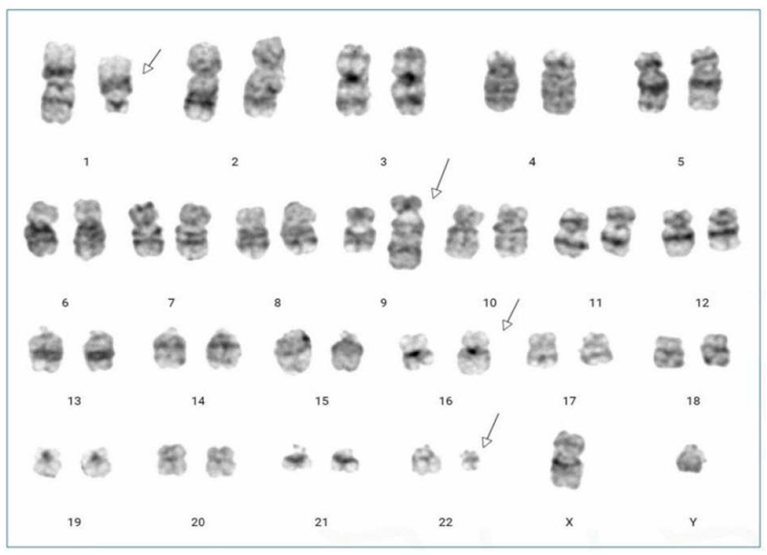
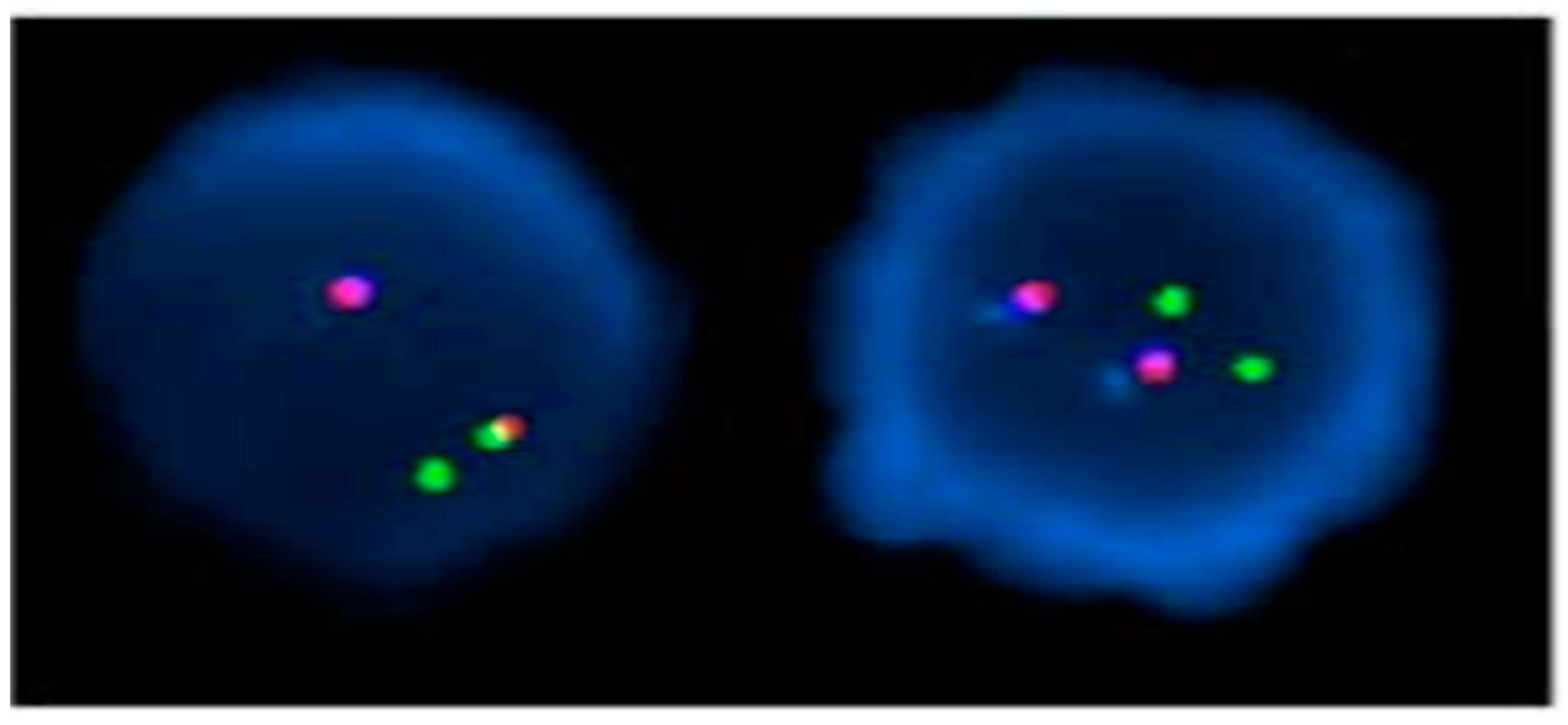
Novel Four-Way Variant Translocation, t(1;9;22;16)(q21;q34;q11.2;q24), in a Patient with Chronic Myeloid Leukemia

1
Department of Laboratory Medicine, Yeungnam University Medical Center, Daegu 42415, Republic of Korea
2
Department of Laboratory Medicine, Yeungnam University College of Medicine, Daegu 42415, Republic of Korea
*
Author to whom correspondence should be addressed.
Diagnostics 2024, 14(3), 303; https://doi.org/10.3390/diagnostics14030303
Submission received: 25 December 2023 / Revised: 25 January 2024 / Accepted: 25 January 2024 / Published: 30 January 2024
(This article belongs to the Section Pathology and Molecular Diagnostics)

Abstract

:
Chronic myeloid leukemia (CML) is characterized by the Philadelphia (Ph) chromosome resulting from the translocation of t(9;22)(q34;q11), producing the BCR::ABL1 fusion gene. Variant Ph chromosome translocations, involving rearrangements in chromosomes other than 9 and 22, occur in 5–10% of CML cases. Herein, we report a unique case of a 36-year-old male with a four-way variant Ph chromosome. Conventional chromosomal analysis performed on bone marrow aspirate samples showed 46, XY, t(1;9;22;16)(q21;q34;q11.2;q24). Nested RT-PCR of the BCR::ABL1 gene revealed a major BCR::ABL rearrangement. The treatment with nilotinib achieved a complete hematologic, cytogenetic, and molecular response after 12 months.

Chronic myeloid leukemia (CML) is a myeloproliferative neoplasm characterized by the Philadelphia (Ph) chromosome arising from the t(9;22)(q34;q11) translocation [1]. The oncogenic BCR::ABL1 gene product, a fusion of the ABL1 gene on chromosome 9q34 and the BCR gene on chromosome 22q11 yields an abnormal tyrosine kinase that dysregulates multiple signaling pathways involved in cell cycle control and apoptosis [2]. Variant Ph chromosomes have additional rearrangements beyond the classic translocation, observed in 5–10% of CML cases [3,4]. The prognosis for patients with variant Ph chromosomes has been variably reported, with some studies indicating poorer outcomes compared to those with the classic Ph chromosome, though others suggest no significant prognostic difference in the imatinib era [4,5,6,7,8]. Very rare are variant Ph chromosomes with complex four-way or five-way rearrangements. We present a novel case of a CML patient with a four-way variant Ph chromosome, t(1;9;22;16)(q21;q34;q11.2;q24).
A 36-year-old male with no significant medical history was evaluated by the hematology department in July 2022 after abnormal blood test results during a routine examination. He had lost approximately 20 kg over the past six months and had developed splenomegaly. His complete blood count (CBC) revealed an Hb level of 7.9 g/dL, a white blood cell (WBC) count of 195 K/μL, and a platelet (PLT) count of 653 K/μL. The differential count detailed 37% segmented neutrophils, 18% band neutrophils, 5% lymphocytes, 2% monocytes, 2% eosinophils, 2% basophils, 8% metamyelocytes, 18% myelocytes, 2% promyelocytes, 2% blasts, and an N-RBC count of 1/100 WBCs. Bone marrow examination revealed hypercellularity with a myeloid-to-erythroid ratio of 13.32:1 due to granulocytic proliferation. The increase in eosinophils, basophils, and monocytes was non-significant. The bone marrow cell differential count included 31.4% segmented neutrophils, 19.6% band neutrophils, 2.6% lymphocytes, 2.6% monocytes, 3.4% eosinophils, 2.0% basophils, 16.4% metamyelocytes, 13.4% myelocytes, 0.6% promyelocytes, 1.2% blasts, and 6.8% erythroid precursors. Conventional chromosomal analysis on bone marrow aspirate samples showed 20 cells with the karyotype 46, XY, t(1;9;22;16)(q21;q34;q11.2;q24) (Figure 1). Nested reverse transcriptase-PCR for the BCR::ABL gene indicated a positive major BCR::ABL rearrangement (b3a2, data not shown). A dual-fusion triple-color fluorescence in situ hybridization (FISH) analysis with whole blood samples detected a 1B1R1G1F pattern in 86.6% of cells (Figure 2). The patient was diagnosed with chronic phase CML with a variant Ph chromosome. Treatment began with hydroxyurea and nilotinib at 600 mg/day starting on the fifth hospital day. At the 12-month follow-up, CBC showed Hb at 14.7 g/dL, WBC at 8.03 K/μL, and PLT at 653 k/μL. No immature cells were noted in the peripheral blood smear. While the marrow aspirate was inadequate, the biopsy showed a normocellular marrow with normal maturation of myeloid and erythroid lineages. Quantitative PCR for BCR::ABL from marrow aspirate yielded an International Scale (IS) value of 0.01711, indicative of major molecular response (MMR). The patient achieved complete hematologic, cytogenetic, and molecular responses and is on nilotinib 600 mg/day as of October 2023.
Approximately 1000 cases of CML with variant Ph chromosomes have been reported to date. Among these, approximately 80 cases exhibit 4-way translocations, this case included [9]. Variant Ph chromosomes have been documented on various chromosomes, particularly at 1p36, 3p21, 5q13, 6p21, 9q22, 11q13, 12p13, 17p13, 17q21, 17q25, 19q13, 21q22, 22q12, and 22q13 [10]. The breakpoints at 1q21 and 16q24, as noted in the patient, are rare. A study encompassing 25 cases with variant Ph chromosomes highlighted chromosomes 1 and 16 as the most commonly implicated [7]. Furthermore, according to the Mitelman Database, the t(1;9;22;16)(q21;q34;q11.2;q24) translocation observed here represents the inaugural reported case of such a four-way translocation [9].
Two hypotheses have been advanced to elucidate the genesis of variant Ph chromosomes: a simultaneous, one-step mechanism involving three or more chromosomes and a sequential, two-step mechanism that follows the initial t(9;22) translocation. This latter multi-step process may signify clonal evolution and potentially portend an unfavorable prognosis [3,11].
Figure 2. FISH analysis using the LSI BCR::ABL dual fusion triple color translocation probe. The method involves using DNA probes that are complementary to the ABL1 gene on chromosome 9 and the BCR gene on chromosome 22. These genes are involved in the t(9;22)(q34;q11.2), and the probes hybridize to the nuclei of either interphase or metaphase cells. The Breakpoint Cluster Region (BCR) at 22q11.2 is identified by a green signal, while the ABL1 oncogene at 9q34 is marked by a red signal, and the ASS1 gene, also located at 9q34, is indicated by a blue signal. A cell negative for the t(9;22) typically displays a pattern of 2 blue, 2 red, and 2 green signals. However, in this study, out of 500 cells examined, 433 cells (86.6%) showed an unusual BCR::ABL rearrangement. The typical pattern for a 4-way variant translocation is 2 blue, 2 red, 2 green, and 1 fusion signals in 1-step mechanism and 2 blue, 1 red, 1 green, and 2 fusion signals in 2-step mechanism [4,11]. However, the atypical pattern observed in this study, characterized by 1 blue and 1 red signal on normal chromosome 9, 1 green signal on normal chromosome 22, and 1 fusion signal on der(22). It is apparent that deletions have occurred in both the ABL1 and BCR genes.
Diagnostics 14 00303 g002
Historical perspectives posited that variant Ph chromosomes could portend a graver outlook compared to the classical Ph chromosome, with deleting der(9) also linked to worse outcomes [6,12,13,14]. Conversely, contemporary research indicates that variant Ph chromosomes do not influence prognosis in the era of imatinib treatment, regardless of the number of chromosomes involved or deletion presence [4,11]. Nevertheless, specific cases, such as two patients with variants involving chromosome 5 but without tyrosine kinase domain mutations, have been associated with poorer prognoses, suggesting that specific chromosomes or breakpoints may indeed affect patient outcomes [7].
The patient under discussion achieved a major molecular response after 12 months of nilotinib treatment and has continued to progress favorably. The influence of variant Ph chromosomes on CML prognosis remains a topic of debate, and it is anticipated that this report of a rare four-way translocation will enrich the collective database, aiding in the refinement of our understanding of these chromosomal variations.

Author Contributions

Conceptualization, J.H.L.; methodology, H.J.S. and J.H.L.; project administration, J.H.L.; validation, J.H.L.; visualization, H.J.S. and J.H.L.; roles/writing—original draft preparation, H.J.S.; writing—review and editing, J.H.L. All authors have read and agreed to the published version of the manuscript.

Funding

This work was supported by the 2024 Yeungnam University Research Grant.

Institutional Review Board Statement

This study was approved by the Institutional Review Board (IRB) of Yeungnam University Hospital (IRB No.: YUMC 2023-11-008, Date of approval: 10 November 2023).

Informed Consent Statement

The Institutional Review Board waived the need for informed consent.

Data Availability Statement

Not applicable.

Conflicts of Interest

The authors declare no conflict of interest.

References

  1. Rowley, J.D. Letter: A new consistent chromosomal abnormality in chronic myelogenous leukaemia identified by quinacrine fluorescence and Giemsa staining. Nature 1973, 243, 290–293. [Google Scholar] [CrossRef]
  2. Quintás-Cardama, A.; Cortes, J. Molecular biology of bcr-abl1-positive chronic myeloid leukemia. Blood 2009, 113, 1619–1630. [Google Scholar] [CrossRef] [PubMed]
  3. Gorusu, M.; Benn, P.; Li, Z.; Fang, M. On the genesis and prognosis of variant translocations in chronic myeloid leukemia. Cancer Genet. Cytogenet. 2007, 173, 97–106. [Google Scholar] [CrossRef] [PubMed]
  4. Marzocchi, G.; Castagnetti, F.; Luatti, S.; Baldazzi, C.; Stacchini, M.; Gugliotta, G.; Amabile, M.; Specchia, G.; Sessarego, M.; Giussani, U.; et al. Variant Philadelphia translocations: Molecular-cytogenetic characterization and prognostic influence on frontline imatinib therapy, a GIMEMA Working Party on CML analysis. Blood 2011, 117, 6793–6800. [Google Scholar] [CrossRef] [PubMed]
  5. El-Zimaity, M.M.; Kantarjian, H.; Talpaz, M.; O’Brien, S.; Giles, F.; Garcia-Manero, G.; Verstovsek, S.; Thomas, D.; Ferrajoli, A.; Hayes, K.; et al. Results of imatinib mesylate therapy in chronic myelogenous leukaemia with variant Philadelphia chromosome. Br. J. Haematol. 2004, 125, 187–195. [Google Scholar] [CrossRef] [PubMed]
  6. Reid, A.G.; Huntly, B.J.; Grace, C.; Green, A.R.; Nacheva, E.P. Survival implications of molecular heterogeneity in variant Philadelphia-positive chronic myeloid leukaemia. Br. J. Haematol. 2003, 121, 419–427. [Google Scholar] [CrossRef]
  7. Shetty, D.; Talker, E.; Jain, H.; Talker, J.; Patkar, N.; Subramanian, P.; Jain, H.; Bonda, A.; Punatar, S.; Gokarn, A.; et al. Evaluation of cytogenetic response in CML patients with variant Philadelphia translocation. Asia Pac. J. Clin. Oncol. 2022, 18, 99–108. [Google Scholar] [CrossRef] [PubMed]
  8. Fabarius, A.; Leitner, A.; Hochhaus, A.; Müller, M.C.; Hanfstein, B.; Haferlach, C.; Göhring, G.; Schlegelberger, B.; Jotterand, M.; Reiter, A.; et al. Impact of additional cytogenetic aberrations at diagnosis on prognosis of CML: Long-term observation of 1151 patients from the randomized CML Study IV. Blood 2011, 118, 6760–6768. [Google Scholar] [CrossRef] [PubMed]
  9. Mitelman, F.; Johansson, B.; Mertens, F. Mitelman Database of Chromosome Aberrations and Gene Fusions in Cancer. 2023. Available online: https://mitelmandatabase.isb-cgc.org (accessed on 1 November 2023).
  10. Johansson, B.; Fioretos, T.; Mitelman, F. Cytogenetic and molecular genetic evolution of chronic myeloid leukemia. Acta Haematol. 2002, 107, 76–94. [Google Scholar] [CrossRef] [PubMed]
  11. Richebourg, S.; Eclache, V.; Perot, C.; Portnoi, M.F.; Van den Akker, J.; Terré, C.; Maareck, O.; Soenen, V.; Viguié, F.; Laï, J.L.; et al. Mechanisms of genesis of variant translocation in chronic myeloid leukemia are not correlated with ABL1 or BCR deletion status or response to imatinib therapy. Cancer Genet. Cytogenet. 2008, 182, 95–102. [Google Scholar] [CrossRef] [PubMed]
  12. Al Achkar, W.; Wafa, A.; Mkrtchyan, H.; Moassass, F.; Liehr, T. Novel complex translocation involving 5 different chromosomes in a chronic myeloid leukemia with Philadelphia chromosome: A case report. Mol. Cytogenet. 2009, 2, 21. [Google Scholar] [CrossRef] [PubMed]
  13. Campbell, L.J. Deletion of the derivative chromosome 9 in chronic myeloid leukemia. Methods Mol. Med. 2006, 125, 107–114. [Google Scholar] [CrossRef] [PubMed]
  14. Sinclair, P.B.; Nacheva, E.P.; Leversha, M.; Telford, N.; Chang, J.; Reid, A.; Bench, A.; Champion, K.; Huntly, B.; Green, A.R. Large deletions at the t(9;22) breakpoint are common and may identify a poor-prognosis subgroup of patients with chronic myeloid leukemia. Blood 2000, 95, 738–743. [Google Scholar] [CrossRef]
Figure 1. GTL-banding karyotypes of cultured bone marrow cells at a resolution of 400. All 20 observed dividing cells exhibit the t(1;9;22;16)(q21;q34;q11.2;q24). The arrow indicates rearranged chromosomes.
Diagnostics 14 00303 g001
Disclaimer/Publisher’s Note: The statements, opinions and data contained in all publications are solely those of the individual author(s) and contributor(s) and not of MDPI and/or the editor(s). MDPI and/or the editor(s) disclaim responsibility for any injury to people or property resulting from any ideas, methods, instructions or products referred to in the content.

