
Using Biosensors to Detect and Map Language Areas in the Brain for Individuals with Traumatic Brain Injury


Abstract
1. Introduction
1.1. Biosensor Types for Language Detection
1.2. Mapping Language Areas: Biosensor Mechanisms
1.3. Measuring Brain Activity: Biosensor Functionality
1.4. Biosensor Efficacy: Accuracy and Limitations
1.5. Purpose of the Present Study
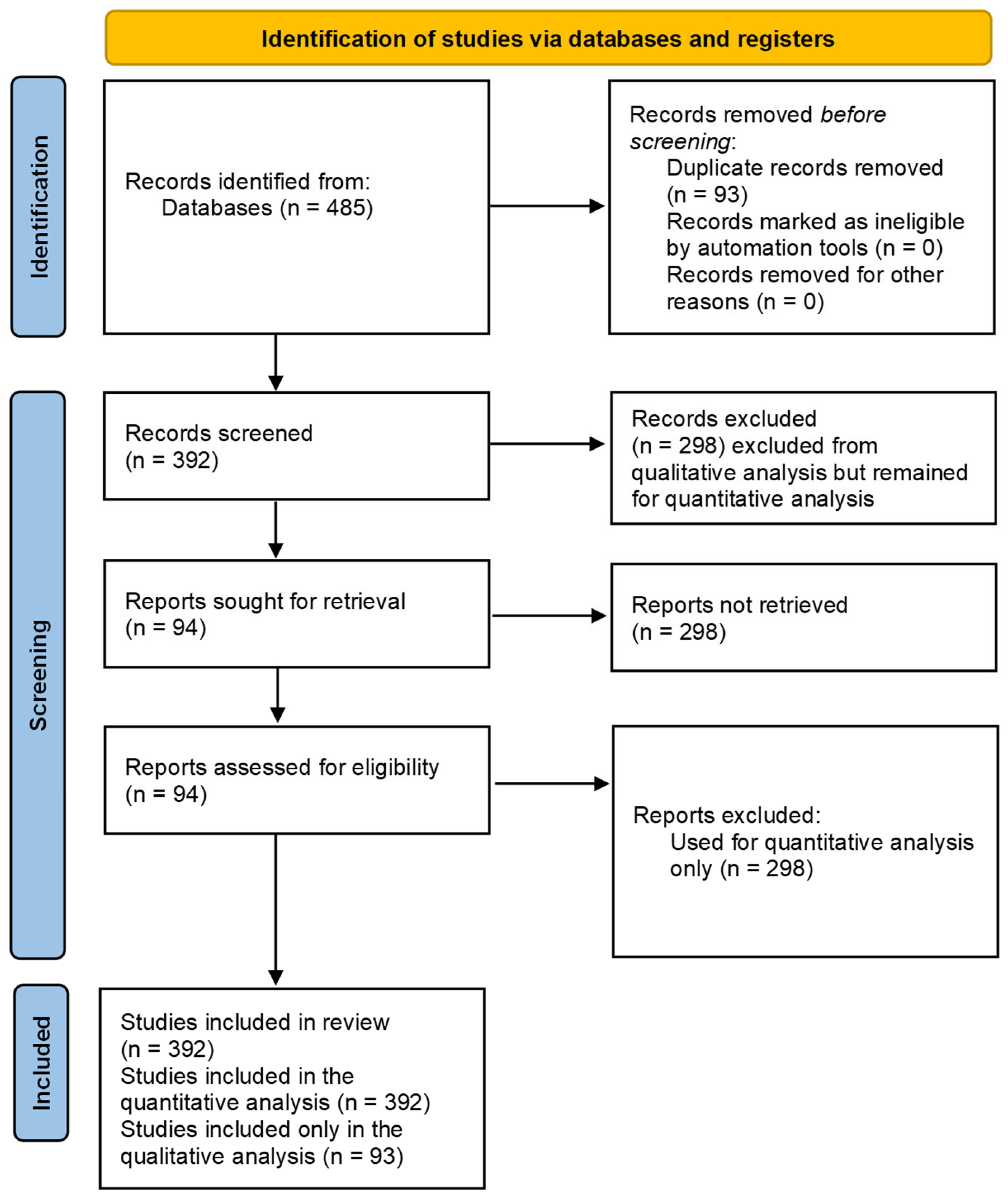
2. Methods
2.1. Sample
2.2. Design
2.3. Measures
2.4. Procedure
3. Results
3.1. Different Frequencies in Post-Stroke Aphasia
3.2. Classical Anterior Regions and Language Production
3.3. Human Brain Language Areas and fMRI Studies
3.4. Preoperative Assessment of Language Functions
3.5. Functional Connectivity and Naming Tasks
3.6. Mirror Neuron Theory and Non-Fluent Aphasia
3.7. Post-Stroke Aphasia and Genetic Contributions
3.8. Fluent Aphasia and Neuroanatomical Insights
3.9. Superior Temporal Gyrus and Speech Production
3.10. Task-Based fMRI Studies and Naming Therapy
3.11. Cluster Analysis Takeaway
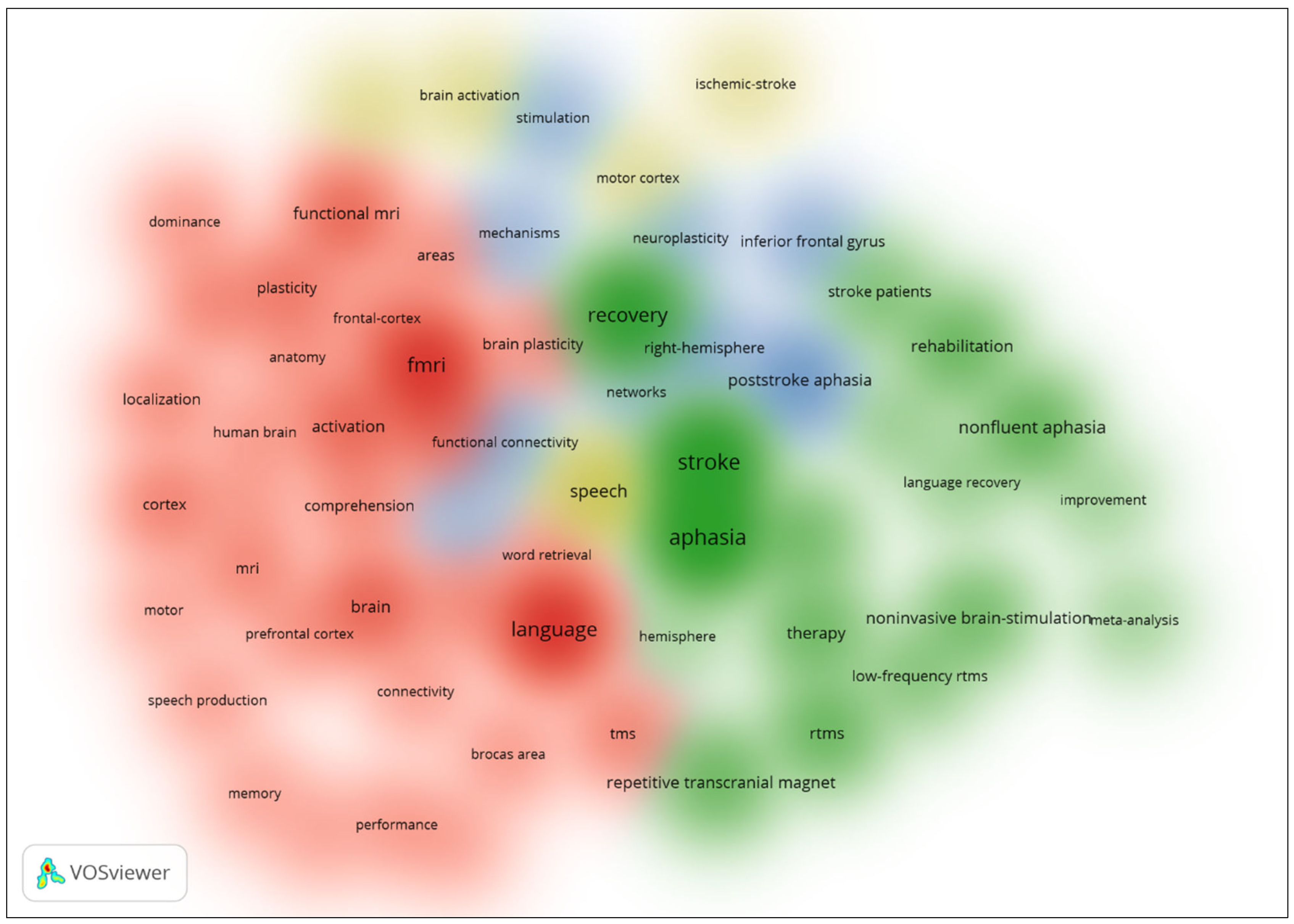
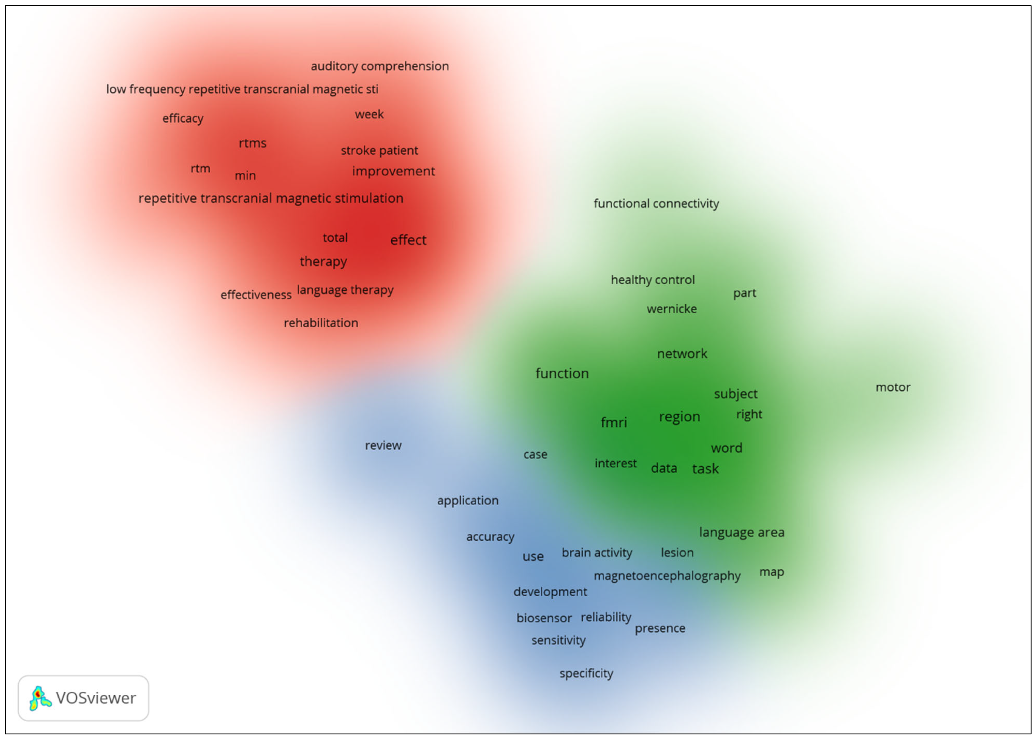
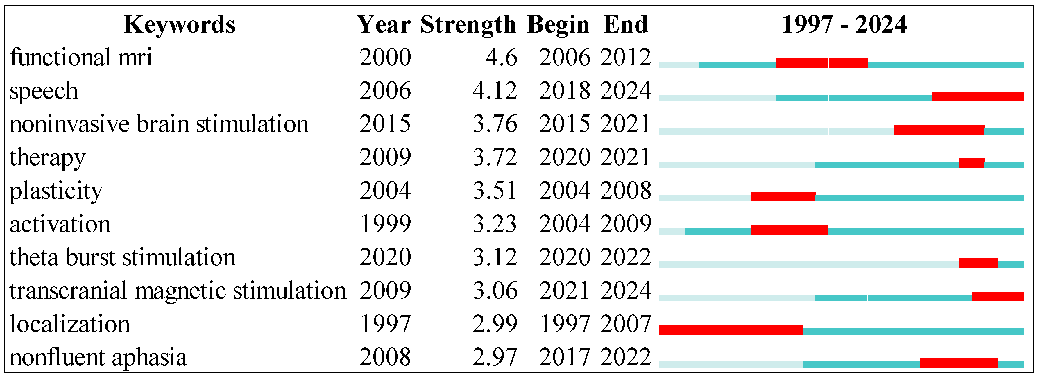
3.12. Thematic Analysis
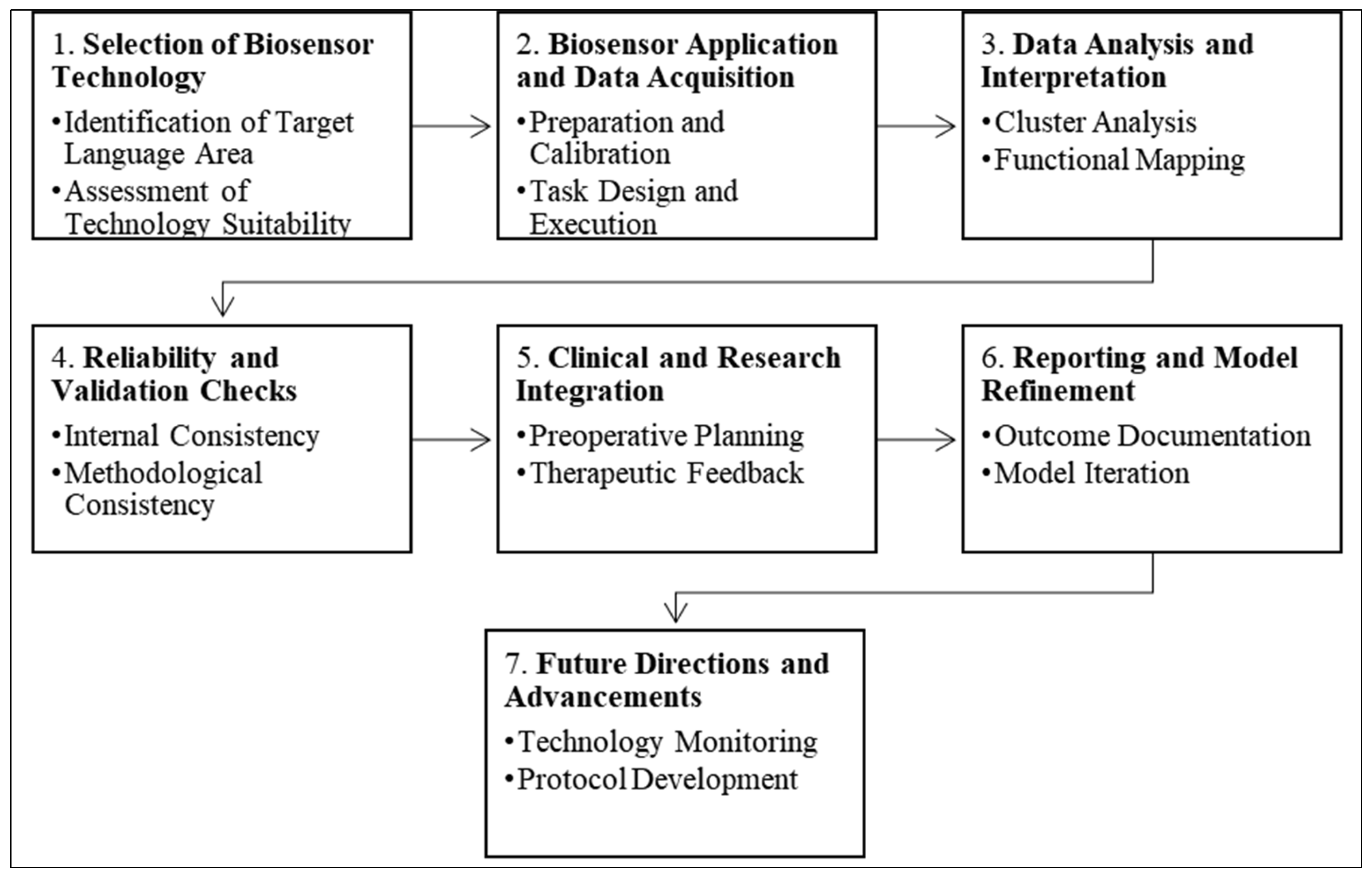
3.13. A Model for Biosensor Use in Language Area Detection
4. Discussion
4.1. Limitations
4.2. Implications
5. Conclusions
Author Contributions
Funding
Institutional Review Board Statement
Informed Consent Statement
Data Availability Statement
Acknowledgments
Conflicts of Interest
References
- Luo, L.; Yang, D.; Yang, Y. New Biosensors Detect Light Deep inside the Brain. Brain-X 2023, 1, e3. [Google Scholar] [CrossRef]
- Perani, D.; Cappa, S.F.; Tettamanti, M.; Rosa, M.; Scifo, P.; Miozzo, A.; Basso, A.; Fazio, F. A FMRI Study of Word Retrieval in Aphasia. Brain Lang. 2003, 85, 357–368. [Google Scholar] [CrossRef]
- Matsumoto, R.; Nair, D.; LaPresto, E.; Najm, I.; Bingaman, W.; Shibasaki, H.; Shibasaki, H.; Lüders, H. Functional Connectivity in the Human Language System: A Cortico-Cortical Evoked Potential Study. Brain 2004, 127, 2316–2330. [Google Scholar] [CrossRef]
- Binder, J.R.; Frost, J.A.; Hammeke, T.A.; Cox, R.W.; Rao, S.M.; Prieto, T.E. Human Brain Language Areas Identified by Functional Magnetic Resonance Imaging. J. Neurosci. 1997, 17, 353–362. [Google Scholar] [CrossRef]
- Kozai, T.D.Y.; Jaquins-Gerstl, A.; Vazquez, A.L.; Michael, A.C.; Cui, X.T. Brain Tissue Responses to Neural Implants Impact Signal Sensitivity and Intervention Strategies. ACS Chem. Neurosci. 2015, 6, 48–67. [Google Scholar] [CrossRef]
- Papanicolaou, A.C.; Simos, P.G.; Breier, J.I.; Zouridakis, G.; Willmore, L.J.; Wheless, J.W.; Constantinou, J.E.C.; Maggio, W.W.; Gormley, W.B. Magnetoencephalographic Mapping of the Language-Specific Cortex. J. Neurosurg. 1999, 90, 85–93. [Google Scholar] [CrossRef]
- Simos, P.G.; Breier, J.I.; Zouridakis, G.; Papanicolaou, A.C. Identification of Language-Specific Brain Activity Using Magnetoencephalography. J. Clin. Exp. Neuropsychol. 1998, 20, 706–722. [Google Scholar] [CrossRef]
- Keirsse, J.; Boussard-Plédel, C.; Loréal, O.; Sire, O.; Bureau, B.; Turlin, B.; Leroyer, P.; Lucas, J. Chalcogenide Glass Fibers Used as Biosensors. J. Non-Cryst. Solids 2003, 326, 430–433. [Google Scholar] [CrossRef]
- Murugaiyan, S.B.; Ramasamy, R.; Gopal, N.; Kuzhandaivelu, V. Biosensors in Clinical Chemistry: An Overview. Adv. Biomed. Res. 2014, 3, 67. [Google Scholar] [CrossRef]
- Vasylieva, N.; Maucler, C.; Meiller, A.; Viscogliosi, H.; Lieutaud, T.; Barbier, D.; Marinesco, S. Immobilization Method to Preserve Enzyme Specificity in Biosensors: Consequences for Brain Glutamate Detection. Anal. Chem. 2013, 85, 2507–2515. [Google Scholar] [CrossRef]
- Malhotra, S.; Verma, A.; Tyagi, N.; Kumar, V. Biosensors: Principle, Types and Applications. Int. J. Adv. Res. Innov. Ideas Educ. 2017, 3, 3639–3644. [Google Scholar]
- Hauck, T.; Tanigawa, N.; Probst, M.; Wohlschlaeger, A.; Ille, S.; Sollmann, N.; Maurer, S.; Zimmer, C.; Ringel, F.; Meyer, B.; et al. Task Type Affects Location of Language-Positive Cortical Regions by Repetitive Navigated Transcranial Magnetic Stimulation Mapping. PLoS ONE 2015, 10, e0125298. [Google Scholar] [CrossRef]
- Pulvermüller, F. Brain Mechanisms Linking Language and Action. Nat. Rev. Neurosci. 2005, 6, 576–582. [Google Scholar] [CrossRef]
- Friederici, A.D. The Brain Basis of Language Processing: From Structure to Function. Physiol. Rev. 2011, 91, 1357–1392. [Google Scholar] [CrossRef]
- Price, C.J. The Anatomy of Language: A Review of 100 FMRI Studies Published in 2009. Ann. N. Y. Acad. Sci. 2010, 1191, 62–88. [Google Scholar] [CrossRef]
- Price, C.J. The Anatomy of Language: Contributions from Functional Neuroimaging. J. Anat. 2000, 197, 335–359. [Google Scholar] [CrossRef]
- Pulvermüller, F.; Fadiga, L. Active Perception: Sensorimotor Circuits as a Cortical Basis for Language. Nat. Rev. Neurosci. 2010, 11, 351–360. [Google Scholar] [CrossRef]
- Ojemann, G.A. Brain Organization for Language from the Perspective of Electrical Stimulation Mapping. Behav. Brain Sci. 1983, 6, 189–206. [Google Scholar] [CrossRef]
- Ward, N.S.; Cohen, L.G. Mechanisms Underlying Recovery of Motor Function after Stroke. JAMA Neurol. 2004, 61, 1844–1848. [Google Scholar] [CrossRef]
- O’Neill, R.D.; Rocchitta, G.G.M.; McMahon, C.P.; Serra, P.A.; Lowry, J.P. Designing Sensitive and Selective Polymer/Enzyme Composite Biosensors for Brain Monitoring in Vivo. Trends Anal. Chem. 2008, 27, 78–88. [Google Scholar] [CrossRef]
- Zhang, L.; Tian, Y. Designing Recognition Molecules and Tailoring Functional Surfaces for In Vivo Monitoring of Small Molecules in the Brain. Acc. Chem. Res. 2018, 51, 688–696. [Google Scholar] [CrossRef]
- Nazir, T.A.; Hrycyk, L.; Moreau, Q.; Frak, V.; Cheylus, A.; Ott, L.; Lindemann, O.; Fischer, M.H.; Paulignan, Y.; Delevoye-Turrell, Y. A Simple Technique to Study Embodied Language Processes: The Grip Force Sensor. Behav. Res. Methods 2017, 49, 61–73. [Google Scholar] [CrossRef]
- Chen, Z.; Truong, T.M.; Ai, H. Illuminating Brain Activities with Fluorescent Protein-Based Biosensors. Chemosensors 2017, 5, 32. [Google Scholar] [CrossRef]
- Amaral, J.; Gaspar, J.; Pinto, V.; Costa, T.; Costa, T.; Sousa, N.; Cardoso, S.; Freitas, P.P. Measuring Brain Activity with Magnetoresistive Sensors Integrated in Micromachined Probe Needles. Appl. Phys. A 2013, 111, 407–412. [Google Scholar] [CrossRef]
- Cheran, L.-E.; Benvenuto, P.; Thompson, M. Coupling of Neurons with Biosensor Devices for Detection of the Properties of Neuronal Populations. Chem. Soc. Rev. 2008, 37, 1229–1242. [Google Scholar] [CrossRef]
- Wang, K.; Cai, C.; Michiharu, Y.; Uchiyama, T. Real-Time Brain Activity Measurement and Signal Processing System Using Highly Sensitive MI Sensor. AIP Adv. 2017, 7, 056635. [Google Scholar] [CrossRef]
- Es’kov, V.M.; Papshev, V.A.; Kulaev, S. V Biosensor Measurements on Diffusion Coefficients of Physiologically Active Substances in Brain Tissue. Meas. Tech. 2004, 47, 715–718. [Google Scholar] [CrossRef]
- Manikandan, N.; Muruganand, S.; Karuppasamy; Subramanian, S. Implantable Multisensory Microelectrode Biosensor for Revealing Neuron and Brain Functions. In Proceedings of the International Workshop on the Physics of Semiconductor and Devices, Delhi, India, 12–15 December 2017; pp. 763–769. [Google Scholar] [CrossRef]
- FitzGerald, D.B.; Cosgrove, G.R.; Ronner, S.; Jiang, H.; Buchbinder, B.R.; Belliveau, J.W.; Rosen, B.R.; Benson, R.R. Location of Language in the Cortex: A Comparison between Functional MR Imaging and Electrocortical Stimulation. Am. J. Neuroradiol. 1997, 18, 1529–1539. [Google Scholar]
- Rajan, N.K.; Duan, X.; Reed, M.A. Performance Limitations for Nanowire/Nanoribbon Biosensors. Wiley Interdiscip. Rev.-Nanomed. Nanobiotechnol. 2013, 5, 629–645. [Google Scholar] [CrossRef]
- Carminati, M.; Vergani, M.; Ferrari, G.; Caranzi, L.; Caironi, M.; Sampietro, M.; Sampietro, M. Accuracy and Resolution Limits in Quartz and Silicon Substrates with Microelectrodes for Electrochemical Biosensors. Sens. Actuators B-Chem. 2012, 174, 168–175. [Google Scholar] [CrossRef]
- Billingsley-Marshall, R.; Simos, P.G.; Papanicolaou, A.C. Reliability and Validity of Functional Neuroimaging Techniques for Identifying Language-Critical Areas in Children and Adults. Dev. Neuropsychol. 2004, 26, 541–563. [Google Scholar] [CrossRef]
- Petrini, G.; Moreva, E.; Bernardi, E.; Traina, P.; Tomagra, G.; Carabelli, V.; Degiovanni, I.P.; Genovese, M. Is a Quantum Biosensing Revolution Approaching. Adv. Quantum Technol. 2020, 3, 2000066. [Google Scholar] [CrossRef]
- Bernal, B.; Guillen, M.; Korman, B. Nontask-Related Brain Lateralization Biomarkers in Children: The Asymmetry of Language Areas on Functional Connectivity Functional Magnetic Resonance Imaging. Brain Connect. 2018, 8, 321–332. [Google Scholar] [CrossRef]
- de Boissezon, X.; Raboyeau, G.; Simonetta-Moreau, M.; Puel, M.; Demonet, J.F.; Cardebat, D. Functional Neuroimaging and the Treatment of Aphasia: Speech Therapy and Repetitive Transcranial Magnetic Stimulation. Rev. Neurol. 2008, 164, S45–S48. [Google Scholar] [CrossRef]
- Enatsu, R.; Kubota, Y.; Kakisaka, Y.; Bulacio, J.; Piao, Z.; O’Connor, T.; Horning, K.; Mosher, J.; Burgess, R.C.; Bingaman, W.; et al. Reorganization of Posterior Language Area in Temporal Lobe Epilepsy: A Cortico-Cortical Evoked Potential Study. Epilepsy Res. 2013, 103, 73–82. [Google Scholar] [CrossRef]
- Giussani, C.; Roux, F.E.; Ojemann, J.; Sganzerla, E.P.; Pirillo, D.; Papagno, C. Is Preoperative Functional Magnetic Resonance Imaging Reliable for Language Areas Mapping in Brain Tumor Surgery? Review of Language Functional Magnetic Resonance Imaging and Direct Cortical Stimulation Correlation Studies. Neurosurgery 2010, 66, 113–120. [Google Scholar] [CrossRef]
- Hara, T.; Abo, M. New Treatment Strategy Using Repetitive Transcranial Magnetic Stimulation for Post-Stroke Aphasia. Diagnostics 2021, 11, 1853. [Google Scholar] [CrossRef]
- Huang, W.; Wu, L.; Ma, H.; Wang, X.; Chen, X.; Sun, S.; Sun, T.; Xia, H. Effectiveness of functional magnetic resonance imaging combined with electrical cortical stimulation under awake craniotomy for lesions involving the eloquent language area of the brain. Chin. J. Clin. Oncol. 2012, 39, 986–989. [Google Scholar] [CrossRef]
- Jung, T.D.; Kim, J.Y.; Lee, Y.S.; Kim, D.H.; Lee, J.J.; Seo, J.H.; Lee, H.J.; Chang, Y. Effect of Repetitive Transcranial Magnetic Stimulation in a Patient with Chronic Crossed Aphasia: FMRI Study. J. Rehabil. Med. 2010, 42, 973–978. [Google Scholar] [CrossRef]
- Kamada, K.; Sawamura, Y.; Takeuchi, F.; Kuriki, S.; Kawai, K.; Morita, A.; Todo, T. Expressive and Receptive Language Areas Determined by a Non-Invasive Reliable Method Using Functional Magnetic Resonance Imaging and Magnetoencephalography. Neurosurgery 2007, 60, 296–305. [Google Scholar] [CrossRef]
- Kapoor, A. Repetitive Transcranial Magnetic Stimulation Therapy for Post-Stroke Non-Fluent Aphasia: A Critical Review. Top. Stroke Rehabil. 2017, 24, 547–553. [Google Scholar] [CrossRef] [PubMed]
- Kassubek, J.; Hickok, G.; Erhard, P. Involvement of Classical Anterior and Posterior Language Areas in Sign Language Production, as Investigated by 4 T Functional Magnetic Resonance Imaging. Neurosci. Lett. 2004, 364, 168–172. [Google Scholar] [CrossRef] [PubMed]
- Kielar, A.; Patterson, D.; Chou, Y.H. Efficacy of Repetitive Transcranial Magnetic Stimulation in Treating Stroke Aphasia: Systematic Review and Meta-Analysis. Clin. Neurophysiol. 2022, 140, 196–227. [Google Scholar] [CrossRef] [PubMed]
- Kim, W.J.; Hahn, S.J.; Kim, W.S.; Paik, N.J. Neuronavigation-Guided Repetitive Transcranial Magnetic Stimulation for Aphasia. J. Vis. Exp. 2016, 111, e53345. [Google Scholar] [CrossRef]
- Kośla, K.; Pfajfer, L.; Bryszewski, B.; Jaskólski, D.; Stefańczyk, L.; Majos, A. Functional Rearrangement of Language Areas in Patients with Tumors of the Central Nervous System Using Functional Magnetic Resonance Imaging. Pol. J. Radiol. 2012, 77, 39–45. [Google Scholar] [CrossRef] [PubMed]
- Kunii, N.; Kamada, K.; Ota, T.; Kawai, K.; Saito, N. A Detailed Analysis of Functional Magnetic Resonance Imaging in the Frontal Language Area: A Comparative Study with Extraoperative Electrocortical Stimulation. Neurosurgery 2011, 69, 590–596. [Google Scholar] [CrossRef]
- Pytel, V.; Cabrera-Martín, M.N.; Delgado-Alvarez, A.; Ayala, J.L.; Balugo, P.; Delgado-Alonso, C.; Yus, M.; Carreras, M.T.; Carreras, J.L.; Matías-Guiu, J.; et al. Personalized Repetitive Transcranial Magnetic Stimulation for Primary Progressive Aphasia. J. Alzheimers Dis. 2021, 84, 151–167. [Google Scholar] [CrossRef] [PubMed]
- Roux, F.E.; Boulanouar, K.; Lotterie, J.A.; Mejdoubi, M.; LeSage, J.P.; Berry, I. Language Functional Magnetic Resonance Imaging in Preoperative Assessment of Language Areas: Correlation with Direct Cortical Stimulation. Neurosurgery 2003, 52, 1335–1345. [Google Scholar] [CrossRef] [PubMed]
- Szaflarski, J.P.; Vannest, J.; Wu, S.W.; DiFrancesco, M.W.; Banks, C.; Gilbert, D.L. Excitatory Repetitive Transcranial Magnetic Stimulation Induces Improvements in Chronic Post-Stroke Aphasia. Med. Sci. Monit. 2011, 17, CR132–CR139. [Google Scholar] [CrossRef]
- Tarta-Arsene, O.; Preoteasa, F.; Magureanu, S.A.; Iliescu, A.; Craiu, D.; Motoescu, C.; Tarta-Arsene, E.; Ciobanu, G. Functional Magnetic Resonance Imaging Contribution to Language Areas Assesmnent in Children with Non-Lesion Focal Epilepsy. Rom. J. Neurol./Rev. Romana Neurol. 2010, 9, 130–136. [Google Scholar] [CrossRef]
- Winhuisen, L.; Thiel, A.; Schumacher, B.; Kessler, J.; Rudolf, J.; Haupt, W.F.; Heiss, W.D. Role of the Contralateral Inferior Frontal Gyrus in Recovery of Language Function in Poststroke Aphasia—A Combined Repetitive Transcranial Magnetic Stimulation and Positron Emission Tomography Study. Stroke 2005, 36, 1759–1763. [Google Scholar] [CrossRef]
- Buchsbaum, B.R.; Baldo, J.; Okada, K.; Berman, K.F.; Dronkers, N.; D’Esposito, M.; Hickok, G. Conduction Aphasia, Sensory-Motor Integration, and Phonological Short-Term Memory—An Aggregate Analysis of Lesion and FMRI Data. Brain Lang. 2011, 119, 119–128. [Google Scholar] [CrossRef] [PubMed]
- Rutten, G.J.M.; Ramsey, N.F.; van Rijen, P.C.; Noordmans, H.J.; van Veelen, C.W.M. Development of a Functional Magnetic Resonance Imaging Protocol for Intraoperative Localization of Critical Temporoparietal Language Areas. Ann. Neurol. 2002, 51, 350–360. [Google Scholar] [CrossRef] [PubMed]
- Binder, J.R.; Gross, W.L.; Allendorfer, J.B.; Bonilha, L.; Chapin, J.; Edwards, J.C.; Grabowski, T.J.; Langfitt, J.T.; Loring, D.W.; Lowe, M.J.; et al. Mapping Anterior Temporal Lobe Language Areas with FMRI: A Multicenter Normative Study. Neuroimage 2011, 54, 1465–1475. [Google Scholar] [CrossRef] [PubMed]
- van der Mark, S.; Klaver, P.; Bucher, K.; Maurer, U.; Schulz, E.; Brem, S.; Martin, E.; Brandeis, D. The Left Occipitotemporal System in Reading: Disruption of Focal FMRI Connectivity to Left Inferior Frontal and Inferior Parietal Language Areas in Children with Dyslexia. Neuroimage 2011, 54, 2426–2436. [Google Scholar] [CrossRef]
- Martin, P.I.; Naeser, M.A.; Ho, M.; Doron, K.W.; Kurland, J.; Kaplan, J.; Wang, Y.Y.; Nicholas, M.; Baker, E.H.; Fregni, F.; et al. Overt Naming FMRI Pre- and Post-TMS: Two Nonfluent Aphasia Patients, with and without Improved Naming Post-TMS. Brain Lang. 2009, 111, 20–35. [Google Scholar] [CrossRef]


| Database | Query: Friday, 29 March 2024 | Result |
|---|---|---|
| Scopus | (TITLE (“biosensor” OR “functional magnetic resonance imaging” OR “fmri” OR “cortico-cortical evoked potentials” OR “magnetoencephalography” OR “repetitive transcranial magnetic stimulation”) AND TITLE (“aphasia” OR “brain damage” OR “language areas”)) AND (LIMIT-TO (DOCTYPE, “ar”) OR LIMIT-TO (DOCTYPE, “re”) OR LIMIT-TO (DOCTYPE, “ch”)) | 168 |
| Web of Science | “biosensor” or “Functional magnetic resonance imaging” or “fMRI” or “cortico-cortical evoked potentials” or “Magnetoencephalography” or “repetitive Transcranial Magnetic Stimulation” (Title) and “aphasia” or “brain damage” or “language areas” (Title) and Article or Review Article or Book Chapters (Document Types) | 127 |
| SciSpace | Biosensors and language areas in the brain | 190 |
| ID | Size | Silhouette | Label (LSI) | Label (LLR) | Label (MI) | Year |
|---|---|---|---|---|---|---|
| 0 | 53 | 0.706 | post-stroke aphasia | different frequencies | extended fMRI-guided anodal | 2012 |
| 1 | 46 | 0.801 | sign language production | classical anterior | sound shape properties | 2012 |
| 2 | 44 | 0.903 | fMRI study | human brain language area | moderate-severe nonfluent aphasia patient | 2005 |
| 3 | 43 | 0.765 | language area | preoperative assessment | constraint-induced language therapy | 2010 |
| 4 | 41 | 0.873 | functional connectivity | functional connectivity | attempted naming | 2009 |
| 5 | 36 | 0.805 | non-fluent aphasia | mirror neuron theory | attempted naming | 2015 |
| 6 | 33 | 0.882 | post-stroke aphasia subject | post-stroke aphasia subject | GRN mutation | 2011 |
| 7 | 28 | 0.825 | language area | fluent aphasia | attempted naming | 2005 |
| 8 | 25 | 0.865 | speech production | superior temporal gyrus | post-stroke aphasia | 2013 |
| 9 | 15 | 0.945 | following naming therapy | task-based fMRI studies | attempted naming | 2015 |
| No. | Authors | Used Biosensor | Methods Used | Language Area Examined | Is It Reliable? | Limitations |
|---|---|---|---|---|---|---|
| 1 | [6] | - Magnes 2500 WH neuromagnetometer from Biomagnetic Technologies, Inc. - Promold ear inserts for auditory stimulation from International Aquatic Trades, Inc. | - Receptive and expressive language cortex, Wernicke’s area, frontal cortex. - Activation through language tasks, MEG mapping, and cortical stimulation. | - MEG-derived maps remain stable and reliable over time. - MEG maps provide valid information on language-specific cortex location. | - Task involves other cognitive operations, not exclusively linguistic. - Different tasks may lead to slightly different maps in the future. | - MEG data collected in normative experiments with healthy volunteers - Reliability, validity, and topographical accuracy assessed in patients with Wada procedure |
| 2 | [29] | - Microwire-based biosensor - Shank-type biosensor coated with enzymes | - Language areas were examined using functional MR imaging and electrocortical stimulation. - Location of language areas varied among subjects and tasks. | - Functional MR imaging is considered a useful presurgical planning tool. - Sensitivity/specificity for identifying language areas ranged from 81%/53% to 92%/0%. | - Specificity of functional MR imaging for language areas was low. - Sensitivity varied based on distance between activated areas. | - Enzyme-based microelectrode array biosensors - Microwire-based biosensor with periodic insertion into cannula |
| 3 | [33] | - Microwire-based biosensor - Shank-type biosensor coated with enzymes | - Rat hippocampal slices and freely moving rat brain regions - In vitro and in vivo brain structures analyzed for language. | - Reliable due to improved selectivity and real-time measurements. - Real-time dynamics observed in vitro and in vivo experiments. | - Non-normally distributed peak concentration values. - Use of independent Wilcoxon test for comparison. | - Innovative techniques for spatial resolution and sensitivity improvement - Application of NV centers for biological electromagnetic field measurement |
| 4 | [4] | - Microwire-based biosensor - Shank-type biosensor coated with enzymes | - Left cerebral hemisphere - Frontal, temporal, and parietal lobes | - Average activation map proved reliable in split-half analysis. - Functional maps obtained from 30 right-handed subjects were reliable. | - Non-normally distributed peak concentration values. - Use of independent Wilcoxon test for comparison. | - FMRI used to identify language processing areas in the human brain. - Language activation task compared with control task involving non-linguistic sounds. |
| 5 | [24] | - Magnetoresistive sensors - Incorporated in micromachined Si probes | - Brain area: Neural tissue - Language element: Magnetoresistive sensors | - Reliable: System combines high sensitivity sensors on micromachined Si probes. - Reliable: Electrical and magnetic behavior of sensors verified in tests. | - Sensitivity to detect nT range magnetic fields. - Limited to detecting ionic currents in electrically active neurons. | - Si-etch-based micromachining process for neural probes - Incorporation of an array of magnetoresistive sensors on probes |
| 6 | [8] | - Chalcogenide glass fibers with tapered sensing zone - Detect metabolic anomalies in hepatic tissues for pathology studies | - Mid-infrared (MIR) range used for spectral analysis - Detection of metabolic anomalies in hepatic tissues through spectroscopy | - Reliable: Spectral differences reflect metabolic alterations in liver tissues. - Reliable: Results confirmed by histologic studies. | - Limited penetration depth of evanescent wave in sample - Fiber can be used only once for each experiment | - Evanescent wave spectroscopy - Transmission infrared spectroscopy |
| 7 | [26] | - Highly sensitive Magneto-Impedance (MI) sensor - Real-time brain activity measurement and signal processing system | - Occipital region measures alpha rhythm. - Frontal, parietal, and temporal regions measure Event-Related Field P300. | - Reliability confirmed by comparing results with relevant research. - MI sensor shows capabilities for brain activity measurement applications. | - MI sensor system requires signal amplification and noise reduction. - MI sensor system may have a limited frequency detection range. | - Real-time brain activity measurement using highly sensitive MI sensor - Signal processing system for monitoring brain activity in real-time |
| 8 | [25] | - Neural biosensor - Silicon microstructure biosensor | - Neuron-based devices for neurological research and drug development. - Collaboration between researchers from different disciplines for future progress. | - Yes, neuron-based devices can measure neurological events with sensitivity. - Potential for testing novel neuron-active drugs and fundamental research. | - Large difference in charge carrier mobilities between ionic and electronic conduction. - Understanding complex neurobiological responses and bioelectronic interface challenges. | - Measure inter-neuron contact, extracellular metabolic products, neuron-small molecule interactions - Evaluate neuron-active compounds, pharmacological activity, and neurological events with sensitivity |
| 9 | [1] | - Light LisNRs - Photosensitive MRI probe | - Light distribution in living rat brain studied using novel sensor. - Sensor detects light intensity in deep brain tissues effectively. | - Reliable: Steady performance in rat brain, consistent light response. - Potential for further optimization through adjustments. | - Limited ability to image signals in deep tissues - Conventional fluorescent sensors affected by absorption and scattering in tissues | - Fluorescent sensors including quantum dots, up conversion nanoparticles, and fluorescent proteins. - Novel sensor converts light into a magnetic signal for MRI. |
| 10 | [21] | - NTA-modified biosensor - Cyt c-based biosensor | - Brain area: Rat brain during ischemia and reperfusion - Language element: Cyt c-based O2- sensors suffer from interference | - High selectivity challenge due to coexisting biological species in the brain. - Difficult to elucidate mechanism due to lack of selective methods. | - Interference from reductants in the brain limits sensor application. - Lack of selective and reliable analytical methods for real-time determination. | - Electrochemical biosensors with high selectivity - ZnOSOD microelectrode for O2- determination in bean sprouts |
| 11 | [31] | - Quartz and silicon substrates with microelectrodes for impedance detection. - Quartz preferred over silicon due to reduced stray capacitances. | - Rat hippocampal slices and freely moving rat brain regions - In vitro and in vivo brain structures analyzed for language. | - Quartz substrate allows accurate measurement of sensor head capacitance. - Silicon substrate with grounding recovers expected sensor head capacitance value. | - High stray capacitances from conductive Si substrate affect biosensor accuracy. - Stray capacitance degrades resolution in biosensor readout. | - Direct experimental comparison of 10 μm disk electrodes - Analysis of accuracy and resolution in impedance detection |
| 12 | [28] | - Multisensory Microelectrode Biosensor - MEMS biosensor | - Frontal cortex: responsible for complex thinking, decision making, social behaviors. - Granule cells: studied in dense knot deep in the brain. | - Yes, PEGDE method shows over 90% positive results. - PEGDE used for specific neurotransmitter detection in CNS. | - Increased internal traffic and potential brain damage - Separate sensors for each brain parameter causing inefficiency | - PEGDE method for neuron identification - Multisensory microelectrode biosensor for neuron activity detection |
| 13 | [10] | - Glutaraldehyde and PEGDE-based biosensors were used. - Glutaraldehyde biosensors overestimated glutamate levels compared to PEGDE biosensors. | - Brain interstitial fluid for neurotransmitter detection - Glutamate and glucose oxidase specificity in biosensors | - Glutaraldehyde-based biosensors overestimated glutamate levels compared to capillary electrophoresis. - PEGDE-based biosensors showed consistent glutamate levels with capillary electrophoresis. | - Glutaraldehyde decreased substrate specificity, overestimating glutamate levels. - PEGDE maintained substrate specificity, providing accurate glutamate detection. | - Glutaraldehyde and PEGDE used for enzyme immobilization in biosensors. - HPLC and CE-LIF used for confirming biosensor specificity. |
| 14 | [19] | - Functional magnetic resonance imaging - Positron emission tomography | - Primary motor cortex (M1) - Secondary motor areas such as PMd and supplementary motor area | - Yes, based on non-invasive brain function study techniques. - Mechanisms identified led to novel motor rehabilitation approaches. | - Weakness: Lack of universally accepted treatment for stroke. - Measure: Non-invasive techniques such as fMRI, PET, TMS, EEG, and MEG used. | - Functional neuroimaging studies - Integration of behavioral, computational, and neurophysiological approaches |
| 15 | [30] | - Field-effect transistor-based biosensors (bioFETs) - Micropurification chip (MPC) for cancer marker detection. | - Charge-based biosensors and their limitations in detection performance. - Strategies to improve sensitivity and signal-to-noise ratio in biosensors. | - Reliability and accuracy are paramount for clinical translation. - Intrinsic device noise and screening by electrolyte limit performance. | - Intrinsic device noise limits smallest measurable signal. - Screening by electrolyte environment reduces measurable signal. | - Signal-to-noise ratio as a universal performance metric. - Alternative functionalization and detection schemes for physiological conditions. |
| 16 | [7] | - Microwire-based biosensor - Shank-type biosensor coated with enzymes | - Rat hippocampal slices and freely moving rat brain regions - In vitro and in vivo brain structures analyzed for language. | - Reliable due to improved selectivity and real-time measurements. - Real-time dynamics observed in vitro and in vivo experiments. | - Non-normally distributed peak concentration values. - Use of independent Wilcoxon test for comparison. | - Magnetoencephalography (MEG) - Event-related magnetic fields (ERFs) recorded for language tasks. |
| 17 | [32] | - Microwire-based biosensor - Shank-type biosensor coated with enzymes | - Rat hippocampal slices and freely moving rat brain regions - In vitro and in vivo brain structures analyzed for language. | - Reliable due to improved selectivity and real-time measurements. - Real-time dynamics observed in vitro and in vivo experiments. | - Non-normally distributed peak concentration values. - Use of independent Wilcoxon test for comparison. | - Enzyme-based microelectrode array biosensors - Microwire-based biosensor with periodic insertion into cannula |
| 18 | [20] | - Microwire-based biosensor - Shank-type biosensor coated with enzymes | - Rat hippocampal slices and freely moving rat brain regions - In vitro and in vivo brain structures analyzed for language. | - Reliable due to improved selectivity and real-time measurements. - Real-time dynamics observed in vitro and in vivo experiments. | - Non-normally distributed peak concentration values. - Use of independent Wilcoxon test for comparison. | - PtGOx-PPD sensors - Incorporating enzyme in monomer solution |
| 19 | [9] | - Enzyme nanoparticle-based biosensors - Cell-based biosensors with immobilized cells and tissues | - Bioaffinity and biocatalytic devices - Cell-based biosensors, enzyme immunosensors, DNA biosensors | - Biosensors meet criteria for sensitive, accurate diagnostic tools. - Nanotechnology optimizes biochips for precise bedside monitoring. | - Amperometric, potentiometric, or optical transducers - Enzyme nanoparticle-based biosensors using nanotechnology | |
| 20 | [11] | - Electrochemical sensors - Multiple analytes | - Biological elements: enzymes, antibodies, micro-organisms, tissues, organelles - Transduction elements: mass-based, electrochemical, optical biosensors | - Biosensors are reliable due to high sensitivity and selectivity. - Stability, reproducibility, and low cost are important for reliability. | - Stability of biological component outside normal environment is critical. - Matching appropriate biological and electronic components is essential for relevance. | - Transducer technologies: Specific for biological sensor interaction. - Enzyme biosensors: Detect organophosphates and carbamates in pesticides. |
| 21 | [5] | - Microdialysis probe for sampling extracellular brain fluid - Fast-scan cyclic voltammetry (FSCV) with carbon fiber microelectrodes | - pH levels and acidosis patterns in brain tissue examined - Time course of insertion-related bleeding and coagulation studied | - Large performance variability observed due to tissue damage - Insertion injury initiates inflammatory tissue response impacting sensor performance | - Chronic tissue response decreases signal sensitivity over time. - Pro-inflammatory molecules adsorb onto implant surface, perpetuating inflammation response. | - Microelectrodes for fast-scan cyclic voltammetry (FSCV) and electrophysiology - Microdialysis probes for sampling and detecting various neurochemicals |
| 22 | [22] | - Grip force sensor - Tool for measuring language-induced activity in motor structures | - Motor brain structures activated by action words - Grip force sensor measures language-induced motor activity | - Grip force sensor provides accurate data on motor brain activity. - Continuous monitoring allows fine-grained estimation of motor activity. | - Idiosyncratic grip force signatures present in some participants. - Noise in data of some participants affecting correct exploitation. | - Grip force sensor for online measurement of language-induced activity - Data recording and stimulus presentation on two distinct computers |
| 23 | [12] | - Glutamate oxidase for central nervous system neurotransmitter detection. - Three-enzyme system for optimal acetylcholine detection. | - pMTG, opIFG, and vPrG involved in word processing during naming. - No response errors mainly located in trIFG, vPrG, and mMTG. | - Object naming task is the most discriminative for language mapping. - rTMS can identify different brain areas for each language task. | - No measurement of naming, reading, or generation latencies. - Hesitation errors only compared to baseline testing, introducing subjectivity. | - 19 subjects performed language tasks during rTMS mapping - 5 Hz/10 pulses applied with 0 ms delay |
| 24 | [27] | - Central neuro-structures - CCZ | - Medulla oblongata - Central chemoreceptor zone (CCZ) | - Results discussed with prospects for using chemical biosensors. - Error range in measurements was 5–8. | - Lack of discussion on potential measurement errors. - Limited information on calibration procedures for accuracy assessment. | - Spherical drop and planar source methods for measuring diffusion coefficient - Planar diffusion method involving changes in C0 or distance R |
| 25 | [23] | - Genetically encoded fluorescent sensors - New bioluminescent sensors for deep-tissue imaging | - Brain activities, neuronal imaging using fluorescent protein-based biosensors. - Protein engineering, optimizations, and experimental applications for imaging brain activities. | - Reliability is influenced by protein engineering efforts and experimental applications. - Future developments aim to enhance reliability and fill technological gaps. | - Factors influencing sensor performance analyzed through protein engineering efforts. - Future developments can fill technological gaps for improved sensor performance. | - Genetically encoded fluorescent sensors - New bioluminescent sensors for deep-tissue imaging |
| No. | Citation | Aim | Method | Biosensors Implication |
|---|---|---|---|---|
| 1 | [34] | To examine if language area connectivity is asymmetric in the brains of normal children. | Functional connectivity analysis using MATLAB CONN toolbox | Suggests biosensors can detect lateralization in language areas, important for understanding development. |
| 2 | [4] | Identify potential language processing areas in the human brain using a broad definition of language. | Functional MRI with phonetic and semantic analysis tasks | Demonstrates the precision of biosensors in mapping language processing networks in the brain. |
| 3 | [35] | Assess the neurobiological impact of aphasia treatment including speech therapy and the role of hemispheres. | Functional imaging and rTMS | Highlights the potential of biosensors to monitor and enhance the effectiveness of speech therapy in aphasia. |
| 4 | [36] | Investigate connectivity in the reorganized language network of temporal lobe epilepsy patients. | CCEP study | Indicates biosensors can reveal functional shifts in language networks due to epilepsy. |
| 5 | [37] | Determine the reliability of preoperative language fMRI for patients with brain tumors. | Review of studies comparing language fMRI with direct cortical stimulation (DCS) | Raises questions about the reliability of biosensors for preoperative language area mapping in tumor-related cases. |
| 6 | [38] | Explore rTMS as a treatment for post-stroke aphasia and its challenges. | rTMS | Suggests biosensors could be essential in optimizing rTMS for language function improvement post-stroke. |
| 7 | [39] | Evaluate fMRI and ECS effectiveness in localizing language functional areas during awake craniotomy. | Functional MRI and electrical cortical stimulation (ECS) | Supports the use of biosensors in enhancing the accuracy of language area localization during surgery. |
| 8 | [40] | Explore rTMS as a language improvement treatment in a patient with chronic crossed aphasia. | Pre- and post-rTMS fMRI for noun generation and sentence completion | Demonstrates the potential of biosensors in monitoring and enhancing rTMS treatment efficacy for aphasia. |
| 9 | [41] | Establish a non-invasive method combining fMRI and MEG for identifying language areas. | Combined fMRI and MEG, validated against the Wada test | Shows biosensors’ reliability in non-invasively identifying language areas, offering an alternative to invasive tests. |
| 10 | [42] | Evaluate the effectiveness of rTMS therapy for post-stroke non-fluent aphasia. | Literature review and analysis of rTMS studies | Suggests biosensors are key in assessing rTMS as a viable treatment for post-stroke language impairments. |
| 11 | [43] | Examine the cerebral organization for sign language production using fMRI. | 4 T fMRI in deaf native ASL users and hearing controls performing a naming task. | Indicates biosensors can capture the neural correlates of language production across different modalities. |
| 12 | [44] | Analyze the efficacy of rTMS in treating stroke-induced aphasia. | Systematic review and meta-analysis of rTMS clinical studies. | Points to biosensors’ role in establishing effective rTMS parameters for aphasia treatment. |
| 13 | [45] | Test neuronavigation-guided rTMS for aphasia and compare its precision to conventional methods. | Neuronavigation-guided rTMS versus conventional TMS with picture naming task | Highlights biosensors’ precision in targeting and potentially improving outcomes in aphasia therapy. |
| 14 | [46] | Investigate the reorganization of language areas in patients with CNS tumors using fMRI. | fMRI prior to surgical treatment in patients with tumors near language centers | Suggests biosensors can detect functional rearrangements in language areas due to CNS tumors. |
| 15 | [47] | Assess the reliability of 3-T fMRI for localizing language-related function by comparing with ECS. | Detailed analysis comparing 3-T fMRI results with extraoperative ECS mapping | Reveals variations in biosensor reliability for language area localization, dependent on brain regions. |
| 16 | [48] | Evaluate the effect of personalized rTMS in patients with primary progressive aphasia. | Randomized, double-blind pilot study with active-versus control-site rTMS | Demonstrates biosensors’ role in personalizing rTMS to enhance language and cognitive functions in PPA. |
| 17 | [49] | Analyse the usefulness of preoperative language fMRI by correlating with intraoperative cortical stimulation results. | Correlation of fMRI data with intraoperative direct cortical stimulation (DCS) findings | Challenges the sole reliance on biosensors for critical surgical decisions, advocating for multiple modalities. |
| 18 | [50] | Explore the efficacy of fMRI-guided rTMS for chronic post-stroke aphasia. | fMRI-guided rTMS using an excitatory stimulation protocol | Suggests biosensors can contribute to language recovery by guiding rTMS to target areas. |
| 19 | [51] | Discuss fMRI’s role in assessing language areas in children with non-lesion focal epilepsy. | Application of fMRI in pre-surgical planning for epileptic patients | Demonstrates biosensors’ utility in non-invasively mapping language areas for surgical planning. |
| No. | Authors | Aim | Method | Biosensors Implication | Citations |
|---|---|---|---|---|---|
| 1 | [4] | To identify brain regions involved in phonological and lexical-semantic language processing. | fMRI with phonetic and semantic analysis tasks. | Demonstrates the capability of fMRI to delineate language processing areas, potentially refining classical language models. | 1043 |
| 2 | [49] | Examine the utility of preoperative fMRI correlated with intraoperative cortical stimulation in patients with brain tumors. | fMRI with naming and verb generation tasks, correlated with direct cortical stimulation. | Indicates the need for multimodal approaches to improve the accuracy of preoperative language mapping with biosensors. | 299 |
| 3 | [37] | Assess the reliability of language fMRI compared with direct cortical stimulation in brain tumor surgery. | Review of studies comparing preoperative language fMRI with intraoperative direct cortical stimulation. | Highlights the variability in biosensor reliability, suggesting the need for methodologically robust studies for validation. | 256 |
| 4 | [52] | Determine the role of the right inferior frontal gyrus in language function recovery post-stroke. | rTMS combined with PET during a semantic task. | Provides evidence of the right hemisphere’s role in language function post-stroke, suggesting a compensatory mechanism detectable by biosensors. | 246 |
| 5 | [53] | Analyze the neural basis of conduction aphasia and its relation to phonological short-term memory. | Aggregate analysis of lesion and fMRI data. | Supports the role of the left temporoparietal region in language processing, with implications for biosensor-based diagnoses. | 209 |
| 6 | [54] | Develop an fMRI protocol for localizing critical language areas as an alternative to intraoperative mapping. | fMRI with language tasks compared to intraoperative electrocortical stimulation mapping. | Suggests that fMRI can reliably predict the presence or absence of language function, with potential to guide surgical decisions. | 203 |
| 7 | [55] | Map language areas in the anterior temporal lobe using fMRI to assist in epilepsy surgery. | Multicenter normative fMRI study comparing narrative comprehension with arithmetic tasks. | Highlights efficacy of biosensors in pre-surgical mapping to minimize language and memory deficits post-surgery. | 179 |
| 8 | [56] | Investigate disruptions in reading-related brain connectivity in children with dyslexia. | fMRI connectivity analysis during a continuous reading task. | Points to the potential of biosensors to unravel the neural underpinnings of dyslexia and inform interventions. | 169 |
| 9 | [2] | To investigate the neural mechanisms of word retrieval in aphasic patients. | fMRI during covert word retrieval tasks in aphasic patients. | Provides insights into the neural adaptations that underpin language recovery in aphasia. | 130 |
| 10 | [57] | To evaluate the impact of rTMS on naming in nonfluent aphasia patients. | Overt naming fMRI before and after a series of rTMS treatments. | Suggests that biosensors can track changes in language processing following rTMS, offering potential therapeutic insights. | 126 |
Disclaimer/Publisher’s Note: The statements, opinions and data contained in all publications are solely those of the individual author(s) and contributor(s) and not of MDPI and/or the editor(s). MDPI and/or the editor(s) disclaim responsibility for any injury to people or property resulting from any ideas, methods, instructions or products referred to in the content. |
© 2024 by the authors. Licensee MDPI, Basel, Switzerland. This article is an open access article distributed under the terms and conditions of the Creative Commons Attribution (CC BY) license (https://creativecommons.org/licenses/by/4.0/).
Share and Cite
Alduais, A.; Alarifi, H.S.; Alfadda, H. Using Biosensors to Detect and Map Language Areas in the Brain for Individuals with Traumatic Brain Injury. Diagnostics 2024, 14, 1535. https://doi.org/10.3390/diagnostics14141535
Alduais A, Alarifi HS, Alfadda H. Using Biosensors to Detect and Map Language Areas in the Brain for Individuals with Traumatic Brain Injury. Diagnostics. 2024; 14(14):1535. https://doi.org/10.3390/diagnostics14141535
Chicago/Turabian StyleAlduais, Ahmed, Hessah Saad Alarifi, and Hind Alfadda. 2024. "Using Biosensors to Detect and Map Language Areas in the Brain for Individuals with Traumatic Brain Injury" Diagnostics 14, no. 14: 1535. https://doi.org/10.3390/diagnostics14141535
APA StyleAlduais, A., Alarifi, H. S., & Alfadda, H. (2024). Using Biosensors to Detect and Map Language Areas in the Brain for Individuals with Traumatic Brain Injury. Diagnostics, 14(14), 1535. https://doi.org/10.3390/diagnostics14141535