Case Report

Persistently Elevated HBV Viral-Host Junction DNA in Urine as a Biomarker for Hepatocellular Carcinoma Minimum Residual Disease and Recurrence: A Pilot Study

1
JBS Science Inc., Doylestown, PA 18901, USA
2
Department of Medicine, Thomas Jefferson University Hospital, Philadelphia, PA 19107, USA
3
Division of Gastroenterology and Hepatology, Department of Medicine, Thomas Jefferson University Hospital, Philadelphia, PA 19107, USA
4
Department of Translational Science, Baruch S. Blumberg Institute, Doylestown, PA 18901, USA
*
Authors to whom correspondence should be addressed.
Diagnostics 2023, 13(9), 1537; https://doi.org/10.3390/diagnostics13091537
Submission received: 13 February 2023 / Revised: 2 March 2023 / Accepted: 21 March 2023 / Published: 25 April 2023
(This article belongs to the Section Pathology and Molecular Diagnostics)

Abstract

:
Hepatitis B virus (HBV)-host junction sequences (HBV-JSs) has been detected in the urine of patients with HBV infection. This study evaluated HBV-JSs as a marker of minimum residual disease (MRD) and tumor recurrence after treatment in HBV-hepatocellular carcinoma (HCC) patients. Archived serial urine DNA from two HBV–HCC with recurrence as confirmed by MRI and four HBV-related cirrhosis (LC) patients were used. Urinary HBV-JSs were identified by an HBV-targeted NGS assay. Quantitative junction-specific PCR assays were developed to investigate dynamic changes of the most abundant urinary HBV-JS. Abundant urinary HBV-JSs were identified in two cases of tumor recurrence. In case 1, a 78-year-old female with HBV- HCC underwent a follow-up MRI following microwave ablation. While MRI results were variable, the unique HBV-JS DNA, HBV-Chr17, steadily increased from initial diagnosis to HCC recurrence. In case 2, a 74-year-old male with HBV–HCC contained two HBV-JS DNA, HBV-Chr11 and HBV-TERT, that steadily increased after initial HCC diagnosis till recurrence. One LC examined had HBV-TERT DNA detected, but transiently in 3.5 years during HCC surveillance. HBV-JS DNA was persistently elevated prior to the diagnosis of recurrent HCC, suggesting the potential of urinary HBV-JS DNA to detect MRD and HCC recurrence after treatment.

1. Introduction

Liver cancer is the sixth most commonly diagnosed cancer and the third most common cause of cancer death worldwide, with an estimated 900,000 new liver cancer cases and 830,000 deaths during 2020 [1]. Hepatocellular carcinoma (HCC), a major form of liver cancer, has a dismal 5-year mortality rate of 85%, mainly due to late detection and a high recurrence rate [2,3,4,5,6]. Recurrence rates range from 15% for liver transplantation to nearly 100% for surgery or ablation [7,8,9,10,11,12,13]. The high HCC recurrence rate can be attributed to (1) incomplete treatment where minimum residual disease (MRD) remains; (2) micro-metastases due to MRD within the liver; and (3) de novo lesions [6,14]. Thus, recurrence monitoring is mandatory after HCC treatment.
Early Detection of MRD is important for disease management to improve prognosis. Currently, there are no useful postoperative recurrence markers or specific guidelines for how HCC recurrence should be monitored. It has been difficult to detect recurrent HCC and MRD using alpha-fetoprotein (AFP) and desgama–carboxy prothrombin (DCP) measurements which have limited sensitivity and specificity in patients with non-tumorous hepatic disorders and post-ablation due to active hepatic necrosis that can result in increased levels [15] and by serial imaging [8,9,10,16,17,18,19]. The gold standard for HCC diagnosis is magnetic resonance imaging (MRI)/computed tomography (CT) imaging but is limited in its ability to detect small tumors (<2 cm), tumors in the presence of previously treated lesions (especially from local ablation), cirrhosis, obesity, and dysplastic nodules [9,10,16]. Thus, there is an urgent unmet medical need to have a sensitive test for the detection of MRD and monitoring of HCC recurrence.
Cell-free tumor DNA (ctDNA), the DNA derived from apoptotic tumor cells released into circulation from tumors, is a promising tumor marker with versatile applications, including monitoring tumor progression or resistance to chemotherapies [20,21,22,23]. We have demonstrated that fragmented cell-free DNA (cfDNA) in urine contains DNA derived from solid tumors, including HCC and colon cancer if such a tumor is present [24,25,26,27,28,29]. By developing assays tailored for short fragmented urine ctDNA (uctDNA) for detecting HCC-associated DNA modifications, we have demonstrated the promise of urine for (1) the effective monitoring for HCC recurrence in a pilot study [30], (2) sensitive measurement of MRD after surgical resection [27], and (3) detection of 30% more HCC as compared to serum AFP alone in a 609-subject study [31].
Hepatitis B virus (HBV) is the leading cause of liver cancer, contributing to 60% of HCC in Africa and East Asia. Integrated HBV DNA has been shown to be in 80–90% of HBV-related HCC [32,33,34,35]. HBV-host junction sequences (HBV-JSs) containing both host and viral sequences serve as markers for infected hepatocytes and thus were used to trace the clonal expansion of HCC [36,37] and emerge as a potential marker for HCC recurrence [38]. Recently tumor-identified HBV-JS have been detected in plasma as a potential ctDNA marker for detecting recurrence [39] and in urine by us [29] in patients with HBV infection, including HBV–HCC. It has been suggested immune surveillance eliminates premalignant or malignant cells before they can develop into detectable tumors [40]. Consequently, it is necessary to monitor dynamic marker levels or marker persistency in liquid biopsy to distinguish a marker’s basal noise level due to immune surveillance from clinically relevant marker detection levels. This six-case study aims to provide pilot evidence to test our hypothesis that persistently elevated urine HBV-JS DNA can serve as a biomarker for HBV–HCC MRD and recurrence monitoring.

2. Case Presentations

2.1. Case Selection

To evaluate the urinary HBV-JS DNA as a potential indicator of minimum residual disease (MRD) and for monitoring tumor recurrence in patients with HBV–HCC, we first identified HBV–HCC cases that met the following criteria for investigation: (1) having HCC recurrence confirmed by MRI, (2) with at least four available serial archived urine cell-free DNA (ucfDNA) with one serial DNA sample containing at least 20 ng DNA that could be used for HBV-targeted NGS assay for HBV-JS detection as previously described [29], and (3) with at least 10 copies of HBV DNA per mL urine as measured by HBV DR2 qPCR assay (JBS Science Inc., Doylestown, PA, USA). Two recurrent HBV–HCC cases, cases 1 and 2 were identified (Table 1). One archived ucfDNA from each case, the first recurrence for case 1 and 3 months prior to recurrence for case 2, with more than 20 ng ucfDNA, were selected for HBV-targeted NGS assay for HBV-JS detection. Four HBV-related cirrhosis (LC) patients whose urine samples were collected under HCC surveillances that had at least 4 serial archived DNA available were selected as negative controls for HCC, listed as cases 3–6 in Table 1.

2.2. Case 1

A 78-year-old female with HBV-related cirrhosis was diagnosed with HCC in 2015. After microwave ablation, the patient was monitored for recurrence by MRI and serum AFP. Follow-up MRI revealed a new LI-RADS 3 lesion 1 year later with serum AFP of 2.65 ng/mL. Subsequent imaging remained radiographically indeterminate until 2018, when the lesion was classified as definite HCC (LI-RADS 5).
The last available archived urine DNA for HBV-JS NGS analysis was collected over 2 years (8/2016) before the MRI diagnosis of recurrence which was in 2018. We detected one HBV-JS integrated in Chromosome 17 non-coding region (HBV-Chr17) with ≥2 SR (Table 2). To assess the dynamics of HBV-Chr17 over time, a short amplicon junction-specific quantitative PCR (<60 bp) was developed as detailed in Supplemental Figure S1 and Table S1 for the limit of detection, sensitivity and specificity, and primer sequences. We calculated the percent of HBV-Chr17 per host Chromosome 1 (Chr1) copies and plotted with serum AFP level (ng/mL) with the time of urine collection, as shown in Figure 1. The details of the six clinical time points are summarized in Table 3 with HCC treatment history.
Interestingly, while serial AFP levels were negligible and MRI results variable, the unique HBV-JS DNA, HBV-Chr17, steadily increased from initial diagnosis of 1.2% in 2015 to 3.0% and then 7.0% in August 2016, prior to diagnosis of HCC recurrence in October 2018. There was no urine collected when HCC was diagnosed. HBV-Chr17 was detected right after treatment and steadily elevated, suggesting the presence of MRD and suspicion of HCC recurrence from the same clone that contained HBV-Chr17. The patient passed away shortly.

2.3. Case 2

A 74-year-old male with HBV-related cirrhosis was diagnosed with HCC in 2014, which recurred with an LI-RADS 5 lesion after 1 year despite loco-regional therapy.
The last available archived ucfDNA used for HBV-JS detection was 3 months before recurrence diagnosis by MRI. Four HBV-JS with ≥2 SRs were detected from the urine specimen collected on 4/2015. Of which, the three most abundant HBV-JSs are integrations in non-coding sequences in Chromosome 11 (HBV-Chr11) with 63 SRs, TERT gene (HBV-TERT_1) with 22 SRs, and GCSHP2 (HBV-GCSHP2) with 3 SRs as listed in Table 2. The fourth HBV-JS is a second TERT integration (HBV-TERT_2) with 2 SRs. Note this less abundant HBV-TERT_2 contains a different host-viral integration breakpoint from HBV-TERT_1.
Our hypothesis is that the tumor-derived viral-host junction DNA should be the most abundant among HBV-JSs detected in urine because of clonal expansion in tumorigenesis. Interestingly, HBV-Chr11 and HBV-TERT_1 junctions are much more abundant than HBV-GCSHP2, suggesting HBV-Chr11 and HBV-TERT_1 junctions might be HCC-derived. We thus selected HBV-Chr11 and HBV-TERT_1 for subsequent study of biomarker dynamics in serially collected urine, with clinical time points and HCC treatment detailed in Table 3 and HBV-JS qPCR assays (HBV-Chr11 and HBV-TERT) detailed in Supplemental Figure S1 and Table S1.
As shown in Figure 1, these two junction DNA were detectable in urine collected right after microwave ablation on 11/2014, suggesting the existence of MRD that was not detected by CT scan. Both HBV-Chr11 and HBV-TERT_1 junctions steadily increased from ~10% to 60% and over 100% three months before diagnosis of recurrence by MRI on 7/2015. There was no urine collected when HCC was diagnosed, and the patient passed away shortly after the recurrence diagnosis.

2.4. Case 3

A 53-year-old male with HBV-related cirrhosis was under HCC surveillance. Urine specimens were collected from each annual visit. In the urine specimen collected on 5/2015, a frequently recurrent HCC-associated HBV-JS, HBV-TERT junction was identified with 9 SRs that were identical to HBV-TERT_1 found in case 2 (Table 2). Interestingly, the detection of HBV-TERT, although initially highly abundant in May 2015, was only transiently detected in the surveillance period of 3.5 years. There were no detectable HBV-TERT junctions detected in the other six archived urine DNA collected 2 years before 2015 and 2 years after 2015 (Figure 1). No evidence of HCC was suggested by ultrasound, and AFP levels were negligible under 2 ng/mL.

2.5. Case 4–6

Of the three remaining LC cases examined, HBV-JSs were detected in two LC archived ucfDNA and were integrations in repeat regions (Table 2), similar to what we previously reported in chronic HBV patients [29]. We have shown most of the HBV-JSs identified in non-HCC chronic HBV-infected patients were found in repeat regions. HBV-JS identified in repeat regions in LC cases were not included in serial monitoring. Thus, in cases 4–6, none of the identified HBV-JS were included in biomarker dynamic analysis.

3. Discussion

For the first time, this pilot study demonstrates the potential applicability of using unique urinary HBV-JS DNA as noninvasive biomarkers for the detection of MRD and for monitoring HCC recurrence in patients with HBV–HCC. In this small 6-case study with two MRI-identified recurrent HCC cases, urinary DNA markers were found to be elevated in urine samples as early as 3 years before MRI confirmation. In other words, the detection of HCC-derived HBV-JS DNA after treatment can indicate the existence of MRD, similar to the detection of CTNNB1 mutation in the urine collected after surgical treatment in our previous study [27]. Interestingly, all three cases selected for HBV-JS assessment were low in AFP, ranging from undetectable to less than 2 ng/mL throughout the study including at the time of recurrence diagnosis by MRI. By assessing the dynamic of HBV-JS in urine collected from cirrhotic patients under HCC surveillance (case 3), we observed a transient elevation of HCC-associated recurrent TERT integration, demonstrating that marker dynamic or persistency is needed to separate a noisy level of biomarkers from a clinically relevant marker detection in a liquid biopsy. This highlights the emerging biomarker application of urinary HBV-JS for HCC MRD and tumor recurrence monitoring.
In case 2, the three most frequent HBV-JS were listed from four HBV-JS identified to have at least 2 SRs. Two, HBV-Chr11 and HBV-TERT_1, have much higher SRs, 63 and 22, than the third one, HBV-GCSHP2, which has 3 SRs. We selected the most abundant HBV-JS detected from urine as a biomarker to monitor for recurrence because we believe that the tumor-derived viral-host junction DNA should be the most abundant among HBV-JS detected in urine because of clonal expansion in tumorigenesis. Interestingly, these two HBV-JS DNA were persistently elevated with the time of study till recurrence, suggesting they were correlated with tumor growth and are HCC-derived HBV-JS.
Although this is a small longitudinal pilot study, the potential of HBV-JS DNA markers for the management of HCC recurrence and detection of MRD is demonstrated. First, for both recurrent cases (cases 1 and 2), HBV-JS DNA markers were elevated before and at the time of diagnosis by MRI imaging. MRI/CT imaging is the gold standard for diagnosing recurrent HCC, but its ability to detect early recurrence, especially after local ablation in the previously treated areas, is limited. This may explain why the DNA markers were found in urine earlier than the MRI diagnosis. Secondly, HCC, like other solid cancers, is a disease of the genome. Detection of HCC genetics should provide not only sensitive and earlier detection for monitoring HCC recurrence and unambiguous assessment for MRD and may also provide HCC genetic information to assist in patient management. Integrated HBV DNA, found in >85% of HBV–HCC, can play a significant role in HBV-related liver disease progression. An HBV-JS created through each integration event provides a molecular signature for each infected hepatocyte and has been used to determine HBV–HCC clonality. Its high abundance can be used as a biomarker for clonal expansion.
Interestingly, in this pilot study of six patients, we detected two identical HBV-TERT junctions, the most frequent recurrently targeted gene by HBV integration found in HCC patients [41]. The majority of TERT junction coordinates should be unique as HBV integration is through a non-sequence-specific mechanism. However, in our previous study, we found that 76% of TERT integrations (n = 477) mainly occur in a small 15 kb region upstream of exon 1, and within that region, over 50% were found in the ~1kb promoter region (Chr5:1,295,162–1,296,162). This small window of frequent TERT integration contained 10% of HBV-TERT integrations that shared identical viral-host breakpoints. It may not be surprising that the HBV-TERT junction identified in case 2 and case 3 of this study coincided with the previously reported repeat HBV-TERT integration.
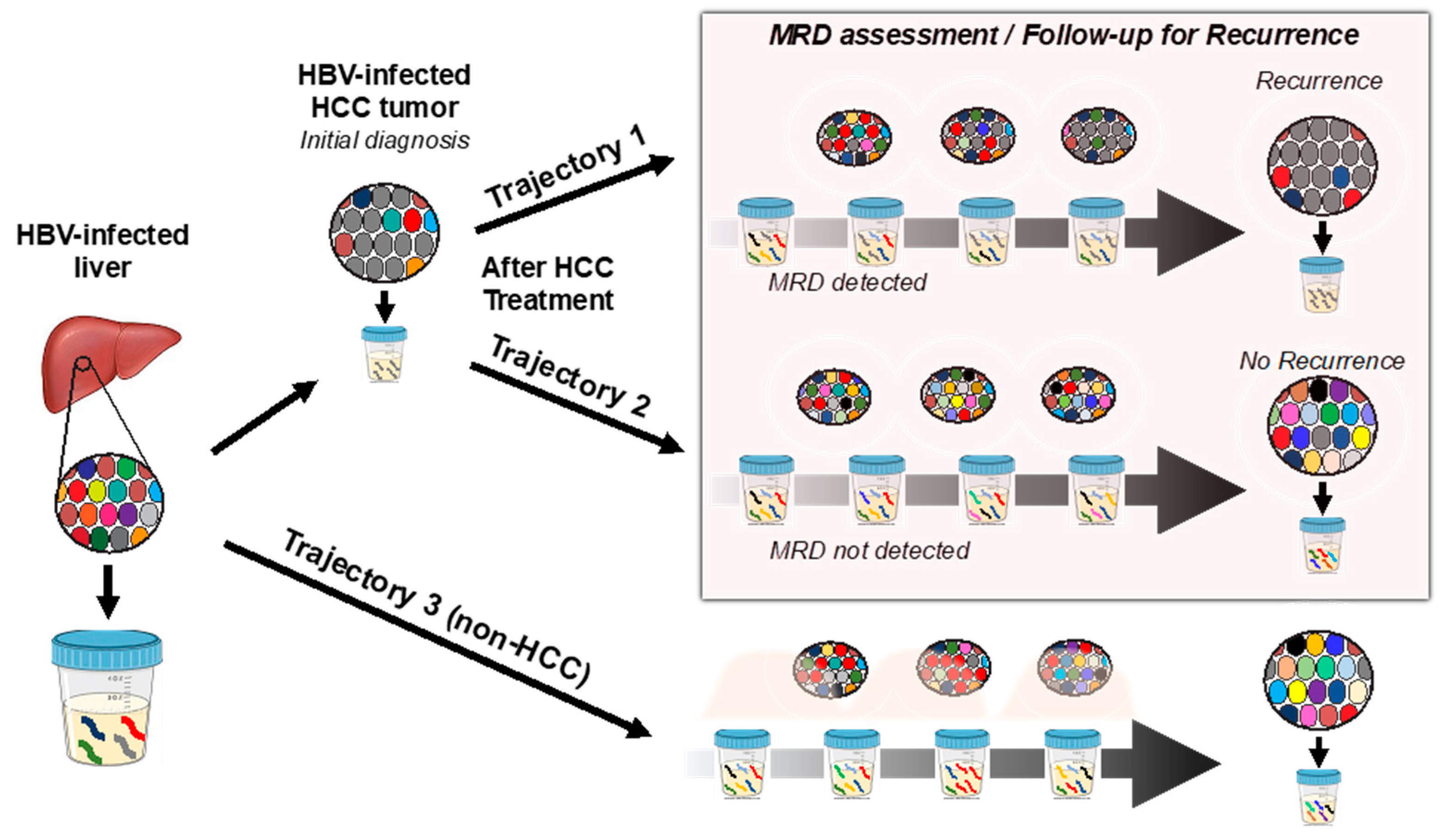
As we’ve demonstrated previously [29] that HBV-JS DNA can be detected in patients with HBV infection, including hepatitis and LC, we also detected HBV-JS DNA in three of four LC patients. Although integration was found in a known HCC-driver gene, TERT, for case 3 LC, it was only detected transiently. This could be due to the interplay of the host immune system, tumorigenesis, and the existence of HBV DNA integration events, as we have previously suggested [42]. Here, nucleos(t)ide analogs were effective in reducing viral replication as indicated by low viral serum load (Table 1) but were not able to eliminate existing integrated HBV DNA in infected hepatocytes. HBV-JSs represent a unique biomarker that can serve as a marker for existing HBV infection as well as a liver-specific marker detectable in urine due to apoptosis of infected hepatocytes. Interestingly, a known HCC-associated recurrent TERT integration was detected in urine from an LC patient (case 3). It is possible that the TERT integration was derived from either neo-antigen expression or indicative of tumorigenesis. Likely, its elimination by the host immune system resulted in a spike of detectable HBV-TERT junction in urine. On the other hand, detection of persistently elevated HBV-JS marker values reflects persistent tumor growth whereby some tumor cells that harbored HCC-derived junctions were eliminated by apoptosis, thus serving as an important biomarker for HCC MRD and recurrence monitoring, as schematically shown in Figure 2.
Although this study is limited by the small cohort size, it provides feasibility evidence that detection of highly abundant HBV-JS in urine from patients with HBV–HCC (for example, HBV-Chr11 and HBV-TERT_1 in case 2) could be HCC-derived and served as an HCC marker for monitoring HCC recurrence and MRD assessment in a noninvasive urine liquid biopsy. Furthermore, since the collection of urine can be done at home and then shipped to certified laboratories for testing, the urine test can result in better compliance without the need for a doctor’s office visit.
It is important to note that the levels of urine DNA markers fluctuate for several reasons, including the hydration of the patient at the time of collection, which can result in diluted DNA in the urine. Further studies are needed to identify a proper internal control for this work, which is currently in progress.
Despite the lack of FDA-approved HCC therapies since Sorafenib, the first FDA-approved HCC chemotherapy over 10 years ago, the recent approval of the immune checkpoint inhibitors (ICIs) PD-L1 inhibitor atezolizumab plus the antiangiogenic agent bevacizumab as front-line treatment for advanced HCC and the IMbrave 150 phase III trial has established a novel standard of care in a relatively short period of time. Despite this unprecedented treatment paradigm shift in HCC treatment, only a proportion of HCC patients benefit from immunotherapy, highlighting the need for biomarkers for assessing MRD and predicting treatment response. Encouragingly, the advent of molecular profiling has produced notable promise in the management of HCC associated with response to ICIs. Genomic aberrations such as Wnt/β-catenin mutations in HCCs have been reported to be significantly associated with resistance to immunotherapy compared to wild-type patients, emerging as potential predictive biomarkers of immunotherapy. The HCC medical community is called to put more effort aimed at evaluating novel biomarkers of response to ICIs, considering tumor-intrinsic (e.g., PD-L1 expression, tumor mutation burden (TMB), microsatellite instability (MSI) status, etc.), immune-specific, and combinatorial biomarkers [43,44,45]. The identification of predictive biomarkers of response to immunotherapy remains a priority in HCC, especially considering that the number of indications and patients receiving ICIs is expected to increase in the near future. Thus, it is critical to identify more genetic markers in addition to PD-L1 expression, TMB, and MSI status to select the right patients to receive immunotherapy and to provide useful information for disease monitoring and treatment-decision making. HBV integration is known to cause insertional mutagenesis of HCC-associated genes and induce chromosomal instability [41]. The mutagenesis resulting from HBV integration should be included in the TMB load and evaluated for its impact on disease management. Although this is a pilot study, the detection of HCC-derived urinary HBV-JS as a biomarker for assessing treatment efficacy or MRD and its persistently increasing levels prior to recurrence is encouraging and consistent with our previous β-catenin mutation study in the urine of HCC patients with recurrence after surgical removal of primary HCC [27]. Together, a large cohort study is warranted to comprehensively determine and develop these potential biomarkers for assessing treatment efficacy, MRD, recurrence, and predicting treatment response.
In conclusion, we have demonstrated the potential of urinary HBV-JS DNA for the early detection of HCC recurrence and MRD assessment. Furthermore, we concluded that marker dynamics, such as persistent elevations of a highly abundant HBV-JS DNA, are important for the clinical relevance of HCC detection.

Supplementary Materials

The following supporting information can be downloaded at: https://www.mdpi.com/article/10.3390/diagnostics13091537/s1, Figure S1. Development of short amplicon HBV-JS quantitative PCR. Table S1: HBV-JS qPCR primers used.

Author Contributions

Conceptualization, H.-W.H., Y.-H.S.; formal analysis, S.Y.L., Y.-L.K., Y.-H.S., P.B. and F.-S.S.; specimens/resources, H.-W.H., D.H.-D. and J.C.; data curation/experimental execution, S.Y.L., Y.-L.K. and F.-S.S.; writing—original draft preparation, H.-W.H., Y.-H.S. and S.Y.L.; writing—review and editing, all authors; funding acquisition, H.-W.H., Y.-H.S., S.Y.L. and W.S. All authors have read and agreed to the published version of the manuscript.

Funding

This work has been supported by the National Institute of Health R44 CA165312 (S.Y.L., H.H., Y.H.S.) and RO1 CA202769 (Y.H.S., H.H. and S.Y.L.).

Institutional Review Board Statement

This study was conducted under the approval of the Institutional Review Board at Thomas Jefferson University Hospital, Philadelphia, PA.

Informed Consent Statement

In this retrospective study, each patient was informed of this study and gave their consent.

Data Availability Statement

The data generated and analyzed during this study are available from the corresponding author upon reasonable request.

Conflicts of Interest

S.Y.L. is an employee of JBS Science, Inc. S.Y.L., Y.S. and W.S. are shareholders of JBS Science, Inc. All other authors declare they have no other competing interests that exist. The funders had no role in the design of the study; in the collection, analyses, or interpretation of data; in the writing of the manuscript; or in the decision to publish the results.

References

  1. Fund, W.C.R. Liver Cancer Statistics. Available online: https://www.wcrf.org/dietandcancer/cancer-trends/liver-cancer-statistics (accessed on 5 June 2022).
  2. Hung, I.F.-N.; Wong, D.K.-H.; Poon, R.T.-P.; Fong, D.Y.-T.; Chui, A.H.-W.; Seto, W.-K.; Fung, J.Y.-Y.; Chan, A.C.-Y.; Yuen, J.C.-H.; Tiu, R.; et al. Risk Factors and Post-Resection Independent Predictive Score for the Recurrence of Hepatitis B-Related Hepatocellular Carcinoma. PLoS ONE 2016, 11, e0148493. [Google Scholar] [CrossRef] [PubMed]
  3. Chong, C.C.-N.; Lee, K.-f.; Ip, P.C.-T.; Wong, J.S.-W.; Cheung, S.Y.-S.; Wong, J.; Ho, S.C.; Lai, P.B.-S. Pre-operative predictors of post-hepatectomy recurrence of hepatocellular carcinoma: Can we predict earlier? Surgeon 2012, 10, 260–266. [Google Scholar] [CrossRef] [PubMed]
  4. Marrero, J.A.; Pelletier, S. Hepatocellular carcinoma. Clin. Liver Dis. 2006, 10, 339–351. [Google Scholar] [CrossRef]
  5. Lok, A.; McMahon, B. Chronic hepatitis B. Hepatology 2001, 34, 1225–1241. [Google Scholar] [CrossRef]
  6. Sherman, M. Recurrence of hepatocellular carcinoma. N. Engl. J. Med. 2008, 359, 2045–2047. [Google Scholar] [CrossRef]
  7. Portolani, N.; Coniglio, A.; Ghidoni, S.; Giovanelli, M.; Benetti, A.; Tiberio, G.A.M.; Giulini, S.M. Early and Late Recurrence After Liver Resection for Hepatocellular Carcinoma: Prognostic and Therapeutic Implications. Ann. Surg. 2006, 243, 229–235. [Google Scholar] [CrossRef] [PubMed]
  8. Kamiyama, T.; Nakanishi, K.; Yokoo, H.; Kamachi, H.; Tahara, M.; Suzuki, T.; Shimamura, T.; Furukawa, H.; Matsushita, M.; Todo, S. Recurrence Patterns After Hepatectomy of Hepatocellular Carcinoma: Implication of Milan Criteria Utilization. Ann. Surg. Oncol. 2009, 16, 1560–1571. [Google Scholar] [CrossRef]
  9. Minami, Y.; Nishida, N.; Kudo, M. Therapeutic response assessment of RFA for HCC: Contrast-enhanced US, CT and MRI. World J. Gastroenterol. 2014, 20, 4160–4166. [Google Scholar] [CrossRef]
  10. Minami, Y.; Kudo, M. Imaging Modalities for Assessment of Treatment Response to Nonsurgical Hepatocellular Carcinoma Therapy: Contrast-Enhanced US, CT, and MRI. Liver Cancer 2015, 4, 106–114. [Google Scholar] [CrossRef]
  11. Piao, C.Y. Lamivudine treatment in patients with HBV-related hepatocellular carcinoma--using an untreated, matched control cohort. Acta Med. Okayama 2005, 59, 217–224. [Google Scholar] [CrossRef] [PubMed]
  12. Kim, S.J.; Kim, J.M. Prediction models of hepatocellular carcinoma recurrence after liver transplantation: A comprehensive review. Clin. Mol. Hepatol. 2022, 28, 739–753. [Google Scholar] [CrossRef] [PubMed]
  13. Lee, J.; Jin, Y.J.; Shin, S.K.; Kwon, J.H.; Kim, S.G.; Suh, Y.J.; Jeong, Y.; Yu, J.H.; Lee, J.W.; Kwon, O.S.; et al. Surgery versus radiofrequency ablation in patients with Child- Pugh class-A/single small (≤3 cm) hepatocellular carcinoma. Clin. Mol. Hepatol. 2022, 28, 207–218. [Google Scholar] [CrossRef] [PubMed]
  14. Kuzuya, T.; Katano, Y.; Kumada, T.; Toyoda, H.; Nakano, I.; Hirooka, Y.; Itoh, A.; Ishigami, M.; Hayashi, K.; Honda, T.; et al. Efficacy of antiviral therapy with lamivudine after initial treatment for hepatitis B virus-related hepatocellular carcinoma. J. Gastroenterol. Hepatol. 2007, 22, 1929–1935. [Google Scholar] [CrossRef] [PubMed]
  15. Park, H.; Park, J.Y. Clinical significance of AFP and PIVKA-II responses for monitoring treatment outcomes and predicting prognosis in patients with hepatocellular carcinoma. BioMed. Res. Int. 2013, 2013, 310427. [Google Scholar] [CrossRef] [PubMed]
  16. Willatt, J.M.; Hussain, H.K.; Adusumilli, S.; Marrero, J.A. MR Imaging of Hepatocellular Carcinoma in the Cirrhotic Liver: Challenges and Controversies. Radiology 2008, 247, 311–330. [Google Scholar] [CrossRef] [PubMed]
  17. Yu, J.-S. Hepatocellular Carcinoma after Transcatheter Arterial Chemoembolization: Difficulties on Imaging Follow-up. Korean J. Radiol. 2005, 6, 134–135. [Google Scholar] [CrossRef] [PubMed]
  18. Zhao, Y.-J.; Ju, Q.; Li, G.-C. Tumor markers for hepatocellular carcinoma. Mol. Clin. Oncol. 2013, 1, 593–598. [Google Scholar] [CrossRef] [PubMed]
  19. Korean Liver Cancer Association. 2022 KLCA-NCC Korea practice guidelines for the management of hepatocellular carcinoma. Clin. Mol. Hepatol. 2022, 28, 583–705. [Google Scholar] [CrossRef]
  20. Dawson, S.J.; Tsui, D.W.; Murtaza, M.; Biggs, H.; Rueda, O.M.; Chin, S.F.; Dunning, M.J.; Gale, D.; Forshew, T.; Mahler-Araujo, B.; et al. Analysis of circulating tumor DNA to monitor metastatic breast cancer. N. Engl. J. Med. 2013, 368, 1199–1209. [Google Scholar] [CrossRef]
  21. Garcia-Murillas, I.; Schiavon, G.; Weigelt, B.; Ng, C.; Hrebien, S.; Cutts, R.J.; Cheang, M.; Osin, P.; Nerurkar, A.; Kozarewa, I. Mutation tracking in circulating tumor DNA predicts relapse in early breast cancer. Sci. Transl. Med. 2015, 7, 302ra133. [Google Scholar] [CrossRef]
  22. Reinert, T.; Schøler, L.V.; Thomsen, R.; Tobiasen, H.; Vang, S.; Nordentoft, I.; Lamy, P.; Kannerup, A.-S.; Mortensen, F.V.; Stribolt, K. Analysis of circulating tumour DNA to monitor disease burden following colorectal cancer surgery. Gut 2016, 65, 625–634. [Google Scholar] [CrossRef] [PubMed]
  23. Misale, S.; Yaeger, R.; Hobor, S.; Scala, E.; Janakiraman, M.; Liska, D.; Valtorta, E.; Schiavo, R.; Buscarino, M.; Siravegna, G.; et al. Emergence of KRAS mutations and acquired resistance to anti-EGFR therapy in colorectal cancer. Nature 2012, 486, 532–536. [Google Scholar] [CrossRef] [PubMed]
  24. Su, Y.-H.; Song, J.; Wang, Z.; Wang, X.; Wang, M.; Brenner, D.E.; Block, T.M. Removal of high molecular weight DNA by carboxylated magnetic beads enhances the detection of mutated K-ras DNA in urine. Ann. N. Y. Acad. Sci. 2008, 1137, 82–91. [Google Scholar] [CrossRef] [PubMed]
  25. Su, Y.-H.; Wang, M.; Aiamkitsumrit, B.; Brenner, D.E.; Block, T.M. Detection of K-ras mutation in urine of patients with colorectal cancer. Cancer Biomark. 2005, 1, 177–182. [Google Scholar] [CrossRef]
  26. Su, Y.-H.; Wang, M.; Brenner, D.E.; Norton, P.A.; Block, T.M. Detection of mutated K-ras DNA in urine, plasma, and serum of patients with colorectal carcinoma or adenomatous polyps. Ann. N. Y. Acad. Sci. 2008, 1137, 197–206. [Google Scholar] [CrossRef]
  27. Lin, S.Y.; Chang, T.-T.; Steffen, J.D.; Chen, S.; Jain, S.; Song, W.; Lin, Y.-J.; Su, Y.-H. Detection of CTNNB1 Hotspot Mutations in Cell-Free DNA from the Urine of Hepatocellular Carcinoma Patients. Diagnostics 2021, 11, 1475. [Google Scholar] [CrossRef]
  28. Lin, S.Y.; Dhillon, V.; Jain, S.; Chang, T.-T.; Hu, C.-T.; Lin, Y.-J.; Chen, S.-H.; Yu, L.; Block, T.M.; Su, Y.-H. A locked nucleic acid clamp-mediated PCR assay for detection of a p53 codon 249 hotspot mutation in urine. J. Mol. Diagn. 2011, 13, 474–484. [Google Scholar] [CrossRef]
  29. Lin, S.Y.; Su, Y.-P.; Trauger, E.R.; Song, B.P.; Thompson, E.G.C.; Hoffman, M.C.; Chang, T.-T.; Lin, Y.-J.; Kao, Y.-L.; Cui, Y.; et al. Detection of Hepatitis B Virus–Host Junction Sequences in Urine of Infected Patients. Hepatol. Commun. 2021, 5, 1649–1659. [Google Scholar] [CrossRef]
  30. Hann, H.W.; Jain, S.; Park, G.; Steffen, J.D.; Song, W.; Su, Y.-H. Detection of urine DNA markers for monitoring recurrent hepatocellular carcinoma. Hepatoma Res. 2017, 3, 105–111. [Google Scholar] [CrossRef]
  31. Kim, A.K.; Hamilton, J.P.; Lin, S.Y.; Chang, T.-T.; Hann, H.-W.; Hu, C.-T.; Lou, Y.; Lin, Y.-J.; Gade, T.P.; Park, G. Urine DNA biomarkers for hepatocellular carcinoma screening. Br. J. Cancer 2022, 126, 1432–1438. [Google Scholar] [CrossRef]
  32. Sung, W.K.; Zheng, H.; Li, S.; Chen, R.; Liu, X.; Li, Y.; Lee, N.P.; Lee, W.H.; Ariyaratne, P.N.; Tennakoon, C.; et al. Genome-wide survey of recurrent HBV integration in hepatocellular carcinoma. Nat. Genet. 2012, 44, 765–769. [Google Scholar] [CrossRef] [PubMed]
  33. Fujimoto, A.; Totoki, Y.; Abe, T.; Boroevich, K.A.; Hosoda, F.; Nguyen, H.H.; Aoki, M.; Hosono, N.; Kubo, M.; Miya, F.; et al. Whole-genome sequencing of liver cancers identifies etiological influences on mutation patterns and recurrent mutations in chromatin regulators. Nat. Genet. 2012, 44, 760–764. [Google Scholar] [CrossRef]
  34. Totoki, Y.; Tatsuno, K.; Covington, K.R.; Ueda, H.; Creighton, C.J.; Kato, M.; Tsuji, S.; Donehower, L.A.; Slagle, B.L.; Nakamura, H. Trans-ancestry mutational landscape of hepatocellular carcinoma genomes. Nat. Genet. 2014, 46, 1267–1273. [Google Scholar] [CrossRef] [PubMed]
  35. Kan, Z.; Zheng, H.; Liu, X.; Li, S.; Barber, T.D.; Gong, Z.; Gao, H.; Hao, K.; Willard, M.D.; Xu, J.; et al. Whole-genome sequencing identifies recurrent mutations in hepatocellular carcinoma. Genome Res. 2013, 23, 1422–1433. [Google Scholar] [CrossRef] [PubMed]
  36. Esumi, M.; Aritaka, T.; Arii, M.; Suzuki, K.; Tanikawa, K.; Mizuo, H.; Mima, T.; Shikata, T. Clonal origin of human hepatoma determined by integration of hepatitis B virus DNA. Cancer Res. 1986, 46, 5767–5771. [Google Scholar] [PubMed]
  37. Esumi, M.; Tanaka, Y.; Tozuka, S.; Shikata, T. Clonal state of human hepatocellular carcinoma and non-tumorous hepatocytes. Cancer Chemother. Pharmacol. 1989, 23, S1–S3. [Google Scholar] [CrossRef] [PubMed]
  38. (Korean Association for the Study of the Liver. KASL clinical practice guidelines for management of chronic hepatitis B. Clin. Mol. Hepatol. 2022, 28, 276–331. [Google Scholar] [CrossRef]
  39. Li, C.L.; Ho, M.C.; Lin, Y.Y.; Tzeng, S.T.; Chen, Y.J.; Pai, H.Y.; Wang, Y.C.; Chen, C.L.; Lee, Y.H.; Chen, D.S.; et al. Cell-Free Virus-Host Chimera DNA From Hepatitis B Virus Integration Sites as a Circulating Biomarker of Hepatocellular Cancer. Hepatology 2020, 72, 2063–2076. [Google Scholar] [CrossRef]
  40. Mohme, M.; Riethdorf, S.; Pantel, K. Circulating and disseminated tumour cells—Mechanisms of immune surveillance and escape. Nat. Rev. Clin. Oncol. 2017, 14, 155–167. [Google Scholar] [CrossRef]
  41. Lin, S.Y.; Zhang, A.; Lian, J.; Wang, J.; Chang, T.-T.; Lin, Y.-J.; Song, W.; Su, Y.-H. Recurrent HBV Integration Targets as Potential Drivers in Hepatocellular Carcinoma. Cells 2021, 10, 1294. [Google Scholar] [CrossRef]
  42. Garrido, D.; Block, P.; Lin, S.; Halegoua-DeMarzio, D.; Hann, H.-W. Survival Disparity between Antiviral-Treated and Antiviral-Naïve Patients Who Develop Their First HBV-Associated Hepatocellular Carcinoma. Arch. Gastroenterol. Res. 2021, 2, 86–94. [Google Scholar] [CrossRef]
  43. Rizzo, A.; Cusmai, A.; Gadaleta-Caldarola, G.; Palmiotti, G. Which role for predictors of response to immune checkpoint inhibitors in hepatocellular carcinoma? Expert Rev. Gastroenterol. Hepatol. 2022, 16, 333–339. [Google Scholar] [CrossRef]
  44. Viscardi, G.; Tralongo, A.C.; Massari, F.; Lambertini, M.; Mollica, V.; Rizzo, A.; Comito, F.; Di Liello, R.; Alfieri, S.; Imbimbo, M.; et al. Comparative assessment of early mortality risk upon immune checkpoint inhibitors alone or in combination with other agents across solid malignancies: A systematic review and meta-analysis. Eur. J. Cancer 2022, 177, 175–185. [Google Scholar] [CrossRef] [PubMed]
  45. Rizzo, A.; Ricci, A.D.; Di Federico, A.; Frega, G.; Palloni, A.; Tavolari, S.; Brandi, G. Predictive Biomarkers for Checkpoint Inhibitor-Based Immunotherapy in Hepatocellular Carcinoma: Where Do We Stand? Front. Oncol. 2021, 11, 803133. [Google Scholar] [CrossRef] [PubMed]
Figure 1. HBV-JS DNA biomarkers levels in serial urine samples from HBV–HCC patients with HCC recurrence and an HBV-cirrhotic patient under HCC surveillance. Both HBV–HCC patients were being monitored for HCC recurrence by MRI and serum AFP. The urine samples were collected prospectively from two HBV–HCC patients (when available) after curative treatment and at follow-up visits and from one cirrhotic patient at HCC surveillance visit. Samples were retrospectively measured for HBV-JS DNA biomarkers. HBV-JS DNA along with serum AFP (ng/mL), were plotted at office visits until the last available visit in which an MRI was performed. The “POS” represents detection of HCC recurrence by MRI. HCC: hepatocellular carcinoma; AFP: alpha-fetoprotein.
Diagnostics 13 01537 g001
Figure 2. Schematic diagram of urinary HBV-JS detection trajectories during serial sampling of HBV-infected patients undergoing HCC surveillance for primary and recurrent HCC. Each colored circle represents an HBV-infected hepatocyte harboring a unique HBV-host junction sequence (HBV-JS). Gray-colored circles and urinary DNA fragments represent HCC cells and HCC-derived HBV-JS, respectively. Trajectory 1 illustrates an HBV–HCC patient undergoing HCC treatment after an initial HCC diagnosis and then follow-up for HCC recurrence, where serial urine sampling reveals detection of a low amount of HCC-derived HBV-JS (gray-colored DNA) after treatment, suggesting MRD and an increasing level of an HCC-derived HBV-JS detected in urine, up to recurrence diagnosis. Trajectory 2 depicts an HBV–HCC patient whose serial urine sampling reveals a highly diverse collection of HBV-JS after HCC treatment and no detection of original HCC-derived HBV-JS that was detected during the initial HCC diagnosis indicating no MRD nor recurrence. Trajectory 3 represents an HBV-infected non-HCC patient under HCC surveillance with serial urine sampling showing transient levels of HBV-JS detected in urine, as indicated by red DNA.
Diagnostics 13 01537 g002
Table 1. Clinical characteristics of patient urine for viral-host junction detection by HBV-targeted NGS assay.
CaseDiagnosisAgeGenderAntiviral TherapyALT (IU/L)Serum AFP (ng/mL)HBV Serum (IU/mL)HBe Ag (−/+)Hbe AbHCC StageTumor Size (cm)
1HCC78FLAM, TDFNA1ND-+I0.74
2HCC72MLAM, TDF301.4<20--NA3.4
3LC53MLAM, TAF, TDF271.5<20NDNDNA
4LC68F-NA9.1<10NDND
5LC60MLAM, ENT183<20--
6LC57MIFN, LAM, TDF831.8<20+-
LAM, lamivudine; TDF, tenofovir disoproxil fumarate, TAF, tenofovir alafenamide; ENT, entecavir; IFN, pegylated interferon α; NA, not available; ND, not done. Note, all the clinical data were recorded as close as possible to the time of urine collection.
Table 2. HBV-JS detected in urine by HBV-targeted NGS assay.
CaseSample Used (Month/Year)Disease StatusHBV-JS *
Gene (Supporting Read)
111/20153 years pre-recur (HCC)Chr17_NC (2)
24/20153 months pre-recurTERT_1 (22), chr11_NC (63), GCSHP2 (3)
35/2015LCTERT_1 (9), Chr7_NC (2)
47/2019LCNA
59/2013LCSimple repeat (9), LTR (6)
67/2015LCSINE (17), Simple repeat (6)
* Up to three most abundant HBV-JS listed; NA: no HBV-JS with at least 2 SR detected. LTR: long terminal repeat; SINE: short interspersed nuclear elements.
Table 3. Clinical characteristics and HCC treatment history over six time points for three cases studied for HBV-JS dynamic.
Case Data Collection Timepoint (Month/Year)Follow-Up (Months)Disease Status (HCC Treatment)Serum AFP (ng/mL)LI-RADSHCC Stage Tumor Size (cm)
12/20150Pre-recur1.0NANANA
9/20157Pre-recur1.21NANANA
11/20159POS (MW)1.21NAI0.74
2/201612Pre-recur2.653NANA
8/201618Pre-recur1.084NANA
10/201844POS1.55I1.2
210/20130Pre-recur1.4NANANA
5/20147Pre-recur1.2NANANA
11/201413Pre-recur1.2NANANA
1/201515POS (MW)1.55NA3.4
4/201518Pre-recur1NANANA
7/201521POS (TACE)1NANA0.6
37/20130Cirrhosis1.5NANANA
8/201414Cirrhosis1.5NANANA
5/201522Cirrhosis1.8NANANA
8/201525Cirrhosis1.8NANANA
1/201631Cirrhosis2.1NANANA
4/201633Cirrhosis1.9NANANA
3/201745Cirrhosis1.9NANANA
Pre-recur indicates timepoint pre-HCC recurrence; POS indicates timepoint at positive HCC recurrence diagnosis; MW, patient underwent microwave ablation; TACE, patient underwent transcatheter arterial chemoembolization.
Disclaimer/Publisher’s Note: The statements, opinions and data contained in all publications are solely those of the individual author(s) and contributor(s) and not of MDPI and/or the editor(s). MDPI and/or the editor(s) disclaim responsibility for any injury to people or property resulting from any ideas, methods, instructions or products referred to in the content.