
Cellular-Level Analysis of Retinal Blood Vessel Walls Based on Phase Gradient Images

 , ,
, , 
Abstract
:1. Introduction
2. Materials and Methods
2.1. The Imaging System
2.2. Human Subjects and Imaging Procedure
2.3. Confocal and Non-Confocal Imaging
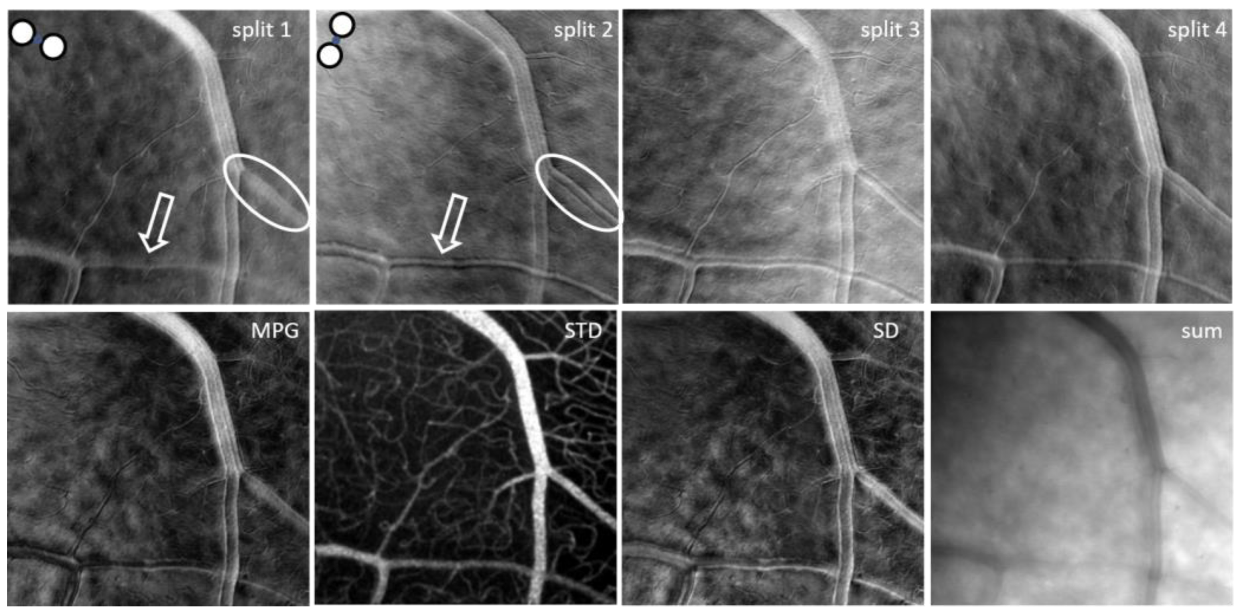
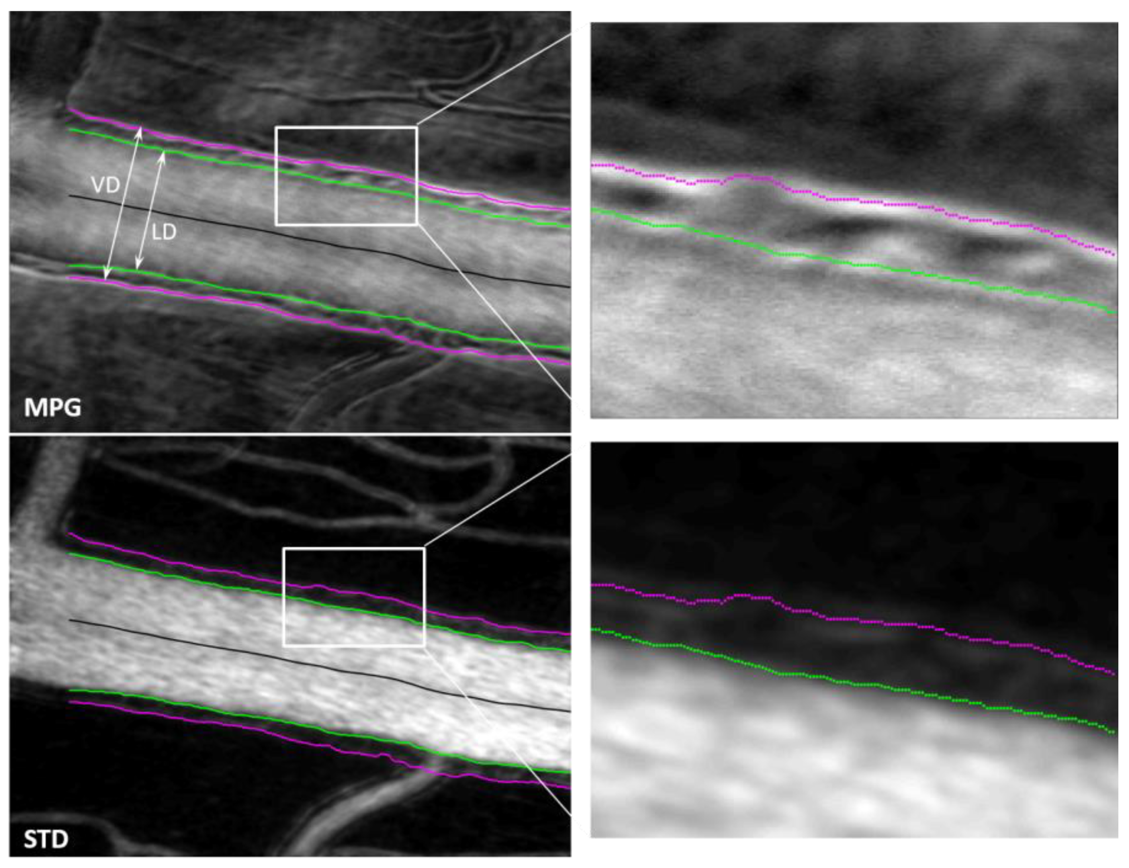
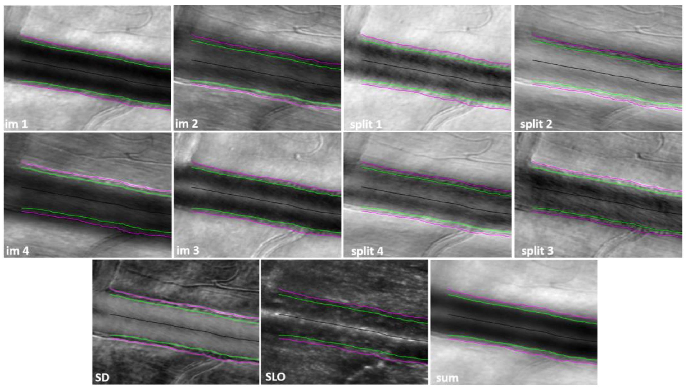
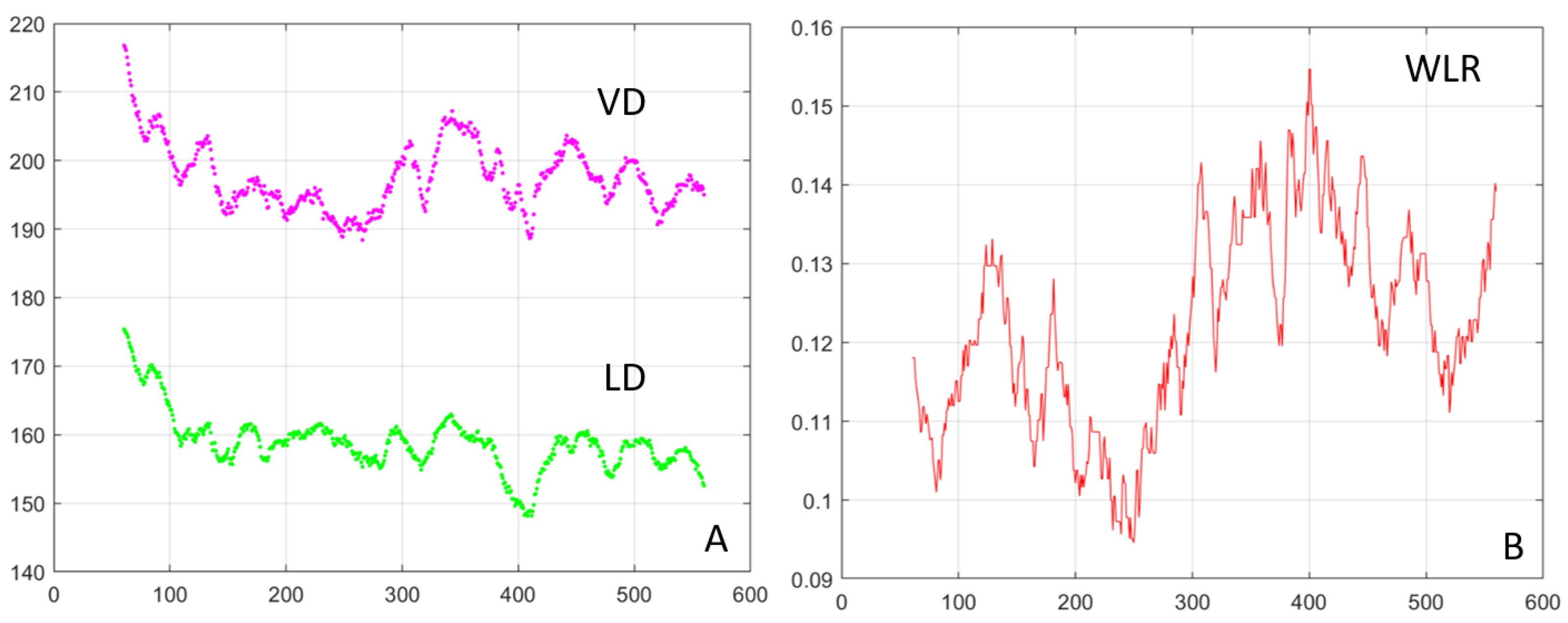
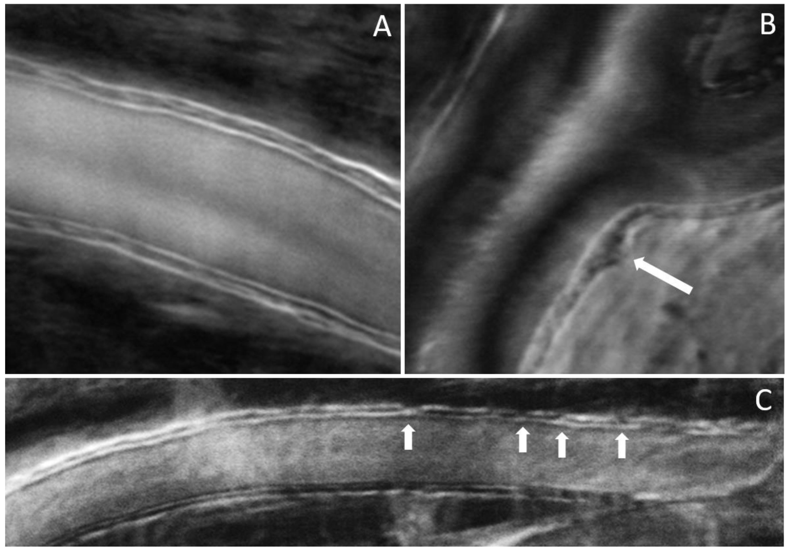
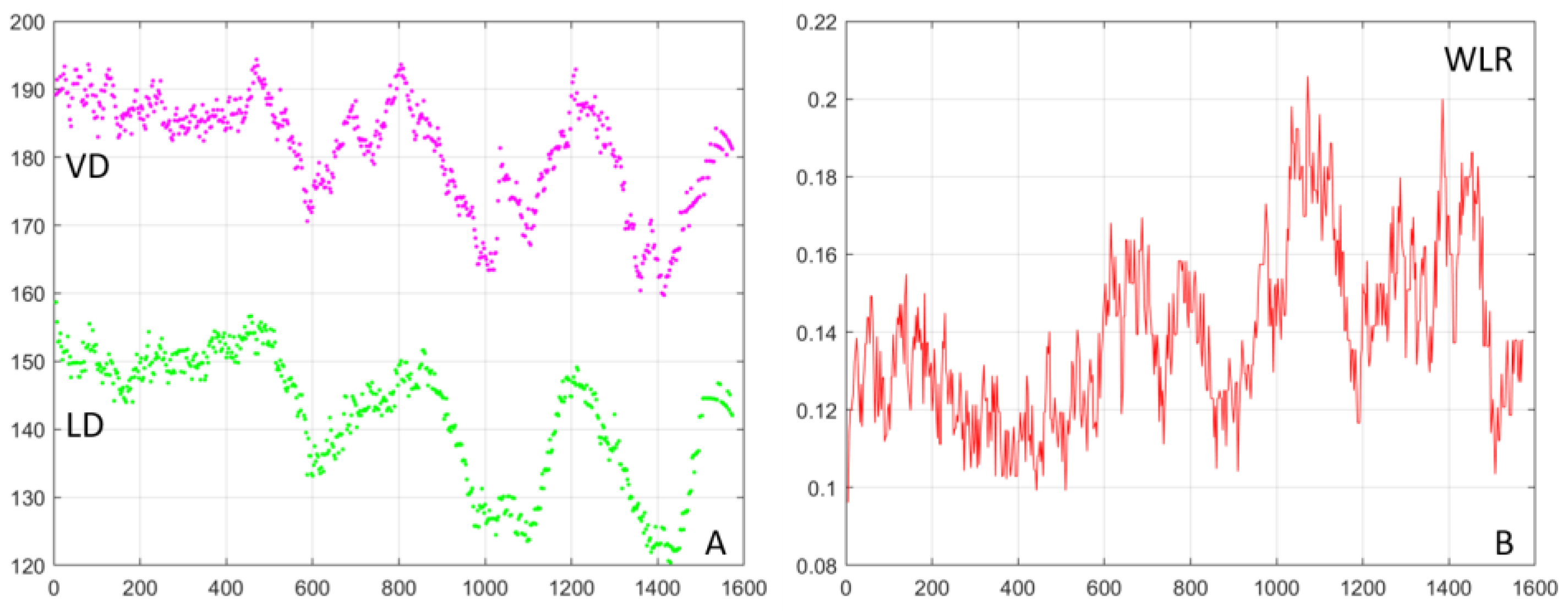
3. Results
4. Discussion
5. Conclusions
Supplementary Materials
Author Contributions
Funding
Institutional Review Board Statement
Informed Consent Statement
Data Availability Statement
Acknowledgments
Conflicts of Interest
References
- Fulton, A.B.; Akula, J.D.; Mocko, J.A.; Hansen, R.M.; Benador, I.Y.; Beck, S.C.; Fahl, E.; Seeliger, M.W.; Moskowitz, A.; Harris, M.E. Retinal degenerative and hypoxic ischemic disease. Doc. Ophthalmol. 2009, 118, 55–61. [Google Scholar] [CrossRef] [PubMed]
- Steinberg, R.H. Monitoring communications between photoreceptors and pigment epithelial cells: Effects of “mild” systemic hypoxia. Friedenwald lecture. Investig. Ophthalmol. Vis. Sci. 1987, 28, 1888–1904. [Google Scholar]
- Shakib, M.; De Oliveira, L.F.; Henkind, P. Development of Retinal Vessels. II. Earliest Stages of Vessel Formation. Investig. Ophthalmol. Vis. Sci. 1968, 7, 689–700. [Google Scholar]
- Bek, T.; Hajari, J.; Jeppesen, P. Interaction between flicker-induced vasodilatation and pressure autoregulation in early retinopathy of Type 2 diabetes. Graefe’s Arch. Clin. Exp. Ophthalmol. 2008, 246, 763–769. [Google Scholar] [CrossRef]
- Bower, B.; Zhao, M.; Zawadzki, R.; Izatt, J. Real-time spectral domain Doppler optical coherence tomography and investigation of human retinal vessel autoregulation. J. Biomed. Opt. 2007, 12, 041214. [Google Scholar] [CrossRef] [PubMed]
- Tilma, K.K.; Bek, T. Topical treatment for 1 week with latanoprost but not diclofenac reduces the diameter of dilated retinal arterioles in patients with type 1 diabetes mellitus and mild retinopathy. Acta Ophthalmol. 2012, 90, 750–755. [Google Scholar] [CrossRef]
- Ye, X.D.; Laties, A.M.; Stone, R.A. Peptidergic innervation of the retinal vasculature and optic nerve head. Investig. Ophthalmol. Vis. Sci. 1990, 31, 1731–1737. [Google Scholar]
- Ciulla, T.A.; Amador, A.G.; Zinman, B. Diabetic Retinopathy and Diabetic Macular Edema: Pathophysiology, screening, and novel therapies. Diabetes Care 2003, 26, 2653–2664. [Google Scholar] [CrossRef]
- IDF Diabetes Atlas Tenth Edition. 2021. Available online: https://diabetesatlas.org/ (accessed on 15 September 2023).
- Tam, J.; Martin, J.A.; Roorda, A. Noninvasive visualization and analysis of parafoveal capillaries in humans. Investig. Ophthalmol. Vis. Sci. 2010, 51, 1691–1698. [Google Scholar] [CrossRef]
- Chui, T.Y.; Zhong, Z.; Song, H.; Burns, S.A. Foveal avascular zone and its relationship to foveal pit shape. Optom. Vis. Sci. 2012, 89, 602–610. [Google Scholar] [CrossRef]
- Spaide, R.F.; Fujimoto, J.G.; Waheed, N.K.; Sadda, S.R.; Staurenghi, G. Optical coherence tomography angiography. Prog. Retin. Eye Res. 2017, 64, 1–55. [Google Scholar] [CrossRef] [PubMed]
- Kashani, A.H.; Chen, C.L.; Gahm, J.K.; Zheng, F.; Richter, G.M.; Rosenfeld, P.J.; Shi, Y.; Wang, R.K. Optical coherence tomography angiography: A comprehensive review of current methods and clinical applications. Prog. Retin. Eye Res. 2017, 60, 66–100. [Google Scholar] [CrossRef] [PubMed]
- Jia, Y.; Bailey, S.T.; Hwang, T.S.; McClintic, S.M.; Gao, S.S.; Pennesi, M.E.; Flaxel, C.J.; Lauer, A.K.; Wilson, D.J.; Hornegger, J.; et al. Quantitative optical coherence tomography angiography of vascular abnormalities in the living human eye. Proc. Natl. Acad. Sci. USA 2015, 112, E2395–E2402. [Google Scholar] [CrossRef] [PubMed]
- Fingler, J.; Zawadzki, R.J.; Werner, J.S.; Schwartz, D.; Fraser, S.E. Volumetric microvascular imaging of human retina using optical coherence tomography with a novel motion contrast technique. Opt. Express 2009, 17, 22190–22200. [Google Scholar] [CrossRef] [PubMed]
- Makita, S.; Hong, Y.; Yamanari, M.; Yatagai, T.; Yasuno, Y. Optical coherence angiography. Opt. Express 2006, 14, 7821–7840. [Google Scholar] [CrossRef] [PubMed]
- Prieto, P.M.; Vargas-Martin, F.; Goelz, S.; Artal, P. Analysis of the performance of the Hartmann-Shack sensor in the human eye. J. Opt. Soc. Am. A-Opt. Image Sci. Vis. 2000, 17, 1388–1398. [Google Scholar] [CrossRef]
- Doble, N.; Yoon, G.; Chen, L.; Bierden, P.; Singer, B.; Olivier, S.; Williams, D.R. Use of a microelectromechanical mirror for adaptive optics in the human eye. Opt. Lett. 2002, 27, 1537–1539. [Google Scholar] [CrossRef]
- Shirai, T. Liquid-crystal adaptive optics based on feedback interferometry for high-resolution retinal imaging. Appl. Opt. 2002, 41, 4013–4023. [Google Scholar] [CrossRef]
- Vargas-Martin, F.; Prieto, P.M.; Artal, P. Correction of the aberrations in the human eye with a liquid-crystal spatial light modulator: Limits to performance. J. Opt. Soc. Am. A-Opt. Image Sci. Vis. 1998, 15, 2552–2562. [Google Scholar] [CrossRef]
- Romero-Borja, F.; Venkateswaran, K.; Roorda, A.; Hebert, T. Optical slicing of human retinal tissue in vivo with the adaptive optics scanning laser ophthalmoscope. Appl. Opt. 2005, 44, 4032–4040. [Google Scholar] [CrossRef]
- Chui, T.Y.; Vannasdale, D.A.; Burns, S.A. The use of forward scatter to improve retinal vascular imaging with an adaptive optics scanning laser ophthalmoscope. Biomed. Opt. Express 2012, 3, 2537–2549. [Google Scholar] [CrossRef] [PubMed]
- Roorda, A.; Duncan, J.L. Adaptive Optics Ophthalmoscopy. Annu. Rev. Vis. Sci. 2015, 1, 19–50. [Google Scholar] [CrossRef] [PubMed]
- Scoles, D.; Sulai, Y.N.; Langlo, C.S.; Fishman, G.A.; Curcio, C.A.; Carroll, J.; Dubra, A. In Vivo Imaging of Human Cone Photoreceptor Inner Segments. Investig. Ophthalmol. Vis. Sci. 2014, 55, 4244–4251. [Google Scholar] [CrossRef] [PubMed]
- Hamilton, D.K.; Sheppard, C.J.R. Differential phase contrast in scanning optical microscopy. J. Microsc. 1984, 133, 27–39. [Google Scholar] [CrossRef]
- Rossi, E.A.; Granger, C.E.; Sharma, R.; Yang, Q.; Saito, K.; Schwarz, C.; Walters, S.; Nozato, K.; Zhang, J.; Kawakami, T.; et al. Imaging individual neurons in the retinal ganglion cell layer of the living eye. Proc. Natl. Acad. Sci. USA 2017, 114, 586–591. [Google Scholar] [CrossRef]
- Mujat, M.; Patel, A.; Maguluri, G.; Ferguson, R.D.; Iftimia, N. Simultaneous Multi-Offset Imaging of Retinal Microstructures Free of Directionality Artifacts; SPIE: Bellingham, WA, USA, 2021; Volume 11623. [Google Scholar]
- Mozaffari, S.; Jaedicke, V.; Larocca, F.; Tiruveedhula, P.; Roorda, A. Versatile multi-detector scheme for adaptive optics scanning laser ophthalmoscopy. Biomed. Opt. Express 2018, 9, 5477–5488. [Google Scholar] [CrossRef]
- Sredar, N.; Kowalski, B.; Razeen, M.M.; Steven, S.; Dubra, A. Non-confocal quad-detection adaptive optics scanning light ophthalmoscopy of the photoreceptor mosaic. Investig. Ophthalmol. Vis. Sci. 2018, 59, 4632. [Google Scholar]
- Mujat, M.; Akula, J.D.; Fulton, A.B.; Ferguson, R.D.; Iftimia, N. Non-rigid registration for high-resolution retinal imaging. Diagnostics 2023, 13, 2285. [Google Scholar] [CrossRef]
- Bronstein, R.; Werman, M.; Peleg, S. Surface reconstruction from derivatives. In Proceedings of the International Conference on Pattern Recognition, The Hague, The Netherlands, 30 August–3 September 1992; IEEE Computer Society Press: Washington, DC, USA, 1992; p. 391. [Google Scholar]
- Dubra, A.; Paterson, C.; Dainty, C. Wave-front reconstruction from shear phase maps by use of the discrete Fourier transform. Appl. Opt. 2004, 43, 1108–1113. [Google Scholar] [CrossRef]
- Freischlad, K.R.; Koliopoulos, C.L. Modal estimation of a wave front from difference measurements using the discrete Fourier transform. J. Opt. Soc. Am. A 1986, 3, 1852–1861. [Google Scholar] [CrossRef]


Disclaimer/Publisher’s Note: The statements, opinions and data contained in all publications are solely those of the individual author(s) and contributor(s) and not of MDPI and/or the editor(s). MDPI and/or the editor(s) disclaim responsibility for any injury to people or property resulting from any ideas, methods, instructions or products referred to in the content. |
© 2023 by the authors. Licensee MDPI, Basel, Switzerland. This article is an open access article distributed under the terms and conditions of the Creative Commons Attribution (CC BY) license (https://creativecommons.org/licenses/by/4.0/).
Share and Cite
Mujat, M.; Sampani, K.; Patel, A.H.; Sun, J.K.; Iftimia, N. Cellular-Level Analysis of Retinal Blood Vessel Walls Based on Phase Gradient Images. Diagnostics 2023, 13, 3399. https://doi.org/10.3390/diagnostics13223399
Mujat M, Sampani K, Patel AH, Sun JK, Iftimia N. Cellular-Level Analysis of Retinal Blood Vessel Walls Based on Phase Gradient Images. Diagnostics. 2023; 13(22):3399. https://doi.org/10.3390/diagnostics13223399
Chicago/Turabian StyleMujat, Mircea, Konstantina Sampani, Ankit H. Patel, Jennifer K. Sun, and Nicusor Iftimia. 2023. "Cellular-Level Analysis of Retinal Blood Vessel Walls Based on Phase Gradient Images" Diagnostics 13, no. 22: 3399. https://doi.org/10.3390/diagnostics13223399
APA StyleMujat, M., Sampani, K., Patel, A. H., Sun, J. K., & Iftimia, N. (2023). Cellular-Level Analysis of Retinal Blood Vessel Walls Based on Phase Gradient Images. Diagnostics, 13(22), 3399. https://doi.org/10.3390/diagnostics13223399