COVID-19 Lung Ultrasound Scores and Lessons from the Pandemic: A Narrative Review
Abstract
1. Introduction
2. Methods
3. Results
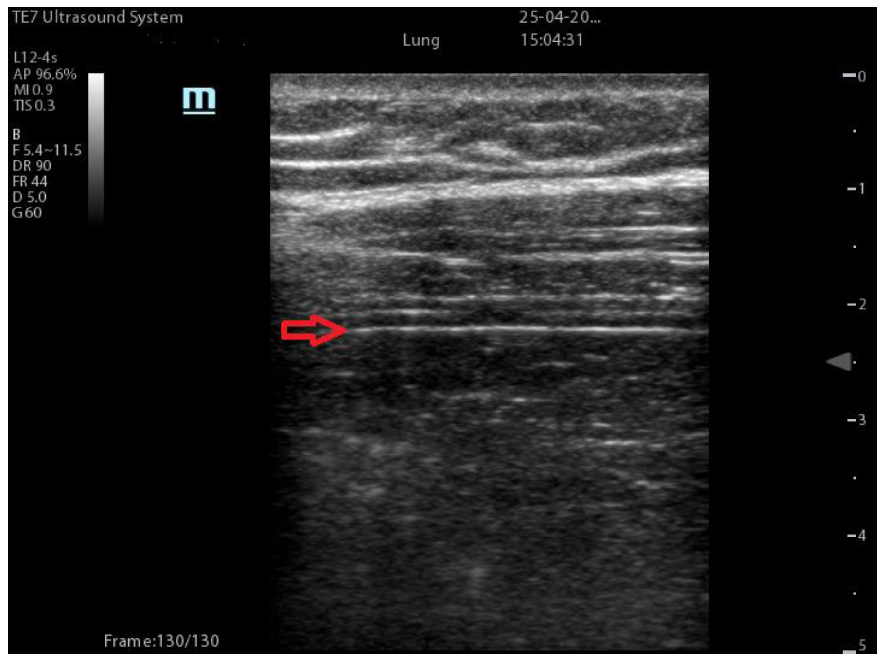
3.1. Lung Findings
3.2. Other Findings
3.3. COVID-19 Characteristics at LUS
3.4. Lung Ultrasound Scores for Predicting Severity, Treatment Response, and Outcomes
| Coalescent Lung score c-LUS [10,11] | Score 0: presence of A-lines, maximum 2 B-lines Score 1: ≥3 well-spaced B-lines Score 2: coalescent B-lines Score 3: tissue-like pattern | 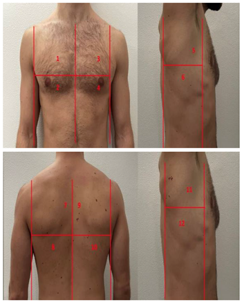 12 AREAS |
| Quantitative LUSS (q-LUS) [12] | Score 0: A-lines, maximum 2 B-lines Score 1: artefacts occupying ≤ 50% of pleura Score 2: artefacts occupying > 50% of pleura Score 3: tissue-like pattern |  12 AREAS |
| Soldati Score [36] | Score 0: continuous and regular pleural line, presence of A-lines. Score 1: indented pleural line, with the presence of vertical white areas. Score 2: broken pleural line with the appearance of small-to-large consolidations associated with white lung. Score 3: dense and largely extended white lung with or without consolidations. |  14 AREAS |
| Falgarone index [35] | 1: normal pleural image with A-lines 2: B lines 3: Multiple B-lines (ground glass) 4: consolidations 5: neo organisation known as hepatisation 6: pleurisy (not found among the patients) Index: a score from 1 to 5 is given to each area, the scores obtained are added up together. The resulting number is divided to the maximal score possible considering only the evaluated areas. |  12 AREAS |
| Simplified LUS score [43] | 1: a small loss of aeration characterised by more than three B-lines or presence of multiple sub-pleuric consolidations separated by normal pleura; 2: a moderate loss of aeration consisting of multiple and coalescent B-lines and/or multiple sub-pleuric consolidations 1 × 2 cm or smaller and separated by thickened or irregular pleura; 3: a severe loss of aeration described as parenchymal consolidation or subpleuric consolidations greater than 1 × 2 cm. |  6 AREAS |
| Integrated ultrasound score (I-LUS) [41] | 0: A-lines or ≤2 B-lines plus regular sliding 1: ≥3 B lines or spaced focal points plus regular sliding 2: coalescing B-lines 3: pulmonary consolidations Plus: Presence of pleural effusion (1: present, 0: absent). Presence of pericardial effusion (1: present, 0: absent). Measurement of the IVC respiratory variation (<0–33%) (1: present, 0: absent). Diaphragm excursion: measured in normal respiration, with M-mode through a right subcostal scan. A value > 2 +/− 0.5 cm is considered normal (0 points), while an inferior value is considered abnormal (1 point). |  12 AREAS |
| Casella score [42] | 0: regular pleural line, presence of horizontal artefacts (A-lines); 1: at least 3 B-lines in at least one scan of the region; the B-lines do not merge one in the other. Small subpleural consolidations ≤1 cm diameter may be present; 2: multiple, converging B-lines, usually determining a so-called “white lung”. Small subpleural consolidations ≤1 cm diameter may be present; 3: presence of at least one consolidation with major axis >1 cm. |  11 AREAS |
| Dargent score [46] | 0 = normal findings 1 = well-defined B-lines 2 = coalescent B-lines 3 = consolidations | 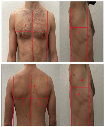 12 AREAS |
| Modified LUS score [47] | 0: well-spaced B-lines <3 1: well-spaced B-lines ≥3 2: multiple coalescent B-lines 3: lung consolidation Plus, plural line is quantitatively ranked as follows: 0: normal 1: irregular pleural line 2: blurred pleural line |  12 AREAS |
| Soldati Score | Falgarone Index | Simplified LUS Score | Integrated LUS Score | Casella Score | C-LUS | |
|---|---|---|---|---|---|---|
| Worsening predictor | AUC = 0.82 | Good correlation p < 0.001 | ||||
| Diagnosis | Sensitivity 99% Specificity 56% | Sensitivity 100% Specificity 40% | ||||
| Correlation with CT scan | Sensitivity 90%Specificity 100% | Positive correlation p < 0.0001 | ||||
| Oxygen requirement | Sensitivity 95% Specificity 67% | |||||
| NIV requirement/NIV failure | Sensitivity 88% Specificity 93% | Positive correlation p = 0.005 | Positive correlation p < 0.0001 | |||
| Outcome | Sensitivity 87% Specificity 82% | Positive correlation p = 0.005 | Positive correlation p = 0.005 | Positive correlation p < 0.05 | ||
| Advantages | Specific for Covid-19 | High correlation with CT-scan | Fast to perform | Takes into account IVC, Diaphragm and Pericardium | Valuable prognostic tool in hospitalized patients | High sensitivity for asymptomatic patients |
4. Discussion
5. Limitations
6. Conclusions
Author Contributions
Funding
Institutional Review Board Statement
Informed Consent Statement
Data Availability Statement
Conflicts of Interest
References
- Statement on the Fifteenth Meeting of the IHR (2005) Emergency Committee on the COVID-19 Pandemic. Available online: https://www.who.int/news/item/05-05-2023-statement-on-the-fifteenth-meeting-of-the-international-health-regulations-%282005%29-emergency-committee-regarding-the-coronavirus-disease-%28covid-19%29-pandemic (accessed on 5 May 2023).
- Wu, Z.; McGoogan, J.M. Characteristics of and Important Lessons from the Coronavirus Disease 2019 (COVID-19) Outbreak in China: Summary of a Report of 72314 Cases from the Chinese Center for Disease Control and Prevention. JAMA 2020, 323, 1239–1242. [Google Scholar] [CrossRef] [PubMed]
- Maggi, L.; Biava, A.M.; Fiorelli, S.; Coluzzi, F.; Ricci, A.; Rocco, M. Lung Ultrasound: A Diagnostic Leading Tool for SARS-CoV-2 Pneumonia: A Narrative Review. Diagnostics 2021, 11, 2381. [Google Scholar] [CrossRef] [PubMed]
- Rizzetto, F.; Perillo, N.; Artioli, D.; Travaglini, F.; Cuccia, A.; Zannoni, S.; Tombini, V.; Di Domenico, S.L.; Albertini, V.; Bergamaschi, M.; et al. Correlation between lung ultrasound and chest CT patterns with estimation of pulmonary burden in COVID-19 patients. Eur. J. Radiol. 2021, 138, 109650. [Google Scholar] [CrossRef] [PubMed]
- Hussain, A.; Via, G.; Melniker, L.; Goffi, A.; Tavazzi, G.; Neri, L.; Villen, T.; Hoppmann, R.; Mojoli, F.; Noble, V.; et al. Multi-organ point-of-care ultrasound for COVID-19 (PoCUS4COVID): International expert consensus. Crit. Care 2020, 24, 702. [Google Scholar] [CrossRef] [PubMed]
- Peng, Q.Y.; Wang, X.T.; Zhang, L.N.; Chinese Critical Care Ultrasound Study Group (CCUSG). Findings of lung ultrasonography of novel corona virus pneumonia during the 2019–2020 epidemic. Intensive Care Med. 2020, 46, 849–850. [Google Scholar] [CrossRef]
- Quarato, C.M.I.; Mirijello, A.; Maggi, M.M.; Borelli, C.; Russo, R.; Lacedonia, D.; Foschino Barbaro, M.P.; Scioscia, G.; Tondo, P.; Rea, G.; et al. Lung Ultrasound in the Diagnosis of COVID-19 Pneumonia: Not Always and Not Only What Is COVID-19 “Glitters”. Front. Med. 2021, 8, 707602. [Google Scholar] [CrossRef]
- Demi, L. Lung ultrasound: The future ahead and the lessons learned from COVID-19. J. Acoust. Soc. Am. 2020, 148, 2146. [Google Scholar] [CrossRef]
- Di Pietro, S.; Mascolo, M.; Falaschi, F.; Brambilla, W.; Ruzga, R.; Mongodi, S.; Perlini, S.; Perrone, T. Lung-ultrasound objective structured assessment of technical skills (LUS-OSAUS): Utility in the assessment of lung-ultrasound trained medical undergraduates. J. Ultrasound 2021, 24, 57–65. [Google Scholar] [CrossRef]
- Bouhemad, B.; Brisson, H.; Le-Guen, M.; Arbelot, C.; Lu, Q.; Rouby, J.J. Bedside ultrasound assessment of positive end-expiratory pressure-induced lung recruitment. Am. J. Respir. Crit. Care Med. 2011, 183, 341–347. [Google Scholar] [CrossRef]
- Soummer, A.; Perbet, S.; Brisson, H.; Arbelot, C.; Constantin, J.M.; Lu, Q.; Rouby, J.J.; Lung Ultrasound Study Group. Ultrasound assessment of lung aeration loss during a successful weaning trial predicts postextubation distress. Crit. Care Med. 2012, 40, 2064–2072. [Google Scholar] [CrossRef]
- Mongodi, S.; Bouhemad, B.; Orlando, A.; Stella, A.; Tavazzi, G.; Via, G.; Iotti, G.A.; Braschi, A.; Mojoli, F. Modified Lung Ultrasound Score for Assessing and Monitoring Pulmonary Aeration. Ultraschall Med. 2017, 38, 530–537. [Google Scholar] [CrossRef] [PubMed]
- Duggan, N.M.; Shokoohi, H.; Liteplo, A.S.; Huang, C.; Goldsmith, A.J. Best Practice Recommendations for Point-of-Care Lung Ultrasound in Patients with Suspected COVID-19. J. Emerg. Med. 2020, 59, 515–520. [Google Scholar] [CrossRef] [PubMed]
- Allinovi, M.; Parise, A.; Giacalone, M.; Amerio, A.; Delsante, M.; Odone, A.; Franci, A.; Gigliotti, F.; Amadasi, S.; Delmonte, D.; et al. Lung Ultrasound May Support Diagnosis and Monitoring of COVID-19 Pneumonia. Ultrasound Med. Biol. 2020, 46, 2908–2917. [Google Scholar] [CrossRef]
- Volpicelli, G.; Elbarbary, M.; Blaivas, M.; Lichtenstein, D.A.; Mathis, G.; Kirkpatrick, A.W.; Melniker, L.; Gargani, L.; Noble, V.E.; Via, G.; et al. International evidence-based recommendations for point-of-care lung ultrasound. Intensive Care Med. 2012, 38, 577–591. [Google Scholar] [CrossRef] [PubMed]
- Lichtenstein, D.; Mézière, G.; Biderman, P.; Gepner, A.; Barré, O. The comet-tail artifact. An ultrasound sign of alveolar-interstitial syndrome. Am. J. Respir. Crit. Care Med. 1997, 156, 1640–1646. [Google Scholar] [CrossRef] [PubMed]
- Lichtenstein, D.A.; Mezière, G.A. Relevance of lung ultrasound in the diagnosis of acute respiratory failure: The BLUE protocol. Chest 2008, 134, 117–125. [Google Scholar] [CrossRef] [PubMed]
- Ciurba, B.E.; Sárközi, H.K.; Szabó, I.A.; Ianoși, E.S.; Grigorescu, B.L.; Csipor-Fodor, A.; Tudor, T.P.; Jimborean, G. Applicability of lung ultrasound in the assessment of COVID-19 pneumonia: Diagnostic accuracy and clinical correlations. Respir. Investig. 2022, 60, 762–771. [Google Scholar] [CrossRef]
- Lichtenstein, D.; Hulot, J.S.; Rabiller, A.; Tostivint, I.; Mezière, G. Feasibility and safety of ultrasound-aided thoracentesis in mechanically ventilated patients. Intensive Care Med. 1999, 25, 955–958. [Google Scholar] [CrossRef]
- Yu, C.J.; Yang, P.C.; Chang, D.B.; Luh, K.T. Diagnostic and therapeutic use of chest sonography: Value in critically ill patients. AJR Am. J. Roentgenol. 1992, 159, 695–701. [Google Scholar] [CrossRef]
- De la Quintana Gordon, F.B.; Nacarino Alcorta, B. Ecografía pulmonar básica. Parte 1. Ecografía pulmonar normal y patología de la pared torácica y la pleura [Basic lung ultrasound. Part 1. Normal lung ultrasound and diseases of the chest wall and the pleura]. Rev. Esp. Anestesiol. Reanim. 2015, 62, 322–336. [Google Scholar] [CrossRef]
- Pinal-Fernandez, I.; Pallisa-Nuñez, E.; Selva-O’Callaghan, A.; Castella-Fierro, E.; Simeon-Aznar, C.P.; Fonollosa-Pla, V.; Vilardell-Tarres, M. Pleural irregularity, a new ultrasound sign for the study of interstitial lung disease in systemic sclerosis and antisynthetase syndrome. Clin. Exp. Rheumatol. 2015, 33, S136–S141. [Google Scholar] [PubMed]
- Smargiassi, A.; Inchingolo, R.; Soldati, G.; Copetti, R.; Marchetti, G.; Zanforlin, A.; Giannuzzi, R.; Testa, A.; Nardini, S.; Valente, S. The role of chest ultrasonography in the management of respiratory diseases: Document II. Multidiscip. Respir. Med. 2013, 8, 55. [Google Scholar] [CrossRef] [PubMed]
- Lichtenstein, D. Lung ultrasound in the critically ill. Curr. Opin. Crit. Care 2014, 20, 315–322. [Google Scholar] [CrossRef] [PubMed]
- Durant, A.; Nagdev, A. Ultrasound detection of lung hepatization. West. J. Emerg. Med. 2010, 11, 322–323. [Google Scholar]
- Noda, Y.; Sekiguchi, K.; Kohara, N.; Kanda, F.; Toda, T. Ultrasonographic diaphragm thickness correlates with compound muscle action potential amplitude and forced vital capacity. Muscle Nerve 2016, 53, 522–527. [Google Scholar] [CrossRef] [PubMed]
- Nason, L.K.; Walker, C.M.; McNeeley, M.F.; Burivong, W.; Fligner, C.L.; Godwin, J.D. Imaging of the diaphragm: Anatomy and function. Radiographics 2012, 32, E51–E70. [Google Scholar] [CrossRef]
- Rocco, M.; Maggi, L.; Ranieri, G.; Ferrari, G.; Gregoretti, C.; Conti, G.; DE Blasi, R.A. Propofol sedation reduces diaphragm activity in spontaneously breathing patients: Ultrasound assessment. Minerva Anestesiol. 2017, 83, 266–273. [Google Scholar] [CrossRef]
- Matamis, D.; Soilemezi, E.; Tsagourias, M.; Akoumianaki, E.; Dimassi, S.; Boroli, F.; Richard, J.C.; Brochard, L. Sonographic evaluation of the diaphragm in critically ill patients. Technique and clinical applications. Intensive Care Med. 2013, 39, 801–810. [Google Scholar] [CrossRef]
- Vivier, E.; Mekontso Dessap, A.; Dimassi, S.; Vargas, F.; Lyazidi, A.; Thille, A.W.; Brochard, L. Diaphragm ultrasonography to estimate the work of breathing during non-invasive ventilation. Intensive Care Med. 2012, 38, 796–803. [Google Scholar] [CrossRef]
- Volpicelli, G.; Gargani, L. Sonographic signs and patterns of COVID-19 pneumonia. Ultrasound J. 2020, 12, 22. [Google Scholar] [CrossRef]
- Kalkanis, A.; Schepers, C.; Louvaris, Z.; Godinas, L.; Wauters, E.; Testelmans, D.; Lorent, N.; Van Mol, P.; Wauters, J.; De Wever, W.; et al. Lung Aeration in COVID-19 Pneumonia by Ultrasonography and Computed Tomography. J. Clin. Med. 2022, 11, 2718. [Google Scholar] [CrossRef] [PubMed]
- Volpicelli, G.; Lamorte, A.; Villén, T. What’s new in lung ultrasound during the COVID-19 pandemic. Intensive Care Med. 2020, 46, 1445–1448. [Google Scholar] [CrossRef] [PubMed]
- Okoye, C.; Calsolaro, V.; Fabbri, A.; Franchi, R.; Antognoli, R.; Zisca, L.; Bianchi, C.; Calabrese, A.M.; Rogani, S.; Monzani, F. Usefulness of lung ultrasound for selecting asymptomatic older patients with COVID 19 pneumonia. Sci. Rep. 2021, 11, 22892. [Google Scholar] [CrossRef] [PubMed]
- Falgarone, G.; Pamoukdjian, F.; Cailhol, J.; Giocanti-Auregan, A.; Guis, S.; Bousquet, G.; Bouchaud, O.; Seror, O. Lung ultrasound is a reliable diagnostic technique to predict abnormal CT chest scan and to detect oxygen requirements in COVID-19 pneumonia. Aging 2020, 12, 19945–19953. [Google Scholar] [CrossRef] [PubMed]
- Soldati, G.; Smargiassi, A.; Inchingolo, R.; Buonsenso, D.; Perrone, T.; Briganti, D.F.; Perlini, S.; Torri, E.; Mariani, A.; Mossolani, E.E.; et al. Proposal for International Standardization of the Use of Lung Ultrasound for Patients With COVID-19: A Simple, Quantitative, Reproducible Method. J. Ultrasound Med. 2020, 39, 1413–1419. [Google Scholar] [CrossRef]
- Gutsche, H.; Lesser, T.G.; Wolfram, F.; Doenst, T. Significance of Lung Ultrasound in Patients with Suspected COVID-19 Infection at Hospital Admission. Diagnostics 2021, 11, 921. [Google Scholar] [CrossRef]
- Perrone, T.; Soldati, G.; Padovini, L.; Fiengo, A.; Lettieri, G.; Sabatini, U.; Gori, G.; Lenore, F.; Garolfi, M.; Palumbo, I.; et al. A New Lung Ultrasound Protocol Able to Predict Worsening in Patients Affected by Severe Acute Respiratory Syndrome Coronavirus 2 Pneumonia. J. Ultrasound Med. 2021, 40, 1627–1635. [Google Scholar] [CrossRef]
- Gualtierotti, R.; Tafuri, F.; Rossio, R.; Rota, M.; Bucciarelli, P.; Ferrari, B.; Giachi, A.; Suffritti, C.; Cugno, M.; Peyvandi, F.; et al. Lung Ultrasound Findings and Endothelial Perturbation in a COVID-19 Low-Intensity Care Unit. J. Clin. Med. 2022, 11, 5425. [Google Scholar] [CrossRef]
- Skopljanac, I.; Ivelja, M.P.; Barcot, O.; Brdar, I.; Dolic, K.; Polasek, O.; Radic, M. Role of Lung Ultrasound in Predicting Clinical Severity and Fatality in COVID-19 Pneumonia. J. Pers. Med. 2021, 11, 757. [Google Scholar] [CrossRef]
- Dell’Aquila, P.; Raimondo, P.; Racanelli, V.; De Luca, P.; De Matteis, S.; Pistone, A.; Melodia, R.; Crudele, L.; Lomazzo, D.; Solimando, A.G.; et al. Integrated lung ultrasound score for early clinical decision-making in patients with COVID-19: Results and implications. Ultrasound J. 2022, 14, 21. [Google Scholar] [CrossRef]
- Casella, F.; Barchiesi, M.; Leidi, F.; Russo, G.; Casazza, G.; Valerio, G.; Torzillo, D.; Ceriani, E.; Del Medico, M.; Brambilla, A.M.; et al. Lung ultrasonography: A prognostic tool in non-ICU hospitalized patients with COVID-19 pneumonia. Eur. J. Intern. Med. 2021, 85, 34–40. [Google Scholar] [CrossRef] [PubMed]
- Biasucci, D.G.; Buonsenso, D.; Piano, A.; Bonadia, N.; Vargas, J.; Settanni, D.; Bocci, M.G.; Grieco, D.L.; Carnicelli, A.; Scoppettuolo, G.; et al. Lung ultrasound predicts non-invasive ventilation outcome in COVID-19 acute respiratory failure: A pilot study. Minerva Anestesiol. 2021, 87, 1006–1016. [Google Scholar] [CrossRef]
- Kichloo, A.; Dettloff, K.; Aljadah, M.; Albosta, M.; Jamal, S.; Singh, J.; Wani, F.; Kumar, A.; Vallabhaneni, S.; Khan, M.Z. COVID-19 and Hypercoagulability: A Review. Clin. Appl. Thromb. Hemost. 2020, 26, 1076029620962853. [Google Scholar] [CrossRef]
- Biasucci, D.G.; Bocci, M.G.; Buonsenso, D.; Pisapia, L.; Consalvo, L.M.; Vargas, J.; Grieco, D.L.; De Pascale, G.; Antonelli, M. Thromboelastography Profile Is Associated with Lung Aeration Assessed by Point-of-Care Ultrasound in COVID-19 Critically Ill Patients: An Observational Retrospective Study. Healthcare 2022, 10, 1168. [Google Scholar] [CrossRef]
- Dargent, A.; Chatelain, E.; Kreitmann, L.; Quenot, J.P.; Cour, M.; Argaud, L.; COVID-LUS study group. Lung ultrasound score to monitor COVID-19 pneumonia progression in patients with ARDS. PLoS ONE 2020, 15, e0236312. [Google Scholar] [CrossRef]
- Taniguchi, H.; Ohya, A.; Yamagata, H.; Iwashita, M.; Abe, T.; Takeuchi, I. Prolonged mechanical ventilation in patients with severe COVID-19 is associated with serial modified-lung ultrasound scores: A single-centre cohort study. PLoS ONE 2022, 17, e0271391. [Google Scholar] [CrossRef] [PubMed]
- Antúnez-Montes, O.Y. Proposal to Unify the Colorimetric Triage System with the Standardized Lung Ultrasound Score for COVID-19. J. Ultrasound Med. 2021, 40, 859–862. [Google Scholar] [CrossRef] [PubMed]
- Boero, E.; Rovida, S.; Schreiber, A.; Berchialla, P.; Charrier, L.; Cravino, M.M.; Converso, M.; Gollini, P.; Puppo, M.; Gravina, A.; et al. The COVID-19 Worsening Score (COWS)-a predictive bedside tool for critical illness. Echocardiography 2021, 38, 207–216. [Google Scholar] [CrossRef]
- Hernández-Píriz, A.; Tung-Chen, Y.; Jiménez-Virumbrales, D.; Ayala-Larrañaga, I.; Barba-Martín, R.; Canora-Lebrato, J.; Zapatero-Gaviria, A.; García De Casasola-Sánchez, G. Usefulness of lung ultrasound in the early identification of severe COVID-19: Results from a prospective study. Med. Ultrason. 2022, 24, 146–152. [Google Scholar] [CrossRef] [PubMed]
- Manivel, V.; Lesnewski, A.; Shamim, S.; Carbonatto, G.; Govindan, T. CLUE: COVID-19 lung ultrasound in emergency department. Emerg. Med. Australas. 2020, 32, 694–696. [Google Scholar] [CrossRef]
- Lugarà, M.; Tamburrini, S.; Coppola, M.G.; Oliva, G.; Fiorini, V.; Catalano, M.; Carbone, R.; Saturnino, P.P.; Rosano, N.; Pesce, A.; et al. The Role of Lung Ultrasound in SARS-CoV-19 Pneumonia Management. Diagnostics 2022, 12, 1856. [Google Scholar] [CrossRef] [PubMed]
- Eltahlawi, M.; Roshdy, H.; Walaa, M.; Manthou, P.; Araiza-Garaygordobil, D.; Elshabrawy, M.; Elkholy, M.; Abdelkhalek Basha, M.; Tharwat, M.; Mansour, W. A New Scoring Model to Diagnose COVID-19 Using Lung Ultrasound in the Emergency Department. Egypt. J. Bronchol. 2022, 16, 9. [Google Scholar] [CrossRef]
- Ji, L.; Cao, C.; Gao, Y.; Zhang, W.; Xie, Y.; Duan, Y.; Kong, S.; You, M.; Ma, R.; Jiang, L.; et al. Prognostic value of bedside lung ultrasound score in patients with COVID-19. Crit. Care 2020, 24, 700. [Google Scholar] [CrossRef] [PubMed]
- Mento, F.; Khan, U.; Faita, F.; Smargiassi, A.; Inchingolo, R.; Perrone, T.; Demi, L. State of the Art in Lung Ultrasound, Shifting from Qualitative to Quantitative Analyses. Ultrasound Med. Biol. 2022, 48, 2398–2416. [Google Scholar] [CrossRef] [PubMed]
- Demi, L.; Mento, F.; Di Sabatino, A.; Fiengo, A.; Sabatini, U.; Macioce, V.N.; Robol, M.; Tursi, F.; Sofia, C.; Di Cienzo, C.; et al. Lung Ultrasound in COVID-19 and Post-COVID-19 Patients, an Evidence-Based Approach. J. Ultrasound Med. 2022, 41, 2203–2215. [Google Scholar] [CrossRef]
- Mento, F.; Perrone, T.; Fiengo, A.; Tursi, F.; Macioce, V.N.; Smargiassi, A.; Inchingolo, R.; Demi, L. Limiting the areas inspected by lung ultrasound leads to an underestimation of COVID-19 patients’ condition. Intensive Care Med. 2021, 47, 811–812. [Google Scholar] [CrossRef]
- Volpicelli, G.; Gargani, L. A simple, reproducible and accurate lung ultrasound technique for COVID-19: When less is more. Intensive Care Med. 2021, 47, 813–814. [Google Scholar] [CrossRef]
- Heldeweg, M.L.A.; Lieveld, A.W.E.; de Grooth, H.J.; Heunks, L.M.A.; Tuinman, P.R.; ALIFE study group. Determining the optimal number of lung ultrasound zones to monitor COVID-19 patients: Can we keep it ultra-short and ultra-simple? Intensive Care Med. 2021, 47, 1041–1043. [Google Scholar] [CrossRef]
- Mento, F.; Perrone, T.; Macioce, V.N.; Tursi, F.; Buonsenso, D.; Torri, E.; Smargiassi, A.; Inchingolo, R.; Soldati, G.; Demi, L. On the Impact of Different Lung Ultrasound Imaging Protocols in the Evaluation of Patients Affected by Coronavirus Disease 2019: How Many Acquisitions Are Needed? J. Ultrasound Med. 2021, 40, 2235–2238. [Google Scholar] [CrossRef]
- Tung-Chen, Y.; Ossaba-Vélez, S.; Acosta Velásquez, K.S.; Parra-Gordo, M.L.; Díez-Tascón, A.; Villén-Villegas, T.; Montero-Hernández, E.; Gutiérrez-Villanueva, A.; Trueba-Vicente, Á.; Arenas-Berenguer, I.; et al. The Impact of Different Lung Ultrasound Protocols in the Assessment of Lung Lesions in COVID-19 Patients: Is There an Ideal Lung Ultrasound Protocol? J. Ultrasound 2022, 25, 483–491. [Google Scholar] [CrossRef]
- Vassalou, E.E.; Karantanas, A.H.; Antoniou, K.M. Proposed Lung Ultrasound Protocol During the COVID-19 Outbreak. J. Ultrasound Med. 2021, 40, 397–399. [Google Scholar] [CrossRef] [PubMed]
- Demi, L.; van Hoeve, W.; van Sloun, R.J.G.; Soldati, G.; Demi, M. Determination of a potential quantitative measure of the state of the lung using lung ultrasound spectroscopy. Sci. Rep. 2017, 7, 12746. [Google Scholar] [CrossRef] [PubMed]
- Mojoli, F.; Bouhemad, B.; Mongodi, S.; Lichtenstein, D. Lung Ultrasound for Critically Ill Patients. Am. J. Respir. Crit. Care Med. 2019, 199, 701–714. [Google Scholar] [CrossRef] [PubMed]
- Chiumello, D.; Mongodi, S.; Algieri, I.; Vergani, G.L.; Orlando, A.; Via, G.; Crimella, F.; Cressoni, M.; Mojoli, F. Assessment of Lung Aeration and Recruitment by CT Scan and Ultrasound in Acute Respiratory Distress Syndrome Patients. Crit. Care Med. 2018, 46, 1761–1768. [Google Scholar] [CrossRef]
- Demi, L.; Wolfram, F.; Klersy, C.; De Silvestri, A.; Ferretti, V.V.; Muller, M.; Miller, D.; Feletti, F.; Wełnicki, M.; Buda, N.; et al. New International Guidelines and Consensus on the Use of Lung Ultrasound. J. Ultrasound Med. 2022, 42, 309–344. [Google Scholar] [CrossRef] [PubMed]
Disclaimer/Publisher’s Note: The statements, opinions and data contained in all publications are solely those of the individual author(s) and contributor(s) and not of MDPI and/or the editor(s). MDPI and/or the editor(s) disclaim responsibility for any injury to people or property resulting from any ideas, methods, instructions or products referred to in the content. |
© 2023 by the authors. Licensee MDPI, Basel, Switzerland. This article is an open access article distributed under the terms and conditions of the Creative Commons Attribution (CC BY) license (https://creativecommons.org/licenses/by/4.0/).
Share and Cite
Maggi, L.; De Fazio, G.; Guglielmi, R.; Coluzzi, F.; Fiorelli, S.; Rocco, M. COVID-19 Lung Ultrasound Scores and Lessons from the Pandemic: A Narrative Review. Diagnostics 2023, 13, 1972. https://doi.org/10.3390/diagnostics13111972
Maggi L, De Fazio G, Guglielmi R, Coluzzi F, Fiorelli S, Rocco M. COVID-19 Lung Ultrasound Scores and Lessons from the Pandemic: A Narrative Review. Diagnostics. 2023; 13(11):1972. https://doi.org/10.3390/diagnostics13111972
Chicago/Turabian StyleMaggi, Luigi, Giulia De Fazio, Riccardo Guglielmi, Flaminia Coluzzi, Silvia Fiorelli, and Monica Rocco. 2023. "COVID-19 Lung Ultrasound Scores and Lessons from the Pandemic: A Narrative Review" Diagnostics 13, no. 11: 1972. https://doi.org/10.3390/diagnostics13111972
APA StyleMaggi, L., De Fazio, G., Guglielmi, R., Coluzzi, F., Fiorelli, S., & Rocco, M. (2023). COVID-19 Lung Ultrasound Scores and Lessons from the Pandemic: A Narrative Review. Diagnostics, 13(11), 1972. https://doi.org/10.3390/diagnostics13111972


