Article

A Helping Hand: Fungi, as Well as Bacteria, Support Ecophysiological Descriptors to Depict the Posidonia oceanica Conservation Status

1
PhD Program in Evolutionary Biology and Ecology, Tor Vergata University of Rome, 00133 Rome, Italy
2
Department of Biology, University of Rome Tor Vergata, 00133 Rome, Italy
3
Institute for Environmental Protection and Research (ISPRA), 00144 Rome, Italy
4
Department of Chemical Engineering, Cyprus & European University of Technology, 3036 Limassol, Cyprus
5
Cyprus Marine and Maritime Institute, CMMI House, Vasileos Pavlou Square, 6023 Larnaca, Cyprus
6
Dead Sea and Arava Science Center (DSASC), Masada National Park, Mount Masada 8698000, Israel
7
Eilat Campus, Ben-Gurion University of the Negev, Hatmarim Blv., Eilat 8855630, Israel
8
Department of Environmental Science, Ben-Gurion University of the Negev, Beersheba 8410501, Israel
9
Faculty of Psychology, eCampus University, 22060 Novedrate, Italy
*
Author to whom correspondence should be addressed.
Water 2025, 17(8), 1151; https://doi.org/10.3390/w17081151
Submission received: 12 March 2025 / Revised: 8 April 2025 / Accepted: 10 April 2025 / Published: 12 April 2025

Abstract

:
The crucial role of plant–microbe interactions in seagrass growth and overall fitness is widely recognized and known to influence plant response to stress. Human-induced changes in coastal ecosystems necessitate efficient descriptors for seagrass monitoring. Recently, for Posidonia oceanica meadows, an integrative approach combining ecophysiological descriptors with bacterial communities has been successfully applied. Conversely, the mycobiota remains largely unexplored and fungal communities cannot be included yet as a putative descriptor. This study aims to evaluate the ecological status of two P. oceanica meadows in the Akrotiri Bay (Cyprus), located under different geomorphological features (depth and seabed type) and degrees of human pressure (port proximity vs. Marine Protected Area). A set of descriptors including morphometry, biochemical markers and bacterial communities collected in 2023 are compared with those collected, at the same sites, in 2017. Furthermore, the investigation of the leaf-associated microbial community included the underrepresented fungal communities, in addition to the bacterial ones, to evaluate their usefulness in evaluating the plant conservation status. Results indicated a good P. oceanica conservation status at both sites, showing an amelioration in the Limassol port meadow from 2017. In 2023, the biometrical/biochemical descriptors were found comparable across sites as the bacterial communities, differing from 2017 results. Noteworthy, fungal communities exhibited significant differences between sites, with a clear reduction, in the Limassol port meadow, of the dominant Posidoniomyces atricolor which is known as a specific colonizer of P. oceanica roots. These results confirm the strong relationship between P. atricolor and P. oceanica host, and suggest its sensitivity to environmental changes, able to keep track of ecological shifts.

1. Introduction

The physiological state of plants is closely linked to the interactions between plants and their associated microbiome [1]. The plant microbiota includes eukaryotic microorganisms, such as fungi, and prokaryotic microorganisms, primarily bacteria, as well as viruses. Together, they form a functional unit referred to as the holobiont (sensu Rosenberg et al. [2]; Zilber-Rosenberg and Rosenberg [3]). The plant holobiont can rapidly respond collectively and adapt to environmental changes, benefiting from the additional metabolic activities provided by its associated microorganisms [3,4].
Interactions between plants, bacteria, and fungi depend on the ability of specific microorganisms to thrive on plant surfaces (microorganisms known as epiphytes) or to colonize the plant’s internal tissues (microorganisms known as endophytes), which can occur in both roots and leaves [5,6,7]. The interactions between microorganisms and their specific plant habitat leads to microbial selection and taxonomic variation across different plant parts [8], this process conditions the sensitivity to environmental factors of each plant part and its associate microbiota [9,10].
The ecological significance of plant–microbe interactions in enhancing plant growth and overall fitness is widely recognized [9,11]. The nature of these microbial interactions, which can range from parasitism to mutualism, and their impact on the plant host largely depend on environmental conditions [12,13], influencing plants response to stress [14,15,16]. While it might take time for the host plant to change its physiology in response to a particular environmental stressor, the microbiota can rapidly alter its community structure (microbial composition and relative abundances) to cope with abiotic or biotic stress, thereby enhancing the adaptive potential of the plant holobiont [16,17].
The marine environment is regarded as a reservoir of microorganisms, but their diversity in the marine realm is still frequently underestimated, although there is a plethora of studies investigating the association of microorganisms with marine hosts such as corals, sponges, kelp and seagrasses [18,19,20,21].
As regards the microorganisms hosted by seagrasses, the focus of the present study, both bacteria and fungi play crucial roles in plant growth and productivity [22,23,24,25,26]. The seagrass-associated epiphytic and endophytic bacteria have attracted significant attention [24,25,26], while the structure and composition of fungal communities remained largely unexplored, despite their widely recognized importance in terrestrial plants [27,28]. In addition, some existing studies show inconsistencies regarding the prevalence of fungal taxa associated with seagrasses [29,30,31,32,33,34]. Likely, these discrepancies are primarily due to the limited representation of marine fungal sequences in public databases, which hinders precise taxonomic identification of fungi associated with seagrasses [28,34]. Nevertheless, it is worth to note that the importance of fungi in the dynamics of marine systems fuels interest on marine fungal symbionts, and new associations are continuously found [35,36,37].
Posidonia oceanica is the endemic iconic seagrass of the Mediterranean Sea, forming dense underwater meadows up to 40 m depth [38]. It has significant ecological importance due to their role in providing essential ecosystem services, such enriching the surrounding waters with oxygen (primary productivity), creating habitats for fish and invertebrates and enhancing biodiversity [39,40]. Furthermore, P. oceanica is particularly important in reducing coastal erosion, by stabilizing sediments, and contributing to carbon sequestration, making it an important ally in mitigating climate change impacts [41,42,43].
P. oceanica is known for hosting a diverse array of bacteria, that contribute to its physiological state [10,15,24], and an associated mycobiota, that is much less studied [29,35,44]. P. oceanica has been found to be associated with a unique fungal endophyte, Posidoniomyces atricolor, that reflects the Dark Septate Endophyte (DSE) colonization pattern, known for improving growth and nutrient uptake in terrestrial plants [35,44]. No association with mycorrhizal fungi were identified in P. oceanica [33,45].
P. oceanica meadows are highly subjected to coastal deterioration and climate change [46,47,48], and the increasing environmental pressure on coastal marine systems has created a demand for effective descriptors, able for monitoring its conservation status. Biometric and biochemical descriptors are commonly used [49,50], while associated microbial colonizers are now considered promising putative tools for seagrass monitoring [15]. If the holobiont is a functional entity, identifying the structure and composition of the P. oceanica-associated microbial communities offers new and efficient approaches for improving monitoring activities. In recent studies assessing P. oceanica conservation status, an integrative approach combining diverse descriptors proved essential for addressing the intricate biotic and abiotic pressure within these systems. Their complexity requires descriptors with distinct temporal responses; for instance, morphometric indicators reflect slowly the plant response to stressors, while biochemical descriptors often capture seagrass response to disturbances more rapidly [51]. Bacterial communities add further depth to this analysis, serving as potential early-warning indicators due to their metabolic plasticity and population dynamics, which can rapidly respond to environmental changes. Their ability to detect subtle fluctuations in environmental conditions—often prior to any noticeable effects on traditional plant descriptors—enhances the robustness of ecological assessment in seagrass ecosystems [50]. By employing an integrated methodology that combines microbial analyses with morphometric and biochemical descriptors, it is possible to gain a comprehensive understanding of seagrass conservation status and the ecological dynamics at play. This integrated approach not only improves early detection of the stressor effects but may also help in identifying the environmental pressures contributing to ecosystem degradation [52]. Recent research has predominantly focused on bacterial colonizers, significantly underrepresenting the fungal communities, essential to both the plant holobiont and the ecosystem.
This study aimed to:
- Evaluate the ecological status of two P. oceanica meadows in the Akrotiri Bay (Cyprus) by using ecophysiological and microbial descriptors;
- Compare the results from the same set of descriptors including morphometry, biochemical markers and bacterial community profiles with those collected, at the same sites, during a previous sampling carried out in December 2017 [53,54];
- Investigate the leaf-associated fungal community to gain new insight into the mycobiota and evaluate its potential as a descriptor of the plant conservation status.

2. Materials and Methods

2.1. Study Sites and Sampling

Sampling was carried out in July 2023 at the same sites of a previous sampling campaign performed in 2017, at two coastal sites in the Akrotiri Bay (Cyprus, Eastern Mediterranean Sea; Figure 1): (i) a station close to Limassol Port (Site 1, 34°42.36′ N, 33°08.38′ E), by SCUBA diving and (ii) a station in the underwater archaeological site at Amathus (Site 2, 34°38′46.80″ N, 33°1′43.95″ E) by snorkeling; sampling was performed in the morning, between 10 and 12 a.m., as in the 2017 sampling. In Site 1, patches of P. oceanica, colonizing infralittoral muddy sand substratum, are located at 7–9 m depth. This site is likely to be highly influenced by human activities occurring in the area, including the neighboring Limassol harbor and aquaculture farm cages. It is also located near the routes of local ferries which repeatedly suspend the fine sediment. In the previous sampling campaigns (2017; Conte et al. [53]), an Halophila stipulacea patch was found at Site 1 separated by around 10 m of sandy channels from the P. oceanica patch. No H. stipulacea plants were detected during this sampling campaign in Site 1 and, in some areas, they were replaced by Caulerpa sp. Site 2 was located in a shallow area (2–3 m depth) of the Amathus Marine Protected Area, characterized by a muddy sand substrate intermingled with historical remnants of the ancient underwater harbor of Amathus, dated to the beginning of the Hellenistic period (end of the 4th century B.C.). P. oceanica thrives very well on the historic walls-remnants and under the low human impact environment of the Amathus Marine Protected Area, established in 2017, when an artificial reef composition was deployed https://map.navigatormap.org/site-detail?site_id=14173 (accessed on 11 April 2025), [55]).
It was not feasible to collect environmental parameters during the sampling activities. Therefore, data from the Bio-ORACLE dataset (v.3.0; mean values over the last ten years) processed with QGIS (v.3.34.9) have been provided in the Supplementary Material (Table S1).

2.2. Morphometry and Biochemical Analyses

For morphometric and biochemical analyses, 15 leaves were sampled from P. oceanica plants at each site. To standardize the samples based on the age of the leaf, which would affect the biofilm associated with this plant part, only the 2nd youngest leaf was sampled. Leaf length and leaf surface were measured using ImageJ platform (version 1.47; [56]). On the same leaves, photosynthetic pigments, i.e., chlorophyll a (Chl a) and b (Chl b), carotenoids (Car) and total phenol content were quantified.
Extraction of chlorophylls a or b, and total carotenoids, was performed according to Conte et al. [53]. Briefly, 50 mg of fresh leaf tissue was grounded in liquid nitrogen using a mortar and pestle, using 2.5 mL of methanol and kept overnight under dark at 4 °C. The extracts were centrifuged (20′ at 4000× g) and pigments measured using a spectrophotometer (PerkinElmer Lambda 25 UV/VIS, Waltham, MA, USA) at wavelengths of 470, 652, 665, and 750 nm. The following equations, based on Wellburn [57], were applied to determine pigment concentrations in each extract (as mg g−1, fresh weight, FW):
Chl a (g mL−1) = 16.72 (A 665 − A 750) − 9.16 (A 652 − A 750);
Chl b (g mL−1) = 34.09 (A 665 − A 750) − 15.28 (A 652 − A750);
Car (g mL−1) = [(1000 A 470 − A 750) − (1.63 Chl a) − (104.96 Chl b)]/221
Extraction of total phenols was performed according to Migliore et al. [58] on 100 mg fresh green leaf tissue, grounded in liquid nitrogen using a mortar and pestle, to facilitate crashing. The resulting powders were first extracted in 4 mL of 0.1 N HCl kept under dark overnight at 4 °C. The samples were then centrifugated (20′; 4000× g) and pellets resuspended in other 4 mL of 0.1 N HCl and centrifuged again. The supernatants from the two centrifugations were pooled and 50 μL were mixed with 475 μL of 0.25 N Folin-Ciocalteau reagent. Then, after 3-min incubation, 475 μL of Na2CO3 (7.5%) were added, according to the Booker and Miller [59] protocol. Quantification of total phenols was performed after 60′ by spectrophotometry at 724 nm. Total phenols were quantified by the external standard plot method: a calibration curve was built by using five different concentrations (0, 25, 50, 100 and 200 μg/mL) of chlorogenic acid (r2 = 0.99) and the phenol concentration quantified as chlorogenic acid equivalents (mg g−1, FW).

2.3. DNA Extraction, 16S rRNA and ITS2-5.8S Sequencing and Bioinformatic Analysis

At both sites three replicates of the entire 2nd leaf (about 20 cm in length), sediment (30 gr), and near-meadow seawater (1 L) were collected to analyze P. oceanica-associated microbial communities (i.e., bacteria and fungi). The volume of collected samples allowed for the extraction of enough DNA for all the microbial analyses except for the seawater sample, which was not enough to collect fungal DNA for metabarcoding analysis.
The microbial community associated with P. oceanica was evaluated only in leaves and not in rhizomes/roots for conservation purposes, i.e., to avoid disruptive sampling of P. oceanica, particularly impactful where meadows are fragmented/discontinuous.
Samples were processed according to Mejia et al. [50]. DNA extraction was performed using a DNeasy PowerSoil DNA kit (QIAGEN) following the manufacturer’s instructions.
The primers 515F (forward primer, 5′-GTGCCAGCMGCCGCG GTAA-3′) and 806R (reverse primer, 5′-GGACTACHVGGGTWTCTAAT-3′) were employed to amplify the highly variable region V4 of the 16S rRNA gene [60]. PCR cycling parameters were as follows: 94 °C for 3 min, followed by 28 cycles of 94 °C for 30 s, 53 °C for 40 s, and 72 °C for 1 min, with a final elongation step at 72 °C for 5 min.
The primers ITS3-mixF (forward, 5′-CAWCGATGAAGAACGCAG-3′, [61]) and ITS4R (reverse, 5′-TCCTCCGCTTATTGATATGC-3′; [62]) were employed to amplify the fungal ITS2-5.8S rDNA gene region. PCR cycling parameters were as follows: an initial denaturation step at 95 °C for 15 min, followed by 35 cycles at 95 °C for 30 s, annealing at 55 °C for 30 s, elongation at 72 °C for 1 min, and a final extension at 72 °C for 10 min. Both primer pairs contained the Illumina MiSeq universal adaptors (Eurofins Genomics, Ebersberg, Germany).
PCR amplicons were checked by a 1.2% agarose gel electrophoresis and purified by using the Geneaid™ DNA/RNA Extraction Kit according to the manufacturer’s instructions. Since the fungal DNA extracted from the seawater samples was not able to produce amplicons, they were excluded from the analysis. The range of the DNA extraction yields, for each sample type and site, are reported in Supplementary Material (Table S2).
Purified samples were sent for Illumina MiSeq sequencing (2 × 300 bp) at Eurofins Genomics Laboratories (Germany). The obtained sequences were deposited in the National Centre for Biotechnology Information (NCBI) under the accession numbers PRJNA1151666 and PRJNA1151697, for the bacterial and fungal community, respectively.
Raw reads were processed using QIIME 2 (Quantitative Insight Into Microbial Ecology to platform v2022.8, [63]). Briefly, sequences were filtered, denoised and checked for quality and chimeras using the DADA2 pipeline [64], to obtain an Amplicon Sequence Variant (ASV) table. For DADA2 processing, the quality filtering thresholds have been set to 15, for both forward and reverse reads in bacterial sequences, and to 20 for fungal sequences. Taxonomic classification of the 16S rRNA gene sequences was performed using a Naïve Bayes classifier trained on the SILVA 138 SSU database [65], while that of ITS2-5.8S rDNA gene sequences was assigned using the Naïve Bayes classifier trained on the UNITE database [66]. Bacterial and fungal sequences were clustered into operational taxonomic units (OTUs) with 97% similarity using the q2-vsearch algorithm. Both the bacterial and fungal OTUs tables were filtered to remove mitochondria, chloroplast or unclassified sequences and normalized to the smallest number of sequences in a dataset (7314 for bacteria, sample SW2_2; 10,000 for fungi, sample SED1_3) using the q2-srs plugin in QIIME2.
The sequencing produced 1,613,027 and 990,908 raw sequences for bacterial and fungal components, respectively. DADA2 and the final filtering resulted in a reduction in sequence count, reported in Supplementary Material (Table S3).

2.4. Statistical Analysis

The Welch Two-Sample t-test (R-Studio platform, v. 4.2.2) was utilized to compare differences in P. oceanica leaf length and surface area, as well as the pigment and total phenol contents (n = 15 for each site).
α and β diversity indexes were calculated based on OTUs richness and used to assess the variability in community composition among sample types and sites. α diversity of bacterial or fungal community samples, associated with P. oceanica leaves, with seawater and sediment samples was evaluated at both sites using the Shannon index (H’) and computed by PAST (v.4.10) software. The Kruskal–Wallis test, followed by Dunn’s post hoc test, was employed to evaluate differences in α diversity between sample types and sites. The PERMANOVA test based on the Bray–Curtis and Unweighted UniFrac similarity index, with 9999 permutations was performed to assess differences in β diversity. The pseudo-F statistic in the QIIME 2 PERMANOVA test has been used to measure the grouping effect-size on β diversity, by comparing the variance between groups to the residual variance within groups. Non-metric multidimensional scaling (NMDS) based on the Bray–Curtis distances was applied using PAST software (v.4.10) to visualize the differences in microbial community composition and structure, while ANOSIM analysis with the Bray–Curtis dissimilarity (PAST 4.10; [67,68]) was performed to evaluate the statistically relevant differences. Pie charts were employed to visualize the bacterial or fungal core agglomerated at the bacterial family level, or at the lowest observed taxonomic level for fungi.

3. Results

3.1. Morphological Parameters

P. oceanica plants showed a mean leaf length of 47.3 ± 14.1 and 47.4 ± 12.2 cm, and a mean leaf area of 30.9 ± 14.1 cm2 and 32.1 ± 16.7 cm2, in Site 1 and Site 2, respectively. No significant differences in both leaf length and surface area were found between the plants from the two sites (Welch Two-Sample t-test, p > 0.05; n = 15 at each site).

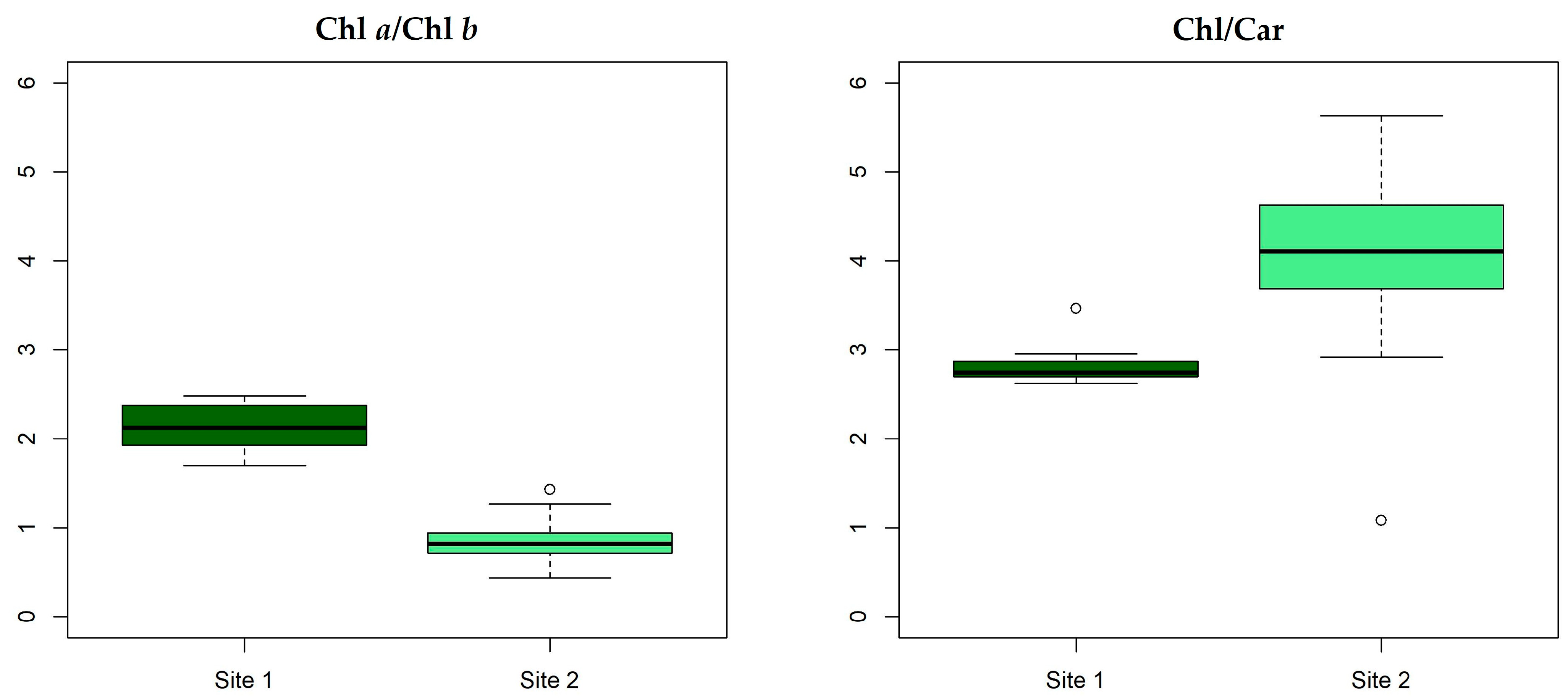
3.2. Biochemical Parameters

The mean total phenol content in P. oceanica leaves was 10.5 ± 3.7 and 11.2 ± 5.0 mg g−1 FW in Site 1 and in Site 2, respectively. No significant differences were found between the plants from the two sites (Welch Two-Sample t-test, p > 0.05).
The mean value of pigments in P. oceanica leaves from the two sites are reported in Figure 2: chlorophyll a (0.41 ± 0.12 and 0.57 ± 0.11 mg g−1 FW, respectively), chlorophyll b (0.20 ± 0.07 and 0.75 ± 0.28 mg g−1 FW, respectively) and carotenoids (0.21 ± 0.06 and 0.32 ± 0.02 mg g−1 FW, respectively) were always lower in Site 1 than in Site 2. Chlorophyll b showed the highest significant difference between the two sites (p = 2.05·10−6), although the Welch Two-Sample t-test revealed significant differences between sites also for chlorophyll a and carotenoids, probably related to the high variability in data distributions found in Site 1 samples (site comparison: Chl a, p = 0.001; Car, p = 1.19 · 10−4).
The chlorophyll ratio (Chl a/Chl b, Figure 3) was significantly higher in Site 1 (2.1 ± 0.26) than in Site 2 (0.9 ± 0.26; Welch Two-Sample t-test: p < 0.001), due to the low values of Chl b in Site 1. Conversely, the total chlorophyll–carotenoids ratio (Chl/Car) was significantly higher in Site 2 than in Site 1 (4.0 ± 0.11 vs. 2.8 ± 0.20, respectively; Welch Two-Sample t-test: p < 0.001; Figure 3).

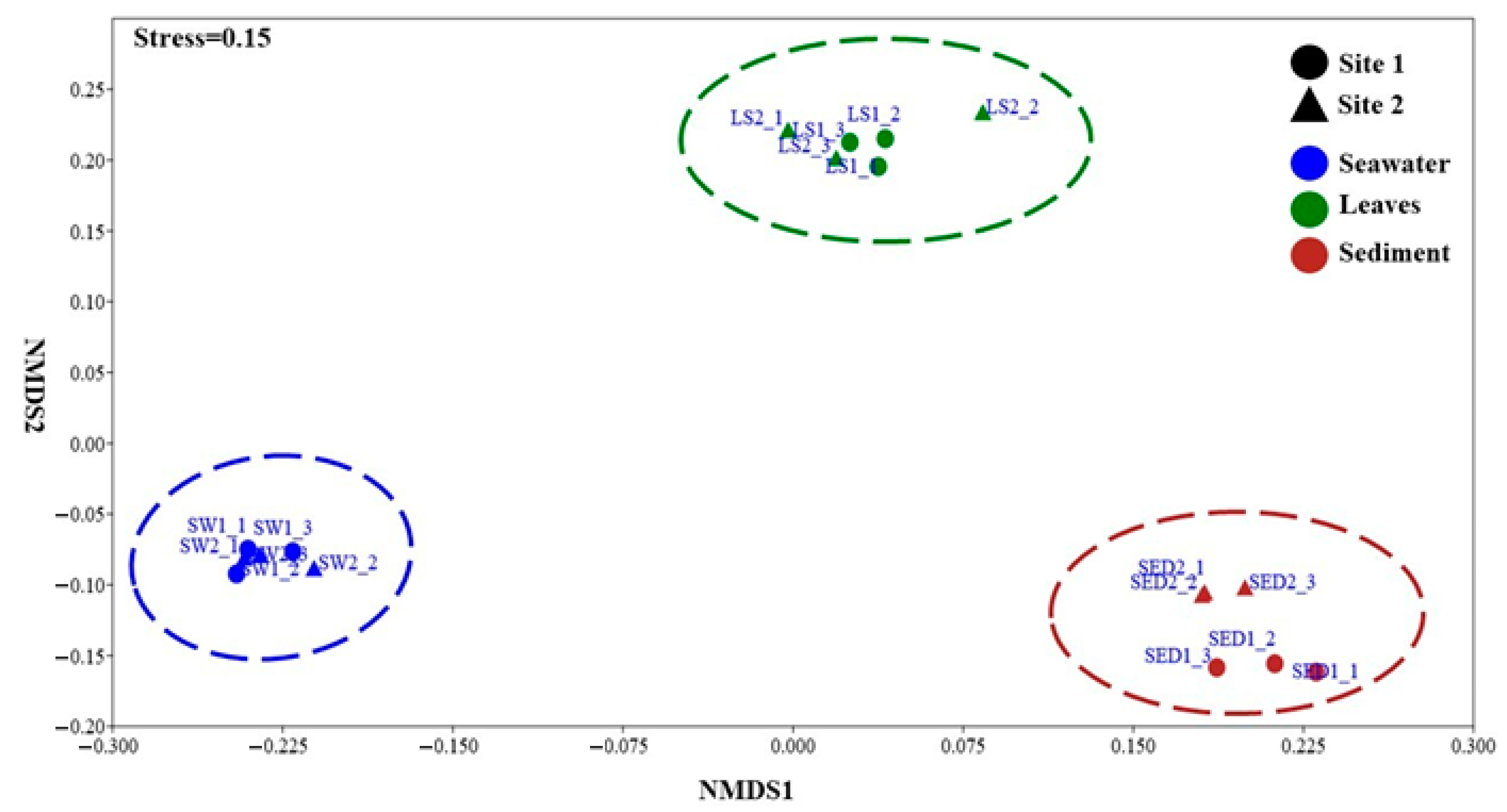
3.3. Microbial Diversity

3.3.1. Bacterial Diversity and Composition

Bacterial OTU clustering (97% similarity) applied to filtered DADA2 ASVs (n = 171,367) resulted in a total of 650 OTUs. Among these sequences, 74,923 (44%), belonged to the sediment samples, 55,619 (32.5%) belonged to seawater samples, while 40,825 (23.8%) belonged to the leaf-associated bacterial communities.
Overall, Site 1 exhibited the highest number of sequences (Table 1) and even the highest Shannon diversity index values. In particular, the highest richness and diversity were observed in the sediment bacterial communities (Limassol port sea stretch). However, α diversity did not showed significant differences between the Site 1 and 2, except for the sediments (the Kruskal–Wallis test followed by Dunn’s post hoc analysis; p = 0.01; Table 1).
The n-MDS plot showed (Figure 4) clearly distinct bacterial communities among seawater, sediment, and leaf. Differences were significant among sample types (PERMANOVA test; Bray–Curtis, pseudo-F = 2.24, p = 0.001; Unweighted UniFrac, pseudo-F = 3.14, p = 0.001), but not between the two sites, whose samples clustered together according to sample type (PERMANOVA test, Site 1 vs. Site 2; Bray–Curtis, pseudo-F = 0.96; p > 0.05; Unweighted UniFrac, pseudo-F = 1.57, p > 0.05). Furthermore, leaf bacterial communities exhibit an intermediate structure between seawater and sediment bacterial communities.
Among the 650 detected OTUs (Figure 5; Supplementary Material Table S4), Pseudomonadota and Bacteroidota phyla were dominant and distributed across all samples, accounting for 60% and 16% of all observed bacterial sequences, respectively (Pseudomonadota: 33% in Site 1 and 27% in Site 2; Bacteroidota: 8% in both sites). Less abundant, the phylum Desulfobacterota (6% relative abundance) was mostly found in sediment samples (5% in Site 1 and 1% in Site 2). Among the bacterial taxa, 295 (42%) were collectively rare, with a relative abundance of less than 2%; they were distributed as follows: 12.3% in seawater Site 1 (SW1; mean number of OTUs = 41.7 ± 21.5; mean number of sequences = 3839.0 ± 2388.2) and 8.8% in seawater Site 2 (SW2; mean number of OTUs = 23.0 ± 5.6; mean number of sequences = 1814.7 ± 513.4), 43.4% in sediment Site 1 (SED1; mean number of OTUs = 98.7 ± 19.5; mean number of sequences = 10,313.3 ± 2525.5) and 22.4% in sediment Site 2 (SED2; mean number of OTUs = 37.0 ± 7.5; mean number of sequences = 3048.0 ± 614.2), 18.2% in leaf Site 1 (LEAF1; mean number of OTUs = 38 ± 30; mean number of sequences = 3237.3 ± 3204.7) and 16.1% in leaf Site 2 (LEAF2; mean number of OTUs = 18.3 ± 1.1; mean number of sequences = 1067 ± 27.6).
Within the Pseudomonadota, the Rhodobacteraceae family was the most abundant across seawater, sediment and leaf samples, with the highest percentage in leaves (28% relative abundance at Site 1 and 23% at Site 2). Other prevalent taxa were differently distributed across samples. In seawater, OTUs including AEGEAN-169_marine_group, SAR86 or SAR116_clade, Flavobacteriaceae, and the Cryomorphaceae family were abundant (Figure 5). Their relative abundances were similar in both sites, except for the Cryomorphaceae family, which showed a relative abundance of less than 1% at Site 1 but 16% at Site 2. Conversely, the OTU AEGEAN-169 had a relative abundance of 27% at Site 1 and 21% at Site 2. The SAR86 and SAR116_clade had both a relative abundance of 13 or 11%, respectively, at Site 1 or 2, while the Flavobacteraceae family had 17% relative abundance at both sites. The sediment-associated bacterial community was mainly represented by Gammaproteobacteria, including Vibrionaceae, Nitrincolaceae, and Fusibacteraceae (Figure 5). These families, alongside Rhodobacteraceae, collectively accounted for approximately 35% and over 70% of the overall relative abundance of the sediment-associated bacterial community at Site 1 and Site 2, respectively. Additionally, specific taxa were observed at each site, with Site 1 exhibiting the highest abundance of unique taxa (45%). Among the leaf-associated bacterial community, OTUs such as Microtrichaceae, Hyphomonadaceae, Saprospiraceae were prevalent across both sites (Figure 5). Their relative abundances differed between the sites: Hyphomonadaceae, 13% and 7%; Microtrichaceae, 9% and 21%; Saprospiraceae, 11% and 14%, respectively, in Site 1 and Site 2. The Granulosicoccaceae family maintained a consistent relative abundance of 4% across both sites.

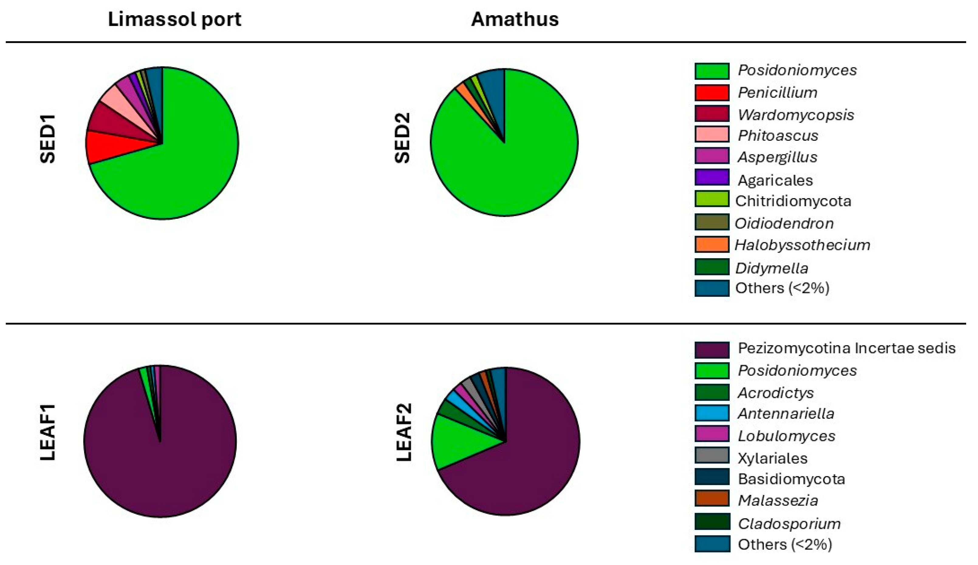
3.3.2. Fungal Diversity and Composition

Fungal OTU clustering (97% similarity) applied to filtered DADA2 ASVs (n = 157,653) resulted in a total of 80 OTUs. Among these sequences, the fungal community associated with the sediment had the highest number of sequences: 94,559 (60%) while 63,094 (40%), belonged to leaf samples. No fungal DNA was detected/extracted in/from seawater samples, probably because of the volume collected (1 L) not enough for yielding sufficient DNA for PCR amplification. Overall, Site 1 had a higher number of fungal sequences than Site 2, as found for the associated bacterial component.
Across samples, the fungal community α diversity showed low H’ values with no significant differences in sediments between Sites 1 and 2; conversely, the leaf-associated fungal communities exhibited the highest Shannon index value at Site 2, significantly different from Site 1 (Table 2).
The n-MDS plot showed (Figure 6) clearly distinct fungal community structure and composition not only between sample types (sediment and leaves) but also between sites, displaying four distinct clusters. The differences were significant both between sediment vs. leaf-associated fungal communities (PERMANOVA test; Bray–Curtis, pseudo-F = 5.38, p = 0.001; Unweighted UniFrac, pseudo-F = 2.69, p = 0.001) and between Site 1 vs. Site 2 only by using the Bray–Curtis similarity index (PERMANOVA test; Bray–Curtis, pseudo-F = 2.68, p = 0.02; Unweighted UniFrac, pseudo-F = 1.14, p > 0.05).
Out of the 80 detected fungal OTUs (Figure 7; Supplementary Material Table S4), 18 had a relative abundance higher than 2% in at least one sample: among these 12 belonged to the Ascomycota phylum (accounting for 96% of all sequences), 4 to the Basidiomycota phylum (2% of all sequences) and 2 belonged to the Chytridiomycota phylum (2% of all sequences). Overall, Basidiomycota and Chytridiomycota were more abundant at Site 2 in both sediment and leaves samples, whereas at Site 1, they were found to be collectively rare (<1%).
Overall, two OTUs were dominant: the species Posidoniomyces atricolor, and an unidentified taxon within the class Pezizomycotina incertae sedis. These fungal OTUs were differently distributed across sample types: as a total, P. atricolor was dominant in sediment, but less represented in leaf samples (70% vs. 2% relative abundance at Site 1 and 88% vs. 13% at Site 2, respectively). The unclassified taxon Pezizomycotina incertae sedis, completely absent in the sediment, in the leaf-associated fungal community had 95% relative abundance at Site 1 and 68% at Site 2. Site 1 also included a 2% of rare taxa, comprising 20 taxa with less than 1% relative abundance, while the Site 2, in addition to Posidoniomyces and the unclassified Pezizomycotina incertae sedis, comprised 3% of rare taxa, together with the genera: Acrodictys, Antennariella, Cladosporium, Lobulomyces, Malassezia, and unclassified taxa belonging to the phylum Basidiomycota, including the order Xylariales; their relative abundances ranged from 2 to 3%. Unfortunately, it is currently impossible to confidently classify the Pezizomycotina incertae sedis taxon. This limits the precise evaluation of the actual diversity of the fungi belonging to this group, dominant in the leaves, as well as their roles in relation to the P. oceanica conservation status.
The sediment-associated fungal community was different in the two sites: Site 1 was characterized by the genera, Penicillium (7%), Wardomycopsis (7%), Pithoascus (5%), Aspergillus (3%), Oidiodendron (2%), and the order Agaricales (2%), almost absent at Site 2, whilst Site 2 included Halobyssothecium and Dydimella genera and taxa of the phylum Chytridiomycota (all at 2% relative abundance).

4. Discussion

We evaluated the ecological status of two Posidonia oceanica meadows in the Akrotiri Bay (Cyprus) by using ecophysiological and microbial descriptors: the plants thrived under different habitat features (muddy sand vs. historic remnants intermingled with muddy sand seabed) and depth (medium vs. shallow depth) and were subjected to different degrees of human pressure. Site 1 is adjacent to Limassol port; hence plants were subjected to the associated activities (i.e., boat traffic, anchoring or mooring), but are also in the proximity of an aquaculture fish farm. Conversely, Site 2 is located within the Amathus Marine Protected Area, a site under low human pressure, as many activities are forbidden (fishing, anchoring or mooring, navigation, etc.).
During a previous sampling carried out in December 2017, at the same sites (Site 1, published by Conte et al. [53]; Site 2 published by Rotini et al. [54]), we assessed the same set of descriptors including morphometry, biochemical analyses and leaf-associated bacterial community. Table 3 summarize the comparison of the morphometric and biochemical data acquired in the two sampling campaigns; results are discussed in Section 4.1. The results regarding the leaf-associated bacterial communities are compared and discussed in Section 4.2.
The novelty introduced in the current study has been to include the fungal component in the set of descriptors as fungi are currently underrepresented in the assessment of P. oceanica-associated microbial communities. The potentiality of fungal community in depicting the conservation status of the plants is discussed in Section 4.3.

4.1. P. oceanica Morphometric and Biochemical Descriptors

In the 2023 sampling campaign, morphometric analyses did not highlight differences between the plants collected in the two sites, but the comparison of the results obtained in the two sampling campaign gave a different picture after 6 years, particularly for the Limassol meadows: while at Site 2 leaf area values were comparable to those collected in 2017 [54], at Site 1 leaf area values were higher than those collected in 2017 [53]. Is worth to note that in 2017 the co-occurrence of the exotic Halophila stipulacea in syntopy (sensu Rivas [69]) was recorded in Site 1. This latter result suggests that morphometric descriptors may have been affected by interspecific interactions, as found by Conte et al. [70] and Mannino et al. [71] in eastern and southern Mediterranean Sea for the interaction between Cymodocea nodosa and H. stipulacea.
Also, the biochemical analyses yielded the same picture of the morphometric descriptors. Total phenol content, a putative marker of environmental stress [58,72], in this study was found quite low at both sites and not significantly different between sites. If compared to the concentrations measured in 2017, the phenol values were found to be reduced in both sites. In Limassol port, phenols were reduced by a factor of 4 (Table 3), suggesting a significant improvement of the conditions evaluated in 2017 [53]. This reduced phenol content may be related to reduced competition: as already reported, in 2023, H. stipulacea plants were found in the area and H. stipulacea may act as a competitor, triggering the increase in phenols in seagrasses [70,71]. Photosynthetic pigment contents (Chl a, Chl b, and Car) were found comparable among sites and with the results obtained in 2017. The main difference was recorded for chlorophyll b, which showed a very high value at Amathus in this study, higher than those found at both sites in 2017. This difference could be attributed to the highest light regime in Amathus, due to the shallow depth (2–3 vs. 7–9 m in Limassol port) and to the sampling season (July in 2023 vs. December in 2017). Both differences may depend on the adaptive response/acclimatization of the P. oceanica photosynthetic system to depth and season [73,74], including a chlorophyll b-carotenoids increase to provide protection from free radicals and oxidative damage [75,76].
Overall, the morphometric and biochemical markers of the P. oceanica plants, in 2023, indicated a plant similar eco-physiological condition in the two sites, notwithstanding the geomorphological differences and the human activities.

4.2. P. oceanica-Associated Bacterial Communities

The bacterial community, proposed as a further descriptor of the conservation status of P. oceanica [15,50,53,54], has been evaluated in the abiotic matrices (seawater and sediment) and leaves but not in rhizomes/roots in both sampling campaigns for a conservation issue, as it implies destructive sampling.
In the 2023 sampling campaign, as expected, differences in the associated bacterial communities were found among the different type of samples, i.e., seawater, sediment and leaves, but the α diversity of each type of associated bacterial community did not change significantly between sites. The unique significant difference in the Shannon diversity index was found in the sediment-associated bacterial component from Site 1, the high value depending on the low-abundant taxa, as the rare bacterial colonizers (each with a relative abundance < 2%) accounted for the majority of the sequences (SED1 = ∼60%). This difference may be due to the different sediment features, and/or to human-related activities which may condition the microbial richness [77]; in the harbor area it is easy to hypothesize an enrichment of the seabed.
Also, the β diversity showed comparable bacterial community composition in both Sites, according to sample type. In seawater, the components mainly belonged to marine phototrophic and heterotrophic bacterioplankton, while in sediment, the dominant associated bacteria belonged to Vibrionaceae, Nitrincolaceae, and Fusibacteraceae, all playing essential roles in nutrient cycling, organic matter degradation in marine sediments and P. oceanica meadows (for the main putative metabolic traits of the dominant taxa, see Table 4). In both sites, the main leaf-associated taxa included Rhodobacteraceae, Hyphomonadaceae, Microtrichaceae, and Saprospiraceae families, already described as P. oceanica bacterial core at the Amathus site in the previous sampling campaign by Rotini et al. [54]. Differently, in the current sampling, Thalassospiraceae and Sphingomonadaceae families were found as rare components of the P. oceanica leaf-associated bacterial community; these taxa were much more abundant in the 2017 sampling [54]. Again, in/on the leaves, the bacterial core families were involved in essential processes for the host plant, as nutrient cycling or organic matter degradation (Table 4).
The most important result of the 2023 sampling campaign regards the change in the leaf-associated bacterial community at Limassol port. In the present study, this community extensively differed from the one found by Conte et al. [53] at the same site, with a dominance of Gammaproteobacteria and the presence of specific taxa, as Burkholderiales, both indicating impacted environment or stressed meadow [95,96,97,98]. These taxa were rare or absent in the 2023 samples. By the same token, the different leaf-associated bacterial community composition and plant descriptor values (i.e., morphometry and total phenol content) may be related to a low environmental quality in 2017, as already inferred by Conte et al. [53]. This low environmental quality may have favored the co-occurrence of H. stipulacea in the site, as it is a small r-strategist species, characterized by high invasive capability, which allows it to rapidly colonize new environments, but also to rapidly disappear. The occurrence of H. stipulacea triggered competitive/negative interaction with P. oceanica and, among the possible environmental conditions favoring H. stipulacea in Site 1, there was an aquaculture fish-farm, known to release ammonium in the seawater column, which is efficiently assimilated by H. stipulacea and may facilitate its expansion [99].
Although no significant changes in harbor or fish farming activities have been made public in the last years, in this sampling campaign, no one H. stipulacea plant was found in Site 1, surmising an overall change in environmental conditions. The absence of the competitor may have triggered the improvement of P. oceanica morphometric and biochemical descriptors. Also the P. oceanica-associated bacterial communities in Limassol port have changed, as seagrass-associated bacterial communities are known to rapidly adapt to plant ecophysiology and environmental changes, modifying their structure and composition [100,101]. They changed significantly in Limassol port to become similar to those found in the Amathus site, both in this and in the previous study. This change further supports the surmise of an improvement of P. oceanica condition at Limassol port, possibly aided by the Cyprus open sea conditions, as water circulation can play a significant role in reducing localized impacts on marine ecosystems [102,103].
The change is clearly one of the main results of the present study, showing clear differences of the P. oceanica-associated bacterial communities in Limassol port, between the two sampling campaigns, six years apart. The concordance of the bacterial community in both the two sites in the present study, and in the Amathus archaeological area in the two sampling campaigns, suggests a comparable conservation status of P. oceanica and further confirms that, in the absence of disturbances, P. oceanica maintains characteristic associated bacterial communities even under different environmental features, as already stated by Rotini et al. [54]. The capability of the associated bacteria to thrive under different environmental features, as found in Limassol port and Amathus, may be related to their metabolic versatility [104], which allows to preserve taxonomic diversity by adjusting metabolic activities under different environmental conditions [104,105].

4.3. P. oceanica-Associated Fungal Communities

Most of the studies on the microorganisms associated with seagrass have focused almost exclusively on bacterial communities, neglecting the fungal counterparts [106,107,108,109,110], notwithstanding the well-known pivotal role of plant–fungal interactions [111,112]. This is critical also for P. oceanica and is the reason for including in this study the fungal communities in the evaluation of the meadow features.
Unfortunately, fungi cannot yet be considered as putative seagrass descriptors, even for the scarcity of information on the mycobiota in marine systems. In fact, there are still some difficulties to pursue this goal, as high-throughput amplicon sequencing of mycobiome is still in its infancy; there is a lack of representation of fungal sequences in public databases, other than the lack of standardized protocol/technical issues: for example, fungal sequencing of seawater samples did not produce outcomes in open sea, probably due to the limited seawater volume sampled (1 L), critical for collecting fungal diversity by DNA metabarcoding techniques [113], although this sampling volume had already worked in other environmental conditions [114].
The analysis of P. oceanica-associated fungal communities returned narrow assemblages, showing low Shannon index values in all samples, as already found by Poli et al. [115]. The most common fungi found at both sites were the key members of P. oceanica fungal community [29,35,115]: Posidoniomyces atricolor, dominant in sediment, and Pezizomycotina incertae sedis class, dominant in leaves (Table 5), together with Cladosporium genus, present in leaves, and Aspergillus, Penicillium or Wardomycopsis genera in the sediment.
Nevertheless, fungal distribution showed interesting features: both in sediment and in leaves, P. atricolor was found more abundant in Site 2 than in Site 1, and significant differences in α diversity were found in leaf-associated communities between sites. In leaves, Site 2 showed the highest Shannon index for fungal communities, depending on the presence of several less abundant fungal colonizers, along with the two dominant fungal taxa. In addition to the different types of seabed, in Site 2, the high α diversity may be related to the shallow and clear waters, which allow for high light penetration, enhancing the P. oceanica photosynthetic activity and synthesis of organic compounds, such as leaf exudates, which might provide a rich substrate for several fungal colonizers [116]. Conversely, in sediment, Shannon index for fungal communities was higher in Site 1 than in Site 2, although difference was not significant, as fungi are known to show direct correlation with organic carbon concentration, consistent with a saprophytic lifestyle [28,117].
As clearly shown in Figure 7, the main differences between sites depended on the less common fungi, unique to each site, defining distinct fungal communities in leaf or sediment samples. This may be related with the site features and/or environmental conditions, influencing composition and abundance of fungal communities, especially in the leaf samples [118]. However, the dominant fungi also contributed to the differences in β diversity, as the relative abundance of Posidoniomyces atricolor was found clearly reduced at Limassol port in both leaf and sediment samples. The observed reduction in the relative abundance of P. atricolor at Site 1 agrees with findings from the Villefranche-sur-Mer bay, showing a similar reduced relative abundance in leaves at the impacted site [114]. The common response across different Mediterranean sea stretch suggests that P. atricolor may be sensitive to environmental changes and could potentially serve as a descriptor of ecological shifts. Furthermore, while P. atricolor was known to be a specific colonizer of P. oceanica roots [35,44], in this study and in the other on the Mediterranean site of Villefranche-sur-Mer (France), it was also found in leaf samples [114], confirming its strong relationship with the P. oceanica host.
The same tight relationship can be hypothesized for Pezizomycotina incertae sedis, the dominant taxon in P. oceanica leaves, whose relative abundance increased alongside the reduction in P. atricolor. This trend has also been found in an impacted site at the Villefranche-sur-Mer bay, indicating that the reduction in P. atricolor may facilitate the prevalence of Pezizomycotina incertae sedis. This taxon has been already found in P. oceanica [35] but is known to inhabit the leaves of terrestrial plants [119,120] and to behave as an opportunistic pathogen of plants or animals [121,122].
Table 5. The two dominant fungal taxa which characterize P. oceanica sediment and leaf samples, and their putative metabolic traits or functions.
Fungal TaxaPutative FunctionsReferences
Posidoniomyces atricolorPutative Dark Septate Endophyte (DSE) not reported from other hosts or ecosystems, indicating a specific adaptation to the marine environment and P. oceanica host. It is supposed to be involved in host growth, nutrient acquisition, abiotic stress tolerance, and also in decomposing organic material. It constitutes one of the most important microorganisms by abundance that degrade P. oceanica tissues within the matte [35,123]
Pezizomycotina incertae sedisFilamentous ascomycetes with septate hypha, known to be saprotrophs involved in the decomposition of plant materials, contributing to the breakdown of complex carbohydrates in leaves. Opportunistic animal or plant pathogens.[122,124]
It is worth to note that all the dominant fungal colonizers belong to the Ascomycota phylum, which is commonly found in marine environments associated with seagrass, including P. oceanica [29,115]. By contrast, in marine habitats, Basidiomycota are under-represented compared to Ascomycota [125,126,127], despite the fact that they were already reported in seagrasses, mangroves, as well as in algae and animals [128].
Overall, unlike bacterial community, fungi showed significant differences in both structure and taxonomic composition between sites, suggesting a major sensitivity to different environmental conditions, as observed in the terrestrial environment [129]. These results further suggest a role for fungal communities as effective putative descriptor of P. oceanica conservation status and highlights the importance of integrating fungal community data into conservation strategies, and the need to implement the core knowledge about these communities. Nevertheless, the critical aspect of estimating fungal diversity and composition—and consequently their use as ecological descriptors—lies in the lack of extensive studies, sampling efforts, classification systems, and reference sequences in public databases. Despite the limitations, emerging research highlighted the potential of fungi as ‘mycoindicators’, a valuable tool for ecological assessment, as these organisms may offer insights into environmental changes, community dynamics, and ecosystem health [130].

5. Conclusions

Due to the increasing human-induced environmental changes in coastal ecosystems, there is a growing concern about the conservation status of seagrass meadows worldwide. The conventional descriptors employed to evaluate P. oceanica ecological status indicated a condition that was largely consistent across the two sites examined, characterized by slight variations that did not suggest notable environmental stressors. This condition is different from that found in Limassol port in the 2017 sampling campaign, and is characterized by the absence of Halophila stipulacea, a signal of reduced competition and environmental stress for P. oceanica. In contrast, the fungal community, which has not previously been included in these assessments, exhibited pronounced changes in response to specific environmental conditions. This underscores a level of sensitivity within the fungal community that is not captured by conventional descriptors, highlighting the importance of gaining a more comprehensive understanding of their variability patterns in relation to host eco-physiological conditions and the surrounding environmental contexts.
Posidoniomyces atricolor showed a detectable reduction in abundance at sites potentially impacted by human activities, independent of the meadow’s acclimatization to specific environmental conditions. The recognized specificity of P. atricolor to P. oceanica, combined with its potential role as an endosymbiont, suggests that P. atricolor may serve as a useful ‘mycoindicator’ in future ecological assessments. While this work is still highly preliminary due to existing limitations, it clearly highlights the need for further investigation.

Supplementary Materials

The following supporting information can be downloaded at: https://www.mdpi.com/article/10.3390/w17081151/s1, Table S1: Environmental parameter values collected from the Bio-ORACLE dataset (v.3.0; mean values over the last ten years) and processed with QGIS (v.3.34.9) at both sampling sites. Table S2: Ranges of DNA concentration (ng/µL) for each sample type and site, extracted using the DNeasy PowerSoil DNA Kit (QIAGEN) and quantified with Nanodrop spectrophotometer. Values represent the lowest and the highest concentration across the three independent replicates per sample type. Table S3: Raw reads, DADA2 output, and final reads obtained after filtering procedures for bacterial (A) and fungal (B) sequences by using QIIME 2. Table S4: OTUs identification. The bacterial (A) or fungal (B) OTUs found at the Limassol Site and the Amathus Site 2 in P. oceanica leaves and abiotic matrices are indicated in bold.

Author Contributions

Conceptualization, L.M. (Luciana Migliore), M.I.V. and G.W.; underwater diving for samples collection: A.A., M.K. and G.W.; material preparation and data collection: S.F., L.M. (Luciana Migliore), A.A., L.M. (Loredana Manfra), M.I.V., E.C., G.W. and M.K.; data analysis: S.F. and M.M.D., particularly for bioinformatics; funding and material acquisition: L.M. (Luciana Migliore), M.I.V. and G.W.; writing—original draft preparation: S.F., L.M. (Luciana Migliore) and A.R. All authors have read and agreed to the published version of this manuscript.

Funding

Sara Frasca and Annamaria Alabiso (from the XXXVII and XXXVI cycles, respectively) were funded by a three-year fellowship from the Ph.D. School of Evolutionary Biology and Ecology at Tor Vergata Rome University. Sara Frasca held a PON doctorate in collaboration with GT50 s.r.l., while Annamaria Alabiso obtained an industrial doctorate in partnership with AlgaRes s.r.l. The ISCRA CINECA Project Class C on the Galileo100 server, titled ’MyTOS,’ and ‘FUN-SEA’ granted to Sara Frasca (HP10CIUIH7 and HP10CQP3QE projects), enabled the data elaboration.

Data Availability Statement

The raw data generated for 16S and ITS2 amplicon project have been deposited in the NCBI Sequence Read Archive (SRA) under BioProject ID: PRJNA1151666 and PRJNA1151697, respectively.

Acknowledgments

The authors are grateful to Fernanda Oliva Pintucci (São Carlos School of Engineering, University of São Paulo, Brasil) for helping us with laboratory activities. We acknowledge the CINECA award under the ISCRA initiative, for the availability of high-performance computing resources and support.

Conflicts of Interest

The authors declare no conflicts of interest.

References

  1. Trivedi, P.; Batista, B.D.; Bazany, K.E.; Singh, B.K. Plant–microbiome interactions under a changing world: Responses, consequences and perspectives. New Phytol. 2022, 234, 1951–1959. [Google Scholar] [CrossRef] [PubMed]
  2. Rosenberg, E.; Koren, O.; Reshef, L.; Efrony, R.; Zilber-Rosenberg, I. The hologenome theory disregards the coral holobiont: Reply from Rosenberg et al. Nat. Rev. Microbiol. 2007, 5, 826–826. [Google Scholar] [CrossRef]
  3. Zilber-Rosenberg, I.; Rosenberg, E. Role of microorganisms in the evolution of animals and plants: The hologenome theory of evolution. FEMS Microbiol. Rev. 2008, 32, 723–735. [Google Scholar] [CrossRef]
  4. Vandenkoornhuyse, P.; Quaiser, A.; Duhamel, M.; Le Van, A.; Dufresne, A. The importance of the microbiome of the plant holobiont. New Phytol. 2015, 206, 1196–1206. [Google Scholar] [CrossRef] [PubMed]
  5. Hardoim, P.R.; Van Overbeek, L.S.; Berg, G.; Pirttilä, A.M.; Compant, S.; Campisano, A.; Sessitsch, A. The hidden world within plants: Ecological and evolutionary considerations for defining the functioning of microbial endophytes. MMBR 2015, 79, 293–320. [Google Scholar] [CrossRef]
  6. Bettenfeld, P.; Canals, J.C.; Jacquens, L.; Fernandez, O.; Fontaine, F.; Van Schaik, E.; Trouvelot, S. The microbiota of the grapevine holobiont: A key component of plant health. J. Adv. Res. 2022, 40, 1–15. [Google Scholar] [CrossRef]
  7. Chen, J.; Zang, Y.; Yang, Z.; Qu, T.; Sun, T.; Liang, S.; Tang, X. Composition and functional diversity of epiphytic bacterial and fungal communities on marine macrophytes in an intertidal zone. Front. Microbiol. 2022, 13, 839465. [Google Scholar] [CrossRef]
  8. Jones, P.; Garcia, B.J.; Furches, A.; Tuskan, G.A.; Jacobson, D. Plant host-associated mechanisms for microbial selection. Front. Plant Sci. 2019, 10, 862. [Google Scholar] [CrossRef]
  9. Bulgarelli, D.; Schlaeppi, K.; Spaepen, S.; Van Themaat, E.V.L.; Schulze-Lefert, P. Structure and functions of the bacterial microbiota of plants. Annu. Rev. Plant Biol. 2013, 64, 807–838. [Google Scholar] [CrossRef]
  10. Ugarelli, K.; Chakrabarti, S.; Laas, P.; Stingl, U. The seagrass holobiont and its microbiome. Microorganisms 2017, 5, 81. [Google Scholar] [CrossRef]
  11. Martin, F.M.; Uroz, S.; Barker, D.G. Ancestral alliances: Plant mutualistic symbioses with fungi and bacteria. Science 2017, 356, 4501. [Google Scholar] [CrossRef] [PubMed]
  12. Thomson, B.C.; Tisserant, E.; Plassart, P.; Uroz, S.; Griffiths, R.I.; Hannula, S.E.; Lemanceau, P. Soil conditions and land use intensification effects on soil microbial communities across a range of European field sites. Soil Biol. Biochem. 2015, 88, 403–413. [Google Scholar] [CrossRef]
  13. Hassani, M.A.; Durán, P.; Hacquard, S. Microbial interactions within the plant holobiont. Microbiome 2018, 6, 58. [Google Scholar] [CrossRef] [PubMed]
  14. Bettenfeld, P.; Fontaine, F.; Trouvelot, S.; Fernandez, O.; Courty, P.E. Woody plant declines: What’s wrong with the microbiome? Trends Plant Sci. 2020, 25, 381–394. [Google Scholar] [CrossRef]
  15. Conte, C.; Rotini, A.; Manfra, L.; D’Andrea, M.M.; Winters, G.; Migliore, L. The seagrass holobiont: What we know and what we still need to disclose for its possible use as an ecological indicator. Water 2021, 13, 406. [Google Scholar] [CrossRef]
  16. Lyu, D.; Zajonc, J.; Pagé, A.; Tanney, C.A.; Shah, A.; Monjezi, N.; Smith, D.L. Plant holobiont theory: The phytomicrobiome plays a central role in evolution and success. Microorganisms 2021, 9, 675. [Google Scholar] [CrossRef]
  17. Omae, N.; Tsuda, K. Plant-microbiota interactions in abiotic stress environments. MPMI 2022, 35, 511–526. [Google Scholar] [CrossRef]
  18. Baharum, S.N.; Beng, E.K.; Mokhtar, M.A.A. Marine microorganisms: Potential application and challenges. J. Biol. Sci. 2010, 10, 555–564. [Google Scholar] [CrossRef]
  19. Batista, D.; Costa, R.; Carvalho, A.P.; Batista, W.R.; Rua, C.P.; de Oliveira, L.; Leomil, L.; Fròes, A.M.; Thompson, F.L.; Coutinho, R.; et al. Environmental conditions affect activity and associated microorganisms of marine sponges. Mar. Environ. Res. 2018, 142, 59–68. [Google Scholar] [CrossRef]
  20. Vitorino, L.C.; Bessa, L.A. Microbial diversity: The gap between the estimated and the known. Diversity 2018, 10, 46. [Google Scholar] [CrossRef]
  21. Qiu, Z.; Coleman, M.A.; Provost, E.; Campbell, A.H.; Kelaher, B.P.; Dalton, S.J.; Thomas, T.; Steinberg, P.D.; Marzinelli, E.M. Future climate change is predicted to affect the microbiome and condition of habitat-forming kelp. Proc. Biol. Sci. 2019, 13, 20181887. [Google Scholar] [CrossRef] [PubMed]
  22. Compant, S.; Duffy, B.; Nowak, J.; Clément, C.; Barka, E.A. Use of plant growth-promoting bacteria for biocontrol of plant diseases: Principles, mechanisms of action, and future prospects. Appl. Environ. Microbiol. 2005, 71, 4951–4959. [Google Scholar] [CrossRef] [PubMed]
  23. Hayat, R.; Ali, S.; Amara, U.; Khalid, R.; Ahmed, I. Soil beneficial bacteria and their role in plant growth promotion: A review. Ann. Microbiol. 2010, 60, 579–598. [Google Scholar] [CrossRef]
  24. Tarquinio, F.; Hyndes, G.A.; Laverock, B.; Koenders, A.; Säwström, C. The seagrass holobiont: Understanding seagrass-bacteria interactions and their role in seagrass ecosystem functioning. FEMS Microbiol. Lett. 2019, 366, fnz057. [Google Scholar] [CrossRef]
  25. Rajakaruna, O.; Wijayawardene, N.N.; Udagedara, S.; Jayasinghe, P.K.; Gunasekara, S.S.; Boonyuen, N.; Bamunuarachchige, T.C.; Ariyawansa, K.G.S.U. Exploring fungal diversity in seagrass ecosystems for pharmaceutical and ecological insights. J. Fungus 2024, 10, 627. [Google Scholar] [CrossRef]
  26. Tasdemir, D.; Scarpato, S.; Utermann-Thüsing, C.; Jensen, T.; Blümel, M.; Wenzel-Storjohann, A.; Echelmeyer, V.A. Epiphytic and endophytic microbiome of the seagrass Zostera marina: Do they contribute to pathogen reduction in seawater? Sci. Total Environ. 2024, 908, 168422. [Google Scholar] [CrossRef]
  27. Jones, E.G. Are there more marine fungi to be described? Bot. Mar. 2011, 54, 343–354. [Google Scholar] [CrossRef]
  28. Amend, A.; Burgaud, G.; Cunliffe, M.; Edgcomb, V.P.; Ettinger, C.L.; Gutiérrez, M.H.; Gladfelter, A.S. Fungi in the marine environment: Open questions and unsolved problems. MBio 2019, 10, 10–1128. [Google Scholar] [CrossRef]
  29. Panno, L.; Bruno, M.; Voyron, S.; Anastasi, A.; Gnavi, G.; Miserere, L.; Varese, G.C. Diversity, ecological role and potential biotechnological applications of marine fungi associated to the seagrass Posidonia oceanica. New Biotechnol. 2013, 30, 685–694. [Google Scholar] [CrossRef]
  30. Torta, L.; Lo Piccolo, S.; Piazza, G.; Burruano, S.; Colombo, P.; Ottonello, D.; Calvo, S. Lulwoana sp., a dark septate endophyte in roots of Posidonia oceanica (L.) Delile seagrass. Plant Biol. 2015, 17, 505–511. [Google Scholar] [CrossRef]
  31. Venkatachalam, A.; Govinda Rajulu, M.B.; Thirunavukkarasu, N.; Suryanarayanan, T.S. Endophytic fungi of marine algae and seagrasses: A novel source of chitin modifying enzymes. Mycosphere 2015, 6, 345–355. [Google Scholar] [CrossRef]
  32. Venkatachalam, A.; Thirunavukkarasu, N.; Suryanarayanan, T.S. Distribution and diversity of endophytes in seagrasses. Fungal Ecol. 2015, 13, 60–65. [Google Scholar] [CrossRef]
  33. Vohník, M.; Borovec, O.; Kolařík, M. Communities of cultivable root mycobionts of the seagrass Posidonia oceanica in the northwest Mediterranean Sea are dominated by a hitherto undescribed pleosporalean dark septate endophyte. Microb. Ecol. 2016, 71, 442–451. [Google Scholar] [CrossRef]
  34. Ettinger, C.L.; Eisen, J.A. Fungi, bacteria and oomycota opportunistically isolated from the seagrass, Zostera marina. PLoS ONE 2020, 15, e0236135. [Google Scholar] [CrossRef] [PubMed]
  35. Vohník, M.; Borovec, O.; Kolaříková, Z.; Sudová, R.; Réblová, M. Extensive sampling and high-throughput sequencing reveal Posidoniomyces atricolor gen. et sp. nov. (Aigialaceae, Pleosporales) as the dominant root mycobiont of the dominant Mediterranean seagrass Posidonia oceanica. MycoKeys 2019, 55, 59–86. [Google Scholar] [CrossRef]
  36. Vohník, M.; Josefiová, J. Novel epiphytic root-fungus symbiosis in the Indo-Pacific seagrass Thalassodendron ciliatum from the Red Sea. Mycorrhiza 2024, 34, 447–461. [Google Scholar] [CrossRef]
  37. Wang, X.; Pecoraro, L.; Chen, J.; Tang, Y.; Lee, S.; Chen, S.; Liu, H. Halophilomyces hongkongensis, a novel species and genus in the Lulworthiaceae with antibacterial potential, colonizing the roots and rhizomes of the seagrass Halophila ovalis. J. Fungi 2024, 10, 474. [Google Scholar] [CrossRef]
  38. Boudouresque, C.F.; Mayot, N.; Pergent, G. The outstanding traits of the functioning of the Posidonia oceanica seagrass ecosystem. Biol. Mar. Medit. 2006, 13, 109–113. [Google Scholar]
  39. Boudouresque, C.F.; Bernard, G.; Bonhomme, P.; Charbonnel, E.; Diviacco, G.; Meinesz, A.; Pergent, G.; Pergent-Martini, C.; Ruitton, S.; Tunesi, L. Protection and Conservation of Posidonia oceanica Meadows; RAMOGE and RAC/SPA: Tunis, Tunisia, 2012. [Google Scholar]
  40. Boudouresque, C.F.; Pergent, G.; Pergent-Martini, C.; Ruitton, S.; Thibaut, T.; Verlaque, M. The necromass of the Posidonia oceanica seagrass meadow: Fate, role, ecosystem services and vulnerability. Hydrobiologia 2016, 781, 25–42. [Google Scholar] [CrossRef]
  41. Pergent-Martini, C.; Pergent, G.; Monnier, B.; Boudouresque, C.F.; Mori, C.; Valette-Sansevin, A. Contribution of Posidonia oceanica meadows in the context of climate change mitigation in the Mediterranean Sea. Mar. Environ. Res. 2021, 165, 105236. [Google Scholar] [CrossRef]
  42. Hendriks, I.E.; Escolano-Moltó, A.; Flecha, S.; Vaquer-Sunyer, R.; Wesselmann, M.; Marbà, N. Mediterranean seagrasses as carbon sinks: Methodological and regional differences. Biogeosciences 2022, 19, 4619–4637. [Google Scholar] [CrossRef]
  43. Litsi-Mizan, V.; Efthymiadis, P.T.; Gerakaris, V.; Serrano, O.; Tsapakis, M.; Apostolaki, E.T. Decline of seagrass (Posidonia oceanica) production over two decades in the face of warming of the Eastern Mediterranean Sea. New Phytol. 2023, 239, 2126–2137. [Google Scholar] [CrossRef] [PubMed]
  44. Vohník, M. Are lulworthioid fungi dark septate endophytes of the dominant Mediterranean seagrass Posidonia oceanica? Plant Biol. 2022, 24, 127–133. [Google Scholar] [CrossRef]
  45. Vohník, M.; Borovec, O.; Kolařík, M.; Sudová, R. Fungal root symbionts of the seagrass Posidonia oceanica in the central Adriatic Sea revealed by microscopy, culturing and 454-pyrosequencing. Mar. Ecol. Prog. Ser. 2017, 583, 107–120. [Google Scholar] [CrossRef]
  46. Boudouresque, C.F.; Bernard, G.; Pergent, G.; Shili, A.; Verlaque, M. Regression of Mediterranean seagrasses caused by natural processes and anthropogenic disturbances and stress: A critical review. Bot. Mar. 2009, 52, 395–418. [Google Scholar] [CrossRef]
  47. Díaz-Almela, E.; Marbà, N.; Álvarez, E.; Santiago, R.; Holmer, M.; Grau, A.; Duarte, C.M. Benthic input rates predict seagrass (Posidonia oceanica) fish farm-induced decline. Mar. Pollut. Bull. 2008, 56, 1332–1342. [Google Scholar] [CrossRef]
  48. Marbà, N.; Díaz-Almela, E.; Duarte, C.M. Mediterranean seagrass (Posidonia oceanica) loss between 1842 and 2009. Biol. Conserv. 2014, 176, 183–190. [Google Scholar] [CrossRef]
  49. Rotini, A.; Belmonte, A.; Barrote, I.; Micheli, C.; Peirano, A.; Santos, R.O.; Migliore, L. Effectiveness and consistency of a suite of descriptors for assessing the ecological status of seagrass meadows (Posidonia oceanica L. Delile). Estuar. Coast. Shelf Sci. 2013, 130, 252–259. [Google Scholar] [CrossRef]
  50. Mejia, A.Y.; Rotini, A.; Lacasella, F.; Bookman, R.; Thaller, M.C.; Shem-Tov, R.; Migliore, L. Assessing the ecological status of seagrasses using morphology, biochemical descriptors and microbial community analyses. A study in Halophila stipulacea (Forsk.) Aschers meadows in the northern Red Sea. Ecol. Indic. 2016, 60, 1150–1163. [Google Scholar] [CrossRef]
  51. Roca, G.; Alcoverro, T.; Krause-Jensen, D.; Balsby, T.J.S.; van Katwijk, M.M.; Marbà, N.; Romero, J. Response of seagrass indicators to shifts in environmental stressors: A global review and management synthesis. Ecol. Indic. 2016, 63, 310–323. [Google Scholar] [CrossRef]
  52. Glasl, B.; Webster, N.S.; Bourne, D.G. Microbial indicators as a diagnostic tool for assessing water quality and climate stress in coral reef ecosystems. Mar. Biol. 2017, 164, 1–18. [Google Scholar] [CrossRef]
  53. Conte, C.; Rotini, A.; Winters, G.; Vasquez, M.I.; Piazza, G.; Kletou, D.; Migliore, L. Elective affinities or random choice within the seagrass holobiont? The case of the native Posidonia oceanica (L.) Delile and the exotic Halophila stipulacea (Forssk.) Asch. from the same site (Limassol, Cyprus). Aquat. Bot. 2021, 174, 103420. [Google Scholar] [CrossRef]
  54. Rotini, A.; Conte, C.; Winters, G.; Vasquez, M.I.; Migliore, L. Undisturbed Posidonia oceanica meadows maintain the epiphytic bacterial community in different environments. Environ. Sci. Pollut. Res. 2023, 30, 95464–95474. [Google Scholar] [CrossRef] [PubMed]
  55. Ramos Esplá, A.A. Artificial Reefs in the Amathus Bay (Limassol, Cyprus); Technical Report N°06/2005; Department of Fisheries and Marine Research of Cyprus—University of Alicante: Alicante, Spain; pp. 1–108. [CrossRef]
  56. Abràmoff, M.D.; Magalhães, P.J.; Ram, S.J. Image processing with ImageJ. Biophotonics Intern. 2004, 11, 36–42. [Google Scholar]
  57. Wellburn, A.R. The spectral determination of chlorophylls a and b, as well as total carotenoids, using various solvents with spectrophotometers of different resolution. J Plant Physiol. 1994, 144, 307–313. [Google Scholar] [CrossRef]
  58. Migliore, L.; Rotini, A.; Randazzo, D.; Albanese, N.N.; Giallongo, A. Phenols content and 2-D electrophoresis protein pattern: A promising tool to monitor Posidonia meadows health state. BMC Ecol. 2007, 7, 1–8. [Google Scholar] [CrossRef]
  59. Booker, F.L.; Miller, J.E. Phenylpropanoid metabolism and phenolic composition of soybean [Glycine max (L.) Merr.] leaves following exposure to ozone. J. Exp. Bot. 1998, 49, 1191–1202. [Google Scholar] [CrossRef]
  60. Caporaso, J.G.; Lauber, C.L.; Walters, W.A.; Berg-Lyons, D.; Lozupone, C.A.; Turnbaugh, P.J.; Knight, R. Global patterns of 16S rRNA diversity at a depth of millions of sequences per sample. PNAS 2011, 108, 4516–4522. [Google Scholar] [CrossRef]
  61. Tedersoo, L.; Bahram, M.; Põlme, S.; Kõljalg, U.; Yorou, N.S.; Wijesundera, R.; Abarenkov, K. Global diversity and geography of soil fungi. Science 2014, 346, 1256688. [Google Scholar] [CrossRef]
  62. White, T.J.; Bruns, T.; Lee, S.W.T.; Taylor, J. Amplification and direct sequencing of fungal ribosomal RNA genes for phylogenetics. PCR Protoc. A Guide Methods Appl. 1990, 18, 315–322. [Google Scholar]
  63. Bolyen, E.; Rideout, J.R.; Dillon, M.R.; Bokulich, N.A.; Abnet, C.C.; Al-Ghalith, G.A.; Caporaso, J.G. Reproducible, interactive, scalable and extensible microbiome data science using QIIME 2. Nat. Biotechnol. 2019, 37, 852–857. [Google Scholar] [CrossRef] [PubMed]
  64. Callahan, B.J.; McMurdie, P.J.; Rosen, M.J.; Han, A.W.; Johnson, A.J.A.; Holmes, S.P. DADA2: High-resolution sample inference from Illumina amplicon data. Nat. Methods 2016, 13, 581–583. [Google Scholar] [CrossRef] [PubMed]
  65. Quast, C.; Pruesse, E.; Yilmaz, P.; Gerken, J.; Schweer, T.; Yarza, P.; Glöckner, F.O. The SILVA ribosomal RNA gene database project: Improved data processing and web-based tools. NAR 2012, 41, D590–D596. [Google Scholar] [CrossRef] [PubMed]
  66. Kõljalg, U.; Nilsson, R.H.; Abarenkov, K.; Tedersoo, L.; Taylor, A.F.; Bahram, M.; Larsson, K.H. Towards a unified paradigm for sequence-based identification of fungi. Mol. Ecol. 2013, 22, 5271–5277. [Google Scholar] [CrossRef] [PubMed]
  67. Hammer, Ø.; Harper, D.A. Past: Paleontological statistics software package for education and data analysis. Palaeontol. Electron. 2001, 4, 1. [Google Scholar]
  68. Legendre, P.; Legendre, L. Numerical Ecology; Elsevier: Amsterdam, 2012. [Google Scholar]
  69. Rivas, L.R. A reinterpretation of the concepts “Sympatric” and “Allopatric” with proposal of the additional terms “Syntopic” and “Allotopic”. Syst. Zool. 1964, 13, 42–43. [Google Scholar] [CrossRef]
  70. Conte, C.; Apostolaki, E.T.; Vizzini, S.; Migliore, L. A tight interaction between the native seagrass Cymodocea nodosa and the exotic Halophila stipulacea in the Aegean Sea highlights seagrass holobiont variations. Plants 2023, 12, 350. [Google Scholar] [CrossRef]
  71. Mannino, A.M.; Balistreri, P.; Mancuso, F.P.; Bozzeda, F.; Pinna, M. Searching for the competitive ability of the alien seagrass Halophila stipulacea with the autochthonous species Cymodocea nodosa. NeoBiota 2023, 83, 155–177. [Google Scholar] [CrossRef]
  72. Mannino, A.M.; Micheli, C. Ecological function of phenolic compounds from Mediterranean fucoid algae and seagrasses: An overview on the genus Cystoseira sensu lato and Posidonia oceanica (L.) Delile. J. Mar. Sci. Eng. 2020, 8, 19. [Google Scholar] [CrossRef]
  73. Casazza, G.; Mazzella, L. Photosynthetic pigment composition of marine angiosperms: Preliminary characterization of Mediterranean seagrasses. Bull. Mar. Sci. 2002, 71, 1171–1181. [Google Scholar]
  74. Madonia, A.; Caporale, G.; Penna, M.; Bonamano, S.; Marcelli, M. Assessment of the photosynthetic response of Posidonia oceanica (Linneaus) Delile, 1813 along a depth gradient in the Northern Tyrrhenian Sea (Latium, Italy). Geosciences 2021, 11, 202. [Google Scholar] [CrossRef]
  75. Demmig-Adams, B.; Adams, W.W.; Barker, D.H.; Logan, B.A.; Bowling, D.R.; Verhoeven, A.S. Using chlorophyll fluorescence to assess the fraction of absorbed light allocated to thermal dissipation of excess excitation. Physiol. Plant 1996, 98, 253–264. [Google Scholar] [CrossRef]
  76. Brain, R.A.; Cedergreen, N. Biomarkers in aquatic plants: Selection and utility. Rev. Environ. Contam. 2008, 49–109. [Google Scholar] [CrossRef]
  77. Banchi, E.; Del Negro, P.; Celussi, M.; Malfatti, F. Sediment features and human activities structure the surface microbial communities of the Venice Lagoon. Front. Mar. Sci. 2021, 8, 762292. [Google Scholar] [CrossRef]
  78. Louca, S.; Parfrey, L.W.; Doebeli, M. Decoupling function and taxonomy in the global ocean microbiome. Science 2016, 353, 1272–1277. [Google Scholar] [CrossRef]
  79. Guo, R.; Ma, X.; Zhang, J.; Liu, C.; Thu, C.A.; Win, T.N.; Wang, P. Microbial community structures and important taxa across oxygen gradients in the Andaman Sea and eastern Bay of Bengal epipelagic waters. Front. Microbiol. 2022, 13, 1041521. [Google Scholar] [CrossRef]
  80. Roda-Garcia, J.J.; Haro-Moreno, J.M.; Huschet, L.A.; Rodriguez-Valera, F.; López-Pérez, M. Phylogenomics of SAR116 clade reveals two subclades with different evolutionary trajectories and an important role in the ocean sulfur cycle. mSystems 2021, 6, e00944-21. [Google Scholar] [CrossRef]
  81. Gómez-Pereira, P.R.; Schüler, M.; Fuchs, B.M.; Bennke, C.; Teeling, H.; Waldmann, J.; Amann, R. Genomic content of uncultured Bacteroidetes from contrasting oceanic provinces in the North Atlantic Ocean. Environ. Microbiol. 2012, 14, 52–66. [Google Scholar] [CrossRef]
  82. Ngugi, D.K.; Stingl, U. High-quality draft single-cell genome sequence of the NS5 marine group from the coastal Red Sea. Genome Announc. 2018, 6, e00565-18. [Google Scholar] [CrossRef]
  83. Bowman, J.P.; McCammon, S.A.; Brown, J.L.; Nichols, P.D.; McMeekin, T.A. Psychroserpens burtonensis gen. nov., sp. nov., and Gelidibacter algens gen. nov., sp. nov., psychrophilic bacteria isolated from Antarctic lacustrine and sea ice habitats. Int. J. Syst. Bacteriol. 1997, 47, 670–677. [Google Scholar] [CrossRef]
  84. Williams, T.J.; De Long, E.; Evans, F.; Demaere, M.Z.; Lauro, F.M.; Raftery, M.J.; Ducklow, H.W.; Grzymski, J.J.; Murray, A.E.; Cavicchioli, R. A metaproteomic assessment of winter and summer bacterioplankton from Antarctic Peninsula coastal surface waters. ISME J. 2012, 6, 1883–1900. [Google Scholar] [CrossRef] [PubMed]
  85. Brenner, D.J.; Krieg, N.R.; Staley, J.R. Bergey’s Manual of Systematic Bacteriology; Springer: New York, NY, USA, 2005. [Google Scholar]
  86. Thompson, F.L.; Tetsuya, L.; Swings, J. Biodiversity of Vibrios. Microbiol. Mol. Biol. Rev. 2004, 68, 403–431. [Google Scholar] [CrossRef] [PubMed]
  87. Park, Y.; Cha, J.; Song, B.; Huang, Y.; Kim, S.; Kim, S.; An, S. Total microbial activity and sulfur cycling microbe changes in response to the development of hypoxia in a shallow estuary. Ocean Sci. 2020, 55, 165–181. [Google Scholar] [CrossRef]
  88. Ravot, G.; Magot, M.; Fardeau, M.L.; Patel, B.K.; Thomas, P.; Garcia, J.L.; Ollivier, B. Fusibacter paucivorans gen. nov., sp. nov., an anaerobic, thiosulfate-reducing bacterium from an oil-producing well. Int. J. Syst. Evol. Microbiol. 1999, 49, 1141–1147. [Google Scholar] [CrossRef]
  89. Plugge, C.M.; Zhang, W.; Scholten, J.C.; Stams, A.J. Metabolic flexibility of sulfate-reducing bacteria. Front. Microbiol. 2011, 2, 81. [Google Scholar] [CrossRef]
  90. Brioukhanov, A.L.; Kadnikov, V.V.; Beletsky, A.V.; Savvichev, A.S. Aerotolerant thiosulfate-reducing bacterium Fusibacter sp. strain WBS isolated from littoral bottom sediments of the White Sea—Biochemical and genome analysis. Microorganisms 2023, 11, 1642. [Google Scholar] [CrossRef]
  91. Pujalte, M.J.; Lucena, T.; Ruvira, M.A.; Arahal, D.R.; Macián, M.C. The family Rhodobacteraceae. In The Prokaryotes Alphaproteobacteria and Betaproteobacteria; Rosenberg, E., DeLong, E.F., Lory, S., Stackebrandt, E., Thompson, F., Eds.; Springer: Berlin/Heidelberg, Germany, 2014. [Google Scholar]
  92. Abraham, W.R.; Rohde, M. The family Hyphomonadaceae. In The Prokaryotes; Rosenberg, E., DeLong, E.F., Lory, S., Stackebrandt, E., Thompson, F., Eds.; Springer: Berlin/Heidelberg, Germany, 2014. [Google Scholar] [CrossRef]
  93. McIlroy, S.J.; Nielsen, P.H. The family Saprospiraceae. In The Prokaryotes; Rosenberg, E., DeLong, E.F., Lory, S., Stackebrandt, S., Thompson, F., Eds.; Springer: Berlin/Heidelberg, Germany, 2014. [Google Scholar]
  94. Li, J.; Zheng, L.; Ye, C.; Ni, B.; Wang, X.; Liu, H. Evaluation of an intermittent aeration constructed wetland for removing residual organics and nutrients from secondary effluent: Performance and microbial analysis. Bioresour. Technol. 2021, 329, 124897. [Google Scholar] [CrossRef]
  95. Coenye, T.; Vandamme, P. Diversity and significance of Burkholderia species occupying diverse ecological niches. Environ. Microbiol. 2003, 5, 719–729. [Google Scholar] [CrossRef]
  96. Kämpfer, P.; Ruppel, S.; Remus, R. Enterobacter radicincitans sp. nov., a plant growth-promoting species of the family Enterobacteriaceae. Syst. Appl. Microbiol. 2005, 28, 213–221. [Google Scholar] [CrossRef]
  97. Nešvera, J.; Rucká, L.; Pátek, M. Catabolism of phenol and its derivatives in bacteria: Genes, their regulation, and use in the biodegradation of toxic pollutants. Adv. Appl. Microbiol. 2015, 93, 107–160. [Google Scholar] [CrossRef]
  98. Rotini, A.; Conte, C.; Seveso, D.; Montano, S.; Galli, P.; Vai, M.; Mejia, A. Daily variation of the associated microbial community and the Hsp60 expression in the Maldivian seagrass Thalassia hemprichii. J. Sea Res. 2020, 156, 101835. [Google Scholar] [CrossRef]
  99. Alexandre, A.; Georgiou, D.; Santos, R. Inorganic nitrogen acquisition by the tropical seagrass Halophila stipulacea. Mar. Ecol. 2014, 35, 387–394. [Google Scholar] [CrossRef]
  100. Qiu, L.; Zhang, Q.; Zhu, H.; Reich, P.B.; Banerjee, S.; van der Heijden, M.G.; Wei, X. Erosion reduces soil microbial diversity, network complexity, and multifunctionality. ISME J. 2021, 15, 2474–2489. [Google Scholar] [CrossRef] [PubMed]
  101. Wang, L.; Han, M.; Li, X.; Yu, B.; Wang, H.; Ginawi, A.; Yan, Y. Mechanisms of niche-neutrality balancing can drive the assembling of microbial community. Mol. Ecol. 2021, 30, 1492–1504. [Google Scholar] [CrossRef]
  102. Mazzola, A.; Favaloro, E.; Sarà, G. Cultivation of the Mediterranean amberjack, Seriola dumerili (Risso, 1810), in submerged cages in the Western Mediterranean Sea. Aquaculture 2000, 181, 257–268. [Google Scholar] [CrossRef]
  103. Crain, C.M.; Kroeker, K.; Halpern, B.S. Interactive and cumulative effects of multiple human stressors in marine systems. Ecol. Lett. 2008, 11, 1304–1315. [Google Scholar] [CrossRef]
  104. Liu, L.; Wang, S.; Chen, J. Transformations from specialists to generalists cause bacterial communities to be more stable than micro-eukaryotic communities under anthropogenic activity disturbance. Sci. Total Environ. 2021, 790, 148141. [Google Scholar] [CrossRef]
  105. Ouyang, L.; Chen, H.; Liu, X.; Wong, M.H.; Xu, F.; Yang, X.; Li, S. Characteristics of spatial and seasonal bacterial community structures in a river under anthropogenic disturbances. Environ. Pollut. 2020, 264, 114818. [Google Scholar] [CrossRef]
  106. Bengtsson, M.M.; Sjøtun, K.; Lanzén, A.; Øvreås, L. Bacterial diversity in relation to secondary production and succession on surfaces of the kelp Laminaria hyperborea. ISME J. 2012, 6, 2188–2198. [Google Scholar] [CrossRef]
  107. Bockelmann, A.C.; Beining, K.; Reusch, T.B. Widespread occurrence of endophytic Labyrinthula spp. in northern European eelgrass Zostera marina beds. Mar. Ecol. Prog. Ser. 2012, 445, 109–116. [Google Scholar] [CrossRef]
  108. Bockelmann, A.C.; Tams, V.; Ploog, J.; Schubert, P.R.; Reusch, T.B. Quantitative PCR reveals strong spatial and temporal variation of the wasting disease pathogen, Labyrinthula zosterae, in northern European eelgrass (Zostera marina) beds. PLoS ONE 2013, 8, e62169. [Google Scholar] [CrossRef] [PubMed]
  109. Michelou, V.K.; Caporaso, J.G.; Knight, R.; Palumbi, S.R. The ecology of microbial communities associated with Macrocystis pyrifera. PLoS ONE 2013, 8, e67480. [Google Scholar] [CrossRef]
  110. Brakel, J.; Werner, F.J.; Tams, V.; Reusch, T.B.; Bockelmann, A.C. Current European Labyrinthula zosterae are not virulent and modulate seagrass (Zostera marina) defense gene expression. PLoS ONE 2014, 9, e92448. [Google Scholar] [CrossRef]
  111. Tedersoo, L.; Anslan, S.; Bahram, M.; Kõljalg, U.; Abarenkov, K. Identifying the ‘unidentified’ fungi: A global-scale long-read third-generation sequencing approach. Fungal Divers. 2020, 103, 273–293. [Google Scholar] [CrossRef]
  112. Priyashantha, A.H.; Dai, D.Q.; Bhat, D.J.; Stephenson, S.L.; Promputtha, I.; Kaushik, P.; Karunarathna, S.C. Plant–fungi interactions: Where it goes? Biology 2023, 12, 809. [Google Scholar] [CrossRef]
  113. Nilsson, R.H.; Anslan, S.; Bahram, M.; Wurzbacher, C.; Baldrian, P.; Tedersoo, L. Mycobiome diversity: High-throughput sequencing and identification of fungi. Nat. Rev. Microbiol. 2019, 17, 95–109. [Google Scholar] [CrossRef]
  114. Frasca, S.; Alabiso, A.; D’Andrea, M.M.; Cattaneo, R.; Migliore, L. Diversity and Composition of Posidonia oceanica-Associated Bacterial and Fungal Communities: Effect of Boat-Induced Mechanical Stress in the Villefranche-sur-Mer Bay (France). Diversity 2024, 16, 604. [Google Scholar] [CrossRef]
  115. Poli, A.; Bovio, E.; Ranieri, L.; Varese, G.C.; Prigione, V. Fungal Diversity in the Neptune Forest: Comparison of the Mycobiota of Posidonia oceanica, Flabellia petiolata, and Padina pavonica. Front. Microbiol. 2020, 11, 933. [Google Scholar] [CrossRef]
  116. Duarte, B.; Martins, I.; Rosa, R.; Matos, A.R.; Roleda, M.Y.; Reusch, T.B.; Jueterbock, A. Climate change impacts on seagrass meadows and macroalgal forests: An integrative perspective on acclimation and adaptation potential. Front. Mar. Sci. 2018, 5, 190. [Google Scholar] [CrossRef]
  117. Orsi, W.; Biddle, J.F.; Edgcomb, V. Deep sequencing of subseafloor eukaryotic rRNA reveals active fungi across marine subsurface provinces. PLoS ONE 2013, 8, e56335. [Google Scholar] [CrossRef]
  118. Kohn, T.; Rast, P.; Kallscheuer, N.; Wiegand, S.; Boedeker, C.; Jetten, M.S.; Jogler, C. The microbiome of Posidonia oceanica seagrass leaves can be dominated by Planctomycetes. Front. Microbiol. 2020, 11, 1458. [Google Scholar] [CrossRef] [PubMed]
  119. Lunghini, D.; Granito, V.M.; Di Lonardo, D.P.; Maggi, O.; Persiani, A.M. Fungal diversity of saprotrophic litter fungi in a Mediterranean maquis environment. Mycologia 2013, 105, 1499–1515. [Google Scholar] [CrossRef] [PubMed]
  120. Maharachchikumbura, S.S.N.; Wang, Y.; Hyde, K.D. An updated account of Fagales-inhabiting Italian Ascomycota and mycogeography, with additions to Pezizomycotina. AJOM 2022, 5, 79–186. [Google Scholar] [CrossRef]
  121. Zhang, N.; Castlebury, L.A.; Miller, A.N.; Huhndorf, S.M.; Schoch, C.L.; Seifert, K.A.; Sung, G.H. An overview of the systematics of the Sordariomycetes based on a four-gene phylogeny. Mycologia 2006, 98, 1076–1087. [Google Scholar] [CrossRef]
  122. Zhang, N.; Wang, Z. 3 Pezizomycotina: Sordariomycetes and Leotiomycetes. In Systematics and Evolution Part B; McLaughlin, D., Spatafora, J., Eds.; Springer: Berlin, Germany, 2015. [Google Scholar]
  123. Lefebvre, L.; Compère, P.; Gobert, S. The formation of aegagropiles from the Mediterranean seagrass Posidonia oceanica (L.) Delile (1813): Plant tissue sources and colonisation by melanised fungal mycelium. Mar. Biol. 2023, 170, 19. [Google Scholar] [CrossRef]
  124. Tedersoo, L.; May, T.W.; Smith, M.E. Ectomycorrhizal lifestyle in fungi: Global diversity, distribution, and evolution of phylogenetic lineages. Mycorrhiza 2010, 20, 217–263. [Google Scholar] [CrossRef]
  125. Raghukumar, S. Fungi in Coastal and Oceanic Marine Ecosystems; Springer: New York, NY, USA, 2017; Volume 378. [Google Scholar]
  126. Poli, A.; Vizzini, A.; Prigione, V.; Varese, G.C. Basidiomycota isolated from the Mediterranean Sea–Phylogeny and putative ecological roles. Fungal Ecol. 2018, 36, 51–62. [Google Scholar] [CrossRef]
  127. Poli, A.; Varese, G.C.; Garzoli, L.; Prigione, V. Seagrasses, seaweeds and plant debris: An extraordinary reservoir of fungal diversity in the Mediterranean Sea. Fungal Ecol. 2022, 60, 101156. [Google Scholar] [CrossRef]
  128. Jones, E.G.; Pang, K.L. Marine Fungi: And Fungal-Like Organisms; Walter de Gruyter: Berlin, Germany, 2012. [Google Scholar]
  129. He, D.; Guo, Z.; Shen, W.; Ren, L.; Sun, D.; Yao, Q.; Zhu, H. Fungal communities are more sensitive to simulated environmental changes than bacterial communities in a subtropical forest: The single and interactive effects of nitrogen addition and precipitation seasonality change. Microb. Ecol. 2023, 86, 521–535. [Google Scholar] [CrossRef]
  130. Warnasuriya, S.D.; Udayanga, D.; Manamgoda, D.S.; Biles, C. Fungi as environmental bioindicators. Sci. Total Environ. 2023, 892, 164583. [Google Scholar] [CrossRef]
Figure 1. Posidonia oceanica sampling sites in the Akrotiri Bay (Cyprus, Eastern Mediterranean Sea): Limassol Port (Site 1; blue dot), and the underwater archaeological site at Amathus (Site 2; green dot).
Water 17 01151 g001
Figure 2. Chlorophyll a (Chl a; light green) or b (Chl b; blue navy), and carotenoids (Car; orange) content (mg g−1 FW) in P. oceanica leaves collected in two sites of the Akrotiri Bay (n = 15 per site; Site 1 = Limassol port; Site 2 = Amathus). Welch Two-Sample t-test, * p = 0.001,** p = 1.19 · 10−4, and *** p = 2.05 · 10−6.
Water 17 01151 g002
Figure 3. Chlorophyll ratio (Chl a/Chl b), and total chlorophyll–carotenoids ratio (Chl/Car) in P. oceanica leaves collected in two sites of the Akrotiri Bay (n = 15 per site; Site 1 = Limassol port, dark green; Site 2 = Amathus, light green).
Water 17 01151 g003
Figure 4. n-MDS of the bacterial communities associated with seawater (in blue), sediment (in brown) and P. oceanica leaves (in green), in two sites of the Akrotiri Bay (Site 1 = Limassol port, dots; Site 2 = Amathus, triangles), based on Bray–Curtis dissimilarity metrics. Stress = 0.15; ANOSIM test R = 0.87; p < 0.05; number of permutations: 9999.
Water 17 01151 g004
Figure 5. Bacterial taxa present in the three replicates of each sample with abundance > 2%. SW = seawater; SED = sediment; LEAF = leaves; 1 = Limassol port site; 2 = Amathus site.
Water 17 01151 g005
Figure 6. n-MDS of the fungal communities associated with sediment (in brown) and P. oceanica leaves (in green), in two sites of the Akrotiri Bay (Site 1 = Limassol port, dots; Site 2 = Amathus, triangles), based on Bray–Curtis dissimilarity metrics. Stress = 0.07; ANOSIM test R = 0.57; p < 0.001; number of permutations: 9999.
Water 17 01151 g006
Figure 7. Relative abundances (%) of fungal taxa present in the samples as more than 2%. SED = sediment, LEAF = leaves; 1 = Limassol port site; 2 = Amathus site.
Water 17 01151 g007
Table 1. Bacterial community α-diversity (Shannon index, as mean H’ of three replicates ± s.d.) and total number of bacterial sequences found in the two sites of the Akrotiri Bay (Site 1 = Limassol port; Site 2 = Amathus).
Site 1Site 2
Shannon (H’)Tot. N SequencesShannon (H’)Tot. N SequencesDunn’s post hoc
Seawater3.2 ± 0.1331,2923.0 ± 0.0724,327p > 0.05
Sediment4.0 ± 0.0845,1903.2 ± 0.0529,733p = 0.01
Leaves3.4 ± 0.2824,8133.1 ± 0.2116,012p > 0.05
Table 2. Fungal community α-diversity (Shannon index, as mean H’ of three replicated ± s.d.) and total number of fungal sequences found in the two sites of the Akrotiri Bay (Site 1 = Limassol port; Site 2 = Amathus).
Site 1Site 2
Shannon (H’)Tot. N SequencesShannon (H’)Tot. N SequencesDunn’s post hoc
Sediment1.1 ± 0.4450,9220.7 ± 0.2243,637p > 0.05
Leaves0.3 ± 0.1138,4611.6 ± 0.0724,633p = 0.03
Table 3. Comparison of the results obtained in the current study (2023) on plant ecophysiological descriptors with the results of the same descriptors obtained in the 2017 sampling campaign at the same sites: (Site 1 = Limassol port; Site 2 = Amathus).
Seagrass DescriptorsLimassol (Site 1)Amathus (Site 2)
December 2017 #July 2023 §December 2017 #July 2023 §
Leaf area
(cm2)
20.7 ± 4.5030.9 ± 14.1036.7 ± 2.1032.1 ± 16.70
Total phenols
(mg g−1 of FW)
52.11 ± 26.5010.5 ± 3.7020.82 ± 4.4911.2 ± 5.00
Chlorophyll a
(mg g−1 of FW)
0.38 ± 0.100.41 ± 0.120.47 ± 0.040.57 ± 0.11
Chlorophyll b
(mg g−1 of FW)
0.24 ± 0.100.20 ± 0.070.29 ± 0.030.75 ± 0.28
Carotenoids
(mg g−1 of FW)
0.18 ± 0.050.21 ± 0.060.21 ± 0.020.32 ± 0.02
Total Chl/Car3.46 ± 0.494.0 ± 0.113.53 ± 0.172.8 ± 0.20
Chl a/Chl b1.62 ± 0.112.1 ± 0.261.61 ± 0.030.9 ± 0.26
Note: Data from: # = previous sampling campaign (Conte et al. [53], Rotini et al. [54]); § = current study.
Table 4. Main bacterial taxa found in seawater, sediment and P. oceanica leaf samples, and their putative metabolic traits or functions. The functional data were collected from both a literature survey and the Functional Annotation of Prokaryotic Taxa database (FAPROTAX v1.2.7, [78]) to assign the putative ecological functional annotations to OTUs.
Bacterial TaxaPutative FunctionsReferences
SEAWATER
AEGEAN_169_marine groupMarine bacterioplankton, involved in marine sulfur cycle[79]
SAR86-SAR116Heterotrophic bacteria, putative involved in the sulfur cycle[80]
Flavobacteraceae (NS4-NS5)Marine bacterioplankton, able to degrade high-molecular-weight organic matter, such as proteins and polysaccharides[81,82]
CryomorphaceaeChemo-organotrophic bacteria, involved in degradation of high-molecular-weight organic matter, such as polysaccharides, and contributing to carbon cycling in marine ecosystems[83,84]
SEDIMENT
Vibrionaceae (Vibrio spp.)Fermentative or aerobic chemoheterotrophs, involved in nitrogen fixation, bioluminescence, and known as pathogens[85,86]
Nitrincolaceae (Amphritea spp.)Aerobic bacteria, involved in degradation of complex organic compounds and sulfur cycle[87]
Fusibacteraceae (Fusibacter spp.)Fermentative or chemo-organotrophic obligate anaerobic bacteria, involved in the breakdown of complex organic substrates in anoxic environments; producing hydrogen, acetate, and other fermentation products that serve as substrates for other microorganisms involved in methanogenesis and sulfate reduction[88,89,90]
LEAVES
RhodobacteraceaeHeterotrophic bacteria, deeply involved in sulfur and carbon biogeochemical cycling and potentially engaged in mutualistic interactions with aquatic micro- and macro-organisms[91]
HyphomonadaceaeChemoheterotrophic bacteria, putatively involved in leaf nitrate supply and biofilm formation[92]
Microtrichaceae
(Sva0996 marine group)
Chemoheterotrophic bacteria, involved in nitrification-anammox systems and able to hydrolyze and metabolize complex organic matter [93,94]
SaprospiraceaeChemoheterotrophic bacteria, able to hydrolyze and metabolize complex organic matter[93]
Disclaimer/Publisher’s Note: The statements, opinions and data contained in all publications are solely those of the individual author(s) and contributor(s) and not of MDPI and/or the editor(s). MDPI and/or the editor(s) disclaim responsibility for any injury to people or property resulting from any ideas, methods, instructions or products referred to in the content.

