Article

Karyotype Characterization of Nine Periwinkle Species (Gastropoda, Littorinidae)

by
Daniel García-Souto
1,2,
Sandra Alonso-Rubido
1,3,
Diana Costa
4,
José M. Eirín-López
5,
Emilio Rolán-Álvarez
1,6,
Rui Faria
4,7,
Juan Galindo
1,6 and
Juan J. Pasantes
1,6,*
1
Departamento Bioquímica, Xenética e Inmunoloxía, Universidade de Vigo, E-36310 Vigo, Spain
2
CIMUS Biomedical Research Institute, University of Santiago de Compostela, E-15706 Santiago de Compostela, Spain
3
Systems Biotechnology Group, Department of Applied Biocatalysis, CSIC-Institute of Catalysis and Petrochemistry, C/Marie Curie 2, E-28049 Madrid, Spain
4
CIBIO, Centro de Investigação em Biodiversidade e Recursos Genéticos, InBIO, Laboratório Associado, Campus Agrário de Vairão, Universidade do Porto, 4485-661 Vairão, Portugal
5
Department of Biological Sciences, Florida International University, Miami, FL 33199, USA
6
CIM-UVIGO, Centro de Investigación Mariña, Universidade de Vigo, E-36331 Vigo, Spain
7
Department of Animal and Plant Sciences, University of Sheffield, Sheffield S10 2TN, UK
*
Author to whom correspondence should be addressed.
Genes 2018, 9(11), 517; https://doi.org/10.3390/genes9110517
Submission received: 14 September 2018 / Revised: 12 October 2018 / Accepted: 19 October 2018 / Published: 23 October 2018
(This article belongs to the Section Animal Genetics and Genomics)

Abstract

:
Periwinkles of the family Littorinidae (Children, 1834) are common members of seashore littoral communities worldwide. Although the family is composed of more than 200 species belonging to 18 genera, chromosome numbers have been described in only eleven of them. A molecular cytogenetic analysis of nine periwinkle species, the rough periwinkles Littorina arcana, L. saxatilis, and L. compressa, the flat periwinkles L. obtusata and L. fabalis, the common periwinkle L. littorea, the mangrove periwinkle Littoraria angulifera, the beaded periwinkle Cenchritis muricatus, and the small periwinkle Melarhaphe neritoides was performed. All species showed diploid chromosome numbers of 2n = 34, and karyotypes were mostly composed of metacentric and submetacentric chromosome pairs. None of the periwinkle species showed chromosomal differences between male and female specimens. The chromosomal mapping of major and minor rDNA and H3 histone gene clusters by fluorescent in situ hybridization demonstrated that the patterns of distribution of these DNA sequences were conserved among closely related species and differed among less related ones. All signals occupied separated loci on different chromosome pairs without any evidence of co-localization in any of the species.

1. Introduction

The family Littorinidae (Children, 1834) comprises more than 200 species of periwinkles grouped in 18 genera that are common members of marine intertidal communities around the world. Although the systematics and evolution of the Littorininae have been widely studied [1,2,3,4], there is still some degree of taxonomic confusion due in part to the high phenotypic polymorphism showed by some of the taxa. This, together with the existence of species complexes where gene flow and/or incomplete lineage sorting has been detected between closely related species, makes their identification difficult for downstream evolutionary and ecological studies. The best known of these complexes of species are those composed by the rough periwinkles Littorina arcana (Hannaford-Ellis, 1978), L. compressa (Jeffreys, 1865), and L. saxatilis (Olivi, 1792), and those including the flat periwinkles L. fabalis (W. Turton, 1825) and L. obtusata (Linnaeus, 1758).
Cytogenetic analyses, mostly limited to the description of chromosome numbers, have been published for a total of 11 species of the family Littorinidae (Table 1) [5,6,7,8,9,10,11,12,13,14,15]. Five of these species belong to the genus Echinolittorina [E. hawaiiensis (Rosewater & Kadolsky, 1981), E. miliaris (Quoy & Gaimard, 1833), E. punctata (Gmelin, 1791), E. radiata (Souleyet, 1852), and E. subnodosa (Molina, 1782)], four to the genus Littorina [L. brevicula (Philippi, 1844), L. keenae (Rosewater, 1978), L. obtusata, and L. saxatilis], and the remaining two are Littoraria strigata (Philippi, 1846) and Melarhaphe neritoides (Linnaeus, 1758). Karyotype compositions have been described for L. saxatilis [7,10,11], M. neritoides [6,9,11], E. subnodosa [13], and L. keenae [15], and the existence of sex chromosome determination systems has been proposed for both M. neritoides (X0) [9,11] and L saxatilis (XY) [12]. Furthermore, the presence of closely associated repetitive DNA sequences on a single chromosome location has been described in M. neritoides [14].
In this work, we characterized the chromosomes of nine periwinkle species by 4′,6-diamidino-2-phenylindole (DAPI) staining and fluorescent in situ hybridization (FISH) mapping of major (45S) rDNA, minor (5S) rDNAs, and H3 histone gene clusters. A fragment of the mitochondrial cytochrome c oxidase subunit I (COI) gene was further amplified and sequenced to add independent genetic information for species identification.

2. Materials and Methods
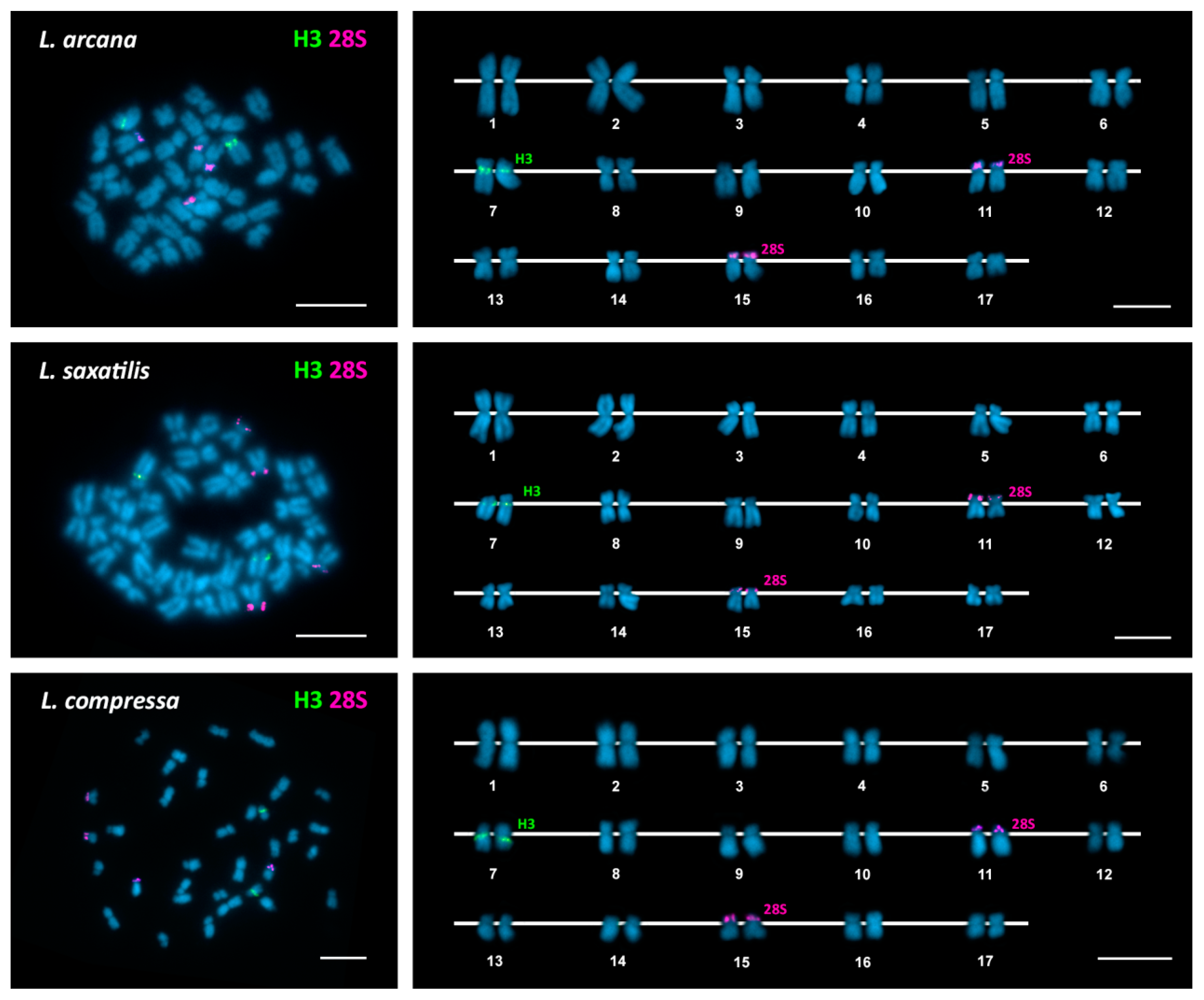
Periwinkles were collected at the localities indicated in Table 2. Attending to shell and internal morphology criteria, the specimens were preliminarily identified as Littorina arcana (Hannaford-Ellis, 1978), Littorina saxatilis (Olivi, 1792), Littorina compressa (Jeffreys, 1865), Littorina fabalis (W. Turton, 1825), Littorina obtusata (Linnaeus, 1758), Littorina littorea (Linnaeus, 1758), Littoraria angulifera (Lamarck, 1822), Cenchritis muricatus (Linnaeus, 1758), and Melarhaphe neritoides (Linnaeus, 1758). The nomenclature used for the taxa follows the World Register of Marine Species database [16]. Representatives of the two sympatric ecotypes of L. saxatilis [4] and the three morphotypes/ecotypes described for the Iberian Peninsula L. fabalis [17] were included.
Periwinkles were transported to the laboratory and processed following a combination of methods previously described for mollusks [18,19,20]. After an overnight colchicine (0.005%) treatment, periwinkles were euthanized and their muscular feet were dissected and preserved in absolute ethanol. The rest of the soft tissues were immersed in 50% (3 h) and 25% (3 h) sea water containing colchicine (0.05%) prior to fixation with ethanol/acetic acid. Small pieces of fixed gills and gonads were immersed in 60% acetic acid and disaggregated to obtain cell suspensions that were spread onto preheated slides [21,22].
Genomic DNA, extracted with the EZNA Mollusc DNA Kit (Omega Bio-Tek, Norcross, GA, USA), was used to amplify the H3 histone gene, 5S rDNA, and 28S rDNA in a GeneAmp PCR system 9700 (Applied Biosystems, Foster City, CA, USA). PCRs were performed in 20 μL total volume containing 1× PCR buffer, 2.5 mmol/L MgCl2, 50 ng DNA, 0.5 mmol/L each dNTP, 1 μmol/L each primer, and 1 U BIOTAQ DNA polymerase (Bioline, London, UK). H3 histone gene and 5S rDNA probes were labeled by supplementing the PCR mixtures either with 20 μM biotin-16-dUTP (Roche Applied Science, Penzberg, Germany) or 5 μM digoxigenin-11-dUTP (10× DIG Labeling Mix, Roche Applied Science), whilst 28S rDNA probes were labeled employing a nick translation kit (Roche Applied Science) [23,24,25,26].
Chromosome preparations were stained with DAPI (0.14 μg/mL) for 8 min, air dried, mounted with antifade (Vectashield, Vector Laboratories Inc., Burlingame, CA, USA), and photographed using a Nikon Eclipse-800 microscope equipped with a DS-Qi1Mc CCD camera (Nikon, Tokyo, Japan) controlled by the NIS-Elements software (Nikon). After visualization and photography, single and/or double FISH experiments using H3 histone gene, 5S rDNA, and/or 28S rDNA probes were performed following previously published methods [23,24,25,26]. In short, chromosome preparations were digested with RNase and pepsin, fixed with formaldehyde and denaturated in 70% formamide at 69 °C (2 min). After overnight hybridization in 50% formamide at 37 °C, chromosome preparations were washed at 45 °C with 50% formamide and 1× SSC. Biotin-labeled probes were detected with fluorescein avidin and biotinylated anti-avidin (Vector Laboratories) whereas digoxigenin-labeled probes were detected with mouse antidigoxigenin, goat antimouse rhodamine, and rabbit antigoat rhodamine (Sigma–Aldrich, St. Louis, MO, USA). Chromosome preparations were counterstained with DAPI, mounted with antifade and the previously recorded metaphase plates photographed again. Chromosome preparations subjected to double FISH experiments were then rehybridized using a third probe, and the same metaphase plates were photographed once more.
Karyotype analyses were performed in a minimum of 10 specimens (5 females, 5 males) from each periwinkle species. A minimum of 10 metaphase plates per specimen was analyzed. For each periwinkle species, the best 10 metaphase plates showing FISH signals, obtained from different individuals, were employed to build karyotypes. Measurements of short and long arm lengths were taken, and relative lengths and centromeric indices from each chromosome pair were determined for each metaphase plate. Mean values per chromosome pair and species were also calculated.
The morphological identification of the specimens was confirmed by amplifying and sequencing a fragment of the mitochondrial cytochrome c oxidase subunit I (COI) gene [27,28]. All sequences were deposited in the NCBI GenBank database under the accession numbers MH809396 to MH809424 (Supplementary materials Figure S1).

3. Results

All male and female specimens of the nine periwinkle species showed diploid chromosome numbers of 2n = 34 and karyotypes characterized by a superabundance of meta/submetacentric chromosome pairs (15 to 17).
As can be seen in Figure 1, Figure 2 and Figure 3, FISH mapping of 28S rDNA probes showed hybridization signals in the nine periwinkle taxa. Five of them displayed a single major (45S) rDNA cluster located at subterminal positions on the short arms of submetacentric chromosome pair 8 in the flat periwinkles L. obtusata and L. fabalis, subtelocentric pair 16 in the mangrove periwinkle L. angulifera and the beaded periwinkle C. muricatus, and metacentric pair 11 in the small periwinkle M. neritoides. In contrast, two clusters were detected at subterminal positions on chromosome pairs 11 and 15 in the rough periwinkles L. arcana, L. saxatilis, and L. compressa, and on chromosome pairs 8 and 16 in the common periwinkle L. littorea.
In regard to H3 histone genes, a single cluster was detected in all periwinkles: close to the centromere on the long arms of chromosome pair 7 in the rough periwinkles Littorina arcana, L. saxatilis, and L. compressa and the flat periwinkles L. obtusata and L. fabalis; intercalary to the long arms of chromosome pair 7 in the common periwinkle L. littorea and the small periwinkle M. neritoides; and at a subterminal location on the long arms of chromosome pair 17 in the mangrove periwinkle L. angulifera and the beaded periwinkle C. muricatus.
In contrast, 5S rDNA hybridization signals were only detected in the flat periwinkles L. obtusata and L. fabalis and the small periwinkle M. neritoides. In all cases, the single 5S rDNA cluster was close to the centromere on the long arms of chromosome pair 4.
Double-color FISH using 28S rDNA and H3 histone gene probes labeled differently, followed by rehybridization with a 5S rDNA probe in L. obtusata, L. fabalis, and M. neritoides, demonstrated that all clusters were located on different chromosome pairs in each of the nine periwinkles (Figure 1, Figure 2 and Figure 3). A summary of the FISH mapping results, including those previously published for the small periwinkle M. neritoides [14], is presented in Table 3.

4. Discussion

Chromosome studies of gastropod mollusks are scarce [29]. This is mainly due to low mitotic indices, small chromosome sizes, and technical difficulties in obtaining good chromosome morphology and spreading. This is particularly evident in Littorinidae, where chromosome numbers were described for only eleven (Table 1) of the around 200 species currently included in the family. The diploid chromosome numbers of 2n = 34 presented by the nine species studied herein, six of them newly described, are coincident with those proposed for seven of the species previously studied and seem to confirm that this is the common number in periwinkles. In contrast, this number differs with those proposed for Echinolittorina hawaiensis (2n = 30), E. miliaris (2n = 36), E. radiata (2n = 36), and E. subnodosa (2n = 16) [5,13]. Although those differences could indicate variations in chromosome numbers among different genera in the family, as the divergent diploid numbers (30 and 36) for three of these species were determined from paraffinned material in a study performed in the early 1960s [5], it would be necessary to confirm them using modern techniques of chromosome preparation. In regard to the highly divergent diploid chromosome number of 2n = 16 proposed for E. subnodosa [13], it is important to take into account that gastropod mollusks are usually intermediary hosts for digenean parasites, many of which present chromosome numbers and karyotypes [30] similar to the one reported for E. subnodosa [13]. Thus, future studies are needed to confirm the chromosome number in this species.
Regarding chromosome morphologies, the superabundance of metacentric and submetacentric chromosome pairs (15 to 17) in the karyotypes of the nine periwinkles described in this work is partially concordant with previous results. The minor discrepancies in karyotype composition can be attributed to differences in the degree of condensation of the chromosomes and the methodology employed [15].
In contrast with previous reports describing differences in chromosome number for male (X0, 33 chromosomes) and female (XX, 34) M. neritoides [9,11] and in chromosome morphology for male (XY) and female (XX) L. saxatilis [12], we did not detect any difference in chromosome number nor karyotype composition between males and females in any of the nine periwinkle species studied here. In this sense, targeted resequencing in L. saxatilis [31] did not reveal any regions with low heterozygosity in males, as expected for an XY sex determination system [32], in agreement with our results. The absence or presence of sex chromosome determinism in these species may have some evolutionary implications. For example, according to Haldane’s rule, intrinsic genetic incompatibilities should affect the heterogametic sex [33]. However, if there are undifferentiated sex chromosomes in L. saxatilis, both males and females should be equally affected by incompatibilities. A small degree of postzygotic isolation was observed in hybrid males after the analysis of sperm quality [34], but other postzygotic effects (embryo abnormalities) were also detected in females with intermediate ecotype characteristics [35]. The observation of postzygotic effects in both male and female hybrids is compatible with the absence of differentiated sex chromosomes we found here.
Pertaining to the FISH mapping results, all major (45S) rDNA clusters detected in periwinkles were subtelomeric, but their number differed between one in flat, mangrove, beaded, and small periwinkles versus two in rough and common periwinkles. This variation in the number of 45S rDNA clusters does not completely agree with their phylogenetic relationships (Supplementary materials Figure S1) [1] as the closely related rough and flat periwinkles displayed a different number of major rDNA clusters, whereas the first presents the same number as the more distantly related common periwinkles. Nevertheless, the location of the two clusters differs between the rough and the common periwinkles. The variation in the number of 45S rDNA clusters is a common observation in many taxonomic groups and in some cases has been related to their subtelomeric location. The presence of repeat sequences and breakpoints at subtelomeric regions [36,37], together with clustering of telomeres in meiosis, could favor sequence exchanges between non-homologous chromosomes [38] and consequently explain the variation in number.
Concerning minor (5S) rDNA clusters, hybridization signals were detected in only three of the nine periwinkles studied: the flat periwinkles L. fabalis and L. obtusata and the small periwinkle M. neritoides. In all cases, the single cluster was located close to the centromere in chromosome pair 4. Even though the absence of signals in the remaining six species could be attributable to technical difficulties or a non-clustered nature of the 5S rDNA repeats, the presence of clustered repeats in a number that is below the resolution power of FISH is a more probable explanation.
In contrast to the proposed adjacent location of 45S rDNA and 5S rDNA in the small periwinkle M. neritoides [14], the single species of Littorinidae in which FISH was previously applied, our results demonstrated that in both the flat periwinkles L. fabalis and L. obtusata and the small periwinkle M. neritoides the signals for 45S rDNA and those for 5S rDNA appeared in different chromosome pairs.
As commonly reported for other invertebrate groups, we detected a single H3 histone gene cluster in all nine periwinkles. In contrast, the chromosomal location of the cluster varied from species to species: subcentromeric signals appeared in rough and flat periwinkles, intercalary ones in common and small periwinkles, and subterminal ones in mangrove and beaded periwinkles. The presence of these clusters at subterminal positions is quite unusual in invertebrate species and has only been described in some grasshoppers [39] and bivalves [25,28,40,41,42,43].
Overall, the different probes show the same chromosomal location within flat periwinkles (L. obtusata and L. fabalis, including three morphotypes/ecotypes in the latter species) and also within the complex L. arcana, L. compressa, and L. saxatilis, in agreement with the monophily of these two groups. However, the differences in the distribution patterns of rDNA and histone gene clusters found in this work indicate that conservation in chromosome number in Littorinidae is not paired by conservation in chromosome structure. This is not a strange phenomenon as demonstrated by the abundance of chromosomal rearrangements (i.e., inversions, translocations) found in sequenced eukaryotic genomes [44,45], including L. saxatilis [46].
Although FISH mapping tandemly repeated gene families is a useful tool for chromosome identification in invertebrates, this technique has only been previously applied to map rDNA clusters in a single species of Littorinidae, the small periwinkle M. neritoides [14]. In this work, we have demonstrated the feasibility of applying those techniques to locate rDNA and histone gene clusters in nine species of periwinkles, thus opening the possibility of mapping other probes in these organisms and contributing to a better understanding of their chromosome evolution. Even more, the viability of applying FISH to periwinkles also opens the possibility of employing the whole genome of one of the members of a species complex as a probe to hybridize chromosomes of other members of the complex (genomic in situ hybridization, GISH) to obtain insights into their differences and/or to detect interspecific hybridization and introgression.
FISH mapping information has provided valuable information to complement sequencing information for genome assembly in different species. The extension of this technique beyond rDNA and histone gene clusters to increase the contiguity of genome assemblies (chromosome level) and to detect chromosomal rearrangements is likely to shed light on the evolutionary relevance of structural variation and adaptation or speciation in gastropods.

Supplementary Materials

The following are available online at https://www.mdpi.com/2073-4425/9/11/517/s1, Figure S1: Maximum likelihood tree based on partial mitochondrial COI gene sequences of nine periwinkles using the TN93 + G + I nucleotide substitution model.

Author Contributions

D.G.-S., S.A.-R. and J.P. conceived and designed the experiments; D.G.-S., D.C., R.F., J.G. and J.M.E.-L. collected the specimens; D.G.-S., S.A.-R., D.C., R.F., J.G. and J.P. performed the experiments; D.G.-S., S.A.-R. and J.P. analyzed the data; J.M.E.-L., E.R.-A. and J.P. contributed reagents/materials/analysis tools; D.G.-S and J.P. wrote the paper.

Funding

This research was funded by the Xunta de Galicia and Fondos FEDER: “Unha maneira de facer Europa” (Axudas do programa de consolidación e estruturación de unidades de investigacións competitivas do SUG: ED431C 2016-037), and by the Fundação para a Ciência e a Tecnologia (Portuguese Foundation for Science and Technology), project number: FCT [PTDC/BIA-EVF/113805/2009].

Acknowledgments

D. García-Souto was partially supported by a FPU fellowship from “Ministerio de Educación, Cultura y Deporte” (Spain). RF was funded by FCT [SFRH/BPD/89313/2012] and is currently funded by the European Union’s Horizon 2020 research and innovation program, under the Marie Sklodowska-Curie grant agreement number 706376.

Conflicts of Interest

The authors declare no conflict of interest. The founding sponsors had no role in the design of the study; in the collection, analyses, or interpretation of data; in the writing of the manuscript, and in the decision to publish the results.

References

  1. Reid, D.G.; Dyal, P.; Williams, S.T. A global molecular phylogeny of 147 periwinkle species (Gastropoda, Littorininae). Zool. Scr. 2012, 41, 125–136. [Google Scholar] [CrossRef]
  2. Galindo, J.; Grahame, J.W. Ecological speciation and the intertidal snail Littorina saxatilis. Adv. Ecol. 2014, 2014, 239251. [Google Scholar] [CrossRef]
  3. Panova, M.; Johansson, T.; Canbäck, B.; Bentzer, J.; Rosenblad, M.A.; Johannesson, K.; Tunlid, A.; André, C. Species and gene divergence in Littorina snails detected by array comparative genomic hybridization. BMC Genom. 2014, 15, 687. [Google Scholar] [CrossRef] [PubMed]
  4. Rolán-Álvarez, E.; Austin, C.J.; Boulding, E.G. The contribution of the genus Littorina to the field of evolutionary ecology. Oceanogr. Mar. Biol. 2015, 53, 157–214. [Google Scholar] [CrossRef]
  5. Nishikawa, S. A comparative study of the chromosomes in marine gastropods, with some remarks on cytotaxonomy and phylogeny. J. Shimonoseki Coll. Fish. 1962, 11, 149–186. [Google Scholar]
  6. Thiriot-Quiévreux, C.; Ayraud, N. Les caryotypes de quelques espéces de bivalves et de gastéropodes marins. Mar. Biol. 1982, 70, 165–172. [Google Scholar] [CrossRef]
  7. Janson, K. Chromosome number in two phenotypically distinct populations of Littorina saxatilis Olivi from the Swedish west coast. J. Mollus. Stud. 1983, 49, 224–227. [Google Scholar] [CrossRef]
  8. Vitturi, R.; Catalano, E. Macaluso, M. Chromosome studies in three species of the gastropod superfamily Littorinoidea. Malacol. Rev. 1986, 19, 53–60. [Google Scholar]
  9. Vitturi, R.; Catalano, E.; Macaluso, M.; Zava, B. The karyology of Littorina neritoides (Linnaeus, 1758) (Mollusca, Prosobranchia). Malacologia 1988, 29, 319–324. [Google Scholar]
  10. Birstein, V.J.; Mikhailova, N.A. On the karyology of trematodes of the genus Microphallus and their intermediate gastropod host, Littorina saxatilis II. Karyological study of Littorina saxatilis (Gastropoda: Prosobranchia). Genetica 1990, 80, 167–170. [Google Scholar] [CrossRef]
  11. Vitturi, R.; Libertini, A.; Panozzo, M.; Mezzapelle, G. Karyotype analysis and genome size in three Mediterranean species of periwinkles (Prosobranchia: Mesogastropoda). Malacologia 1995, 37, 123–132. [Google Scholar]
  12. Rolán-Alvarez, E.; Buño, I.; Gonsalvez, J. Sex is determinated by sex chromosomes in Littorina saxatilis (Olivi) (Gastropoda, Prosobranchia). Hereditas 1996, 124, 261–267. [Google Scholar] [CrossRef]
  13. Ebied, A.M.; Hassan, H.A.; Abu-Almaaty, A.H.; Yaseen, A.E. Cytogenetic studies on metaphase chromosomes of eight gastropod species of orders Mesogastropoda and Neogastropoda from the Red Sea (Prosobranchia–Mollusca). J. Egypt. Ger. Soc. Zool. 2000, 33, 317–336. [Google Scholar]
  14. Colomba, M.S.; Vitturi, R.; Castriota, L.; Bertoni, R.; Libertini, A. FISH mapping of 18S–28S and 5S ribosomal DNA, periwinkle Melarhaphe neritoides (Prosobranchia, Gastropoda, Cenogastropoda). Heredity 2002, 88, 381–384. [Google Scholar] [CrossRef] [PubMed]
  15. Libertini, A.; Trisolini, R.; Edmands, S. A cytogenetic study of the periwinkle Littorina keenae Rosewater, 1978 (Gastropoda: Littorinidae). J. Mollus. Stud. 2004, 70, 299–301. [Google Scholar] [CrossRef]
  16. World Register of Marine Species. Available online: http://www.marinespecies.org/ (accessed on 18 July 2018).
  17. Rolán, E.; Templado, J. Consideraciones sobre el complejo Littorina obtusata-mariae (Mollusca, Gastropoda, Littorinidae) en el Noroeste de la península Ibérica. Thalassas 1987, 5, 71–85. [Google Scholar]
  18. Méndez, J.; Pasantes, J.J.; Martínez-Expósito, M.J. Banding pattern of mussel (Mytilus galloprovincialis) chromosomes induced by 2× SSC/Giemsa-stain treatment. Mar. Biol. 1990, 106, 375–377. [Google Scholar] [CrossRef]
  19. Dixon, D.R.; Pascoe, P.L.; Gibbs, P.E.; Pasantes, J. The nature of Robertsonian chromosomal polymorphism in Nucella lapillus: A re-examination. In Genetics and Evolution of Aquatic Organisms; Beaumont, A.R., Ed.; Chapman and Hall: London, UK, 1994; pp. 389–399. ISBN 9780412493706. [Google Scholar]
  20. Martínez-Expósito, M.J.; Pasantes, J.J.; Méndez, J. Proliferation kinetics of mussel (Mytilus galloprovincialis) gill cells. Mar. Biol. 1994, 120, 41–45. [Google Scholar] [CrossRef]
  21. Pérez-García, C.; Cambeiro, J.M.; Morán, P.; Pasantes, J.J. Chromosomal mapping of rDNAs, core histone genes and telomeric sequences in Perumytilus purpuratus (Bivalvia: Mytilidae). J. Exp. Mar. Biol. Ecol. 2010, 395, 199–205. [Google Scholar] [CrossRef]
  22. Pérez-García, C.; Guerra-Varela, J.; Morán, P.; Pasantes, J.J. Chromosomal mapping of rRNA genes, core histone genes and telomeric sequences in Brachidontes puniceus and Brachidontes rodriguezi (Bivalvia: Mytilidae). BMC Genet. 2010, 11, 109. [Google Scholar] [CrossRef] [PubMed]
  23. Pérez-García, C.; Morán, P.; Pasantes, J.J. Cytogenetic characterization of the invasive mussel species Xenostrobus securis Lmk. (Bivalvia: Mytilidae). Genome 2011, 54, 771–778. [Google Scholar] [CrossRef] [PubMed]
  24. Pérez-García, C.; Morán, P.; Pasantes, J.J. Karyotypic diversification in Mytilus mussels (Bivalvia: Mytilidae) inferred from chromosomal mapping of rRNA and histone gene clusters. BMC Genet. 2014, 15, 84. [Google Scholar] [CrossRef] [PubMed]
  25. García-Souto, D.; Pérez-García, C.; Morán, P.; Pasantes, J.J. Divergent evolutionary behavior of H3 histone gene and rDNA clusters in venerid clams. Mol. Cytogenet. 2015, 8, 40. [Google Scholar] [CrossRef] [PubMed] [Green Version]
  26. García-Souto, D.; Pérez-García, C.; Kendall, J.; Pasantes, J.J. Molecular cytogenetics in trough shells (Mactridae, Bivalvia): Divergent GC-rich heterochromatin content. Genes 2016, 7, 47. [Google Scholar] [CrossRef] [PubMed]
  27. García-Souto, D.; Pérez-García, C.; Pasantes, J.J. Are pericentric inversions reorganizing wedge shell genomes? Genes 2017, 8, 370. [Google Scholar] [CrossRef] [PubMed]
  28. García-Souto, D.; Pasantes, J.J. Cytogenetics in Arctica islandica (Bivalvia, Arctiidae): The longest lived non-colonial metazoan. Genes 2018, 9, 299. [Google Scholar] [CrossRef] [PubMed]
  29. Thiriot-Quiévreux, C. Advances in chromosomal studies of gastropod Mollusc. J. Mollus. Stud. 2002, 69, 187–201. [Google Scholar] [CrossRef]
  30. García-Souto, D.; Pasantes, J.J. Molecular cytogenetics in digenean parasites: Linked and unlinked Major and 5S rDNAs, B chromosomes and karyotype diversification. Cytogenet. Genome Res. 2015, 147, 195–207. [Google Scholar] [CrossRef] [PubMed]
  31. Westram, A.M.; Rafajlovic, M.; Chaube, P.; Faria, R.; Larsson, T.; Panova, M.; Ravinet, M.; Blomberg, A.; Mehlig, B.; Johannesson, K.; et al. Clines on the seashore: The genomic architecture underlying rapid divergence in the face of gene flow. Evol. Lett. 2018, 2, 297–309. [Google Scholar] [CrossRef] [PubMed]
  32. Westram, A.M.; University of Sheffield, Sheffield, UK; Faria, R.; University of Sheffield, Sheffield, UK; Butlin, R.K.; University of Sheffield, Sheffield, UK; Johannesson, K.; University of Gothenburg, Gothenburg, Sweden. Personal communication, 2018.
  33. Coyne, J.A.; Orr, H.A. Speciation; Sinauer Associates, Inc.: Sunderland, MA, USA, 2004; pp. 284–311. ISBN 9780878930890. [Google Scholar]
  34. Sá-Pinto, A.; Martínez-Fernández, M.; López-Fernández, C.; Ferreira, Z.; Pereira, R.; Gosálvez, J.; Rolán-Alvarez, E. Incipient post-zygotic barrier in a model system of ecological speciation with gene flow. J. Evol. Biol. 2013, 26, 2750–2756. [Google Scholar] [CrossRef] [PubMed] [Green Version]
  35. Hull, S.L.; Grahame, J.; Mill, P.J. Morphological divergence and evidence for reproductive isolation in Littorina saxatilis (Olivi) in northeast England. J. Mollus. Stud. 1996, 62, 89–99. [Google Scholar] [CrossRef]
  36. Stankiewicz, P.; Lupski, J.R. Genome architecture, rearrangements and genomic disorders. Trends Genet. 2002, 18, 74–81. [Google Scholar] [CrossRef]
  37. Torres, G.A.; Gong, Z.; Iovene, M.; Hirsh, C.D.; Buell, C.R.; Bryan, G.J.; Novák, P.; Macas, J.; Jiang, J. Organization and evolution of subtelomeric satellite repeats in the potato genome. G3 (Bethesda) 2011, 1, 85–92. [Google Scholar] [CrossRef] [PubMed]
  38. Linardopoulou, E.V.; Williams, E.M.; Fan, Y.X.; Friedman, C.; Young, J.M.; Trask, B.J. Human subtelomeres are hot spots of interchromosomal recombination and segmental duplication. Nature 2005, 437, 94–100. [Google Scholar] [CrossRef] [PubMed] [Green Version]
  39. Cabrero, J.; López-León, M.D.; Teruel, M.; Camacho, J.P.M. Chromosome mapping of H3 and H4 histone gene clusters in 35 species of acridid grasshoppers. Chromosome Res. 2009, 17, 397–404. [Google Scholar] [CrossRef] [PubMed]
  40. Zhang, L.; Bao, Z.; Wang, S.; Huang, X.; Hu, J. Chromosome rearrangements in Pectinidae (Bivalvia; Pteriomorphia) implied based on chromosomal localization of histone H3 gene in four scallops. Genetica 2007, 130, 193–198. [Google Scholar] [CrossRef] [PubMed]
  41. Carrilho, J.; Pérez-García, C.; Leitão, A.; Malheiro, I.; Pasantes, J.J. Cytogenetic characterization and mapping of rDNAs, core histone genes and telomeric sequences in Venerupis aurea and Tapes rhomboides (Bivalvia: Veneridae). Genetica 2011, 139, 823–830. [Google Scholar] [CrossRef] [PubMed]
  42. García-Souto, D.; Sumner-Hempel, A.; Fervenza, S.; Pérez-García, C.; Torreiro, A.; González-Romero, R.; Eirín-López, J.M.; Morán, P.; Pasantes, J.J. Detection of invasive and cryptic species in marine mussels (Bivalvia, Mytilidae): A chromosomal perspective. J. Nat. Conserv. 2017, 39, 58–67. [Google Scholar] [CrossRef]
  43. García-Souto, D.; Rios, G.; Pasantes, J.J. Karyotype differentiation in tellin shells (Bivalvia: Tellinidae). BMC Genet. 2017, 18, 66. [Google Scholar] [CrossRef] [PubMed]
  44. Coghlan, A.; Eichler, E.E.; Oliver, S.G.; Paterson, A.H.; Stein, L. Chromosome evolution in eukaryotes: A multi-kingdom perspective. Trends Genet. 2005, 21, 673–682. [Google Scholar] [CrossRef] [PubMed]
  45. Peichel, C.L. Chromosome evolution: Molecular mechanisms and evolutionary consequences. J. Hered. 2017, 108, 1–2. [Google Scholar] [CrossRef] [PubMed]
  46. Faria, R.; Chaube, P.; Morales, H.; Larsson, T.; Lemmon, A.R.; Lemmon, E.M.; Rafajlović, M.; Panova, M.; Ravinet, M.; Johannesson, K.; et al. Multiple chromosomal rearrangements in a hybrid zone between Littorina saxatilis ecotypes. Mol. Ecol. 2018, in press. [Google Scholar]
Figure 1. Chromosomal mapping of major rDNA and histone gene clusters in rough periwinkles. Fluorescent in situ hybridization mapping of H3 histone gene (H3, green) and major rDNA (28S, magenta) probes to mitotic chromosomes of the rough periwinkles Littorina arcana, Littorina saxatilis, and Littorina compressa counterstained with 4′-6-diamidino-2-phenylindole (DAPI). Scale bars, 5 μm.
Genes 09 00517 g001
Figure 2. Chromosomal mapping of rDNA and histone gene clusters in flat and common periwinkles. Fluorescent in situ hybridization mapping of minor rDNA (5S, red), H3 histone gene (H3, green) and major rDNA (28S, magenta) probes to mitotic chromosomes of the flat periwinkles Littorina obtusata and Littorina fabalis and the common periwinkle Littorina littorea counterstained with 4′-6-diamidino-2-phenylindole (DAPI). No minor (5S) rDNA signals were detected in the common periwinkle. Scale bars, 5 μm.
Genes 09 00517 g002
Figure 3. Chromosomal mapping of rDNA and histone gene clusters in mangrove, beaded, and small periwinkles. Fluorescent in situ hybridization mapping of H3 histone gene (H3, green) and major rDNA (28S, magenta) probes to mitotic chromosomes of the mangrove periwinkle Littoraria angulifera, the beaded periwinkle Cenchritis muricatus, and the small periwinkle Melarhaphe neritoides counterstained with 4′-6-diamidino-2-phenylindole (DAPI). Minor rDNA (5S, red) signals were also detected in the small periwinkle. Scale bars, 5 μm.
Genes 09 00517 g003
Table 1. Haploid (n) and diploid (2n) chromosome numbers and karyotypes in Littorinidae.
Speciesn2nKaryotypeReference
Echinolittorina hawaiensis (Rosewater & Kadolsky, 1981)1530 [5]
Echinolittorina miliaris (Quoy & Gaimard, 1833)18 [5]
Echinolittorina punctata (Gmelin, 1791)16 [8]
1734 [11]
Echinolittorina radiata (Souleyet, 1852)18 [5]
Echinolittorina subnodosa (Molina, 1782)8168 m[13]
Littoraria strigata (Philippi, 1846) 17 [5]
Littorina brevicula (Philippi, 1844)17 [5]
Littorina keena Rosewater, 1978 3410 m/sm, 7 st/t[15]
Littorina obtusata (Linnaeus, 1758) 34 [7]
Littorina saxatilis (Olivi, 1792) 348 m/sm, 9 st/t[7]
3410 m/sm, 7 st/t[10]
346 m, 9 sm, 2 st[11]
34 XX ♀/34 XY ♂10 m/sm, 7 st[12]
Melarhaphe neritoides (Linnaeus, 1758)17348 m, 2 sm, 7 t[6]
1734 XX ♀/33 X0 ♂10 m, 3 sm, 3 st, 1 t[9]
34 XX ♀/33 X0 ♂10 m, 3 sm, 3 st, 1 t[11]
Notes: m: metacentric; sm: submetacentric; st: subtelocentric; t: telocentric.
Table 2. Collection localities of the species of Littorinidae.
SpeciesLocalityCoordinates
Littorina arcanaHoly Island, UK53.299781, −4.679846
Littorina saxatilisCabo Estai, Ría de Vigo, Spain42.182181, −8.813204
Cabo Silleiro, Spain42.104987, −8.898929
Littorina compressaHoly Island, UK53.299781, −4.679846
Littorina obtusataRande, Ría de Vigo, Spain42.284311, −8.658661
Littorina fabalisSanto André, Portugal41.412417, −8.787778
Praia da Borna, Ría de Vigo, Spain42.280867, −8.697019
O Grove, Ría de Arousa, Spain42.460500, −8.872028
Littorina littoreaMarín, Ría de Pontevedra, Spain42.396351, −8.695599
Littoraria anguliferaMiami, FL, USA25.810107, −80.164063
Cenchritis muricatusMiami, FL, USA25.889330, −80.150129
Melarhaphe neritoidesCabo Udra, Ría de Aldán, Spain42.333599, −8.827797
Table 3. Chromosomal location of rDNA and histone gene clusters in Littorinidae.
Species2n45S rDNA5S rDNAH3 Histone GenesReference
Littorina arcana3411p ter (st) 7q cen (sm)This work
15p ter (sm)
Littorina saxatilis3411p ter (sm) 7q cen (sm)This work
15p ter (sm)
Littorina compressa3411p ter (sm) 7q cen (sm)This work
15p ter (sm)
Littorina obtusata348p ter (sm)4q cen (m)7q cen (m)This work
Littorina fabalis348p ter (sm)4q cen (sm)7q cen (sm)This work
Littorina littorea348p ter (m) 7q ic (sm)This work
16p ter (sm)
Littoraria angulifera3416p ter (st) 17q ter (m)This work
Cenchritis muricatus3416p ter (st) 17q ter (st)This work
Melarhaphe neritoides3411p ter (m)4p cen (m)7q ic (st)This work
34/33p ter (m) * [14]
Notes: p: short arm; q: long arm; cen: subcentromeric; ic: intercalary; ter: subterminal; (m): metacentric; (sm): submetacentric; (st): subtelocentric; (t): telocentric; *: unidentified chromosome pair.

