Article

Genetic Analysis Reveals a Protective Effect of Sphingomyelin on Cholelithiasis

1
Department of Epidemiology and Biostatistics, School of Public Health, Xi’an Jiaotong University Health Science Center, Xi’an 710061, China
2
Xi’an Jiaotong University Health Science Center, Xi’an 710061, China
3
School of Life Science and Technology, Xi’an Jiaotong University, Xi’an 710049, China
4
The Key Laboratory of Biomedical Information Engineering of Ministry of Education, Xi’an Jiaotong University, Xi’an 710049, China
5
The School of Mathematics and Statistics, Xi’an Jiaotong University, Xi’an 710049, China
6
Department of Geriatric Endocrinology Metabolism, The Second Affiliated Hospital of Xi’an Jiaotong University, Xi’an 710004, China
*
Authors to whom correspondence should be addressed.
These authors contributed equally to this work.
Genes 2025, 16(5), 523; https://doi.org/10.3390/genes16050523
Submission received: 4 April 2025 / Revised: 26 April 2025 / Accepted: 26 April 2025 / Published: 29 April 2025
(This article belongs to the Section Molecular Genetics and Genomics)

Abstract

:
Background: Cholelithiasis is the most common disorder affecting the biliary system. Choline is an essential nutrient in the human diet and is crucial for the synthesis of neurotransmitters. Previous studies have suggested an association between choline metabolites and cholelithiasis. However, the underlying mechanisms remain unclear. This research aims to fill the knowledge gap regarding the role of choline metabolites in cholelithiasis. Methods: Genetic data related to choline metabolites and other covariates were retrieved from the U.K. Biobank and IEU OpenGWAS database. Two-sample (TSMR) and multivariate Mendelian randomization (MVMR) analyses, mediation analysis, linkage disequilibrium score regression (LDSC), colocalization analysis, and enrichment analysis were performed. Results: A significant causal relationship was identified between serum level of sphingomyelin and cholelithiasis (p-value = 0.0002). A protective causal effect was identified in MVMR analysis. The following mediated MR analysis indicated that only LDL mediated a large part of the causal relationship (59.18%). Seven genes, including GCKR, SNX17, ABCG8, MARCH8, FUT2, APOH, and HNF1A, were revealed to be colocalized with the causal signal between sphingomyelin and cholelithiasis. Conclusion: The present study has identified a protective effect between sphingomyelin and cholelithiasis. This effect is largely mediated by LDL. The findings of this study offer valuable information for further exploration of the molecular mechanisms of cholelithiasis.

1. Introduction

Cholelithiasis is a common condition characterized by the accumulation of elevated levels of cholesterol or bilirubin—a breakdown product of hemoglobin—in the bile, which leads to the formation of stones within the biliary system, including the gallbladder and bile ducts. Approximately 20% of adults are affected by cholelithiasis, with more than 20% of these individuals developing symptoms and complications. Consequently, cholelithiasis imposes a substantial socioeconomic burden as a prevalent and costly digestive disorder [1]. Extensive research has identified several risk factors for cholelithiasis. Non-modifiable factors, including race, female sex, pregnancy, and age over 40 years, play a particularly significant role. Modifiable risk factors for cholelithiasis include obesity, a high-calorie diet, metabolic syndrome, and dyslipidemia. At present, surgical intervention constitutes the primary treatment for symptomatic cholelithiasis [2].
Choline, an essential nutrient in the human diet, is crucial for the synthesis of neurotransmitters such as acetylcholine, as well as for the production of methyl donors, betaine, and phospholipids. It plays vital roles in cell maintenance and growth throughout life, including neurotransmission, membrane synthesis, lipid transport, and single-carbon metabolism [3]. It is imperative to note that essential choline exists in several forms, including water-soluble free choline, fat-soluble phosphatidylcholine, and sphingomyelin. These forms are absorbed and metabolized through distinct pathways, which influence their bioavailability [4]. Previous studies have suggested an association between sphingolipids and cholelithiasis [5]. However, the underlying mechanisms between choline metabolites and cholelithiasis remain unclear.
Mendelian randomization (MR) is a highly efficacious tool for the identification of causal risk factors that underpin complex traits and diseases by means of genetic data. By employing single-nucleotide polymorphisms (SNPs) from genome-wide association studies (GWAS) as instrumental variables (IVs) for exposure, MR controls confounding factors and avoids reverse causation, strengthening causal inferences [6,7,8,9]. In the present study, the causal relationship between choline metabolite levels and cholelithiasis was examined using MR analysis. This current research aims to fill the knowledge gap regarding the role of choline metabolites in cholelithiasis and provide valuable insights for developing more effective preventive and treatment strategies. The graphical abstract is presented in Supplementary Figure S1.

2. Materials and Methods

2.1. Study Design

The study design and the assumptions of an MR study are presented in Figure 1. The risk factors under consideration include choline metabolites and other covariates, including LDL, HDL, triglycerides, and coronary artery disease (CAD), which were retrieved from specific data sources. The analyses include MR, LDSC, colocalization analysis, and enrichment analysis. The selection of genetic markers adheres to the three fundamental principles of instrumental variables. Firstly, the genetic markers must demonstrate a strong association with the exposure. Secondly, they must not demonstrate a direct association with the outcome. Thirdly, they must influence the outcome exclusively through their effect on the exposure. This study was conducted in strict accordance with the STROBE-MR checklist [10]. The code employed in this article is available in Supplementary Material S1.

2.2. Data Sources

The serum levels of three choline metabolites—total choline, phosphatidylcholine, and sphingomyelin—were selected as exposure factors from the U.K. Biobank (n = 114,999). The GWAS summary statistics for these metabolites are accessible via the IEU OpenGWAS database (https://gwas.mrcieu.ac.uk, accessed on 10 November 2024), with identifiers of met-d-cholines, met-d-phosphatidylcholines, and met-d-sphingomyelins (Borges CM) [11]. The genetic data of cholelithiasis from the Neale Lab within the U.K. Biobank were used for endpoints (http://www.nealelab.is/uk-biobank, accessed on 10 November 2024). The IEU OpenGWAS database (GWAS identifier: ukb-a-559, n = 337,199) was utilized for the purpose of data retrieval, and cholelithiasis was diagnosed according to ICD-10 (K80). For MVMR, four additional covariates were selected and adjusted in the analyses: LDL (ieu-b-5089, n = 201,678), HDL (ieu-b-109, n = 403,943), triglycerides (ieu-b-111, n = 441,016) [12], and CAD (ebi-a-GCST90013868, n = 352,063) [13]. The serum levels of three choline metabolites—total choline, phosphatidylcholine, and sphingomyelin—were selected as exposure factors from the U.K. Biobank (n = 114,999). The GWAS summary statistics for these metabolites are accessible via the IEU OpenGWAS database (https://gwas.mrcieu.ac.uk, accessed on 10 November 2024), with identifiers of met-d-cholines, met-d-phosphatidylcholines, and met-d-sphingomyelins (Borges CM) [11]. The genetic data of cholelithiasis from the Neale Lab within the U.K. Biobank were used for endpoints (http://www.nealelab.is/uk-biobank, accessed on 10 November 2024). The IEU OpenGWAS database (GWAS identifier: ukb-a-559, n = 337,199) was utilized for the purpose of data retrieval, and the diagnosis of cholelithiasis was made in accordance with the International Classification of Diseases, Tenth Revision (ICD-10, K80). In the context of multivariate Mendelian randomization (MVMR), the analysis incorporated four additional covariates, which were selected and adjusted as follows: LDL (ieu-b-5089, n = 201,678), HDL (ieu-b-109, n = 403,943), triglycerides (ieu-b-111, n = 441,016) [12], and CAD (ebi-a-GCST90013868, n = 352,063) [13]. The genetic data were derived exclusively from European populations and had undergone rigorous quality control measures and ethical approval, thus ensuring direct applicability.

2.3. Genetic Instrument Selection

SNPs with strong correlations (p-value < 5 × 10⁻⁸) were initially selected as candidate IVs. Linkage disequilibrium (LD) among them was estimated. One genetic marker was excluded on the basis that the r2 values of the corresponding SNP pair were greater than 0.001 (with a distance of less than 10,000 kb). To remove weak instrumental variables, an F-test was performed for each SNP, and SNPs with F-values less than 10 were excluded. Phenoscaner (https://github.com/phenoscanner/phenoscanner, accessed on 10 November 2024) was utilized to search and remove confounding SNPs [14,15]. Finally, a total of 49, 48, and 48 SNPs were identified as IVs for total choline, phosphatidylcholine, and sphingomyelin, respectively.

2.4. TSMR and MVMR Analyses

Mendelian randomization satisfies the relevance assumption, the independence assumption, and the exclusion restriction [16]. Five MR methods, including MR-Egger [17], weighted median [18], inverse variance weighted (IVW) [19], simple mode, and weighted mode [20], were utilized for MR analyses using the TwoSampleMR package (http://www.r-project.org, accessed on 10 November 2024). The IVW estimates were selected as the primary approach, while the other four methods were employed to enhance robustness across diverse scenarios. A significant causal relationship was considered when the p-value of IVW was less than 0.05, and the results of all five methods had the same direction. Heterogeneity was assessed using the MR-Egger Cochran Q test [21]. The MR-Egger intercept [22] and the MR-Presso global test [23] were utilized to test for pleiotropy. In instances where heterogeneity and pleiotropy were detected, the MR-Presso outlier method [23] was employed to remove outliers, and the analysis was repeated. Furthermore, leave-one-out tests for each SNP were conducted using the IVW results [24], and the SNP effect values were visualized in a funnel plot [25] to ensure that no outliers were interfering with the model results.
MVMR was carried out including LDL, HDL, triglycerides, and CAD as covariates. Results were evaluated using the IVW random effects model and the MR-Egger model.

2.5. Reverse and Mediated Mendelian Randomization Analysis

Reverse and mediated MR analysis was conducted for sphingomyelin, which was identified as a significant positive causal factor for cholelithiasis. The analysis incorporated four factors as mediators. In the analysis of the mediator Mendelian randomization, the effect of sphingomyelin on LDL was β1, the effects of the mediators on cholelithiasis were β2, and the indirect effect was calculated using β1 × β2. The significance of the indirect effect was then tested using a stepwise testing method.

2.6. LDSC and Colocalization Analysis

LDSC and colocalization analyses were performed to investigate genetic correlations between the three exposure factors and cholelithiasis [26,27,28]. The coloc package was used for colocalization analysis (p1 = 1 × 10⁻⁴, p2 = 1 × 10⁻⁴, p12 = 1 × 10⁻⁵) [28]. SNPs associated with exposure factors were selected to define the colocalization regions based on a significance threshold of p-value < 1 × 10⁻⁶. Linkage disequilibrium (LD) pruning was performed within a 100 kb window by removing SNPs with r2 < 0.001. For the remaining SNPs, regions of colocalization were defined by extending 50 kb on either side of the selected SNPs. Subsequently, a full Bayesian colocalization analysis was conducted, utilizing Bayes factors. The threshold for posterior probability for hypothesis 4 (PPH4) was set at > 0.9.

2.7. Gene Enrichment Analysis

Gene enrichment analysis was further conducted for loci obtained from colocalization analysis using the NCBI website (https://www.ncbi.nlm.nih.gov/, accessed on 10 November 2024) and the STRING database (https://string-db.org/, accessed on 10 November 2024) [29]. Enrichment analysis was then performed using GO and KEGG databases [30].

3. Results

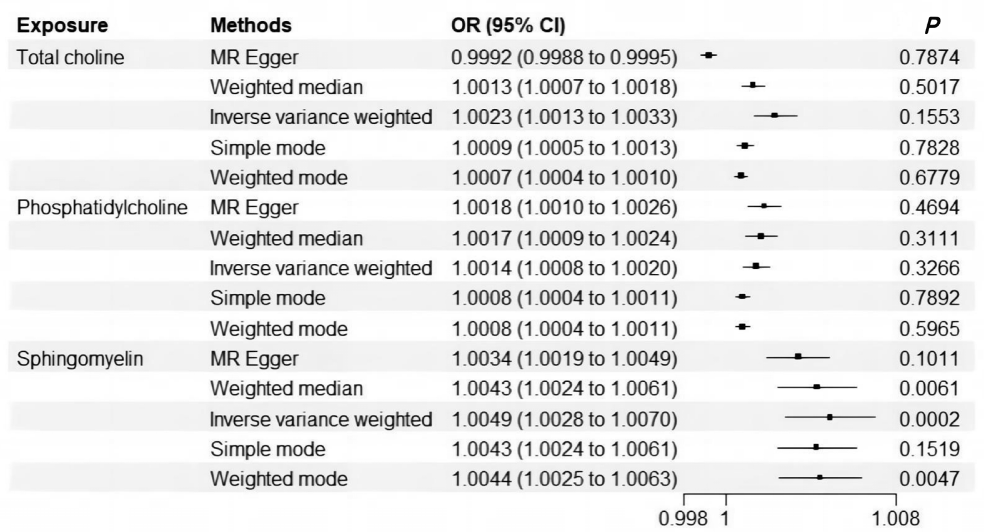
3.1. Significant Signals Were Identified for Causal Relationship Between Sphingomyelin and Cholelithiasis

In TSMR analysis, heterogeneity was detected (p-value < 0.05). The MR-Presso method excluded specific SNPs for total choline, phospholipids, and sphingolipids, respectively. Subsequently, a significant causal relationship was identified between serum level of sphingomyelin and cholelithiasis (OR [95%CI] = 1.0049 [1.0028–1.0070], p-value = 0.0002) (Figure 2). No horizontal pleiotropy was detected by MR-Egger intercepts, while the MR-Presso global test indicated some presence (Supplementary Table S1). Leave-one-out tests and funnel plots did not identify SNPs with a major impact on the results (Supplementary Figure S2).

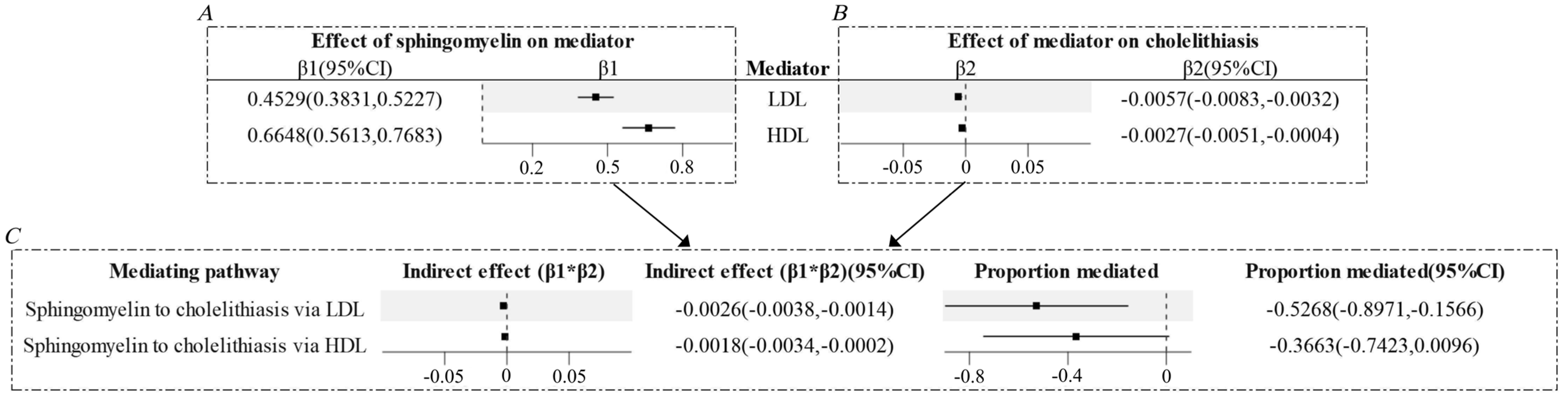
3.2. Low-Density Lipoprotein (LDL) and High-Density Lipoprotein (HDL) Might Mediate Part of the Causal Relationship Between Sphingomyelin and Cholelithiasis

A protective causal effect was identified in the MVMR analysis (Table 1). The following mediated MR analysis indicated that only LDL and HDL mediated part of the causal relationship between sphingomyelin and cholelithiasis (Figure 3, Supplementary Table S2). The indirect effects of LDL were found to be −0.0029, and the indirect effects of HDL were −0.0018. The LDL accounted for 59.18% of the total effect of sphingomyelin on cholelithiasis; however, the mediated proportion of HDL did not reach statistical significance. Reverse MR analysis revealed that the causal relationship between cholelithiasis and sphingolipids was not significant (p-value for IVW = 0.2520, p-value for MR-Egger intercept = 0.1790, p-value for Cochran Q test = 3.7161 × 10⁻²⁸).

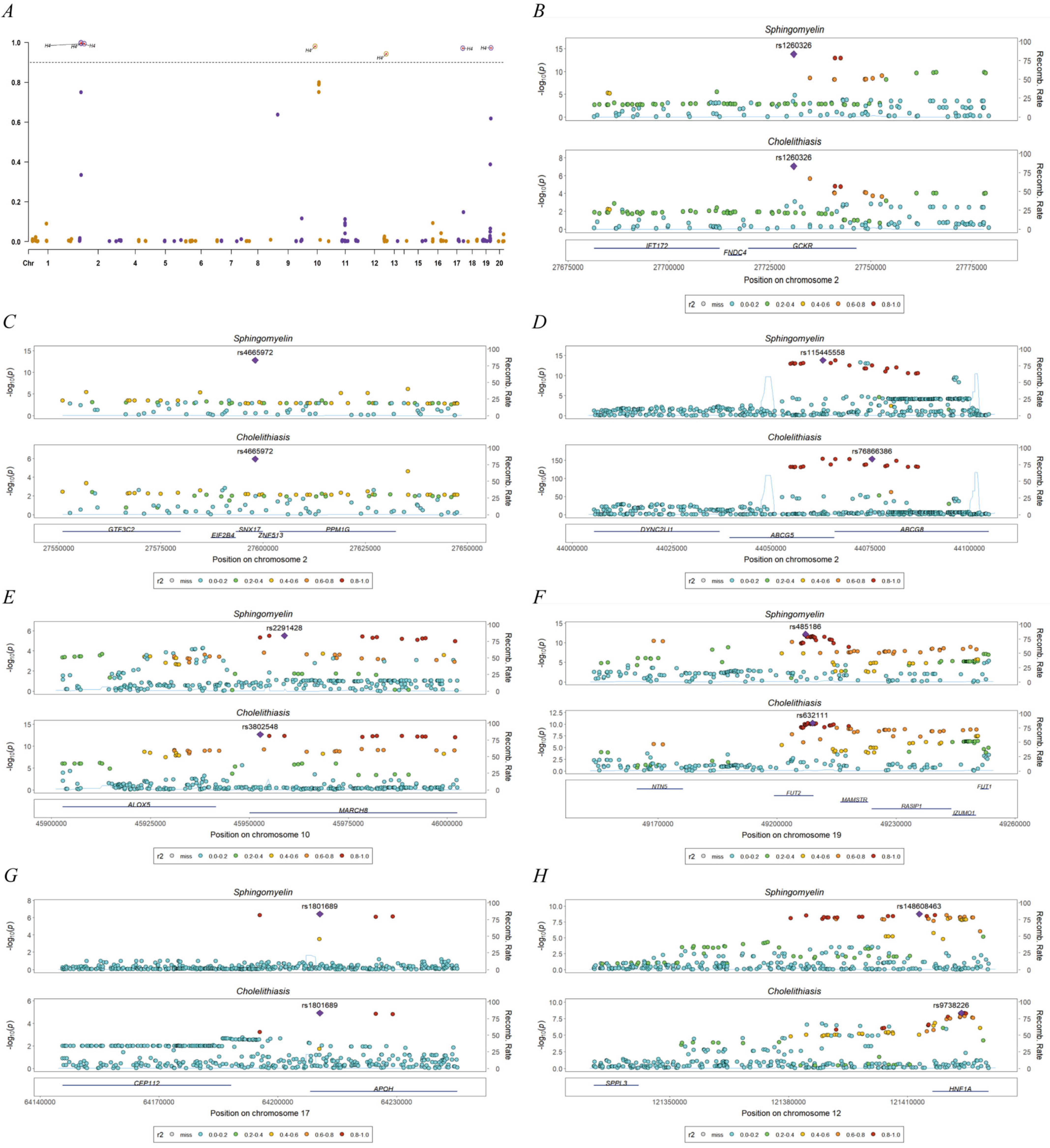
3.3. Bioinformatics Evidence for Unraveling the Significant Causal Signal Between Sphingomyelin and Cholelithiasis

LDSC analysis demonstrated genetic correlations between serum levels of three risk factors (total choline, phosphatidylcholine, sphingomyelin) and cholelithiasis at the whole-genome level (Supplementary Table S3). Colocalization analysis revealed abundant colocalization signals, thereby identifying specific SNPs mapped to genes, including GCKR (glucokinase regulatory protein), SNX17 (sorting nexin-17), ABCG8 (ATP-binding cassette sub-family G member 5), MARCH8 (membrane-associated ring-CH-type finger 8), FUT2 (fucosyltransferase 2), APOH (apolipoprotein H) and HNF1A (hepatocyte nuclear factor 1α) (Figure 4, Supplementary Tables S4 and S5).
These seven genes were found to be enriched in multiple biological processes, cellular components, and molecular functions in the GO database. In the KEGG database, these genes were identified to be enriched in glycosphingolipid biosynthesis—lacto and neolacto series, cholesterol metabolism, maturity-onset diabetes of the young, glycosaminoglycan biosynthesis—keratan sulfate, and glycosphingolipid biosynthesis—globo and isoglobo series (Supplementary Figure S3).

4. Discussion

Choline, a trace element in plasma that performs crucial physiological functions in the human body, is often overlooked in the development of disease. In the present study, a significant causal relationship was identified between sphingomyelin, important choline metabolites, and cholelithiasis. Further analysis suggested that this protective causal effect may be mediated by serum levels of HDL and LDL. In addition, seven genes enriched in glycosphingolipid biosynthesis were also identified as contributing to sphingomyelin and cholelithiasis through colocalization analysis.
TSMR analysis revealed a significant causal relationship between sphingomyelin and cholelithiasis after the elimination of outliers. MVMR analysis was used to adjust for suspected confounders and account for pleiotropy. A protective effect was identified through MVMR between serum levels of sphingomyelin and cholelithiasis. These findings were inconsistent with a previous study performed by Jiarui et al. [5] This finding was further illustrated in followed mediating analysis. LDL and HDL levels play an important mediating role between sphingolipids and cholelithiasis. TSMR showed that sphingolipids directly increased the risk of cholelithiasis but also increased LDL. The indirect effect of sphingolipids in reducing the risk of cholelithiasis by raising LDL was significantly greater than their direct effect, as shown by the results of MVMR. Thus, the overall effect of sphingolipids is protective against cholelithiasis. The effect of HDL was similar to that of LDL, but its effect was smaller. This partly explains why adjusting for HDL in MVMR did not change the causal relationship between sphingolipids and cholelithiasis.
LDSC analysis identified genome-wide genetic correlations between three choline metabolites and cholelithiasis. Subsequent colocalization analysis revealed seven potential colocalized loci between sphingomyelin and cholelithiasis, suggesting that multiple genes may contribute to this genetic correlation. It provides a brief discussion of the functions of the seven identified genes that would help elucidate their potential roles in the genetic correlation between sphingomyelin and cholelithiasis. Among the colocalized genes, GCKR functions in the liver through protein–protein interactions, facilitating its nuclear localization [31]. HNF1A is involved in glycolipid metabolism [32]. ABCG5 encodes a transporter protein involved in the excretion of cholesterol and promotes cholesterol excretion [33]. Common mutations in ABCG5 confer the majority of the genetic risk for cholelithiasis, accounting for approximately 25% of the total risk [1]. Previous studies have shown that patients with cholesterol stone disease and cholecystitis have increased levels of ABCG5 expression [34]. In conclusion, choline metabolites may combat cholelithiasis by promoting cholesterol excretion and transport. Multiple lines of evidence indicate that these three genes are associated with diabetes, as reported in previous studies [35,36,37]. However, the present study is constrained by its reliance on public databases; further experimental validation is required to enhance its credibility. Subsequent studies may contribute to the enhancement of the study’s credibility. Cholelithiasis is also a prevalent gastrointestinal manifestation of diabetes, suggesting a genetic correlation between the two conditions [38]. Sphingomyelin has been identified to be associated with diabetes [39] and has also been demonstrated to influence the progression of diabetic kidney disease [40]. Enrichment analysis also demonstrated that the localized genes are implicated in lipid and carbohydrate metabolism and are associated with maturity-onset diabetes in young people. These findings suggest that the underlying molecular mechanisms responsible for sphingomyelin levels may contribute to the development of both cholelithiasis and diabetes. This study suffers from several limitations. Firstly, the analysis was restricted to European ancestry samples, which limits the generalisability of the findings. Further validation studies are still needed to generalize the findings to other populations. Additionally, due to limitations in the data availability, not all choline metabolites were analyzed. In order to obtain more robust results, larger data sets from multiple sources are required. While the study provides insights into the role of choline metabolites in cholelithiasis etiology, replication in more diverse populations is needed for definitive conclusions to be drawn. Furthermore, the current research reveals causal reference, and would not have great significance for clinical practice. Related clinical translational research is needed in the future.

5. Conclusions

In conclusion, the present study has identified a protective effect between sphingomyelin and cholelithiasis. This effect is largely mediated by LDL. The findings of this study offer valuable information for further exploration of the molecular mechanisms of cholelithiasis.

Supplementary Materials

The following supporting information can be downloaded at https://www.mdpi.com/article/10.3390/genes16050523/s1: Figure S1: The graphical abstract; Figure S2: Sensitivity analysis for Mendelian randomization; Figure S3: Bubble plots for enrichment analysis of genes and their related genes obtained from colocalization analysis; Table S1: Results of Mendelian randomization analysis in five methods and tests of heterogeneity (Cochran Q test) and tests of multiple validity (MR-Egger intercept and global test); Table S2: Results of multivariable Mendelian randomization in IVW method and MR-Egger method and tests of multiple validity (MR-Egger intercept) and F-value; Table S3: Genetic correlation between choline metabolites and cholelithiasis; Table S4: Results of colocalization analysis between sphingomyelin and cholelithiasis; Table S5: PPH4 values for each SNP within significant regions in the results of colocalization analysis between sphingomyelin and cholelithiasis.

Author Contributions

Conceptualization, methodology, validation, and formal analysis, Y.G. and K.M.; data curation, X.W., J.Q. and H.L.; writing—original draft preparation, A.L., K.M. and Z.W.; writing—review and editing, T.Z., Y.L. and S.L.; visualization, Z.G., M.S., L.S. and A.L.; supervision, T.Z., B.Z. and S.L.; project administration, T.Z. and B.Z. All authors have read and agreed to the published version of the manuscript.

Funding

This study was supported by the Hygiene and Health Care Scientific Research Program of Shaanxi Province (2022D010) and the Scientific Research Program of Shaanxi Provincial Centre for Disease Control and Prevention (HXDSH20232686).

Institutional Review Board Statement

U.K. Biobank data have approval from the North West Multicenter Research Ethics Committee (MREC) (REC reference: 21/NW/0157) (18 June 2021). According to restrictions of U.K. Biobank, research projects that use UKB data directly do not need to apply for additional ethical review.

Informed Consent Statement

We used data from publicly available databases that have been ethically reviewed without individual information, so no additional informed consent was required.

Data Availability Statement

The original data presented in the study are openly available. The summarized GWAS data were extracted from IEU OpenGWAS (https://gwas.mrcieu.ac.uk/, accessed on 10 November 2024), U.K. Biobank (http://www.nealelab.is/uk-biobank, accessed on 10 November 2024), and GWAS catalog (https://www.ebi.ac.uk/gwas/, accessed on 10 November 2024).

Acknowledgments

All authors would kindly give thanks to the funding body for supporting this research.

Conflicts of Interest

All authors declare that the research was conducted in the absence of any commercial or financial relationships that could be construed as a potential conflict of interest. The funders had no role in the design of the study; in the collection, analyses, or interpretation of data; in the writing of the manuscript; or in the decision to publish the results.

Abbreviations

The following abbreviations are used in this manuscript:
MRMendelian randomization
SNPsSingle-nucleotide polymorphisms
GWASGenome-wide association studies
IVsInstrumental variables
LDLLow-density lipoprotein
HDLHigh-density lipoprotein
CADCoronary artery disease
LDSCLinkage disequilibrium score regression
LDLinkage disequilibrium
TSMRTwo-sample Mendelian randomization
MVMRMultivariate Mendelian randomization
IVWInverse variance weighted
GCKRGlucokinase regulatory protein
SNX17Sorting nexin-17
ABCG8ATP-binding cassette sub-family G member 5
MARCH8Membrane-associated ring-CH-type finger 8
FUT2Fucosyltransferase 2
APOHApolipoprotein H
HNF1AHepatocyte nuclear factor 1α

References

  1. Lammert, F.; Gurusamy, K.; Ko, C.W.; Miquel, J.F.; Méndez-Sánchez, N.; Portincasa, P.; Van Erpecum, K.J.; Van Laarhoven, C.J.; Wang, D.Q.H. Gallstones. Nat. Rev. Dis. Primers 2016, 2, 16024. [Google Scholar] [CrossRef] [PubMed]
  2. Littlefield, A.; Lenahan, C. Cholelithiasis: Presentation and Management. J. Midwifery Womens Health 2019, 64, 289–297. [Google Scholar] [CrossRef]
  3. Wiedeman, A.M.; Barr, S.I.; Green, T.J.; Xu, Z.M.; Innis, S.M.; Kitts, D.D. Dietary Choline Intake: Current State of Knowledge Across the Life Cycle. Nutrients 2018, 10, 1513. [Google Scholar] [CrossRef] [PubMed]
  4. Zeisel, S.H. Dietary choline—Biochemistry, physiology, and pharmacology. Annu. Rev. Nutr. 1981, 1, 95–121. [Google Scholar] [CrossRef] [PubMed]
  5. Mi, J.R.; Jiang, L.J.; Liu, Z.Y.; Wu, X.; Zhao, N.; Wang, Y.Z.; Bai, X.Y. Identification of blood metabolites linked to the risk of cholelithiasis: A comprehensive Mendelian randomization study. Hepatol. Int. 2022, 16, 1484–1493. [Google Scholar] [CrossRef]
  6. Lin, J.F.; Zhou, J.W.; Xu, Y. Potential drug targets for multiple sclerosis identified through Mendelian randomization analysis. Brain 2023, 146, 3364–3372. [Google Scholar] [CrossRef]
  7. Li, Y.J.; Li, Q.X.; Cao, Z.Q.; Wu, J.H. Multicenter proteome-wide Mendelian randomization study identifies causal plasma proteins in melanoma and non-melanoma skin cancers. Commun. Biol. 2024, 7, 857. [Google Scholar] [CrossRef]
  8. Li, H.R.; Du, S.; Dai, J.L.; Jiang, Y.K.; Li, Z.M.; Fan, Q.H.; Zhang, Y.X.; You, D.F.; Zhang, R.Y.; Zhao, Y.; et al. Proteome-wide Mendelian randomization identifies causal plasma proteins in lung cancer. Iscience 2024, 27, 108985. [Google Scholar] [CrossRef]
  9. Li, H.B.; Zhang, Z.; Qiu, Y.T.; Weng, H.Y.; Yuan, S.; Zhang, Y.X.; Zhang, Y.; Xi, L.F.; Xu, F.Y.; Ji, X.F.; et al. Proteome-wide mendelian randomization identifies causal plasma proteins in venous thromboembolism development. J. Hum. Genet. 2023, 68, 805–812. [Google Scholar] [CrossRef]
  10. Yeung, S.L.A.; Gill, D. Standardizing the reporting of Mendelian randomization studies. BMC Med. 2023, 21, 187. [Google Scholar] [CrossRef]
  11. Lyon, M.S.; Andrews, S.J.; Elsworth, B.; Gaunt, T.R.; Hemani, G.; Marcora, E. The variant call format provides efficient and robust storage of GWAS summary statistics. Genome Biol. 2021, 22, 32. [Google Scholar] [CrossRef] [PubMed]
  12. Richardson, T.G.; Sanderson, E.; Palmer, T.M.; Ala-Korpela, M.; Ference, B.A.; Smith, G.D.; Holmes, M.V. Evaluating the relationship between circulating lipoprotein lipids and apolipoproteins with risk of coronary heart disease: A multivariable Mendelian randomisation analysis. PLoS Med. 2020, 17, e1003062. [Google Scholar] [CrossRef]
  13. Mbatchou, J.; Barnard, L.; Backman, J.; Marcketta, A.; Kosmicki, J.A.; Ziyatdinov, A.; Benner, C.; O’Dushlaine, C.; Barber, M.; Boutkov, B.; et al. Computationally efficient whole-genome regression for quantitative and binary traits. Nat. Genet. 2021, 53, 1097–1103. [Google Scholar] [CrossRef] [PubMed]
  14. Staley, J.R.; Blackshaw, J.; Kamat, M.A.; Ellis, S.; Surendran, P.; Sun, B.B.; Paul, D.S.; Freitag, D.; Burgess, S.; Danesh, J.; et al. PhenoScanner: A Database of Human Genotype-Phenotype Associations. Bioinformatics 2016, 32, 3207–3209. [Google Scholar] [CrossRef]
  15. Kamat, M.A.; Blackshaw, J.A.; Young, R.; Surendran, P.; Burgess, S.; Danesh, J.; Butterworth, A.S.; Staley, J.R. PhenoScanner V2: An expanded tool for searching human genotype-phenotype associations. Bioinformatics 2019, 35, 4851–4853. [Google Scholar] [CrossRef] [PubMed]
  16. Emdin, C.A.; Khera, A.V.; Kathiresan, S. Mendelian Randomization. JAMA-J. Am. Med. Assoc. 2017, 318, 1925–1926. [Google Scholar] [CrossRef]
  17. Bowden, J.; Smith, G.D.; Burgess, S. Mendelian randomization with invalid instruments: Effect estimation and bias detection through Egger regression. Int. J. Epidemiol. 2015, 44, 512–525. [Google Scholar] [CrossRef]
  18. Bowden, J.; Smith, G.D.; Haycock, P.C.; Burgess, S. Consistent Estimation in Mendelian Randomization with Some Invalid Instruments Using a Weighted Median Estimator. Genet. Epidemiol. 2016, 40, 304–314. [Google Scholar] [CrossRef]
  19. Lawlor, D.A.; Harbord, R.M.; Sterne, J.A.C.; Timpson, N.; Smith, G.D. Mendelian randomization: Using genes as instruments for making causal inferences in epidemiology. Stat. Med. 2008, 27, 1133–1163. [Google Scholar] [CrossRef]
  20. Hartwig, F.P.; Smith, G.D.; Bowden, J. Robust inference in summary data Mendelian randomization via the zero modal pleiotropy assumption. Int. J. Epidemiol. 2017, 46, 1985–1998. [Google Scholar] [CrossRef]
  21. Araujo, H.A.; Cooper, A.B.; Hassan, M.A.; Venditti, J. Estimating suspended sediment concentrations in areas with limited hydrological data using a mixed-effects model. Hydrol. Process. 2012, 26, 3678–3688. [Google Scholar] [CrossRef]
  22. Burgess, S.; Thompson, S.G. Interpreting findings from Mendelian randomization using the MR-Egger method. Eur. J. Epidemiol. 2017, 32, 377–389. [Google Scholar] [CrossRef]
  23. Verbanck, M.; Chen, C.Y.; Neale, B.; Do, R. Detection of widespread horizontal pleiotropy in causal relationships inferred from Mendelian randomization between complex traits and diseases. Nat. Genet. 2018, 50, 693–698. [Google Scholar] [CrossRef] [PubMed]
  24. Hemani, G.; Tilling, K.; Smith, G.D. Orienting the causal relationship between imprecisely measured traits using GWAS summary data. PLoS Genet. 2017, 13, e1007081. [Google Scholar] [CrossRef]
  25. Egger, M.; Smith, G.D.; Schneider, M.; Minder, C. Bias in meta-analysis detected by a simple, graphical test. BMJ-Br. Med. J. 1997, 315, 629–634. [Google Scholar] [CrossRef] [PubMed]
  26. Ni, G.Y.; Moser, G.; Schizophrenia Working Group of the Psychiatric Genomics Consortium; Wray, N.R.; Lee, S. Estimation of Genetic Correlation via Linkage Disequilibrium Score Regression and Genomic Restricted Maximum Likelihood. Am. J. Hum. Genet. 2018, 102, 1185–1194. [Google Scholar] [CrossRef]
  27. Bulik-Sullivan, B.K.; Loh, P.R.; Finucane, H.K.; Ripke, S.; Yang, J.; Patterson, N.; Daly, M.J.; Price, A.L.; Neale, B.M.; Schizophrenia Working, G. LD Score regression distinguishes confounding from polygenicity in genome-wide association studies. Nat. Genet. 2015, 47, 291–295. [Google Scholar] [CrossRef]
  28. Giambartolomei, C.; Vukcevic, D.; Schadt, E.E.; Franke, L.; Hingorani, A.D.; Wallace, C.; Plagnol, V. Bayesian Test for Colocalisation between Pairs of Genetic Association Studies Using Summary Statistics. PLoS Genet. 2014, 10, e1004383. [Google Scholar] [CrossRef]
  29. Szklarczyk, D.; Gable, A.L.; Nastou, K.C.; Lyon, D.; Kirsch, R.; Pyysalo, S.; Doncheva, N.T.; Legeay, M.; Fang, T.; Bork, P.; et al. The STRING database in 2021: Customizable protein-protein networks, and functional characterization of user-uploaded gene/measurement sets. Nucleic Acids Res. 2021, 49, D605–D612. [Google Scholar] [CrossRef]
  30. Kanehisa, M.; Goto, S. KEGG: Kyoto Encyclopedia of Genes and Genomes. Nucleic Acids Res. 2000, 28, 27–30. [Google Scholar] [CrossRef]
  31. Jin, L.; Guo, T.T.; Li, Z.X.; Lei, Z.; Li, H.; Mao, Y.Q.; Wang, X.; Zhou, N.; Zhang, Y.Z.; Hu, R.B.; et al. Role of Glucokinase in the Subcellular Localization of Glucokinase Regulatory Protein. Int. J. Mol. Sci. 2015, 16, 7377–7393. [Google Scholar] [CrossRef]
  32. Liu, Y.L.; Zhao, F.N.; Tan, F.S.; Tang, L.; Du, Z.Y.; Mou, J.; Zhou, G.; Yuan, C.F. HNF1A-AS1: A Tumor-associated Long Non-coding RNA. Curr. Pharm. Des. 2022, 28, 1720–1729. [Google Scholar] [CrossRef]
  33. Fong, V.; Patel, S.B. Recent advances in ABCG5 and ABCG8 variants. Curr. Opin. Lipidol. 2021, 32, 117–122. [Google Scholar] [CrossRef] [PubMed]
  34. Yoon, J.H.; Choi, H.S.; Jun, D.W.; Yoo, K.S.; Lee, J.; Yang, S.Y.; Kuver, R. ATP-Binding Cassette Sterol Transporters Are Differentially Expressed in Normal and Diseased Human Gallbladder. Dig. Dis. Sci. 2013, 58, 431–439. [Google Scholar] [CrossRef] [PubMed]
  35. Ellard, S.; Colclough, K. Mutations in the genes encoding the transcription factors hepatocyte nuclear factor 1 alpha (HNF1A) and 4 alpha (HNF4A) in maturity-onset diabetes of the young. Hum. Mutat. 2006, 27, 854–869. [Google Scholar] [CrossRef]
  36. Bonetti, S.; Trombetta, M.; Boselli, M.L.; Turrini, F.; Malerba, G.; Trabetti, E.; Pignatti, P.F.; Bonora, E.; Bonadonna, R.C. Variants of GCKR Affect Both β-Cell and Kidney Function in Patients with Newly Diagnosed Type 2 Diabetes The Verona Newly Diagnosed Type 2 Diabetes Study 2. Diabetes Care 2011, 34, 1205–1210. [Google Scholar] [CrossRef] [PubMed]
  37. Bloks, V.W.; Bakker-van Waarde, W.M.; Verkade, H.J.; Kema, I.P.; Wolters, H.; Vink, E.; Groen, A.K.; Kuipers, F. Down-regulation of hepatic and intestinal Abcg5 and Abcg8 expression associated with altered sterol fluxes in rats with streptozotocin-induced diabetes. Diabetologia 2004, 47, 104–112. [Google Scholar] [CrossRef]
  38. Katz, L.A.; Spiro, H.M. Gastrointestinal Manifestations of Diabetes. N. Engl. J. Med. 1966, 275, 1350–1361. [Google Scholar] [CrossRef]
  39. Lee, K.S.; Rim, J.H.; Lee, Y.H.; Lee, S.G.; Lim, J.B.; Kim, J.H. Association of circulating metabolites with incident type 2 diabetes in an obese population from a national cohort. Diabetes Res. Clin. Pract. 2021, 180, 109077. [Google Scholar] [CrossRef]
  40. Barlovic, D.P.; Harjutsalo, V.; Sandholm, N.; Forsblom, C.; Groop, P.H.; FinnDiane Study Group. Sphingomyelin and progression of renal and coronary heart disease in individuals with type 1 diabetes. Diabetologia 2020, 63, 1847–1856. [Google Scholar] [CrossRef]
Figure 1. Conceptual framework of this study.
Genes 16 00523 g001
Figure 2. Mendelian randomization analysis of the relationship between choline metabolites and cholelithiasis (results corresponding to five different methods).
Genes 16 00523 g002
Figure 3. Forest plots of mediated Mendelian randomization analysis. (A) MR-estimated effects of sphingomyelin on LDL and HDL, presented as β1 with 95% CI. (B) MR-estimated effects of LDL and HDL on cholelithiasis, presented as β2 with 95% CI. (C) Indirect effects and proportions mediated of each mediator separately, by product of coefficients method with delta method estimated 95% CIs.
Genes 16 00523 g003
Figure 4. Manhattan plots for colocalization analysis. (A) Manhattan plot of selected SNPs associations with PPH4 at the genome-wide scale, the vertical coordinate indicates PPH4. Yellow and purple dots are used to distinguish between different chromosomes. (BH) Locus comparison plot for the colocalization regions (PPH4 > 0.9) that were identified through the implementation of the COLOC analysis. The purple dot is used to denote the independent SNPs associated with genetic liability. The corresponding “selected SNPs” are rs1260326, rs4665972, 1s56266464, 1s3802548, 1s35106244, 1575003668 and rs2649999 in order.
Genes 16 00523 g004
Table 1. Multivariable Mendelian randomization analysis between choline metabolites and cholelithiasis.
OutcomeExposurenSNPsMVMR-IVWMVMR-Eggerp for MR-Egger InterceptF-Value
OR (95% CI)p-ValueOR (95% CI)p-Value
CholelithiasisHDL3151.0000 (1.0000 to 1.0000)0.9990 1.0052 (1.0029 to 1.0074)0.01220.000217.6444
Total choline0.9961 (0.9944 to 0.9978)0.0590 0.9956 (0.9937 to 0.9975)0.0308 11.2519
LDL900.9931 (0.9901 to 0.9961)0.06710.9932 (0.9903 to 0.9962)0.1200 0.955952.8175
Total choline0.9977 (0.9967 to 0.9987)0.58980.9978 (0.9969 to 0.9988)0.6240 47.1951
Triglyceride2751.0002 (1.0001 to 1.0002)0.8990 0.9964 (0.9948 to 0.9979)0.0450 0.0039116.1858
Total choline0.9984 (0.9978 to 0.9991)0.3330 0.9982 (0.9974 to 0.9990)0.2580 22.2594
CAD840.9990 (0.9985 to 0.9994)0.40170.9976 (0.9965 to 0.9986)0.17620.2870 30.1093
Total choline0.9956 (0.9936 to 0.9975)0.00830.9951 (0.9930 to 0.9972)0.0047 78.4745
HDL3160.9994 (0.9991 to 0.9996)0.6820 1.0043 (1.0024 to 1.0061)0.03880.000420.3171
Phosphatidylcholine0.9976 (0.9965 to 0.9986)0.2170 0.9971 (0.9958 to 0.9984)0.1313 12.9910
LDL910.9926 (0.9894 to 0.9958)0.04040.9924 (0.9891 to 0.9957)0.07790.9270 68.9316
Phosphatidylcholine0.9989 (0.9985 to 0.9994)0.77960.9988 (0.9983 to 0.9993)0.7696 58.3947
Triglyceride2771.0004 (1.0002 to 1.0005)0.7690 0.9964 (0.9949 to 0.9980)0.05020.0027108.2092
Phosphatidylcholine0.9986 (0.9980 to 0.9992)0.3500 0.9984 (0.9977 to 0.9991)0.2956 25.0179
CAD840.9988 (0.9982 to 0.9993)0.34770.9973 (0.9962 to 0.9985)0.16690.310529.6764
Phosphatidylcholine0.9971 (0.9958 to 0.9984)0.08680.9967 (0.9952 to 0.9981)0.0577 88.3187
HDL3091.0028 (1.0016 to 1.0040)0.31191.0096 (1.0054 to 1.0137)0.00510.00099.2775
Sphingomyelin0.9907 (0.9866 to 0.9947)0.01460.9901 (0.9858 to 0.9944)0.0089 6.8220
LDL900.9945 (0.9921 to 0.9969)0.2890 0.9947 (0.9924 to 0.9970)0.3420 0.936823.9513
Sphingomyelin0.9969 (0.9956 to 0.9983)0.5530 0.9970 (0.9957 to 0.9983)0.5710 24.7277
Triglyceride2730.9989 (0.9984 to 0.9994)0.55290.9953 (0.9933 to 0.9973)0.09720.089566.8256
Sphingomyelin0.9943 (0.9919 to 0.9968)0.02970.9940 (0.9914 to 0.9966)0.0212 19.0968
CAD900.9982 (0.9974 to 0.9990)0.43670.9983 (0.9976 to 0.9991)0.62460.962728.3273
Sphingomyelin0.9923 (0.9889 to 0.9957)0.02310.9923 (0.9890 to 0.9957)0.0273 67.9384
MVMR-Egger: multivariable Mendelian randomization using Egger regression; MVMR-IVW: multivariable Mendelian randomization using inverse variance-weighted approach; nSNPs: number of SNPs used in MR; LDL: low-density lipoprotein; HDL: high-density lipoprotein; CAD: coronary artery disease.
Disclaimer/Publisher’s Note: The statements, opinions and data contained in all publications are solely those of the individual author(s) and contributor(s) and not of MDPI and/or the editor(s). MDPI and/or the editor(s) disclaim responsibility for any injury to people or property resulting from any ideas, methods, instructions or products referred to in the content.

