Article

Integrating Artificial Intelligence and Bioinformatics Methods to Identify Disruptive STAT1 Variants Impacting Protein Stability and Function

1
Department of Basic Medical Sciences, College of Medicine, Prince Sattam bin Abdulaziz University, Al Kharj 16278, Saudi Arabia
2
Department of Physiology, Faculty of Medicine, King Abdul-Aziz University, Rabigh 25724, Saudi Arabia
3
Plastic Surgery, Department of Surgery, College of Medicine, Prince Sattam bin Abdulaziz University, Al Kharj 16278, Saudi Arabia
*
Author to whom correspondence should be addressed.
Genes 2025, 16(3), 303; https://doi.org/10.3390/genes16030303
Submission received: 23 January 2025 / Revised: 11 February 2025 / Accepted: 14 February 2025 / Published: 1 March 2025
(This article belongs to the Section Bioinformatics)

Abstract

:
Background: The Signal Transducer and Activator of Transcription 1 (STAT1) gene is an essential component of the JAK-STAT signaling pathway. This pathway plays a pivotal role in the regulation of different cellular processes, including immune responses, cell growth, and apoptosis. Mutations in the STAT1 gene contribute to a variety of immune system dysfunctions. Objectives: We aim to identify disease-susceptible single-nucleotide polymorphisms (SNPs) in STAT1 gene and predict structural changes associated with the mutations that disrupt normal protein–protein interactions using different computational algorithms. Methods: Several in silico tools, such as SIFT, Polyphen v2, PROVEAN, SNAP2, PhD-SNP, SNPs&GO, Pmut, and PANTHER, were used to determine the deleterious nsSNPs of the STAT1. Further, we evaluated the potentially deleterious SNPs for their effect on protein stability using I-Mutant, MUpro, and DDMUT. Additionally, we predicted the functional and structural effects of the nsSNPs using MutPred. We used Alpha-Missense to predict missense variant pathogenicity. Moreover, we predicted the 3D structure of STAT1 using an artificial intelligence system, alphafold, and the visualization of the 3D structures of the wild-type amino acids and the mutant residues was performed using ChimeraX 1.9 software. Furthermore, we analyzed the structural and conformational variations that have resulted from SNPs using Project Hope, while changes in the biological interactions between wild type, mutant amino acids, and neighborhood residues was studied using DDMUT. Conservational analysis and surface accessibility prediction of STAT1 was performed using ConSurf. We predicted the protein–protein interaction using STRING database. Results: In the current study, we identified six deleterious nsSNPs (R602W, I648T, V642D, L600P, I578N, and W504C) and their effect on protein structure, function, and stability. Conclusions: These findings highlight the potential of approaches to pinpoint pathogenic SNPs, providing a time- and cost-effective alternative to experimental approaches. To the best of our knowledge, this is the first comprehensive study in which we analyze STAT1 gene variants using both bioinformatics and artificial-intelligence-based model tools.

1. Introduction

Signal Transducer and Activator of Transcription (STAT) proteins are a family of transcription factors latently present in the cytoplasm and participate in a variety of cellular events following cytokines and growth factors signaling [1,2]. STAT proteins are involved in intracellular signaling downstream of the type I and type II cytokine receptors. Upon activation, translocation to the nucleus, binding to their specific promoter regions of target genes and regulation of their transcription subsequently takes place [3,4]. Seven proteins have been identified (STAT1, -2, -3, -4, -5a, -5b, and -6) and share a common structure consisting of an SH2 domain that mediates STAT interactions through homo- or heterodimers, a coiled-coil domain, which is important for dimer nuclear localization, a DNA-binding domain, which leads to target gene transcription, and a transactivation domain [5,6].
The Signal Transducer and Activator of Transcription 1 (STAT1) gene is composed of 25 exons and 7 domains, located on chromosome 2q32.2 [7,8,9]. STAT1 is an essential mediator of the JAK-STAT signaling pathway in response to interferons [8,10,11,12]. It plays a crucial role in the biological immune response against intracellular mycobacterial infection as well as viral infections [8,13,14]. Upon type I IFN-gamma (IFN-γ) binding to cell surface receptors, there is a signaling pathway through protein kinases then activation of Jak kinases (TYK2 and JAK1) with tyrosine phosphorylation of STAT1, dimerization of phosphorylated STAT1, and association with ISGF3G/IRF-9 forming ISGF3 transcription factor [15]. ISGF3 enters the nucleus and binds to the IFN-stimulated response element (ISRE) to activate the transcription of IFN-stimulated genes (ISG), which bring the cell into an antiviral state [16]. Moreover, in response to type II IFN, STAT1 is tyrosine- and serine-phosphorylated; it then forms a homodimer termed IFN-gamma-activated factor (GAF) [17] that migrates into the nucleus and binds to the IFN-gamma-activated sequence (GAS) to drive the expression of the target genes, inducing a cellular antiviral state [18].
Genetic variants within STAT1 gene lead to loss-of-function (LOF) and gain-of-function (GOF) phenotypes, with a wide range of clinical presentations, including autoimmunity and life-threatening mycobacterial, severe viral, and bacterial infections [19,20,21]. STAT1 amorphic alleles cause severe viral and bacterial infections, while hypomorphic alleles cause mild disseminated mycobacterial disease [22]. Moreover, hypermorphic mutations are responsible for a variety of clinical presentations such as chronic mucocutaneous candidiasis (CMC), arterial aneurysms, autoimmunity, and squamous cell cancers [23]. STAT1 gain-of-function (GOF) mutation, mostly located at coiled-coil (CCD) and DNA-binding domains (DBD) causing hyper-phosphorylation of STAT1 protein, thus enhanced STAT1-dependent responses to interferons (IFNs) and IL-27, with sequential impairment of Th17 cell development [24,25,26]. GOF mutation is associated with chronic mucocutaneous candidiasis [10,27,28], while patients with LOF mutations display an increased susceptibility to intracellular bacteria, including a Mendelian susceptibility to mycobacterial disease (MSMD) [10,22].
Single-nucleotide polymorphisms (SNPs) constitute a common form of genetic variation in humans [29]. The nonsynonymous SNPs (nsSNPs) cause alteration in the amino acid residues because of variation in the sequence of DNA at a single position of a nucleotide (A, T, C, or G), which contributes to the functional diversity of the related proteins [30,31,32].
Recently, bioinformatics tools have played a significant role in the prediction of damaging SNPs and their relationship with diseases [33]. The influence of STAT1 nsSNPs on protein structure and function has not been thoroughly investigated, despite their potential importance; this indicates a substantial scientific gap. Nonetheless, limited published articles have systematically examined STAT1 SNPs by bioinformatics approaches.
The objective of this study is to define the structural and functional characterization of the most pathogenic variations of the STAT1 gene. We performed a comprehensive STAT1-SNPs analysis using bioinformatics prediction tools combined with artificial intelligence models to identify the pathogenic and deleterious SNPs, providing novel insights into their involvement in immune dysregulation and establishing a foundation for subsequent functional and clinical research.

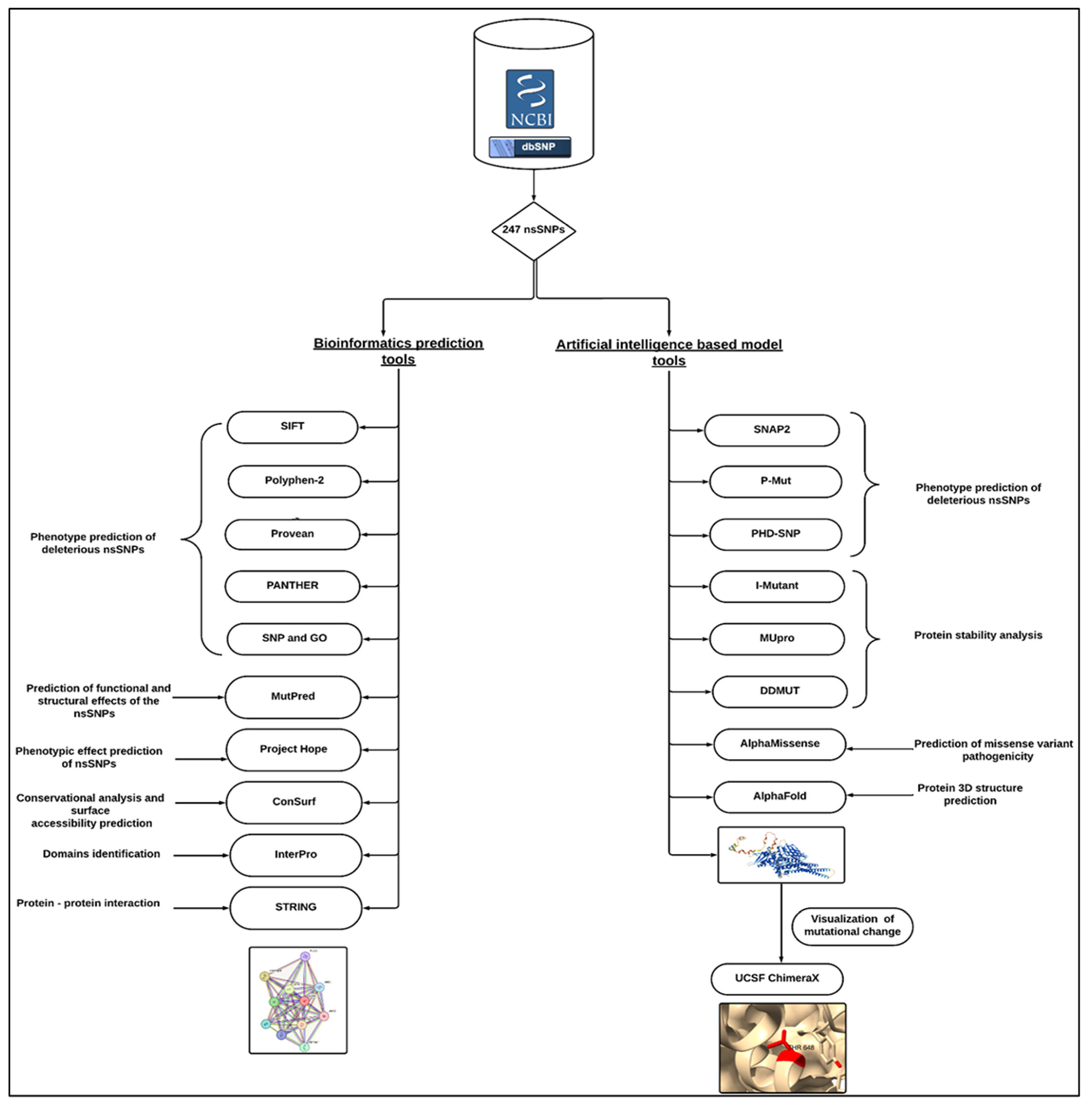
2. Materials and Method

An overview of the complete methodological approach is shown in Figure 1.

2.1. Data Retrieval

We gathered the data for the human STAT1 gene from the National Center for Biological Information (NCBI) website (https://www.ncbi.nlm.nih.gov/) (accessed on 20 April 2024). While the SNP information (SNP ID) of the STAT1 gene was obtained from the NCBI dbSNP (https://www.ncbi.nlm.nih.gov/gene/?term=STAT1, accessed on 20 April 2024), the protein ID and its sequence were extracted from UniProtKB in Swiss-Prot databases with the accession number P42224 (https://www.expasy.org/search/uniprot, accessed on 20 April 2024) [34].

2.2. Phenotype Prediction of Deleterious ns SNPs

We predicted the deleterious nsSNPs by using eight different tools. Sorting Intolerant from Tolerant (SIFT) (http://sift.bii.a-star.edu.sg/, accessed on 20 April 2024) predicts whether the replacement of an amino acid alters protein function. We downloaded nsSNP IDs from the online databases of NCBI and then uploaded them to SIFT. Results were documented as damaging (deleterious) or benign (tolerated), depending on the cutoff value of 0.05, as values less than or equal to (0.0–0.04) were predicted to be damaging or intolerant, while (0.05_1) is benign or tolerated [35].
Polyphen-2 (http://genetics.bwh.harvard.edu/pph2/, accessed on 20 April 2024) analyzes multiple sequence alignments and the protein’s three-dimensional structure, then predicts the possible impact of amino acid substitutions on the stability and function of human proteins using structural and comparative evolutionary considerations. The prediction outcomes are classified as probably damaging, possibly damaging, or benign based on the position-specific independent counts value (PSIC), which ranges from 0 to 1. Values near zero are regarded as benign, while values near one are considered probably damaging [36].
Provean (https://www.jcvi.org/research/provean/, accessed on 20 April 2024) is a software tool that predicts whether an amino acid substitution or indel has an impact on the biological function of a protein. Variants with a score equal to or below −2.5 are considered deleterious, while variants with a score above −2.5 are neutral [37].
SNAP2 (https://rostlab.org/services/snap2web/, accessed on 20 April 2024) is a trained classifier that uses the “neural network” machine learning tool to predict the functional effects of mutations by utilizing several sequence and variant properties to discriminate between effect and neutral variants/nonsynonymous SNPs [38].
PHD-SNP (https://snps.biofold.org/phd-snp/phd-snp.html, accessed on 20 April 2024) uses a support vector machine (SVM)-based method trained to determine disease-associated nsSNPs using sequence information. PHD-SNP classifies mutations either to be disease-related (disease) or a neutral polymorphism [39].
SNP and GO (https://snps-and-go.biocomp.unibo.it/snps-and-go/, accessed on 20 April 2024) is a server for the prediction of single-point protein mutations likely to be involved in the development of diseases in humans [40].
P-Mut is a web-based tool for the annotation of pathological variants on proteins. It allows fast and accurate prediction of the pathological properties of single-point amino acid mutations based on the use of a neural network. It is available at (http://mmb.irbbarcelona.org/PMut, accessed on 20 April 2024) [41].
Protein Analysis through Evolutionary Relationships (PANTHER) (http://pantherdb.org/, accessed on 20 April 2024) uses a position-specific evolutionary preservation (PSEP) score to measure the length of time (in millions of years), with <200 my “probably benign”, <450 my “possibly damaging”, and 450 my “probably damaging” [42].

2.3. Predicting Functional and Structural Effects of the nsSNP

MutPred v1.2 (http://mutpred.mutdb.org/, accessed on 20 April 2024) is used for sorting disease-associated or neutral amino acid substitutions in humans. MutPred is an efficient web-based application tool that screens amino acid substitutions and predicts the molecular base of the disease [43].

2.4. Protein Stability Analysis of Predicted STAT1 nsSNPs

I-Mutant 3.0 is available at (https://gpcr2.biocomp.unibo.it/cgi/predictors/I-Mutant3.0/I-Mutant3.0.cgi, accessed on 20 April 2024); it is a neural-network-based tool for routinely analyzing protein stability and change while taking single-site mutations into consideration [44]. The FASTA sequence of proteins retrieved from UniProt is used as an input to predict the mutational effect on protein stability.
MUpro, a group of machine learning methods, predicts the effects of single amino acid substitutions on protein stability [45]. It uses both support vector machines and neural networks; the output is either increased or decreased stability [45]. MUpro also interprets the result based on Gibbs free energy (ΔΔG), with a confidence score between −1 and 11. It is available at http://mupro.proteomics.ics.uci.edu, accessed on 20 April 2024.
DDMUT (https://biosig.lab.uq.edu.au/ddmut/, accessed on 20 April 2024) is a fast and accurate network using deep learning models to predict changes in Gibbs free energy (ΔΔG) upon single- and multiple-point mutations [46]. DDMut achieved a Pearson’s correlation of up to 0.70 (RMSE: 1.37 kcal/mol) on predicting single-point mutations on cross-validation and 0.74 (RMSE: 1.67 kcal/mol) on multiple mutations.

2.5. Prediction of Missense Variant Pathogenicity

Alpha Missense is an adaptation of alphafold fine-tuned on human and primate variant population frequency databases to predict missense variant pathogenicity. It works by combining structural context and evolutionary conservation. This model achieves state-of-the-art results across a wide range of genetic and experimental benchmarks, all without explicitly training on such data [47].

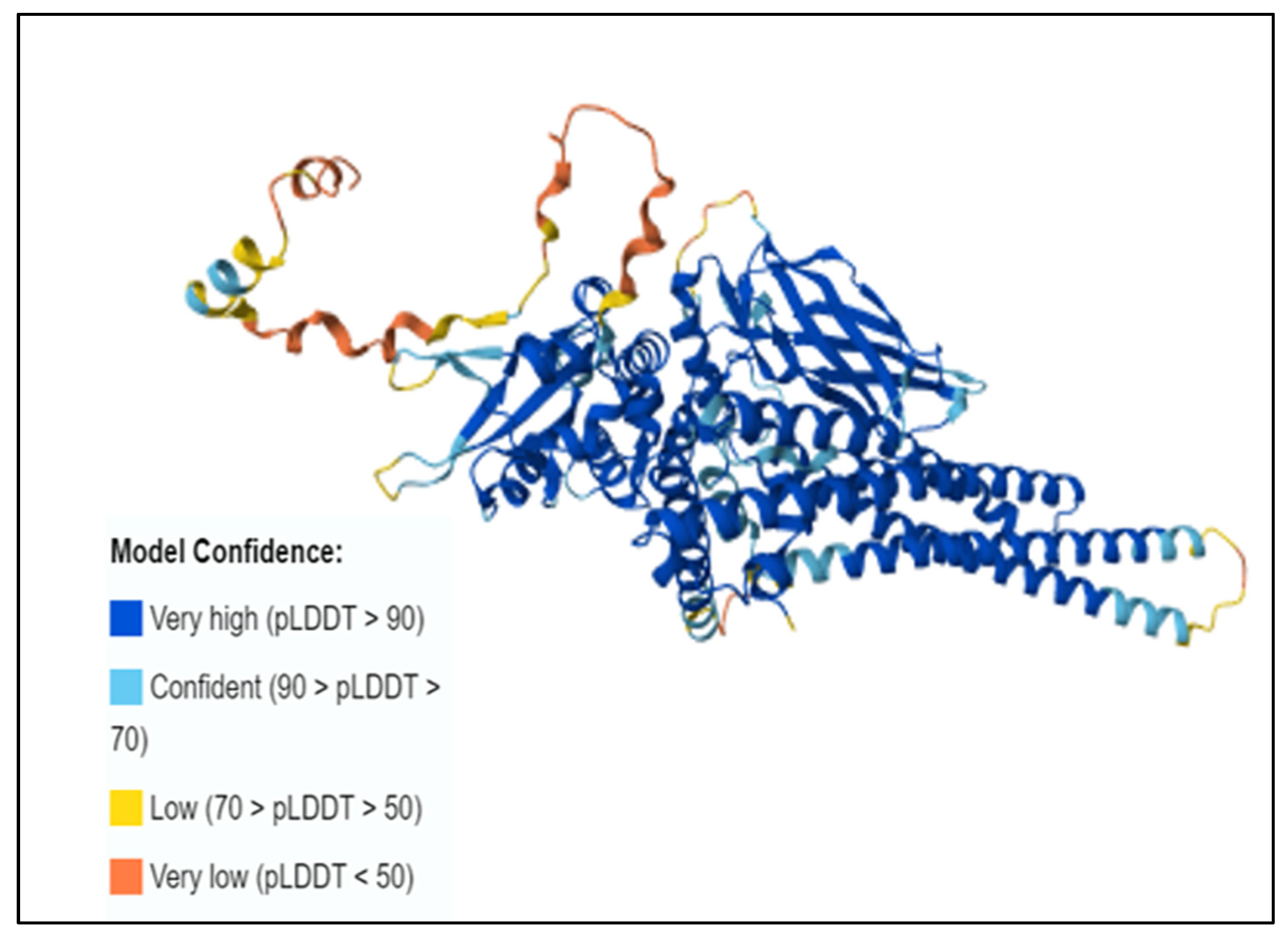
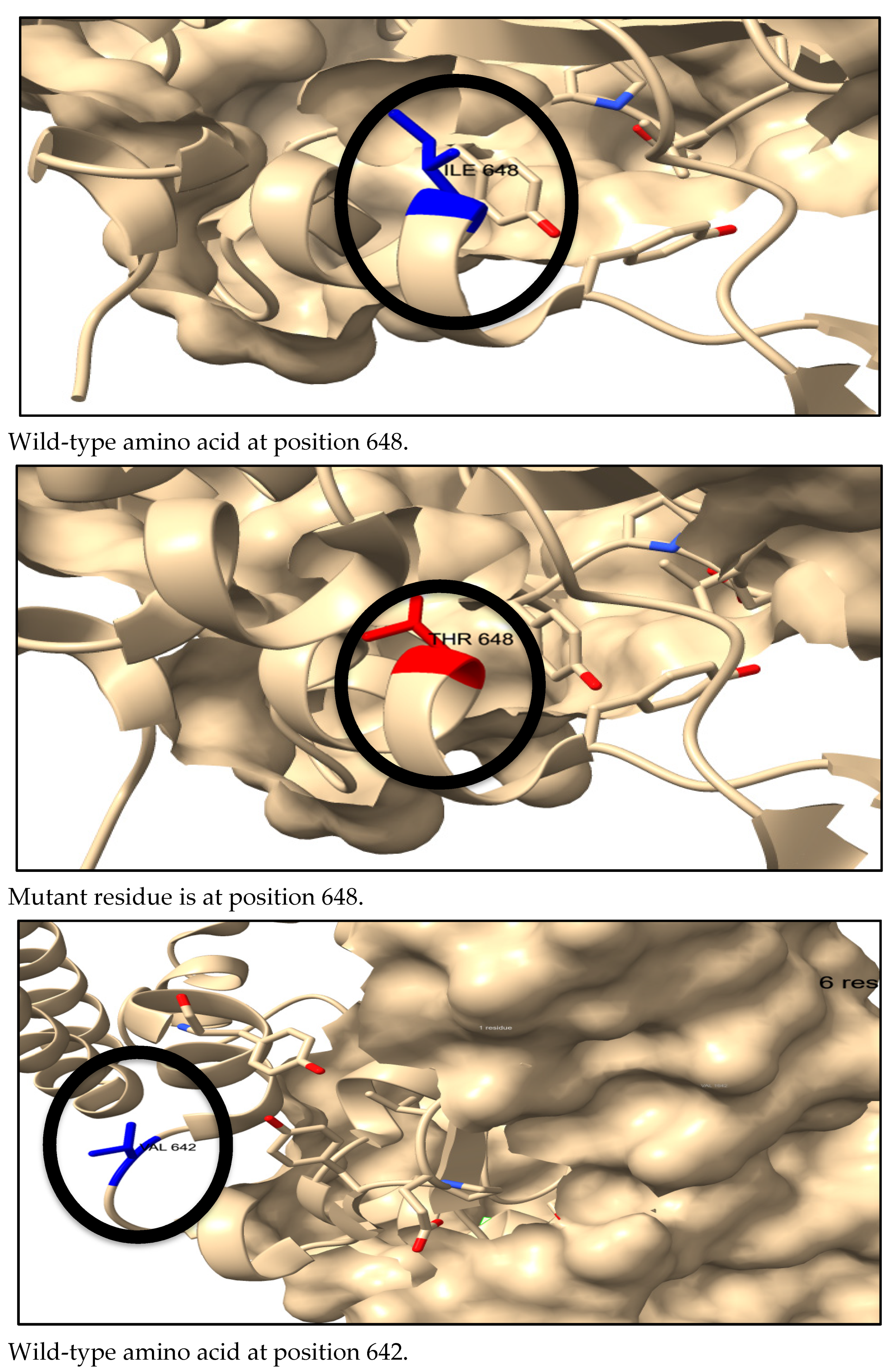
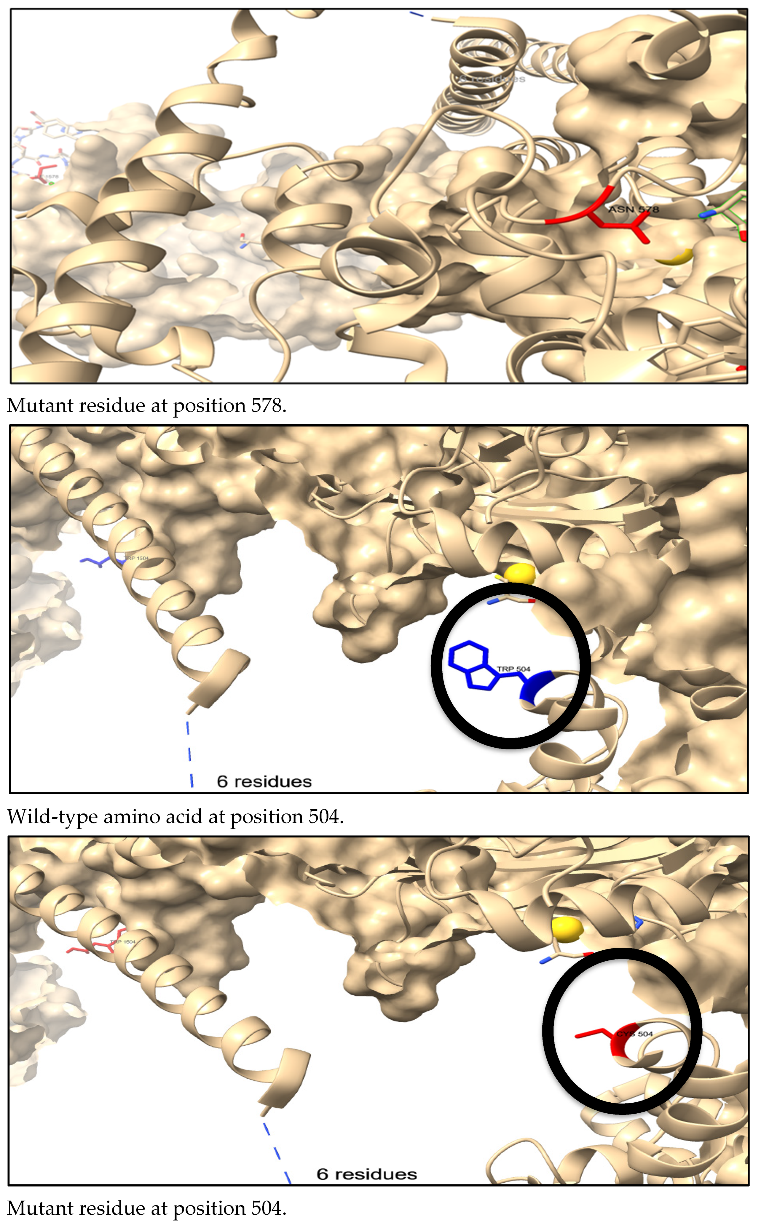
2.6. Three-Dimensional Structure Prediction and Visualization

We predicted the 3D structure using an artificial intelligence system, AlphaFold (https://alphafold.ebi.ac.uk, accessed on 20 April 2024). Alphafold is an artificial intelligence system developed by google DeepMind. It predicts a protein’s 3D structure from its amino acid sequence. It can predict protein structures computationally with high accuracy [48]. We used the UniProt sequence of the STAT1 protein as an input to obtain the alphafold model.
UCSF ChimeraX 1.9 is a robust application that enables interactive viewing and analysis of various molecular structures and related data, including density maps, sequence alignments, and supramolecular assemblies [49]. It allows the mapping and visualization of amino acid substitutions. Chimera X is available at https://www.rbvi.ucsf.edu/chimerax/, accessed on 20 April 2024.

2.7. Phenotypic Effects Prediction

Project Hope (version 1.0) is an online web server used to analyze the structural and conformational variations that have resulted from single amino acid substitutions [50]. We uploaded STAT1 protein sequence, wild-type amino acids, and mutants. The results provided describe the change in the physiochemical properties of the amino acid in the given SNPs. It is available at (https://www3.cmbi.umcn.nl/hope/method/, accessed on 20 April 2024).
DDMUT can also detect changes in the biological interactions between wild-type amino acids and neighborhood residues in comparison with mutant residues [46].

2.8. Conservational Analysis and Surface Accessibility Prediction of STAT1

The ConSurf bioinformatics tool (https://consurf.tau.ac.il, accessed on 20 April 2024) was used to study the evolutionary conservation of nsSNP positions in a protein sequence [51]. We submitted the FASTA sequence of the STAT1 protein to the server, and we screened out the highly conserved residues, exposed and buried residues.

2.9. Identification of nsSNPs in STAT1 Protein Domains

We submitted the FASTA sequence of the STAT1 protein to the InterPro server (https://www.ebi.ac.uk/interpro, accessed on 20 April 2024). It predicts protein families and conserved domains, and then we manually pinpointed the positions of nsSNPs within these domains [52].

2.10. Prediction of Protein–Protein Interactions

A precomputed database, STRING (https://string-db.org/, accessed on 20 April 2024), is used to determine protein–protein interactions to understand the function, structure, molecular action, and regulation of the protein [53]. We submitted the protein sequence as an input query.

3. Results

3.1. Distribution of STAT1 Gene SNP Datasets

The total number of SNPs was 10,989. There were 888 frame shift mutations; 480 SNPs located in the coding region, of which 247 were nsSNPs and 233 were synonymous SNPs (sSNPs), while 9.621 SNPs were in noncoding regions, of which 375 occurred in the 3′UTR, 131 in the 5′UTR region, and the rest (9115) were in the intronic region, as shown in Figure 2. We chose nonsynonymous coding SNPs for our investigation.

3.2. Identification of Deleterious Missense Mutation

All 247 nsSNPs were retrieved and subjected to pathogenicity prediction web servers. Sixty-four nsSNPs were found to be deleterious by SIFT and were further subjected to crosschecking by using three different tools (Poly-Phen-2, PROVEAN, and SNAP2).
The shortlisted 33 nsSNPs passed the first four tools, presented in Table 1, then were submitted to another set of four tools: P Mut, PhD-SNP, SNPs and GO, and PANTHER. In total, 29 SNPs out of the 33 predicted by the first set of tools are disease-causing by P mut, 21 out of 33 are disease-causing by Panther, 20 are disease-causing by PhD-SNP, and 14 out of 33 by SNP and GO. A final nine nsSNPs passed all eight tools shown in Table 2. We further analyzed the final set of SNPs for the functional and structural modifications.

3.3. MutPred Prediction for Functional and Structural Modifications

We submitted the shortlisted nine nsSNPs to the MutPred server, along with the resultant probability scores and their p values in Table 3. The structural and functional alterations predicted include loss of disorder, catalytic residue, glycosylation, gain of phosphorylation, solvent accessibility, ubiquitination, and molecular recognition features (MoRF) binding. According to these predictions, several nsSNPs might be the reason behind any possible structural and functional modifications of STAT1 protein.

3.4. Prediction of Change in STAT1 Stability Due to Mutation

We used I mutant, MUpro, and DDMUT servers to predict the effect of the nsSNPs on protein stability. The result revealed that six variants destabilized the STAT 1 protein, namely (I648T) rs759271255, (V642D) rs752542806, (R602W) rs 1209841496, (L600P) rs137852678, (I578N) rs767475430, and (W504C) rs916580554. The results are presented in Table 4.

3.5. Pathogenicity Prediction Results

We analyzed STAT1 nsSNP by Alpha-Missense, and we found that all the pathogenic nsSNPs that were predicted by the previous tools were also classified as pathogenic in Alpha-Missense, presented in Table 5. The heat map represented the mutations in STAT1, as shown in Figure 3.

3.6. The Conservational Status and Surface Accessibility Analysis of STAT1 Protein

Highly conserved residues are most likely to be involved in proteins’ structural integrity and functions. We evaluated the conservational profile for the STAT1 protein. The ConSurf algorithm represented the structural and functional conservation levels of all the amino acid residues of the STAT1 protein. Four SNPs (I648T, L600P, W504C, and I578N) are predicted to be located in a conserved region. L600P and I578N are predicted to be structural residues (highly conserved and buried). V642D is predicted as buried, and R602W is predicted as a functional residue (highly conserved and exposed), presented in Table 6.

3.7. Three-Dimensional Structure Prediction by AlphaFold and SNP Visualization by ChimeraX

An individual residue confidence score (pLDDT) between 0 and 100 is generated by the AlphaFold algorithm. Alphafold produces a per residue confidence score (pLDDT) 1–100. Regions with low pLDDT may be unstructured in isolation. The majority of the 3D structural region corresponds to alpha-helical domains and has extremely high confidence (pLDDT > 90). The remaining components of the model are depicted as unresolved loops with low (70 > pLDDT > 50) and extremely low (pLDDT > 50) scores, as in Figure 4.
We used ChimeraX to visualize the 3D structures of the wild-type amino acids in blue and the mutant residues in red, as shown in Figure 5.

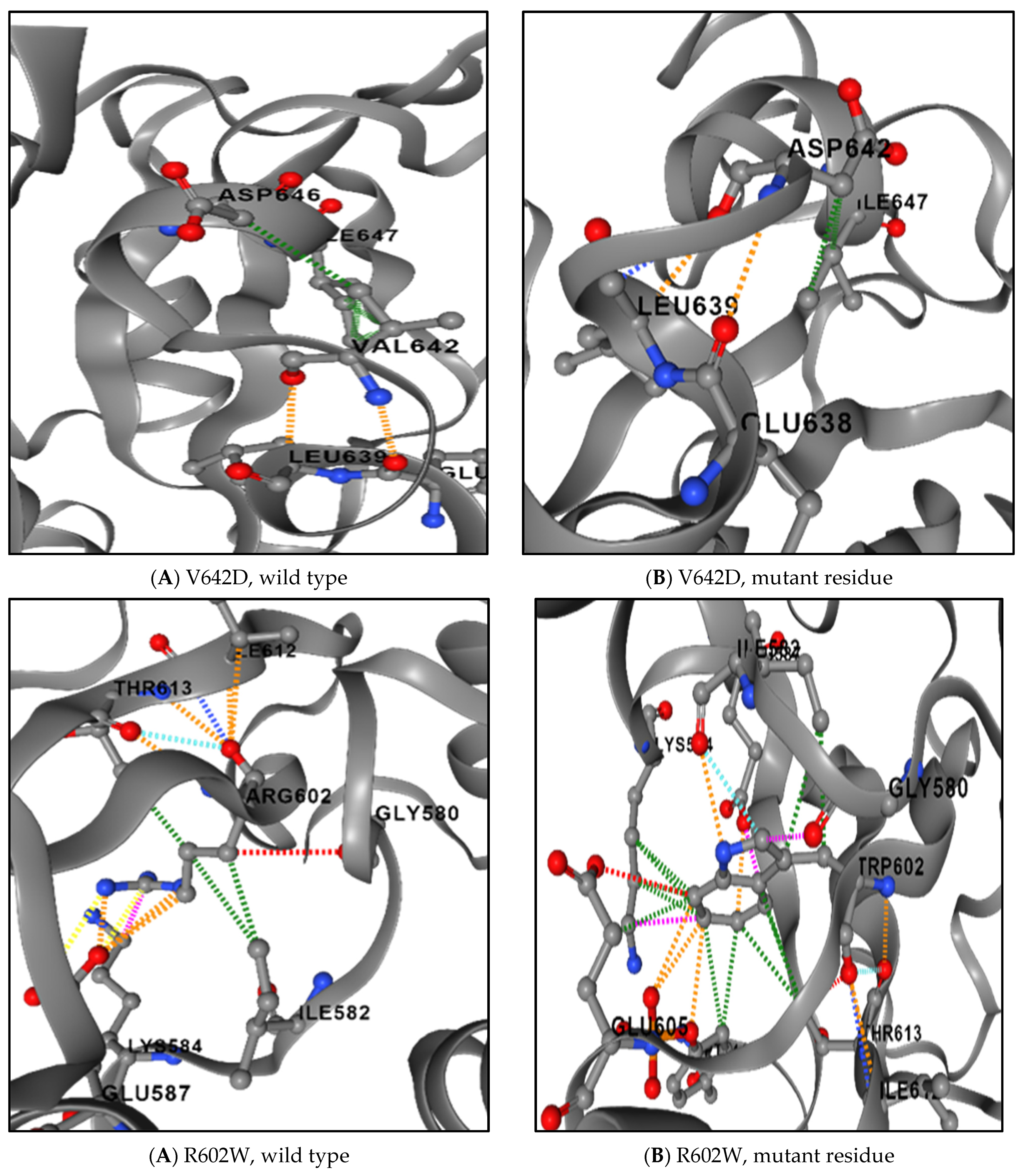
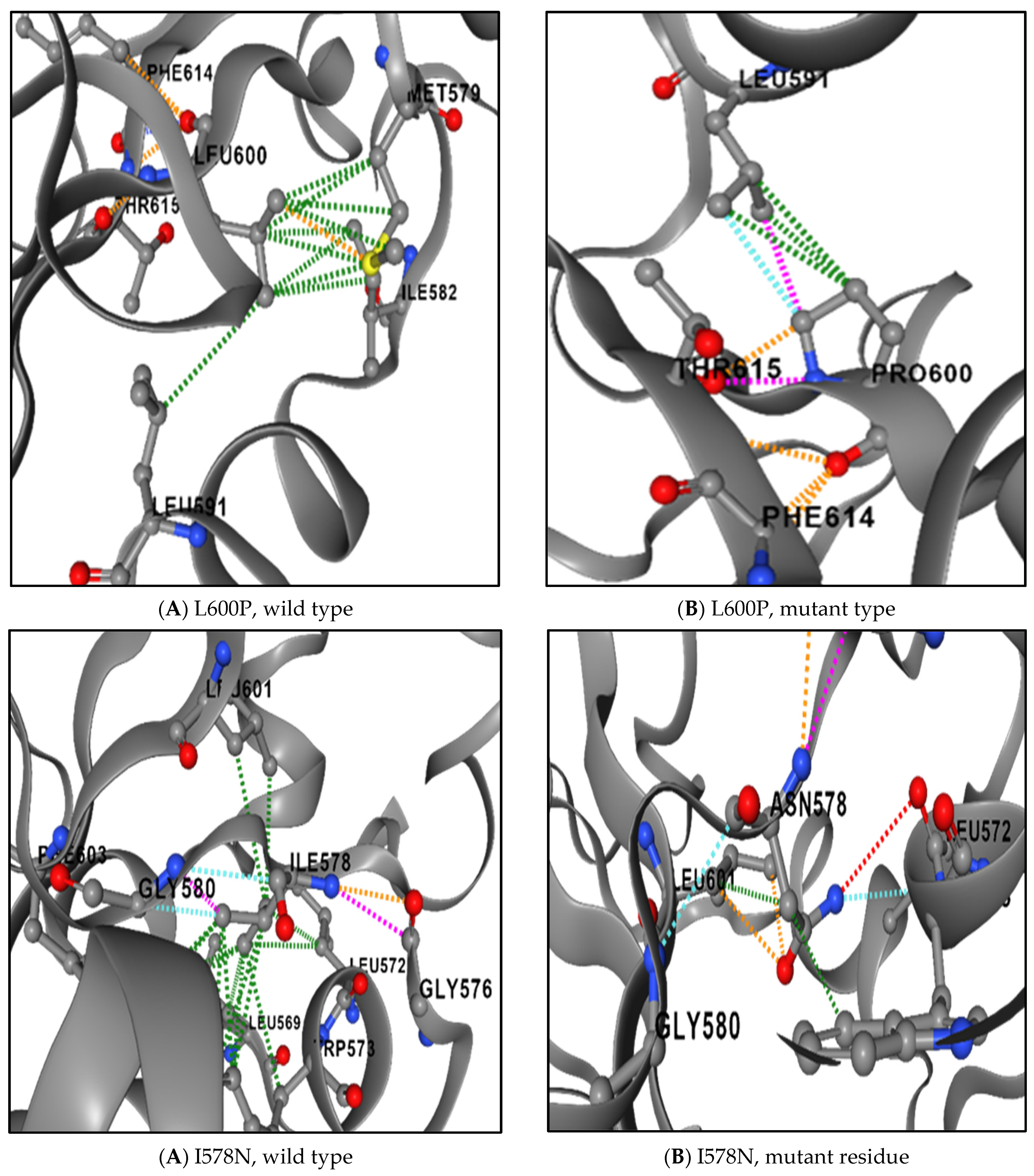
3.8. The Physical Outcome of Predicted SNPs

We examined the impact of the generated damaging SNPs on the three-dimensional structure of STAT1 using the HOPE server. The server predicted that all the mutated amino acids were different in size; one had a different charge, and six had different hydrophobicity. The results are in Table 7.
Loss of the interactions between the wild-type amino acid and other amino acids in the protein and/or development of new interactions or bonds between the mutant residue of the protein and the other amino acids in the protein were predicted by DDMUT, as presented in Figure 6.

3.9. Domain Identification of the STAT1 Protein by the InterPro Server

The InterPro tool predicted the domain regions of the STAT1 protein. The STAT1, SH2 domain (a phosphotyrosine binding pocket) at position (557–707), STAT transcription factor, DNA binding domain at (323–458), and STAT1_TAZ2-binding domain (715–739) are conserved sites. Src homology 2 (SH2) domain profile (573–670), SH2 domain (578–638), STAT1 transcription factor, all alpha domain (144–305), and STAT transcription factor protein interaction (2–12) are as in Table 8.

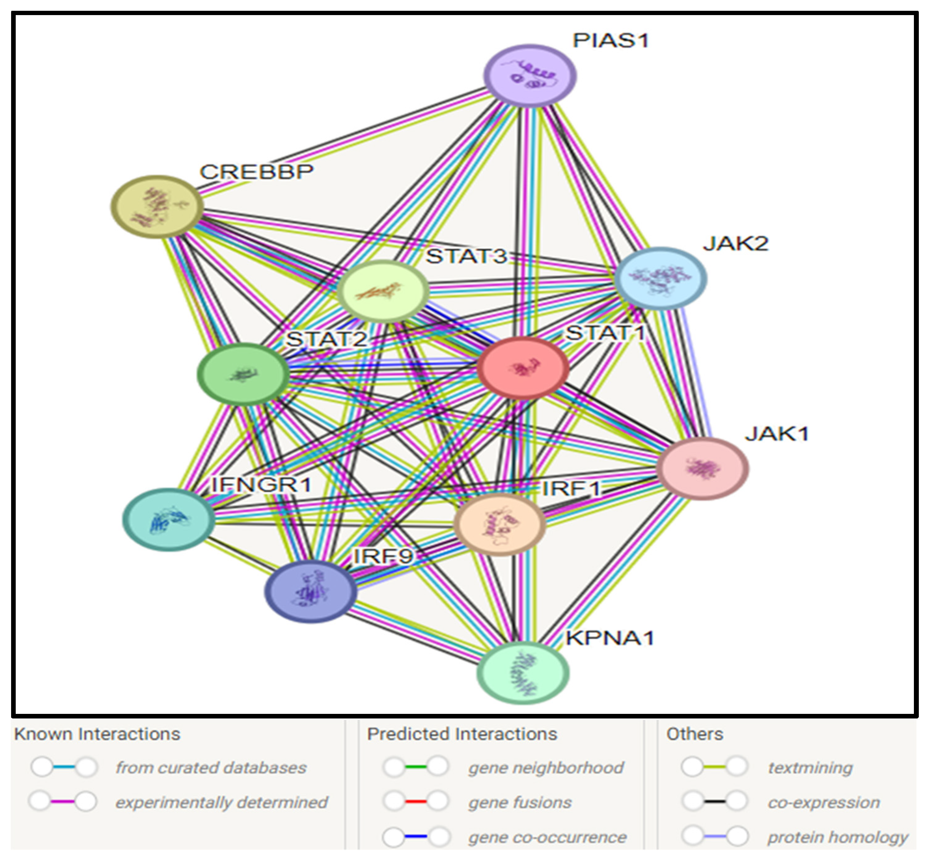
3.10. STAT1–Protein Interaction

Analysis of protein–protein interaction using the STRING network revealed that STAT1 interacts with 10 proteins, which include other proteins of the same STAT family (STAT2 and STAT3), proteins of the JAK family (JAK1 and JAK2), IFR1, IFR9, IFNGR1, CREBBP, KBNA1, and PIAS1, as presented in Figure 7.

4. Discussion

We evaluated the functional and pathogenic sequences of missense SNPs of the human STAT1 gene, utilizing 12 diverse in silico prediction tools (SIFT, PolyPhen2, PROVEAN, PANTHER, P MUT, PhD-SNP, SNPs&GO, SNAP2, and MutPred2). In silico prediction analysis identified six variants (I648T, V642D, R602W, L600P, I578N, and W504C) considered pathogenic and deleterious. These mutations have a major impact on the protein’s physicochemical characteristics, such as its size and charge hydrophobicity, which ultimately affect the protein’s stability and function and may have an impact on disease. Furthermore, we assessed the effect of missense SNPs on the stability of the STAT1 structure utilizing three stability prediction algorithms: I-Mutant3, MUpro, and DDMUT. All the variants revealed a reduction in stability by the three stability prediction tools (I-Mutant3, MUpro, and DDMUT). In general, we assumed that all missense SNPs in the STAT1 gene were highly unstable in their protein structures, so they were selected for further structural bioinformatics analysis utilizing various tools to explore the consequences of tentatively destructive missense SNPs on STAT1 protein function. To evaluate the conservation profile, we used the ConSurf algorithm to represent the structural and functional conservation levels of all the amino acid residues of STAT1 protein. The ConSurf analysis revealed that the variant in position 602 is a functional residue in a highly conserved and exposed position. Structural residues in highly conserved and buried positions were identified in positions 600 and 578. The identified variants were found in a highly conserved region; this finding suggests that they might be involved in modifications of molecular mechanisms such as bond gain or loss.
STAT1 GOF mutations with CMC were first described in 2001 and 2011, respectively; later, studies confirmed that STAT1-GOF mutations cause immunodeficiency and immune dysregulation, with a wide clinical spectrum [54].
Among the six SNPs identified linked to STAT1 gene mutations in this study, some of these SNPs have been associated with diseases in previous studies, while others were projected to be so in this study using various computational tools. Population genetics and clinical studies are crucial to verifying the results of such research, even though utilizing computational techniques to analyze the impact of the SNPs may aid in identifying disease-related SNPs.
One mutation, namely L600P, has already been previously reported as a mutation in the STAT1 gene in an infant who died of a viral-like illness associated with complete STAT1 deficiency and carried a homozygous nucleotide substitution (T→C) in exon 20, resulting in the substitution of a proline for a leucine at amino acid position 600 [55]. This mutation was found to be pathogenic using all the bioinformatics tools. I648T, V642D, R602W, I578N, and W504C were not reported previously.
Three mutations, namely L706S (rsRCV000009610), Q463H (VAR_065817), and E320Q (VAR_065816), have been reported as mutations in the STAT1 gene. The two previously reported types of autosomal-dominant (AD) Mendelian susceptibility to mycobacterial disease (AD-MSMD) causing STAT1 mutations are located in the tail segment domain (p.L706S) or in the DNA-binding domain (p.E320Q and p.Q463H) [56]. These mutations were not available in the dbSNP database. Two other SNPs (K637E) and (K673R) affecting the SH2 domain, which has been previously reported in two cases with AD-STAT1 deficiency in two unrelated patients from Japan and Saudi Arabia, were also not available in the dbSNPs database at the time of the analysis [56].
Two mutations linked to chronic mucocutaneous candidiasis are (T437I) and (Q271P). Q271P occurred within a specific pocket of the STAT1 coiled-coil domain, near residues essential for dephosphorylation, and was identified in a German patient who presented at 1 year of age with autosomal dominant chronic mucocutaneous candidiasis, showed signs of thyroid autoimmunity, and died at age 41 from squamous cell carcinoma [57,58]. These mutations were not available in the dbSNPs database.
The A267V variant in STAT1 has been reported in >10 individuals with chronic mucocutaneous candidiasis (CMC) and segregated with disease in 16 individuals from nine families [59]. This mutation was not present in the dbSNP database.
Interestingly, nsSNPs in the STAT1 gene will ultimately affect and may disturb the normal function of other interacting genes. As our study was in detail, it provides all the information and analysis needed for the identification of the most damaging nsSNPs. Like ours, there are certain limitations in every study. Utilizing in silico technologies is now a crucial method for identifying disease-related SNPs. In this study, the STAT1 gene underwent a thorough analysis utilizing 18 genetics analysis tools (10 computational tools and 8 AI-based methods) to determine the impact of nsSNPs on the protein’s structure and function.
Our study is based on computer tools and web servers, which are based on mathematical and statistical algorithms. Therefore, to confirm these results, experimental investigation is necessary.

5. Conclusions

Our study provides an insight about nsSNPs of the STAT1 gene, its protein 3D structure, and its interactions with other genes, which might be helpful in future studies of STAT1 in order to better understand its role in immunity and all related diseases.

Supplementary Materials

The following supporting information can be downloaded at: https://www.mdpi.com/article/10.3390/genes16030303/s1, Figure S1: Shows the percentages of the SNPs in STAT1 gene; Figure S2: Heat map generated by alpha-missense, shows the variations in STAT1 gene; Figure S3: Conservation profile of amino acids in STAT1protein; Figure S4: Protein 2D structure of human STAT1predicted by AlphaFold2; Figure S5: Effect of the six most deleterious nsSNPs on the STAT1 protein structure; Figure S6: Difference in ionic interactions between the wild-type (A) and mutant residues (B) in I648T; Table S1: List of nsSNPs that were predicted to have deleterious effect by SIFT, PolyPhen-2, Provean and SNAP2; Table S2: MutPred probability values of deleterious and pathogenic nsSNPs identified in STAT1; Table S3: Deleterious and pathogenic ns SNPs were predicted to have significant decrease on protein stability by I-MUTANT 3.0 algorithm, MUpro, and DDMUT; Table S4: Shows Alpha-missense prediction of the pathogenic nsSNPs in STAT1; Table S5: Conservation profile of most damaging nsSNPs of STAT1; Table S6: Changes in physical properties between wild-type and mutant residues predicted by project hope; Table S7: Domain regions of the selected most damaging nsSNPs in STAT1.

Author Contributions

Conceptualization, E.K., L.A.K. and M.A.; Data curation, E.K., A.A. and M.A.; Funding acquisition, E.K.; Investigation, E.K., M.A., L.A.K. and A.A.; Methodology, E.K. and M.A.; Software, E.K. and L.A.K.; Supervision, A.A.; Validation, E.K. and M.A.; Writing—original draft, E.K., L.A.K., M.A. and A.A.; Writing—review and editing, E.K., L.A.K., M.A. and A.A. All authors have read and agreed to the published version of the manuscript.

Funding

This study is supported via funding from Prince Sattam bin Abdulaziz University Grant Number: 2024/03/28313.

Institutional Review Board Statement

Not applicable.

Informed Consent Statement

Not applicable.

Data Availability Statement

The original contributions presented in this study are included in the article/Supplementary Materials. Further inquiries can be directed to the corresponding author.

Conflicts of Interest

The authors declare no conflicts of interest.

References

  1. Awasthi, N.; Liongue, C.; Ward, A.C. STAT proteins: A kaleidoscope of canonical and non-canonical functions in immunity and cancer. J. Hematol. Oncol. 2021, 14, 198. [Google Scholar] [CrossRef] [PubMed]
  2. Calò, V.; Migliavacca, M.; Bazan, V.; Macaluso, M.; Buscemi, M.; Gebbia, N.; Russo, A. STAT proteins: From normal control of cellular events to tumorigenesis. J. Cell. Physiol. 2003, 197, 157–168. [Google Scholar] [CrossRef] [PubMed]
  3. Zhong, M.; Henriksen, M.A.; Takeuchi, K.; Schaefer, O.; Liu, B.; ten Hoeve, J.; Ren, Z.; Mao, X.; Chen, X.; Shuai, K.; et al. Implications of an antiparallel dimeric structure of nonphosphorylated STAT1 for the activation-inactivation cycle. Proc. Natl. Acad. Sci. USA 2005, 102, 3966–3971. [Google Scholar] [CrossRef]
  4. Mao, X.; Ren, Z.; Parker, G.N.; Sondermann, H.; Pastorello, M.A.; Wang, W.; McMurray, J.S.; Demeler, B.; Darnell, J.E.; Chen, X. Structural bases of unphosphorylated STAT1 association and receptor binding. Mol. Cell 2005, 17, 761–771. [Google Scholar] [CrossRef] [PubMed]
  5. Metwally, H.; Kishimoto, T. Distinct Phosphorylation of STAT1 Confers Distinct DNA Binding and Gene-regulatory Properties. J. Cell. Signal. 2020, 1, 50–55. [Google Scholar] [CrossRef]
  6. Lorenzini, T.; Dotta, L.; Giacomelli, M.; Vairo, D.; Badolato, R. STAT mutations as program switchers: Turning primary immunodeficiencies into autoimmune diseases. J. Leukoc. Biol. 2017, 101, 29–38. [Google Scholar] [CrossRef]
  7. Asano, T.; Utsumi, T.; Kagawa, R.; Karakawa, S.; Okada, S. Inborn errors of immunity with loss- and gain-of-function germline mutations in STAT1. Clin. Exp. Immunol. 2023, 212, 96–106. [Google Scholar] [CrossRef]
  8. Mizoguchi, Y.; Okada, S. Inborn errors of STAT1 immunity. Curr. Opin. Immunol. 2021, 72, 59–64. [Google Scholar] [CrossRef]
  9. Verhoeven, Y.; Tilborghs, S.; Jacobs, J.; De Waele, J.; Quatannens, D.; Deben, C.; Prenen, H.; Pauwels, P.; Trinh, X.B.; Wouters, A.; et al. The potential and controversy of targeting STAT family members in cancer. Semin. Cancer Biol. 2020, 60, 41–56. [Google Scholar] [CrossRef]
  10. Liongue, C.; Sobah, M.L.; Ward, A.C. Signal transducer and activator of transcription proteins at the nexus of immunodeficiency, autoimmunity and cancer. Biomedicines 2023, 12, 45. [Google Scholar] [CrossRef]
  11. Reich, N.C. STATs get their move on. Jak-stat 2013, 2, e27080. [Google Scholar] [CrossRef] [PubMed]
  12. de Prati, A.C.; Ciampa, A.R.; Cavalieri, E.; Zaffini, R.; Darra, E.; Menegazzi, M.; Suzuki, H.; Mariotto, S. STAT1 as a new molecular target of anti-inflammatory treatment. Curr. Med. Chem. 2005, 12, 1819–1828. [Google Scholar] [CrossRef] [PubMed]
  13. Tolomeo, M.; Cavalli, A.; Cascio, A. STAT1 and its crucial role in the control of viral infections. Int. J. Mol. Sci. 2022, 23, 4095. [Google Scholar] [CrossRef] [PubMed]
  14. Asano, T.; Noma, K.; Mizoguchi, Y.; Karakawa, S.; Okada, S. Human STAT1 gain of function with chronic mucocutaneous candidiasis: A comprehensive review for strengthening the connection between bedside observations and laboratory research. Immunol. Rev. 2024, 322, 81–97. [Google Scholar] [CrossRef]
  15. Shuai, K.; Schindler, C.; Prezioso, V.R.; Darnell, J.E. Activation of transcription by IFN-γ: Tyrosine phosphorylation of a 91-kD DNA binding protein. Science 1992, 258, 1808–1812. [Google Scholar] [CrossRef]
  16. Heim, M.H. The Jak-STAT pathway: Cytokine signalling from the receptor to the nucleus. J. Recept. Signal Transduct. 1999, 19, 75–120. [Google Scholar] [CrossRef]
  17. Eilers, A.; Georgellis, D.; Klose, B.; Schindler, C.; Ziemiecki, A.; Harpur, A.G.; Wilks, A.F.; Decker, T. Differentiation-regulated serine phosphorylation of STAT1 promotes GAF activation in macrophages. Mol. Cell. Biol. 1995, 15, 3579–3586. [Google Scholar] [CrossRef]
  18. Decker, T.; Lew, D.J.; Mirkovitch, J.; Darnell, J.E. Cytoplasmic activation of GAF, an IFN-gamma-regulated DNA-binding factor. EMBO J. 1991, 10, 927–932. [Google Scholar] [CrossRef]
  19. Meesilpavikkai, K.; Hirankarn, N.; Dalm, V.A.S.H.; van Hagen, P.M.; Dik, W.A.; IJspeert, H. Unraveling the Immunogenetics of STAT Proteins: Clinical Perspectives on Gain-of-Function and Loss-of-Function Variants. Asian Pac. J. Allergy Immunol. 2024, 42, 105–122. [Google Scholar] [CrossRef]
  20. Chen, X.; Chen, J.; Chen, R.; Mou, H.; Sun, G.; Yang, L.; Jia, Y.; Zhao, Q.; Wen, W.; Zhou, L.; et al. Genetic and Functional Identifying of Novel STAT1 Loss-of-Function Mutations in Patients with Diverse Clinical Phenotypes. J. Clin. Immunol. 2022, 42, 1778–1794. [Google Scholar] [CrossRef]
  21. Boisson-Dupuis, S.; Kong, X.-F.; Okada, S.; Cypowyj, S.; Puel, A.; Abel, L.; Casanova, J.-L. Inborn errors of human STAT1: Allelic heterogeneity governs the diversity of immunological and infectious phenotypes. Curr. Opin. Immunol. 2012, 24, 364–378. [Google Scholar] [CrossRef] [PubMed]
  22. Tsumura, M.; Okada, S.; Sakai, H.; Yasunaga, S.; Ohtsubo, M.; Murata, T.; Obata, H.; Yasumi, T.; Kong, X.-F.; Abhyankar, A.; et al. Dominant-negative STAT1 SH2 domain mutations in unrelated patients with Mendelian susceptibility to mycobacterial disease. Hum. Mutat. 2012, 33, 1377–1387. [Google Scholar] [CrossRef] [PubMed]
  23. Uzel, G.; Sampaio, E.P.; Lawrence, M.G.; Hsu, A.P.; Hackett, M.; Dorsey, M.J.; Noel, R.J.; Verbsky, J.W.; Freeman, A.F.; Janssen, E.; et al. Dominant gain-of-function STAT1 mutations in FOXP3 wild-type immune dysregulation-polyendocrinopathy-enteropathy-X-linked-like syndrome. J. Allergy Clin. Immunol. 2013, 131, 1611–1623. [Google Scholar] [CrossRef] [PubMed]
  24. Hartono, S.P.; Vargas-Hernández, A.; Ponsford, M.J.; Chinn, I.K.; Jolles, S.; Wilson, K.; Forbes, L.R. Novel STAT1 Gain-of-Function Mutation Presenting as Combined Immunodeficiency. J. Clin. Immunol. 2018, 38, 753–756. [Google Scholar] [CrossRef]
  25. Henrickson, S.E.; Dolan, J.G.; Forbes, L.R.; Vargas-Hernández, A.; Nishimura, S.; Okada, S.; Kersun, L.S.; Brodeur, G.M.; Heimall, J.R. Gain-of-Function STAT1 Mutation With Familial Lymphadenopathy and Hodgkin Lymphoma. Front. Pediatr. 2019, 7, 160. [Google Scholar] [CrossRef]
  26. Okada, S.; Asano, T.; Moriya, K.; Boisson-Dupuis, S.; Kobayashi, M.; Casanova, J.-L.; Puel, A. Human STAT1 Gain-of-Function Heterozygous Mutations: Chronic Mucocutaneous Candidiasis and Type I Interferonopathy. J. Clin. Immunol. 2020, 40, 1065–1081. [Google Scholar] [CrossRef]
  27. Wang, X.; Zhao, W.; Chen, F.; Zhou, P.; Yan, Z. Chinese Pedigree of Chronic Mucocutaneous Candidiasis Due to STAT1 Gain-of-Function Mutation: A Case Study and Literature Review. Mycopathologia 2023, 188, 87–97. [Google Scholar] [CrossRef]
  28. Egri, N.; Esteve-Solé, A.; Deyà-Martínez, À.; Ortiz de Landazuri, I.; Vlagea, A.; García, A.P.; Cardozo, C.; Garcia-Vidal, C.; Bartolomé, C.S.; Español-Rego, M.; et al. Primary immunodeficiency and chronic mucocutaneous candidiasis: Pathophysiological, diagnostic, and therapeutic approaches. Allergol. Immunopathol. 2021, 49, 118–127. [Google Scholar] [CrossRef]
  29. Collins, F.S.; Brooks, L.D.; Chakravarti, A. A DNA polymorphism discovery resource for research on human genetic variation. Genome Res. 1998, 8, 1229–1231. [Google Scholar] [CrossRef]
  30. Arshad, S.; Ishaque, I.; Mumtaz, S.; Rashid, M.U.; Malkani, N. In-Silico Analyses of Nonsynonymous Variants in the BRCA1 Gene. Biochem. Genet. 2021, 59, 1506–1526. [Google Scholar] [CrossRef]
  31. Yazar, M.; Özbek, P. In Silico Tools and Approaches for the Prediction of Functional and Structural Effects of Single-Nucleotide Polymorphisms on Proteins: An Expert Review. OMICS J. Integr. Biol. 2021, 25, 23–37. [Google Scholar] [CrossRef] [PubMed]
  32. Allemailem, K.S.; Almatroudi, A.; Alrumaihi, F.; Makki Almansour, N.; Aldakheel, F.M.; Rather, R.A.; Afroze, D.; Rah, B. Single nucleotide polymorphisms (SNPs) in prostate cancer: Its implications in diagnostics and therapeutics. Am. J. Transl. Res. 2021, 13, 3868–3889. [Google Scholar] [PubMed]
  33. Clifford, R.J.; Edmonson, M.N.; Nguyen, C.; Scherpbier, T.; Hu, Y.; Buetow, K.H. Bioinformatics tools for single nucleotide polymorphism discovery and analysis. Ann. N. Y. Acad. Sci. 2004, 1020, 101–109. [Google Scholar] [CrossRef]
  34. Artimo, P.; Jonnalagedda, M.; Arnold, K.; Baratin, D.; Csardi, G.; de Castro, E.; Duvaud, S.; Flegel, V.; Fortier, A.; Gasteiger, E.; et al. ExPASy: SIB bioinformatics resource portal. Nucleic Acids Res. 2012, 40, W597–W603. [Google Scholar] [CrossRef]
  35. Kumar, P.; Henikoff, S.; Ng, P.C. Predicting the effects of coding non-synonymous variants on protein function using the SIFT algorithm. Nat. Protoc. 2009, 4, 1073–1081. [Google Scholar] [CrossRef]
  36. Adzhubei, I.; Jordan, D.M.; Sunyaev, S.R. Predicting functional effect of human missense mutations using PolyPhen-2. Curr. Protoc. Hum. Genet. 2013, 76, 7.20.1–7.20.41. [Google Scholar] [CrossRef]
  37. Choi, Y.; Chan, A.P. PROVEAN web server: A tool to predict the functional effect of amino acid substitutions and indels. Bioinformatics 2015, 31, 2745–2747. [Google Scholar] [CrossRef]
  38. Bromberg, Y.; Yachdav, G.; Rost, B. SNAP predicts effect of mutations on protein function. Bioinformatics 2008, 24, 2397–2398. [Google Scholar] [CrossRef]
  39. Capriotti, E.; Calabrese, R.; Casadio, R. Predicting the insurgence of human genetic diseases associated to single point protein mutations with support vector machines and evolutionary information. Bioinformatics 2006, 22, 2729–2734. [Google Scholar] [CrossRef]
  40. Capriotti, E.; Calabrese, R.; Fariselli, P.; Martelli, P.L.; Altman, R.B.; Casadio, R. WS-SNPs&GO: A web server for predicting the deleterious effect of human protein variants using functional annotation. BMC Genom. 2013, 14 (Suppl. S3), S6. [Google Scholar] [CrossRef]
  41. López-Ferrando, V.; Gazzo, A.; de la Cruz, X.; Orozco, M.; Gelpí, J.L. PMut: A web-based tool for the annotation of pathological variants on proteins, 2017 update. Nucleic Acids Res. 2017, 45, W222–W228. [Google Scholar] [CrossRef] [PubMed]
  42. Mi, H.; Muruganujan, A.; Thomas, P.D. PANTHER in 2013: Modeling the evolution of gene function, and other gene attributes, in the context of phylogenetic trees. Nucleic Acids Res. 2013, 41, D377–D386. [Google Scholar] [CrossRef] [PubMed]
  43. Pejaver, V.; Urresti, J.; Lugo-Martinez, J.; Pagel, K.A.; Lin, G.N.; Nam, H.-J.; Mort, M.; Cooper, D.N.; Sebat, J.; Iakoucheva, L.M.; et al. Inferring the molecular and phenotypic impact of amino acid variants with MutPred2. Nat. Commun. 2020, 11, 5918. [Google Scholar] [CrossRef]
  44. Capriotti, E.; Fariselli, P.; Calabrese, R.; Casadio, R. Predicting protein stability changes from sequences using support vector machines. Bioinformatics 2005, 21 (Suppl. S2), ii54–ii58. [Google Scholar] [CrossRef]
  45. Cheng, J.; Randall, A.; Baldi, P. Prediction of protein stability changes for single-site mutations using support vector machines. Proteins Struct. Funct. Bioinform. 2006, 62, 1125–1132. [Google Scholar] [CrossRef]
  46. Zhou, Y.; Pan, Q.; Pires, D.E.V.; Rodrigues, C.H.M.; Ascher, D.B. DDMut: Predicting effects of mutations on protein stability using deep learning. Nucleic Acids Res. 2023, 51, W122–W128. [Google Scholar] [CrossRef]
  47. Cheng, J.; Novati, G.; Pan, J.; Bycroft, C.; Žemgulytė, A.; Applebaum, T.; Pritzel, A.; Wong, L.H.; Zielinski, M.; Sargeant, T.; et al. Accurate proteome-wide missense variant effect prediction with AlphaMissense. Science 2023, 381, eadg7492. [Google Scholar] [CrossRef]
  48. Varadi, M.; Anyango, S.; Deshpande, M.; Nair, S.; Natassia, C.; Yordanova, G.; Yuan, D.; Stroe, O.; Wood, G.; Laydon, A.; et al. AlphaFold Protein Structure Database: Massively expanding the structural coverage of protein-sequence space with high-accuracy models. Nucleic Acids Res. 2022, 50, D439–D444. [Google Scholar] [CrossRef]
  49. Meng, E.C.; Goddard, T.D.; Pettersen, E.F.; Couch, G.S.; Pearson, Z.J.; Morris, J.H.; Ferrin, T.E. UCSF ChimeraX: Tools for structure building and analysis. Protein Sci. 2023, 32, e4792. [Google Scholar] [CrossRef]
  50. Venselaar, H.; Te Beek, T.A.H.; Kuipers, R.K.P.; Hekkelman, M.L.; Vriend, G. Protein structure analysis of mutations causing inheritable diseases. An e-Science approach with life scientist friendly interfaces. BMC Bioinform. 2010, 11, 548. [Google Scholar] [CrossRef]
  51. Ashkenazy, H.; Erez, E.; Martz, E.; Pupko, T.; Ben-Tal, N. ConSurf 2010: Calculating evolutionary conservation in sequence and structure of proteins and nucleic acids. Nucleic Acids Res. 2010, 38, W529–W533. [Google Scholar] [CrossRef] [PubMed]
  52. Mulder, N.J.; Apweiler, R.; Attwood, T.K.; Bairoch, A.; Bateman, A.; Binns, D.; Biswas, M.; Bradley, P.; Bork, P.; Bucher, P.; et al. InterPro Consortium InterPro: An integrated documentation resource for protein families, domains and functional sites. Brief Bioinform. 2002, 3, 225–235. [Google Scholar] [CrossRef] [PubMed]
  53. Szklarczyk, D.; Franceschini, A.; Kuhn, M.; Simonovic, M.; Roth, A.; Minguez, P.; Doerks, T.; Stark, M.; Muller, J.; Bork, P.; et al. The STRING database in 2011: Functional interaction networks of proteins, globally integrated and scored. Nucleic Acids Res. 2011, 39, D561–D568. [Google Scholar] [CrossRef] [PubMed]
  54. Zhang, W.; Chen, X.; Gao, G.; Xing, S.; Zhou, L.; Tang, X.; Zhao, X.; An, Y. Clinical Relevance of Gain- and Loss-of-Function Germline Mutations in STAT1: A Systematic Review. Front. Immunol. 2021, 12, 654406. [Google Scholar] [CrossRef]
  55. Dupuis, S.; Jouanguy, E.; Al-Hajjar, S.; Fieschi, C.; Al-Mohsen, I.Z.; Al-Jumaah, S.; Yang, K.; Chapgier, A.; Eidenschenk, C.; Eid, P.; et al. Impaired response to interferon-α/β and lethal viral disease in human STAT1 deficiency. Nat. Genet. 2003, 33, 388–391. [Google Scholar] [CrossRef]
  56. Chapgier, A.; Boisson-Dupuis, S.; Jouanguy, E.; Vogt, G.; Feinberg, J.; Prochnicka-Chalufour, A.; Casrouge, A.; Yang, K.; Soudais, C.; Fieschi, C.; et al. Novel STAT1 alleles in otherwise healthy patients with mycobacterial disease. PLoS Genet. 2006, 2, e131. [Google Scholar] [CrossRef]
  57. Liu, L.; Okada, S.; Kong, X.-F.; Kreins, A.Y.; Cypowyj, S.; Abhyankar, A.; Toubiana, J.; Itan, Y.; Audry, M.; Nitschke, P.; et al. Gain-of-function human STAT1 mutations impair IL-17 immunity and underlie chronic mucocutaneous candidiasis. J. Exp. Med. 2011, 208, 1635–1648. [Google Scholar] [CrossRef]
  58. Wang, X.; Zhang, R.; Wu, W.; Wang, A.; Wan, Z.; van de Veerdonk, F.L.; Li, R. New and recurrent STAT1 mutations in seven Chinese patients with chronic mucocutaneous candidiasis. Int. J. Dermatol. 2017, 56, e30–e33. [Google Scholar] [CrossRef]
  59. Breuer, O.; Daum, H.; Cohen-Cymberknoh, M.; Unger, S.; Shoseyov, D.; Stepensky, P.; Keller, B.; Warnatz, K.; Kerem, E. Autosomal dominant gain of function STAT1 mutation and severe bronchiectasis. Respir. Med. 2017, 126, 39–45. [Google Scholar] [CrossRef]
Figure 1. Workflow of the analysis.
Genes 16 00303 g001
Figure 2. Shows the distribution of the SNPs in STAT1 gene.
Genes 16 00303 g002
Figure 3. Heat map generated by Alpha-Missense shows the variations in STAT1 gene.
Genes 16 00303 g003
Figure 4. Protein 3D structure of human STAT1 predicted by AlphaFold2.
Genes 16 00303 g004
Figure 5. Effect of the six most deleterious nsSNPs on the STAT1 protein structure. ChimeraX software was used to visualize the 3D structure of the wild-type (blue), mutant residues (red) and gold ion (yellow).
Genes 16 00303 g005aGenes 16 00303 g005bGenes 16 00303 g005cGenes 16 00303 g005d
Figure 6. Difference in ionic interactions between the wild-type (A) and mutant residues (B).
Genes 16 00303 g006aGenes 16 00303 g006bGenes 16 00303 g006cGenes 16 00303 g006d
Figure 7. STAT1–protein interactions by STRING database.
Genes 16 00303 g007
Table 1. List of nsSNPs that were predicted to have deleterious effect by SIFT, PolyPhen-2, Provean, and SNAP.
SNP IDAmino Acid ChangeSIFTPoly-Phen-2ProveanSNAP2
PredictionTIEffectScoreEffectScorePredictionScore
1rs1173266737P728ADeleterious0PB0.974Deleterious−3.14E15
2rs1374373369D674VDeleterious0.01PB0.989Deleterious−7.327E47
3rs771679419Y668FDeleterious0PS0.609Deleterious−3.583E67
4rs759271255I648TDeleterious0.01PB1Deleterious−4.197E56
5rs752542806V642DDeleterious0PB0.994Deleterious−5.149E60
6rs1387961263L639FDeleterious0PB1Deleterious−3.489E7
7rs1209841496R602WDeleterious0PB1Deleterious−7.4E87
8rs137852678L600PDeleterious0PB1Deleterious−6.472E91
9rs1398307167P596QDeleterious0.01PS0.951Deleterious−3.974E57
10rs1398307167P596LDeleterious0.01PB0.966Deleterious−5.853E58
11rs767475430I578NDeleterious0PB0.1Deleterious−6.392E80
12rs113988352I561TDeleterious0PB0.981Deleterious−4.143E38
13rs1803838P538LDeleterious0.03PS0.588Deleterious−3.097E10
14rs916580554W504CDeleterious0PB0.999Deleterious−11,937E48
15rs1185249247S503NDeleterious0PS0.949Deleterious−2.621E50
16rs866554932P481RDeleterious0.04PB0.1Deleterious−6.451E6
17rs935654762V455ADeleterious0PB0.997Deleterious−3.255E35
18rs527393923T450MDeleterious0PB0.1Deleterious−4.499E56
19rs760409880L448FDeleterious0PS0.816Deleterious−3.135E33
20rs776192196P326LDeleterious0PB0.996Deleterious−6.947E29
21rs763976174R304HDeleterious0.02PS0.850Deleterious−2.809E52
22rs751403509R304CDeleterious0PB0.1Deleterious−4.767E39
23rs779371351I248NDeleterious0PB0.1Deleterious−5.877E38
24rs779371351I248TDeleterious0PB0.1Deleterious−4.218E42
25rs1017740241C247YDeleterious0PB0.1Deleterious−9.192E49
26rs763588438V149GDeleterious0.01PB0.987Deleterious−4.942E48
27rs1482374494A119TDeleterious0PB0.1Deleterious−3.113E26
28rs756147217P98SDeleterious0PB0.1Deleterious−5.885E48
29rs865962653S51LDeleterious0.01PS0.883Deleterious−4.4E34
30rs781389511A46TDeleterious0PB0.1Deleterious−2.543E1
31rs34255470I30TDeleterious0.02PB0.1Deleterious−3.391E17
32rs11549696P27TDeleterious0PB0.1Deleterious−6.503E64
33rs1233778383W4CDeleterious0PB1Deleterious−10.563E23
PB: probably damaging, PS: possibly damaging, E: effect.
Table 2. List of pathological nsSNPs predicted by PhD-SNP, SNPs and GO, P Mut, and PANTHER.
nsSNPAmino Acid
Change
P MUTPhD-SNPSNP&GOPANTHER
PredictionScorePredictionRIPredictionRIEffectPreservation
Time
1rs1374373369D674VDisease90%Disease9Disease8probably damaging 1036
2rs759271255I648TDisease92%Disease6Disease6probably damaging842
3rs752542806V642DDisease87%Disease5Disease8probably damaging455
4rs1209841496R602WDisease93%Disease8Disease3probably damaging1237
5rs137852678L600PDisease93%Disease9Disease8probably damaging1036
6rs767475430I578NDisease93%Disease8Disease6probably damaging 1237
7rs916580554W504CDisease91%Disease6Disease8probably damaging750
8rs527393923T450MDisease86%Disease1Disease2probably damaging1036
9rs865962653S51LDisease88%Disease5Disease1probably damaging750
Table 3. MutPred probability values of deleterious and pathogenic nsSNPs identified in STAT1.
SNP IDAmino Acid
Change
MutPred 2 ScoreAffected PROSITE and ELM MotifsMolecular MechanismsProbabilityp-Value
1rs1374373369D674V0.813 Gain of Strand
Gain of Acetylation at K673
0.26
0.25
0.04
0.01
2rs759271255I648T0.893 Altered Stability0.160.02
3rs752542806V642D0.867ELME000063 ELME000085
ELME000147
ELME000155
ELME000220
ELME000233
Altered Ordered interface0.354.2 × 10−3
Gain of Relative solvent
Accessibility
0.307.3 × 10−3
Altered Transmembrane protein0.188.6 × 10−3
Altered DNA binding0.150.04
4rs1209841496R602W0.896-
ELME000328
ELME000052
ELME000062
Gain of Strand0.270.02
Altered Stability0.090.05
5rs137852678L600P0.965ELME000052
ELME000328
Gain of Intrinsic disorder0.310.04
Altered Stability0.286.6 × 10−3
6rs767475430I578N0.936PS00008
7rs916580554W504C0.807ELME000197
8rs527393923T450M0.373
9rs865962653S51L0.665ELME000063
ELME000147
ELME000336
Altered transmembrane protein0.232.4 × 10−3
p-values ≤ 0.05.
Table 4. Deleterious and pathogenic nsSNPs predicted to have a significant decrease on protein stability by I-MUTANT 3.0 algorithm, MUpro, and DDMUT.
SNP IDAmino Acid
Change
I Mutant 3MUproDDMUT
StabilityRIDDG
(kcal/mol)
StabilityDDG (kcal/mol)StabilityDDG (kcal/mol)
1rs759271255I648TDecrease9−2.43Decrease−2.4802937Destabilizing−2.93
2rs752542806V642DDecrease8−1.85Decrease−1.8071037Destabilizing−1.11
3rs1209841496R602WDecrease3−0.20Decrease−1.0486884Destabilizing−0.19
4rs137852678L600PDecrease3−1.54Decrease−1.6074419Destabilizing−3.06
5rs767475430I578NDecrease5−1.92Decrease−0.98144877Destabilizing−0.84
6rs916580554W504CDecrease8−1.41Decrease−0.86533645Destabilizing−0.73
Table 5. Alpha-Missense prediction of the pathogenic nsSNPs in STAT1.
SNP IDSubstitutionAlpha-Missense PathogenicityAlpha-Missense Prediction
1rs759271255I648T0.9875Likely Pathogenic
2rs752542806V642D0.9916Likely Pathogenic
3rs1209841496R602W0.9982Likely Pathogenic
4rs137852678L600P0.9998Likely Pathogenic
5rs767475430I578N0.9986Likely Pathogenic
6rs916580554W504C0.9815Likely Pathogenic
Table 6. Conservation profile of most damaging nsSNPs of STAT1.
SNP IDAmino Acid
Change
Conservation
Score
Prediction
1rs759271255I648T8Conserved and buried
2rs752542806 V642D6Buried
3rs120984149R602W9(functional residues), highly conserved and exposed
4rs137852678L600P9(structural residues), highly conserved and buried
5rs767475430I578N9(structural residues), highly conserved and buried
Table 7. Changes in physical properties between wild-type and mutant residues predicted by project hope.
SNPsDifference in SizeDifference in ChargeDifference in HydrophobicityDisrupt Hydrogen BondAffect Contact with Ligand Molecules
1I648TYesNoYesNoYes
2V642DYesNoYesNoYes
3R602WYesNoYesNoYes
4L600PYesNoYesNoNo
5I578NYesNoYesYesYes
6W504CYesNoYesNoYes
Table 8. Domain regions of the selected most damaging nsSNPs in STAT1.
STAT1 Domains (Position)SNPs
STAT1, SH2 domain (557–707)Y668F, I648T, V642D, R602W, and L600P
STAT1 transcription factor, DNA binding domain (323–458)R304C
SH2 domain (578–638)I578N
Src homology 2 (SH2) domain profile (573–670)I578N
Disclaimer/Publisher’s Note: The statements, opinions and data contained in all publications are solely those of the individual author(s) and contributor(s) and not of MDPI and/or the editor(s). MDPI and/or the editor(s) disclaim responsibility for any injury to people or property resulting from any ideas, methods, instructions or products referred to in the content.

