Article

The Prevalence and Clinical Characteristics of MYO3A-Associated Hearing Loss in 15,684 Hearing Loss Patients

by
Karuna Maekawa
1,
Shin-ya Nishio
1,
Hiromitsu Miyazaki
2,
Yoko Ohta
3,
Naoki Oishi
4,
Misato Kasai
5,
Ai Yamamoto
6,
Mayuri Okami
6,
Koichiro Wasano
6,
Akihiro Sakai
7 and
Shin-ichi Usami
1,*
1
Department of Hearing Implant Sciences, Shinshu University School of Medicine, Matsumoto 390-8621, Japan
2
Department of Otolaryngology-Head and Neck Surgery, Tohoku University School of Medicine, Sendai 980-8575, Japan
3
Department of Otorhinolaryngology-Head and Neck Surgery, Tokyo Medical University, Tokyo 160-0023, Japan
4
Department of Otorhinolaryngology-Head and Neck Surgery, Keio University School of Medicine, Tokyo 160-8582, Japan
5
Department of Otorhinolaryngology, Juntendo University, Tokyo 113-8421, Japan
6
Department of Otorhinolaryngology, Tokai University School of Medicine, Isehara 259-1193, Japan
7
Department of Ear Nose and Throat-Head and Neck Surgery, Wakayama Medical University, Wakayama 641-0012, Japan
*
Author to whom correspondence should be addressed.
Genes 2025, 16(1), 92; https://doi.org/10.3390/genes16010092
Submission received: 26 December 2024 / Revised: 11 January 2025 / Accepted: 14 January 2025 / Published: 16 January 2025
(This article belongs to the Section Human Genomics and Genetic Diseases)

Abstract

:
Background/Objectives: MYO3A belongs to the unconventional myosin superfamily, and the myosin IIIa protein localizes on the tip of the stereocilia of vestibular and cochlear hair cells. Deficiencies in MYO3A have been reported to cause the deformation of hair cells into abnormally long stereocilia with an increase in spacing. MYO3A is a rare causative gene of autosomal recessive sensorineural hearing loss (DFNB30), with only 13 cases reported to date. In this study, we aimed to elucidate the phenotypes caused by MYO3A variations. Methods: Massively parallel DNA sequencing was performed on 15,684 Japanese hearing loss patients (mean age 27.5 ± 23.1 years old, 6574 male, 8612 female and 498 patients for whom information was unavailable), identifying nine candidate patients with MYO3A variants. Results: We identified eight causative MYO3A variants by massively parallel DNA sequencing, including six novel variants, and reported nine individuals possessing MYO3A gene variants, which is the largest group of non-related patients yet to be detected. Our findings confirmed that MYO3A variants cause progressive hearing loss, with its onset varying from birth to the second decade, eventually leading to severe-to-profound hearing loss. Conclusions: We clarified that patients with MYO3A gene variants present with late-onset, progressive hearing loss. Our findings have enabled us to predict the outcomes of hearing loss in patients with candidate MYO3A gene variants and to provide intervention in a timely manner.

1. Introduction

Hearing loss (HL) is one of the most common sensory impairments, with congenital cases affecting 1.62 in 1000 newborns [1]. Around 70% of prelingual HL cases have been reported to have a genetic cause, with autosomal recessive sensorineural HL (ARSNHL) being the most common type of genetic HL. Sixty-one genes have been identified to cause this type of HL [2,3]. Children born to consanguineous parents have been found to have a higher incidence of autosomal recessive disorders, including HL. The loci associated with inherited non-syndromic HL are designated as “DFN” (for “DeaFNess”), with the letter “B” indicating autosomal recessive inheritance patterns (DFNB) [4]. The major causative genes of ARSNHL in Japanese patients are GJB2 (16%), SLC26A4 (5%) and CDH23 (4%), and the detection of causative genes for HL has become significantly more achievable through the introduction of massively parallel DNA sequencing (MPS) analysis [5]. However, the elucidation of HL causation becomes more challenging when related to late-onset HL, as multiple factors, such as presbycusis, idiopathic sudden SNHL, environmental risk factors, etc., can be involved. The detection rate of genetic causes in late-onset HL patients is reported to be roughly 20 to 30% [5,6]. Identifying the cause of HL is crucial, particularly if a causative gene is involved, as it allows us to predict its outcome, such as progression, thus enabling timely intervention.
The MYO3A gene encodes the myosin Ⅲa protein, which belongs to the unconventional myosin superfamily, and localizes on the tip of the stereocilia of vestibular and cochlear hair cells. Stereocilia are composed of a tightly bundled and crosslinked cytoskeleton actin core and contain mechanosensitive cellular protrusions. The tip region is located at the tip of the stereocilia shaft, containing protein complexes that maintain the link between adjacent stereocilia. The myosin Ⅲa protein is important for structural dynamics, with regular turnover. The maintenance of the length and morphology of stereocilia are crucial to normal hearing, and it is speculated that there is an auto-regulatory mechanism that uses phosphorylation to balance kinase activity and ATPase activity to precisely control myosin Ⅲa concentration at the protrusion tips [7,8,9]. Myosin Ⅲa consists of one extension protein kinase domain, an N-terminal conserved motor domain (head) that contains the ATP and actin-binding regions, a light chain-binding 2 calmodulin-binding IQ neck domain and a C-terminal region tail domain [10]. Several excellent illustrations showing the localization and function of myosin Ⅲa protein in the hair cell stereocilia are available elsewhere [7,8,10,11]. Previous studies have strongly suggested that the myosin Ⅲa protein requires an intact motor and tail domain for tip localization, as well as to induce and elongate actin protrusions [11,12].
The MYO3A gene is a rare causative gene of ARNSHL (DFNB30), with only 13 cases reported to date [13]. A MYO3A deficit in mice has been reported to cause stereocilia of abnormal length and an increase in spacing between stereocilia rows [7,8,9]. It has been proposed that myosin Ⅲa moves toward the plus end of actin protrusions, docks at the tips, and produces a plus-end directed force that elongates the actin protrusion [14]. Studies in model mice have also shown that loss of function of both MYO3A and MYO3B leads to profound deafness, whereas the loss of MYO3A function alone causes progressive HL similar to DFNB30, the type of HL associated with MYO3A variants in humans [15,16]. Although many basic studies have been performed, the detailed clinical characteristics of MYO3A-associated HL remain unclear. The clinical phenotypes of MYO3A-associated HL varied among cases in previous reports [17,18,19,20,21,22,23,24,25,26], with some cases showing congenital HL, whereas others showed late-onset HL. The severity of HL also varied from mild to profound. Thus, further study is needed to clarify the clinical characteristics of MYO3A-associated HL.
In this study, we performed MPS analysis for 15,684 HL patients and identified the patients with biallelic MYO3A variants, which is the largest group of patients to be reported to date. In this study, we were able to further our understanding of the clinical characteristics of MYO3A-associated HL and evaluated genotype–phenotype correlations through an analysis of the clinical data.

2. Materials and Methods

2.1. Subjects

In this study, 15,684 Japanese HL patients (mean age 27.5 ± 23.1 years old, 6574 male, 8612 female and 498 patients for whom information was unavailable) were recruited from 102 otolaryngology departments across the country, as detailed in our previous paper [5]. Among these subjects, we selected patients with biallelic MYO3A variants through MPS of 158 target genes. Prior to participating in this study, all patients (or from their next of kin, caretaker, or guardian in case of minors or children) provided written informed consent. This study was approved by the Shinshu University Ethical Committee and the ethical committee of each participating institution. Clinical information was obtained from medical charts. Peripheral blood samples were obtained from each individual and from their consenting relatives. This study was conducted according the Declaration of Helsinki. The study protocol was approved by the Ethics Committee of Shinshu University School of Medicine (no.387—4 September 2012, no.576—2 May 2017 and no.718—7 March 2022). The participants were enrolled between September 2012 and November 2024, and MPS analysis was performed between April 2021 and December 2024.

2.2. Variant Analysis

Sequencing libraries of the 158 target genes reported to cause nonsyndromic or syndromic HL were prepared with an Ion AmpliSeq Custom Panel (ThermoFisher Scientific, Waltham, MA, USA) using the Ion AmpliSeq Library Kit 2.0 (ThermoFisher Scientific) and the Ion Xpress Barcode Adapter 1–96 Kit (ThermoFisher Scientific), according to the manufacturer’s instructions. The detailed protocol has been described in our previous paper [27]. After the sequencing libraries were prepared, sequencing was performed using the Ion S5 plus system with the Ion 540 Kit-Chef and Ion 540 Chip Kit (ThermoFisher Scientific) according to the manufacturer’s instructions. The sequencing read data were mapped against the human genome sequence (build GRCh37/hg19) using the Torrent Mapping Alignment Program. Following mapping, the DNA variants were picked up using the Torrent Variant Caller plug-in software version 5.16.0.0. After variant calling, their impacts were assessed using ANNOVAR software version 2020-06-08 [28]. The protein-affecting variants (including the missense, nonsense, insertion/deletion and splicing variants) with a minor allele frequency of less than 1% of the 1000 genome database [29], the Genome Aggregation Database [30], the 54,000 Japanese genome variation database (ToMMo 54KJPN) [31], and the 333 in-house controls were selected. All filtering procedures were performed as described in our previous paper [4]. Direct Sanger sequencing was utilized to validate the identified variants. PCR and sequencing primers used in this study were shown in Supplemental Table (Table S1). We also performed copy number analysis based on the read depth data obtained from NGS analysis for all 158 genes as described in our previous paper [32]. The pathogenicity of identified variants was evaluated according to the American College of Medical Genetics (ACMG) standards and guidelines [33] with the ClinGen Hearing Loss Clinical Domain Working Group Expert Specification [34].

2.3. Clinical Evaluations

We collected information on onset age, the progressiveness of HL, pedigree and episodes of vertigo from medical charts. Pure-tone audiometry was used to assess hearing thresholds for patients aged 5 years and above, whereas auditory steady state response (ASSR), conditioned orientation response audiometry (COR: one type of the behavioral audiometry), or play audiometry were used for individuals under 5 years old. The pure-tone average (PTA) was determined using the audiometric thresholds at four different frequencies (0.5, 1, 2 and 4 kHz). We divided their HL into four categories: mild (>25 dB and ≤40 dB HL), moderate (>40 dB and ≤70 dB HL), severe (>70 dB and ≤90 dB HL) and profound (>90 dB HL). The type of HL was classified as flat, low-frequency HL, mid-frequency HL, sloping high-frequency HL (gradually lowering 10 dB for high frequency) or precipitous high-frequency HL (higher-frequency thresholds that worsened by at least 20dB per octave) as described elsewhere [35].

3. Results

3.1. Detected Variations

We identified eight possibly disease-causing MYO3A variants, six of which were novel (Table 1). The novel variants consisted of two missense variants, three frame-shift deletion variants and one truncating variant. Two variations were on the kinase domain, three on the motor domain and one was on the tail domain of MYO3A (Table 1). The c.1450T>C variant was detected in four patients from three different hospitals (Table 2). There was no apparent consanguinity amongst these four patients, and the identified patients belonged to independent pedigrees (Figure 1).
The allele frequency of all variants identified in this study was less than 0.0007, supporting the threshold defined by the ClinGen HL Clinical Domain Working Group, based on the aforementioned database (Table 1). Based on the ACMG guidelines, four of these novel variants were categorized as likely pathogenic, and two as variants of uncertain significance. As we were not able to conduct segregation analyses on the families of the probands due to a lack of peripheral blood samples, it is possible that the HL was due to other causes, but no other biallelic recessive gene variations were detected by our filtering of the MPS results. The low carrier frequencies in the Japanese control population database (ToMMo 54KJPN) also support the idea that their HL was caused by pathogenic MYO3A variants.

3.2. Clinical Characteristics of MYO3A-Associated HL

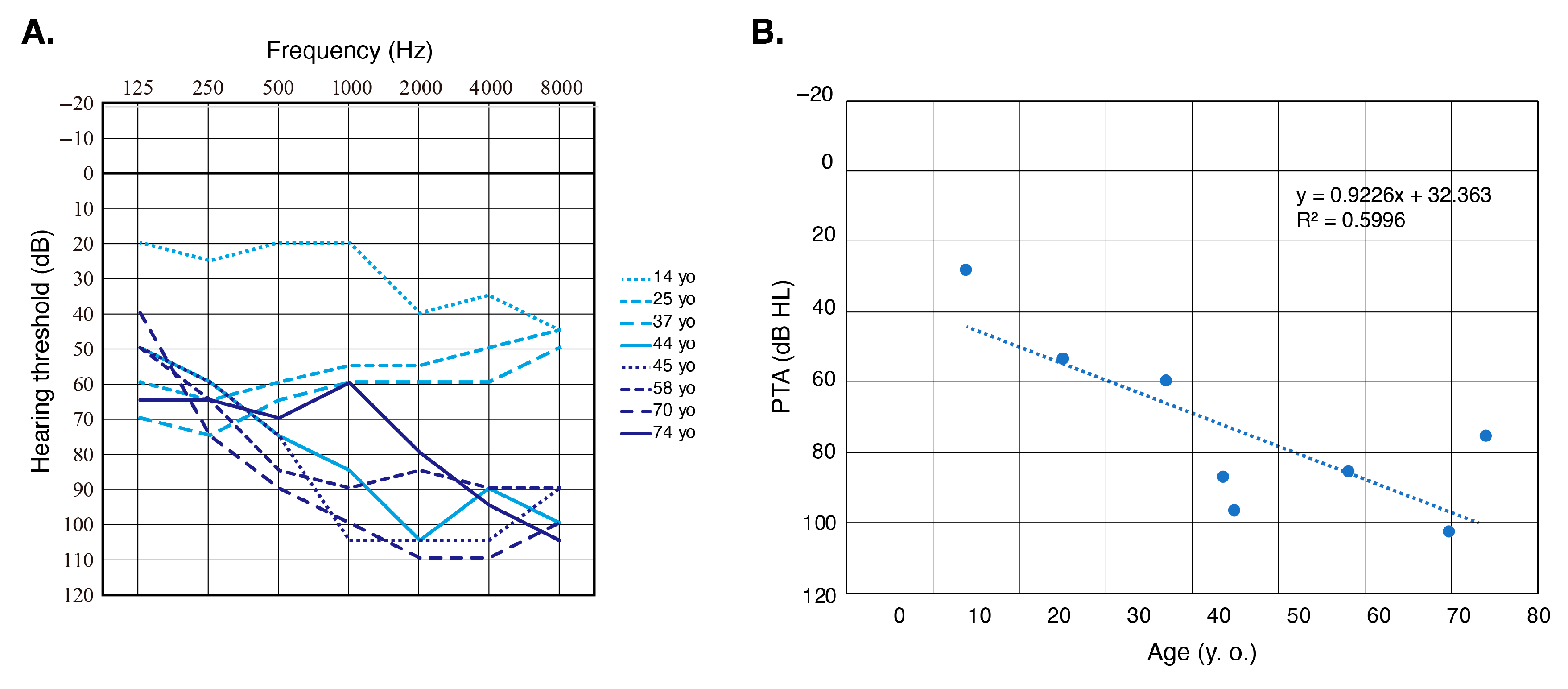
Nine affected individuals from nine independent pedigrees were detected in this study. MYO3A is a relatively rare causative gene, and the prevalence of MYO3A-associated HL among Japanese HL patients is 0.06% (9/15,684). We were able to obtain clinical information from eight of these patients, which is summarized in Table 2. The onset age of their HL varied from 10 to 30 years old (mean age: 19.6 years old), and all of them presented with post-lingual deterioration in hearing. The severity of their HL varied from mild to profound, and all patients were aware of HL progression at the time of their genetic testing. Although we were not able to obtain serial audiograms from individual patients, the correlation between age and HL progression was observable on the overlapping audiograms of all individuals (Figure 2A). Assessment of HL progression by scatter plotting the pure-tone average data for our patients clearly indicated the progression of HL with age (Figure 2B). The types of HL were categorized as down-sloping in six, and flat in two patients. Two individuals complained of vertigo, but the specifics are unknown.

4. Discussion

Among the eight possibly pathogenic MYO3A variants identified in this study, six were novel. The localization of the identified variants is shown in Figure 3. The position of the variations may influence the genotype–phenotype correlations, but none were observed in this study. For example, it has been reported that damaging effects in the kinase domain cause congenital profound HL [18], yet all patients detected in our study, including those with homozygous variants in the kinase domain, presented with HL onset at over 10 years of age. Further, no effect on phenotypes was observed with the type of amino acid change caused by nucleotide changes.
In this study, we identified nine candidate individuals with HL caused by MYO3A variants, which is the largest number of patients to be detected to date. Based on the HGMD professional database [13], there are 41 MYO3A variants currently listed as HL causal variants. However, 15 of those variants were not identified as a cause for HL, but were only detected through MPS analysis (i.e., the variant was detected only in the heterozygous state, in vitro fertilization (IVF) screening, IVF donor screening). On the other hand, 13 cases have been reported to have ARNSHL caused by MYO3A deficiency (DFNB30), including 15 HL causal variants. We summarized the identified variants and clinical information from previous reports for comparison with the results of this study (Table 3). In our study, all patients presented with late-onset HL and were aware of the progression of their HL, with the overlapping audiograms also showing age-related deterioration in hearing (Figure 2). Unlike the findings in our study, in five of nine reported cases for which information on onset age was available, HL onset was congenital or pre-lingual. The exact ages at which their audiograms were obtained remains unclear, but all patients in the previous reports presented with moderate-to-profound HL (Table 3).
It should be noted that variations in the MYO3A gene had been thought to cause ARNSHL (DFNB30), but were reported as a genetic cause of autosomal dominant HL [36,37,38]. To date, most MYO3A variants causing ADNSHL have been reported from Brazilian families or from families of South African decent, but this sheds new light on the evaluation of our database, indicating that heterozygous variations might be the cause of HL. However, we face challenges in the verification of this conclusion as segregation analysis is difficult in Japan due to the increase in nuclear families and reduction in the number of births.
In this study, two patients noted their experience of vestibular symptoms. Unfortunately, we could not obtain detailed vestibular testing results for these cases. Due to this limitation of this study, further study is needed, including comprehensive vestibular assessment (caloric testing, cervical vestibular evoked myogenic potential, ocular vestibular evoked myogenic potential and video head impulse testing), to conclude the effects of MYO3A gene variants on vestibular function.
It is crucial that we provide adequate information, support and treatment to patients that present with late-onset, progressive HL. Patients will have to adjust their lifestyles, and regardless of the provision of hearing aids and cochlear implantation, deterioration in their quality of life is inevitable. In Japan, patients diagnosed with late-onset or juvenile-onset progressive HL due to pathogenic variations in the ACTG1, CDH23, COCH, KCNQ4, TECTA, TMPRSS3, WFS1, EYA4, MYO6, MYO15A and POU4F3 genes, and who have progressed to severe-to-profound HL, are provided with medical support from the government throughout their life, and we aim to further our research and increase the number of genes for which late-onset progressive HL patient support is applicable.
As future prospects, we believe that the clinical phenotypes of MYO3A-associated HL patients clarified in this study will be useful in providing more appropriate clinical management. In particular, most of the cases identified in this study and previous reports showed progressive HL, eventually progressing to severe-to-profound HL. Thus, frequent follow-up and clinical intervention, including the provision of hearing aids and cochlear implantation, should be considered. A further prospective study will be useful in evaluating hearing deterioration through the use of the serial audiometric testing results from the same patient. In addition, the evaluation of the outcomes of hearing aids or cochlear implantation will also be useful.
Table 3. Clinical characteristics of MYO3A-associated hearing loss patients in previous reports.
Nucleotide ChangeAA ChangeInheritanceOnsetSeverityConfigurationProgressionPopulationReference
c.[149A>G];[149A>G]p.[K50R];[K50R]ARCongenitalProfoundNANATunisia[26]
c.[424C>T];[424C>T]p.[H142Y];[H142Y]ARCongenital/
Pre-lingual
Severe-to-profoundNANASouth Africa[23]
c.[580C>A];[1582_1583insT]p.[P194T];[Y530Lfs*9]ARNASevere-to-profound HF_precipitousNAKorea[18]
c.[732-2A>G];[732-2A>G]p.[IVS8 as A-G -2];[IVS8 as A-G -2]ARSecond decadeModerate-to-profoundHF_precipitousNAUS[17]
c.[824G>A];[3737_3738delAG]p.[R275H];[E1246Gfs*5]ARNAModerate-to-severeNANAChina[24]
c.[1370_1371delGA];[1370_1371delGA]p.[R457Nfs*25];[R457Nfs*25]ARPre-lingualNANAYesPalestine[22]
c.[1450T>C];[3093G>A]p.[S484P];[W1031X]AR6 yoSevereHF_gentleYesChina[25]
c.[1777-12G>A];[3126T>G]p.[IVS17 as G-A -12];[Y1042X]ARSecond decadeModerate-to-profoundHF_precipitousNAUS[17]
c.[1841C>T];[1841C>T}p.[S614F];[S614F]ARCongenitalProfoundNANoChina[20]
c.[3126T>G];[3126T>G]p.[Y1042X];[Y1042X]ARSecond decadeModerate-to-profoundHF_precipitousNAUS[17]
c.[4462A>G];[[4681C>T]p.[K1488E];[R1561X]ARCongenital/
Pre-lingual
ProfoundNANATaiwan[19]
c.716T>Cp.L239PADPre-lingualModerate-to-profoundHF_gentleYesEurope[38]
c.1463G>Ap.G488EADPost-lingual early-onsetModerate-to-profoundNAYesAfrican American[36]
c.2090T>Gp.L697WAD10-60Mild-to-profoundHF_gentleYesBrazil. Portugal[37]
AA: amino acid, AR: autosomal recessive, AD: autosomal dominant, HF_gentle: sloping high frequency HL, HF_precipitous: precipitous high-frequency HL, *: stop codon.

5. Conclusions

In this study, we utilized MPS to identify nine candidate individuals with HL caused by MYO3A variations, which is the largest number of patients to be detected to date. MYO3A variations cause late-onset HL without any accompanying symptoms. Our findings have confirmed that although onset age may vary from birth to the second decade, the HL will progress, eventually leading to severe-to-profound HL. Although the frequency of MYO3A- associated HL is lower than that of other ARNSHL genes, it is essential that we take MPS into consideration for early onset detection to better allow for timely intervention.

Supplementary Materials

The following supporting information can be downloaded at: https://www.mdpi.com/article/10.3390/genes16010092/s1, Table S1: MYO3A primers used in this study.

Author Contributions

Conceptualization, K.M., S.-y.N. and S.-i.U.; methodology, K.M., S.-y.N. and S.-i.U.; software, S.-y.N.; validation, K.M. and S.-y.N.; resources, H.M., Y.O., N.O., M.K., A.Y., M.O., K.W. and A.S.; data curation, K.M. and S.-y.N.; writing—original draft preparation, K.M.; writing—review and editing, K.M., S.-y.N. and S.-i.U.; visualization, K.M. and S.-y.N.; supervision, S.-i.U.; project administration, S.-i.U.; funding acquisition, S.-i.U. All authors have read and agreed to the published version of the manuscript.

Funding

This study was funded by a Health and Labor Sciences Research Grant for Research on Rare and Intractable Diseases and Comprehensive Research on Disability Health and Welfare from the Ministry of Health, Labor and Welfare of Japan (S.U. H29-Nanchitou(Nan)-Ippan-031, 20FC1048, 23FC0201); and Grants-in-Aid from the Japan Agency for Medical Research and Development (AMED) (S.U. JP16kk0205010, JP18ek0109363, JP21ek0109542, JP24ek0109741).

Institutional Review Board Statement

The study was conducted according to the guidelines of the Declaration of Helsinki, and was approved by the Institutional Ethics Committee of Shinshu University School of Medicine (No.387—4 September 2012, No.576—2 May 2017 and No.718—7 March 2022).

Informed Consent Statement

Written informed consent was obtained from all patients (or from their next of kin, caretaker or legal guardian in the cases of minors or children).

Data Availability Statement

The datasets used during the current study are available from the corresponding author on reasonable request due to ethical reasons.

Acknowledgments

We thank all participants in the present study. We also thank Fumiko Tomioka and Sachiko Matsuda for their technical assistance with this research.

Conflicts of Interest

All authors declare no conflicts of interest in this study.

References

  1. Yoshimura, H.; Okubo, T.; Shinagawa, J.; Nishio, S.Y.; Takumi, Y.; Usami, S.I. Epidemiology, aetiology and diagnosis of congenital hearing loss via hearing screening of 153913 newborns. Int. J. Epidemiol. 2024, 53, dyae052. [Google Scholar] [CrossRef] [PubMed]
  2. Sheffield, A.M.; Smith, R.J.H. The Epidemiology of Deafness. Cold Spring Harb. Perspect. Med. 2019, 9, a033258. [Google Scholar] [CrossRef]
  3. Hereditary Hearing Loss Homepage. Available online: https://hereditaryhearingloss.org (accessed on 20 November 2024).
  4. Aldè, M.; Cantarella, G.; Zanetti, D.; Pignataro, L.; La Mantia, I.; Maiolino, L.; Ferlito, S.; Di Mauro, P.; Cocuzza, S.; Lechien, J.R.; et al. Autosomal Dominant Non-Syndromic Hearing Loss (DFNA): A Comprehensive Narrative Review. Biomedicines 2023, 11, 1616. [Google Scholar] [CrossRef] [PubMed]
  5. Usami, S.I.; Nishio, S.Y. The genetic etiology of hearing loss in Japan revealed by the social health insurance-based genetic testing of 10K patients. Hum. Genet. 2022, 141, 665–681. [Google Scholar] [CrossRef]
  6. Sloan-Heggen, C.M.; Bierer, A.O.; Shearer, A.E.; Kolbe, D.L.; Nishimura, C.J.; Frees, K.L.; Ephraim, S.S.; Shibata, S.B.; Booth, K.T.; Campbell, C.A.; et al. Comprehensive genetic testing in the clinical evaluation of 1119 patients with hearing loss. Hum. Genet. 2016, 135, 441–450. [Google Scholar] [CrossRef] [PubMed]
  7. Quintero, O.A.; Moore, J.E.; Unrath, W.C.; Manor, U.; Salles, F.T.; Grati, M.; Kachar, B.; Yengo, C.M. Intermolecular autophosphorylation regulates myosin IIIa activity and localization in parallel actin bundles. J. Biol. Chem. 2010, 285, 35770–35782. [Google Scholar] [CrossRef] [PubMed]
  8. Quintero, O.A.; Unrath, W.C.; Stevens, S.M., Jr.; Manor, U.; Kachar, B.; Yengo, C.M. Myosin 3A kinase activity is regulated by phosphorylation of the kinase domain activation loop. J. Biol. Chem. 2013, 288, 37126–37137. [Google Scholar] [CrossRef] [PubMed]
  9. Salles, F.T.; Merritt, R.C., Jr.; Manor, U.; Dougherty, G.W.; Sousa, A.D.; Moore, J.E.; Yengo, C.M.; Dosé, A.C.; Kachar, B. Myosin IIIa boosts elongation of stereocilia by transporting espin 1 to the plus ends of actin filaments. Nat. Cell Biol. 2009, 11, 443–450. [Google Scholar] [CrossRef] [PubMed]
  10. Lelli, A.; Michel, V.; Boutet de Monvel, J.; Cortese, M.; Bosch-Grau, M.; Aghaie, A.; Perfettini, I.; Dupont, T.; Avan, P.; El-Amraoui, A.; et al. Class III myosins shape the auditory hair bundles by limiting microvilli and stereocilia growth. J. Cell Biol. 2016, 212, 231–244. [Google Scholar] [CrossRef]
  11. Merritt, R.C.; Manor, U.; Salles, F.T.; Grati, M.; Dose, A.C.; Unrath, W.C.; Quintero, O.A.; Yengo, C.M.; Kachar, B. Myosin IIIB uses an actin-binding motif in its espin-1 cargo to reach the tips of actin protrusions. Curr. Biol. 2012, 22, 320–325. [Google Scholar] [CrossRef]
  12. Dosé, A.C.; Hillman, D.W.; Wong, C.; Sohlberg, L.; Lin-Jones, J.; Burnside, B. Myo3A, one of two class III myosin genes expressed in vertebrate retina, is localized to the calycal processes of rod and cone photoreceptors and is expressed in the sacculus. Mol Biol. Cell 2003, 14, 1058–1073. [Google Scholar] [CrossRef] [PubMed]
  13. HGMD Professional. Available online: http://www.hgmd.org (accessed on 20 November 2024).
  14. Cirilo, J.A., Jr.; Gunther, L.K.; Yengo, C.M. Functional Role of Class III Myosins in Hair Cells. Front. Cell Dev. Biol. 2021, 25, 643856. [Google Scholar] [CrossRef] [PubMed]
  15. Ebrahim, S.; Avenarius, M.R.; Grati, M.; Krey, J.F.; Windsor, A.M.; Sousa, A.D.; Ballesteros, A.; Cui, R.; Millis, B.A.; Salles, F.T.; et al. Stereocilia-staircase spacing is influenced by myosin III motors and their cargos espin-1 and espin-like. Nat. Commun. 2016, 1, 10833. [Google Scholar] [CrossRef] [PubMed]
  16. Les Erickson, F.; Corsa, A.C.; Dosé, A.C.; Burnside, B. Localization of a class III myosin to filopodia tips in transfected HeLa cells requires an actin-binding site in its tail domain. Mol. Biol. Cell 2003, 14, 4173–4180. [Google Scholar] [CrossRef] [PubMed]
  17. Walsh, T.; Walsh, V.; Vreugde, S.; Hertzano, R.; Shahin, H.; Haika, S.; Lee, M.K.; Kanaan, M.; King, M.C.; Avraham, K.B. From flies’ eyes to our ears: Mutations in a human class III myosin cause progressive nonsyndromic hearing loss DFNB30. Proc. Natl. Acad. Sci. USA 2002, 99, 7518–7523. [Google Scholar] [CrossRef] [PubMed]
  18. Choi, B.Y.; Park, G.; Gim, J.; Kim, A.R.; Kim, B.J.; Kim, H.S.; Park, J.H.; Park, T.; Oh, S.H.; Han, K.H.; et al. Diagnostic application of targeted resequencing for familial nonsyndromic hearing loss. PLoS ONE 2013, 8, e68692. [Google Scholar] [CrossRef]
  19. Wu, C.C.; Lin, Y.H.; Liu, T.C.; Lin, K.N.; Yang, W.S.; Hsu, C.J.; Chen, P.L.; Wu, C.M. Identifying children with poor cochlear implantation outcomes using massively parallel sequencing. Medicine 2015, 94, e1073. [Google Scholar] [CrossRef]
  20. Sommen, M.; Schrauwen, I.; Vandeweyer, G.; Boeckx, N.; Corneveaux, J.J.; van den Ende, J.; Boudewyns, A.; De Leenheer, E.; Janssens, S.; Claes, K.; et al. DNA Diagnostics of Hereditary Hearing Loss: A Targeted Resequencing Approach Combined with a Mutation Classification System. Hum. Mutat. 2016, 37, 812–819. [Google Scholar] [CrossRef] [PubMed]
  21. Qu, R.; Sang, Q.; Xu, Y.; Feng, R.; Jin, L.; He, L.; Wang, L. Identification of a novel homozygous mutation in MYO3A in a Chinese family with DFNB30 non-syndromic hearing impairment. Int. J. Pediatr. Otorhinolaryngol. 2016, 84, 43–47. [Google Scholar] [CrossRef] [PubMed]
  22. Abu Rayyan, A.; Kamal, L.; Casadei, S.; Brownstein, Z.; Zahdeh, F.; Shahin, H.; Canavati, C.; Dweik, D.; Jaraysa, T.; Rabie, G.; et al. Genomic analysis of inherited hearing loss in the Palestinian population. Proc. Natl. Acad. Sci. USA 2020, 117, 20070–20076. [Google Scholar] [CrossRef]
  23. Wonkam, A.; Manyisa, N.; Bope, C.D.; Dandara, C.; Chimusa, E.R. Whole exome sequencing reveals pathogenic variants in MYO3A, MYO15A and COL9A3 and differential frequencies in ancestral alleles in hearing impairment genes among individuals from Cameroon. Hum. Mol. Genet. 2021, 29, 3729–3743. [Google Scholar] [CrossRef] [PubMed]
  24. Wu, J.; Cao, Z.; Su, Y.; Wang, Y.; Cai, R.; Chen, J.; Gao, B.; Han, M.; Li, X.; Zhang, D.; et al. Molecular diagnose of a large hearing loss population from China by targeted genome sequencing. J. Hum. Genet. 2022, 67, 643–649. [Google Scholar] [CrossRef]
  25. Guan, J.; Li, J.; Chen, G.; Shi, T.; Lan, L.; Wu, X.; Zhao, C.; Wang, D.; Wang, H.; Wang, Q. Family trio-based sequencing in 404 sporadic bilateral hearing loss patients discovers recessive and De novo genetic variants in multiple ways. Eur. J. Med. Genet. 2021, 64, 104311. [Google Scholar] [CrossRef] [PubMed]
  26. Souissi, A.; Abdelmalek Driss, D.; Chakchouk, I.; Ben Said, M.; Ben Ayed, I.; Mosrati, M.A.; Elloumi, I.; Tlili, A.; Aifa, S.; Masmoudi, S. Molecular insights into MYO3A kinase domain variants explain variability in both severity and progression of DFNB30 hearing impairment. J. Biomol. Struct. Dyn. 2022, 40, 10940–10951. [Google Scholar] [CrossRef] [PubMed]
  27. Nishio, S.Y.; Hayashi, Y.; Watanabe, M.; Usami, S.I. Clinical application of a custom AmpliSeq library and ion torrent PGM sequencing to comprehensive mutation screening for deafness genes. Genet. Test Mol. Biomark. 2015, 19, 209–217. [Google Scholar] [CrossRef] [PubMed]
  28. Wang, K.; Li, M.; Hakonarson, H. ANNOVAR: Functional annotation of genetic variants from high-throughput sequencing data. Nucleic. Acids. Res. 2010, 38, e164. [Google Scholar] [CrossRef]
  29. 1000 Genome Project. Available online: https://www.internationalgenome.org/1000-genomes-summary/ (accessed on 10 April 2020).
  30. Genome Aggregation Database. Available online: https://gnomad.broadinstitute.org (accessed on 11 November 2024).
  31. ToMMo 54KJPN-Integrative Japanese Genome Variation Database. Available online: https://jmorp.megabank.tohoku.ac.jp (accessed on 9 July 2023).
  32. Nishio, S.Y.; Moteki, H.; Usami, S.I. Simple and efficient germline copy number variant visualization method for the Ion AmpliSeq custom panel. Mol. Genet. Genom. Med. 2018, 6, 678–686. [Google Scholar] [CrossRef] [PubMed]
  33. Richards, S.; Aziz, N.; Bale, S.; Bick, D.; Das, S.; Gastier-Foster, J.; Grody, W.W.; Hegde, M.; Lyon, E.; Spector, E.; et al. ACMG Laboratory Quality Assurance Committee. Standards and guidelines for the interpretation of sequence variants: A joint consensus recommendation of the American College of Medical Genetics and Genomics and the Association for Molecular Pathology. Genet. Med. 2015, 17, 405–424. [Google Scholar] [CrossRef] [PubMed]
  34. Oza, A.M.; DiStefano, M.T.; Hemphill, S.E.; Cushman, B.J.; Grant, A.R.; Siegert, R.K.; Shen, J.; Chapin, A.; Boczek, N.J.; Schimmenti, L.A.; et al. ClinGen Hearing Loss Clinical Domain Working Group. Expert specification of the ACMG/AMP variant interpretation guidelines for genetic hearing loss. Hum. Mutat. 2018, 39, 1593–1613. [Google Scholar] [CrossRef] [PubMed]
  35. Mazzoli, M.; Van Camp, G.; Newton, V.; Giarbini, N.; Declau, F.; Parving, A. Recommendations for the Description of Genetic and Audiological Data for Families with Nonsyndromic Hereditary Hearing Impairment. Audiol. Med. 2003, 1, 148–150. [Google Scholar]
  36. Grati, M.; Yan, D.; Raval, M.H.; Walsh, T.; Ma, Q.; Chakchouk, I.; Kannan-Sundhari, A.; Mittal, R.; Masmoudi, S.; Blanton, S.H.; et al. MYO3A causes human dominant deafness and interacts with Protocadherin 15-CD2 isoform. Hum. Mutat. 2016, 37, 481–487. [Google Scholar] [CrossRef]
  37. Dantas, V.G.L.; Raval, M.H.; Ballesteros, A.; Cui, R.; Gunther, L.K.; Yamamoto, G.L.; Alves, L.U.; Bueno, A.S.; Lezirovitz, K.; Pirana, S.; et al. Characterization of a novel MYO3A missense mutation associated with a dominant form of late onset hearing loss. Sci. Rep. 2018, 8, 8706. [Google Scholar] [CrossRef] [PubMed]
  38. Doll, J.; Hofrichter, M.A.H.; Bahena, P.; Heihoff, A.; Segebarth, D.; Müller, T.; Dittrich, M.; Haaf, T.; Vona, B. A novel missense variant in MYO3A is associated with autosomal dominant high-frequency hearing loss in a German family. Mol. Genet. Genomic. Med. 2020, 8, e1343. [Google Scholar] [CrossRef] [PubMed]
Figure 1. Pedigree and audiograms for the families of each MYO3A-associated HL patient identified in this study. The variants identified in this study are indicated in the figure. Pedigrees have been enumerated #1 to #9 for clarification. Solid line: hearing threshold in the right ear; Dashed line: hearing threshold in the left ear.
Genes 16 00092 g001
Figure 2. (A) Overlapping audiograms from all MYO3A-associated HL patients identified in this study. (B) Detailed progression analysis of hearing deterioration for patients with MYO3A-associated HL. Each dot indicates the pure-tone average (PTA; average of hearing thresholds for 500 Hz, 1000 Hz, 2000 Hz and 4000 Hz) and age of each patient. Dotted line indicates the linear regression.
Genes 16 00092 g002
Figure 3. All reported pathogenic MYO3A variants and their locations in the MYO3A gene. Novel variants identified in this study are indicated in red, variants identified in this study and previous reports are indicated in orange and variants reported to have an AD inheritance pattern are indicated in blue. *: stop codon.
Genes 16 00092 g003
Table 1. The MYO3A variants identified in this study.
Nucleotide ChangeAA ChangeExonDomainSIFTPP2LRTMutTasterMutAssessorREVELCADDToMMo 54KJPNgnomAD AllPathogenicityReference
c.409-2A>- exon6Kinase.......1.80E-05.Likely pathogenic
(PVS1, PM2)
This study
c.712G>Ap.Ala238Threxon8KinaseTDDDL0.45133..VUS
(PM2, PM3_Supporting)
This study
c.893dupp.Gln300Thrfs*21exon10Motor.......0.000221.Pathogenic
(PVS1, PM2_Supporting, PM3_Supporting)
This study
c.991C>Tp.Arg331*exon11Motor..DA...9.00 × 10−63.18 × 10⁻⁵Pathogenic
(PVS1, PM2_Supporting, PM3_Supporting)
[20]
c.1450T>Cp.Ser484Proexon15MotorDDDDH0.90527.84.60 × 10−53.98 × 10⁻⁶Pathogenic
(PM3_Strong, PP1_Strong, PM2_Supporting, PP3)
[25]
c.1464delp.Lys489Asnfs*3exon15Motor.......0.000129.Likely pathogenic
(PVS1, PM2_Supporting)
This study
c.2308G>Ap.Glu770Lysexon21MotorTPDDL0.491241.80 × 10−5.VUS
(PM2)
This study
c.4164dupp.Asn1389Lysfs*4exon30Tail.......0.000101.Likely pathogenic
(PVS1, PM2_Supporting)
This study
All variants are indicated on NM_173591. AA: amino acid, PP2: PolyPhen2, MutTaster: mutation taster, MutAssessor: mutation assessor, T (in SIFT): tolerated, D (in SIFT): deleterious, D (in PP2): probably damaging, P (in PP2): possibly damaging, D (in LRT): deleterious, D (in MutTaster): disease causing, A (in MutTaster): disease causing automatic, L (in MutAssessor): low, H (in MutAssessor): high, VUS: variant of uncertain significance, *: stop codon.
Table 2. Clinical characteristics of the MYO3A-associated hearing loss patients identified in this study.
Family NumberIDBase Change
Allele 1
AA Change
Allele 1
Base Change
Allele 2
AA Change
Allele 2
Inheritance PatternOnsetAgeGenderSeverity of HLType of HLProgressionVestibular Symptoms
1JHLB-3058c.409-2A>- c.2308G>Ap.Glu770LysSporadic1215MModerateHF_gentleYY
2JHLB-14312c.893dupAp.Gln300Thrfs*21c.893dupAp.Gln300Thrfs*21Sporadic2274FSevereHF_gentleYN
32402c.991C>Tp.Arg331*c.991C>Tp.Arg331*UnknownNANAFNANANANA
4O-3349c.712G>Ap.Ala238Thrc.712G>Ap.Ala238ThrAD/Mit1337FModerateFlatYY
5JHLB-5865c.1450T>Cp.Ser484Proc.1450T>Cp.Ser484ProAR2058FProfoundHF_gentleYN
6JHLB-12633c.1450T>Cp.Ser484Proc.1450T>Cp.Ser484ProAD/Mit2544FProfoundHF_gentleYN
7JHLB-13481c.1450T>Cp.Ser484Proc.1450T>Cp.Ser484ProUnknown2545FProfoundHF_gentleYN
8JHLB-14690c.1450T>Cp.Ser484Proc.1450T>Cp.Ser484ProSporadic3070FProfoundHF_gentleYN
9JHLB-889c.1464delAp.Lys489Asnfs*3c.4164dupAp.Asn1389Lysfs*4Sporadic1025FModerateFlatYN
AA: amino acid, AR: autosomal recessive, AD: autosomal dominant, M: male, F: female, HF_gentle: sloping high frequency HL, Y: yes, N: no, *: stop codon.
Disclaimer/Publisher’s Note: The statements, opinions and data contained in all publications are solely those of the individual author(s) and contributor(s) and not of MDPI and/or the editor(s). MDPI and/or the editor(s) disclaim responsibility for any injury to people or property resulting from any ideas, methods, instructions or products referred to in the content.