Review

22q11.21 Deletions: A Review on the Interval Mediated by Low-Copy Repeats C and D

1
Section of Cytogenetics, Oncology Department, Azienda Ospedaliero-Universitaria Pisana, 56126 Pisa, Italy
2
Section of Clinical and Laboratory Immunology, Pediatric Unit, Department of Clinical and Experimental Medicine, University of Pisa, 56126 Pisa, Italy
3
Section of Pediatric Hematology and Oncology, Azienda Ospedaliero-Universitaria Pisana, 56126 Pisa, Italy
*
Author to whom correspondence should be addressed.
Genes 2025, 16(1), 72; https://doi.org/10.3390/genes16010072
Submission received: 29 November 2024 / Revised: 7 January 2025 / Accepted: 7 January 2025 / Published: 9 January 2025
(This article belongs to the Section Human Genomics and Genetic Diseases)

Abstract

:
22q11.2 is a region prone to chromosomal rearrangements due to the presence of eight large blocks of low-copy repeats (LCR22s). The 3 Mb 22q11.2 “typical deletion”, between LCR22-A and D, causes a fairly well-known clinical picture, while the effects of smaller CNVs harbored in this interval are still to be fully elucidated. Nested deletions, flanked by LCR22B-D, LCR22B-C, or LCR22C-D, are very rare and are collectively described as “central deletions”. The LCR22C-D deletion (CDdel) has never been separately analyzed. In this paper, we focused only on CDdel, evaluating its gene content and reviewing the literature and public databases in order to obtain new insights for the classification of this CNV. At first glance, CDdels are associated with a broad phenotypic spectrum, ranging from clinically normal to quite severe phenotypes. However, the frequency of specific clinical traits highlights that renal/urinary tract abnormalities, cardiac defects, and neurological/behavioral disorders are much more common in CDdel than in the general population. This frequency is too high to be fortuitous, indicating that CDdel is a predisposing factor for these phenotypic traits. Among the genes present in this interval, CRKL is an excellent candidate for cardiac and renal defects. Even if further data are necessary to confirm the role of CDdels, according to our review, this CNV fits into the class of ‘likely pathogenic’ CNVs.

1. Introduction

22q11.2 is a region prone to chromosomal rearrangements (i.e., deletions and duplications) due to the presence of eight large blocks of low-copy repeats (LCR22s: LCR22-A to LCR22-H). In this paper, we focused on 22q11.2 deletions flanked by LCR22-C and D, since their clinical meaning has not yet been fully clarified.
The interpretation of the effects of a copy number variant (CNV), such as the 22q11.2C-D deletion, is a multi-step process that requires mainly the evaluation of its genomic content and the analysis of the similar imbalances in the medical literature. This process leads to a five-tier classification (pathogenic, likely pathogenic, uncertain significance, likely benign, and benign).
The study of 22q11.2C-D deletion was performed according to the recent semi-quantitative point-based scoring system developed by the American College of Medical Genetics and Genomics (ACMG) and the Clinical Genome Resource (ClinGen) [1].

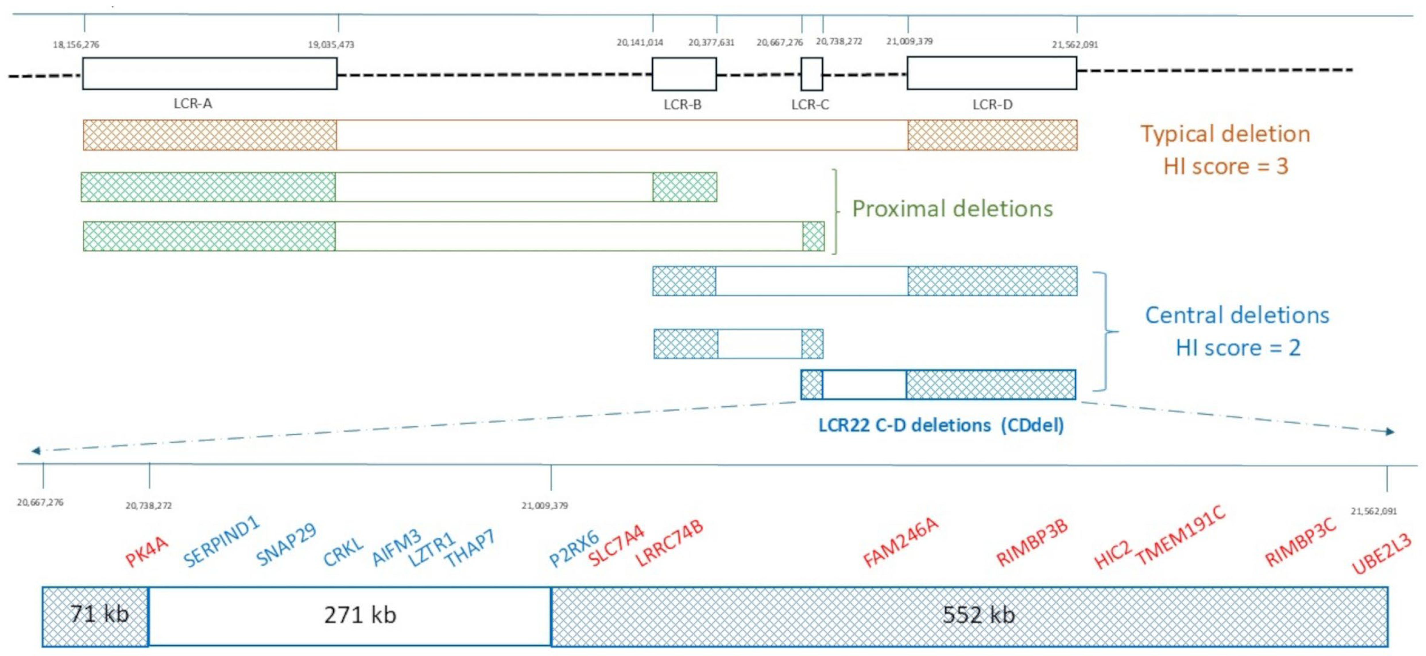
2. Architecture of the 22q11.2 Region

The 22q11.2 chromosome region contains eight large blocks of low-copy repeats (LCR22s: LCR22-A to LCR22-H) that are predisposed to meiotic non-allelic homologous recombination and lead to genomic imbalances (Figure 1) [2,3].
Deletions involving the four proximal LCRs (from LCR22A to LCR22D) cause the “22q11.2 deletion syndrome” (22q11.2DS), a heterogeneous multisystemic condition related to an impaired development of the third and fourth pharyngeal arches [2,3,4].
The vast majority of 22q11.2DS cases have a ~3 Mb deletion, known as the “typical or proximal typical deletion”, flanked by LCR22A-D (Figure 1). Individuals with atypical deletions due to a different combination of LCR22s or with breakpoints not falling into LCRs have also been reported [3].
About 5–10% of the cases show “proximal nested deletions” flanked by LCR22A-B or LCR22A-C, whose phenotype is indistinguishable from the “proximal typical deletion” (Figure 1). The clinical meaning of these intervals (LCR22A-D, LCR22A-B, and LCR22A-C) is also well-documented by ClinGen (https://www.ncbi.nlm.nih.gov/projects/dbvar/clingen/) (accessed on 28 November 2024), a publicly available resource that collects evidence supporting and/or refuting the “dosage sensitivity” (DS) for genomic regions and provides a “haploinsufficiency” (HI) DS score. For these intervals, the HI DS score is 3, which corresponds to “sufficient evidence for dosage pathogenicity”.
Nested deletions, flanked by LCR22B-D, LCR22B-C, or LCR22C-D, are very rare and collectively described as “central deletions” with a HI DS score of 2, corresponding to “emerging evidence for haploinsufficiency” (https://www.ncbi.nlm.nih.gov/projects/dbvar/clingen/) (accessed on 28 November 2024) (Figure 1).
The LCR22C-D deletion (hereinafter called “CDdel”) has never been separately analyzed. In this paper, we focused only on CDdel, evaluating its gene content and reviewing the literature and public databases in order to obtain new insights for the classification of this CNV.

3. Gene Content

A crucial step for classifying a CNV is to verify if it overlaps with any established or predicted HI score gene, and, in particular, with any gene associated with dominantly inherited disorders (https://www.ncbi.nlm.nih.gov/projects/dbvar/clingen/) (accessed on 28 November 2024) [1].
If we exclude the LCR22C and D flanking sequences, the LCR22C-D deletion spans approximately 271 kb (GRCh38/hg38, chr22: 20,738,272–21,009,379) and harbors seven protein-coding genes (Figure 1). Identifying the exact number of genes harbored in the LCR22C-D deletions may be a challenge, since it is difficult to exactly locate the breakpoints into the LCRC and D, due to the oligo/SNP low coverage of these regions in the array platforms. In view of this, the genes harbored in LCR22C (71 kb) and LCR22D (552 kb) were also evaluated, since it cannot be excluded that their deletion, if present, may affect the phenotype [2] (track “NCBI RefSeq genes” of UCSC https://genome.ucsc.edu/cgi-bin/hgGateway) (accessed on 28 November 2024) (Table 1).
Only two genes, LZTR (leucine zipper-like transcription regulator 1) and SERPIND1 (heparin cofactor II), are associated with dominantly inherited disorders and for none of them has the HI score been recorded (https://www.ncbi.nlm.nih.gov/projects/dbvar/clingen/) (accessed on 28 November 2024). Regarding LZTR, its pathogenicity results from mutational mechanisms different from the dosage sensitivity: “Noonan syndrome 10” is caused by dominant negative mutations, and “Schwannomatosis-2” is the result of a multi-step pathway involving several others genes [5,6]. Nothing is known about the mechanism underlying the pathogenicity of SERPIND1.
None of the few available HI of CDdel genes reaches a score of 3 (sufficient evidence for dosage pathogenicity) (Table 1).
Even if it reaches a HI score of only 1 (little evidence of pathogenicity), CRLK (CRK like proto-oncogene, adaptor protein) is the most interesting gene, according to studies in mice and humans (https://www.ncbi.nlm.nih.gov/projects/dbvar/clingen/) (accessed on 28 November 2024) (Table 1).
Knockout mice with Crkl haploinsufficiency shows some clinical features observed in the 22q11.2 DS, such as cardiovascular and genitourinary defects [7,8,9]. Moreover, in a vast cohort of individuals with 22q11.2DS, common variants on the not-deleted allele are associated with a moderate increased risk for conotruncal heart defects (CTDs). These variants are located in a 350 kb region, largely within the LCR22C-D interval; the top associated variants lie in putative regulatory regions of CRKL, strengthening the involvement of this gene in cardiac pathologies [10]. A role of CRKL in renal anomalies has been suggested by variants in domains evolutionarily conserved, found in five individuals with renal agenesis or hypodysplasia [9].
Since there are no genes with a significant HI score in the region, we evaluated two other HI predictors, the gnomAD probability of loss-of-function intolerance score (pLI) and the DECIPHER HI index (%HI) [1]. Due to the intrinsic limitations of these tools, only genes with a pLI score ≥ 0.9 and %HI index of ≤10% can be considered to have supportive evidence of HI. As shown in Table 1, none of the genes in the LCR22C-D interval fall into this category.
Taking into account all these data (the HI score evaluation, HI predictors, and mechanisms of pathogenicity of genes associated with dominantly inherited disorders), neither the pathogenicity nor benignity of CDdel can be established.

4. Long-Distance Effects of CNVs

As known, CNVs may have long-distance effects through chromatin structure alterations or epigenetic modifications [11,12].
Topologically associated domains (TADs) can be defined as linear units of chromatin that fold, as discrete three-dimensional (3D) structures tending to favor internal chromatin interactions [11]. TADs constitute functional units in the genome, important for the correct regulation of gene expression, since they co-localize regulatory elements with their target genes. TAD boundaries are structural barriers necessary to ensure this 3D genomic organization and their disruption is found to be associated with a wide range of diseases [11]. The LCRC-D interval mostly overlaps with a 350 kb region (GRCh38/hg38, chr22: 20,607,741–20,958,141) within a single TAD, as determined by an analysis of the chromatin structure by high-throughput chromatin conformation capture (Hi-C), (track “HapMap” of UCSC, https://genome.ucsc.edu/cgi-bin/hgGateway) (accessed on 28 November 2024). According to these data, CDdel disrupts a TAD boundary, but whether this has any long-range effect on gene expression has not yet been studied.
Recently, a DNA methylation episignature in the 22q11.2DS has been identified [12]. Both the typical and proximal 22q11.2 deletions show a unique and highly specific episignature, that may impact the expression of genes outside the 22q11.2 region, potentially relevant in the pathogenesis of the 22q11.2DS phenotype; however, this altered methylation pattern is not present in either the LCR22B-D or LCR22C-D deletions, suggesting that central deletions are entities distinct from 22q11.2DS, at least on the basis of the episignature.

5. CDdel Cases from Published Literature and Public Databases

A thorough review of public databases and medical literature was performed to identify cases with CDdel and to evaluate clinical features reported along with their relative frequencies.
Cases were excluded from our analysis if they showed (1) deletions overlapping CDdel, but with at least one breakpoint beyond LCR-C or LCR-D; (2) additional CNVs or gene variants in other genome regions; or (3) no clinical phenotype description. Cohorts of cases with “central deletions” in which CDdel patients were indistinguishable from those with different intervals of deletions were not included (Figure 1) [13,14,15,16,17,18,19,20].
Taking into consideration these criteria, a cohort of 65 individuals with CDdel, including 56 symptomatic and 9 asymptomatic cases, were selected. Only the individuals with pathological features were reported in the Table 2 and identified with a progressive “P number” [8,9,19,21,22,23,24,25,26,27,28,29] (Decipher: https://www.deciphergenomics.org/) (accessed on 28 November 2024).
All their clinical features are described in Supplementary Tables S1 and S2, as well as the pathological signs investigated but not found (Supplementary Table S3). CDdels are associated with a broad phenotypic spectrum, ranging from a mild to a quite severe phenotype. It is likely that cases with significant and profound disorders are a consequence of additional genetic variants, since neither Whole-Exon Sequencing (WES) or Whole-Genome Sequencing (WGS) were performed in any of these patients.
The nine asymptomatic individuals, including four parents (parents of P24, P25, P43, and P46) and five cases (nsv588305, esv3893441, esv2678175, and nsv4277899) reported in DGV (https://dgv.tcag.ca/) (accessed on 28 November 2024) suggest that the frequency of this alteration is probably underestimated (Table 2).
As reported in Table 3, the pathological traits have been grouped in macro-areas and their frequency is reported as the ratio between the number of affected cases and the total number of selected cases. A detailed description of each clinical sign was also reported along with the number of individuals where the clinical trait has actually been evaluated and excluded.
A rigorous statistical analysis of the frequency of clinical features is precluded due to the small number of individuals, the variable accuracy of the phenotypic description, and the presence of familiar cases where the genetic background may infer the phenotypic outcome.
Nevertheless, this careful analysis of the CDdel phenotypes allowed us to identify clinical traits present with a higher or lower frequency.

5.1. More Frequent Pathological Traits

The most frequent pathological traits include (1) renal/urinary tract abnormalities, (2) cardiac defects, and (3) neurological/behavioral disorders.
The total number of CDdel cases presenting with these three phenotypic categories is higher than 65, since each of these aspects has been evaluated singularly in studies (or databases) where no other clinical characteristics were examined [9,24].
These pathological aspects have a different ‘weight’ on CNV classification: congenital cardiac or renal/urinary tract defects are considered “highly specific and well-defined” features, because they represent distinct phenotypic traits, often with a genetic etiology and a limited genetic heterogeneity; on the other hand, neurological/behavioral aspects are “non-specific” since they are more common in the general population, have more considerable genetic heterogeneity, and often have a non-genetic etiology. For these reasons, their contribution in defining the pathogenicity of a CNV is lower [1].

5.1.1. Renal/Urinary Tract Abnormalities

Renal/urinary tract defects represent the most frequently evaluated clinical feature in CDdel cases. Some of these cases (P7, P8, P9, P28, P29, P30, and P34) were recruited through the search for structural variants in large cohorts affected by congenital anomalies of the kidney and urinary tract [9,24]. Querying the Children’s Hospital of Philadelphia’s “22q and You” database (https://www.chop.edu/centers-programs/22q-and-you-center/news) (accessed on 28 November 2024), two additional individuals (P35, P36) were found [9]. These works of research also led to the identification of nine CDdel people without renal and urinary tract abnormalities, including eight cases collected in the “22q and You” database and one person recovered from the population used as a control by Lopez-Riveira et al., 2017 [9]. Since the clinical features of these nine individuals were not known except for the absence of the renal–urinary phenotype, they were included in only this phenotypic category.
To summarize, as far as the renal/urinary anomaly group is concerned, a total of 74 people were considered (65 + 9). In 23/74 cases, they were present (31.1%), and they were excluded in 21.
The renal/urinary anomalies reported are quite heterogeneous; notably, most of the cases (15/23) show a quite severe phenotype, including renal agenesis (N = 9) or hypodysplasia (N = 6).
As mentioned above, according to studies in humans and mice, the CRKL gene could be responsible for the urinary tract defects [7,9]. The rather high frequency of these abnormalities, probably due to CRKL, makes CDdel a risk factor for renal/urinary tract defects.

5.1.2. Cardiac Defects

As in 22.11.2DS, conotruncal defects (CTDs) are the most frequently reported alterations in CDdel cases (Table 3), when a cardiological evaluation is performed. T-box transcription factor 1 (TBX1) was identified as the main gene responsible for cardiac abnormalities in 22q11.2DS. However, this gene is not included in the CDdel region; thus, there should be other genes responsible for these defects. A good candidate is CKRL, since the Ckrl−/− knockout mice showed an altered heart development [7,8].
To summarize, cardiac defects were reported in 10/69 cases (14.5%) and excluded in 21. This frequency is lower than in 22q11.2DS syndrome (25–35%) [30], but higher than in the general population (approximately from 0.4 to 5% in live births) [31], highlighting that CDdels may also have a role as a predisposing factor for cardiac anomalies.

5.1.3. Neurological/Behavioral Disorders

Among the selected 65 cases, the neurological/behavioral disorders were reported in 31 individuals. However, 6 additional cases (nssv3466483, nssv3482339, nssv3472863, nssv3482015, nssv13648688, and nssv580067) were added after a search in public databases that collect CNVs found in patients with Developmental Delay (DD) and/or Intellectual Disability (ID), such as the Copy Number Morbidity map and Clinical Genome Resource CNVs (track “Development Delay” and “ClinGen CNVs” of UCSC, https://genome.ucsc.edu/cgi-bin/hgGateway) (accessed on 28 November 2024).
This group collects a wide spectrum of clinical signs, ranging from mild to severe phenotypes. As stated above, these disorders are usually considered to have less “weight” in defining the pathogenicity of a CNV. Nevertheless, it can be inferred that CDdels represent a predisposing factor for the manifestation of neurological/behavior disorders, since they have been described in about half of the cases (37/71, 52.1%) (Table 3). In order to better analyze our data, this group was split into six macro-areas: (1) Developmental Delay (DD) and/or Intellectual Disability (ID), found in 27/71 (38%); (2) autism spectrum disorder (ASD) in 3/71 (4.2%); (3) attention-deficit/hyperactivity disorder (ADHD) spectrum disorder in 6/71 (8.5%); (4) behavioral and mood disorders in 5/71 (7%); (5) movement disorders in 7/71(9.9%); and (6) “heterogeneous” neurological signs in 12/71 (16.9%) (Table 3).
It is interesting to note that, in our cohort, different Neuro-Developmental Disorders (NDDs), including ID, DD, ASD, and ADHD, may concur in the same patient. This comorbidity is almost a constant feature of NDDs, with different NDDs probably being related to the variable expressivity of the same genetic alteration [32].
A search for candidate genes within the interval LCR22B-D highlighted that PI4KA (phosphatidylinositol 4-kinase alpha) is highly expressed in fetal and adult brain tissues, with particularly high levels in adult cerebral cortical tissues [33], but further data are necessary to confirm its role.

5.2. Less Frequent Pathological Traits

This section includes a miscellany of traits that are present in more than one person without reaching a significant frequency.
Most CDdel individuals show facial dysmorphisms, but their heterogeneity does not allow us to identify a shared gestalt as in the 22q11.2DS ones (Table S2). Palatal defects were reported only in six people (P2, P3, P18, P19, P22, and P27).
Various genital defects are showed in 7/65 cases (10.8%), with the cryptorchidism as the most prevalent anomaly (P14, P16, P28, and P33).
Skeletal abnormalities, observed in 13 individuals, were too heterogeneous to be considered a distinctive clinical trait.
Frequent infections, particularly otitis media (P2, P4, P18, and PX), and an inadequate vaccine response (PX, PX) were described, but, in CDdel cases, a complete analysis of the immune system has never been performed like in 22q11.2 DS [34].

6. Conclusions

The 3 Mb 22q11.2 “typical deletion” causes a fairly well-known clinical picture, while the effects of smaller CNVs harbored in this interval are yet to be fully elucidated.
In this review, we have collected all the information currently available on the deletion in the LCR22C-D interval, in order to obtain new insights for the classification of this CNV.
Thus far, data available on the genes, such as the HI score evaluation, HI predictors. and mechanisms of pathogenicity of genes associated with dominantly inherited disorders, are not exhaustive. The data on chromatin structure alterations due to this CNV are interesting but incomplete.
The review of the medical literature can give more clues about the role of this CNV. At first glance, CDdels are associated with a broad phenotypic spectrum, ranging from clinically normal to quite severe phenotypes; thus, predicting the clinical consequences of this CNV is still challenging. However, a thorough analysis of the frequency of specific clinical features highlights that renal/urinary tract abnormalities, cardiac defects, and neurological/behavioral disorders are much more common in CDdel than in the general population. This frequency is too high to be fortuitous, indicating that CDdel should not be considered a benign variant, but rather a predisposing factor for these phenotypic traits. The clinical management of CDdel cases include a careful evaluation of these aspects.
Among the genes present in this interval, CRKL is an excellent candidate for cardiac and renal defects, according to studies in humans and mice, whereas PI4KA could be a candidate for the neurological/behavioral disorder aspects. Even if further data are necessary to confirm the role of CDdels, according to our review, this CNV fits into the class of ‘likely pathogenic’ CNVs.

Supplementary Materials

The following supporting information can be downloaded at: https://www.mdpi.com/article/10.3390/genes16010072/s1, Table S1: Detailed clinical features of 56 symptomatic individuals with CDdel; Table S2: Craniofacial dysmorphisms in CDdel cases; Table S3: Pathological signs that had been researched but not found in the cohort of 56 symptomatic individuals.

Author Contributions

Conceptualization, V.B.; methodology, F.C., A.L., V.B. and G.C.; software, V.B.; validation, F.C. and V.B.; formal analysis, A.L., F.C. and V.B.; investigation, V.B.; data curation, A.L., V.B., G.C., A.V. and R.C.; writing—original draft preparation, V.B.; writing—review and editing, V.B., A.V. and R.C.; visualization, V.B., F.C., A.L., R.C., G.C. and A.V.; supervision, R.C., V.B. and A.V. All authors have read and agreed to the published version of the manuscript.

Funding

This research received no external funding.

Institutional Review Board Statement

Ethical review and approval were waived for this study since all the data for this paper were obtained reviewing literature data and public databases.

Informed Consent Statement

Not applicable.

Data Availability Statement

Data presented in this review are available since taken from published literature and public databases.

Conflicts of Interest

The authors declare no conflicts of interest.

References

  1. Riggs, E.R.; Andersen, E.F.; Cherry, A.M.; Kantarci, S.; Kearney, H.; Patel, A.; Raca, G.; Ritter, D.I.; South, S.T.; Thorland, E.C.; et al. Technical standards for the interpretation and reporting of constitutional copy-number variants: A joint consensus recommendation of the American College of Medical Genetics and Genomics (ACMG) and the Clinical Genome Resource (ClinGen). Genet. Med. 2020, 22, 245–257. [Google Scholar] [CrossRef] [PubMed]
  2. Vervoort, L.; Vermeesch, J.R. The 22q11.2 Low Copy Repeats. Genes 2022, 13, 2101. [Google Scholar] [CrossRef] [PubMed]
  3. Szczawińska-Popłonyk, A.; Schwartzmann, E.; Chmara, Z.; Głukowska, A.; Krysa, T.; Majchrzycki, M.; Olejnicki, M.; Ostrowska, P.; Babik, J. Chromosome 22q11.2 Deletion Syndrome: A Comprehensive Review of Molecular Genetics in the Context of Multidisciplinary Clinical Approach. Int. J. Mol. Sci. 2023, 24, 8317. [Google Scholar] [CrossRef] [PubMed]
  4. Bertini, V.; Azzarà, A.; Legitimo, A.; Milone, R.; Battini, R.; Consolini, R.; Valetto, A. Deletion Extents Are Not the Cause of Clinical Variability in 22q11.2 Deletion Syndrome: Does the Interaction between DGCR8 and miRNA-CNVs Play a Major Role? Front. Genet. 2017, 8, 47. [Google Scholar] [CrossRef]
  5. Steklov, M.; Pandolfi, S.; Baietti, M.F.; Batiuk, A.; Carai, P.; Najm, P.; Zhang, M.; Jang, H.; Renzi, F.; Cai, Y.; et al. Mutations in LZTR1 drive human disease by dysregulating RAS ubiquitination. Science 2018, 362, 1177–1182. [Google Scholar] [CrossRef]
  6. Motta, M.; Fidan, M.; Bellacchio, E.; Pantaleoni, F.; Schneider-Heieck, K.; Coppola, S.; Borck, G.; Salviati, L.; Zenker, M.; Cirstea, I.C.; et al. Dominant Noonan syndrome-causing LZTR1 mutations specifically affect the Kelch domain substrate-recognition surface and enhance RAS-MAPK signaling. Hum. Mol. Genet. 2019, 28, 1007–1022. [Google Scholar] [CrossRef]
  7. Guris, D.L.; Fantes, J.; Tara, D.; Druker, B.J.; Imamoto, A. Mice lacking the homologue of the human 22q11. 2 gene CRKL phenocopy neurocristopathies of DiGeorge syndrome. Nat. Genet. 2001, 27, 293–298. [Google Scholar] [CrossRef]
  8. Racedo, S.E.; McDonald-McGinn, D.M.; Chung, J.H.; Goldmuntz, E.; Zackai, E.; Emanuel, B.S.; Zhou, B.; Funke, B.; Morrow, B.E. Mouse and human CRKL is dosage sensitive for cardiac outflow tract formation. Am. J. Hum. Genet. 2015, 96, 235–244. [Google Scholar] [CrossRef]
  9. Lopez-Rivera, E.; Liu, Y.P.; Verbitsky, M.; Anderson, B.R.; Capone, V.P.; Otto, E.A.; Yan, Z.; Mitrotti, A.; Martino, J.; Steers, N.J.; et al. Genetic Drivers of Kidney Defects in the DiGeorge Syndrome. N. Engl. J. Med. 2017, 376, 742–754. [Google Scholar] [CrossRef]
  10. Zhao, Y.; Diacou, A.; Johnston, H.R.; Musfee, F.I.; McDonald-McGinn, D.M.; McGinn, D.; Crowley, T.B.; Repetto, G.M.; Swillen, A.; Breckpot, J.; et al. Complete Sequence of the 22q11.2 Allele in 1,053 Subjects with 22q11.2 Deletion Syndrome Reveals Modifiers of Conotruncal Heart Defects. Am. J. Hum. Genet. 2020, 106, 26–40. [Google Scholar] [CrossRef]
  11. Bhattacharya, M.; Lyda, S.F.; Lei, E.P. Chromatin insulator mechanisms ensure accurate gene expression by controlling overall 3D genome organization. Curr. Opin. Genet. Dev. 2024, 87, 102208. [Google Scholar] [CrossRef] [PubMed]
  12. Rooney, K.; Levy, M.A.; Haghshenas, S.; Kerkhof, J.; Rogaia, D.; Tedesco, M.G.; Imperatore, V.; Mencarelli, A.; Squeo, G.M.; Di Venere, E.; et al. Identification of a DNA Methylation Episignature in the 22q11.2 Deletion Syndrome. Int. J. Mol. Sci. 2021, 22, 8611. [Google Scholar] [CrossRef] [PubMed]
  13. Jalali, G.R.; Vorstman, J.A.; Errami, A.; Vijzelaar, R.; Biegel, J.; Shaikh, T.; Emanuel, B.S. Detailed analysis of 22q11.2 with a high density MLPA probe set. Hum. Mutat. 2008, 29, 433–440. [Google Scholar] [CrossRef]
  14. Ogilvie, C.M.; Ahnn, J.W.; Mann, K.; Roberts, R.G.; Flinter, F. A novel deletion in proximal 22q associated with cardiac septal defects and microcephaly: A case report. Mol. Cytogenet. 2009, 2, 9. [Google Scholar] [CrossRef]
  15. Fernández, L.; Nevado, J.; Santos, F.; Heine-Suñer, D.; Martinez-Glez, V.; García-Miñaur, S.; Palomo, R.; Delicado, A.; Pajares, I.L.; Palomares, M.; et al. A deletion and a duplication in distal 22q11.2 deletion syndrome region. Clinical implications and review. BMC Med. Genet. 2009, 10, 48. [Google Scholar] [CrossRef]
  16. Breckpot, J.; Thienpont, B.; Bauters, M.; Tranchevent, L.C.; Gewillig, M.; Allegaert, K.; Vermeesch, J.R.; Moreau, Y.; Devriendt, K. Congenital heart defects in a novel recurrent 22q11.2 deletion harboring the genes CRKL and MAPK1. Am. J. Med. Genet. A 2012, 158A, 574–580. [Google Scholar] [CrossRef]
  17. Garavelli, L.; Rosato, S.; Wischmeijer, A.; Gelmini, C.; Esposito, A.; Mazzanti, L.; Franchi, F.; De Crescenzo, A.; Palumbo, O.; Carella, M.; et al. 22q11.2 Distal Deletion Syndrome: Description of a New Case with Truncus Arteriosus Type 2 and Review. Mol. Syndr. 2011, 2, 35–44. [Google Scholar] [CrossRef]
  18. Zhao, W.; Niu, G.; Shen, B.; Zheng, Y.; Gong, F.; Wang, X.; Lee, J.; Mulvihill, J.J.; Chen, X.; Li, S. High-Resolution Analysis of Copy Number Variants in Adults with Simple-to-Moderate Congenital Heart Disease. Am. J. Med. Genet. Part A 2013, 161A, 3087–3094. [Google Scholar] [CrossRef]
  19. Rump, P.; de Leeuw, N.; van Essen, A.J.; Verschuuren-Bemelmans, C.C.; Veenstra-Knol, H.E.; Swinkels, M.E.; Oostdijk, W.; Ruivenkamp, C.; Reardon, W.; de Munnik, S.; et al. Central 22q11.2 deletions. Am. J. Med. Genet. A 2014, 164A, 2707–2723. [Google Scholar] [CrossRef]
  20. Burnside, R.D. 22q11.21 Deletion Syndromes: A Review of Proximal, Central, and Distal Deletions and Their Associated Features. Cytogenet. Genome Res. 2015, 146, 89–99. [Google Scholar] [CrossRef]
  21. Kurahashi, H.; Nakayama, T.; Osugi, Y.; Tsuda, E.; Masuno, M.; Imaizumi, K.; Kamiya, T.; Sano, T.; Okada, S.; Nishisho, I. Deletion mapping of 22q11 in CATCH22 syndrome: Identification of a second critical region. Am. J. Hum. Genet. 1996, 58, 1377–1381. [Google Scholar] [PubMed]
  22. D’Angelo, C.S.; Jehee, F.S.; Koiffmann, C.P. An inherited atypical 1 Mb 22q11.2 deletion within the DGS/VCFS 3 Mb region in a child with obesity and aggressive behavior. Am. J. Med. Genet. A 2007, 143A, 1928–1932. [Google Scholar] [CrossRef] [PubMed]
  23. Yu, S.; Graf, W.D.; Ramalingam, A.; Brawner, S.J.; Joyce, J.M.; Fiedler, S.; Zhou, X.G.; Liu, H.Y. Identification of Copy Number Variants on Human Chromosome 22 in Patients with a Variety of Clinical Findings. Cytogenet. Genome Res. 2011, 134, 260–268. [Google Scholar] [CrossRef] [PubMed]
  24. Sanna-Cherchi, S.; Kiryluk, K.; Burgess, K.E.; Bodria, M.; Sampson, M.G.; Hadley, D.; Nees, S.N.; Verbitsky, M.; Perry, B.J.; Sterken, R.; et al. Copy-Number Disorders Are a Common Cause of Congenital Kidney Malformations. Am. J. Hum. Genet. 2012, 91, 987–997. [Google Scholar] [CrossRef]
  25. Verhagen, J.M.; Diderich, K.E.; Oudesluijs, G.; Mancini, G.M.; Eggink, A.J.; Verkleij-Hagoort, A.C.; Groenenberg, I.A.; Willems, P.J.; du Plessis, F.A.; de Man, S.A.; et al. Phenotypic variability of atypical 22q11.2 deletions not including TBX1. Am. J. Med. Genet. A 2012, 158A, 2412–2420. [Google Scholar] [CrossRef]
  26. Williams, C.L.; Nelson, K.R.; Grant, J.H.; Mikhail, F.M.; Robin, N.H. Cleft palate in a patient with the nested 22q11.2 LCR C to D deletion. Am. J. Med. Genet. A 2016, 170A, 260–262. [Google Scholar] [CrossRef]
  27. Clements, C.C.; Wenger, T.L.; Zoltowski, A.R.; Bertollo, J.R.; Miller, J.S.; de Marchena, A.B.; Mitteer, L.M.; Carey, J.C.; Yerys, B.E.; Zackai, E.H.; et al. Critical region within 22q11.2 linked to higher rate of autism spectrum disorder. Mol. Autism 2017, 8, 58. [Google Scholar] [CrossRef]
  28. Gavril, E.C.; Popescu, R.; Nucă, I.; Ciobanu, C.G.; Butnariu, L.I.; Rusu, C.; Pânzaru, M.C. Different Types of Deletions Created by Low-Copy Repeats Sequences Location in 22q11.2 Deletion Syndrome: Genotype-Phenotype Correlation. Genes 2022, 13, 2083. [Google Scholar] [CrossRef]
  29. Stefekova, A.; Capkova, P.; Capkova, Z.; Curtisova, V.; Srovnal, J.; Mracka, E.; Klaskova, E.; Prochazka, M. MLPA analysis of 32 foetuses with a congenital heart defect and 1 foetus with renal defects—Pilot study. The significant frequency rate of presented pathological CNV. Biomed. Pap. Med. Fac. Univ. Palacky. Olomouc Czech Repub. 2022, 166, 187–194. [Google Scholar] [CrossRef]
  30. Unolt, M.; Versacci, P.; Anaclerio, S.; Lambiase, C.; Calcagni, G.; Trezzi, M.; Carotti, A.; Crowley, T.B.; Zackai, E.H.; Goldmuntz, E.; et al. Congenital heart diseases and cardiovascular abnormalities in 22q11.2 deletion syndrome: From well-established knowledge to new frontiers. Am. J. Med. Genet. A 2018, 176, 2087–2098. [Google Scholar] [CrossRef]
  31. Hoffman, J.I.; Kaplan, S. The incidence of congenital heart disease. J. Am. Coll. Cardiol. 2002, 39, 1890–1900. [Google Scholar] [CrossRef] [PubMed]
  32. Bertini, V.; Milone, R.; Cristofani, P.; Cambi, F.; Bosetti, C.; Barbieri, F.; Bertelloni, S.; Cioni, G.; Valetto, A.; Battini, R. Enhancing DLG2 Implications in Neuropsychiatric Disorders: Analysis of a Cohort of Eight Patients with 11q14.1 Imbalances. Genes 2022, 12, 859. [Google Scholar] [CrossRef] [PubMed]
  33. Woodwar, K.J.; Stampalia, J.; Vanyai, H.; Rijhumal, H.; Potts, K.; Taylor, F.; Peverall, J.; Grumball, T.; Sivamoorthy, S.; Alinejad-Rokny, H.; et al. Atypical nested 22q11.2 duplications between LCR22B and LCR22D are associated with neurodevelopmental phenotypes including autism spectrum disorder with incomplete penetrance. Mol. Genet. Genomic. Med. 2019, 7, e00507. [Google Scholar] [CrossRef] [PubMed]
  34. Costagliola, G.; Legitimo, A.; Bertini, V.; Alberio, A.M.Q.; Valetto, A.; Consolini, R. Distinct Immunophenotypic Features in Patients Affected by 22q11.2 Deletion Syndrome with Immune Dysregulation and Infectious Phenotype. J. Clin. Med. 2023, 12, 7579. [Google Scholar] [CrossRef]
Figure 1. Schematic representation of typical, proximal, and central 22q11.2 deletions. Low-copy repeats (LCRs) from A to D are depicted as dark squares. Their localization refers to GRCh38/hg38. The orange bar represents the typical 22q11.2 deletion, the green ones the proximal deletions. and the blue ones the central deletions. For each group of deletions, the genomic haploinsufficiency (HI) score is given (https://www.ncbi.nlm.nih.gov/projects/dbvar/clingen/) (accessed on 28 November 2024). At the bottom, a detailed representation of the LCR22C-D deletion (CDdel) and its gene content is shown.
Genes 16 00072 g001
Table 1. Genes included in LCR22C-D region.
GeneDescriptionMORBID (Phenotype MIM Number)HI Score%HIpLI
PI4KA
*600286
phosphatidylinositol 4-kinase alphaAR #619708 Gastrointestinal defects and immunodeficiency syndrome
AR #616531 Polymicrogyria, perisylvian, with cerebellar hypoplasia and arthrogryposis
AR #619621 Spastic paraplegia
N35.170
SERPIND1
*142360
heparin cofactor II
(serpin family D member1)
AD #612356 Thrombophilia 10 due to heparin cofactor II deficiencyN68.880
SNAP29
*604202
synaptosomal-associated protein, 29 kdAR # 609528 Cerebral dysgenesis, neuropathy, ichthyosis, and palmoplantar keratoderma syndrome (CEDNIK)3061.360.01
CRKL
*602007
CRK like proto-oncogene, adaptor proteinN14.350.45
AIFM3
*617298
apoptosis-inducing factor, mitochondrion-associated, 3NN38.590
LZTR1
*600574
leucine zipper-like transcription regulator 1AD # 616564 Noonan syndrome 10
AR #605275 Noonan syndrome 2
AD #615670{Schwannomatosis-2, susceptibility to}
N32.820
THAP7
*609518
THAP domain- containing protein 7NN50.870.01
P2RX6
*608077
purinergic receptor P2X-like 1NN76.260
SLC7A4
*603752
solute carrier family 7, member 4NN74.020
LRRC74B
N
leucine rich repeat containing 74BNNN0
FAM246A
N
family with sequence similarity 246 member ANNNN
RIMBP3B
*612700
RIMS-binding protein 3BNN89.370.41
HIC2
*607712
hypermethylated in cancer 2NN59.731
TMEM191C
N
transmembrane protein 191CNNNN
RIMBP3C
*612701
RIMS- binding protein 3CNN88.68N
UBE2L3
*603721
ubiquitin-conjugating enzyme E2 L3NN3.650.87
For each gene, MIM number (*), phenotype MIM number (#), HI score evaluation, HI%, and pLI are reported. The phenotype associated with OMIM/MORBID genes (red) and the pattern of inheritance are showed. N = not available/reported, HI = haploinsufficiency score, HI% = DECIPHER haploinsufficiency index, pLI gnomAD v4.0 pLI score (https://www.ncbi.nlm.nih.gov/projects/dbvar/clingen/) (accessed on 28 November 2024) AR = autosomal recessive; AD = autosomal dominant.
Table 2. CDdel cases with pathological features.
Reference/
ID DECIPHER
SubjectsSex/AgeInheritancePosition (Mb) Extent (kb)
Kurahashi et al., 1996 [21]P1NR/NRNRNRNR
D’Angelo et al., 2007 [22]P2
P3: mother
F/8y
F/NR
mat
NR
NR About 1000
Yu et al., 2012 [23]P4: case11M/NRNR20.725–21.205480
P5: case12F/NRNR20.725–21.205480
P6: case13M/NRNR20.725–21.205480
Sanna-Cherchi et al., 2012 [24]P7: ITA_25 inheritedNR370-410
P8: ITA-13 inheritedNR370-410
P9: Czec_1 dnNR370-410
Verhagen et al., 2012 [25](family A) P10: case 1F/32yNR20.654–21.274620
(family C) P11: case 4
       P12: case 5 (mother)
F/TP
F/33y
mat
NR
20.683–21.274591
(family D) P13: case 6F/18ydn20.706–21.107401
(family E) P14: case 7
       P15: case 8
M/5y
F/37y
mat
dn
20.706–21.107401
Rump et al., 2014 [19](family 2) P16: case 4
      P17: case 5 (mother)
M/7y
F (47,XXX)/31y
mat
NR
20.711–21.107396
(family 4) P18: case 7
      P19: case 8 (father)
      P20: case 9 (brother)
      P21: case 10 (brother)
      P22: case 11 (grandfather)
M/8y
M/38y
M/5y
M/13y
M/68y
pat
pat
pat
pat
NR
20.699–21.151452
(family 8) P23: case16M/12yNR20.656–21.574918
(family 10) P24: case18M/4ymat *20.706–21.107401
(family 12) P25: case 21M/11ymat *20.691–21.105414
Racedo et al., 2015 [8]P26NRNRNRNR
Williams et al., 2016 [26]P27F/6 wNR20.726–21.086360
Lopez- Rivera et al., 2017
[9]
P28: patient 5M/1mNR20.695–21.115420
P29: patient 6M/TPNR20.705–21.115410
P30: patient 7F/16yNR20.705–21.105400
P31: patient 8F/9yNR20.705–21.105400
P32: patient 9M/birthNR20.715–21.105390
P33: patient 10M/birthNR20.725–21.115380
P34: patient 11F/21yNR20.735–21.115370
P35: 12164-AM/13mNR20.715–21.105390
P36: 12283-AM/1yNR20.715–21.105390
Clements et al., 2017 [27]P37, P38, P39, P40 (PX)3M-1F/
2P <3y; 2P 15+y
NRNRNR
Gavril et al., 2022 [28]P41F/14ydnNRNR
Stefekova et al., 2022 [29]P42NR/TPNR20.695–21.111416
249397 (DECIPHER)P43Finherited *20.678–21.585907
249400 (DECIPHER)P44FNR20.721–21.095374
251146 (DECIPHER)P45MNR20.706–21.107401
255749 (DECIPHER)P46Minherited *20.740–21.109368
262738 (DECIPHER)P47FNR20.721–21.025304
273516 (DECIPHER)P48Minherited20.400–21.086686
279514 (DECIPHER)P49MNR20.713–21.111397
504011 (DECIPHER)P50FNR20.727–21.086359
519448 (DECIPHER)P51Mmat20.740–21.109368
424297 (DECIPHER)P52Fpat20.721–21.100379
501528 (DECIPHER)P53Fpat20.722–21.103380
501567 (DECIPHER)P54MNR20.722–21.103380
339858 (DECIPHER)P55Fmat20.721–21.109388
390043 (DECIPHER)P56Fpat20.721–21.109388
For each case (P), age, sex and inheritance are reported along with the literature reference or the DECIPHER identification number (https://www.deciphergenomics.org) (accessed on 28 November 2024). In familiar cases, the proband is highlighted in bold. CDdel position (first and last abnormal probe) and extent are shown according to GRCh38/hg38. PX refers generically to one of the 4 individuals (P37, P38, P39, and P40) that is not possible to distinguish individually [26]. Two of these patients are under 3 years old and two are over 15 years old (2P < 3 y; 2P 15 + y) [26]. F: female; M: male; m: months; mat: maternal; pat: paternal; NR = not reported; asterisk (*) = inherited by asymptomatic parent; TP = terminated pregnancy.
Table 3. Macro-areas of pathological traits.
Macro-AreasFrequencyExcluded
in N Cases
Anomaly Description
MORE FREQUENT
Renal/urinary
abnormalities
31.1%
(23/74)
65 + 9 = 74
N = 21mono/bilateral renal agenesis (P16, P29, P33, P34, P35, PX, P42, P47, P54); renal hypodysplasia (P7, P8, P9, P28, P31, P32); hydronephrosis (P18, P19, P36, P52); vesicoureteral reflux (P30, P32, P33); renal cyst/s (P10, P22, P28); uretero-pelvic junction stenosis (P20, P32) pyelonephritis (P20); megaureter (P28); renal hyperchogenicity (P36).
Cardiac defects14.5%
(10/69)
65 + 4 = 69
N = 21tetralogy of Fallot (P1, P10, P26, P36, PX); ventricular septal defect (P41, P51); atrio-ventricular septal defect (P17); pulmonary atresia (P1), pulmonary artery stenosis (P36), subpulmonary stenosis (P51), mitral valve incompetence (P17), dilated cardiomyopathy (P49), supraventricular tachycardia (P49), ventricular extra systole (P22).
Neurological/behavioral disorders52.1%
(37/71)
65 + 6 = 71
DD/ID38%
(27/71)
27/65 + 6
N = 8DD/ID (P4, P21, P54, P46, P48, nssv3466483, nssv3482339, nssv3472863, nssv3482015, nssv13648688, nssv580067); mild DD/ID (P10, P12, P17, P18, P25); severe DD/ID (P13, P14, P24); speech delay (P2, P5, P20, P35, P46); dyslexia (P18, P21, P23); learning disability (P2, P6, P19); motor delay (P15, P20, P25).
ASD4.2%
(3/71)
ASD(P25); PDD (P14); PDD-NOS (P21, P25).
ADHD spectrum disorder8.5%
(6/71)
ADHD (PX); hyperactivity (P2, P43, P44, P48); ADD (P18); short attention span (P43).
Behavioral and mood disorders7%
(5/71)
N = 9psychosis (P43); aggressive and self-injurious behavior (P2, P18); hyperphagia, sleep problems (P2); OCD (PX); ODD (PX); depressive disorder (P3, PX); anxiety (P3, P18, PX).
Movement disorders 9.9%
(7/71)
stereotypic movements (P13); chorea (P13, P25); limb ataxia (P13); clumsiness (P17, P18, P19, P21); tremor (P17, P18, P19, P22); cramps (P17).
“heterogeneous”
neurological signs
16.9%
(12/71)
hypotonia (P14, P18, P21, P25, P47); joint hypermobility (P13, P51, P56); hyperreflexia (P15, P17, P18); Babinski reflexes (P17); mild ptosis (P14, P15, P18); dysphagia (P36); sensorineural hearing impairment (P50).
LESS FREQUENT
Cleft palate/high arched palate9.2%
(6/65)
N = 12high arched palate (P2, P3); high narrow palate (P18, P19, P22); bilateral cleft lip and palate (P27).
Genital anomalies10.8%
(7/65)
N = 9mono/bilateral cryptorchidism (P14, P16, P28, P33); phymosis (P20, P32); epididymis and ductus deferens agenesis (P16); uterus bicornis (P10).
Skeletal anomalies-N = 13short stature (P12, P13, P23, P43, P45, P52); microcephaly (P5, P13, P27, P47); kyphosis (P17); scoliosis (P24); 6 lumbar vertebrae (PX); pes planus (P2); pes cavus (P17); 6 thoracic ribs (PX); missing canine (P23); hammered toes (P17); small hands (P2); craniosynostosis, ridged cranial sutures (P47); delay in bone (P23).
Morbidity- ear infections (P2, P18); recurrent infections (P14, PX, PX); inadequate vaccine response (PX, PX); low Ig (PX, PX).
The frequency of each trait is reported as the ratio between the number of affected cases and the total number of selected cases. A detailed description of each clinical sign and the number of individuals for whom the clinical trait has been actually evaluated and excluded is reported. PX refers generically to one of the 4 individuals (P37, P38, P39, and P40) that is not possible to distinguish individually [26]. ID: intellectual disability; DD: developmental delay; ADHD: attention deficit/hyperactivity disorder; OCD: obsessive compulsive disorder; ODD: oppositional defiant disorder; PDD: pervasive developmental disorder; PDD-NOS: pervasive developmental disorder not otherwise specified; ADD: attention deficit disorder; ASD: autism spectrum disorder.
Disclaimer/Publisher’s Note: The statements, opinions and data contained in all publications are solely those of the individual author(s) and contributor(s) and not of MDPI and/or the editor(s). MDPI and/or the editor(s) disclaim responsibility for any injury to people or property resulting from any ideas, methods, instructions or products referred to in the content.

