Article

Impact of High-to-Moderate Penetrance Genes on Genetic Testing: Looking over Breast Cancer

by
Antonella Turchiano
1,†,
Marilidia Piglionica
1,†,
Stefania Martino
1,
Rosanna Bagnulo
1,
Antonella Garganese
1,
Annunziata De Luisi
1,
Stefania Chirulli
1,
Matteo Iacoviello
1,
Michele Stasi
1,
Ornella Tabaku
1,
Eleonora Meneleo
1,
Martina Capurso
1,
Silvia Crocetta
1,
Simone Lattarulo
1,
Yevheniia Krylovska
1,
Patrizia Lastella
2,
Cinzia Forleo
3,
Alessandro Stella
1,
Nenad Bukvic
1,
Cristiano Simone
1,4 and
Nicoletta Resta
1,*
add Show full author list remove Hide full author list
1
Medical Genetic, Department of Precision and Regenerative Medicine and Ionian Area (DiMePRe-J), University of Bari “Aldo Moro”, 70124 Bari, Italy
2
Rare Disease Center, Internal Medicine Unit “C. Frugoni”, AOU Policlinico di Bari, 70124 Bari, Italy
3
Cardiology Unit, Department of Precision and Regenerative Medicine and Ionian Area (DiMePRe-J), University of Bari “Aldo Moro”, 70124 Bari, Italy
4
Medical Genetics, National Institute of Gastroenterology, “S. de Bellis” Research Hospital, Via Turi 27, Castellana Grotte, 70013 Bari, Italy
*
Author to whom correspondence should be addressed.
These authors contributed equally to this work.
Genes 2023, 14(8), 1530; https://doi.org/10.3390/genes14081530
Submission received: 23 June 2023 / Revised: 19 July 2023 / Accepted: 24 July 2023 / Published: 26 July 2023

Abstract

:
Breast cancer (BC) is the most common cancer and the leading cause of cancer death in women worldwide. Since the discovery of the highly penetrant susceptibility genes BRCA1 and BRCA2, many other predisposition genes that confer a moderate risk of BC have been identified. Advances in multigene panel testing have allowed the simultaneous sequencing of BRCA1/2 with these genes in a cost-effective way. Germline DNA from 521 cases with BC fulfilling diagnostic criteria for hereditary BC were screened with multigene NGS testing. Pathogenic (PVs) and likely pathogenic (LPVs) variants in moderate penetrance genes were identified in 15 out of 521 patients (2.9%), including 2 missense, 7 non-sense, 1 indel, and 3 splice variants, as well as two different exon deletions, as follows: ATM (n = 4), CHEK2 (n = 5), PALB2 (n = 2), RAD51C (n = 1), and RAD51D (n = 3). Moreover, the segregation analysis of PVs and LPVs into first-degree relatives allowed the detection of CHEK2 variant carriers diagnosed with in situ melanoma and clear cell renal cell carcinoma (ccRCC), respectively. Extended testing beyond BRCA1/2 identified PVs and LPVs in a further 2.9% of BC patients. In conclusion, panel testing yields more accurate genetic information for appropriate counselling, risk management, and preventive options than assessing BRCA1/2 alone.

1. Introduction

Breast cancer (BC) is the most common diagnosed cancer and the leading cause of cancer death in women worldwide [1]. A combination of genetic and nongenetic factors affects BC occurrence: about 5–10% of all cases of BC are related to genetic predisposition or family history, and the remaining 90–95% of cases are connected to environmental factors and lifestyle.
BRCA1 and BRCA2 are the genes most commonly associated with hereditary BC, whose proteins are involved in the double-strand DNA damage reparation mechanism [2]. With an average cumulative lifetime risk of about 70% for BRCA1/2 carriers [3], genetic testing is clinically indicated in individuals with hereditary BC [4]. Despite BRCA1 and BRCA2 being the two high-penetrance genes mainly correlated with increased risk of hereditary BC, many other predisposition genes have been discovered [5]. Mutations in the TP53, PTEN, STK11, and CDH1 genes, which are known to play a role in well-described syndromes (Li-Fraumeni syndrome, Cowden syndrome, Peutz-Jeghers syndrome, and hereditary diffuse gastric cancer syndrome) confer an increased risk of BC [5,6]. Furthermore, the role of moderate-risk susceptibility genes in BC predisposition is constantly emerging. Among those genes, PALB2, CHEK2, ATM, RAD51C, and RAD51D encode proteins involved in BRCA protein stability and/or function which altogether form a part of the homologous recombination (HR) DNA repair pathway [2]. BC risk increases to 15–35% in moderate-risk susceptibility gene carriers, and the NCCN recommends annual mammogram and breast magnetic resonance imaging (MRI) for all these individuals [4,7]. Conversely, insufficient evidence has been collected so far to establish a risk reduction option, except for PALB2 mutation carriers [4].
In the era of multigene panel testing, next-generation sequencing (NGS) provides a unique platform to study several genes in a short time in a cost-effective way and in a huge number of patients [8]. This approach yields more accurate genetic information for appropriate counselling and risk management for patients with PVs and LPVs in other genes than assessing BRCA1/2 alone, and may lead to improve therapeutic and preventive options [9]. In addition, family members at high risk for cancer may be eligible for genetic testing and medical interventions.
In this study, the data of 521 BC cases who underwent a multigene NGS test were reviewed to assess the mutation occurrences in genes other than BRCA1/2 and to define a common clinicopathologic profile. Finally, a segregation analysis of detected variants in first-degree family members was performed.

2. Materials and Methods

2.1. Patients’ Recruitment

A group of 521 patients diagnosed with BC were referred for diagnostic purposes to the Medical Genetic Unit at the University of Bari from January 2018 to April 2023. Since they fulfilled the criteria concerning cancer personal and family history according to the NCNN guideline, genetic testing was proposed [4]. Posttest counselling was offered to patients carrying genetic variants, and segregation analysis of PVs and LPVs into first-degree relatives was performed. All the individuals provided informed consent for the study.

2.2. Next-Generation Sequencing (NGS)

Peripheral blood (PB) was collected from all the patients. Genomic DNA extraction was performed using a QIAamp Mini Kit (Qiagen, Hilden, Germany), following the manufacturer’s instructions. Its concentration and quality were evaluated using a Qubit ds DNA HS Assay Kit on a Qubit 2.0 Fluorimeter (Invitrogen, Carlsbad, CA, USA), according to the manufacturer’s instructions. Two multigene panels were used to test the genes involved in cancer predisposition. In detail, the genomic DNA of patients enrolled from January 2018 to May 2022 was investigated with a custom Illumina AmpliSeq Panel (Illumina, San Diego, CA, USA) which included the coding sequencing (CDS) and ±25 bp of intronic flanking regions of 23 genes associated with familial cancer (EPCAM, TP53, RAD51D, RAD51C, RAD50, PTEN, ATM, MSH2, MSH6, CHEK2, STK11, APC, MLH1, MUTYH, PALB2, BARD1, CDH1, BRIP1, PMS2, XRCC2, NBN, BRCA1, and BRCA2). The panel was designed online using the Design Studio tool provided by Illumina (designstudio.illumina.com (accessed on 11 November 2020)). Library preparation was carried out by following the Illumina Ampliseq workflow (Illumina, San Diego, CA, USA). Sequencing runs were conducted on an Illumina Miseq instrument (Illumina, San Diego, CA, USA) following the manufacturer’s instructions using a standard flow cell (Illumina, San Diego, CA, USA) and a V2 300 cycle cartridge (Illumina, San Diego, CA, USA). Conversely, for the remaining individuals of the cohort, the TruSight Hereditary Cancer Panel provided by Illumina (Illumina, San Diego, CA, USA), including 113 genes related to cancer predisposition, was utilized. The panel comprehended the CDSs (±20 bp of intronic flanking regions) of the selected 113 genes. The Nextera Flex for Enrichment protocol (Illumina, San Diego, CA, USA) was performed to prepare the libraries. A NextSeq instrument (Illumina, San Diego, CA, USA) was used to perform the sequencing run by using a mid-output flow cell and cartridge.

2.3. Genetic Variant Classification

Data analysis was performed using the Miseq control software (Illumina, San Diego, CA, USA) and the NextSeq control software (Illumina, San Diego, CA, USA), respectively. The BAM were generated using the BWA Aligner software [10,11]. The variant calling was performed using the Genome Analysis Toolkit (GATK) software [12]. For the former panel, a mean read per sample of about 800,000 with a mean read length of 150 was generated. The mean coverage was about 200× with a uniformity of base coverage of about 95%. The latter panel sequencing produced a mean read per patient of about 4,000,000 with a mean read length of 150. The average base coverage depth was about 800× with a uniformity of base coverage of 98.8%. To visually inspect the BAM file, an ALAMUT Visual Plus Genome Viewer (Sophia Genetics, Lausanne, Switzerland) was used. The retained variants were categorized into pathogenic (PVs) or likely pathogenic variants (LPVs) by means of several online databases (e.g., ClinVar, LOVD, Varsome, Franklin) and according to the ACMG criteria [13]. All the detected variants were described in accordance with the Human Genome Variants Society (HGVS) recommendations [14].

2.4. Sanger Sequencing

The detected PVs and LPVs were confirmed and segregated by Sanger sequencing on a SeqStudio Genetic Analyzer instrument (Applied Biosystems, Waltham, MA, USA) according to the manufacturer’s instructions.

2.5. Copy Number Variants (CNVs) Detection

Fastq data generated from the Trusight Hereditary Cancer panel were analyzed with the DRAGEN Enrichment software v4.0.3 in order to detect the CNVs. All the deleterious CNVs identified were confirmed by multiplex ligation-dependent probe amplification (MLPA) MRC Holland® (Amsterdam, The Netherlands). In detail, SALSA MLPA Probemixes P045 BRCA2/CHEK2- D1-0519 (MRC-Holland, Amsterdam, The Netherlands) and P041 ATM-1-B1-0220 (MRC-Holland, Amsterdam, The Netherlands) were used. The MLPA data were analyzed by Coffalyser.Net-MRC Holland software® (Amsterdam, The Netherlands).

3. Results

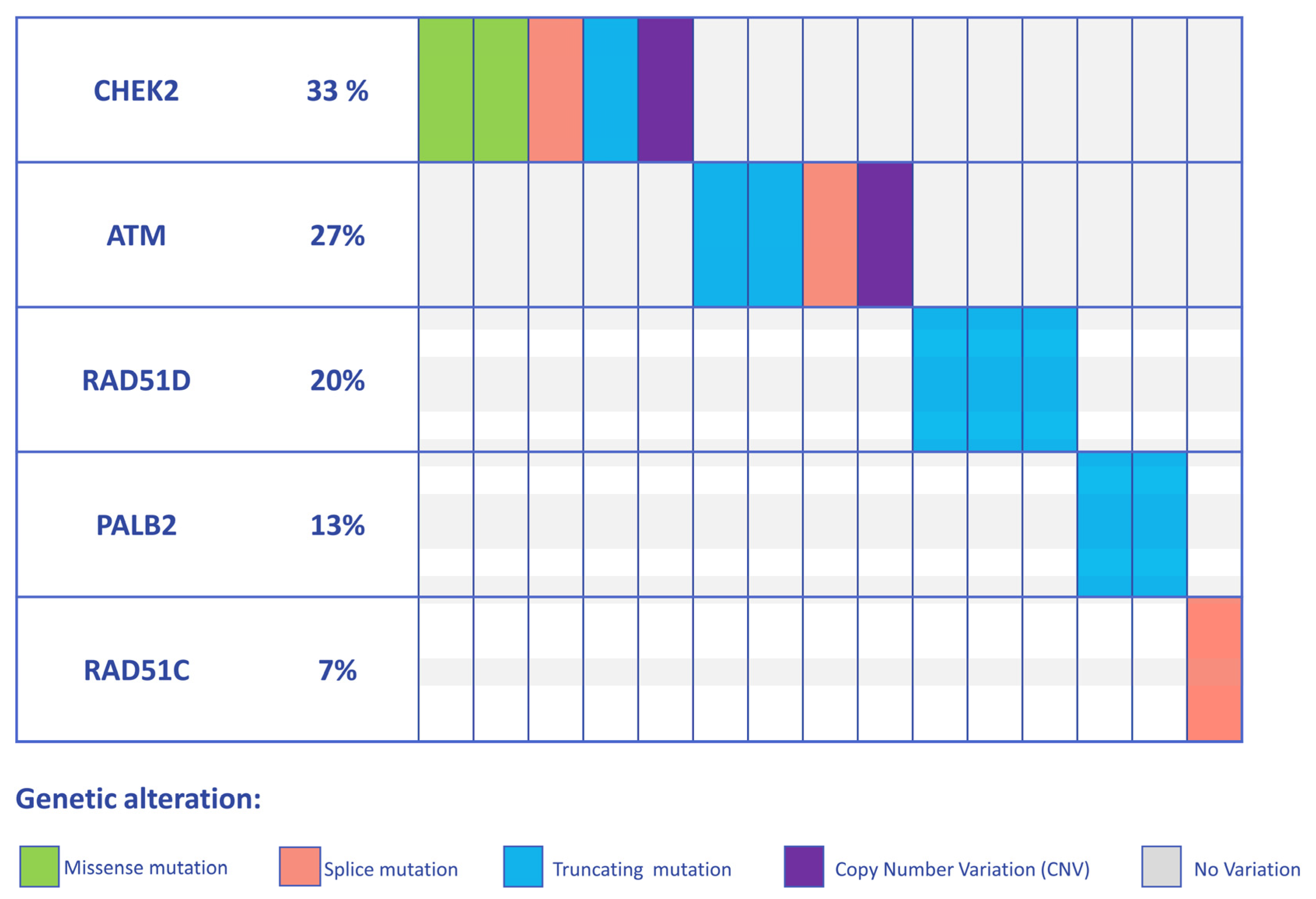
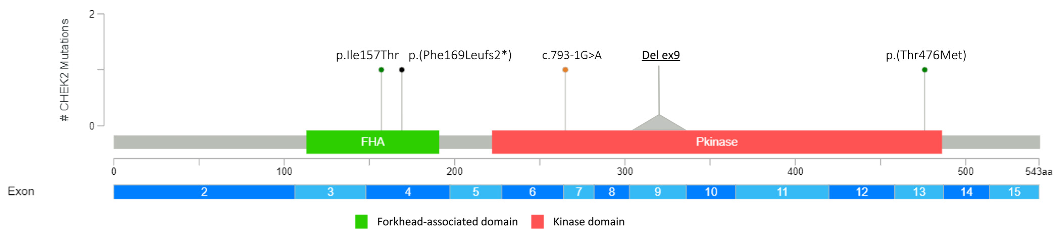
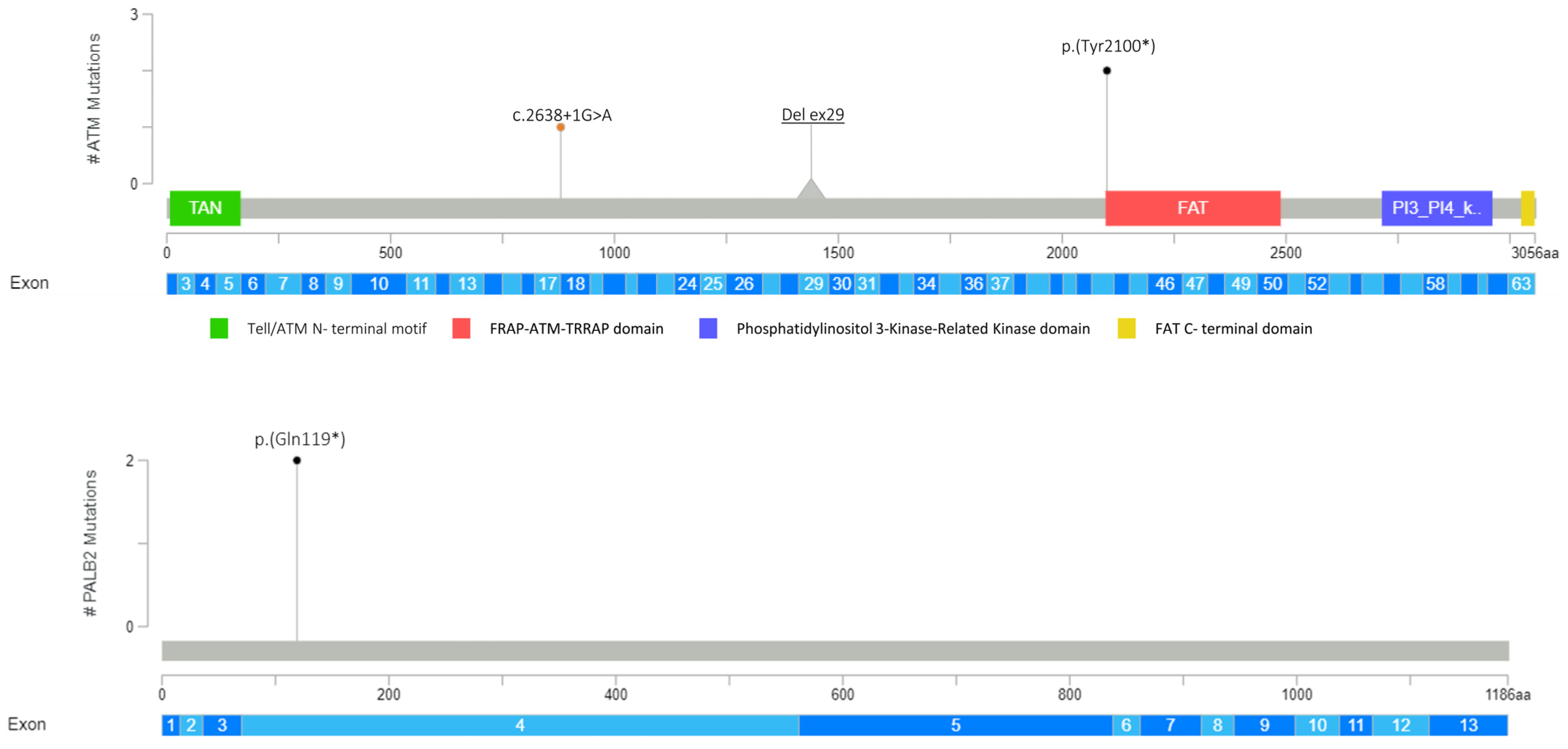
3.1. Detection of PVs and LPVs in BC Moderate Penetrance Genes

Germline DNA from 521 patients diagnosed with BC were retrospectively analyzed to assess PV and LPV frequency in cancer susceptibility genes other than BRCA1/2. The analysis revealed 15 out of 521 (2.9%) individuals harboring PVs/LPVs in five BC predisposing genes (PALB2, CHEK2, ATM, RAD51C, and RAD51D). In detail, 4 individuals (0.76%) were shown to carry PVs and LPVs in the ATM gene, 5 individuals (0.95%) in the CHEK2 gene, 2 individuals (0.38%) in the PALB2 gene, and 3 individuals (0.58%) in the RAD51D gene, and 1 PV was disclosed in the RAD51C gene (0.19%). The distribution of the PVs/LPVs is showed in Figure 1.
Overall, as regards the mutational impact upon protein, 2 missense variants out of 15 (13.3%), 7 (46.6%) non-sense, 1 (6.7%) indel, and 3 (20%) splicing variants were detected (Figure 1, Figure 2, Figure 3 and Figure 4). To note, 2 samples (13.3%) showed that two microdeletions comprehended CHEK2 exon 9 and ATM exon 29, respectively (Figure 2, Figure 3 and Figure 4). Overall, three variants occurring in PALB2 (p.(Gln119*)), ATM (p.(Tyr2100*)), and RAD51D (p.(Trp268*)) were identified as recurrent in two samples each. All the variants detected are listed in Table 1.

3.2. Clinical and Demographic Characteristics of High-to-Moderate Penetrance Gene Positive Patients

Table 2 and Table S1 summarize the clinical and demographic characteristics of the patients, showing deleterious variants in BC susceptibility genes. The mean age at diagnosis was 46 years (ranging from 29 to 69 years). The majority of the patients (73.3%) harbored a noninvasive ductal carcinoma of no special type (IDC-NST). Only one patient developed another type of tumor beside BC (in situ melanoma), and three patients developed multiple BCs. It is noteworthy that the patient with a RAD51C PV experienced two BCs characterized by different HER2, estrogen receptor (ER), and progesterone receptor (PR) expression profiles. More than half of the selected individuals (73%) showed a family history of BC. On the other end, approximately 50% of the selected individuals displayed a family history of cancer other than BC. Unfortunately, a HER2, ER, and PR profile was not available for all the patients. Interestingly, all the patients carrying ATM PVs and LPVs showed the same HER2, ER, and PR status.

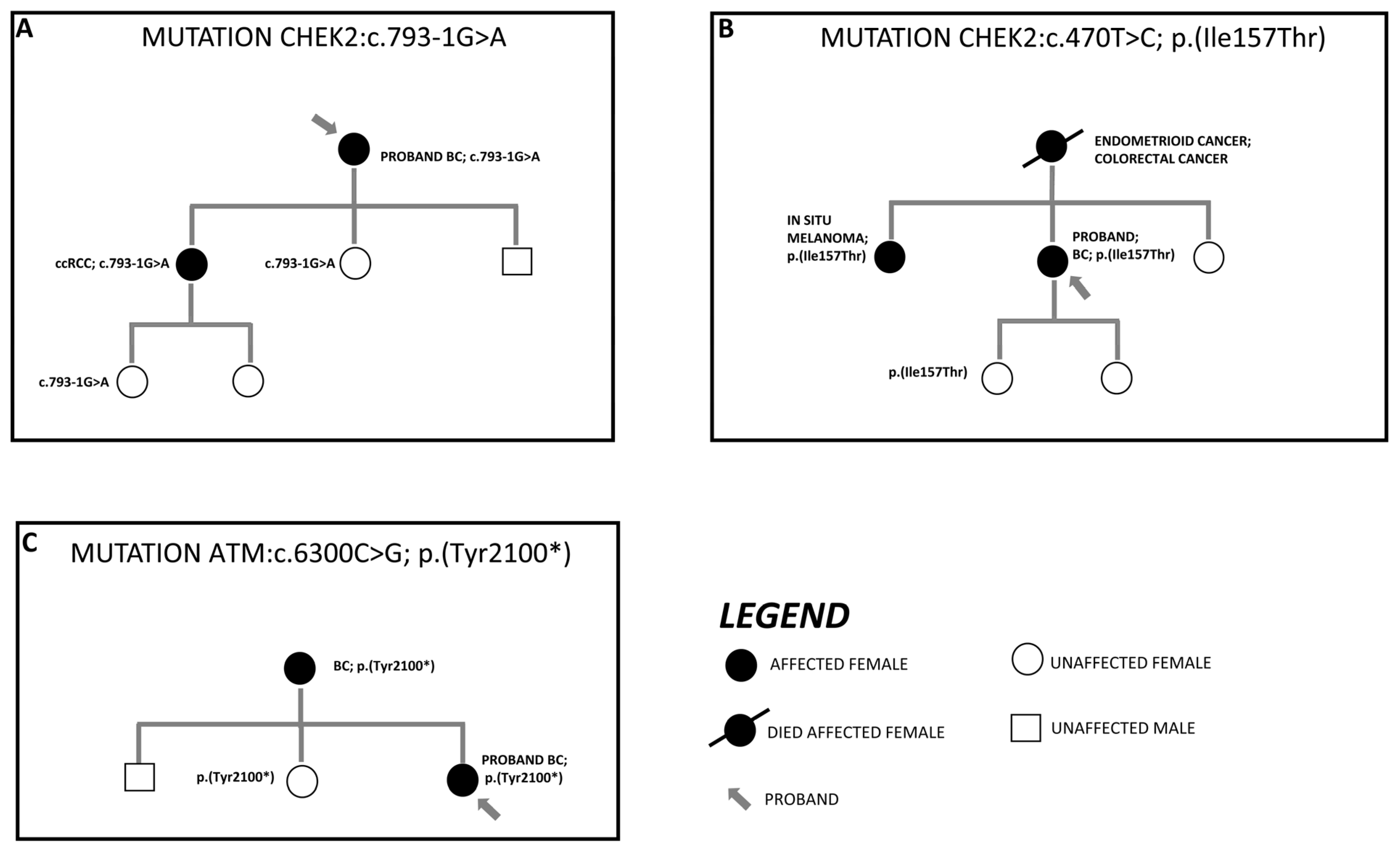
3.3. Segregation Analysis of Detected Variant in First-Degree Family Members

In agreement with the guideline, the detected PVs and LPVs were segregated into first-degree relatives. To date, segregation data are only available for six variants. The most significant results of segregation analysis are shown in Figure 5. It is noteworthy that two CHEK2 mutations occurred in two individuals with a diagnosis of clear cell renal cell carcinoma (ccRCC) and in situ melanoma, respectively. Additionally, a pathogenic ATM variant was also detected in a subject who had developed BC.

4. Discussion

In the present study, a population of 521 patients diagnosed with BC who underwent NGS-based multigene panel testing were retrospectively analyzed. All the patients were selected according to their BC personal history and cancer family history [4]. This study aimed to assess the frequency of PVs and LPVs in BC moderate-risk cancer genes beyond BRCA1/2, in order to ascertain a possible correlation between deleterious variants in those genes and the genesis of cancer other than BC, and to explore the clinical, histological, and molecular BC profile. Overall, 15 patients out of 521 (2.9%) showed PVs and LPVs in five cancer-predisposing genes other than BRCA1/2 (PALB2, CHEK2, ATM, RAD51C, and RAD51D). All these genes play a pivotal role in the HR mechanism [2]. In detail, the distribution of PVs and LPVs was as follows: ATM (0.76%), CHEK2 (0.95%), PALB2 (0.38%), RAD51D (0.58%), and RAD51C (0.19%). In our population study, the RAD51C PVs and LPVs occurrence are discovered to be in line with previous publications [7,15]. In contrast, the RAD51D variant frequency detected was slightly higher than previous studies, probably due to the small size of our population study [7,15]. Moreover, the frequency of the ATM, PALB2, and CHEK2 mutations is overall lower than those reported in similar studies [16,17,18,19,20,21]. Nevertheless, many of these studies focused on BRCA1/2 negative BC. Herein, we examined BC patients with NGS multigene panels including BRCA1/2, thus not excluding those carrying deleterious variants in the two high-risk genes. Among all 521 subjects, 37 showed a PVs or/and LPVs in BRCA1/2 (7%). It is noteworthy that none of the 15 subjects displayed additional deleterious variants in other genes, making the identified PVs and LPVs the main genetic cause of BC onset.
Three variants were identified as recurrent in two patients; these were, respectively, (PALB2:p.(Gln119*), ATM:p.(Tyr2100*), and RAD51D:p.(Trp268*)). To our knowledge, p.(Gln119*) and p.(Tyr2100*) have been never described in the literature in individuals affected by BC. The deleterious effect of these two mutations was predicted, since ATM and PALB2 loss-of-function is a known mechanism of pathogenicity, resulting in an absent or disrupted protein product. Regarding the p.(Trp268*) mutation, it has already been reported in subjects diagnosed with ovarian cancer [22,23,24], but never in BC.
Two interesting variants were detected in CHEK2 [p.(Ile157Thr) and p.(Thr476Met)]. Both these variants were associated with attenuated BC risk [7,25,26] and have been identified in the population with a high frequency. The p.(Ile157Thr) variant also had a more favorable prognosis among BC patients [27]. This variant lies in a weakly conserved amino acid of the forkhead-associated domain, and conflicting interpretations of its pathogenicity have been published [28]. There were also discordant results on CHEK2 protein function affected by this variant; some studies showed that p.(Ile157Thr) does not inhibit several steps in protein activation [29,30,31,32,33,34], and other studies showed disruption of the same activation [35,36]. The p.(Thr476Met) variant falls in the kinase domain of CHEK2 protein in a moderately conserved domain. As with p.(Ile157Thr), the pathogenic classification of p.(Thr476Met) is discordant, and a previous study showed that this variant has been classified as pathogenic, likely pathogenic, and VUS by different authors [37]. By in silico analysis, the variant has been predicted to be deleterious. Kleiblova et al. (2019) showed a deleterious effect in vitro and a likely benign effect in vivo by functional studies of KAP1 phosphorylation [30]. In addition, Desrichard et al. (2011) showed reduced SOX phosphorylation equal to that of the pathogenic c.1100delC variant, suggesting that this mutation affects the ability of the recombinant CHEK2 to recognize, bind, and phosphorylate its substrate [38].
Interestingly, within the overall mutational spectrum, two microdeletions (CHEK2 exon 9 and ATM exon 29) were also identified. CHEK2 deletions of exon 8 and of exon 9 and 10 have already been found in BC patients [39,40]. More specifically, the latter deletion is a founder mutation in the Northern and Eastern European population, accounting for a frequency of 0.9–1% in BC cases and of 0.4–0.5% in the controls, thus contributing to the BC burden [39,41]. Additionally, the CHEK2 microdeletion was unveiled in a young patient with a strong family history of cancer. The patient’s father died of ampullary cancer, preventing us to segregate the same alterations and find a possible correlation.
As regards ATM exon 29 deletion, no research has been reported so far about the same deletion in cancer. Nonetheless, distinct types of ATM deletion have been associated with BC [42,43], also in patients revealing a BC family history. The individual in our cohort also showed a family history of BC and prostate cancer. Unfortunately, again, a segregation analysis was not available.
The contribution of the RAD51C and RAD51D deleterious variants to ovarian cancer has been previously established by numerous studies, but their role in BC predisposition is now emerging [22,44,45,46]. Several studies suggested a correlation between RAD51C/D PV and BC susceptibility, given their related functions in the DNA repair process via HR. Nonetheless, this remained controversial until the study of Yang et al. (2020) [47]. They estimated the relative risk of developing BC to be 1.83 for RAD51D PV carriers and 1.99 for RAD51C PV carriers, leading to the categorization of these genes as BC moderate-risk genes. Of note, one of the truncating variants [p.(Arg253*)] in the RAD51D gene identified in our population study has been previously reported in a BC patient [48]. Concerning the RAD51C gene, the c.1026+5_1026+7delGTA variant was identified in one patient diagnosed with BC at age 49, without a family history of cancer. Many authors classified this variant as likely pathogenic, as it impacts splicing by causing aberrant exon 8 skipping, resulting in a frame shift and a premature stop codon (p.R322Sfs22*) [49,50,51,52,53]. However, in the comprehensive splicing study of the RAD51C germline variants by Sanoguera-Miralles et al. (2020) [54], this alteration is reported to totally disrupt splicing, and the authors therefore proposed a pathogenic classification based on their functional study.
In addition, among all the patients tested, 5.8% had at least one variant of uncertain significance (VUS) identified in the PALB2, CHEK2, ATM, RAD51C, and RAD51D genes. Although there are conflicting points of view in the literature regarding the clinical management of VUS carriers, due to the unknown impact on gene function and cancer risk, the NCCN guidelines recommend screening and risk reduction strategies on the basis of personal and family history [4,55,56], which can also be evaluated by means of BC risk prediction model tools.
The segregation analysis, when available, highlighted two major results: CHEK2 variants c.793-1G>A and p.(Ile157Thr) were found in a ccRCC case and in a subject diagnosed with in situ melanoma, respectively.
To date, the association between CHEK2 alteration and skin cancer (both melanoma and nonmelanoma) is still unclear. Further studies are necessary to explore the involvement of CHEK2 in the development of skin cancer. Nevertheless, CHEK2 is reported to be a key protein in response to UV damage through its upregulation and its phosphorylation by the ATM and ATR proteins [57].
The finding of CHEK2 PVs in a ccRCC patient is not unprecedented. Indeed, a large number of studies have been pointing out a possible connection between CHEK2 alteration and the onset of kidney cancer (especially renal cell carcinoma) [58,59,60,61,62]. In 2004, Cybulski et al. identified an increased risk of kidney cancer associated with the p.(Ile157Thr) variant [63]. Subsequent studies with wider cohorts highlighted the peculiar occurrence of the CHEK2 mutation in patients affected by renal cell carcinoma [58,59,60,61,62]. Additionally, Brooks et al. [64] found a CHEK2 splicing variant in two individuals who had a diagnosis of ccRCC. Therefore, our finding adds an important piece of evidence to the literature, leading to the need of new more in-depth research to evaluate the role of CHEK2 in renal cancer.
Regarding the clinical and demographic features of the 15 selected patients, the median age of BC onset was about 46 years. This result is approximately in line with previous reports [16,17,21,65]. Notably, Chen et al. found that RAD51D mutation carriers were more likely to predispose to early-onset BC, with a mean age at diagnosis of 45.4 years, similar to patients with BRCA1 mutation (44.8 years) [66]. The mean age of 41 years in our RAD51D mutation carriers further confirm this finding.
Most of the cases (73.3%) exhibited an IDC-NST histology, confirming this as the most common BC histotype [67].
Furthermore, 73% and 53.3% of the cohort showed, respectively, BC and cancer other than BC in their family history. Again, these results are in line with previous studies [16,21,65].
It is noteworthy that all four subjects displaying an ATM-mutated BC are characterized by the same HER2, ER, and PR expression levels (HER2 negative, PR, and ER positives). This observation further confirms previous studies [68,69,70] which highlighted a strong association with an ER-positive profile [15,71]. For variants in the other four genes, we were not able to determine a common ER, PR, and HER2 molecular profile, also due to the limitation of the available histopathological data. Nevertheless, a PALB2-mutated BC exhibited a triple-negative profile, which is consistent with the literature [68,71]. Also, the RAD51C and RAD51D variants have shown a strong association with triple-negative cancer [68,70]. Here, only one case displayed a triple-negative BC. Intriguingly, a subject carrying a RAD51D PV developed two BCs with an opposite receptor profile. Furthermore, a link between the protein-truncating variants in RAD51D and ER-negative breast cancers other than the ER-positive disease has been recently defined [7,15]. In our population study, although 2 cases out of 4 confirm the subtype-specific association with PVs in RAD51D, the small size of our population study does not allow for strong evidence. Concerning the CHEK2 gene, the variants have been associated with all BC histotypes except for the triple-negative [15]. Based on the limited available information, three out of five CHEK2 PVs carriers did not show a triple-negative molecular status.

5. Conclusions

Given the rise in the use of multigene panel testing in diagnostic procedure, the detection of PVs and/or LPVs in a moderate-risk penetrance gene is constantly increasing. In this study, we provide estimates of PV and/or LPV occurrence in genes besides BRCA1/2 and their association either with additional forms of tumors (especially ccRCC) or with a peculiar molecular signature. Taken all together, these results may help to guide counseling, cancer screening, and other risk management strategies for women and families with PVs and/or LPVs in BC predisposition genes.

Supplementary Materials

The following supporting information can be downloaded at: https://www.mdpi.com/article/10.3390/genes14081530/s1, Table S1: Histology and ER, PR, and HER molecular profile correlated with each variant.

Author Contributions

Conceptualization, N.R., A.T. and M.P.; investigation, A.T., M.P., S.M., S.C. (Stefania Chirulli), M.I., M.S., R.B., A.G., A.D.L., E.M., S.C. (Silvia Crocetta), S.L., M.C., Y.K. and O.T.; resources, C.S., M.I., M.S., E.M., O.T., S.C. (Silvia Crocetta), S.L., M.C., Y.K. and P.L.; supervision, N.R., A.S., N.B., C.S. and C.F.; visualization, A.T., M.P. and S.M.; writing—original draft, A.T. and M.P.; writing—review and editing, A.T., M.P., N.R., C.S., A.S. and C.F. All authors have read and agreed to the published version of the manuscript.

Funding

This study was supported by the funding MUR program NextGenerationEU DM 737/2021-CUP H91121001660006 “Fondo promozione e sviluppo DM 737/2021-CUP H91121001660006; Horizon Europe Seeds S55 Identificative Code”.

Institutional Review Board Statement

The study was conducted according to the guidelines of the Declaration of Helsinki. All the individuals provided informed consent for genetic analysis and publication of the results. Ethical review and approval were waived for this study because all the procedures were conducted as part of a clinical assessment and in line with local ethical policy and any other legal requirements.

Informed Consent Statement

Informed consent was obtained from all the subjects involved in the study.

Data Availability Statement

The data presented in this study are available on request from the corresponding author.

Conflicts of Interest

The authors declare no conflict of interest.

References

  1. Sung, H.; Ferlay, J.; Siegel, R.L.; Laversanne, M.; Soerjomataram, I.; Jemal, A.; Bray, F. Global Cancer Statistics 2020: GLOBOCAN Estimates of Incidence and Mortality Worldwide for 36 Cancers in 185 Countries. CA Cancer J. Clin. 2021, 71, 209–249. [Google Scholar] [CrossRef]
  2. Roy, R.; Chun, J.; Powell, S.N. BRCA1 and BRCA2: Different Roles in a Common Pathway of Genome Protection. Nat. Rev. Cancer 2012, 12, 68–78. [Google Scholar]
  3. Kuchenbaecker, K.B.; Hopper, J.L.; Barnes, D.R.; Phillips, K.A.; Mooij, T.M.; Roos-Blom, M.J.; Jervis, S.; Van Leeuwen, F.E.; Milne, R.L.; Andrieu, N.; et al. Risks of Breast, Ovarian, and Contralateral Breast Cancer for BRCA1 and BRCA2 Mutation Carriers. JAMA 2017, 317, 2402–2416. [Google Scholar] [CrossRef] [PubMed] [Green Version]
  4. Daly, M.B.; Pal, T.; Berry, M.P.; Buys, S.S.; Dickson, P.; Domchek, S.M.; Elkhanany, A.; Friedman, S.; Goggins, M.; Hutton, M.L.; et al. Genetic/Familial High-Risk Assessment: Breast, Ovarian, and Pancreatic, Version 2.2021, NCCN Clinical Practice Guidelines in Oncology. J. Natl. Compr. Cancer Netw. 2021, 19, 77–102. [Google Scholar]
  5. Fanale, D.; Incorvaia, L.; Filorizzo, C.; Bono, M.; Fiorino, A.; Calò, V.; Brando, C.; Corsini, L.R.; Barraco, N.; Badalamenti, G.; et al. Detection of Germline Mutations in a Cohort of 139 Patients with Bilateral Breast Cancer by Multi-Gene Panel Testing: Impact of Pathogenic Variants in Other Genes beyond BRCA1/2. Cancers 2020, 12, 2415. [Google Scholar] [PubMed]
  6. Shah, P.D.; Patil, S.; Dickler, M.N.; Offit, K.; Hudis, C.A.; Robson, M.E. Twenty-One-Gene Recurrence Score Assay in BRCA-Associated versus Sporadic Breast Cancers: Differences Based on Germline Mutation Status. Cancer 2016, 122, 1178–1184. [Google Scholar] [CrossRef]
  7. Hu, C.; Hart, S.N.; Gnanaolivu, R.; Huang, H.; Lee, K.Y.; Na, J.; Gao, C.; Lilyquist, J.; Yadav, S.; Boddicker, N.J.; et al. A Population-Based Study of Genes Previously Implicated in Breast Cancer. N. Engl. J. Med. 2021, 384, 440–451. [Google Scholar] [CrossRef]
  8. Fountzilas, C.; Kaklamani, V.G. Multi-Gene Panel Testing in Breast Cancer Management. In Optimizing Breast Cancer Management; Cancer Treatment and Research Series; Springer: Cham, Switzerland, 2018; Volume 173, pp. 121–140. [Google Scholar]
  9. Neben, C.L.; Zimmer, A.D.; Stedden, W.; van den Akker, J.; O’Connor, R.; Chan, R.C.; Chen, E.; Tan, Z.; Leon, A.; Ji, J.; et al. Multi-Gene Panel Testing of 23,179 Individuals for Hereditary Cancer Risk Identifies Pathogenic Variant Carriers Missed by Current Genetic Testing Guidelines. J. Mol. Diagn. 2019, 21, 646–657. [Google Scholar] [CrossRef] [Green Version]
  10. Li, H.; Durbin, R. Fast and Accurate Short Read Alignment with Burrows-Wheeler Transform. Bioinformatics 2009, 25, 1754–1760. [Google Scholar] [CrossRef] [Green Version]
  11. Li, H.; Durbin, R. Fast and Accurate Long-Read Alignment with Burrows-Wheeler Transform. Bioinformatics 2010, 26, 589–595. [Google Scholar] [CrossRef] [Green Version]
  12. Van der Auwera, G.A.; O’Connor, B.D. Genomics in the Cloud: Using Docker, GATK, and WDL in Terra, 1st ed.; O’Reilly Media: Sebastopol, CA, USA, 2020. [Google Scholar]
  13. Richards, S.; Aziz, N.; Bale, S.; Bick, D.; Das, S.; Gastier-Foster, J.; Grody, W.W.; Hegde, M.; Lyon, E.; Spector, E.; et al. Standards and Guidelines for the Interpretation of Sequence Variants: A Joint Consensus Recommendation of the American College of Medical Genetics and Genomics and the Association for Molecular Pathology. Genet. Med. 2015, 17, 405–424. [Google Scholar] [CrossRef] [PubMed] [Green Version]
  14. den Dunnen, J.T.; Dalgleish, R.; Maglott, D.R.; Hart, R.K.; Greenblatt, M.S.; Mcgowan-Jordan, J.; Roux, A.F.; Smith, T.; Antonarakis, S.E.; Taschner, P.E.M. HGVS Recommendations for the Description of Sequence Variants: 2016 Update. Hum. Mutat. 2016, 37, 564–569. [Google Scholar] [CrossRef] [PubMed] [Green Version]
  15. Dorling, L.; Carvalho, S.; Allen, J.; González-Neira, A.; Luccarini, C.; Wahlström, C.; Pooley, K.A.; Parsons, M.T.; Fortuno, C.; Wang, Q.; et al. Breast Cancer Risk Genes—Association Analysis in More than 113,000 Women. N. Engl. J. Med. 2021, 384, 428–439. [Google Scholar] [CrossRef] [PubMed]
  16. Toss, A.; Tenedini, E.; Piombino, C.; Venturelli, M.; Marchi, I.; Gasparini, E.; Barbieri, E.; Razzaboni, E.; Domati, F.; Caggia, F.; et al. Clinicopathologic Profile of Breast Cancer in Germline ATM and CHEK2 Mutation Carriers. Genes 2021, 12, 616. [Google Scholar] [CrossRef]
  17. Hauke, J.; Horvath, J.; Groß, E.; Gehrig, A.; Honisch, E.; Hackmann, K.; Schmidt, G.; Arnold, N.; Faust, U.; Sutter, C.; et al. Gene Panel Testing of 5589 BRCA1/2-Negative Index Patients with Breast Cancer in a Routine Diagnostic Setting: Results of the German Consortium for Hereditary Breast and Ovarian Cancer. Cancer Med. 2018, 7, 1349–1358. [Google Scholar] [CrossRef]
  18. Weitzel, J.N.; Neuhausen, S.L.; Adamson, A.; Tao, S.; Ricker, C.; Maoz, A.; Rosenblatt, M.; Nehoray, B.; Sand, S.; Steele, L.; et al. Pathogenic and Likely Pathogenic Variants in PALB2, CHEK2, and Other Known Breast Cancer Susceptibility Genes among 1054 BRCA-Negative Hispanics with Breast Cancer. Cancer 2019, 125, 2829–2836. [Google Scholar] [CrossRef]
  19. Park, J.S.; Shin, S.; Lee, Y.J.; Lee, S.T.; Nam, E.J.; Han, J.W.; Lee, S.H.; Kim, T.I.; Park, H.S. Implication and Influence of Multigene Panel Testing with Genetic Counseling in Korean Patients with BRCA1/2 Mutation-Negative Breast Cancer. Cancer Res. Treat. 2022, 54, 1099–1110. [Google Scholar] [CrossRef]
  20. Bono, M.; Fanale, D.; Incorvaia, L.; Cancelliere, D.; Fiorino, A.; Calò, V.; Dimino, A.; Filorizzo, C.; Corsini, L.R.; Brando, C.; et al. Impact of Deleterious Variants in Other Genes beyond BRCA1/2 Detected in Breast/Ovarian and Pancreatic Cancer Patients by NGS-Based Multi-Gene Panel Testing: Looking over the Hedge. ESMO Open 2021, 6, 100235. [Google Scholar] [CrossRef]
  21. Kraus, C.; Hoyer, J.; Vasileiou, G.; Wunderle, M.; Lux, M.P.; Fasching, P.A.; Krumbiegel, M.; Uebe, S.; Reuter, M.; Beckmann, M.W.; et al. Gene Panel Sequencing in Familial Breast/Ovarian Cancer Patients Identifies Multiple Novel Mutations Also in Genes Others than BRCA1/2. Int. J. Cancer 2017, 140, 95–102. [Google Scholar] [CrossRef]
  22. Loveday, C.; Turnbull, C.; Ramsay, E.; Hughes, D.; Ruark, E.; Frankum, J.R.; Bowden, G.; Kalmyrzaev, B.; Warren-Perry, M.; Snape, K.; et al. Germline Mutations in RAD51D Confer Susceptibility to Ovarian Cancer. Nat. Genet. 2011, 43, 879–882. [Google Scholar] [CrossRef] [PubMed]
  23. Baker, J.L.; Schwab, R.B.; Wallace, A.M.; Madlensky, L. Breast Cancer in a RAD51D Mutation Carrier: Case Report and Review of the Literature. Clin. Breast Cancer 2015, 15, e71–e75. [Google Scholar] [CrossRef]
  24. Thompson, E.R.; Rowley, S.M.; Sawyer, S.; Eccles, D.M.; Trainer, A.H.; Mitchell, G.; James, P.A.; Campbell, I.G. Analysis of RAD51D in Ovarian Cancer Patients and Families with a History of Ovarian or Breast Cancer. PLoS ONE 2013, 8, e54772. [Google Scholar] [CrossRef]
  25. Akcay, I.M.; Celik, E.; Agaoglu, N.B.; Alkurt, G.; Kizilboga Akgun, T.; Yildiz, J.; Enc, F.; Kir, G.; Canbek, S.; Kilic, A.; et al. Germline Pathogenic Variant Spectrum in 25 Cancer Susceptibility Genes in Turkish Breast and Colorectal Cancer Patients and Elderly Controls. Int. J. Cancer 2021, 148, 285–295. [Google Scholar] [CrossRef] [PubMed]
  26. Muranen, T.A.; Blomqvist, C.; Dörk, T.; Jakubowska, A.; Heikkilä, P.; Fagerholm, R.; Greco, D.; Aittomäki, K.; Bojesen, S.E.; Shah, M.; et al. Patient Survival and Tumor Characteristics Associated with CHEK2:P.I157T—Findings from the Breast Cancer Association Consortium. Breast Cancer Res. 2016, 18, 98. [Google Scholar] [CrossRef] [PubMed]
  27. Bychkovsky, B.L.; Agaoglu, N.B.; Horton, C.; Zhou, J.; Yussuf, A.; Hemyari, P.; Richardson, M.E.; Young, C.; Laduca, H.; McGuinness, D.L.; et al. Differences in Cancer Phenotypes among Frequent CHEK2 Variants and Implications for Clinical Care—Checking CHEK2. JAMA Oncol. 2022, 8, 1598–1606. [Google Scholar] [CrossRef] [PubMed]
  28. Mundt, E.; Nix, P.; Brown, K.; Bowles, K.R.; Manley, S. Complexities of Variant Classification in Clinical Hereditary Cancer Genetic Testing. J. Clin. Oncol. 2017, 35, 3796–3799. [Google Scholar] [CrossRef] [PubMed]
  29. Boonen, R.A.C.M.; Wiegant, W.W.; Celosse, N.; Vroling, B.; Heijl, S.; Kote-Jara, Z.; Mijuskovic, M.; Cristea, S.; Solleveld-Westerin, N.; Van Wezel, T.; et al. Functional Analysis Identifies Damaging CHEK2 Missense Variants Associated with Increased Cancer Risk. Cancer Res. 2022, 82, 615–631. [Google Scholar] [CrossRef] [PubMed]
  30. Kleiblova, P.; Stolarova, L.; Krizova, K.; Lhota, F.; Hojny, J.; Zemankova, P.; Havranek, O.; Vocka, M.; Cerna, M.; Lhotova, K.; et al. Identification of Deleterious Germline CHEK2 Mutations and Their Association with Breast and Ovarian Cancer. Int. J. Cancer 2019, 145, 1782–1797. [Google Scholar] [CrossRef]
  31. Falck, J.; Mailand, N.; Syljuåsen, R.G.; Bartek, J.; Lukas, J. The ATM-Chk2-Cdc25A Checkpoint Pathway Guards against Radioresistant DNA Synthesis. Nature 2001, 410, 842–847. [Google Scholar] [CrossRef]
  32. Falck, J.; Lukas, C.; Protopopova, M.; Lukas, J.; Selivanova, G.; Bartek, J. Functional Impact of Concomitant versus Alternative Defects in the Chk2-P53 Tumour Suppressor Pathway. Oncogene 2001, 20, 5503–5510. [Google Scholar] [CrossRef] [Green Version]
  33. Ahn, J.Y.; Li, X.; Davis, H.L.; Canman, C.E. Phosphorylation of Threonine 68 Promotes Oligomerization and Autophosphorylation of the Chk2 Protein Kinase via the Forkhead-Associated Domain. J. Biol. Chem. 2002, 277, 19389–19395. [Google Scholar] [CrossRef] [PubMed] [Green Version]
  34. Kilpivaara, O.; Vahteristo, P.; Falck, J.; Syrjäkoski, K.; Eerola, H.; Easton, D.; Bartkova, J.; Lukas, J.; Heikkilä, P.; Aittomäki, K.; et al. CHEK2 Variant I157T May Be Associated with Increased Breast Cancer Risk. Int. J. Cancer 2004, 111, 543–547. [Google Scholar] [CrossRef]
  35. Schwarz, J.K.; Lovly, C.M.; Piwnica-Worms, H. Regulation of the Chk2 Protein Kinase by Oligomerization-Mediated Cis- and Trans-Phosphorylation. Mol. Cancer Res. 2003, 1, 598–609. [Google Scholar]
  36. Cai, Z.; Chehab, N.H.; Pavletich, N.P. Structure and Activation Mechanism of the CHK2 DNA Damage Checkpoint Kinase. Mol. Cell 2009, 35, 818–829. [Google Scholar] [CrossRef]
  37. Agaoglu, N.B.; Unal, B.; Akgun Dogan, O.; Kanev, M.O.; Zolfagharian, P.; Ozemri Sag, S.; Temel, S.G.; Doganay, L. Consistency of Variant Interpretations among Bioinformaticians and Clinical Geneticists in Hereditary Cancer Panels. Eur. J. Hum. Genet. 2022, 30, 378–383. [Google Scholar] [CrossRef] [PubMed]
  38. Desrichard, A.; Bidet, Y.; Uhrhammer, N.; Bignon, Y.J. CHEK2 Contribution to Hereditary Breast Cancer in Non-BRCA Families. Breast Cancer Res. 2011, 13, R119. [Google Scholar] [CrossRef] [PubMed]
  39. Cybulski, C.; Wokołorczyk, D.; Huzarski, T.; Byrski, T.; Gronwald, J.; Górski, B.; Dębniak, T.; Masojć, B.; Jakubowska, A.; Van De Wetering, T.; et al. A Deletion in CHEK2 of 5,395 Bp Predisposes to Breast Cancer in Poland. Breast Cancer Res. Treat. 2007, 102, 119–122. [Google Scholar] [CrossRef]
  40. Nizic-Kos, T.; Krajc, M.; Blatnik, A.; Stegel, V.; Skerl, P.; Novakovic, S.; Gazic, B.; Besic, N. Bilateral Disease Common among Slovenian CHEK2-Positive Breast Cancer Patients. Ann. Surg. Oncol. 2021, 28, 2561–2570. [Google Scholar] [CrossRef]
  41. Dennis, J.; Tyrer, J.P.; Walker, L.C.; Michailidou, K.; Dorling, L.; Bolla, M.K.; Wang, Q.; Ahearn, T.U.; Andrulis, I.L.; Anton-Culver, H.; et al. Rare Germline Copy Number Variants (CNVs) and Breast Cancer Risk. Commun. Biol. 2022, 5, 65. [Google Scholar] [CrossRef]
  42. Bandeira, G.; Rocha, K.; Lazar, M.; Ezquina, S.; Yamamoto, G.; Varela, M.; Takahashi, V.; Aguena, M.; Gollop, T.; Zatz, M.; et al. Germline Variants of Brazilian Women with Breast Cancer and Detection of a Novel Pathogenic ATM Deletion in Early-Onset Breast Cancer. Breast Cancer 2021, 28, 346–354. [Google Scholar] [CrossRef]
  43. Parenti, S.; Rabacchi, C.; Marino, M.; Tenedini, E.; Artuso, L.; Castellano, S.; Carretta, C.; Mallia, S.; Cortesi, L.; Toss, A.; et al. Characterization of New ATM Deletion Associated with Hereditary Breast Cancer. Genes 2021, 12, 136. [Google Scholar] [CrossRef]
  44. Golmard, L.; Castéra, L.; Krieger, S.; Moncoutier, V.; Abidallah, K.; Tenreiro, H.; Laugé, A.; Tarabeux, J.; Millot, G.A.; Nicolas, A.; et al. Contribution of Germline Deleterious Variants in the RAD51 Paralogs to Breast and Ovarian Cancers /631/208/68 /631/67/1347 Article. Eur. J. Hum. Genet. 2017, 25, 1345–1353. [Google Scholar] [CrossRef] [Green Version]
  45. Vuorela, M.; Pylkäs, K.; Hartikainen, J.M.; Sundfeldt, K.; Lindblom, A.; Von Wachenfeldt Wäppling, A.; Haanpää, M.; Puistola, U.; Rosengren, A.; Anttila, M.; et al. Further Evidence for the Contribution of the RAD51C Gene in Hereditary Breast and Ovarian Cancer Susceptibility. Breast Cancer Res. Treat. 2011, 130, 1003–1010. [Google Scholar] [CrossRef]
  46. Schnurbein, G.; Hauke, J.; Wappenschmidt, B.; Weber-Lassalle, N.; Engert, S.; Hellebrand, H.; Garbes, L.; Becker, A.; Neidhardt, G.; Rhiem, K.; et al. RAD51C Deletion Screening Identifies a Recurrent Gross Deletion in Breast Cancer and Ovarian Cancer Families. Breast Cancer Res. 2013, 15, R120. [Google Scholar]
  47. Yang, X.; Song, H.; Leslie, G.; Engel, C.; Hahnen, E.; Auber, B.; Horváth, J.; Kast, K.; Niederacher, D.; Turnbull, C.; et al. Ovarian and Breast Cancer Risks Associated with Pathogenic Variants in RAD51C and RAD51D. J. Natl. Cancer Inst. 2020, 112, 1242–1250. [Google Scholar] [CrossRef] [Green Version]
  48. Sun, J.; Meng, H.; Yao, L.; Lv, M.; Bai, J.; Zhang, J.; Wang, L.; Ouyang, T.; Li, J.; Wang, T.; et al. Germline Mutations in Cancer Susceptibility Genes in a Large Series of Unselected Breast Cancer Patients. Clin. Cancer Res. 2017, 23, 6113–6119. [Google Scholar] [CrossRef] [Green Version]
  49. Tsai, G.J.; Rañola, J.M.O.; Smith, C.; Garrett, L.T.; Bergquist, T.; Casadei, S.; Bowen, D.J.; Shirts, B.H. Outcomes of 92 Patient-Driven Family Studies for Reclassification of Variants of Uncertain Significance. Genet. Med. 2019, 21, 1435–1442. [Google Scholar] [CrossRef] [PubMed]
  50. Golmard, L.; Caux-Moncoutier, V.; Davy, G.; Al Ageeli, E.; Poirot, B.; Tirapo, C.; Michaux, D.; Barbaroux, C.; d’Enghien, C.D.; Nicolas, A.; et al. Germline Mutation in the RAD51B Gene Confers Predisposition to Breast Cancer. BMC Cancer 2013, 13, 484. [Google Scholar] [CrossRef]
  51. Casadei, S.; Gulsuner, S.; Shirts, B.H.; Mandell, J.B.; Kortbawi, H.M.; Norquist, B.S.; Swisher, E.M.; Lee, M.K.; Goldberg, Y.; O’Connor, R.; et al. Characterization of Splice-Altering Mutations in Inherited Predisposition to Cancer. Proc. Natl. Acad. Sci. USA 2019, 116, 26798–26807. [Google Scholar] [CrossRef]
  52. Balmaña, J.; Digiovanni, L.; Gaddam, P.; Walsh, M.F.; Joseph, V.; Stadler, Z.K.; Nathanson, K.L.; Garber, J.E.; Couch, F.J.; Offit, K.; et al. Conflicting Interpretation of Genetic Variants and Cancer Risk by Commercial Laboratories as Assessed by the Prospective Registry of Multiplex Testing. J. Clin. Oncol. 2016, 34, 4071–4078. [Google Scholar] [CrossRef] [PubMed]
  53. Jarhelle, E.; Riise Stensland, H.M.F.; Hansen, G.Å.M.; Skarsfjord, S.; Jonsrud, C.; Ingebrigtsen, M.; Strømsvik, N.; Van Ghelue, M. Identifying Sequence Variants Contributing to Hereditary Breast and Ovarian Cancer in BRCA1 and BRCA2 Negative Breast and Ovarian Cancer Patients. Sci. Rep. 2019, 9, 19986. [Google Scholar] [CrossRef] [PubMed] [Green Version]
  54. Sanoguera-Miralles, L.; Valenzuela-Palomo, A.; Bueno-Martínez, E.; Llovet, P.; Díez-Gómez, B.; Caloca, M.J.; Pérez-Segura, P.; Fraile-Bethencourt, E.; Colmena, M.; Carvalho, S.; et al. Comprehensive Functional Characterization and Clinical Interpretation of 20 Splice-Site Variants of the Rad51c Gene. Cancers 2020, 12, 3771. [Google Scholar] [CrossRef] [PubMed]
  55. Van Dijk, S.; Van Asperen, C.J.; Jacobi, C.E.; Vink, G.R.; Tibben, A.; Breuning, M.H.; Otten, W. Variants of Uncertain Clinical Significance as a Result of BRCA1/2 Testing: Impact of an Ambiguous Breast Cancer Risk Message. Genet. Test. 2004, 8, 235–239. [Google Scholar] [CrossRef] [PubMed]
  56. Culver, J.; Brinkerhoff, C.; Clague, J.; Yang, K.; Singh, K.; Sand, S.; Weitzel, J. Variants of Uncertain Significance in BRCA Testing: Evaluation of Surgical Decisions, Risk Perception, and Cancer Distress. Clin. Genet. 2013, 84, 464–472. [Google Scholar] [CrossRef] [PubMed] [Green Version]
  57. Bui, A.N.; LeBoeuf, N.R.; Nambudiri, V.E. Skin Cancer Risk in CHEK2 Mutation Carriers. J. Eur. Acad. Dermatol. Venereol. 2021, 35, 353–359. [Google Scholar] [CrossRef]
  58. Smith, P.S.; West, H.; Whitworth, J.; Castle, B.; Sansbury, F.H.; Warren, A.Y.; Woodward, E.R.; Tischkowitz, M.; Maher, E.R. Pathogenic Germline Variants in Patients with Features of Hereditary Renal Cell Carcinoma: Evidence for Further Locus Heterogeneity. Genes Chromosomes Cancer 2021, 60, 5–16. [Google Scholar] [CrossRef]
  59. Hartman, T.R.; Demidova, E.V.; Lesh, R.W.; Hoang, L.; Richardson, M.; Forman, A.; Kessler, L.; Speare, V.; Golemis, E.A.; Hall, M.J.; et al. Prevalence of Pathogenic Variants in DNA Damage Response and Repair Genes in Patients Undergoing Cancer Risk Assessment and Reporting a Personal History of Early-Onset Renal Cancer. Sci. Rep. 2020, 10, 13518. [Google Scholar] [CrossRef]
  60. Ged, Y.; Chaim, J.L.; DInatale, R.G.; Knezevic, A.; Kotecha, R.R.; Carlo, M.I.; Lee, C.H.; Foster, A.; Feldman, D.R.; Teo, M.Y.; et al. DNA Damage Repair Pathway Alterations in Metastatic Clear Cell Renal Cell Carcinoma and Implications on Systemic Therapy. J. Immunother. Cancer 2020, 8, e000230. [Google Scholar] [CrossRef]
  61. Carlo, M.I.; Mukherjee, S.; Mandelker, D.; Vijai, J.; Kemel, Y.; Zhang, L.; Knezevic, A.; Patil, S.; Ceyhan-Birsoy, O.; Huang, K.C.; et al. Prevalence of Germline Mutations in Cancer Susceptibility Genes in Patients with Advanced Renal Cell Carcinoma. JAMA Oncol. 2018, 4, 1228–1235. [Google Scholar] [CrossRef] [Green Version]
  62. Stolarova, L.; Kleiblova, P.; Janatova, M.; Soukupova, J.; Zemankova, P.; Macurek, L.; Kleibl, Z. CHEK2 Germline Variants in Cancer Predisposition: Stalemate Rather than Checkmate. Cells 2020, 9, 2675. [Google Scholar] [CrossRef]
  63. Cybulski, C.; Górski, B.; Huzarski, T.; Masojć, B.; Mierzejewski, M.; Dȩbniak, T.; Teodorczyk, U.; Byrski, T.; Gronwald, J.; Matyjasik, J.; et al. CHEK2 Is a Multiorgan Cancer Susceptibility Gene. Am. J. Hum. Genet. 2004, 75, 1131–1135. [Google Scholar] [CrossRef] [Green Version]
  64. Brooks, K.; Holman, M.; Steding, C.; Tucker, M. A Founder CHEK2 Pathogenic Variant in Association with Kidney Cancer. Cancer Genet. 2022, 262–263, 40–42. [Google Scholar] [CrossRef]
  65. Bergstrom, C.; Pence, C.; Berg, J.; Partain, N.; Sadeghi, N.; Mauer, C.; Pirzadeh-Miller, S.; Gao, A.; Li, H.; Unni, N.; et al. Clinicopathological Features and Outcomes in Individuals with Breast Cancer and ATM, CHEK2, or PALB2 Mutations. Ann. Surg. Oncol. 2021, 28, 3383–3393. [Google Scholar] [CrossRef] [PubMed]
  66. Chen, X.; Li, Y.; Ouyang, T.; Li, J.; Wang, T.; Fan, Z.; Fan, T.; Lin, B.; Xie, Y. Associations between RAD51D Germline Mutations and Breast Cancer Risk and Survival in BRCA1/2-Negative Breast Cancers. Ann. Oncol. 2018, 29, 2046–2051. [Google Scholar] [CrossRef] [PubMed]
  67. Alizart, M.; Saunus, J.; Cummings, M.; Lakhani, S.R. Molecular Classification of Breast Carcinoma. Diagn. Histopathol. 2012, 18, 97–103. [Google Scholar] [CrossRef]
  68. Sokolova, A.; Johnstone, K.J.; McCart Reed, A.E.; Simpson, P.T.; Lakhani, S.R. Hereditary Breast Cancer: Syndromes, Tumour Pathology and Molecular Testing. Histopathology 2023, 82, 70–82. [Google Scholar] [CrossRef] [PubMed]
  69. Renault, A.L.; Mebirouk, N.; Fuhrmann, L.; Bataillon, G.; Cavaciuti, E.; Le Gal, D.; Girard, E.; Popova, T.; La Rosa, P.; Beauvallet, J.; et al. Morphology and Genomic Hallmarks of Breast Tumours Developed by ATM Deleterious Variant Carriers. Breast Cancer Res. 2018, 20, 28. [Google Scholar] [CrossRef] [Green Version]
  70. Mavaddat, N.; Dorling, L.; Carvalho, S.; Allen, J.; González-Neira, A.; Keeman, R.; Bolla, M.K.; Dennis, J.; Wang, Q.; Ahearn, T.U.; et al. Pathology of Tumors Associated with Pathogenic Germline Variants in 9 Breast Cancer Susceptibility Genes. JAMA Oncol. 2022, 8, e216744. [Google Scholar] [CrossRef]
  71. Zhou, J.; Wang, H.; Fu, F.; Li, Z.; Feng, Q.; Wu, W.; Liu, Y.; Wang, C.; Chen, Y. Spectrum of PALB2 Germline Mutations and Characteristics of PALB2-Related Breast Cancer: Screening o;f 16,501 Unselected Patients with Breast Cancer and 5890 Controls by next-Generation Sequencing. Cancer 2020, 126, 3202–3208. [Google Scholar] [CrossRef]
Figure 1. Distribution of germline PVs/LPVs in BC susceptibility genes detected with NGS multi-gene panel test in 15 BC patients. The heatmap displays the mutational spectrum identified. The graph shows both SNVs and CNVs: missense mutations, splicing mutations, and truncating mutations are represented by green, salmon pink, and light blue rectangles, respectively; exonic deletions (CHEK2, ATM) are represented by purple rectangles.
Genes 14 01530 g001
Figure 2. Germline PVs/LPVs identified in CHEK2 gene. Lolliplot shows deleterious germline variants throughout the whole protein sequence of CHEK2. The horizontal scale bar represents the length (amino acids) of the protein sequence, and the light-blue bar below represents the corresponding exons. The vertical bar represents the number of patients affected by the specific mutation (#). Each variant identified in this study is depicted by a lolliplot. In detail, the orange lolliplot identifies the splicing mutation, the green lolliplot indicates missense variants, and the black lolliplot displays a frameshift mutation. Deletion of exon 9 is represented by the grey triangle. The lolliplot graph was obtained by the informatics tool Mutation Mapper (https://www.cbioportal.org/ (accessed on 30 May 2023) and modified.
Genes 14 01530 g002
Figure 3. Germline PVs/LPVs identified in ATM and PALB2 genes. Lolliplots show deleterious germline variants throughout the whole protein sequences of ATM (above) and PALB2 (below). The horizontal scale bar represents the length (amino acids) of the protein sequence, and the light-blue bar below represents the corresponding exons. The vertical bar represents the number of patients affected by the specific mutation (#). Each variant identified in this study is depicted by a lolliplot. In detail, the orange lolliplot identifies the splicing mutation, and the black ones display non-sense mutations. Deletion of exon 29 in ATM is represented by the grey triangle. The lolliplot graph was obtained by the informatics tool Mutation Mapper (https://www.cbioportal.org/ (accessed on 30 May 2023) and modified.
Genes 14 01530 g003
Figure 4. Germline PVs/LPVs identified in RAD51C and RAD51D genes. Lolliplot shows deleterious germline variants throughout the whole protein sequences of RAD51C (above) and RAD51D (below). The horizontal scale bar represents the length (amino acids) of the protein sequence, the light blue bar below represents the corresponding exons. The vertical bar represents the number of patients affected by the specific mutation (#). Each variant identified in this study is depicted by a lolliplot. In detail, the orange lolliplot identifies the splicing mutation, and the black ones display non-sense mutation. The lolliplot graph was obtained by the informatics tool Mutation Mapper (https://www.cbioportal.org/ (accessed on 30 May 2023) and modified.
Genes 14 01530 g004
Figure 5. Figure illustrates three family trees showing segregation of PVs and LPVs in affected relatives. The arrows indicate the probands from each family. Panel (A) depicts the segregation of the CHEK2 c.793-1G>A in a patient diagnosed with ccRCC; panel (B) displays the finding of the CHEK2 p.(Ile157Thr) in an in situ melanoma case. The third family tree (C) illustrates the ATM p.(Tyr2100*) in the mother’s proband suffering from BC.
Genes 14 01530 g005
Table 1. List of PVs and LPVs detected by multigene panel testing in BC moderate-risk genes.
C DNA ChangeProtein ChangeClassificationrsID
ATM (NM_000051.3)c.6300C>Gp.(Tyr2100*)PVrs1591789955
c.6300C>Gp.(Tyr2100*)PVrs1591789955
EXON 29 DELETION LPVNot applicable
c.2638+1G>A LPVNot referenced
CHEK2 (NM_007194.3)c.1427C>Tp.(Thr476Met)LPVrs142763740
c.793-1G>A PVrs730881687
c.507delp.(Phe169Leufs2*)PVrs587780183
c.470T>Cp.(Ile157Thr)LPVrs17879961
EXON 9 DELETION LPVNot applicable
PALB2 (NM_024675.3)c.355C>Tp.(Gln119*)PVNot referenced
c.355C>Tp.(Gln119*)PVNot referenced
RAD51D (NM_002878.3)c.803G>Ap.(Trp268*)PVrs750219200
c.803G>Ap.(Trp268*)PVrs750219200
c.757C>Tp.(Arg253*)PVrs137886232
RAD51C (NM_058216.2)c.1026+5_1026+7delGTA PVrs587781410
Table 2. Demographic and clinical characteristics of the selected cohort.
Characteristics%
Age at diagnosis
21–40426.7
41–60960
61–80213.3
Histology
IDC-NST1173.3
INVASIVE MUCINOUS 16.7
INVASIVE LOBULAR213.3
DUCTAL INTRAEPITHELIAL16.7
HER2 profile
POSITIVE318.75
NEGATIVE956.25
UNKNOWN425
ER profile
POSITIVE956.25
NEGATIVE531.25
UNKNOWN212.5
PR profile
POSITIVE743.75
NEGATIVE743.75
UNKNOWN212.5
Family history of BC
YES1173.3
NO426.7
Family history of other cancer
YES853.3
NO746.7
Disclaimer/Publisher’s Note: The statements, opinions and data contained in all publications are solely those of the individual author(s) and contributor(s) and not of MDPI and/or the editor(s). MDPI and/or the editor(s) disclaim responsibility for any injury to people or property resulting from any ideas, methods, instructions or products referred to in the content.

