Article

The Second Highest Prevalence of Celiac Disease Worldwide: Genetic and Metabolic Insights in Southern Brazilian Mennonites

by
Luana Caroline Oliveira
1,2,
Amanda Coelho Dornelles
1,
Renato Mitsunori Nisihara
3,
Estevan Rafael Dutra Bruginski
4,
Priscila Ianzen dos Santos
1,5,
Gabriel Adelman Cipolla
1,2,
Stefanie Epp Boschmann
3,5,
Iara José de Messias-Reason
3,5,
Francinete Ramos Campos
4,
Maria Luiza Petzl-Erler
1,2 and
Angelica Beate Winter Boldt
1,2,*
1
Laboratory of Human Molecular Genetics, Department of Genetics, Federal University of Paraná (UFPR), Centro Politécnico, Jardim das Américas, Curitiba 81531-990, Paraná, Brazil
2
Postgraduate Program in Genetics, Department of Genetics, Federal University of Paraná (UFPR), Centro Politécnico, Jardim das Américas, Curitiba 81531-990, Paraná, Brazil
3
Laboratory of Molecular Immunopathology, Department of Clinical Pathology, Clinical Hospital, Federal University of Paraná (UFPR), Rua General Carneiro, 181 Prédio Central, 11° Andar, Alto da Glória, Curitiba 80060-240, Paraná, Brazil
4
Postgraduate Program in Pharmaceutical Sciences, Laboratory of Bioscience and Mass Spectrometry, Department of Pharmacy, Federal University of Paraná (UFPR), Av. Pref. Lothário Meissner, 632, Jardim Botânico, Curitiba 80210-170, Paraná, Brazil
5
Postgraduate Program in Internal Medicine, Federal University of Paraná (UFPR), Rua General Carneiro, 181 Prédio Central, 11° Andar, Alto da Glória, Curitiba 80060-240, Paraná, Brazil
*
Author to whom correspondence should be addressed.
Genes 2023, 14(5), 1026; https://doi.org/10.3390/genes14051026
Submission received: 20 January 2023 / Revised: 26 April 2023 / Accepted: 27 April 2023 / Published: 30 April 2023
(This article belongs to the Special Issue Genetic Epidemiology of Human Complex Diseases)

Abstract

:
Celiac disease (CD), despite its high morbidity, is an often-underdiagnosed autoimmune enteropathy. Using a modified version of the Brazilian questionnaire of the 2013 National Health Survey, we interviewed 604 Mennonites of Frisian/Flemish origin that have been isolated for 25 generations. A subgroup of 576 participants were screened for IgA autoantibodies in serum, and 391 participants were screened for HLA-DQ2.5/DQ8 subtypes. CD seroprevalence was 1:29 (3.48%, 95% CI = 2.16–5.27%) and biopsy-confirmed CD was 1:75 (1.32%, 95% CI = 0.57–2.59%), which is superior to the highest reported global prevalence (1:100). Half (10/21) of the patients did not suspect the disease. HLA-DQ2.5/DQ8 increased CD susceptibility (OR = 12.13 [95% CI = 1.56–94.20], p = 0.003). The HLA-DQ2.5 carrier frequency was higher in Mennonites than in Brazilians (p = 7 × 10⁻⁶). HLA-DQ8 but not HLA-DQ2.5 carrier frequency differed among settlements (p = 0.007) and was higher than in Belgians, a Mennonite ancestral population (p = 1.8 × 10⁻⁶), and higher than in Euro-Brazilians (p = 6.5 × 10⁻⁶). The glutathione pathway, which prevents reactive oxygen species-causing bowel damage, was altered within the metabolic profiles of untreated CD patients. Those with lower serological positivity clustered with controls presenting close relatives with CD or rheumatoid arthritis. In conclusion, Mennonites have a high CD prevalence with a strong genetic component and altered glutathione metabolism that calls for urgent action to alleviate the burden of comorbidities due to late diagnosis.

1. Introduction

Celiac Disease (CD) is a chronic, immune-mediated enteropathy of the small intestine caused by exposure to dietary gluten (insoluble gliadin polypeptides found in wheat, rye, barley, and other closely related grains) in genetically predisposed individuals [1]. CD has a strong genetic association with the human leukocyte antigens (HLA) HLA-DQ2.5 (DQA1*05-DQB1*02 haplotype) and HLA-DQ8 (DQA1*03-DQB1*03:02 haplotype), which are estimated to account for up to 40% of the heritability [2]. However, only 3% of HLA-DQ2.5-positive and/or HLA-DQ8-positive Europeans consuming gluten develop the disorder, indicating that other genetic and/or environmental factors are involved in CD pathogenesis [3].
The clinical presentation varies from typical gastrointestinal symptoms such as diarrhea, steatorrhea, weight loss, bloating, flatulence, and abdominal pain to extraintestinal manifestations such as abnormal liver function, iron deficiency anemia, bone disease, and skin disorders. Affected individuals may also remain asymptomatic for a long time, despite the growing loss of their intestinal villi [4,5,6]. They are often detected through serologic testing of CD-specific autoantibodies and definitively diagnosed with a duodenal mucosal biopsy [7]. Nevertheless, clinical heterogeneity complicates the diagnostic work-up, delaying the diagnosis or allowing the disease to remain unrecognized, causing CD to be highly underdiagnosed. A gluten-free diet (GFD) is the main form of treatment. Affected individuals who are non-compliant with a GFD and/or have been lately diagnosed may develop polyautoimmunity, osteoporosis, sterility, sexual dysfunction, and dermatologic, neurological and psychiatric disorders, as well as small bowel adenocarcinoma, lymphoma, and carcinoma of the esophagus [8,9,10]. CD prevalence is higher in females, individuals with certain disorders (type 1 diabetes mellitus, autoimmune thyroiditis, Down syndrome, for example), and first-degree relatives of CD patients [11]. The global CD seroprevalence is 1.4% (95% CI = 1.1–1.7%), with the highest seroprevalence reported to date being 5.6% in the isolated Saharawi people of Western Sahara [12]. On the other hand, the global prevalence of biopsy-confirmed CD is 0.7% (95% CI = 0.5–0.9%), with the highest prevalence in Europe (0.8%) and Oceania (0.8%) and the lowest prevalence in South America (0.4%); the prevalence is 0.24% (1:417) in Curitiba, the capital city of Paraná state in Brazil [13,14]. This city also harbors three Mennonite settlements.
Mennonite is the name given to people belonging to a pacifist religious group that emerged in the 16th century during the Anabaptist Movement in Europe. They have passed through at least three bottlenecks, living in isolated communities for more than 20 generations. In Brazil, approximately 1.200 Mennonites (200 families) arrived in the state of Santa Catarina in 1930. From there, several families moved to Curitiba (CTB/PR). Another 86 families moved to Rio Grande do Sul state in 1949 and founded Nova Colony (CN/Aceguá-RS). In 1951, 74 families, totaling 455 people, settled in the Witmarsum Colony (CW/Palmeira-PR) [15]. Although marriages were mostly random within the Mennonite colonies, approximately five centuries of isolation favored consanguinity [15]. Inbreeding contributes to the increase in homozygosity above the level predicted under the Hardy–Weinberg equilibrium and to the expression of specific recessive phenotypes due to allele sharing within families [16,17,18]. Thus, the Mennonites offer an excellent model for studies of complex diseases, thanks also to the accuracy of their genealogical records [19].
We performed a pilot study in January 2013 including 93 individuals from the Witmarsum Mennonite community that were previously enrolled in a genetic population study for lactose intolerance [20]. We found serologically positive anti-endomysial results in three individuals who unsuspected the disease (3.2%). This prompted us to launch an epidemiological, transversal field study lasting from October 2016 to December 2018. The daily consumption of gluten reported in the previous pilot study further justified a tracking study of celiac disease in this population. In this work, we identified one of the highest CD prevalence rates worldwide, which was associated with the distribution of predisposing HLA-DQ variants in Mennonites. Given that the prevalence of CD in first-degree relatives (FDR) of celiac patients increases by 2–21%, depending on the population, gender, and HLA-DQ genotype [21], populational screening of Mennonites is of high public health value regarding preventive medicine, allowing the identification of individuals who will benefit from early dietary intervention, i.e., a GFD.

2. Materials and Methods

2.1. Patient and Public Involvement

Representatives of the Mennonite population, concerned with the apparent high frequency of gastrointestinal symptoms and cancer in their communities, personally directed a request for the identification of the underlying causes and prevention of chronic diseases to the current coordinator of the project. This prompted us to start a survey of lactose intolerance, which was followed by the current epidemiological survey of celiac disease. We previously discussed the strategy of contact, personal interviews, and timing and location of recruitment with the leaders of all communities. We invited the population to participate through an article published in German in the local newspaper, letters, and emails distributed by the residents’ associations and through short oral communications given during the service in the local churches. Mennonite health workers (physicians, nutritionists, nurses, pharmacists, psychologists), educators, and preachers were engaged in the project from the very beginning, both to motivate participation in the study and to support the deliverance of general results (regarding findings about prevalence and prevention of celiac disease) in talks given during special meetings. All community leaders are in close contact with the project coordinator.

2.2. Epidemiological Study

We aimed at 200 individuals from each of the three largest settlements to reach a sample that might be representative of the Mennonite Brazilian population and with enough statistical power to identify an HLA genetic effect (higher than 80%). Inclusion criteria: Mennonite origin for at least one of the parents (sharing a common migratory route from the Netherlands to Poland, then to Ukraine, and from there again to Germany and later to Brazil or Paraguay), more than 12 years of age, capacity to understand and answer the questions of the interviews. Exclusion criteria: ceasing participation in the study.
Thirty-eight individuals who participated in the pilot study were also included in this study; they were interviewed and screened a second time. In total, we interviewed 604 Mennonites: 200 from Nova Colony (CON), a Mennonite settlement near the Uruguayan border (Aceguá-RS); 210 from Witmarsum Colony (CWI), cc. 70 Km from Curitiba (Palmeira-PR); and 194 from Curitiba (CTB), the capital city from Paraná state. To this end, we added some questions to the basic questionnaire of the National Health Survey (PNS) of 2013. Each interview lasted approximately one hour and included questions about parental ancestry, place of birth of grandparents and migratory route, eating and life habits, language, family atmosphere, exposure to mutagens, medical diagnosis of chronic diseases (confirmed by medical reports), and familial disease aggregation. Genealogical records and information given by participants were used to confirm their Mennonite ancestry. Weight, height, waist circumference, and blood pressure were measured according to the guidelines available at www.pns.icict.fiocruz.br and in the Manual of Anthropometry used in the 2013 PNS.

2.3. Serological Screening

Serum samples of 576 individuals were collected in tubes containing thixotropic gel, then centrifuged, aliquoted, and immediately stored at -20 °C. This represents nearly 95% of those interviewed in each community: 200 individuals from CON (100%); 207 from CWI (98.6%); and 169 from CTB (87.1%). Twenty-eight individuals reported having a previous result, obtained by an anti-tissue transglutaminase (tTG) or anti-deamidated gliadin-related peptide (DGP) serological test, 11 being positive. Among them, three individuals on a strict GFD were also included in the serological screening and found to be negative, as expected (Supplementary Table S1). Eight of the 11 positive cases also underwent biopsy exams that confirmed the diagnosis.
We further screened 93 individuals from CON, 80 from CWI, and 17 from CTB with a test that has a specificity of about 99% (97–100%) [22], namely indirect immunofluorescence with anti-human endomysial IgA antibody screening (IgA-EmA, Southern Biotech) on human umbilical cord cryostat sections [23]. This same test was also performed in 98 individuals from CWI and 25 from CTB that were later screened and found to be negative for tTG or DGP (see below). This double testing confirmed the diagnostic sensitivity and specificity [22]. We further performed enzyme-linked immunosorbent assay (ELISA) serological screening with tTG IgA (Euroimmun, Germany) in 27 individuals from CON, 67 from CWI, and 64 from CTB; and with DGP IgA (ORGENTEC Diagnostika Mainz, DE) in 80 individuals from CON, 58 from CWI, and 88 from CTB. None were screened for both. Both assays were performed according to the manufacturer’s instructions, using calibration curves. The results were given using positive, negative, and cut-off controls (20 units for tTG). There were no false positives. The only two tTG positive results were also IgA-EMA positive (Supplementary Table S1). Importantly, DGP-IgA is not an autoantibody but a marker of an ongoing celiac autoimmune response.
Positive results were given to the participants, or to their legal guardians, during personal visits accompanied by a Mennonite nutritionist, with instructions to adhere to a GFD. In collaboration with the physicians serving the communities, all positive cases after screening for autoantibodies (anti-DGP and/or IgA-EmA) were advised to confirm the diagnosis through duodenal biopsy, based on the histological findings of the duodenal mucosa in accordance with the Marsh classification [24]. This approach followed the European guidelines, since biopsy-sparing diagnoses are still challenging in adults, despite almost 100% sensitivity and specificity [22].

2.4. HLA-DQ2.5/DQ8 Genetic Screening

Blood from 391 individuals (64.7% of those interviewed) was collected in tubes containing ethylenediaminetetraacetic acid (EDTA) anticoagulant. Of those, 115 individuals were from CON (57.5%), 155 from CWI (73.8%), and 121 from CTB (62.4%). Genomic DNA was extracted from whole blood according to the Wizard® Genomic DNA Purification kit protocol (Promega, Madison, WI, USA). We identified CD-predisposing HLA genotypes—DQA1*05-DQB1*02 (DQ2.5) and DQA1*03-DQB1*03:02 (DQ8)—without discriminating homozygotes from heterozygotes, by analyzing the melting curve of sequence-specific real-time PCR products using a Viia7 Real-Time PCR system (Thermo Fisher Scientific, Waltham, MA, USA). Sequence-specific primers for DQA1*05, DQB1*02, DQA1*03, and DQB1*03:02 were described by others [25,26]. A fragment of the galactosylceramidase gene (GALC) was used as an internal amplification control. The amplification protocols and conditions differed for each CD-predisposing HLA allele (Supplementary Tables S2 and S3).

2.5. Metabolomic Analysis by LC-HRMS

In this study, an untargeted liquid chromatography–high-resolution mass spectrometry (LC-HRMS) metabolomics approach was used to compare the metabolic state of healthy controls (Mennonites without CD—control group, CT) and Mennonites with CD (serologically positive, newly diagnosed in this study but not under a gluten free diet) to identify differences between their metabolite profiles. This approach was based on the concentration profile of all measurable free low molecular weight metabolites (without targeting a particular analyte/groups of analytes).
We compared five newly diagnosed Mennonites with CD (two 1:5 IgA-EMA positive, two 1:80 IgA-EMA positive, and one positive for anti-tTG IgA) with five CT (serologically negative). Sample preparation and analysis followed a modified version of an already described method [27] (Appendix A). LC-HRMS-based untargeted metabolomics analysis was carried out on an Ultra-Performance Liquid Chromatograph (UPLC) (Acquity UPLC System, Waters Corporation, Milford, MA, USA) coupled to a quadrupole time-of-flight mass spectrometer (QTOF MS) (Xevo G2-S TOF MS, Waters Corporation, Milford, MA, USA) with electrospray ionization (ESI) in positive ion mode. Chromatographic separation was performed on a Waters Acquity column HSS T3 C18 (100 × 2.1 mm, I.D. 1.8 µm). Mobile phase A was HPLC grade water containing 0.1% formic acid and mobile phase B was acetonitrile containing 0.1% formic acid. The column and the autosampler were maintained at temperatures of 25°C and 4°C, respectively. An 18 min linear gradient elution was performed as follows: 100% mobile phase A for the first 5 min, changing to 100% B over 14 min, holding at 100% B for 1 min, and finally back to 100% A at 15 min, holding for 3 min. HRMS source parameters were set with a capillary voltage of 3.2 kV in positive mode, with a cone voltage of 30 V. The cone and desolvation temperatures and gas flows (nitrogen) were of 120°C and 350°C and 20 L/h and 900 L/h, respectively. MS spectra were acquired in full scan analysis over an m/z range of 100–1000 Da. The data station operating software was MassLynx 4.1. A LockSpray solution (leucine enkephalin—m/z 556.2771) was used to maintain mass accuracy during the run time.
LC-HRMS analyses from the CD and CT groups resulted in one dataset (ESI+ mode). All LC-HRMS data (.raw files) were converted to the .mzML format using Proteowizard’s MSConvert software. After conversion, the spectra were uploaded to R 4.0.5 (Cran, https://cran.r-project.org/, accessed on 29 November 2022) and pre-processed with the MetaboAnalystR 3.0 package [28]. The feature matrix was exported to the web platform MetaboAnalyst 5.0 to perform the statistical analysis.

2.6. Statistical and Bioinformatic Analyses

Comparisons of quantitative variables were made with ANOVA or Kruskal–Wallis tests, depending on the normality of the distribution (tested with the Shapiro–Wilk test), using PRISM v.5.0 software (GraphPad Software, San Diego, CA, USA). We directly counted HLA-DQ2.5/DQ8 carrier frequencies in the Mennonite population and compared them with carrier frequencies calculated from the data available in the Allele Frequency Net Database (www.allelefrequencies.net, accessed on 20 August 2021) [29]. Belgians and Euro-Brazilians were the two most comparable populations with sufficient HLA haplotype and allele data to enable these comparisons (Supplementary Table S4).
In the association analysis, independence tests were performed between the variables using Fisher’s two-tailed or chi-square tests. All associations were corrected for possible confounding factors using logistic regression. When appropriate, the odds ratio was calculated with a 95% confidence interval. Two-tailed p values lower than 0.05 and significant after the Benjamini–Hochberg correction for multiple comparisons were considered significant. The associations found with univariate and multivariate binary logistic regression were obtained using VassarStats (VassarStats: Website for Statistical Computation; available at http://vassarstats.net, accessed on 1 March 2020) and STATA v.9.2 (StataCorp LP, College Station, TX, USA).
For the metabolomic analyses, principal component analysis (PCA), hierarchical cluster analysis (HCA), heat maps, and the metabolic pathway were evaluated using the MetaboAnalyst® platform (https://www.metaboanalyst.ca/, accessed on 29 November 2022). Data filtering was performed by interquartile range (IQR), log transformed and normalized by auto scaling for the multivariate analysis. For t-test analysis, a p value < 0.05 was used. The p values and the t-score were used to obtain the enriched pathways through the functional analysis of the MetaboAnalyst tool [28]. The mummichog and gene set enrichment analysis (GSEA) algorithms were further used for pathway analyses, with a p value cutoff of 0.2 (only the top 10% of peaks were used).

3. Results

More than a half (60.1%) of the participants were female. The median age was 49 (14–89.2) years and differed between the communities for female participants, who were older in CON (p = 0.003). BMI values were also much higher in this last settlement, for both females and males (p < 1 × 10⁻⁴ and p < 4 × 10⁻⁴, respectively) (Table 1).
Approximately 19% of individuals reported having at least one clinically diagnosed autoimmune disease (AD), reporting type 1 diabetes, rheumatoid arthritis, autoimmune thyroiditis, and/or celiac disease. Among CD-related symptoms and diseases, joint pain was more frequent in CON (p = 1 × 10⁻⁶) and clinically diagnosed lactose intolerance was more frequent in CTB (p = 0.003). Among 453 individuals who were confident to answer about CD familial history, 26 (5.74%) presented affected FDRs and 15 (3.34%), affected second-degree relatives (SDRs). There were more reports of FDRs affected with celiac disease in CWI (p = 0.015) (Table 1).
The prevalence of CD did not differ among the investigated settlements. Eleven individuals were aware of their CD diagnosis, eight through both positive serology and biopsy exams. Ten individuals were IgA-EmA positive and newly diagnosed in our study (among those tested and found positive with other methods; there were no false positives; all were confirmed by positive IgA-EMA results). Two of them were identified as celiac in the pilot study. They were still not compliant with treatment and remained positive after a time span of almost five years. After a renewed contact during the second phase of our study, one of them adhered to a GFD and turned negative for anti-tTG (personal communication). It was not possible to evaluate the extent of intestinal epithelial injury through biopsies in these newly diagnosed Mennonite patients since most resisted to go through this invasive process. Nevertheless, given the high sensitivity and specificity of the employed serological exams, all ten individuals were considered CD patients in downstream statistical analyses. This represents 10/21 or 47.6% of undiagnosed CD cases (taking into account the whole sample, it would be 1.66%). The estimated CD seroprevalence was of 21/604 or 3.48% (95% CI = 2.16–5.27%), approximately 1:29. The prevalence of biopsy-confirmed CD was of 8/604 or 1.32% (95% CI = 0.57–2.59%), cc. 1:75.
Likely due to the high rate of undiagnosed CD patients, there was no difference between gluten consumption in the CD and non-CD groups. The median age of Mennonite CD patients was of 32 (17–69) and 64 (54–75) years for women (n = 16) and men (n = 5), respectively. Compared with non-CD female participants, female CD patients were younger (p = 0.015). The frequency of reported relatives diagnosed with CD differed between individuals with and without CD (p = 3 × 10⁻⁵). As expected, CD development was strongly associated with the presence of affected relatives (OR = 9.55 [95% CI = 3.28–27.75], p < 10−3). This was also evident in our study, since all six affected FDRs belonged to the group of patients who were already aware of their condition (biopsy proven). Even after the exclusion of four affected FDRs (two from one family and two from another, without excluding first-degree relatives from the non-affected group), prevalence remained high: 17/604 (2.81% or 1:36 [95% CI = 1.65–4.47%]). The distribution of non-CD ADs did not differ between the groups with and without CD (Table 1). As expected, chronic diarrhea, iron-deficiency anemia, and chronic abdominal pain were more common among CD patients, increasing between 4–7 times the odds for a CD diagnosis (Table 2).
Up to 391 individuals were screened for the main CD-predisposing HLA variants (Table 1). The frequency of HLA-DQ2.5- and/or HLA-DQ8-positive individuals was 49.7%, being 26% only HLA-DQ2.5-positive and 21.5% only HLA-DQ8-positive. Their distribution did not differ between the three investigated communities (Figure 1).
Based on data of the Allele Frequency Net Database (http://www.allelefrequencies.net/, accessed on 20 August 2021), we compared the frequencies of HLA-DQ2.5 and HLA-DQ8 carriers in the Mennonite population with those in the Belgian (715 individuals) and Euro-Brazilian (641 individuals) populations, considering both cis and trans allelic combinations (DQA1*05 and DQB1*02 for HLA-DQ2.5 and DQA1*03 and DQB1*03:02 for HLA-DQ8). The frequency of HLA-DQ2.5 carriers among the investigated Mennonites (29.67%) is almost identical to the one reported for Belgians (28.72%) but different from the one reported in Euro-Brazilians (17.49%, Fisher p = 7 × 10⁻⁶). Among 13 investigated CD patients, 12 were HLA-DQ2.5-positive. We did not identify HLA-DQ8-positive CD patients. Even so, the frequency of HLA-DQ2.5- or HLA-DQ8-positive individuals was lower in healthy individuals (49.74% vs. 92.31%). This status was associated with an increased susceptibility to CD (OR = 12.13 [95% CI = 1.56–94.20], p = 0.003). However, the frequency of HLA-DQ8 carriers within the Mennonite population (24.81%) was higher than in Belgians (14.07%, Fisher p = 1.8.x10−6). It was also higher in Mennonites compared to Euro-Brazilians (16%, Fisher p = 6.5 × 10⁻⁶).
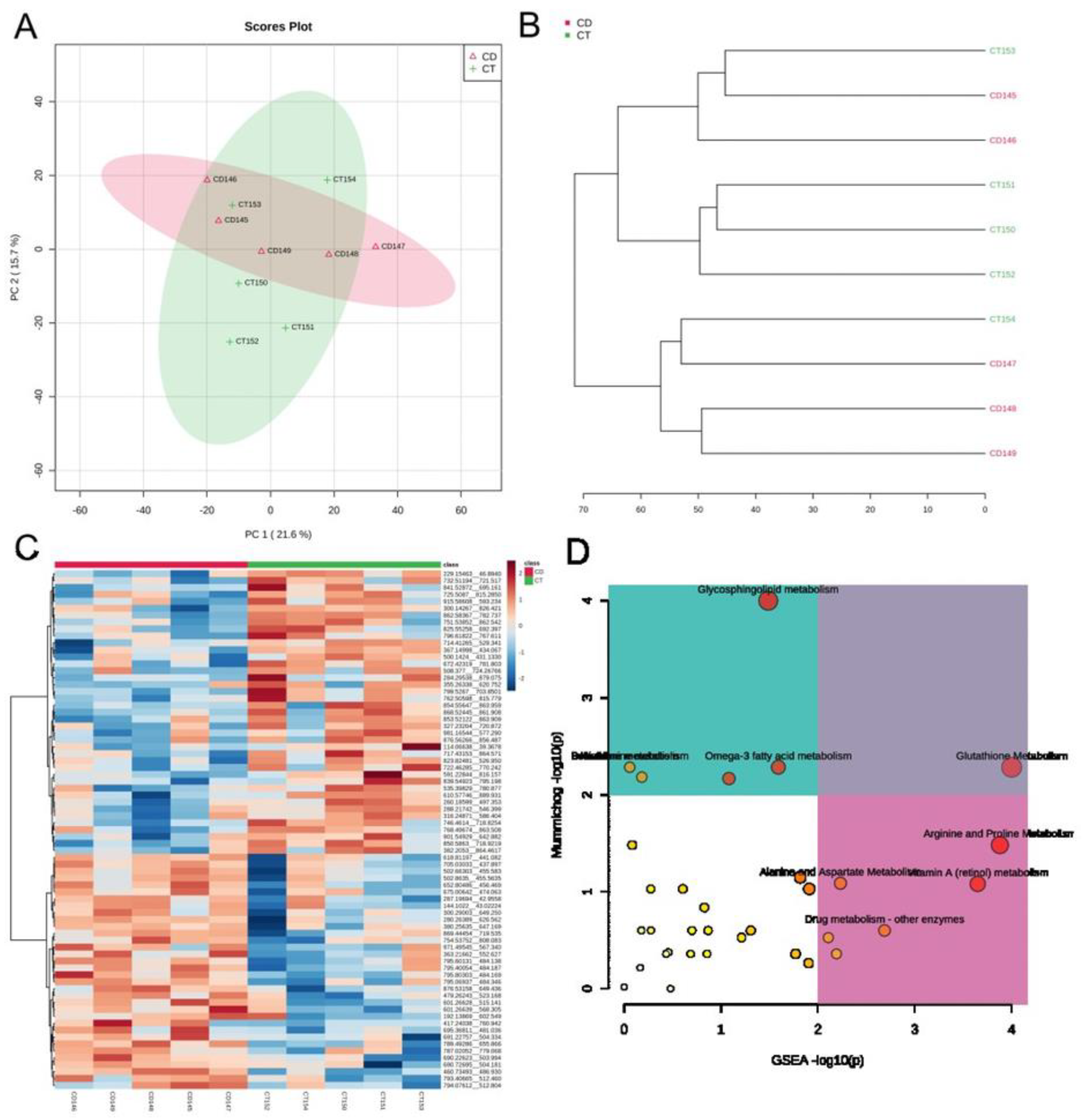
Based on the metabolomic exploratory analysis, both PCA and HCA separated CD Mennonite patients without GFD from CTs; however, two low 1:5 IgA-EMA positive samples clustered with a control having close relatives with rheumatoid arthritis, whereas one control with a close relative with CD clustered with highly positive CD patients with 1:80 IgA-EMA values (Figure 2A,B). PCs 1 and 2 explain 21.6% and 15.7% of the total variance, respectively. The concentration of 31 metabolic features differed in CD patients (p < 0,05) (Figure 2C). Five metabolic pathways were altered: glutathione metabolism; glycosphingolipid metabolism; alanine metabolism; aspartate metabolism; and vitamin A (retinol) metabolism. The glutathione pathway was significantly altered using both mummichog and GSEA approaches (Figure 2D).

4. Discussion

Due to a peculiar demographic history, the genomic architecture of the Mennonite population has suffered the effects of genetic drift, which has altered the frequency of rare Mendelian, as well as chronic diseases [15,19]. Indeed, cc. 18% of the participating individuals in our study reported diagnosis of at least one AD, such as type 1 diabetes, rheumatoid arthritis, autoimmune thyroiditis, and/or celiac disease. This prevalence (17.9%) is in clear contrast with the worldwide AD prevalence of less than 5% [30]. Furthermore, the share of predisposing genetic variants within families is probably much higher than in the general population, increasing the prevalence of these diseases. More importantly, the numbers of reported FDRs and SDRs affected by CD allowed us to get an impression about previous knowledge of the disease in these rather closed communities (of 604 participants, 453 had previous knowledge about the disease in their families, e.g., 75%).
The CD seroprevalence among Southern Brazilian Mennonites was impressively high: 3.47% (approximately 1:29). Among all the populations analyzed, only the isolated Saharawi people of Western Sahara have a higher CD prevalence (5.6%) [12]. It is much higher than the 1.22% seroprevalence reported in the Dutch population [31], which is thought to be one of the main contributors to the current genetic pool of the Mennonites, together with Germans (1.57% seroprevalence) and Belgians (0.86%) [32,33]. Even if taking into account only biopsy-confirmed CD, the 1.32% frequency in Mennonites contrasts with the global prevalence of the disease, which ranges from 0.5% (1:200) to 1.0% (1:100) [34]. More specifically, CD seroprevalence was of 1.4%, and biopsy-based CD diagnosis was of 0.7% in a recent meta-analysis, ranging from 0.4% in South America to 0.8% in Europe and Oceania [13] and reaching 0.24% (1:417) in Curitiba [14] and 0.35% (1:286) in São Paulo [35], two Brazilian cities, and 0.37% in Germany [36]. The Mennonite CD prevalence is actually close to that in Finland (2.13%), which is also genetically isolated [37]. It should be noted that biopsy-proven CD prevalence among Mennonites was calculated based on those eight individuals that were willing to take the biopsy. Thus, it may actually be higher.
Ten individuals were identified as celiac by our serological screening, representing 47.6% of undiagnosed cases (almost 1:2). Worldwide, the proportion of diagnosed and undiagnosed cases varies from one country to another, spanning from 1:2 in Finland [38] to 1:20 in Argentina [39]. The failure to detect the disease, coupled with failure to treat it, may lead to severe comorbidities such as osteoporosis, sterility, neurological and psychiatric disorders, small bowel adenocarcinoma, lymphoma, and carcinoma of the esophagus, as well as increased morbidity [4,7,8,9,10]. Two individuals previously identified as celiac in the pilot study remained positive in the second serological screening, indicating non-compliance with the gluten-free diet. The reason for non-compliance may be related to sociocultural aspects of the Mennonite community, whose eating habits rely heavily on wheat consumption.
Through our questionnaire, we identified predisposed individuals who may benefit from early intervention, such as those with iron-deficiency anemia, weight loss, and chronic abdominal pain [40]. Approximately 9% of individuals reported a family history of CD, 5.79% for FDRs and 3.25% for SDRs. This agrees with the literature: prevalence of CD in FDRs of celiac patients increases between 2–21% depending on gender and HLA-DQ genotype and the association with other ADs is frequent in both patients and relatives [41,42,43,44]. Through a meta-analysis, others estimated a CD prevalence of 7.6% among FDRs and 2.3% among SDRs [45].
Worldwide, HLA typing has a high negative predictive value for the diagnosis of CD, since at least one of the susceptibility variants, either HLA-DQ2.5 or HLA-DQ8, occurs in 86–93% of patients with CD [2,46]. Interestingly, the frequency of HLA-DQ8 in Mennonites differed from the one reported for the Belgian population—which significantly contributed to the current Mennonite gene variant pool. This is most probably the result of a founder effect. Moreover, more than half of HLA-DQ2.5/DQ8 Mennonite carriers do not have CD (50.3%, compared with 40% among Europeans without CD) [3,43]. These individuals may also bear protective genetic/epigenetic/microbiota variants that are yet to be discovered.
Furthermore, HLA-DQ2.5 and HLA-DQ8 frequencies were significantly higher in Mennonites than in Euro-Brazilians. This is a result of prime epidemiological relevance for the design of public health policies, indicating a higher general susceptibility to the disease than most Brazilians with European ancestry.
The HLA-DQ2.5 carrier frequency among Mennonite patients (92.31%) was higher than that reported in 102 Turkish pediatric patients (76%) [47]. As in our study, none of the Turkish patients were HLA-DQ8 positive. One of our CD patients was neither HLA-DQ2.5 nor HLA-DQ8 positive, indicating the influence of yet unknown predisposing genetic variants in HLA-DQ2.5/DQ8-negative CD Mennonite patients. The absence of HLA-DQ2 or HLA-DQ8 was also formerly observed by others in Brazilian patients [48]. In fact, several non-HLA genes, whose products influence the integrity of the intestinal barrier and the quality of the immune response, modulate the susceptibility to the disease [34]. About 15% of the genetic susceptibility to CD is explained by 57 single nucleotide polymorphisms located in 39 non-HLA loci, identified through genome-wide association studies [49,50]. Furthermore, HLA-DQ2.5/DQ8 contribution to CD in monozygotic twins only ranges from 25 to 40%. Thus, it is important to not exclude strongly suspicious cases from diagnostic workup based solely on HLA-DQ2.5/DQ8 negativity.
Interestingly, low IgA-EMA-positive individuals presented a metabolic profile similar to controls with close relatives with either CD or rheumatoid arthritis, diseases that share pathological pathways [51]. In all patients, the glutathione pathway was consistently altered using both algorithms (mummichog and GSEA) in the pilot metabolomic investigation. This pathway acts in the antioxidant defense and turns endogenous and exogenous compounds more soluble and non-toxic [52,53]. Indeed, glutathione was reduced in CD patients compared with controls. The levels of cysteine, the main amino acid in the synthesis of glutathione, were also reduced in children with CD before a GFD [54,55,56]. Furthermore, a high concentration of reactive oxygen species is associated with bowel damage (Marsh score). This corroborates the relationship between celiac disease and the glutathione pathway, one of the main processes to inhibit the damage caused by ROS [57].
Our study has limitations. First, we could not confirm the positive serological IgA-EMA results with positive biopsies. However, given the high specificity of IgA-EMA testing, false positives are rather unexpected. Second, IgA-deficient individuals may appear negative in our serological screening. Despite being uncommon, they may occur more often among CD patients. Missed CD individuals, in this case, would have increased the prevalence values that were already very high. Third, we did not genotype individuals for HLA-DQ2.2, which also contributes to CD susceptibility. In a next phase of our genetic–epidemiological investigation, we will screen for other HLA alleles and also for IgA deficiency in the Mennonite population as recommended [22].
Finally, our results indicate that roughly one in thirty Mennonites are expected to present with CD. Of those affected, half are unaware of the disease. Most are HLA-DQ2.5-positive, but a role for HLA-DQ8, whose frequency is cc. 1.8 times higher than in original contributing populations, cannot be excluded because the CD patient sample is small. Both the HLA-DQ2.5 and HLA-DQ8 carrier frequencies are cc. 1.6 times higher in Mennonites than in Euro-Brazilians, highlighting the epidemiological relevance of this work for public health policies. The glutathione pathway seems more affected in Mennonites with CD before compliance to a GFD. Further identification of other associated genetic, epigenetic, and environmental factors shall help define the CD risk of this population and lay the foundations of future strategies of preventive medicine.

Supplementary Materials

The following supporting information can be downloaded at https://www.mdpi.com/article/10.3390/genes14051026/s1, Table S1: Serological screening in the Mennonite population. Table S2: PCR reagents for the identification of CD-associated HLA-DQ allele carriers; Table S3: PCR conditions for the identification of CD-associated HLA-DQ allele carriers; Table S4: HLA-DQ2 and HLA-DQ8 carrier frequencies estimated for Brazilians and Belgians compared with the Mennonite population.

Author Contributions

Conceptualization, A.B.W.B. and L.C.O.; Formal analysis, L.C.O., E.R.D.B. and A.B.W.B.; Funding acquisition, F.R.C., M.L.P.-E. and A.B.W.B.; Investigation, L.C.O., E.R.D.B. and S.E.B.; Methodology, L.C.O., A.C.D., R.M.N., E.R.D.B., P.I.d.S., G.A.C. and S.E.B.; Project administration, A.B.W.B.; Resources, I.J.d.M.-R. and M.L.P.-E.; Supervision, G.A.C. and A.B.W.B.; Visualization, L.C.O.and A.C.D.; Writing—original draft, L.C.O.; Writing—review and editing, L.C.O., R.M.N., G.A.C., F.R.C., M.L.P.-E. and A.B.W.B. All authors have read and agreed to the published version of the manuscript.

Funding

This work was supported by grants of the following funding agencies: Fundação Araucária (PRONEX/FA—Convênio 116/2018, Protocolo 50530); Programa Pesquisa para o Sistema Único de Saúde: Gestão Compartilhada em Saúde—PPSUS Edição 2015; and Projeto 48.025—Chamada de Projetos 01/2016 (Fundação Araucária-PR/SESA-PR/CNPq/MS-Decit). L.C.O. received a scholarship from Coordenação de Aperfeiçoamento de Pessoal de Nível Superior (CAPES); G.A.C. received a postdoctoral scholarship (CAPES protocol 88882.306040/2018-01); M.L.P.-E and A.B.W.B. received CNPq research scholarships (protocols 303538/2015-5 and 308783/2019-0 for M.L.P.-E.; 314288/2018-0 and 313741/2021 for A.B.W.B.).

Institutional Review Board Statement

The study was conducted according to the guidelines of the Declaration of Helsinki and approved by The Ethics Committee of the Health Sciences Sector of the Federal University of Paraná (CEP SCS-UFPR) with the following Certificate of Presentation of Ethical Appreciation: 55297916.6.0000.0102, protocol numbers: 1.545.447 on 16 May 2016, updated in 2.204.113 on 7 August 2017 and again in 5.215.924 on 28 January 2022.

Informed Consent Statement

Informed consent was obtained from all subjects involved in the study.

Data Availability Statement

The data presented in this study are available on request from the corresponding author. The data are not publicly available due to privacy restrictions given by the General Law of Data Protection (LGPD) in Brazil.

Acknowledgments

We thank the participants for volunteering for this study, the staff of the ACELPAR (Associação dos Celíacos do Brasil—Paraná), and the Residents’ Association of Witmarsum for their support. Deep thanks also to Heidi Martens Wall and Ana Sawatsky Epp and to the staff of the Laboratório de Genética Molecular Humana/UFPR, especially to Hellen Caroline Issler, Leonardo Maldaner Amorim, Wilian Aparecido Vieira Junior, Verónica Calonga Solís, Gabriela Canalli Kretzschmar, and Igor Kalil Meireles for technical assistance and helpful discussions. We extend our thanks to Maique Weber Biavatti, Layzon Antonio da Silva, and Luiz Pollo from Universidade Federal de Santa Catarina for technical support in UPLC-HRMS.

Conflicts of Interest

The authors declare no conflict of interest.

Appendix A

In brief, the blood from individuals with CD but without a GFD and individuals without CD (controls—CT) were collected in tubes without anticoagulant and kept at room temperature for 60 min for serum separation. The tubes were centrifuged at 1500 rpm for 10 min and the supernatants (serum) were aliquoted in 250 µL and stored in 2 mL microtubes, being kept at −80 °C until the preparation of samples. Aliquots of serum (250 µL) were thawed on ice, and methanol (750 µL) was added at −20 °C for protein depletion. The samples were mixed in a vortex agitator and kept at −20 °C for 20 min for residual protein depletion. After this, the samples were mixed again in a vortex agitator and centrifuged at 14,000 rpm for 10 min at 4 °C. The supernatant (900 µL) was taken and dried in the vacuum concentrator at 40 °C. After this, the dried samples were stored at −20 °C until metabolomic analysis. The dried samples were reconstituted with 250 µL of acetonitrile/water (1:1, v/v) and centrifuged for 5 min at 14,000 rpm at 4 °C. Then, 100 µL of supernatant was transferred into a glass autosampler vial to ultra-high performance liquid chromatography–high-resolution mass spectrometry (UPLC-HRMS) analysis.

References

  1. Ludvigsson, J.F.; Leffler, D.A.; Bai, J.C.; Biagi, F.; Fasano, A.; Green, P.H.R.; Hadjivassiliou, M.; Kaukinen, K.; Kelly, C.P.; Leonard, J.N.; et al. The Oslo definitions for coeliac disease and related terms. Gut 2013, 62, 43–52. [Google Scholar] [CrossRef] [PubMed]
  2. Bourgey, M.; Calcagno, G.; Tinto, N.; Gennarelli, D.; Margaritte-Jeannin, P.; Greco, L.; Limongelli, M.G.; Esposito, O.; Marano, C.; Troncone, R.; et al. HLA related genetic risk for coeliac disease. Gut 2007, 56, 1054–1059. [Google Scholar] [CrossRef] [PubMed]
  3. Liu, E.; Rewers, M.; Eisenbarth, G.S. Genetic testing: Who should do the testing and what is the role of genetic testing in the setting of celiac disease? Gastroenterology 2005, 128, S33–S37. [Google Scholar] [CrossRef] [PubMed]
  4. Kotze, L.M.S. Celiac disease in Brazilian patients: Associations, complications and causes of death. Forty years of clinical experience. Arq. Gastroenterol. 2009, 46, 261–269. [Google Scholar] [CrossRef] [PubMed]
  5. Fasano, A.; Catassi, C. Current approaches to diagnosis and treatment of celiac disease: An evolving spectrum. Gastroenterology 2001, 120, 636–651. [Google Scholar] [CrossRef]
  6. Utiyama, S.R.R.; Nass, F.R.; Kotze, L.M.S.; Nisihara, R.M.; Ambrosio, A.R.; de Messias-Reason, I.T. Triagem sorológica de familiares de pacientes com doença celíaca: Anticorpos anti-endomísio, antitransglutaminase ou ambos? Arq. Gastroenterol. 2007, 44, 156–161. [Google Scholar] [CrossRef]
  7. Green, P.H.R.; Lebwohl, B.; Greywoode, R. Celiac disease. J. Allergy Clin. Immunol. 2015, 135, 1099–1106. [Google Scholar] [CrossRef]
  8. Romano, L.; Pellegrino, R.; Sciorio, C.; Barone, B.; Gravina, A.G.; Santonastaso, A.; Mucherino, C.; Astretto, S.; Napolitano, L.; Aveta, A.; et al. Erectile and sexual dysfunction in male and female patients with celiac disease: A cross-sectional observational study. Andrology 2022, 10, 910–918. [Google Scholar] [CrossRef]
  9. Aaron, L.; Torsten, M.; Patricia, W. Autoimmunity in celiac disease: Extra-intestinal manifestations. Autoimmun. Rev. 2019, 18, 241–246. [Google Scholar] [CrossRef]
  10. Rodrigo, L.; Beteta-Gorriti, V.; Alvarez, N.; Gómez de Castro, C.; De Dios, A.; Palacios, L.; Santos-Juanes, J. Cutaneous and Mucosal Manifestations Associated with Celiac Disease. Nutrients 2018, 10, 800. [Google Scholar] [CrossRef]
  11. Lindfors, K.; Ciacci, C.; Kurppa, K.; Lundin, K.E.; Makharia, G.K.; Mearin, M.L.; Murray, J.A.; Verdu, E.F.; Kaukinen, K. Coeliac disease. Nat. Rev. Dis. Prim. 2019, 5, 3. [Google Scholar] [CrossRef] [PubMed]
  12. Catassi, C.; Ratsch, I.M.; Gandolfi, L.; Pratesi, R.; Fabiani, E.; El Asmar, R.; Frijia, M.; Bearzi, I.; Vizzoni, L. Why is coeliac disease endemic in the people of the Sahara? Lancet 1999, 354, 647–648. [Google Scholar] [CrossRef] [PubMed]
  13. Singh, P.; Arora, A.; Strand, T.A.; Leffler, D.A.; Catassi, C.; Green, P.H.; Kelly, C.P.; Ahuja, V.; Makharia, G.K. Global Prevalence of Celiac Disease: Systematic Review and Meta-analysis. Clin. Gastroenterol. Hepatol. 2018, 16, 823–836. [Google Scholar] [CrossRef] [PubMed]
  14. Pereira, M.A.G. Prevalence of celiac disease in an urban area of Brazil with predominantly European ancestry. WJG 2006, 12, 6546. [Google Scholar] [CrossRef] [PubMed]
  15. Lopes, F.L.; Hou, L.; Boldt, A.B.W.; Kassem, L.; Alves, V.M.; Nardi, A.E.; McMahon, F.J. Finding Rare, Disease-Associated Variants in Isolated Groups: Potential Advantages of Mennonite Populations. Hum. Biol. 2016, 88, 109–120. [Google Scholar] [CrossRef]
  16. Hou, L.; Kember, R.L.; Roach, J.C.; O’connell, J.R.; Craig, D.W.; Bucan, M.; Scott, W.K.; Pericak-Vance, M.; Haines, J.L.; Crawford, M.H.; et al. A population-specific reference panel empowers genetic studies of Anabaptist populations. Sci. Rep. 2017, 7, 6079. [Google Scholar] [CrossRef]
  17. Pardo-Seco, J.; Llull, C.; Berardi, G.; Gómez, A.; Andreatta, F.; Martinón-Torres, F.; Toscanini, U.; Salas, A. Genomic continuity of Argentinean Mennonites. Sci. Rep. 2016, 6, 36392. [Google Scholar] [CrossRef]
  18. Toscanini, U.; Brisighelli, F.; Llull, C.; Berardi, G.; Gómez, A.; Andreatta, F.; Pardo-Seco, J.; Gómez-Carballa, A.; Martinón-Torres, F.; Álvarez-Iglesias, V.; et al. Charting the Y-chromosome ancestry of present-day Argentinean Mennonites. J. Hum. Genet. 2016, 61, 507–513. [Google Scholar] [CrossRef]
  19. Jaworski, M.A.; Slater, J.D.; Severini, A.; Hennig, K.R.; Mansour, G.; Mehta, J.G.; Jeske, R.; Schlaut, J.; Pak, C.Y.; Yoon, J.W. Unusual clustering of diseases in a Canadian Old Colony (Chortitza) Mennonite kindred and community. CMAJ 1988, 138, 1017–1025. [Google Scholar]
  20. Boschmann, S.E.; Boldt, A.B.; de Souza, I.R.; Petzl-Erler, M.L.; Messias-Reason, I.J. The Frequency of the LCT*-13910C>T Polymorphism Associated with Lactase Persistence Diverges among Euro-Descendant Groups from Brazil. Med. Princ. Pract. 2016, 25, 18–20. [Google Scholar] [CrossRef]
  21. Tursi, A.; Elisei, W.; Giorgetti, G.M.; Gaspardone, A.; Lecca, P.G.; Di Cesare, L.; Brandimarte, G. Prevalence of celiac disease and symptoms in relatives of patients with celiac disease. Eur. Rev. Med. Pharm. Sci. 2010, 14, 567–572. [Google Scholar]
  22. Al-Toma, A.; Volta, U.; Auricchio, R.; Castillejo, G.; Sanders, D.S.; Cellier, C.; Mulder, C.J.; Lundin, K.E.A. European Society for the Study of Coeliac Disease (ESsCD) guideline for coeliac disease and other gluten-related disorders. United Eur. Gastroenterol. J. 2019, 7, 583–613. [Google Scholar] [CrossRef] [PubMed]
  23. Volta, U.; Molinaro, N.; De Franceschi, L.; Fratangelo, D.; Bianchi, F.B. IgA anti-endomysial antibodies on human umbilical cord tissue for celiac disease screening. Dig. Dis. Sci. 1995, 40, 1902–1905. [Google Scholar] [CrossRef] [PubMed]
  24. Marsh, M.N. Gluten, major histocompatibility complex, and the small intestine. Gastroenterology 1992, 102, 330–354. [Google Scholar] [CrossRef] [PubMed]
  25. Olerup, O.; Aldener, A.; Fogdell, A. HLA-DQB1 and -DQA1 typing by PCR amplification with sequence-specific primers (PCR-SSP) in 2 hours. Tissue Antigens 1993, 41, 119–134. [Google Scholar] [CrossRef]
  26. Profaizer, T.; Eckels, D.; Delgado, J.C. Celiac disease and HLA typing using real-time PCR with melting curve analysis. Tissue Antigens 2011, 78, 31–37. [Google Scholar] [CrossRef]
  27. Vorkas, P.A.; Isaac, G.; Anwar, M.A.; Davies, A.H.; Want, E.J.; Nicholson, J.K.; Holmes, E. Untargeted UPLC-MS Profiling Pipeline to Expand Tissue Metabolome Coverage: Application to Cardiovascular Disease. Anal. Chem. 2015, 87, 4184–4193. [Google Scholar] [CrossRef]
  28. Pang, Z.; Zhou, G.; Ewald, J.; Chang, L.; Hacariz, O.; Basu, N.; Xia, J. Using MetaboAnalyst 5.0 for LC–HRMS spectra processing, multi-omics integration and covariate adjustment of global metabolomics data. Nat. Protoc. 2022, 17, 1735–1761. [Google Scholar] [CrossRef]
  29. González-Galarza, F.F.; Takeshita, L.Y.; Santos, E.J.; Kempson, F.; Maia, M.H.T.; Da Silva, A.L.S.; Silva, A.L.T.E.; Ghattaoraya, G.; Alfirevic, A.; Jones, A.; et al. Allele frequency net 2015 update: New features for HLA epitopes, KIR and disease and HLA adverse drug reaction associations. Nucleic Acids Res. 2015, 43, D784–D788. [Google Scholar] [CrossRef]
  30. Fairweather, D.; Frisancho-Kiss, S.; Rose, N.R. Sex Differences in Autoimmune Disease from a Pathological Perspective. Am. J. Pathol. 2008, 173, 600–609. [Google Scholar] [CrossRef]
  31. Csizmadia, C.G.; Mearin, M.L.; von Blomberg, B.M.; Brand, R.; Verloove-Vanhorick, S.P. An iceberg of childhood coeliac disease in the Netherlands. Lancet 1999, 353, 813–814. [Google Scholar] [CrossRef] [PubMed]
  32. Vijgen, S.; Alliet, P.; Gillis, P.; Declercq, P.; Mewis, A. Seroprevalence of celiac disease in Belgian children and adolescents. Acta Gastroenterol. Belg. 2012, 75, 325–330. [Google Scholar] [PubMed]
  33. Händel, N.; Mothes, T.; Petroff, D.; Baber, R.; Jurkutat, A.; Flemming, G.; Kiess, W.; Hiemisch, A.; Körner, A.; Schlumberger, W.; et al. Will the Real Coeliac Disease Please Stand Up? Coeliac Disease Prevalence in the German LIFE Child Study. J. Pediatr. Gastroenterol. Nutr. 2018, 67, 494–500. [Google Scholar] [CrossRef] [PubMed]
  34. Gujral, N. Celiac disease: Prevalence, diagnosis, pathogenesis and treatment. World J. Gastroenterol. WJG 2012, 18, 6036. [Google Scholar] [CrossRef] [PubMed]
  35. Alencar, M.L.; Ortiz-Agostinho, C.L.; Damião, A.O.; Abrantes-Lemos, C.P.; Leite, A.Z.; Brito, T.D.; Chamone, D.D.; Silva, M.E.; Giannella-Neto, D.; Sipahi, A.M. Prevalence of celiac disease among blood donors in SÃO PAULO—The most populated city in Brazil. Clinics 2012, 67, 1013–1018. [Google Scholar] [CrossRef]
  36. Kratzer, W. Prevalence of celiac disease in Germany: A prospective follow-up study. World J. Gastroenterol. WJG 2013, 19, 2612. [Google Scholar] [CrossRef]
  37. Vilppula, A.; Collin, P.; Mäki, M.; Valve, R.; Luostarinen, M.; Krekelä, I.; Patrikainen, H.; Kaukinen, K. Undetected coeliac disease in the elderly. Dig. Liver Dis. 2008, 40, 809–813. [Google Scholar] [CrossRef]
  38. Mäki, M.; Mustalahti, K.; Kokkonen, J.; Kulmala, P.; Haapalahti, M.; Karttunen, T.; Ilonen, J.; Laurila, K.; Dahlbom, I.; Hansson, T.; et al. Prevalence of Celiac Disease among Children in Finland. N. Engl. J. Med. 2003, 348, 2517–2524. [Google Scholar] [CrossRef]
  39. Gomez, J.C.; Selvaggio, G.S.; Viola, M.; Pizarro, B.; Motta, G.; Barrio, S.; Castelletto, R.; Echeverria, R.; Sugai, E.; Vazquez, H.; et al. Prevalence of celiac disease in argentina: Screening of an adult population in the La Plata area. Am. J. Gastroenterol. 2001, 96, 2700–2704. [Google Scholar] [CrossRef]
  40. Cárdenas-Roldán, J.; Rojas-Villarraga, A.; Anaya, J.M. How do autoimmune diseases cluster in families? A systematic review and meta-analysis. BMC Med. 2013, 11, 73. [Google Scholar] [CrossRef]
  41. Parra-Medina, R.; Molano-Gonzalez, N.; Rojas-Villarraga, A.; Agmon-Levin, N.; Arango, M.-T.; Shoenfeld, Y.; Anaya, J.-M. Prevalence of Celiac Disease in Latin America: A Systematic Review and Meta-Regression. PLoS ONE 2015, 10, e0124040. [Google Scholar] [CrossRef] [PubMed]
  42. Mearin, M.L. The prevention of coeliac disease. Best Pract. Res. Clin. Gastroenterol. 2015, 29, 493–501. [Google Scholar] [CrossRef]
  43. Högberg, L.; Fälth-Magnusson, K.; Grodzinsky, E.; Stenhammar, L. Familial Prevalence of Coeliac Disease: A Twenty-Year Follow-up Study. Scand. J. Gastroenterol. 2003, 38, 61–65. [Google Scholar] [CrossRef]
  44. Ciclitira, P.J.; Johnson, M.W.; Dewar, D.H.; Ellis, H.J. The pathogenesis of coeliac disease. Mol. Asp. Med. 2005, 26, 421–458. [Google Scholar] [CrossRef] [PubMed]
  45. Singh, P.; Arora, S.; Lal, S.; Strand, T.A.; Makharia, G.K. Risk of Celiac Disease in the First- and Second-Degree Relatives of Patients with Celiac Disease: A Systematic Review and Meta-Analysis. Am. J. Gastroenterol. 2015, 110, 1539–1548. [Google Scholar] [CrossRef] [PubMed]
  46. Karell, K.; Louka, A.S.; Moodie, S.J.; Ascher, H.; Clot, F.; Greco, L.; Ciclitira, P.J.; Sollid, L.M.; Partanen, J. HLA types in celiac disease patients not carrying the DQA1*05-DQB1*02 (DQ2) heterodimer: Results from the European genetics cluster on celiac disease. Hum. Immunol. 2003, 64, 469–477. [Google Scholar] [CrossRef]
  47. Basturk, A.; Artan, R.; Yilmaz, A. The incidence of HLA-DQ2/DQ8 in Turkish children with celiac disease and a comparison of the geographical distribution of HLA-DQ. Gastroenterol. Rev. 2017, 4, 256–261. [Google Scholar] [CrossRef]
  48. Kotze, L.M.S.; Nisihara, R.; Utiyama, S.R.R.; Kotze, L.R. Absence of HLA-DQ2 and HLA-DQ8 does not exclude celiac disease in Brazilian patients. Rev. Esp. Enferm. Dig. 2014, 106, 561–562. [Google Scholar]
  49. Ricaño-Ponce, I.; Wijmenga, C.; Gutierrez-Achury, J. Genetics of celiac disease. Best Pract. Res. Clin. Gastroenterol. 2015, 29, 399–412. [Google Scholar] [CrossRef]
  50. Trynka, G.; Hunt, K.A.; Bockett, N.A.; Romanos, J.; Mistry, V.; Szperl, A.; Bakker, S.F.; Bardella, M.T.; Bhaw-Rosun, L.; Castillejo, G.; et al. Dense genotyping identifies and localizes multiple common and rare variant association signals in celiac disease. Nat. Genet. 2011, 43, 1193–1201. [Google Scholar] [CrossRef]
  51. Hua, L.; Xiang, S.; Xu, R.; Xu, X.; Liu, T.; Shi, Y.; Wu, L.; Wang, R.; Sun, Q. Causal association between rheumatoid arthritis and celiac disease: A bidirectional two-sample mendelian randomization study. Front. Genet. 2022, 13, 976579. [Google Scholar] [CrossRef] [PubMed]
  52. Wang, C.; Cui, C.; Li, N.; Sun, X.; Wen, L.; Gao, E.; Wang, F. Antioxidant activity and protective effect of wheat germ peptides in an in vitro celiac disease model via Keap1/Nrf2 signaling pathway. Food Res. Int. 2022, 161, 111864. [Google Scholar] [CrossRef] [PubMed]
  53. Wu, G.; Fang, Y.Z.; Yang, S.; Lupton, J.R.; Turner, N.D. Glutathione Metabolism and Its Implications for Health. J. Nutr. 2004, 134, 489–492. [Google Scholar] [CrossRef] [PubMed]
  54. Wahab, P.J.; Peters, W.H.M.; Roelofs, H.M.J.; Jansen, J.B.M.J. Glutathione S-Transferases in Small Intestinal Mucosa of Patients with Coeliac Disease. Jpn. J. Cancer Res. 2001, 92, 279–284. [Google Scholar] [CrossRef]
  55. Coles, B.F.; Chen, G.; Kadlubar Fred, F.; Radominska-Pandya, A. Interindividual variation and organ-specific patterns of glutathione S-transferase alpha, mu, and pi expression in gastrointestinal tract mucosa of normal individuals. Arch. Biochem. Biophys. 2002, 403, 270–276. [Google Scholar] [CrossRef]
  56. Sevinc, E.; Sevinc, N.; Akar, H.H.; Ozelcoskun, B.D.; Sezgin, G.C.; Arslan, D.; Kendirci, M. Plasma glutamine and cystine are decreased and negatively correlated with endomysial antibody in children with celiac disease. Asia Pac. J. Clin. Nutr. 2016, 25, 452–456. [Google Scholar]
  57. Moretti, S.; Mrakic-Sposta, S.; Roncoroni, L.; Vezzoli, A.; Dellanoce, C.; Monguzzi, E.; Branchi, F.; Ferretti, F.; Lombardo, V.; Doneda, L.; et al. Oxidative stress as a biomarker for monitoring treated celiac disease. Clin. Transl. Gastroenterol. 2018, 9, e157. [Google Scholar] [CrossRef]
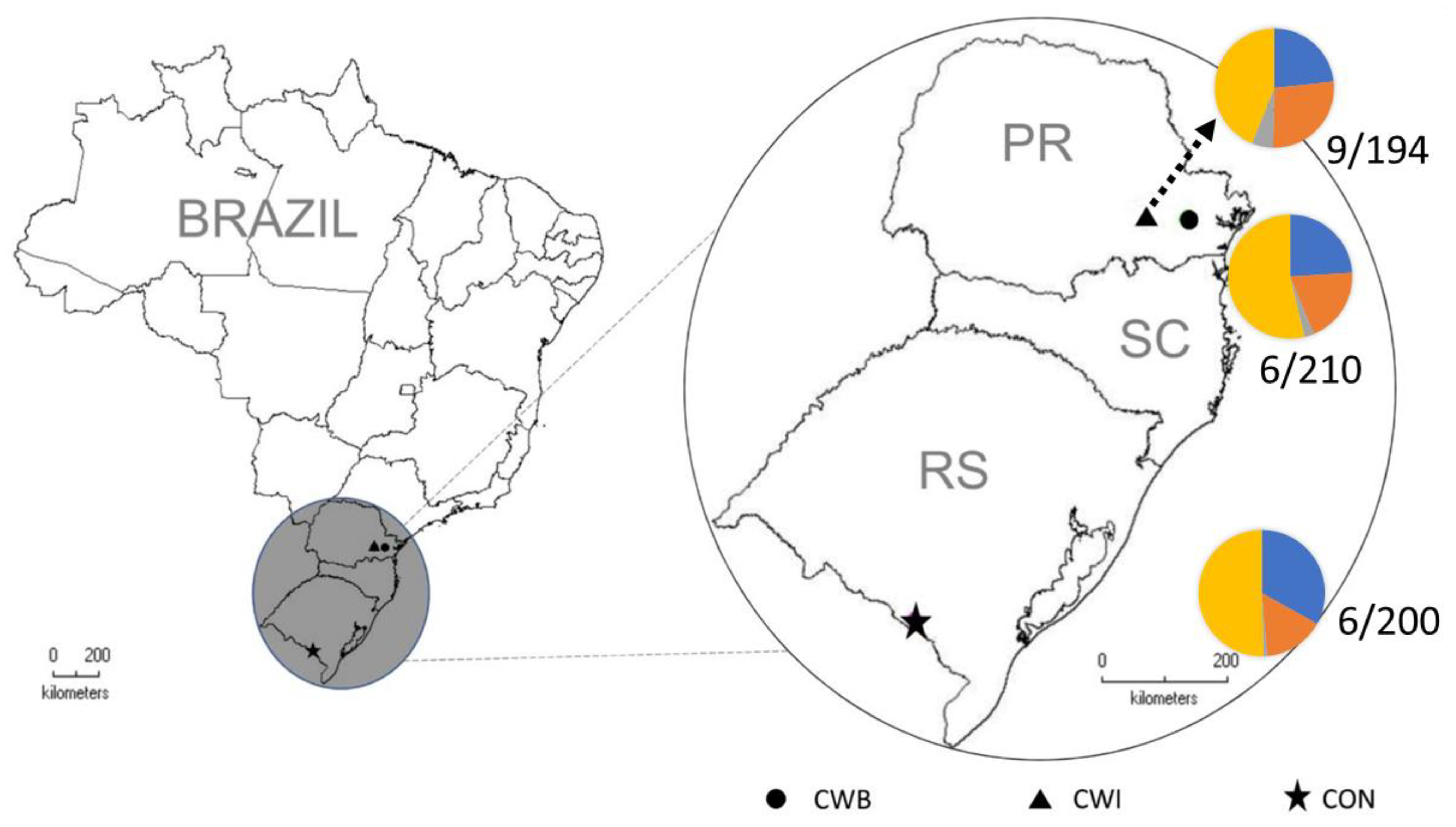
Figure 1. Frequencies of positive serological IgA-EMA results and of HLA-DQ2.5 and DQ8 carriers in each of the investigated Southern Brazilian Mennonite communities (HLA-DQ2.5- DQ8- in yellow, HLA-DQ2.5- DQ8+ in red, HLA-DQ2.5+ DQ8- in blue, HLA-DQ2.5+ DQ8+ in gray). PR = Paraná State, SC = Santa Catarina State, RS = Rio Grande do Sul State, CON = Nova Colony, CWI = Witmarsum Colony, CTB = Curitiba.
Genes 14 01026 g001
Figure 2. Exploratory metabolomics in the sera of CD Mennonites without a GFD. (A) Principal component analysis of serum metabolites identified in celiac patients (CD) and controls (CT). DC146 and DC145 had a 1:5 EMA positivity and clustered together with CT153, who is unaffected but has close relatives with rheumatoid arthritis. CT154 did not group with the others using this approach. Despite being serologically negative, this individual has close relatives with CD. (B) Hierarchical clustering of metabolic CD and CT profiles. Here, DC146, DC145, and CT153 also grouped together, and CT154 grouped with CD patients presenting high IgA-EMA positivity (1:80). (C) Heat map analysis of hierarchical clustering of CD and CT individuals. In blue: decreased metabolite levels; in red: increased metabolite levels. (D) Scatter plots of pathway enrichment analysis provided by mummichog and its integration with GSEA. The color and size of each circle correspond to its p value and enrichment factor, respectively. Darker tones indicate more statistically relevant predicted pathways. The size of each dot represents the ratio between significant pathway hits and the expected number of compound hits within the pathways. The glutathione pathway (upper square, right) was the only pathway found to be significant using both algorithms.
Genes 14 01026 g002
Table 1. Distribution of demographic characteristics, symptoms, diagnoses, and HLA subtypes in the Mennonite population samples.
CONCWICTBp ValueTotalCD PatientsNon-CD Patientsp Value
NN = 200N = 210N = 194 N = 604N = 21N = 583
   Gender (F/M)120/80136/74107/870.143363/24116/05347/2360.173
Median
(min-max)
Median
(min-max)
Median
(min-max)
Median
(min-max)
Median
(min-max)
Median
(min-max)
   Age–female (years)57.0 (14.0–83.0)46.0 (14.4–89.0)44.2 (14.0–89.2)0.003
q = 0.026
49.0 (14.0–89.2)32.0 (17.1–69.0)50.0 (14.0–89.2)0.015
q = 0.039
   Age–male (years)54.0 (17.0–82.0)53.7 (12.0–92.1)49.5 (15.9–88.0)0.24151.0 (12.0–92.1)64.6 (54.9–75.0)51.0 (12.0–92.1)0.095
   BMI–female27.6 (15.8–45.8)24.6 (16.2–35.2)23.6 (15.1–40.6)1 × 10⁻⁴
q = 0.013
25.2 (15.1–45.8)22.4 (19.6–45.8)25.5 (15.1–44.5)0.235
   BMI–male28.4 (19.2–42.9)26.2 (16.1–47.6)26.3 (16.6–38.7)4 × 10⁻⁴
q = 0.015
27.1 (16.1–47.6)27.4 (26.0–28.9)26.9 (16.1–47.6)0.724
% (n/total N)% (n/total N)% (n/total N) % (n/total N)% (n/total N)% (n/total N)
Symptoms/diagnoses
   Chronic diarrhea2.11 (4/190)7.14 (13/182)5.70 (9/158)0.0684.90 (26/530)19.05 (4/21)4.32 (22/509)0.015
q = 0.010
   Weight loss3.68 (7/190)7.94 (10/126)5.06 (8/158)0.2515.27 (25/474)15.00 (3/20)4.85 (22/454)0.081
   Iron-deficiency anemia7.37 (14/190)10.94 (14/128)8.23 (13/158)0.5278.61 (41/476)38.09 (8/21)7.25 (33/455)1 × 10−4
q = 0.018
   Bloating31.58 (60/190)28.96 (53/183)27.21 (43/158)0.66329.37 (156/531)38.09 (8/21)29.02 (148/510)0.462
   Constipation12.63 (24/190)19.04 (24/126)20.88 (33/158)0.09917.09 (81/474)10.00 (2/20)17.40 (79/454)0.549
   Chronic abdominal pain16.32 (31/190)16.48 (30/182)15.28 (24/157)0.95116.07 (85/529)42.85 (9/21)14.96 (76/508)0.002
q = 0.021
   Joint pain26.84 (51/190)9.52 (12/126)8.33 (13/156)1 × 10−6
q = 0.002
16.10 (76/472)5.00 (1/20)16.59 (75/452)0.223
   Diabetes mellitus type 11.06 (2/188)0.79 (1/126)2.46 (3/122)n.c.1.38 (6/436)0 (0/19)1.44 (6/417)nc
   Irritable bowel syndrome5.82 (11/189)5.00 (6/120)4.04 (4/99)0.8874.90 (20/408)6.25 (1/16)5.10 (20/392)1
   Lactose intolerance1.62 (3/185)3.35 (6/179)9.59 (14/146)0.003
q = 0.028
4.51 (23/510)4.76 (1/21)4.50 (22/489)1
   Autoimmune thyroiditis10.47 (20/191)11.29 (14/124)14.74 (23/156)0.45412.10 (57/471)15.00 (3/20)12.36 (56/453)0.727
   Rheumatoid arthritis11.30 (20/177)4.00 (5/125)5.52 (9/163)0.031
q = 0.047
7.31 (34/465)0 (0/19)7.62 (34/446)nc
   Celiac disease3.00 (6/200)2.86 (6/210)4.64 (9/194)0.5603.48 (21/604)--
   Any autoimmune disease *20.90 (37/177)15.08 (19/126)20.24 (33/163)0.40019.10 (89/466)15.79 (3/19) *19.24 (86/447) *1
Familiar aggregation
   FDR with CD3.21 (6/187)11.21 (12/107)5.03 (8/159)0.015
q = 0.044
5.74 (26/453)31.58 (6/19)4.61 (20/434)3 × 10−4
q = 0.015
   SDR with CD1.60 (3/187)4.81 (5/104)4.43 (7/158)0.2213.34 (15/449)21.05 (4/19)2.56 (11/430)0.002
q = 0.023
   Any relative with CD5.88 (11/187)14.95 (16/107)6.29 (10/159)0.013
q = 0.036
8.17 (37/453)36.84 (7/19)4.61 (20/434)3 × 10−5
q = 0.010
HLAN = 115N = 155N = 121 N = 391N = 13N = 378
   DQ2.5 (+)33.91 (39)29.03 (45)26.45 (32)0.55929.67 (116)92.31 (12)27.51 (104)3 × 10⁻⁶
q = 0.007
   Only DQ2.5 (+)33.04 (38)23.23 (36)23.97 (29)0.15926.09 (102)92.31 (12)24.07 (90)1 × 10⁻⁶
q = 0.005
   DQ8 (+)16.52 (19)32.90 (51)22.31 (27)0.007
q = 0.034
24.81 (97)0 25.66 (97)0.044
q = 0.05
   Only DQ8 (+)15.65 (18)27.10 (42)19.83 (24)0.07221.48 (84)0 22.22 (84)0.080
   DQ2.5 (+) and/or DQ8 (+)49.57 (57)56.13 (87)46.28 (56)0.24851.15 (200)92.31 (12)49.74 (188)0.003
q = 0.031
p values were calculated through the chi-square test for most variables, except for BMI and age distributions (non-parametric Mann–Whitney or Kruskal–Wallis tests), gender, type 1 diabetes mellitus, and first- and second-degree relatives (Fisher exact t-test). q = correction with the Benjamini–Hochberg method for multiple comparisons. * autoimmune disease (AD): type 1 diabetes, arthritis, and/or autoimmune thyroiditis, excluding CD for the analysis in this group. Age is in years. BMI = body mass index, CD = celiac disease, CON = Nova Colony, CWI = Witmarsum Colony, CTB = Curitiba, F = female, FDR = first-degree relative, M = male, p = p value, SDR = second-degree relative, n = number of individuals with the evaluated characteristic, N = total number of individuals.
Table 2. Common symptoms associated with CD. CD = celiac disease, Non-CD = non-celiac disease, n = number of individuals with the evaluated characteristic, N = total number of individuals, CI = 95% confidence interval, OR = odds ratio, p value = probability of significance. All associations were corrected for possible confounding factors (gender and age) using logistic regression.
SymptomsCD%
(n/N)
Non-CD%
(n/N)
OR95% CIp Value
Chronic diarrhea19.05 (4/21)4.32 (22/509)5.201.61–16.780.006
Iron-deficiency anemia38.09 (8/21)7.25 (33/455)7.863.04–20.33<10−3
Chronic abdominal pain42.85 (9/21)14.96 (76/508)4.261.73–10.460.002
Disclaimer/Publisher’s Note: The statements, opinions and data contained in all publications are solely those of the individual author(s) and contributor(s) and not of MDPI and/or the editor(s). MDPI and/or the editor(s) disclaim responsibility for any injury to people or property resulting from any ideas, methods, instructions or products referred to in the content.