Article

NUMTs Can Imitate Biparental Transmission of mtDNA—A Case in Drosophila melanogaster

by
Maria-Eleni Parakatselaki
1,
Chen-Tseh Zhu
2,
David Rand
2 and
Emmanuel D. Ladoukakis
1,*
1
Department of Biology, University of Crete, Voutes University Campus, 70013 Iraklio, Greece
2
Department of Ecology, Evolutionary and Organismal Biology, Brown University, Providence, RI 02912, USA
*
Author to whom correspondence should be addressed.
Genes 2022, 13(6), 1023; https://doi.org/10.3390/genes13061023
Submission received: 20 April 2022 / Revised: 27 May 2022 / Accepted: 2 June 2022 / Published: 6 June 2022
(This article belongs to the Special Issue Feature Papers in Population and Evolutionary Genetics and Genomics)

Abstract

:
mtDNA sequences can be incorporated into the nuclear genome and produce nuclear mitochondrial fragments (NUMTs), which resemble mtDNA in their sequence but are transmitted biparentally, like the nuclear genome. NUMTs can be mistaken as real mtDNA and may lead to the erroneous impression that mtDNA is biparentally transmitted. Here, we report a case of mtDNA heteroplasmy in a Drosophila melanogaster DGRP line, in which the one haplotype was biparentally transmitted in an autosomal manner. Given the sequence identity of this haplotype with the mtDNA, the crossing experiments led to uncertainty about whether heteroplasmy was real or an artifact due to a NUMT. More specific experiments revealed that there is a large NUMT insertion in the X chromosome of a specific DGRP line, imitating biparental inheritance of mtDNA. Our result suggests that studies on mtDNA heteroplasmy and on mtDNA inheritance should first exclude the possibility of NUMT interference in their data.

1. Introduction

Two decades ago, it was observed that fragments of mitochondrial DNA can be located in the nuclear DNA. These sequences have been called NUMTs, which is an acronym for nuclear mitochondrial DNA [1]. NUMTs have been mapped in the nuclear genome of several organisms, such as humans [2], Daphnia [3], Parus [4], fig wasps [5], Hydra, [6] and Drosophila [7]. In most cases, NUMTs have the characteristics of pseudogenes, i.e., they can be fragments of gene(s), they might have stop codons and/or frameshift mutations, and increased dN/dS ratio compared to functional mtDNA genes [8].
As expected, the discovery of NUMTs has triggered a discussion on whether they could compromise the interpretation of mtDNA data in phylogenetic studies. It is possible that mitochondrial pseudogenes are co-amplified with real mitochondrial genes, leading to false estimates of coalescent times and intraspecific variation due to different evolutionary rates and modes of inheritance between mtDNA and nDNA [1,9]. Recent studies suggest that the insertion rate of NUMTs can differ among taxa, but generally they tend to be more frequent in taxa with larger nuclear genome sizes [10,11].
NUMTs were thought to be short-sized sequences, with a mean length varying from 100 to 300 bp [12,13], suggesting that sequencing of larger mtDNA fragments followed by searching for stop codons and frameshift mutations could be a way to exclude NUMT sequences from phylogenetic studies [14]. The recent discovery of very large NUMTs in organisms such as bats [15], birds [16], mammals [17,18] including humans [19] indicates that size alone is not sufficient to distinguish between a NUMT and a real mtDNA sequence. Moreover, if the translocation of the mtDNA to the nucleus is recent, it is likely that the mtDNA may have not yet been pseudogenised and therefore, the NUMT sequence might be free of stop codons or frameshift mutations.
Apart from phylogeny, NUMTs can create noise in studies of mtDNA inheritance and heteroplasmy. Given that nuclear DNA is biparentally inherited, a NUMT can be transmitted by both parents and give the erroneous impression that an individual has paternal mtDNA along with true, maternal cytoplasmic mtDNA. A recent study reported biparental transmission of mtDNA in three human families [20] and provided data that the observed pattern was not caused by NUMTs [21]. Some researchers suggested that the possibility of NUMTs had not been exhaustively ruled out in the original study because the observed pattern of biparental transmission of mtDNA can be caused if there are huge mtDNA sequences translocated recently in the nuclear genome (mega-NUMTs) [22,23]. This debate triggered an extensive study in humans which confirmed the presence of mega-NUMTs in the human genome [19].
In this study, we present evidence that a large NUMT has been recently translocated to the nuclear DNA in a Drosophila melanogaster DGRP line. We show that this can imitate biparental mtDNA transmission and produce a signal for false heteroplasmy. We also show that this type of NUMT is technically difficult to distinguish from real paternal mtDNA transmission, particularly because it is located in the X chromosome and thus it partially imitates maternal transmission.

2. Materials and Methods

2.1. Crosses

All lines used in the crosses have nuclear chromosomes from D. melanogaster. Most of them are mitonuclear replacement lines, i.e., they possess mtDNA from Drosophila simulans. Specifically, they carry the siI, sm21, and mau12 mitotypes [24,25] that correspond to siI, siII, and siIII mitotypes [26]. A detailed list of the lines used is shown in Table 1. For each cross, 4–5 pairs of flies were used in each vial, while for each type of cross we had at least 10 replicates. All crosses were performed in a standard Drosophila medium [27] at 25 °C with a 12 h light period.

2.2. DNA Extraction and PCR Assays

For routine PCR assays, DNA was extracted from single flies, as previously described [28]. Before the extraction, the sex of the flies was determined under a stereoscope along with other phenotypic traits, where needed. Each individual was tested for the presence of both maternal and paternal mitotype with primers that are specific to each mtDNA sequence. For long PCR assays, we pooled 10 flies from each of the following lines; siI;w1118, sm21;w1118, siI;820, sm21;820, DGRP-820, and D. melanogaster Oregon R. DNA was extracted and purified with a standard protocol [29].
For isolation of pure mtDNA from unfertilized eggs, we set up two cages, one with siI;820 unmated females and one with sm21;820 unmated females. Cages contained an appropriate egg-laying medium to facilitate the collection of the eggs (5 g sucrose, 2.25 g agar, 50 mL red-fruit juice, and 50 mL H2O). Each egg was homogenized in 15 uL lysis buffer (10 mM Tris-HCl pH 8.0, 1 mM EDTA, 25 mM NaCl). One uL Proteinase K 20 mg/mL was added to each sample and then samples were incubated for 2 h at 55 °C. The presence of specific mtDNAs and the absence of specific nDNAs were verified with template-specific primers (Table 2).
For each PCR reaction, we added 1×  Taq polymerase buffer (EnzyQuest Biotechnology, IMBB, Heraklion, Greece), 0.4 mΜ of each primer, 1.5 mM MgCl2, 0.2 mM dNTPs, 0.4 mg/mL BSA, 0.5 U Taq DNA polymerase, 1 uL DNA, and Nanopure H2O up to 15 uL. The PCR conditions were: 2 min at 95 °C followed by 35 cycles of 15 s at 95 °C, 15 s at the appropriate annealing temperature for each of the primer sets (Table 2), and 30 s at 72 °C. A final extension step of 5 min at 72 °C followed.
For the long PCR assays, the conditions were the following: 1× KAPA LongRange Buffer (KAPA Biosystems), 0.5 mM of each primer, 1.75 mM MgCl2, 0.2 mM dNTPs, 0.4 mg/mL BSA, 0.625 U KAPA LongRange DNA Polymerase, 100 ng of purified DNA, and Nanopure H2O up to 25 uL. Two long PCR assays took place, one with the primer pair 7339+/mel11353R and another with the primer pair mel2057F/7597− (Table 2). The PCR conditions for the primer pair 7339+/mel11353R were 5 min at 95 °C followed by 35 cycles of 30 s at 95 °C, 15 sec at 56 °C, and 4.5 min at 68 °C. A final extension step of 6 min at 72 °C followed. The PCR conditions for the primer pair mel2057F/7597− were 5 min at 95 °C, 35 cycles of 30 s at 95 °C, 15 s at 54 °C, and 6 min at 68 °C, and a final extension step of 6 min at 72 °C.
In all PCR reactions, positive and negative controls were used at all times, to check for the specificity of the primers’ binding. For instance, in PCRs with mel-specific primers, we used DNA from si lines as negative controls and mel lines as positive controls. On the contrary, in PCRs with si-specific primers, we used DNA from mel lines as negative controls and si lines as positive controls. All PCR products were visualized on 1% agarose gel stained with EtBr under UV light.

3. Results

In a previous study, the mtDNA from 12 DGRP lines of D. melanogaster was replaced by six different mtDNAs, three from D. melanogaster (mitotypes OreR, AutW132, Zim53) and three from D. simulans (mitotypes siI, siII, and siIII), creating 72 chimeric lines each containing nuclear DNA from a DGRP line and one of the six mitotypes [24]. In this study, we focused on three lines containing the DGRP-820 nuclear background and one of the following D. simulans mitotypes; siI, sm21, mau12 [24,25], which are, respectively, siI, siII, and siIII mitotypes [26]. In all three chimeric lines (siI;820, sm21;820, mau12;820), we detected the presence of D. melanogaster mtDNA in heteroplasmy with the D. simulans mtDNA using species-specific PCR. The rest of the chimeric lines which contained 11 DGRP nuclear backgrounds and the three D. simulans mitotypes were homoplasmic for the maternal D. simulans mitotypes.
Sequencing of a fragment from the CoxI gene revealed that the recovered D. melanogaster mtDNA sequence (~790 bp) was 99.5% identical with the Canton-S D. melanogaster line. They only differed in two synonymous substitutions. Given that putative heteroplasmy has been recurrently observed in all chimeric lines with the DGRP-820 nuclear background, we hypothesized that it was related to this particular DGRP-820 nuclear background. In order to investigate this hypothesis, we designed specific sets of crosses starting from the original DGRP-820 line rather than the chimeric lines, for two reasons, first to repeat step by step the chromosomal replacement in order to locate the factors responsible for the putative heteroplasmy and second to avoid biases regarding the processes that produced the chimeric lines in the first place.
In the first set of crosses, we replaced each of the three major chromosome pairs (X, 2nd, 3rd chromosome) of a w1118 nuclear background containing the siII mitotype (as sm21 in [32]) with the corresponding chromosome pair from the DGRP-820. We, therefore, created three lines, each containing a different chromosome pair from the DGRP-820 line and the other chromosome pairs from the w1118 line (Table 3, Figures S1 and S2). We found that only the stable line 1 carrying the X chromosome of the DGRP-820 (XDGRP-820) appeared as heteroplasmic, while the others (stable lines 2 and 3), carrying the autosomal chromosomes of the DGRP-820, were homoplasmic for the maternal siII mitotype. This observation suggests that the putative heteroplasmy was associated with the XDGRP−820, but we could not exclude the possibility that the siII mitotype was somehow involved in creating heteroplasmy. For this reason, we repeated the crosses generating a similar set of the three stable lines of Table 3 but using a w1118 line containing the siI instead of the siII mitotype (Figures S1 and S2, [32]). Apparent heteroplasmy was again related to the XDGRP-820. We concluded that the XDGRP-820 was solely responsible for the observed heteroplasmy and that the mtDNA was not responsible for this pattern.
In order to specify the locus (or the loci) of the X chromosome that was causing presumed heteroplasmy, we designed and performed standard mapping crosses using a line with four phenotypic markers (y v f malbz) in the X chromosome (Figure S3). The female progeny from these crosses carried a recombinant X chromosome, containing some regions from the XDGRP-820 and some from the mapping chromosome, and those regions could be identified phenotypically. We observed that all progeny containing the region between the markers f and malbz from the XDGRP-820 were putatively heteroplasmic for mel and siII mitotypes. All the other progeny which did not have this particular region were homoplasmic for the siII mitotype. According to FlyBase, the distance between the f and malbz markers is 3.15 Mb. The locus (or loci) responsible for the presumed heteroplasmy should be located within this region.
With another set of crosses, we investigated whether the putative heteroplasmy was created once and was repeatedly transmitted through the maternal lineage or whether it was emerging at every generation through paternal leakage. For this purpose, we first crossed mel; XDGRP-820 females with males siII; FM6/Y (Figure S4A). All progeny were homoplasmic for the mel mitotype. We then crossed siII;FM6/w1118 females with DGRP-820 males (Figure S4B). We found that all female progeny were putatively heteroplasmic for the siII mitotype from their mother and for the mel mitotype from their fathers, while all males were homoplasmic carrying only the maternal mtDNA. We concluded that the putative paternal mtDNA can be transmitted to the progeny carrying the paternal XDGRP-820.
In order to investigate whether the presumed heteroplasmic females could transmit both their mitotypes to the next generation, we crossed the female progeny from the latter cross (siII/mel;FM6/XDGRP-820) with siIII;FM6/Y flies (Figure S5). With this cross, we could also investigate if paternal mtDNA leakage occurs, because the fathers carried a different D. simulans mitotype (siIII). Based on the FM6 marker, we could distinguish the individuals possessing the XDGRP-820 and search for an association between the transmission of heteroplasmy and transmission of the XDGRP-820. The progeny from this cross that carried the XDGRP-820 (50%) were putatively heteroplasmic for both mitotypes of their mothers. The rest of the progeny were homoplasmic for the siII mitotype of their mothers. No paternal mtDNA leakage was observed at any progeny.
Overall, the results from the crosses showed that a sequence matching the mel mitotype is co-transmitted with the XDGRP-820 in the DGRP-820 chimeric lines. Particularly, the mel mitotype showed a biparental mode of transmission, and in cases where both mitotypes were present in males, the mel mitotype was only transmitted, while the si mitotypes were always transmitted maternally, as expected. There were two possible scenarios for this peculiar pattern. The first scenario suggests that there are actually two mitotypes coexisting in the chimeric lines siI;820 and sm21;820, which are transmitted by both males and females, a scenario that agrees with the biparental transmission of the mtDNA. In this case, there should be a genetic locus on the X chromosome that is malfunctioning and therefore is allowing transmission of the paternal mitotype. The second scenario indicates that a NUMT exists on the XDGRP-820 chromosome that masqueraded as the paternal transmission of the mel mitotype, resulting in false heteroplasmy with the si mitotypes. Specifically, this would mean that either part or the whole mel mtDNA of the DGRP-820 has migrated to the nucleus, which would explain its transmission pattern. Each scenario could not be easily excluded because they both had contradictory observations. The first scenario is very rarely observed in nature, but at that time there was a recent study reporting biparental inheritance of mtDNA in humans [20]. Moreover, as mentioned above, we saw that the fragment we amplified was almost identical to the mel mitotype and contained no stop codons or indel mutations. On the other hand, despite the fact that the existence of a NUMT is more parsimonious than biparental transmission, our sequences were not included in either the annotated NUMTs of the D. melanogaster reference genome [7] or in the specific DGRP-820 genome [33]. The length of annotated NUMTs in D. melanogaster varied from 19 bp to 371 bp [7,33], whereas the fragment that we have amplified was more than double the size of the largest NUMT (the PCR fragment we used for the detection of the mel mitotype was 791 bp, fragment 1 in Figure 1).
To test these two scenarios, we set up a series of experiments. We first designed another pair of mel-specific primers which amplified a 722 bp fragment of the Cytb gene (fragment 2), approximately 8 kb away from the amplified fragment 1 (Figure 1). We chose this region for two reasons. First, there were enough nucleotide differences between the mel and si mitotypes to design specific primers. Second, there was no annotated NUMT in this region. We expected two alternative outcomes from this experiment. If we could not amplify fragment 2 from the mel mitotype, it would certainly mean that there was a NUMT in the DGRP-820 line, because in the case of real heteroplasmy we would be able to amplify any region of the mel mtDNA. On the other hand, if we managed to amplify fragment 2, we could exclude neither the biparental mtDNA transmission nor the presence of a large NUMT. We successfully verified the presence of the mel mitotype in all putative heteroplasmic progeny tested.
We then asked whether males from the chimeric DGRP-820 lines (siI;820, sm21;820) could transmit both their mitotypes (mel and si) to the next generation. We performed this experiment based on the assumption that if both mitotypes were transmitted then there would be true heteroplasmy and not a NUMT. When crossing these males with w1118 females carrying the siIII mitotype (as mau12 in [32], we observed that we could only detect the mel mitotype in the female progeny that also inherited an XDGRP-820 from their father (Figure S6). On the contrary, we did not manage to detect the second paternal mitotype (either siI or siII depending on the cross) in any of the F1 individuals. This result was an indirect indication that we were actually dealing with a NUMT, because in the case of true heteroplasmy we would expect that the transmission of the two mitotypes could not be discriminated in the sperm.
In another experiment, we attempted to PCR amplify the mel mitotype from single unfertilized eggs of females from the chimeric lines siI;820 and sm21;820, in which the mel mitotype was found. The rationale of this experiment was based on two facts. Firstly, D. melanogaster unmated females lay a small number of unfertilized eggs per day [34]. Secondly, each unfertilized egg carries a large quantity of mtDNA, varying between 8 and 12 million copies, but only a few copies of nDNA, which means that we have pure mtDNA extractions with only little contamination from nuclear DNA [35,36]. As PCR is a semiquantitative method, we expect that the PCR product of mtDNA would be overwhelmingly more than that of the nDNA. To verify this, we used two primer pairs, the pair 7339+/7597−, which exclusively amplified a fragment of both mel and si mitotypes, and the pair 1002(F)/2653(R), which amplified a fragment on the X chromosome (Table 2). We successfully detected mtDNA, but we did not manage to detect nDNA. Hence, we used these preparations to test if the observed mel fragments were truly mtDNA or were NUMTs. We tested 20 single unfertilized eggs from siI;820 or sm21;820. We were not able to PCR amplify the mel fragment in any of these eggs. This result suggests that the mel fragments actually belong to a NUMT, located in the undetectable X chromosome of the eggs. The alternative explanation would be that the mel fragment was a small minority in the mtDNA pool of the eggs, and therefore undetectable from the mel-specific primers we have used. This does not sound plausible, because the mel fragment was easily detected in PCRs from whole animal DNA preparations.
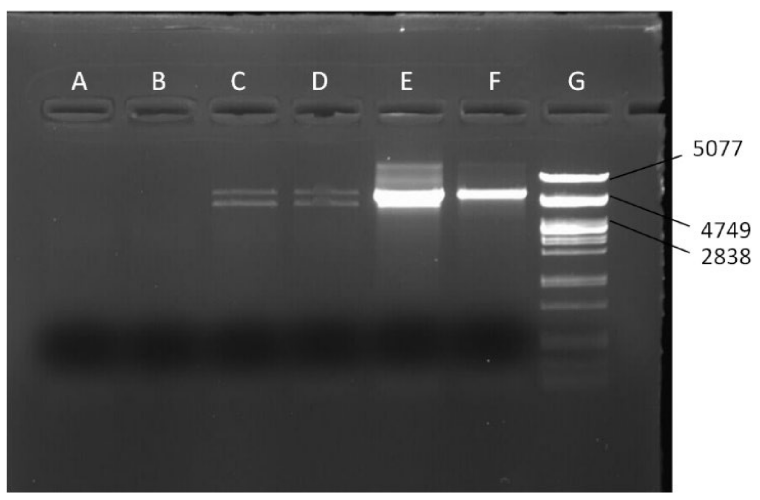
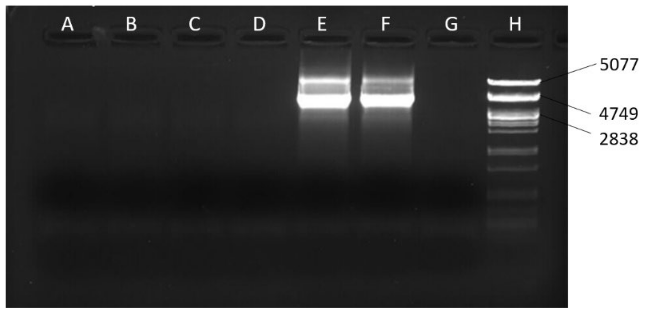
Based on the fact that mtDNA is circular but the NUMTs are linear, we designed long PCR assays to amplify two long overlapping mel mtDNA fragments (fragments 3 and 4, Figure 1A,B), with mel-specific primers. We tested the chimeric lines siI;820 and sm21;820 for the presence of mel sequences. We used DNA from the chimeric lines siI;w1118 and sm21;w1118 as negative controls, since they do not contain mel mtDNA, and DNA from lines Oregon R and DGRP-820 as positive controls, since they contained mel mtDNA, only. If the mel sequences were actually NUMTs, we would not recover one of the fragments, because of the incapability of the PCR to amplify the linear fragment (illustrated in Figure 1B). The first 5.5 kb fragment (fragment 3 from 2057 to 7597 bp) was successfully amplified (Figure 2), but the other 4 kb fragment (fragment 4 from 7339 to 11,353 bp) was not (Figure 3). This result suggests that the heteroplasmy observed in the DGRP-820 line was due to a large NUMT embedded within a 3.15 Mb region of the X chromosome, between the f and malbz genes. The inability to amplify fragment 4 indicates that the junctions of the NUMT are located within this mtDNA fragment. We had already amplified the region between 10,631 and 11,353 bp (fragment 2) using mel-specific primers, which is part of fragment 4. Therefore, the breakpoint that linearized the mtDNA should be in a fragment of 3292 bp, between the sites 7339 and 10,631 bp. Given that the length of D. melanogaster mtDNA is 19,517 bp and the breakpoint appears to be within 3292 bp, we estimate that the size of the NUMT is at least 16,225 bp.

4. Discussion

The attempts to identify NUMTs in the melanogaster species group of Drosophila revealed nine unique NUMTs in D. melanogaster with their size ranging from 19 bp to 371 bp [7,33]. With the exception of D. simulans, all other species of this group have more and, on average, larger NUMTs. Interestingly, almost the whole mtDNA has been introgressed in the nDNA in Drosophila yakuba [7], which belongs to the D. melanogaster species subgroup. The total length of different NUMTs in D. melanogaster’s nuclear genome has been estimated at approximately 10.3 kb [9] because NUMTs can be multiplicated after their introgression [13]. The size of the NUMT that we report here is larger than 16.2 kb, which is more than five times the size of the largest NUMT described in the genus, and it is almost the whole mtDNA which is 19,517 kb [37]. This estimate refers to a single-copy sequence. We cannot exclude the possibility that this NUMT has been duplicated in the nDNA, forming a mega-NUMT with several concatemers, as has been proposed by [22]. If present, the duplications must have happened tandemly, because our experiments for NUMT location have shown that it is embedded in a single rather than multiple chromosomal regions. Based on the current observation, the reported insertion rate of mtDNA fragments into nDNA, which has been estimated at 0.12 per million years [7], should be reconsidered. We deduced that the NUMT has been recently introgressed into the nuclear genome, first because its length is much larger than the previously described NUMTs, second because the sequence divergence between the NUMT and the actual mtDNA was negligible, and third because it has only been found in one out of the twelve nuclear DGRP backgrounds in the chimeric lines created by [24].
A recent study tried to identify new NUMTs in the sequenced DGRP genomes [38] using a bioinformatic approach [33]. This method recovered six NUMTs in all DGRP genomes. Two of them had been previously reported [7], while four of them were newly described. It is interesting that this sophisticated method failed to identify three already annotated NUMTs, as well as the one we are describing here. This might have to do with the pipeline they applied, which identifies chimeric reads at the breakpoints of a translocation. These reads may have been discarded as unmapped due to the absence of matching sequences in the reference genome [33]. As difficult as it is to identify a NUMT in the nuclear genome using bioinformatic methods, in our case it was also difficult to distinguish the NUMT from the mtDNA because of their high sequence similarity, perhaps suggesting that the NUMT was recently translocated to the nuclear DNA. This became possible only because the original mtDNA had been replaced by mtDNA from D. simulans in the chimeric lines that we initially used. Such polymorphic NUMTs have been systematically identified in other species, as well, such as bats [39], horses [40], pigs [41], and even humans [42,43]. It remains to be tested whether the presence of polymorphic NUMTs suggests that the population of origin (for example, the population that the DGRP-820 came from) has a higher effective size and therefore can maintain a higher level of polymorphism. Our study brings out an important difficulty in distinguishing NUMTs from true mtDNA, which regards the position of the NUMTs in the genomes. A NUMT located in the X chromosome mirrors partially maternal transmission which can be easily confused with the mtDNA transmission. In our case, the combination of the low divergence between NUMT and mtDNA as well as NUMT’s position on the X chromosome produced inconclusive results from the crosses and we needed well-targeted experiments to conclude that the putative heteroplasmy we observed was due to a NUMT.
The presence of recent NUMTs in the genomes is not expected to distort the phylogenetic signal of the sequences, because of their sequence similarity with the original mtDNA. It can cause severe problems, though, when studying the inheritance of mtDNA. It is obvious from this study that the heteroplasmy produced by the presence of NUMTs can be easily mistaken for biparental transmission of mtDNA. Such a debate was started by Luo et al. in 2018 in humans [20] and still holds. The evidence they provided for biparental transmission of mtDNA in three human families prompted several researchers to study in depth the validity of this claim. First, Salas et al. [44], reanalyzing the data by Luo et al., estimated that the probability of biparental inheritance is below 10−37. Second, Bai et al. [45] showed that, in four human families, heteroplasmic fathers transmitted only one mitotype to their progeny. This observation is similar to what we have observed in the present study, which turned out to be consistent with a NUMT transmission. Third, Wei et al. [46] observed transmission of mega-NUMTs from fathers in seven families, which could erroneously lead to the conclusion that mtDNA is biparentally transmitted. The present study in D. melanogaster contributes to the discussion described above for humans. These studies revealed that recent translocations of mtDNA into the nDNA can be more prevalent than previously thought. More importantly, while they cannot disprove the possibility of biparental transmission of mtDNA, they clearly show that such a claim should first exclude the probability that such an observation could be due to NUMTs which are mimicking biparental transmission of mtDNA.

Supplementary Materials

The following supporting information can be downloaded at: https://www.mdpi.com/article/10.3390/genes13061023/s1, Figure S1: Crossing scheme for the replacement of the X chromosome in a w1118 background by the X chromosome from the DGRP-820 nuclear background. The crosses were performed twice, once with a sm21;w1118 line (shown here), and once with a siI;w1118 line (not shown).pdf; Figure S2: Crossing scheme for the replacement of the autosome chromosomes in a w1118 background by the autosome chromosomes from the DGRP-820 nuclear background. The crosses were performed twice, once with a sm21;w1118 line (shown here), and once with a siI;w1118 line (not shown).pdf; Figure S3: Crossing scheme for mapping the loci responsible for heteroplasmy. The progeny siIII;FM6/recXDGRP-820 were phenotyped to determine which XDGRP-820 regions they contained. The individuals were then PCR-tested for the presence of the mel mitotype.pdf; Figure S4: Crosses performed to investigate whether the observed, putative heteroplasmy was created once or whether it was emerging at every generation through paternal leakage. The genotypes of the progeny produced after each cross are shown. (A) All progeny were homoplasmic for the maternal mitotype, while no paternal mtDNA leakage was observed. (B) Progeny that carried the XDGRP-820 carried also both the mel and siII mitotypes.pdf; Figure S5: Crosses performed to show whether the putatively heteroplasmic females produced by the crosses from Figure S4-B could transmit their presumed heteroplasmy to the next generation. All progeny that inherited an XDGRP-820 from their mothers had also inherited both maternal mitotype sequences (siII and mel). On the contrary, the rest of the progeny inherited only the siII mitotype.pdf; Figure S6: Cross performed to test whether males, putatively heteroplasmic (mel and siII), with XDGRP-820 chromosome could transmit both their mitotypes to the next generation. Only the mel mitotype was transmitted from father, and exclusively to the progeny who also inherited the XDGRP-820. The same cross was repeated with siI/mel; XDGRP-820/Y giving the same results (not shown).pdf.

Author Contributions

M.-E.P., E.D.L. and D.R. designed the experiments; M.-E.P. and C.-T.Z. performed the experiments; M.-E.P., D.R. and E.D.L. wrote the manuscript. All authors have read and agreed to the published version of the manuscript.

Funding

The project was supported by Special Account of Research, University of Crete (KA10137). M-E.P. has been supported by a scholarship from the Hellenic Foundation for Research and Innovation (HFRI). Additional funding was provided by NIH grant R01GM067862 to D.M.R.

Institutional Review Board Statement

Not applicable.

Conflicts of Interest

The authors declare no conflict of interest.

References

  1. Bensasson, D.; Zhang, D.X.; Hartl, D.L.; Hewitt, G.M. Mitochondrial Pseudogenes: Evolution’s Misplaced Witnesses. Trends Ecol. Evol. 2001, 16, 314–321. [Google Scholar] [CrossRef]
  2. Ricchetti, M.; Tekaia, F.; Dujon, B. Continued Colonization of the Human Genome by Mitochondrial DNA. PLoS Biol. 2004, 2, e273. [Google Scholar] [CrossRef]
  3. Kowal, K.; Tkaczyk, A.; Pierzchała, M.; Bownik, A.; Ślaska, B. Identification of Mitochondrial DNA (NUMTs) in the Nuclear Genome of Daphnia Magna. Int. J. Mol. Sci. 2020, 21, 8725. [Google Scholar] [CrossRef]
  4. Laine, V.N.; Gossmann, T.I.; Van Oers, K.; Visser, M.E.; Groenen, M.A.M. Exploring the Unmapped DNA and RNA Reads in a Songbird Genome. BMC Genom. 2019, 20, 19. [Google Scholar] [CrossRef] [Green Version]
  5. Wang, J.-X.; Liu, J.; Miao, Y.-H.; Huang, D.-W.; Xiao, J.-H. Tracking the Distribution and Burst of Nuclear Mitochondrial DNA Sequences (NUMTs) in Fig Wasp Genomes. Insects 2020, 11, 680. [Google Scholar] [CrossRef] [PubMed]
  6. Song, S.; Jiang, F.; Yuan, J.; Guo, W.; Miao, Y. Exceptionally High Cumulative Percentage of NUMTs Originating from Linear Mitochondrial DNA Molecules in the Hydra Magnipapillata Genome. BMC Genom. 2013, 14, 447. [Google Scholar] [CrossRef] [PubMed] [Green Version]
  7. Rogers, H.H.; Griffiths-Jones, S. Mitochondrial Pseudogenes in the Nuclear Genomes of Drosophila. PLoS ONE 2012, 7, e32593. [Google Scholar] [CrossRef] [Green Version]
  8. Baldo, L.; de Queiroz, A.; Hedin, M.; Hayashi, C.Y.; Gatesy, J. Nuclear–Mitochondrial Sequences as Witnesses of Past Interbreeding and Population Diversity in the Jumping Bristletail Mesomachilis. Mol. Biol. Evol. 2011, 28, 195–210. [Google Scholar] [CrossRef]
  9. Hazkani-Covo, E.; Zeller, R.M.; Martin, W. Molecular Poltergeists: Mitochondrial DNA Copies (Numts) in Sequenced Nuclear Genomes. PLoS Genet. 2010, 6, e1000834. [Google Scholar] [CrossRef]
  10. Kaya, S.; Çıplak, B. Possibility of Numt Co-Amplification from Gigantic Genome of Orthoptera: Testing Efficiency of Standard PCR Protocol in Producing Orthologous COI Sequences. Heliyon 2018, 4, e00929. [Google Scholar] [CrossRef] [PubMed] [Green Version]
  11. Pereira, R.J.; Ruiz-Ruano, F.J.; Thomas, C.J.E.; Pérez-Ruiz, M.; Jiménez-Bartolomé, M.; Liu, S.; de la Torre, J.; Bella, J.L. Mind the Numt: Finding Informative Mitochondrial Markers in a Giant Grasshopper Genome. J. Zool. Syst. Evol. Res. 2021, 59, 635–645. [Google Scholar] [CrossRef]
  12. Pamilo, P.; Viljakainen, L.; Vihavainen, A. Exceptionally High Density of NUMTs in the Honeybee Genome. Mol. Biol. Evol. 2007, 24, 1340–1346. [Google Scholar] [CrossRef] [PubMed]
  13. Richly, E.; Leister, D. NUMTs in Sequenced Eukaryotic Genomes. Mol. Biol. Evol. 2004, 21, 1081–1084. [Google Scholar] [CrossRef] [Green Version]
  14. Calvignac, S.; Konecny, L.; Malard, F.; Douaby, C. Preventing the Pollution of Mitochondrial Datasets with Nuclear Mitochondrial Paralogs (Numts). Mitochondrion 2011, 11, 246–254. [Google Scholar] [CrossRef] [PubMed]
  15. Shi, H.; Xing, Y.; Mao, X. The Little Brown Bat Nuclear Genome Contains an Entire Mitochondrial Genome: Real or Artifact? Gene 2017, 629, 64–67. [Google Scholar] [CrossRef]
  16. Nacer, D.F.; Raposo do Amaral, F. Striking Pseudogenization in Avian Phylogenetics: Numts Are Large and Common in Falcons. Mol. Phylogenet. Evol. 2017, 115, 1–6. [Google Scholar] [CrossRef] [PubMed]
  17. Verscheure, S.; Backeljau, T.; Desmyter, S. In Silico Discovery of a Nearly Complete Mitochondrial Genome Numt in the Dog (Canis lupus familiaris) Nuclear Genome. Genetica 2015, 143, 453–458. [Google Scholar] [CrossRef]
  18. Wang, B.; Zhou, X.; Shi, F.; Liu, Z.; Roos, C.; Garber, P.A.; Li, M.; Pan, H. Full-Length Numt Analysis Provides Evidence for Hybridization between the Asian Colobine Genera Trachypithecus and Semnopithecus. Am. J. Primatol. 2015, 77, 901–910. [Google Scholar] [CrossRef] [PubMed]
  19. Lutz-Bonengel, S.; Niederstätter, H.; Naue, J.; Koziel, R.; Yang, F.; Sänger, T.; Huber, G.; Berger, C.; Pflugradt, R.; Strobl, C.; et al. Evidence for Multi-Copy Mega-NUMT s in the Human Genome. Nucleic Acids Res. 2021, 49, 1517–1531. [Google Scholar] [CrossRef]
  20. Luo, S.; Valencia, C.A.; Zhang, J.; Lee, N.-C.; Slone, J.; Gui, B.; Wang, X.; Li, Z.; Dell, S.; Brown, J.; et al. Biparental Inheritance of Mitochondrial DNA in Humans. Proc. Natl. Acad. Sci. USA 2018, 115, 13039–13044. [Google Scholar] [CrossRef] [Green Version]
  21. Luo, S.; Valencia, C.A.; Zhang, J.; Lee, N.-C.; Slone, J.; Gui, B.; Wang, X.; Li, Z.; Dell, S.; Brown, J.; et al. Reply to Lutz-Bonengel et Al.: Biparental MtDNA Transmission Is Unlikely to Be the Result of Nuclear Mitochondrial DNA Segments. Proc. Natl. Acad. Sci. USA 2019, 116, 1823–1824. [Google Scholar] [CrossRef] [Green Version]
  22. Balciuniene, J.; Balciunas, D. A Nuclear MtDNA Concatemer (Mega-NUMT) Could Mimic Paternal Inheritance of Mitochondrial Genome. Front. Genet. 2019, 10, 2018–2020. [Google Scholar] [CrossRef] [PubMed]
  23. Lutz-Bonengel, S.; Parson, W. No Further Evidence for Paternal Leakage of Mitochondrial DNA in Humans Yet. Proc. Natl. Acad. Sci. USA 2019, 116, 1821–1822. [Google Scholar] [CrossRef] [PubMed] [Green Version]
  24. Mossman, J.A.; Biancani, L.M.; Zhu, C.-T.; Rand, D.M. Mitonuclear Epistasis for Development Time and Its Modification by Diet in Drosophila. Genetics 2016, 203, 463–484. [Google Scholar] [CrossRef] [Green Version]
  25. Montooth, K.L.; Meiklejohn, C.D.; Abt, D.N.; Rand, D.M. Mitochondrial-Nuclear Epistasis Affects Fitness within Species but Does Not Contribute to Fixed Incompatibilities between Species of Drosophila. Evolution 2010, 64, 3364–3379. [Google Scholar] [CrossRef] [PubMed] [Green Version]
  26. Solignac, M.; Monnerot, M.; Mounolou, J.C. Mitochondrial DNA Evolution in the Melanogaster Species Subgroup of Drosophila. J. Mol. Evol. 1986, 23, 31–40. [Google Scholar] [CrossRef]
  27. Ashburner, M.; Golic, K.G.; Hawley, R.S. Drosophila: A Laboratory Handbook, 2nd ed.; Cold Spring Harbor Laboratory Press: Cold Spring Harbor, NY, USA, 2005; ISBN 9780879697068. [Google Scholar]
  28. O’neill, S.L.; Giordano, R.; Colbert, A.M.E.; Karrf, T.L.; Robertson, H.M. 16S RRNA Phylogenetic Analysis of the Bacterial Endosymbionts Associated with Cytoplasmic Incompatibility in Insects. Proc. Natl. Acad. Sci. USA 1992, 89, 2699–2702. [Google Scholar] [CrossRef] [PubMed] [Green Version]
  29. Miller, S.A.; Dykes, D.D.; Polesky, H.F. A Simple Salting out Procedure for Extracting DNA from Human Nucleated Cells. Nucleic Acids Res. 1988, 16, 1215. [Google Scholar] [CrossRef] [Green Version]
  30. Dokianakis, E.; Ladoukakis, E.D. Different Degree of Paternal MtDNA Leakage between Male and Female Progeny in Interspecific Drosophila Crosses. Ecol. Evol. 2014, 4, 2633–2641. [Google Scholar] [CrossRef] [PubMed]
  31. Polovina, E.S.; Parakatselaki, M.E.; Ladoukakis, E.D. Paternal Leakage of Mitochondrial DNA and Maternal Inheritance of Heteroplasmy in Drosophila Hybrids. Sci. Rep. 2020, 10, 2599. [Google Scholar] [CrossRef]
  32. Zhu, C.T.; Ingelmo, P.; Rand, D.M. GxGxE for Lifespan in Drosophila: Mitochondrial, Nuclear, and Dietary Interactions That Modify Longevity. PLoS Genet. 2014, 10, e1004354. [Google Scholar] [CrossRef] [PubMed] [Green Version]
  33. Bevers, R.P.J.; Litovchenko, M.; Kapopoulou, A.; Braman, V.S.; Robinson, M.R.; Auwerx, J.; Hollis, B.; Deplancke, B. Mitochondrial Haplotypes Affect Metabolic Phenotypes in the Drosophila Genetic Reference Panel. Nat. Metab. 2019, 1, 1226–1242. [Google Scholar] [CrossRef] [PubMed]
  34. Wolfner, M.F. The Gifts That Keep on Giving: Physiological Functions and Evolutionary Dynamics of Male Seminal Proteins in Drosophila. Heredity 2002, 88, 85–93. [Google Scholar] [CrossRef] [Green Version]
  35. Ma, H.; O’Farrell, P.H. Selections That Isolate Recombinant Mitochondrial Genomes in Animals. eLife 2015, 4, e07247. [Google Scholar] [CrossRef] [PubMed]
  36. Doane, W.W. Completion of Meiosis in Uninseminated Eggs of Drosophila melanogaster. Science 1960, 132, 677–678. [Google Scholar] [CrossRef]
  37. Lewis, O.L.; Farr, C.L.; Kaguni, L.S. Drosophila melanogaster Mitochondrial DNA: Completion of the Nucleotide Sequence and Evolutionary Comparisons. Insect Mol. Biol. 1995, 4, 263–278. [Google Scholar] [CrossRef]
  38. Mackay, T.F.C.; Richards, S.; Stone, E.A.; Barbadilla, A.; Ayroles, J.F.; Zhu, D.; Casillas, S.; Han, Y.; Magwire, M.M.; Cridland, J.M.; et al. The Drosophila melanogaster Genetic Reference Panel. Nature 2012, 482, 173–178. [Google Scholar] [CrossRef] [Green Version]
  39. Zhang, G.; Geng, D.; Guo, Q.; Liu, W.; Shufen, L.I.; Gao, W.; Wang, Y.; Zhang, M.; Wang, Y.; Yanzhen, B.U.; et al. Genomic Landscape of Mitochondrial DNA Insertions in 23 Bat Genomes: Characteristics, Loci, Phylogeny, and Polymorphism. Integr. Zool. 2021; Online ahead of print. [Google Scholar] [CrossRef]
  40. Nergadze, S.G.; Lupotto, M.; Pellanda, P.; Santagostino, M.; Vitelli, V.; Giulotto, E. Mitochondrial DNA Insertions in the Nuclear Horse Genome. Anim. Genet. 2010, 41, 176–185. [Google Scholar] [CrossRef]
  41. Schiavo, G.; Hoffmann, O.I.; Ribani, A.; Utzeri, V.J.; Ghionda, M.C.; Bertolini, F.; Geraci, C.; Bovo, S.; Fontanesi, L. A Genomic Landscape of Mitochondrial DNA Insertions in the Pig Nuclear Genome Provides Evolutionary Signatures of Interspecies Admixture. DNA Res. 2017, 24, 487–498. [Google Scholar] [CrossRef]
  42. Lang, M.; Sazzini, M.; Calabrese, F.M.; Simone, D.; Boattini, A.; Romeo, G.; Luiselli, D.; Attimonelli, M.; Gasparre, G. Polymorphic NumtS Trace Human Population Relationships. Hum. Genet. 2012, 131, 757–771. [Google Scholar] [CrossRef] [PubMed]
  43. Dayama, G.; Emery, S.B.; Kidd, J.M.; Mills, R.E. The Genomic Landscape of Polymorphic Human Nuclear Mitochondrial Insertions. Nucleic Acids Res. 2014, 42, 12640–12649. [Google Scholar] [CrossRef] [Green Version]
  44. Salas, A.; Schönherr, S.; Bandelt, H.J.; Gómez-Carballa, A.; Weissensteiner, H. Extraordinary Claims Require Extraordinary Evidence in Asserted MtDNA Biparental Inheritance. Forensic Sci. Int. Genet. 2020, 47, 102274. [Google Scholar] [CrossRef]
  45. Bai, R.; Cui, H.; Devaney, J.M.; Allis, K.M.; Balog, A.M.; Liu, X.; Schnur, R.E.; Shapiro, F.L.; Brautbar, A.; Estrada-Veras, J.I.; et al. Interference of Nuclear Mitochondrial DNA Segments in Mitochondrial DNA Testing Resembles Biparental Transmission of Mitochondrial DNA in Humans. Genet. Med. 2021, 23, 1514–1521. [Google Scholar] [CrossRef] [PubMed]
  46. Wei, W.; Pagnamenta, A.T.; Gleadall, N.; Sanchis-Juan, A.; Stephens, J.; Broxholme, J.; Tuna, S.; Odhams, C.A.; Ambrose, J.C.; Baple, E.L.; et al. Nuclear-Mitochondrial DNA Segments Resemble Paternally Inherited Mitochondrial DNA in Humans. Nat. Commun. 2020, 11, 1740. [Google Scholar] [CrossRef] [PubMed] [Green Version]
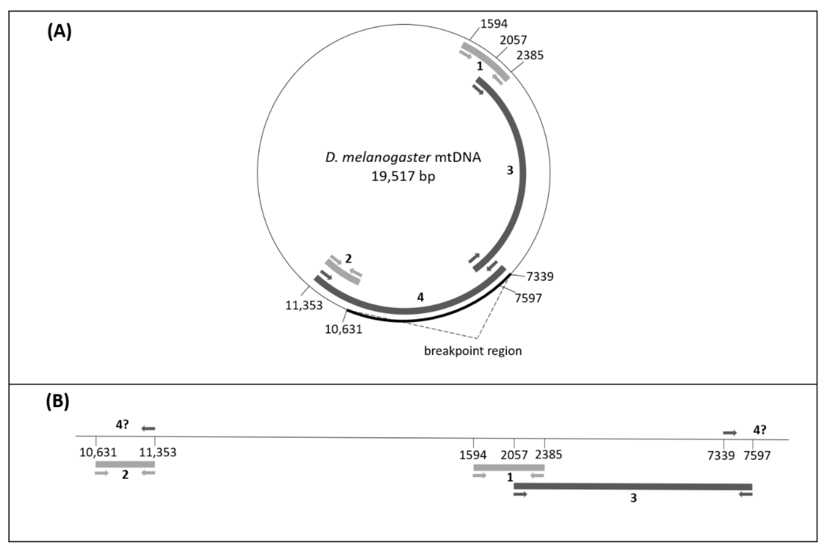
Figure 1. PCR assays designed to detect the mel mtDNA and the NUMT. (A) The circle represents the Drosophila melanogaster mtDNA reference genome. The arrows show the direction of the primers and the thick lines inside the circle represent the potentially amplified fragments, whose coordinates are shown by the numbers. The two short fragments in light grey (CoxI 790 bp fragment 1 from 1594 to 2385 and Cytb 722 bp fragment 2 from 10,631 to 11,353) represent the PCR fragments that were initially used to detect putative heteroplasmy, while the two long PCR fragments in dark grey lines (fragments 3 and 4) were used to distinguish between true heteroplasmy and NUMT. The bold line on the circle marks the region within which the breakpoint for NUMT insertion resides. (B) The NUMT as it should have been inserted in the nuclear genome. The thick lines and the coordinates of the primers and the number of the fragments are similar to part A of the figure. The direction of the primers indicates why the 7339–11,353 fragment was not amplified in the linear molecule. Using the coordinates of the primers and based on the amplified and the non-amplified fragments we estimated the size of the NUMT.
Genes 13 01023 g001
Figure 2. Electrophoresis of the products from long-PCR with primers mel2057F and 7597− (fragment 3). (A) siI;w1118, (B) sm21;w1118, (C) siI;820, (D) sm21;820, (E) DGRP-820, (F) D. melanogaster Oregon R line, (G) λ100 marker, PstI digest. (A,B) are used as negative controls, while (E,F) are the positive controls. The mel product was amplified for lines siI;820 and sm21;820.
Genes 13 01023 g002
Figure 3. Electrophoresis of the products from long-PCR with primers 7339+ and mel11353R (fragment 4). (A) siI;w1118, (B) sm21;w1118, (C) siI;820, (D) sm21;820, (E) DGRP-820, (F) D. melanogaster Oregon R line, (G) no template control, (H) λ100 marker, PstI digest. (A,B) are used as negative controls, while (E,F) are the positive controls. Absence of the bands in (C,D) shows that there is no mel mtDNA to be amplified in lines siI;820 and sm21;820, a result consistent with the NUMT scenario.
Genes 13 01023 g003
Table 1. List of lines used in this study and their mitotypes.
LinesMitotype
mel;DGRP-820mel
siI;820siI
sm21;820siII
siI;w1118;+/+;+/+siI
sm21;w1118;+/+;+/+siII
mau12;w1118;+/+;+/+siIII
D. melanogaster Oregon Rmel
mel;y v f malbz/ y v f malbz; +/+;+/+mel
mel;FM6 B1/+;+/+;+/+mel
siI;w1118;CyO/Sp;TM6B/DrsiI
siII;w1118;CyO/Sp;TM6B/DrsiII
Table 2. Detailed list of primers used.
ReferencePrimersSequence (5′ -> 3′)SpecificityPCR Product Size (bp)Tm (°C)
[30]mel1594FGCTGAATTAGGACATCCTGGAGCmel against siI, siII, siIII79161
mel2385RTCGAGTATCTACATCTATTCCAACG
Present studymel10631FCGAAATTCCCATCCTCmel against siI, siII, siIII72252
mel11353RTTATCAGGGTCTCCCA
[30]siI_1737FTCCTGATATAGCATTTCCAsiI against mel, siIII79458
siI_2531RGTTAATCCTCCTACTGTG
[31]siII_1737FCCCTGATATAGCATTCCCGsiII against mel, siIII79458
siII_2531RGTTAACCCCCCTACTGTA
Present study1588+GAATTAGGACATCCTGGAGCATsiIII against mel, siI, siII78661
2374−GAGTATCAACGTCTATTCCAACTGTG
Kindly provided by Maria Monastirioti1002(F)TCGGAATAAGTTGAAGGATGX chromosome against mtDNA76752
2653(R)TGCCATCCTGACTGCTCAGC
Present study7339+AAGCATGAGTTAATAAATGAAAmtDNA universal primers25854
7597−CCGTTTCTGCTTTAGTTC
Present study7339+AAGCATGAGTTAATAAATGAAAUsed for the long PCR assay401456
mel11353RTTATCAGGGTCTCCCA
Present studymel2057FTATTATTATCACTTCCAGTACUsed with 7597− for the long PCR assay554054
7597−CCGTTTCTGCTTTAGTTC
Table 3. Genotypes of three lines produced after the chromosome replacements. Similar lines were produced with the mitotype siI.
LineGenotype
Stable line 1 s i I I m e l ; X D G R P 820 X D G R P 820 ; C y O S p ; T M 6 B D r
Stable line 2 s i I I ; w 1118 w 1118 ; 2 D G R P 820 2 D G R P 820 ; T M 6 B D r
Stable line 3 s i I I ; w 1118 w 1118 ; C y O S p ; 3 D G R P 820 3 D G R P 820
Publisher’s Note: MDPI stays neutral with regard to jurisdictional claims in published maps and institutional affiliations.