Article

An Identification and Characterization of the Axolotl (Ambystoma mexicanum, Amex) Telomerase Reverse Transcriptase (Amex TERT)

1
Department of Plastic, Aesthetic, Hand and Reconstructive Surgery, Hannover Medical School, 30625 Hannover, Germany
2
Department of Oral and Maxillofacial Surgery, Diakovere Henriettenstift, 30171 Hannover, Germany
*
Author to whom correspondence should be addressed.
Genes 2022, 13(2), 373; https://doi.org/10.3390/genes13020373
Submission received: 14 January 2022 / Revised: 11 February 2022 / Accepted: 16 February 2022 / Published: 18 February 2022
(This article belongs to the Section Animal Genetics and Genomics)

Abstract

:
The Mexican axolotl is one of the few vertebrates that is able to replace its lost body parts during lifespan. Due to its remarkable regenerative abilities, the axolotl emerged as a model organism especially for limb regeneration. Telomeres and the telomerase enzyme are crucial for regeneration and protection against aging processes and degenerating diseases. Despite its relevance for regeneration, the axolotl telomerase and telomere length have not yet been investigated. Therefore, in the present paper, we reveal the sequence of the axolotl telomerase reverse transcriptase gene (Tert) and protein (TERT). Multiple sequence alignment (MSA) showed the known conserved RT- and TERT-specific motifs and residues found in other TERTs. In addition, we establish methods to determine the Tert expression (RT-PCR) and telomerase activity (Q-TRAP) of adult axolotl and blastema tissues. We found that both differentiated forelimb tissue and regenerating blastema tissue express Tert and show telomerase activity. Furthermore, blastema tissue appears to exhibit a higher Tert expression and telomerase activity. The presence of active telomerase in adult somatic cells is a decisive difference to somatic cells of non-regenerating vertebrates, such as humans. These findings indicate that telomere biology may play a key role in the regenerative abilities of cells.

1. Introduction

A healthy organism manages to equilibrate degenerative and regenerative processes. Aging usually causes the decline of regenerative potential as we diagnose soaring degenerative diseases (e.g., chronic internal or neurodegenerative diseases) in the elderly population [1]. Furthermore, the ability to repair acute tissue injury is limited even at young age and is declined during aging. Understanding the mechanisms of the existing regenerative abilities, along with strategies to activate and enhance this potential, would be a great advantage for patients who suffer from degenerative diseases or are affected by serious injuries, such as amputations or burns. Additionally, these chronic diseases and acute injuries have a significant economic impact on our healthcare and general systems, as they produce high costs (e.g., treatment expenses and duration, the loss of working ability) [2]. Against the background of demographic changes and the aging population, the need to discover new therapeutic strategies facing degeneration and tissue damage is urgently required. The human organism’s regenerative potential is strongly limited. In response to acute injury, some tissues, such as bone [3,4], muscle [3,5] and epithelial tissues [6], can be repaired, provided that the defect does not exceed a critical size. Most human regenerative strategies are based upon the replacement of lost cells by means of adult stem cell proliferation. However, humans do not have the ability to regenerate complex structures, such as limbs [7].
In contrast, there are some organisms that can regenerate complex body parts during lifespan [8]. The Mexican axolotl (Ambystoma mexicanum) belongs to the salamander species and evolved to a model organism for regenerative biology [9]. Axolotls are neotenic, meaning they are permanently aquatic and do not undergo metamorphosis. This life cycle distinguishes the axolotl from other salamanders and, furthermore, differences regarding the regenerative potential and mechanisms have been reported [10].
The axolotl is capable of rebuilding its entire limbs and tail, the upper and lower jaw, the brain, spinal cord and myocardial muscle after amputation or severe damage. Deep skin wounds are described as healing without the appearance of scars [11]. Limb regeneration can be subclassified into three overlapping phases. Following amputation and rapid hemostasis, epidermal cells migrate across the amputation surface and close the wound within hours [12]. During the next days, blastema cells accumulate beneath the new built wound epidermis. These cells originate from stem cells or derive from dedifferentiation of adjacent cells [10,13]. The blastema cells re-enter the cell cycle and become highly proliferative, so that the blastema grows [14]. Once the blastema has reached a critical size (“palette” stage), the redifferentiation of the various tissues takes place and a structurally and functionally perfect limb is rebuilt [15,16].
Each chromosome’s end is capped by a DNA–protein complex known as the telomere. The denomination “telomere” derives from the greek “télos" (end) and “méros” (part) and was provided by Hermann Müller in 1938 [17]. In vertebrates, the telomeres consist of thousands of TTAGGG-repeats that build a protective cap in collaboration with associated proteins to ensure genomic stability and integrity [18,19]. During genome duplication, telomeres shorten by about 50–200 bp, due to the incomplete replication of chromosome ends (end replication problem). This progressive loss of telomeric DNA limits the number of cell cycle divisions [20]. The cell loses its proliferative potential and achieves cellular senescence [21].
Telomerase is a reverse transcriptase (RT) catalyzing the elongation of telomeres [22]. Its catalytic core consists of the protein subunit (telomerase reverse transcriptase, TERT) and the RNA subunit (telomerase RNA, TR). The TR provides the template for telomeric repeats synthesis and contains motifs essential for telomerase activity. The TERT-protein is composed of three domains: the N-terminal part, the reverse-transcriptase domain (RT-domain) and the C-terminal extension (CTE). In addition, different accessory proteins adjust telomerase’s localization, regulation and function [23]. Telomerase stabilizes telomere length by adding telomeric repeats to its ends and thus protects cells against senescence. In humans, active telomerase can only be found during embryonic development [24] in an adult’s highly proliferative cells (e.g., germ cells [24], stem cells and progenitor cells [25], and activated lymphocytes [26]) and in many tumor cells [27]. Adult somatic tissues reveal no detectable telomerase activity [28]. In several animal investigations, a correlation between remarkable regenerative capacities and high telomerase activity could be shown without noticing many cancer diseases or even a higher carcinogenic risk at all [29,30]. These findings emphasize the growing interest in developing and exploring new therapeutic strategies to improve regeneration by recovering stable telomeres [31].
Telomeres and the telomerase enzyme are important elements for regeneration and protection against premature aging and degenerating diseases. One of the best-known molecular aging mechanisms is the progressive telomere attrition and proliferative senescence when reaching a critical telomere length. Highly proliferative cells are crucial for regeneration, so telomere biology is supposed to play a significant role in regenerative processes [31]. Related to the axolotl’s excellent regenerative abilities, we also suspect this decisive role of telomere biology.
This study aims to provide the first insights into the axolotl’s telomere biology. We identify and characterize the axolotl Tert gene (Amex Tert) for the first time. Subsequently, the methods used to measure Tert mRNA expression and telomerase activity are evolved and established. This information is useful for further studies concerning axolotl’s telomere biology and for the discovery of new telomere-based regeneration strategies for humans.

2. Materials and Methods

2.1. Animal Husbandry and Handling

For all the experiments, the axolotl’s adult limb tissue and blastema limb tissue were used. The 15 animals overall were bred and kept at the Ambystoma Mexicanum Bioregeneration Center of the Department of Plastic, Aesthetic, Hand and Reconstructive Surgery, Hanover Medical School. The experiments were approved by the Lower Saxony State Office for Consumer Protection and Food Safety under approval number: AZ 33.14-42502-04-13/1212 and in accordance with the German Animal Welfare Care. Adult axolotls (age: 1–5 years; length: 15–25 cm) were maintained in small groups (3–5 animals) in aquariums with fresh, warm tap water (max. 20 °C) without artificial illumination. They were protected against direct sun exposure and were fed 2 times a week with 4–5 pellets, “Axobalance” (Aquaterratec, Broeckel, Germany). Each tank was equipped with ground substrate, artificial plants, hiding places and a filter.
Animals were anesthetized in 0.1% Tricaine (MS-222®) in fresh cold tap water until deep anesthesia was reached (proofed by expiration of the ventral reflex). One forelimb per animal was amputated distal to the elbow with sterile sharp scissors. Afterwards, the animals were transferred into boxes filled with fresh cold tap water. After awakening, the animals were left for one night in quarantine and then transferred back to their tanks. When the regenerating blastema reached the palette stage [15,16], a second amputation was carried out under anesthesia, as described above. Each amputated limb was cut into three slices, directly frozen into liquid nitrogen and stored at −80 °C for the following procedures. After successful regeneration, the animals were examined by a veterinarian and re-homed to private keepers.

2.2. Cloning of Amex Tert Gene Fragments

Contigs with sequence identity to human TERT were searched in the Ambystoma Mexicanum V4.0 (contigs) database, via its website, Sal-SiteTM [32]. Five Contigs (contig282000, contig 346319, FUQAVB301DCODH, GFW9XCP01AHQ6K, and GHXAJEM01EV71Q) were assembled and used for Primer derivation. The primers’ sequences were (5′-AAATGGTTTGCGCCCGATAGTC-3′ and 5′-ATAAAGGCATGGTAGCTAAGCCACTG-3′). Axolotl embryonic cDNA isolated for other projects was obtained from laboratory stock for PCR establishment. PCR was performed utilizing the Advantage® 2 PCR Kit (Takara Bio Inc., Kusatsu, Japan). The amplificate was purified by Nucleo Spin® PCR Clean up Kit (Macherey Nagel) and cloned into pUC118 vector [33] using the Mighty Cloning Reagent Set (Blunt end) (Takara Bio Inc., Kusatsu, Japan) according to the manufacturer’s protocol. The cloned DNA was sequenced by Sanger sequencing using an M13 primer (GATC Services, Eurofins Genomics, Ebersberg, Germany). A BLASTn [34] search in the Nucleotide Collection (nr/nt) database exhibited a high concordance with other TERT nucleotide sequences, so this cloned fragment was supposed to be part of Amex Tert.

2.3. Completion and Sequence Analysis of the Amex TERT Amino Acid Sequence

The predicted Amex TERT fragment was searched in the Transcriptome Shotgun Assembly (TSA) database using BLASTn and could be verified as part of the transcript Ambymex_c1080993_g3_i1 (GenBank Accession number GFBM010789021). The nucleotide sequence was translated into an amino acid sequence [35]. Multiple sequence alignments were performed with the Clustal Omega program (European Bioinformatic Institute, EMBL-EBI) [36]. A phylogenetic tree was constructed by the neighbor-joining method using the FigTree v1.4.3 software. The GenBank accession numbers for multiple sequence alignment and phylogeny are as follows: hTERT, Homo sapiens TERT (accession: NP_937983.2); mTERT, Mus musculus TERT (accession: AAC09323.1); TmtTERT, Terrapene mexicana triunguis TERT (accession: XP_024050859.1); PvTERT, Pogona vitticeps TERT (accession: XP_020652818.1); xTERT, Xenopus laevis TERT (accession: AAI70415.1); ZfTERT, Danio rerio TERT (accession: NP_001077335.1); OlaTERT, Oryzias latipes TERT (accession: NP_001098286.1); TtTERT, Tetrahymena thermophila TERT (accession: AAC39135.1); AtTERT, Arabidopsis thaliana TERT (accession: AAD54777.1); Sc_Est2p, Saccharomyces cerevisiae TERT (Accession: PJP08600.1); and HIV-1 RT (accession: AAB24839.1).

2.4. Analysis of Amex Tert mRNA Expression

Total RNA was prepared from axolotl limbs and limb blastemas by the ArrayPureTM Nano-scale RNA Purification Kit (Epicentre®, Madison, WI, USA) with little adaptions (quadruple amount lysis solution and higher concentrated Proteinase K (50 μg/μL), double amount MCP reagent and isopropranol). A total of 1 μg of total RNA was used for reverse transcription by iScriptTM cDNA Synthesis Kit (Bio-Rad, Hercules, CA, USA) according to the manufacturer’s protocol. For real-time PCR analysis, the following primer pair was established and validated: TeloExp For (5′-CCTCAAGCGTTTGTGTGTCA) and TeloExp Rev (5′-TGGCTGCTCCATAAGCCTAA). The gene ornithine decarboxylase was used as positive control and housekeeping gene [37] for 2−ΔΔCt method. Therefore, the primer pair of axolotl ornithine decarboxylase (sequence listed in Table S1) was included in the PCR reaction. Real-time PCR was performed with an iCyclerTM using the SsoFastTM EvaGreen® Supermix (Bio-Rad, Hercules, CA, USA). PCR conditions: 1 s at 95 °C; 35 cycles: 10 s at 95 °C and 30 s at 56.1 °C. Afterwards, a melting curve was generated to analyze the specificity of PCR products. Data analysis was performed by 2−ΔΔCt method [38].

2.5. Analysis of Telomerase Activity

Frozen tissue samples of axolotl limbs and limb blastemas were dispersed in a mortar and transferred into 200 μL of NP-40 lysis buffer. All samples were incubated for 30 min on ice followed by centrifugation at 16.000× g for 20 min at 4 °C. The clean supernatant was collected in a fresh Eppendorf tube and the total protein concentration was quantified using the Bradford assay. Telomerase activity was detected by the real-time quantitative telomerase repeat amplification protocol (Q-TRAP), according to Herbert et al. [39]. Telomerase positive samples (human MCF-7 breast carcinoma cells [40]) were used as positive control and for generation of a standard curve. For this purpose, a 1:5 dilution series was created. PCR amplification was performed as indicated by the authors [39]. With each PCR run, three negative controls were tested (lysis buffer only, RNase-treated sample, heat-inactivated sample) and each sample was analyzed at least in duplicates. After PCR, the real-time data was collected, the standard curve was determined and converted into Relative Telomerase Activity (RTA) units performing the following calculation: RTA of an unknown sample = 10[(Ct sample–Y int)/slope]. The standard curve obtained was: y = −6.192x + 16.336.

3. Results

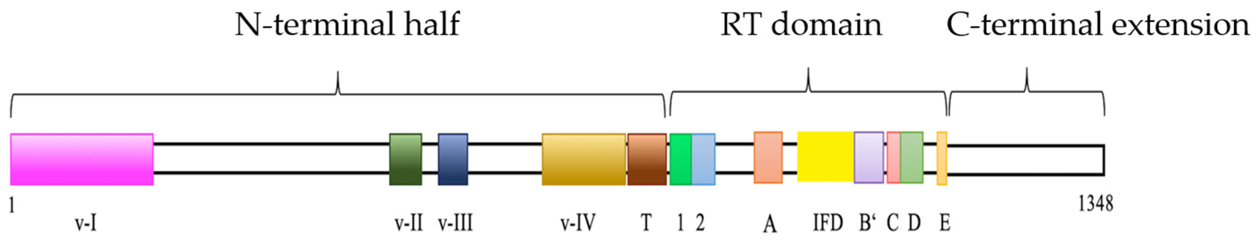
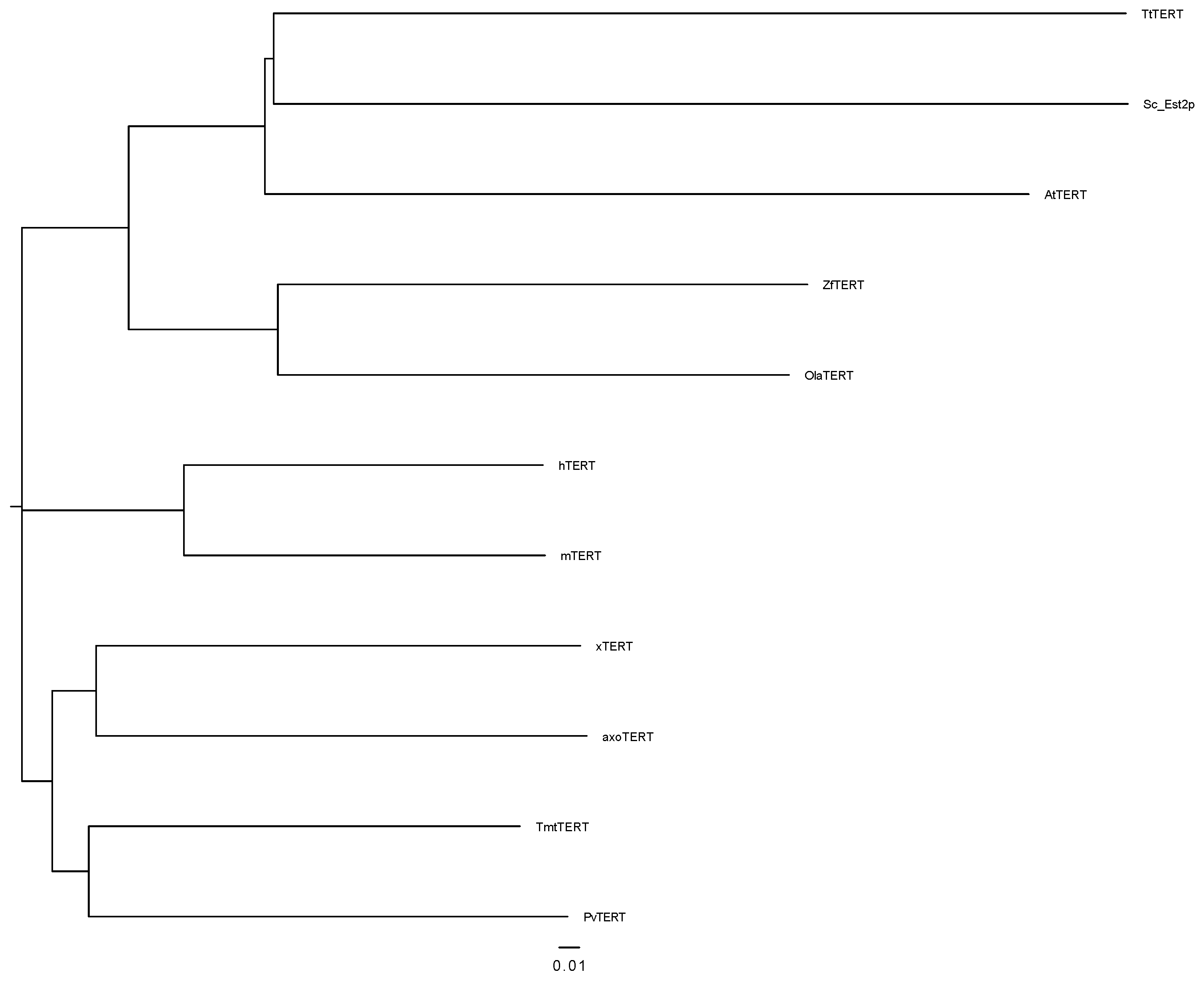
3.1. Identification and Characterization of Amex TERT

Using the database “Ambystoma mexicanum V4.0 (contigs)”, six contigs with concordance to hTERT could be located. Based on the resulting sequence, primers were generated and PCRs amplifying axolotl cDNA performed. The PCR product was cloned and sequenced. A BLASTn search confirmed a high consensus with other TERTs, so the sequence was supposed to be part of the axolotl TERT nucleotide sequence. In the database “transcriptome shotgun assembly”, the sequence could be found as part of the transcript Ambymex_c1080993_g3_i1 (GenBank Accession number GFBM010789021) [41] with 99.41% identity. This transcript was confirmed as an axolotl TERT nucleotide sequence (as is shown below) and collected in GenBank (GenBank Accession number MK702005) [42]. Therefore, we designated this gene as Amex Tert. The 4848 bp nucleotide sequence encoded a protein sequence composed of 1348 amino acids (aa) (GenBank Accession number GQJ02387) [43]. Figure 1 shows the schematic structure of the Amex TERT protein. The entire TERT amino acid sequence shows 53.34% identity to Terrapene mexicana triunguis TERT (TmtTERT); 52.17% to Xenopus laevis TERT (xTERT); 46.13% to Mus musculus TERT (mTERT); 45.99% to Homo sapiens TERT (hTERT) and Pogona vitticeps TERT; 35.37% to Oryzias latipes TERT (OlaTERT); 34.71 to Danio rerio TERT (ZfTERT); 22.35% to Arabidopsis thaliana TERT (AtTERT); 19.62% to Tetrahymena thermophila TERT (TtTERT); and 19.02% to Saccharomyces cerevisiae Est2p (Sc_Est2p).
Multiple sequence alignment (MSA) revealed that TERT contains several RT- and telomerase-specific motifs. At its C-terminal half, TERT possesses the telomerase-specific motif T [44] (aa 763 to 810) followed by the 7 RT motifs 1, 2, A, B’, C, D and E [45] (aa 818 to 1151). The RT motifs are found in diverse groups of RT, but there are short telomerase-specific signatures within these RT motifs that distinguish TERT from other RT [44], which can also be found in Amex TERT. They include a conserved arginine (R) in motif 1 and a conserved phenylalanine (F) behind two aspartic acids (D) in motif C and tryptophan (W)-x-glycine (G)-x-leucine (L) in motif E. Amex TERT also contains amino acid residues essential for RT activity [46]. Between motif A and B’, the TERT-specific domain called “insertion in fingers domain” (IFD) is shown [47]. The domain behind motif E to the C-terminal end of the sequence is called “C-terminal extension”. This area does not reveal many similarities to conventional RT, but has a high concordance to other vertebrate TERT [23,48]. The analysis of the N-terminal half of Amex TERT revealed four known regions conserved among vertebrate TERTs (v-I to v-IV) [49].
The MSA of complete TERT protein and all the mentioned RT- and telomerase-specific motifs is presented in the Supplementary Materials (Figures S1 and S2). Based on the multiple sequence alignment of different TERT proteins, a phylogenetic tree is constructed according to the neighbor-joining method (Figure 2). As expected, Amex TERT shows a higher sequence identity and similarity to those of vertebrates than to plant, yeast and ciliates.

3.2. Expression Analysis of Amex Tert mRNA by RT-PCR in Somatic and Blastema Tissues

We established a method to determine the Tert mRNA expression by RT-PCR. Therefore, the primer TeloExp was created and validated. The gene encoding ornithine decarboxylase was used as a positive control and reference gene for relative quantification. RT-PCR revealed that Amex Tert mRNA was expressed in both adult limbs and limb blastemas. All samples exhibit Tert expression, whereas a higher expression could be detected in blastemas (Table 1). Raw PCR data are shown in the Supplementary Materials (Table S1).

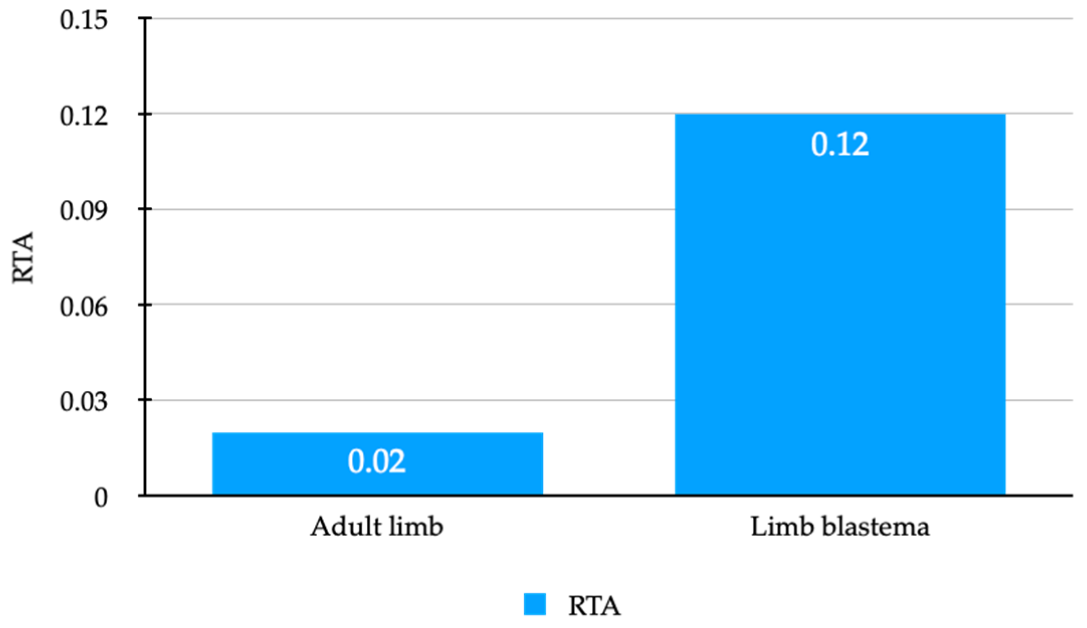
3.3. Active Telomerase Is Detectable in Axolotl Adult Limbs and Limb Blastemas

Simultaneous to the expression analysis, we adapted Q-TRAP [39] to axolotl tissue for examining a possible correlation between Amex Tert expression levels and telomerase activity. Protein extracts of adult limbs and blastema limb tissues were used as samples and MCF-7 protein extracts served as positive controls and references (the standard curve is shown in the Supplementary Materials (Figure S3)). All negative controls (heat-inactivated and RNase-treated) remained negative. In contrast to most non-regenerating vertebrates, axolotl somatic limb tissue exhibits detectable telomerase activity. According to the findings for Tert expression, the blastemas reveal a higher telomerase activity, so telomerase activity seems to be upregulated during limb regeneration (Table 2, Figure 3).

4. Discussion

Although the importance of telomeres and telomerase for mammalian aging, cancer and regeneration and the axolotl’s outstanding regenerative abilities were extensively discussed, to date, there is a lack of research investigating its telomere biology. In this study, we identified and characterized the axolotl TERT gene. The existence of the 7 RT motifs (1, 2, A, B’, C, D, E) and telomerase specific motifs (motif T, IFD, short TERT-specific signatures in RT motifs, vertebrate-specific domains in the amino-terminal half) confirmed the identification of Amex TERT. The nucleotide and amino acid sequence can be found in GenBank® under accession MK702005 [42] and QGJ02387 [43]. The sequence of Amex Tert allowed for the examination of Tert mRNA expression by real-time PCR.
Real-time PCR and TRAP assay demonstrated that limb blastemas, as well as differentiated adult limbs, express TERT mRNA and show telomerase activity. Both appear to be at higher levels in blastemas. This concordant relationship between TERT expression and telomerase activity was already found in other species (e.g., human [44], Xenopus laevis [49]). The presence of telomerase activity in somatic tissues of axolotl is a clear contrast to mammalian tissues, in which telomerase activity can be found in embryonic tissues, germline cells and stem cells, but is not detectable in most adult somatic tissues [24]. Other organisms (e.g., nonvertebrate [50], fish [51,52], some amphibian [49] and reptiles [18]) reveal TERT expression and telomerase activity, even in somatic tissues. At the same time, these animals exhibit notably regenerative abilities and hardly any signs of senescence during lifespan (age-related diseases, loss of reproductive abilities). Therefore, it is reasonable that the presence of TERT expression and telomerase activity is associated with high regenerative capabilities. The higher levels of TERT expression and telomerase activity in limb blastemas indicate that during regeneration, TERT expression is upregulated. Similar observations were made in axolotl tails [53], similar to other regenerative species (e.g., Aeolosoma viride [50]), Danio rerio [54]). Regeneration in the axolotl is performed by the dedifferentiation of somatic cells. Our results show that during the process of dedifferentiation, the upregulation of Tert mRNA expression occurs. The underlying mechanisms have to be investigated in the future. In the next step of regeneration, the dedifferentiated blastema cells start to proliferate. Because of active telomerase, the telomeres persist and cellular senescence is avoided. The length of the axolotl’s telomeres has not yet been investigated, but the high variable telomere lengths of other highly regenerative organisms suggest that an active telomerase is crucial for regeneration, rather than the length of the telomeres.
The axolotl differs from other mentioned species with regenerative abilities. It is one of the few vertebrates able to regenerate complex structures (e.g., limbs) throughout its lifespan. In contrast, Xenopus laevis regenerates whole limbs prior to metamorphosis, but progressively loses this ability, simultaneously with the initiation of metamorphosis [55]. Similar observations were made in mammals: in the early development stage, structures, such as the heart muscle, spinal cord and digits, can be rebuilt. During further development, these abilities certainly expire [31]. Congruent to these findings, in early development stages, higher levels of telomerase activity are detectable in human cells, in which the telomerase activity is suppressed later on. A possible explanation for axolotl’s lifelong regenerative abilities may be based on the neoteny (meaning they become sexually mature without metamorphosis) and preservation of some embryonic-like characteristics [56]. To what extent telomere biology in the axolotl is associated with neoteny has to be examined in further studies. In mammals, the absence of telomerase activity in somatic cells is explained by the need for efficient protection against tumor formation. Without telomerase activity, the telomeres shorten with every cell cycle and proliferation is limited [20,57]. This protection against tumor formation seems to be so important that these organisms accept deficient regenerative capabilities.
Humans possess very limited regenerative abilities that decline with age. Most tissues respond to trauma or diseases with inoperable scar formation. Complex structures cannot be replaced at all. During lifespan, age-related diseases and changes appear [7]. Nowadays, therapeutic strategies that counteract these defects in regenerative capacities are lacking. For developing an opportunity to induce regeneration in adult human tissues, it is useful to explore the already existing mechanisms that enable perfect regeneration. The axolotl uses a mechanism of dedifferentiation, which returns adult somatic cells to a more youthful, undifferentiated status. Interestingly, this type of regeneration can be observed in mammals, too. Mice are able to regenerate their digits by building a blastema at all stages of development (including adults). Human fingertip regeneration parallels digit tip regeneration in mice [58,59]. This fact strengthens the hypothesis that humans own the essential requirements for perfect regeneration and it raises the relevance of the axolotl as a model organism for regenerative biology. In this study, we focused on telomere biology’s role in regenerative processes in the axolotl, and demonstrated the importance of TERT expression and telomerase activity for limb regeneration. Our findings, combined with the known relationship between short telomeres and age-related diseases, point out the possible therapeutic potential of active telomerase. It was already shown that reactivation of telomerase in mice prohibits age-related diseases [60]. The major concern and thereby main obstacle to use telomerase activation as a therapeutic strategy is the possible increase in cancer risk. Over 80% of malignant tumors exhibit high telomerase activity. Studies from telomerase-deficient mice suggest that telomere shortening leads to chromosomal instability and cell transformation. A following telomerase activation allows for cell immortalization and cancer progression. However, telomerase activation in cells with stable chromosomes can prevent telomere shortening and thus tumor initiation. The telomerase itself is not an oncogenetic factor, but enables the unlimited proliferation of already transformed cells [28].

5. Conclusions

To develop a therapeutic strategy based on the axolotl’s regeneration mechanisms, further investigation is required. In this study, we identified and characterized the axolotl TERT gene (Amex Tert). To date, this is the first report of the axolotl Tert gene and TERT protein. Additionally, we established methods to determine Tert mRNA expression and telomerase activity in axolotl limb and blastema tissues. This enables further studies with an increased number of animals to be used to examine telomerase expression, regulation and function during regeneration in different tissues and organs. Additionally, attention should be paid to the telomerase’s RNA subunit (TR), which is essential for telomerase activity. The TR has to be identified and examined to extensively understand the reconstitution of telomerase activity. Furthermore, studies concerning telomere length in axolotl cells are required to elucidate axolotl’s telomere biology.

Supplementary Materials

The following supporting information can be downloaded at: https://www.mdpi.com/article/10.3390/genes13020373/s1; Table S1: RT-PCR raw data using primer TeloExp and adult limb mRNA, respectively, limb blastema mRNA; Table S2: Primer sequences; Figure S1: Multiple sequence alignment (complete TERT protein); Figure S2: Comparison of different TERT motifs of representative species using MSA; Figure S3: Determination of the standard curve and linear relationship for Q-TRAP.

Author Contributions

Conceptualization, S.S. (Sina Springhetti), P.M.V. and S.S. (Sarah Strauß); Formal analysis, S.S. (Sina Springhetti); Methodology, S.S. (Sina Springhetti), V.B., C.L., A.L. and S.S. (Sarah Strauß); Project administration, V.B. and S.S. (Sarah Strauß); Resources, P.M.V. and S.S. (Sarah Strauß); Supervision, S.S. (Sarah Strauß); Validation, S.S. (Sina Springhetti) and A.L.; Visualization, S.S. (Sina Springhetti); Writing—original draft, S.S. (Sina Springhetti); Writing—review and editing, V.B., C.L., A.L., P.M.V. and S.S. (Sarah Strauß). All authors have read and agreed to the published version of the manuscript.

Funding

We acknowledge support by the German Research Foundation (DFG) and the Open Access Publication Fund of Hannover Medical School (MHH).

Institutional Review Board Statement

The study was conducted according to the guidelines of the German Animal Welfare Act, the German Tierschutz-Versuchstierverordnung and the directive 2010/63/EU of the European Parliament and of the Council on the protection of animals used for scientific purposes, and approved by the Institutional Review Board of the Lower Saxony State Office for Consumer Protection and Food Safety (AZ 33.14-42502-04-13/1212 22 July 2013).

Informed Consent Statement

Not applicable.

Data Availability Statement

The Amex TERT nucleotide and amino acid sequences were deposited in NCBI GenBank under the accession number MK702005 and GQJ02387.

Acknowledgments

In memory of Kerstin Reimers-Fadhlaoui (†23 December 2015), from the Experimental Department of the Department of Plastic, Aesthetic, Hand and Reconstructive Surgery at Hannover Medical School, founder of the Ambystoma Mexicanum Bioregeneration Center.

Conflicts of Interest

The authors declare no conflict of interest. The funders had no role in the design of the study; in the collection, analyses, or interpretation of data; in the writing of the manuscript, or in the decision to publish the results.

References

  1. McCusker, C.; Gardiner, D.M. The axolotl model for regeneration and aging research: A mini-review. Gerontology 2011, 57, 565–571. [Google Scholar] [CrossRef] [PubMed]
  2. Robert Koch Institut. Demografischer Wandel. Available online: https://www.rki.de/DE/Content/Gesundheitsmonitoring/Themen/Demografischer_Wandel/Demografischer_Wandel_node.html;jsessionid=07B58C657E419E282F5153EF8AC7B88D.1_cid363 (accessed on 15 June 2019).
  3. Stocum, D.L. Regenerative Biology and Medicine, 2nd ed.; Regeneration of Musculoskeletal Tissues; Academic Press: Cambridge, MA, USA, 2012; Chapter 6; pp. 127–160. [Google Scholar]
  4. Einhorn, T.A. The cell and molecular biology of fracture healing. Clin. Orthop. Relat. Res. 1998, 355, S7–S21. [Google Scholar] [CrossRef] [PubMed]
  5. Allbrook, D. Skeletal muscle regeneration. Muscle Nerve 1981, 4, 234–245. [Google Scholar] [CrossRef]
  6. Gurtner, G.C.; Werner, S.; Barrandon, Y.; Longaker, M.T. Wound repair and regeneration. Nature 2008, 453, 314–321. [Google Scholar] [CrossRef]
  7. Stocum, D.L. Regenerative Biology and Medicine, 2nd ed.; An Overview of Regenerative Biology; Academic Press: Cambridge, MA, USA, 2012; Chapter 1; pp. 3–18. [Google Scholar]
  8. Kumar, A.; Brockes, J.P. Plasticity and reprogramming of differentiated cells in amphibian regeneration. Nat. Rev. Mol. Cell Biol. 2002, 3, 566–574. [Google Scholar] [CrossRef]
  9. Reiß, C.; Olsson, L.; Hoßfeld, U. The history of the oldest self-sustaining laboratory animal: 150 years of axolotl research. J. Exp. Zool. B. Mol. Dev. Evol. 2015, 324, 393–404. [Google Scholar] [CrossRef]
  10. Joven, A.; Elewa, A.; Simon, A. Model systems for regeneration: Salamanders. Development 2019, 146. [Google Scholar] [CrossRef] [Green Version]
  11. Stocum, D.L. Regenerative Biology and Medicine, 2nd ed.; Regeneration of Appendages; Academic Press: Cambridge, MA, USA, 2012; Chapter 8; pp. 183–226. [Google Scholar]
  12. Campbell, L.J.; Crews, C.M. Wound epidermis formation and function in urodele amphibian limb regeneration. Cell. Mol. Life Sci. 2008, 65, 73–79. [Google Scholar] [CrossRef]
  13. Muneoka, K.; Fox, W.F.; Bryant, S.V. Cellular contribution from dermis and cartilage to the regenerating limb blastema in axolotls. Dev. Biol. 1986, 116, 256–260. [Google Scholar] [CrossRef] [Green Version]
  14. Stocum, D.L. The relation of mitotic index, cell density, and growth to pattern regulation in regenerating Ambystoma maculatum forelimbs. J. Exp. Zool. 1980, 212, 233–242. [Google Scholar] [CrossRef]
  15. Haas, B.J.; Whited, J.L. Advances in decoding axolotl limb regeneration. Trends Genet. 2017, 33, 553–565. [Google Scholar] [CrossRef] [PubMed]
  16. Tank, P.W.; Carlson, B.M. A staging system for forelimb regeneration in the axolotl, Ambystoma mexicanum. J. Morph. 1976, 150, 117–128. [Google Scholar] [CrossRef] [PubMed] [Green Version]
  17. Shay, J.W.; Wright, W.E. Telomeres and telomerase: Three decades of progress. Nat. Rev. Genet. 2019, 20, 299–309. [Google Scholar] [CrossRef] [PubMed]
  18. Gomes, N.M.V.; Shay, J.W.; Wright, W.E. Telomere biology in metazoa. FEBS Lett. 2010, 584, 3741–3751. [Google Scholar] [CrossRef] [PubMed] [Green Version]
  19. Meyne, J.; Ratliff, R.L.; Moyzis, R.K. Conservation of the human telomere sequence (TTAGGG)n among vertebrates. Proc. Natl. Acad. Sci. USA 1989, 86, 7049–7053. [Google Scholar] [CrossRef] [Green Version]
  20. Harley, C.B.; Greider, C.W.; Futcher, A.B. Telomeres shorten during ageing of human fibroblasts. Nature 1990, 345, 458–460. [Google Scholar] [CrossRef]
  21. Fagagna, F.; Reaper, P.M.; Clay-Farrace, L.; Fiegler, H.; Carr, P.; von Zglinicki, T.; Saretzki, G.; Carter, N.P.; Jackson, S.P. A DNA damage checkpoint response in telomere-initiated senescence. Nature 2003, 426, 194–198. [Google Scholar] [CrossRef]
  22. Greider, C.W.; Blackburn, E.H. The telomere terminal transferase of tetrahymena is a ribonucleoprotein enzyme with two kinds of primer specificity. Cell 1987, 51, 887–898. [Google Scholar] [CrossRef]
  23. Sykorova, E.; Fajkus, J. Structure-function relationships in telomerase genes. Biol. Cell. 2009, 101, 375–392. [Google Scholar] [CrossRef]
  24. Wright, W.E.; Piatyszek, M.A.; Rainey, W.E.; Byrd, W.; Shay, J.W. Telomerase activity in human germline and embryonic tissues and cells. Dev. Genet. 1996, 18, 173–179. [Google Scholar] [CrossRef]
  25. Chiu, C.P.; Dragowska, W.; Kim, N.W.; Vaziri, H.; Yui, J.; Thomas, T.E.; Harley, C.B.; Lansdorp, P.M. Differential expression of telomerase activity in hematopoietic progenitors from adult human bone marrow. Stem. Cells 1996, 14, 239–248. [Google Scholar] [CrossRef] [PubMed]
  26. Martens, U.M.; Brass, V.; Sedlacek, L.; Pantic, M.; Exner, C.; Guo, Y.; Engelhardt, M.; Lansdorp, P.M.; Waller, C.F.; Lange, W. Telomere maintenance in human B lymphocytes. Br. J. Haematol. 2002, 119, 810–818. [Google Scholar] [CrossRef] [PubMed]
  27. Kim, N.W.; Piatyszek, M.A.; Prowse, K.R.; Harley, C.B.; West, M.D.; Ho, P.L.; Coviello, G.M.; Wright, W.E.; Weinrich, S.L.; Shay, J.W. Specific association of human telomerase activity with immortal cells and cancer. Science 1994, 266, 2011–2015. [Google Scholar] [CrossRef] [PubMed]
  28. Djojosubroto, M.W.; Choi, Y.S.; Lee, H.; Rudolph, K.L. Telomeres and telomerase in aging, regeneration and cancer. Mol. Cells 2003, 15, 164–175. [Google Scholar]
  29. Francis, N.; Gregg, T.; Owen, R.; Ebert, T.; Bodnar, A. Lack of age-associated telomere shortening in long- and short-lived species of sea urchins. FEBS Lett. 2006, 580, 4713–4717. [Google Scholar] [CrossRef] [Green Version]
  30. Tan, T.C.J.; Rahman, R.; Jaber-Hijazi, F.; Felix, D.A.; Chen, C.; Louis, E.J.; Aboobaker, A. Telomere maintenance and telomerase activity are differentially regulated in asexual and sexual worms. Proc. Natl. Acad. Sci. USA 2012, 109, 4209–4214. [Google Scholar] [CrossRef] [Green Version]
  31. Yun, M.H. Changes in regenerative capacity through lifespan. Int. J. Mol. Sci. 2015, 16, 25392–25432. [Google Scholar] [CrossRef] [Green Version]
  32. Sal-Site. Ambystoma.uky.edu. Assembly V4.0. Available online: https://ambystoma.uky.edu/quick-links/est-database (accessed on 12 December 2019).
  33. Vieira, J.; Messing, J. Production of single-stranded plasmid DNA. Methods Enzymol. 1987, 153, 3–11. [Google Scholar] [CrossRef]
  34. Basic Local Alignment Search Tool. National Center for Biotechnology Information, U.S. National Library of Medicine. Available online: https://blast.ncbi.nlm.nih.gov/Blast.cgi (accessed on 12 December 2019).
  35. ExPASy Bioformatics Resources Portal. Swiss Institute of Bioinformatics. Available online: https://web.expasy.org/translate/ (accessed on 3 November 2018).
  36. Clustal Omega: Multiple Sequence Alignment. European Molecular Biology Laboratory (EMBL-EBI). Available online: https://www.ebi.ac.uk/Tools/services/web_clustalo/toolform.ebi (accessed on 23 March 2019).
  37. Guelke, E.; Bucan, V.; Liebsch, C.; Lazaridis, A.; Radtke, C.; Vogt, P.M.; Reimers, K. Identification of reference genes and validation for gene expression studies in diverse axolotl (ambystoma mexicanum) tissues. Gene 2015, 560, 114–123. [Google Scholar] [CrossRef]
  38. Livak, K.J.; Schmittgen, T.D. Analysis of relative gene expression data using real-time quantitative PCR and the 2(-delta delta C(T)) method. Methods 2001, 25, 402–408. [Google Scholar] [CrossRef]
  39. Herbert, B.S.; Hochreiter, A.E.; Wright, W.E.; Shay, J.W. Nonradioactive detection of telomerase activity using the telomeric repeat amplification protocol. Nat. Protoc. 2006, 1, 1583–1590. [Google Scholar] [CrossRef] [PubMed]
  40. Aldous, W.K.; Marean, A.J.; DeHart, M.J.; Matej, L.A.; Moore, K.H. Effects of tamoxifen on telomerase activity in breast carcinoma cell lines. Cancer 1999, 85, 1523–1529. [Google Scholar] [CrossRef]
  41. Haas, B.J.; Bryant, D.; Di Tommaso, T.; Lee, T.; Tickle, T.; Couger, M.B.; Guzikowski, A.; Tsai, S.; Coyne, S.; Freeman, R.; et al. TSA: Ambystoma Mexicanum Ambymex_c1080993_g3_i1 Transcribed RNA Sequence. National Center for Biotechnology Information, U.S. National Library of Medicine. Available online: https://www.ncbi.nlm.nih.gov/nuccore/GFBM010789021.1/ (accessed on 16 November 2016).
  42. Springhetti, S.; Strauss, S.; Reimers, K.; Lazaridis, A.; Liebsch, C.; Vogt, P.M. Ambystoma Mexicanum Telomerase Reverse Transcriptase (TERT) mRNA, Complete cds. National Center for Biotechnology Information. U.S. National Library of Medicine. Available online: https://www.ncbi.nlm.nih.gov/nuccore/MK702005.1 (accessed on 19 November 2019).
  43. Springhetti, S.; Strauss, S.; Reimers, K.; Lazaridis, A.; Liebsch, C.; Vogt, P.M. Telomerase Reverse Transcriptase [Ambystoma Mexicanum]. National Center for Biotechnology Information. U.S. National Library of Medicine. Available online: https://www.ncbi.nlm.nih.gov/protein/1775571859 (accessed on 19 November 2019).
  44. Nakamura, T.M.; Morin, G.B.; Chapman, K.B.; Weinrich, S.L.; Andrews, W.H.; Lingner, J.; Harley, C.B.; Cech, T.R. Telomerase catalytic subunit homologs from fission yeast and human. Science 1997, 277, 955–959. [Google Scholar] [CrossRef] [PubMed]
  45. Xiong, Y.; Eickbush, T.H. Origin and evolution of retroelements based upon their reverse transcriptase sequences. EMBO J. 1990, 9, 3353–3362. [Google Scholar] [CrossRef]
  46. Lingner, J.; Hughes, T.R.; Shevchenko, A.; Mann, M.; Lundblad, V.; Cech, T.R. Reverse transcriptase motifs in the catalytic subunit of telomerase. Science 1997, 276, 561–567. [Google Scholar] [CrossRef]
  47. Lue, N.F.; Lin, Y.; Mian, I.S. A conserved telomerase motif within the catalytic domain of telomerase reverse transcriptase is specifically required for repeat addition processivity. Mol. Cell. Biol. 2003, 23, 8440–8449. [Google Scholar] [CrossRef] [Green Version]
  48. Hossain, S.; Singh, S.; Lue, N.F. Functional analysis of the C-terminal extension of telomerase reverse transcriptase. A putative "thumb" domain. J. Biol. Chem. 2002, 277, 36174–36180. [Google Scholar] [CrossRef] [Green Version]
  49. Kuramoto, M.; Ohsumi, K.; Kishimoto, T.; Ishikawa, F. Identification and analyses of the xenopus TERT gene that encodes the catalytic subunit of telomerase. Gene 2001, 277, 101–110. [Google Scholar] [CrossRef]
  50. Chen, C.; Sung, T.; Chen, L.; Chen, J. Telomere maintenance during anterior regeneration and aging in the freshwater annelid aeolosoma viride. Sci. Rep. 2018, 8, 18078. [Google Scholar] [CrossRef] [Green Version]
  51. Lau, B.W.; Wong, A.O.; Tsao, G.S.; So, K.; Yip, H.K. Molecular cloning and characterization of the zebrafish (danio rerio) telomerase catalytic subunit (telomerase reverse transcriptase, TERT). J. Mol. Neurosci. 2008, 34, 63–75. [Google Scholar] [CrossRef]
  52. Pfennig, F.; Kind, B.; Zieschang, F.; Busch, M.; Gutzeit, H.O. Tert expression and telomerase activity in gonads and somatic cells of the japanese medaka (oryzias latipes). Dev. Growth Differ. 2008, 50, 131–141. [Google Scholar] [CrossRef] [PubMed]
  53. Alibardi, L. Immunodetection of telomerase-like immunoreactivity in normal and regenerating tail of amphibians suggests it is related to their regenerative capacity. J. Exp. Zool. A Ecol. Genet. Physiol. 2015, 323, 757–766. [Google Scholar] [CrossRef] [PubMed]
  54. Anchelin, M.; Murcia, L.; Alcaraz-Pérez, F.; García-Navarro, E.M.; Cayuela, M.L. Behaviour of telomere and telomerase during aging and regeneration in zebrafish. PLoS ONE 2011, 6, e16955. [Google Scholar] [CrossRef] [PubMed]
  55. Suzuki, M.; Yakushiji, N.; Nakada, Y.; Satoh, A.; Ide, H.; Tamura, K. Limb regeneration in xenopus laevis froglet. Sci. World J. 2006, 6 (Suppl. 1), 26–37. [Google Scholar] [CrossRef] [Green Version]
  56. Galliot, B.; Ghila, L. Cell plasticity in homeostasis and regeneration. Mol. Reprod. Dev. 2010, 77, 837–855. [Google Scholar] [CrossRef] [Green Version]
  57. Blasco, M.A. Telomere length, stem cells and aging. Nat. Chem. Biol. 2007, 3, 640–649. [Google Scholar] [CrossRef]
  58. Muneoka, K.; Allan, C.H.; Yang, X.; Lee, J.; Han, M. Mammalian regeneration and regenerative medicine. Birth Defects Res. C Embryo Today 2008, 84, 265–280. [Google Scholar] [CrossRef]
  59. Seifert, A.W.; Muneoka, K. The blastema and epimorphic regeneration in mammals. Dev. Biol. 2018, 433, 190–199. [Google Scholar] [CrossRef]
  60. Samper, E.; Flores, J.M.; Blasco, M.A. Restoration of telomerase activity rescues chromosomal instability and premature aging in terc −/− mice with short telomeres. EMBO Rep. 2001, 2, 800–807. [Google Scholar] [CrossRef] [Green Version]
Figure 1. Schematic structure of the Amex TERT protein. Tert cDNA contains a 4848 bp nucleotide sequence encoding a 1348 amino acid sequence. Four regions conserved among vertebrates (v-I to v-IV) and motif T are located in the N-terminal half of TERT. The middle of the protein contains the seven RT motifs 1, 2, A, B’, C, D and E and the IFD. The C-terminal end is formed by the CTE. All highlighted regions were examined using multiple sequence alignment (MSA) and are shown in the Supplementary Materials (Figure S2).
Genes 13 00373 g001
Figure 2. Based on the MSA (Figure S2), a phylogenetic tree of TERT proteins was created according to the neighbor-joining method. Scale bar indicates 0.1 changes per amino acid.
Genes 13 00373 g002
Figure 3. Difference in RTA for adult limbs and limb blastemas.
Genes 13 00373 g003
Table 1. Difference in Amex Tert gene expression for adult limbs and limb blastemas. The mRNA levels were determined by real-time RT-PCR in adult limbs and limb blastemas, and related to the reference gene ornithine decarboxylase in both tissues.
Amex TERT Average CtOrnithine
Average Ct
△Ct
(Ct Amex TERT − Ct Ornithin)
△△Ct
(△Ct Blastema − △Ct Limb)
Fold Difference
(2−ΔΔCt)
Adult limb
(n = 7)
31.19 ± 1.8131 ± 1.670.19 ± 2.4601
Limb blastema
(n = 6)
30.42 ± 1.2230.93 ± 1.53−0.51 ± 1.96−0.7 ± 1.961.62 (0.42–6.32)
Table 2. Difference in RTA for adult limbs and limb blastemas. Telomerase activity was measured quantitatively in adult limbs and limb blastemas with Q-TRAP assay using 1 μg of protein extract.
Average CtRTA
Adult limb (n = 4)26.6 ± 2.230.02 (0.01–0.05)
Limb blastema (n = 3)22.02 ± 1.690.12 (0.06–0.23)
Publisher’s Note: MDPI stays neutral with regard to jurisdictional claims in published maps and institutional affiliations.

