Review

Mechanism of Human Telomerase Reverse Transcriptase (hTERT) Regulation and Clinical Impacts in Leukemia

Regenerative Medicine Sciences Cluster, Advanced Medical and Dental Institute, Universiti Sains Malaysia, Kepala Batas 13200, Malaysia
*
Author to whom correspondence should be addressed.
Genes 2021, 12(8), 1188; https://doi.org/10.3390/genes12081188
Submission received: 6 April 2021 / Revised: 9 May 2021 / Accepted: 17 May 2021 / Published: 30 July 2021
(This article belongs to the Special Issue Genetics of Leukemia and Myelodysplastic Syndromes)

Abstract

:
The proliferative capacity and continuous survival of cells are highly dependent on telomerase expression and the maintenance of telomere length. For this reason, elevated expression of telomerase has been identified in virtually all cancers, including leukemias; however, it should be noted that expression of telomerase is sometimes observed later in malignant development. This time point of activation is highly dependent on the type of leukemia and its causative factors. Many recent studies in this field have contributed to the elucidation of the mechanisms by which the various forms of leukemias increase telomerase activity. These include the dysregulation of telomerase reverse transcriptase (TERT) at various levels which include transcriptional, post-transcriptional, and post-translational stages. The pathways and biological molecules involved in these processes are also being deciphered with the advent of enabling technologies such as next-generation sequencing (NGS), ribonucleic acid sequencing (RNA-Seq), liquid chromatography-mass spectrometry (LCMS/MS), and many others. It has also been established that TERT possess diagnostic value as most adult cells do not express high levels of telomerase. Indeed, studies have shown that prognosis is not favorable in patients who have leukemias expressing high levels of telomerase. Recent research has indicated that targeting of this gene is able to control the survival of malignant cells and therefore offers a potential treatment for TERT-dependent leukemias. Here we review the mechanisms of hTERT regulation and deliberate their association in malignant states of leukemic cells. Further, we also cover the clinical implications of this gene including its use in diagnostic, prognostic, and therapeutic discoveries.

1. Introduction

The human telomerase reverse transcriptase gene (hTERT) spans over a 40 kb DNA region and consists of 16 exons and 15 introns [1]. This gene produces a polypeptide with a length of 1132 amino acids which is then transformed to a 130kD functional TERT protein [2]. The four critical functional domains in TERT comprise the N-terminal regulatory domain, the RNA binding domain, the reverse transcriptase domain, and the C-terminal dimerization domain [3]. Owing to its complexity, TERT is regulated at various levels which include transcriptional, post-transcriptional, and post-translational mechanisms [4,5,6].
The primary function of this protein is in the maintenance of telomere length. Telomeres cap the ends of chromosomes with repetitive 5′-TTAGGG-3′ conserved sequences to maintain the integrity of genetic information [7]. With each successive round of cell proliferation, 50 to 200 base pairs of DNA material are dissociated from the ends of the chromosomes due to the loss of RNA primers situated on the lagging strand of Okazaki fragments. This phenomenon leads to the progressive shortening of telomeres which limits the potential of somatic cells to divide [8,9]. Certain cell types, such as stem cells and cancer cells, are able to express telomerase to replenish telomeric ends, thereby permitting continuous self-renewal [10,11,12]. Telomerase exerts its DNA polymerizing function through the formation of a complex consisting of two main subunits: the catalytic protein hTERT and the human telomerase RNA component (hTERC) subunit [13].
TERT dysregulation has been identified as a common aberration in leukemogenesis [14]. All major forms of leukemia including acute myeloid (AML), acute lymphoid (ALL), chronic myeloid (CML), and chronic lymphoid (CLL) leukemia have implicated TERT as an essential factor for the development and progression of the disease [15,16,17,18]. The mediators involved in the dysregulation of TERT have been extensively studied and the mapping of their contribution to pathological pathways have been reported in the various forms of leukemia as well [19].
This article reviews the latest information in the field of TERT regulation and its influences on leukemogenesis. This would include detailed information on the transcriptional and translational regulation of TERT, the dysregulation of TERT in leukemias, and the clinical implications of this gene in the treatment of these disorders.

2. Transcriptional Regulation of TERT

The promoter of TERT is a very dynamic region. It is GC rich and lacks the TATA or CCAAT recognition sequence for the transcription start site (TSS) location by RNA polymerase 11. However, this is compensated for by an initiator like CCTCTCC sequence. It also contains two E boxes for Myc binding, several GC boxes for Sp1 binding, and a CCAC box [6]. These motifs make the promoter region an excellent hub for the binding of various repressors and activators [5,6].
TERT expression in adult humans are limited to stem cell, germline, and tissues with high turnover rates. It has been disclosed that the highest levels are found in testicular tissues which give rise to spermatocytes [10,20], intestinal crypts where shedding of cells are excessive [21], and hepatocytes which repopulate the liver during homeostatic conditions as well as in mitigation of injury [12]. TERT expression is not normally observed in adult somatic cells and their atypical occurrence is usually due to TERT promoter mutations leading to the abrogation of telomerase silencing [22]. This phenomenon is highly associated to the development of various forms of cancers including leukemia.

2.1. Negative Transcriptional Regulators of TERT

Negative transcriptional regulators of TERT can either bind directly to the promoter or interact by forming complexes to downregulate TERT expression. Direct acting repressors include retinoblastoma (RB), myeloid-specific zinc finger protein (MZF2), activator protein 1 (AP1), Wilms tumor 1 (WT1), and menin [23,24,25]. Transcriptional repressors exert their effect on TERT expression by forming complexes and interacting with other proteins including Mad1, the HTLV-1 oncogene TAX, tumor suppressors BRCA1/Nmi and p53, interferon regulatory factor 1 (IRF1), and transforming growth factor β (TGFβ) [26,27,28,29,30]. Mad1 has been identified to form a complex with Max (Mad1/Max) and binds to the E box region located at the promoter to prevent TERT expression [26,31,32]. Similarly, it was found that TAX also represses TERT expression by E-box binding [27]. Receptor Ck, on the other hand, has been determined to regulate TERT via interactions with protein kinase C [33]. In other studies, BRCA1/Nmi was found to interfere with c-Myc binding while p53 disrupts Sp1 binding [28,34,35]. IRF1 suppresses TERT expression via tumor suppressor CDKN1B (p27) [29], while TGFβ was found to interact with SMad3 and SIP1 to downregulate TERT [30,36,37].

2.2. Positive Transcriptional Regulators of TERT

Positive transcriptional regulators also function via direct and indirect interactions with the promoter region to activate TERT expression. Positive regulators that act directly include human N-acetyltransferase like protein (hALP), transcriptional elements interacting factor (TEIF), signal transducer and activation of transcription 3 (STAT3), the oncoproteins Ews-ETS, hypoxia inducible factor 1 (HIF1α), and the transcription factor c-Jun [5]. Activators that act indirectly include the c-Myc oncogene, the apoptosis inhibitor survivin, and the viral oncogene E6/NFX1. The oncogene c-Myc activates TERT expression by forming a complex with max (c-Myc/Max) and binding to the E-box region whilst survivin acts via SP1/c-Myc binding. The viral oncogene E6/NFX1 on the other hand induces TERT expression by p53 degradation, activation of c-Myc, and disruption of upstream stimulatory factor (USF) based repression [38,39,40].

2.3. Conditional Transcriptional Regulators of TERT

There are some regulators, such as transcription factors E2F and specificity protein 1 (Sp1), upstream stimulatory factors (USF1/2), and tumor suppressor p73, which act as both repressors and activators of TERT expression depending on the condition. E2F acts as a TERT repressor in cancer cells; however, the condition is reversed in normal cells where it activates the expression of TERT. This transcription factor controls TERT regulation by binding to the GC box region on the promoter [41,42]. USF1/2 on the other hand acts as a repressor in normal cells and activator in cancer cells. This is achieved by E box binding on the promoter [43,44]. Sp1 acts as a repressor of TERT in cancer cells by forming a complex with Sp3, while in Kaposi sarcoma associated herpes virus (KSHV), Sp1 becomes an activator via interactions with latency-associated antigen—briefly known as LANA [45,46,47]. The tumor suppressor p73 also interacts with Sp1 to suppress TERT while activation of TERT is mediated by p73β expression [48,49,50].

3. Post Transcriptional Regulation of TERT

hTERT has been widely studied for the past few decades and the discoveries relating to its mechanisms of action have established the importance of hTERT in cancer biology. In line with these observations, the regulation of hTERT at post-transcriptional level has demonstrated close association with mRNA alternative splicing and non-coding RNA functions [13,51,52,53,54]. However, the regulatory mechanisms and functions of alternative splicing in hTERT are not well understood [53]. Regulation of hTERT by microRNA (miRNA) has recently contributed to the understanding of the diverse regulatory mechanisms of hTERT [53,54,55]. MicroRNAs play a key role in post-transcriptional regulation as gene regulators of their target gene [13,51,53]. Based on previous reports on miRNAs targeting hTERT, miRNAs are able to act as tumor suppressors or oncogenes depending on their target gene [13,51,52]. Interestingly, hTERT regulation in different cancers has been shown to be regulated by different miRNAs. Collectively, it has been established that every miRNA involved with hTERT regulation works synchronously with different regulators of the target genes.

TERT Regulation by microRNAs

MicroRNAs (miRNAs) are short endogenous non-coding RNA molecules consisting of 20–24 nucleotides loaded onto Argonaute (Ago) proteins to regulate gene expression [51,56]. They are transcribed by RNA polymerase II and processed by nuclear and cytoplasmic enzymatic complexes to yield their mature forms. The matured miRNA strand that remains incorporated into the Argonaute-containing RNA-induced silencing complex (RISC) modulates the post-transcriptional gene silencing of target genes [57,58,59]. These complexes provide imperfectly complementary sites for base-pair interactions with the targeted mRNA molecules inducing translational repression or mRNA cleavage [13,60]. The mechanisms associated with miRNA biogenesis and its influence on the hTERT gene have made substantial progress as evidenced by recently published studies. It has been reported that miRNAs can post-transcriptionally alter hTERT transcripts directly or indirectly [13,54]. MiRNAs can directly bind to conserved hTERT complementary sites located in the 3′-untranslated region (3′UTR) to restrict hTERT expression by negatively regulating their translation [61,62,63]. Specifically, expression of specific miRNAs have been shown to exert changes in tumor cell proliferation, cell cycle control, apoptosis, metastatic invasion, and protein expression levels [53,59]. Each miRNA has the potential to mediate the expression of their target genes. miR-1182 and miR-491-5p are two miRNAs that directly target the transcript of hTERT [64,65]. These interactions have been confirmed via luciferase assay. Further, it has been elucidated that miR-491-5p is involved in the regulation of the PI3K/AKT signaling pathway. Therefore, together these epigenetic gene regulators influence hTERT gene expression in cancers. In contrast, miRNAs may also indirectly target the transcription factors involved in hTERT regulation [13]; however, the precise molecular mechanisms that underlie post-transcriptional repression are still unclear.

4. Post Translational Regulation of TERT

Post-translational modifications of hTERT could modify protein stability, subcellular localization, and ultimately, enzyme activity [53]. To date, studies have identified that post-translational regulation of hTERT mainly involve phosphorylation and ubiquitination by kinases and ligases respectively [4,53,66]. Both phosphorylation and ubiquitination give rise to prominent effects by positively and negatively regulating hTERT activity [67].
In phosphorylation, kinases such as protein phosphatase 2A (PP2A) interact with c-Abl tyrosine kinase protein, leading to a threefold reduction of telomerase activity by dephosphorylation [53,68,69]. On the contrary, protein kinase B (an AKT kinase) increases telomerase activity through TERT phosphorylation-dependent activation of the PI3K/Akt/mTOR pathway [68,69]. Similar to protein kinase C, they phosphorylate hTERT by PKC isoenzymes α, β, δ, ε, and ζ to enhance telomerase activity, which is increased along with nuclear accumulation of hTERT [61,66,69,70,71]. In addition, kinases including kinase interacting protein (KIP), c-jun, and mitogen activated protein kinase (MAPK) also regulate post-translational modifications of hTERT. KIP regulation involves interactions with the upstream kinase domain of DNA–PKcs to improve telomerase activity in human cells, while, MAPK upregulate hTERT in a serum and pH-dependent manner within the hypoxic environment of solid tumors [5,72].
Among the regulators reported to modify hTERT translation through ubiquitination are Makorin RING finger protein 1 (MKRN1), SMAD specific E ubiquitin protein ligase 2 (Smurf2), CHIP (C terminus of Hsc70-interacting protein), E6AP, and the E6 ubiquitin ligase proteins. MKRN1 and Smurf2 are E3 ubiquitin ligases, which play a role as negative regulators of hTERT. Overexpression of MKRN1 results in decreased telomerase activity and shortens telomeres through ubiquitination and subsequently dampens hTERT expression in vitro and in vivo [53,67,69]. Similar to MKRN1, Smurf2 suppresses telomerase activity through ubiquitination [5,73,74]. C terminus of Hsc70-interacting protein (CHIP), a co-chaperone of E3 ubiquitin ligase, has been identified through its interaction with Hsc70. Through their interaction with hTERT in the cytoplasm, their cellular abundance has also been shown to mediate ubiquitination and degradation, thus suppressing telomerase activity [53,69,75]. In addition, E6AP and the E6 ubiquitin ligase proteins have been shown to function in tandem with hTERT to induce and repress telomerase activity. The interaction between E6AP and NFX-123 mediates ubiquitination which subsequently leads to the reduction of telomerase activity, while E6 ubiquitin ligase proteins reverses repression of the hTERT gene by targeting NFX1091 for degradation [5]. In addition, new discoveries have identified sumoylation, methylation, and acetylation as emerging modifications involved in the post-translational regulation of hTERT [53]. DNA methylation is a complex topic with recent discoveries proposing different dysregulation of hTERT in leukemia especially on the promoter and proximity regions, which is further discussed in Section 5.
More recent studies on non-coding RNAs have identified that this class of regulatory RNAs play vital roles in influencing the dysregulation of hTERT. Regulation of hTERT via miRNAs are often mediated through indirect mechanisms by targeting upstream proteins. The work of Pourbagheri-Sigaroodi et al. (2019) revealed that TERT expression is regulated by miRNAs, which control the inhibition of NF-κB [76]. Additionally, Liu and colleagues (2012) elucidated that hsa-miR-101 regulates the expression of p23, a heat shock protein 90 cochaperone essential for telomerase assembly and telomere length maintenance [77]. Another miRNA, miR-196b was also identified which exerts its regulatory effects on hTERT via downregulation of c-myc [78]. In terms of long non-coding RNAs, H19 was found to regulate hTERT via disrupting the interaction between protein moieties of hTERT and hTR [79].

5. TERT Dysregulation in Leukemias

Studies on the various forms of leukemia have disclosed the dysregulation of TERT gravely affects the prognosis of the disease and is known to exert its mechanism of action via a plethora of modifications including epigenetics, mutations, amplifications, structural variants, and influences on oncogenes. The prevalence of TERT dysregulation remains to be determined in each subtype of leukemia and is also highly dependent on the population and recruitment criteria of each study. An early study has indicated that it affects approximately half of AML (53.3%) patients [80], while another report indicated that TERT dysregulation is present in all acute promyelocytic leukemia (APL, AML-M3) patients in their cohort [81]. Other studies have shown that hTERT dysregulation was only observed in limited cases of relapsed childhood ALL [82], while a study in Saudi Arabia disclosed that hTERT mutations are not linked to predisposition to childhood acute leukemia [83]. In terms of relevance in disease pathogenesis, studies have shown that in adult T-cell leukemia/lymphoma (ATLL), the activation of telomerase is required for the disease development and progression, while in AML and CML it is only required for maintenance and not initiation [15,84,85]. In recent years, there have been many reports highlighting clinical implications and the importance of this enzyme, which includes targeting of TERT by drugs and biological molecules and the interactions of TERT with chemotherapy drugs.

5.1. Chronic Myeloid Leukemia (CML)

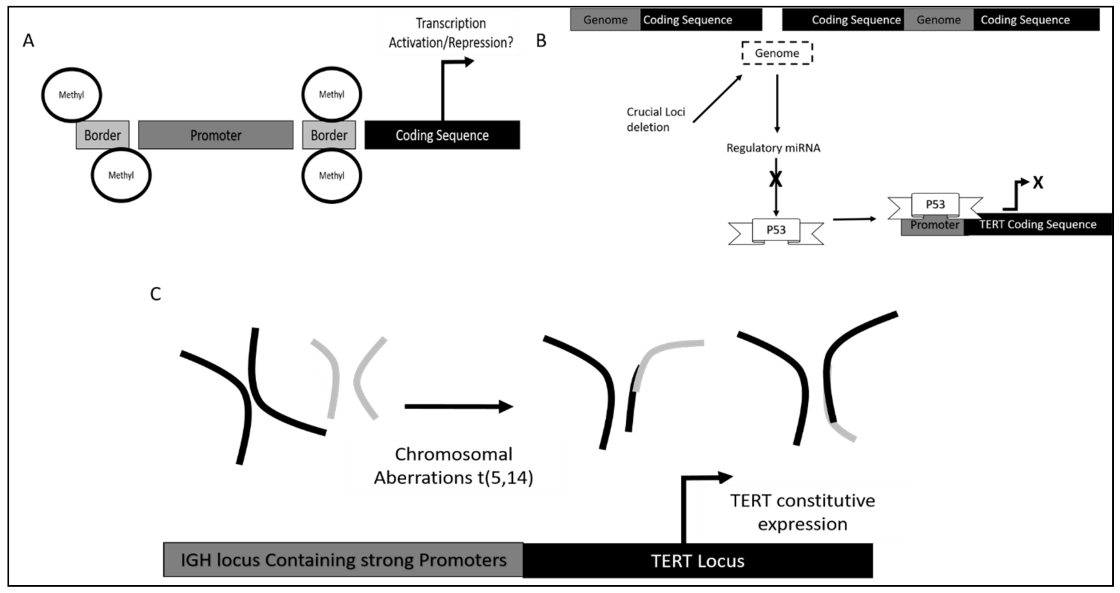
Mechanism of TERT dysregulation was reported to occur via TERT promoter methylation. However, contrary to canonical research, DNA methylation would increase TERT activity, as methylation occurs on the genomic region bordering the TERT promoter. This results in the inaccessibility of repressor on this site, which is proximal to the TERT promoter, thereby increasing the affinity of TERT promoter binding of crucial activators leading to TERT hyper-expression (Figure 1A). The observation mentioned above occurred when the CML model cell line K562 was treated with 5-azacytidine (DAC), a known DNA demethylase. Treatment reduced the methylation of TERT promoter regions, which are sites for CTCF (Transcription Repressor) binding. The result suggested an increase in CTCF binding, which could be seen from the reduction of C-Myc (Transcription Activator) binding and subsequent downregulation of TERT transcription. This further led to TERT reduction induced senescence in chronic myeloid leukemia [86].
Dysregulation of TERT also occur via gene amplification, which results in an increase in gene copy number (GCP). This phenomenon translates to increased gene expression levels due to relatively higher abundance of mRNA resulting from amplification (Figure 1B). It was observed that TERT catalytic components were amplified in patients with CML. Interestingly, TERC amplification does not correlate with resistance towards imatinib mesylate, although the copy number was calculated to be above 3 [87].
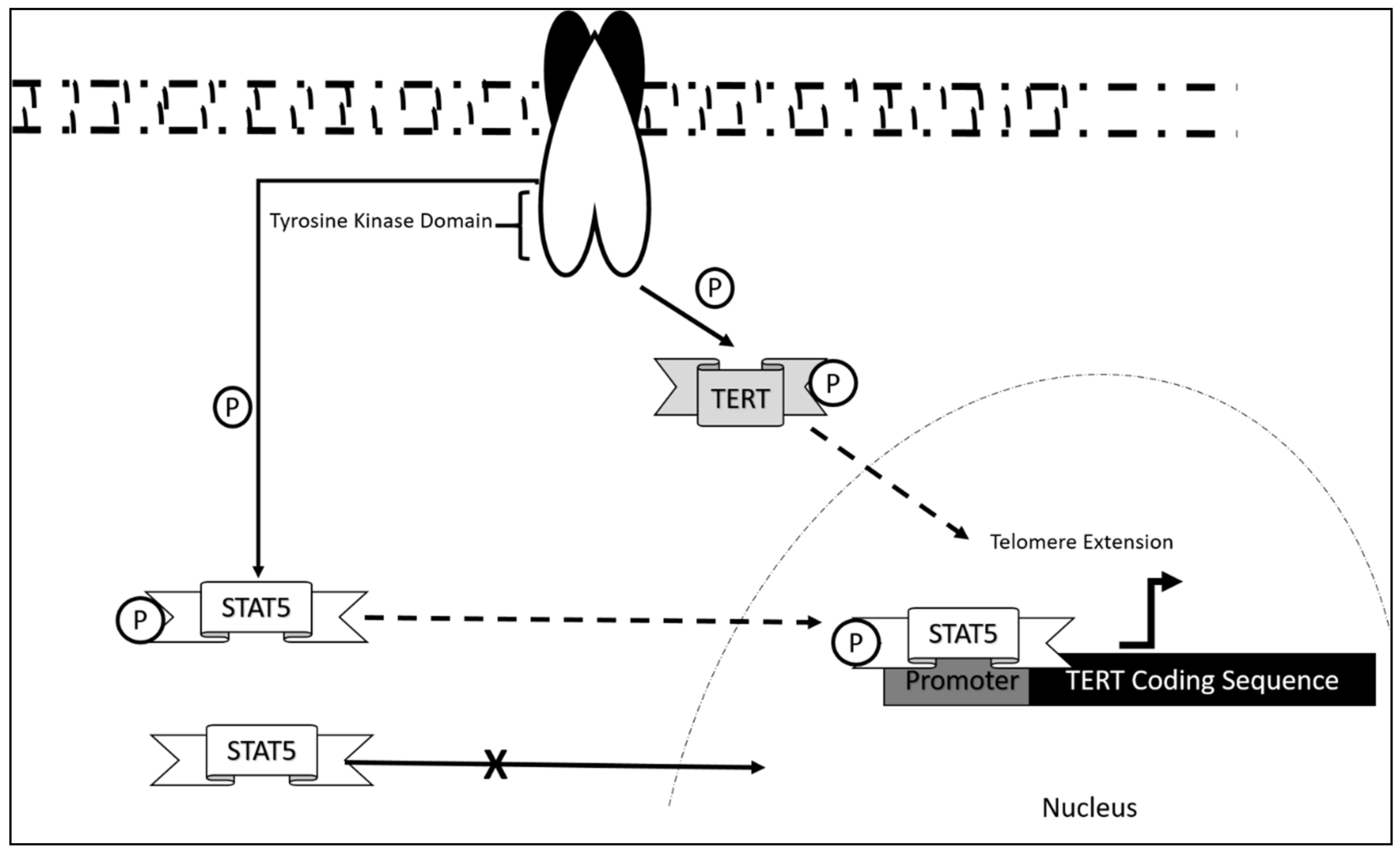
The CML fusion oncogene BCR-ABL was also found to contribute to the overall activity of TERT in aiding disease progression. It was reported that the fusion construct controls hTERT at different levels, namely at the nuclear entry level, direct TERT activity regulation, and TERT mRNA expression levels. BCR-ABL tyrosine kinase activity inhibition via Gleevec (Tyrosine Kinase Inhibitor) treatment resulted in the accumulation of TERT in the nucleoplasm when compared to the nontreated group in which TERT accumulated within the nucleolus. Furthermore, telomeric length was also found to be implicated following Gleevec treatment of CML cells. It was observed that K562 accumulates short telomeric ends (1 Kb<) when comparing cells treated with Gleevec to non-treated cells. The results were also consistent with hTERT phosphorylation levels where a decrease in phosphorylation states were observed post Gleevec treatment suggesting that hTERT activity is induced by BCR-ABL via direct hTERT phosphorylation. Additionally, transcription of hTERT was also found to be mediated via BCR-ABL tyrosine kinase activity as it was found that Gleevec treatment reduced hTERT mRNA abundance in K562. Further experimentation reported that, this was mediated via BCR-ABL activation of STAT5a through phosphorylation, which resulted in STAT5a-Phos binding to the TERT promoter. This was further confirmed via STAT5a inhibitor treatment, which significantly reduced TERT expression. Luciferase activity assay confirmed that the full length contains STAT5a binding as co-expression of the reporter with constitutive plasmid baring STAT5a resulted in over a twofold increase in hTERT expression [88]. Additionally, the same mechanism of STAT activation via BCR-ABL was also observed in CML treated with pimazode, which deactivated STAT5 by inhibiting tyrosine kinase activity of BCR-ABL [89].
TERT was targeted indirectly through upstream factors crucial for TERT activation via inhibition of kinase activity. Imatinib mesylate (IM) is currently one of the major drugs used in CML treatment. However, treatment of CML using IM was found to potentiate CML towards leukemogenesis as it was reported that it enhances expression of hTERT via phosphorylation of STAT5 [90] (Figure 2). Furthermore, knockdown of STAT5 attenuated leukemogenesis and increased cells sensitivity towards IM. It was also observed that patients with CML have different levels of phosphorylated STAT, further reducing the efficacy of IM. Further experimentation also reported that this resistance was conferred by the expression of MDR1 activation via STAT5 phosphorylation. Therefore, it is crucial to determine the expression levels of MDR1 and the phosphorylation state of STAT5 for proper treatment of IM in patients with CML [91].
Interestingly, it was also reported that CML cell line K562 with hTERT depletion does not result in telomere shortening post IM treatment. However proliferative potential of CML was significantly attenuated when telomerase depleted cells were treated with IM, further confirming the role of TERT in conferring treatment resistance towards IM [92]. However, treatment of CML with herbamycin A, also a tyrosine kinase inhibitor, was found to be ineffective in TERT suppression, which is likely due to the compensatory effects of TERT regulators. Nevertheless, herbamycin A treatment of K562 positive TERT cells post cyclin D1 transfection attenuated telomerase activity, while c-myc transfection resulted in partial restoration of hTERT activity [93]. It is therefore crucial to determine the mechanistics of TERT activators before IM prescription as differential expression of transcription factors are crucial in CML progression.
Since it is crucial to target TERT in senescence induction of CML, indirect regulation via IM would result in the ineffective inactivation of upstream factors as TERT is a downstream effector of BCR-ABL, STAT5, and c-MYc. Although the activity of these upstream factors could be effectively modulated by IM, it is of utmost importance to understand that TERT is not an exclusive target for these activators. Therefore, CML treatment with IM would be rendered ineffective via other downstream C-Myc or STAT5 target compensation. In this case, TERT direct downregulation could be synergistically employed along with IM treatment in order to improve CML treatment. Direct modulation of TERT could be employed via the use of BIBR1532, a potent TERT inhibitor which binds directly to TERT core preventing TERT binding onto telomeres. Although lacking studies in CML, it was proven that BIBR1532 is effective in suppressing disease progression in Burkitt’s lymphoma (BL) via direct TERT suppression. Co-treatment with fludarabine was also found to potentiate apoptosis in BL treated with BIBR1532 [94]. This synergistic approach of targeting TERT with BIBR1532 and IM could serve as a potential treatment for CML.

5.2. Chronic Lymphoid Leukemia (CLL)

The mechanism of dysregulation of TERT in CLL is still largely unexplored. However, correlation studies report that telomeric length serves as an initial prognostic marker in CLL disease progression [95]. This was also associated with telomerase/telomere associated proteins expression, which also contribute as markers in the diagnosis of CLL onset. Research showed that significant decrease in telomeric length was observed in chromosomes 13p, 12, 11p, and 17p in which hypomethylation was also present. These aberrations were significant in CLL patients as compared to normal controls. Upregulation of telomerase/telomere-associated proteins was also observed in patients at early disease onset [96]. As in the case of most leukemia, TERT activation occurs via STAT5 mediated binding and its phosphorylation state [89,97,98]. This mechanism, although not studied extensively in CLL, may be a contributing mechanism as to how CLL progress in addition to the upregulated telomerase/telomere-associated proteins and telomeric length dysregulation.
Furthermore, in terms of DNA methylation status it was found that methylation of TERT promoter was crucial in different stages of leukemia development as it confers different functional advantages. As mentioned earlier, telomeric shortening is a contributing factor in disease onset. It was found that this is crucial in telomerase overexpression as promoter methylation contributes to increase in initial telomeric activity in order to repair shortened telomerase during disease onset, a mechanism of immortalization in cancers [99]. This asserts the importance of TERT studies in CLL and its contributing mechanism via promoter methylation, which would allow us to better understand and properly treat this malignancy via direct TERT targeting. It is interesting to note that TERT dysregulation leads to disease development as previous studies in other types of leukemia associates this with chemo-resistance, disease progression, and poor prognosis [100,101].
Dysregulation of TERT in CLL was also observed via single nucleotide polymorphisms (SNPs). Different SNPS led to different potential in TERT induced self-renewal [102]. It was reported that SNPs rs2736100, rs2853690, rs33954691, and rs35033501 resulted in different outcomes in affected patients. SNP rs35033501 was found to be most prominent in patients bearing CLL and was associated with longer telomeres. This may implicate TERT hyperactivity in these SNPs bearing patients in which may contribute to disease development. The same report also mentioned that the rs2736100 SNP were associated with early stage of disease (I-II), while rs35033501 was associated with late stages. Whether this mutation is acquired following disease progression was not mentioned, however it is interesting to note that different SNPs result in different prognosis in patients [103].
Additionally, variation within the TERT and TERT C gene also contributed to an increase in telomeric ends. It was found that TERTC rs10936599 SNP with C allele had a significant association with CLL development as high allele frequency was observed in CLL patients. The same study also observed that TERT rs2736100 occurring in patients bearing CLL was associated with increased CLL risk [104]. How this contributes to leukemogenesis is still understudied but we could link this to an increase in TERT activity as observed in colorectal cancer [105].
Chromosomal aberrations were also found to contribute to TERT dysregulation in CLL. This includes the TERT/CLPTM1L locus in chromosome 5p15 and t(5,14) translocation, which resulted in the fusion of TERT to the IGH gene [106]. The IGH locus, which contains strong promoters, may have enhanced TERT activation as a significant increase in TERT expression was observed in CLL harboring this translocation [107]. Besides that, it was also found that deletion on chromosome 13 at locus q14 (13q14del) also contributed to better prognosis in patients. This locus contains the coding sequence for miR-15a/16-1 cluster which target TERT suppressor p53 [108,109]. The mechanism is illustrated in Figure 3.
In the case of drug studies against TERT in CLL it was found that targeting factors crucial in telomerase assembly may contribute to increase in sensitivity of CLL treatment with fludarabine. A study showed that using a telomerase inhibitor Imelstate (IS) with fludarabine does not lead to reduction in telomerase activity. However, another report on the association between NOP17 and telomerase assembly suggested that IS and fludarabine treatment dysregulate NOP17, resulting in impairment of TERT assembly at telomeric ends [110]. Furthermore, IS was also found to suppress telomerase activity, suggesting potential of IS treatment of CLL via synergy between chemo-drug and the compound [111]. Though studies on drug directly targeting TERT are not extensive, we believe that co-treatment may result in better efficacy of chemotherapy. Co-treatment may be done via directly targeting TERT as mentioned earlier in CML or by targeting indirect factors crucial of telomeric extension. For example, BRD4 inhibitor could significantly reduce telomeric ends length which may contribute to cellular senescence [112]. Using this drug alone may not significantly contribute to CLL inhibition, therefore co-treatment with chemo drug fludarabine may be applied which may result in a synergistic effect of CLL suppression as observed with IS. As the mechanisms of drug action on direct TERT regulation in CLL is not extensively studied, this field of study may be worth exploring in order to better understand TERT dysregulation in CLL.

5.3. TERT Dysregulation and Clinical Implications in Acute Leukemias

Acute leukemia refers to an aggressive form of leukemia that typically progresses rapidly over a short period of time. It can be further subdivided into acute myeloid (AML) and acute lymphoid leukemia (ALL) [113]. TERT dysregulation that has been identified as one of the contributors to the pathogenesis of leukemia and has been reported in both AML and ALL.

5.3.1. TERT Dysregulation and Clinical Implications in Acute Myeloid Leukemia

Acute myeloid leukemia (AML) is a type of leukemia that arises from the myeloid progenitor of hematopoiesis and gives rise to immature blasts in the bone marrow and/or peripheral blood. Lin et al. (2016) reported that TERT overexpression could promote the development of t(8;21) AML by working in tandem with AML-ETO possibly through two main mechanism: telomere integrity maintenance and inhibition of replicative senescence [114]. In addition, gene amplification which is a common characteristic of cancer malignancy was also detected in AML. TERT gene amplification was identified through fluorescence in-situ hybridization (FISH) in AML samples and was positively correlated with higher telomerase activity, which may contribute to AML leukemogenesis [80,115,116,117]. Single nucleotide variants (SNV) have also been identified as a common finding in cancers [118]. In a study by Mosrati et al. (2015), two TERT SNVs of CC genotypes (rs2853669 and rs2736100) that are associated with an increased risk of AML were identified [119]. rs2736100 is located at intron 2 of the TERT gene, whereas rs2853669 is positioned at the TERT promoter. Inflammation is one of the hallmarks of cancer and it was found that CC variant of rs2853669 was linked to increased level of inflammatory cytokines IL-6 and TNF-α, which may contribute to cancer malignancy. In the past decade, many studies have sought to identify TERT variants in various types of cancer, as these variants could be the driver of carcinogenesis. One of the most commonly found mutation in AML is G>A conversion in codon 1062 situated at exon 15 of the TERT gene, resulting in alanine>threonine substitution (A1062T) [120,121,122,123]. AML patients with A1062T TERT mutation had a relatively poorer prognosis as this mutation is associated with shorter overall survival, higher relapse incidence, and greater rate of treatment-related toxicity [120,121]. Epigenetic regulation of TERT has been the focus of many studies as it is believed to aid in the malignant transformation of cells [54]. Hypermethylation of the TERT promoter results in TERT transcription activation that subsequently leads to higher telomerase activity [124]. According to a study by Zhao et al. (2016), TERT proximal promoter and a partial exon 1 (TERTpro/Ex1) region exhibits CpG site hypermethylation in AML cell lines and primary blasts [125]. This distinct methylation profile could serve as a prognostic factor in AML as it is associated with shorter overall survival and drug resistance. Apart from the above regulation, TERT is also regulated by oncogenic signaling FLT3-ITD (FMS-Like Tyrosine Kinase 3 Internal Tandem Duplication). FLT3, a type of receptor tyrosine kinase is often mutated in AML with internal tandem duplications within the juxtamembrane domain. Compelling evidence has implicated the role of FLT3-ITD mutation in leukemogenesis especially in the case of AML [126]. Recently, it has come to light that FLT3-ITD mutation could induce the expression of TERT, resulting in increased telomerase activity that contributes to AML pathogenesis [127]. Another method of telomeric elongation identified in promyelocytic leukemia (PML) (a form of AML) is alternative lengthening of telomeres (ALT) [128]. The study by Osterwald et al. (2015) elucidated that the formation of nuclear bodies at telomeres leading to ALT were a result of concerted effects by 29 proteins involved in DNA repair, protein sumoylation, and organization of chromatins and telomeres. Changes mediated by these nuclear bodies include the increase in phosphorylated ataxia telangiectasia mutated kinase and stimulation of DNA damage responses, which promote maintenance of telomeres. Furthermore, in acute promyelocytic leukemia (APL), it was discovered that disease outcome correlated with alterations on the distal domain of the hTERT promoter [129]. In this study, hypomethylation and deposition of H3K4Me3 at the approximate region of 5kb upstream of the transcription start site was associated with activation of hTERT. These results, therefore, point to the combinatory interaction of hTERT promoter methylation, histone modifications, and chromatin accessibility in dysregulation of hTERT transcription. A study on childhood AML further delineated that there exist a correlation between telomerase activity and methylation of cyclin-dependent kinase inhibitor 2B (CDKN2B) CpG islands [130]. It was revealed that in the cohort of patients with methylated CDKN2B, their mean levels of telomerase were 49.09 ± 44.43 total product generated (TPG), as compared to 29.99 ± 32.43 TPG in patients without CDKN2B methylation. This finding suggests that hypermethylation in CDKN2B CpG islands is positively linked to the increase of telomerase expression.
Conventional chemotherapy is one of the therapeutic approaches used to treat AML patients, whereby it is common that a combination of chemotherapeutic drugs is used. Each of the chemotherapeutic drug aims to target and inhibit the cancer cells via various mechanisms. One of the mechanisms targeted by a commonly used drug for AML, 5-azacytidine (5-AZA) is DNA methylation. 5-AZA, a DNA methyltransferase inhibitor (DNMTI), has been shown to downregulate TERT expression and decrease telomerase activity in AML patient samples and cell lines [131]. Similarly, another study reported that DNMTI 5-Aza-2′-deoxycytidine (DAC) treatment on AML patient samples and cell lines demonstrated a reduction in TERT expression and telomerase activity [132]. However, the mechanism by which 5-AZA and DAC regulates TERT expression in AML has not been fully described to date.
In addition, small molecule inhibitors have been emerging as potential therapeutic alternatives to treat AML patients. This is exemplified by Triptonide (diterpene triepoxide), a small biological molecule derived from the herb Tripterygium wilfordii, that demonstrated antileukemic activity by inhibiting c-Myc transcription and its downstream target, TERT in AML cells [133]. It is also important to note that triptonide not only suppressed the expression of senescence-inhibiting genes but also activated the expression of senescence-promoting genes, such as P16 (CDKN2A) and P21 (CDKN1A). Thus, triptonide is known as “multiple-hits” senescence-promoting agent. Furthermore, a tyrosine kinase inhibitor (TKI), PKC412 exerts its inhibitory action on TERT via FLT3-ITD in a c-Myc dependent manner that functions as the transcription factor of TERT [127].
TERT-targeted immunotherapy has also become the subject of many cancer therapeutic studies. A study by Sandri et al. (2017) demonstrated that TERT865-873-specific, TCR-engineered T-cell lymphocytes were able to halt AML progression in vivo [134]. The main idea behind this therapy, known as adoptive T-cell therapy (ACT), is to increase the affinity and specificity of T-cell receptor (TCR) of T cells to tumor associated antigen (TAA). It is rather interesting to note that TERT865-873-specific, TCR-engineered T-cell lymphocytes were able to distinguish between AML blasts and peripheral blood mononuclear cells (PBMC), indicating its high cell recognition specificity. Khoury et al. (2017) found that treatment of AML patients with hTERT expressing autologous dendritic cells (TERT-DC) was able to improve their overall survival rate associated with favorable recurrence-free survival [135]. hTERT-DC is comprised of dendritic cells (DC) coupled with mRNA-encoding telomerase (hTERT) and the lysosomal targeting signal 38 lysosomal-associated membrane protein (LAMP). Thus, it can be seen that TERT-specific immunotherapies are an effective therapeutic approach in combating AML.

5.3.2. TERT Dysregulation and Clinical Implications in Acute Lymphoblastic Leukemia

Acute lymphoblastic leukemia (ALL) is a type of leukemia that arises from the lymphoid progenitor of hematopoiesis and gives rise to immature blasts in the bone marrow and/or peripheral blood. Previous studies have reported that the expression of TERT is generally upregulated in ALL. This indicates that TERT overexpression could play a role in leukemogenesis [116,136]. TERT gene amplification was detected through FISH analysis in ALL patient samples coupled with increase in telomerase activity [116]. Single nucleotide polymorphism (SNP) is present at a frequency greater than 1% in the general population [137]. Sheng et al. (2013) discovered that in the TERT promoter region, SNP, rs2735940 with C to T substitution, is able to induce TERT expression by upregulating its transcriptional activity in ALL [138]. Not surprisingly, this specific SNP is associated with increased risk of pediatric ALL among Chinese population thus making it as a biomarker in ALL diagnosis. However, contradicting result was reported by Eskandari et al. (2018) whereby it was found that TERT promoter region SNP, rs2735940 with C to T substitution, does not increase the risk of ALL [139]. Furthermore, epigenetic regulation of TERT in pediatric ALL has been demonstrated by Borssén et al. (2011) [140]. TERT promoters in 24% of ALL patient samples shows greater methylation pattern as compared to normal bone marrow samples. However, findings from this study have established that there is a negative correlation between TERT promoter methylation status and TERT gene expression. It is also important to note that the incidence of methylation is higher in T cell-ALL than B-cell precursor ALL, indicating that TERT promoter methylation can be used to differentiate between specific ALL subtypes.
TERT-targeted therapies are quickly gaining popularity as a therapeutic option due to the role of TERT as a universal tumor associated antigen (TAA). BIBR1532, a small biological molecule specific for telomerase inhibition, has been studied by Bashash et al. (2017) for its application in pre-B ALL [141]. BIBR1532 was able to suppress TERT expression and telomerase activity in vitro in a dose-dependent manner. In addition, BIBR1532 suppressed the expression of c-Myc that acts as a transcriptional activator of TERT. Doxorubicin, a commonly used chemotherapeutic drug, was found to exert the same inhibitory action as BIBR1532 on TERT and c-Myc. This study also reported that the combination of doxorubicin together with BIBR1532 showed a greater inhibition of TERT and c-Myc transcription as compared to its individual counterparts. Thus, this combination therapy can be an effective therapeutic approach for pre-B ALL.
TERT-targeted immunotherapy is being studied for its application in ALL. According to a study by Sandri et al. (2017), TERT865-873-specific, TCR-engineered T-cell lymphocytes were able to halt ALL progression in vivo [134]. Similar to AML, hTERT865-873-specific, TCR-engineered T-cell lymphocytes were able to distinguish between ALL blasts and B cells from peripheral blood mononuclear cells (PBMC). Therefore, it can be concluded that TERT specific T cells have shown promising outcomes as next generation treatments for ALL.

6. Challenges and Opportunities in Targeting hTERT in Cancer Therapy

To date, a vast majority of experimental treatments against hTERT have not been able to display promising clinical benefits in studies on leukemia. This is primarily due to several unresolved issues, including the absence of relevant animal models, the inability to obtain high resolution molecular structure of the human telomerase enzyme, and the development of resistance towards inhibitor-based therapies [142].
The challenge in developing mouse models for targeting TERT mainly stems from global expression of telomerase in adult somatic cells [143]. Currently available strains exhibit far longer telomeres than observed in humans and would prove unsuitable for replicative senescence studies relating to TERT.
Elucidation of the molecular structure of telomerase has also been hampered by failure to isolate the enzyme in large quantities due to low cellular abundance, which therefore impedes downstream purification and crystallographic works [142]. This has inevitably hindered mechanistic studies and drug design efforts.
In addition, studies have revealed that drugs targeting hTERT require extended durations to manifest clinical effects as they rely on the attrition of telomeres to arrest the cell cycle [142]. Off target effects could also affect stem and progenitor cells which express hTERT. Furthermore, malignant cells may also escape treatment due to their ability to survive with low levels of hTERT and potential to evolve resistant clones, such as adapting telomere maintenance to the ALT pathway as earlier mentioned, before treatment becomes effective.
In order to address the aforementioned pitfalls, potential strategies have been improvised to overcome each of the shortcomings. TERT inhibitors with high potency, which are capable of fully depleting telomerase activity, have been proposed to exert greater selective pressure and overcome resistance in cancer cells [142]. This strategy may also be more effective against leukemic cells with characteristically shorter telomeres as replicative senescence could be manifested sooner. Furthermore, increasing efforts have also been made to acquire higher resolution molecular structure of TERT including a recent study employing cryo-electron microscopy which successfully identified that human telomerase is able to take on a monomeric structure when bound to its substrate [144]. In terms of preclinical studies, it has been proposed that using patient xenografts derived from early passages could work as an effective model which more closely recapitulate the genetic complexity in human cancers. This would also be able to overcome discrepancies arising from species-specific TERT expression [142].

7. Approaches Targeting Telomerase in Preclinical and Clinical Trials

7.1. Immunotherapies

The revelation that cancer cells express TERT epitopes as a surface marker enabled the development of immunotherapies to target telomerase [145]. The devised strategies thus far include vaccines, adoptive cell transfer, and oncolytic viruses.
TERT-based vaccines rely largely on eliciting recognition and adaptive immune responses in T-cells against telomerase epitopes via MHC class I or II molecules [142]. A phase I/IIa clinical trial in patients with metastatic hormone-naïve prostate cancer demonstrated that the UV1 TERT vaccine was able to elicit measurable immune responses in 18/21 or 85.7% of the cohort [146]. Noteworthy, in 45% of patients, MRI indicated no persisting tumor in the prostatic gland. However, in another phase III trial on advanced pancreatic cancer patients, the TERT vaccine GV1001 did not display significant survival advantage as compared to chemotherapy [147]. These trials exemplify that TERT vaccines alone may not be sufficient in controlling disease progression. More recently, a preclinical study evaluating the co-administration of TERT vaccine with immune checkpoint blockade (anti-CTLA-4) has revealed that a synergistic effect could be achieved, resulting in suppression of tumor growth accompanied by increased survival (Table 1) [148]. It was also highlighted that checkpoint blockade alone or vaccine alone were far from achieving the outcome of the combined treatment, thus rationalizing further development of this strategy.
Adoptive cell therapy was developed to use the patient’s own T lymphocytes with anti-cancer activities (either primed or engineered) in an ex vivo expanded manner. Recent studies have shown that adoptive cell transfer with high avidity telomerase-specific T-cells were able to suppress prostate cancer progression and improve survival of preclinical cancer models [149]. However, autoimmunity was present as evident by depletion of B-cell, albeit the transient observation. Further studies on AML/CML demonstrated that T-cells engineered to express receptors with high avidity to hTERT resulted in suppression of the disease in humanized mouse models [150].
Oncolytic adenoviruses such as telomelysin have been developed to infect and replicate selectively in malignant cells through the control of hTERT promoter driven E1 gene expression [142]. Preclinical assessments found that treatment with telomelysin repressed xenographic gastrointestinal tumor growth via induction of cancer cell death [151]. Additionally, it was also found in the same study that non-immunogenic cancer cells were sensitized to anti-PD1 immunotherapy upon treatment with telomelysin. This finding justifies further investigations into combinational therapy of oncolytic viruses with immune checkpoint blockades.

7.2. Oligonucleotide Inhibitors

Oligonucleotide inhibitors such as imetelstat work by complementary binding to the TERC template, thereby acting through competitive inhibition of telomerase. Its mechanism of action lead to attrition of telomeres, which prompts DNA damage response and ensuing cell death. However, hematological side effects were observed in a dose-dependent manner when imetelstat was administered into patients with solid tumors [152]. A later phase II clinical trial further found that progression-free survival (PFS) and overall survival (OS) were not improved with treatment [153]. Despite these findings, it was observed that patients with the shortest telomeres benefited from imetelstat as indicated by a trend towards improving survival. Accordingly, imetelstat has been repurposed for myeloproliferative disorders such as myelofibrosis, which are characteristically known to have shorter telomeres [111].

7.3. Small Molecule Inhibitors

BIBR1532 is a small molecule inhibitor that works by non-competitive binding to a conserved hydrophobic pocket (FVYL motif), which impairs interactions between TERT and TERC [154]. Treatment with BIBR1532 has produced promising preclinical results despite requiring extended treatment due to its reliance on telomere shortening to induce replicative senescence and characteristically poor pharmacokinetics. Moreover, treatment on leukemic cells lead to observation of acute cytotoxicity which was found independent of telomere shortening. Further investigations revealed the mechanism of action involved p53 dependent DNA damage response, which mediated rapid induction of telomeric dysfunction [155].

7.4. Indirect Inhibitors

7.4.1. G-Quadruplex Stabilizers

Telomeric regions rich in guanine have been observed to form secondary structures known as G-quadruplex via Hoogsteen hydrogen bonding. G-quadruplex stabilizers are able to prevent access of DNA helicases to telomeres and therefore increase propensity towards DNA damage response and subsequent cell death [156]. One such drug tested in preclinical studies using leukemic xenograft models is telomestatin which have shown to inhibit telomerase activity, increased apoptosis of cancer cells and reduced the size of tumors [156]. However, concerns have been raised about the safety of G-quadruplex stabilizers as it has been found through computational prediction that the human genome could form more than 300,000 G-quadruplex structures at various sites including non-telomeric regions [157]. Further studies in this area should endeavor to elucidate the conformation of G-quadruplex in telomeres and design specific G-quadruplex ligands that selectively target telomeric regions [158].

7.4.2. Nucleoside Analogues

Nucleoside analogues including 5-fluoro-2′-deoxyuridine thiophosphate (5-FdU) and 6-thio-2′-deoxyguanosine (6-thio-dG) function as uncapping agents which prevent recruitment and formation of the shelterin complex on telomeres [159,160]. The incorporation of these analogues into the telomeric structure induces dysfunction of the telomere ends leading to activation of the DNA damage response. Nucleoside analogues have been shown to induce minimal side effects on normal human cells whilst exerting potent cytotoxicity on xenografts of lung cancer [161], medulloblastoma [162], and melanoma [163] in preclinical models. Considering these agents function independently of telomere lengths, their use is not limited by heterogeneity of this factor found in most cancer cells [142]. Furthermore, it has been discovered that nucleoside analogues acutely induce apoptosis in transformed cells which excludes prolonged use and undesired suppression of telomerase in stem and progenitor cells. However, due to its rapid response, neutropenia has been documented [159]. Further, it has been found that 6-thio-dG also causes mitochondrial antioxidant adaptive response, which is able to prevent apoptosis in melanoma cells [164]. This effect can be counteracted by including mitochondrial Hsp90 inhibitor alongside nucleoside analogue treatment to induce higher apoptotic effect in cancer cells.

8. Conclusions and Future Perspectives

In summary, many preclinical and clinical studies on leukemia have concluded that TERT plays a pivotal role in disease progression and deteriorating prognosis of patients. Studies aimed at determining the factors influencing TERT expression during leukemogenesis and the stage where this gene is upregulated has contributed immensely to the understanding of the fundamental roles of TERT in various forms of leukemias. Additionally, it has also been proven that different mechanisms are involved in the expression of this gene including transcriptional, genetic, and epigenetic regulators. These mechanisms of activation and dysregulation of TERT in onco-hematological diseases are progressively being elucidated and in recent years, researchers have deciphered many novel variants, modifications, pathways, and mediators leading to its overexpression. The latest findings have identified mutations in the hTERT gene, methylation of its promoter, and microRNAs targeting its transcript as leading areas of fundamental studies and highly promising for therapeutic development. Indeed, progress in clinical trials have determined that chemical drugs and biomolecules targeting various regulators of TERT have immense potential in controlling the proliferation and metastasis of leukemias, primarily via arrest of cellular division. Noteworthy, in recent years there have been renewed interests in targeting hTERT as a therapeutic strategy due to developments in several areas including immunotherapies, drug discovery, gene therapy, and radiotherapy. Combination of agents specific to TERT and other anti-cancer drugs have also been widely explored with many studies indicating synergistic effects, reduced dose requirements and decreased side effects (Table 1). The survival outcomes of leukemic patients have improved in recent years, and it is expected that therapies targeting TERT and their combinations with other anti-cancer agents could further improve the prognosis of patients especially those with TERT-dependent leukemias.

Author Contributions

Conceptualization, E.J.M., M.Y.Y. and N.M.Y.; Writing—Original Draft Preparation, E.J.M., M.Y.Y., A.A., Y.R. and A.R.; Writing—Review and Editing, E.J.M., M.Y.Y. and N.M.Y.; Supervision, E.J.M. and N.M.Y. All authors have read and agreed to the published version of the manuscript.

Funding

Studies related to this topic were funded and supported by Fundamental Research Grant Scheme (FRGS) 203/CIPPT/6711672 from Ministry of Higher Education (MOHE), Malaysia and Universiti Sains Malaysia Research Grant, USM–RUI: 1001/CIPPT/8012265.

Institutional Review Board Statement

Not applicable.

Informed Consent Statement

Not applicable.

Data Availability Statement

Not applicable.

Conflicts of Interest

The authors declare no conflict of interest.

References

  1. Cong, Y.-S.; Wright, W.E.; Shay, J.W. Human Telomerase and Its Regulation. Microbiol. Mol. Biol. Rev. 2002, 66, 407–425. [Google Scholar] [CrossRef] [Green Version]
  2. Ly, H. Telomere Dynamics in Induced Pluripotent Stem Cells: Potentials for Human Disease Modeling. World J. Stem Cells 2011, 3, 89–95. [Google Scholar] [CrossRef] [PubMed]
  3. Kelleher, C.; Teixeira, M.T.; Förstemann, K.; Lingner, J. Telomerase: Biochemical Considerations for Enzyme and Substrate. Trends Biochem. Sci. 2002, 27, 572–579. [Google Scholar] [CrossRef]
  4. Cifuentes-Rojas, C.; Shippen, D.E. Telomerase Regulation. Mutat. Res. 2012, 730, 20–27. [Google Scholar] [CrossRef] [Green Version]
  5. Depcrynski, A.N.; Sachs, P.C.; Elmore, L.W.; Holt, S.E. Regulation of Telomerase Through Transcriptional and Posttranslational Mechanisms. In Telomeres and Telomerase in Cancer; Cancer Drug Discovery and Development; Hiyama, K., Ed.; Humana Press: Totowa, NJ, USA, 2009; pp. 47–85. ISBN 978-1-60327-879-9. [Google Scholar]
  6. Dwyer, J.; Li, H.; Xu, D.; Liu, J.-P. Transcriptional Regulation of Telomerase Activity: Roles of the the Ets Transcription Factor Family. Ann. N. Y. Acad. Sci. 2007, 1114, 36–47. [Google Scholar] [CrossRef]
  7. Relitti, N.; Saraswati, A.P.; Federico, S.; Khan, T.; Brindisi, M.; Zisterer, D.; Brogi, S.; Gemma, S.; Butini, S.; Campiani, G. Telomerase-Based Cancer Therapeutics: A Review on Their Clinical Trials. Curr. Top. Med. Chem. 2020, 20, 433–457. [Google Scholar] [CrossRef]
  8. Bernal, A.; Tusell, L. Telomeres: Implications for Cancer Development. Int. J. Mol. Sci. 2018, 19, 294. [Google Scholar] [CrossRef] [PubMed] [Green Version]
  9. Ramlee, M.K.; Wang, J.; Toh, W.X.; Li, S. Transcription Regulation of the Human Telomerase Reverse Transcriptase (HTERT) Gene. Genes 2016, 7, 50. [Google Scholar] [CrossRef] [PubMed]
  10. Garbuzov, A.; Pech, M.F.; Hasegawa, K.; Sukhwani, M.; Zhang, R.J.; Orwig, K.E.; Artandi, S.E. Purification of GFRα1+ and GFRα1- Spermatogonial Stem Cells Reveals a Niche-Dependent Mechanism for Fate Determination. Stem Cell Rep. 2018, 10, 553–567. [Google Scholar] [CrossRef] [Green Version]
  11. Jaiswal, R.K.; Kumar, P.; Kumar, M.; Yadava, P.K. HTERT Promotes Tumor Progression by Enhancing TSPAN13 Expression in Osteosarcoma Cells. Mol. Carcinog. 2018, 57, 1038–1054. [Google Scholar] [CrossRef]
  12. Lin, S.; Nascimento, E.M.; Gajera, C.R.; Chen, L.; Neuhöfer, P.; Garbuzov, A.; Wang, S.; Artandi, S.E. Distributed Hepatocytes Expressing Telomerase Repopulate the Liver in Homeostasis and Injury. Nature 2018, 556, 244–248. [Google Scholar] [CrossRef] [PubMed]
  13. Leão, R.; Apolónio, J.D.; Lee, D.; Figueiredo, A.; Tabori, U.; Castelo-Branco, P. Mechanisms of Human Telomerase Reverse Transcriptase (HTERT) Regulation: Clinical Impacts in Cancer. J. Biomed. Sci. 2018, 25, 22. [Google Scholar] [CrossRef]
  14. Xia, F.; Yuan, Y.; Chen, R.; Liu, P.; Zhou, H.; Wang, J. Study of telomerase activity and its related genes expression in leukemia cells. Zhonghua Xue Ye Xue Za Zhi 2001, 22, 86–89. [Google Scholar] [PubMed]
  15. Bruedigam, C.; Lane, S.W. Telomerase in Hematologic Malignancies. Curr. Opin. Hematol. 2016, 23, 346–353. [Google Scholar] [CrossRef]
  16. Davison, G.M. Telomeres and Telomerase in Leukaemia and Lymphoma. Transfus. Apher. Sci. 2007, 37, 43–47. [Google Scholar] [CrossRef]
  17. Keller, G.; Brassat, U.; Braig, M.; Heim, D.; Wege, H.; Brümmendorf, T.H. Telomeres and Telomerase in Chronic Myeloid Leukaemia: Impact for Pathogenesis, Disease Progression and Targeted Therapy. Hematol. Oncol. 2009, 27, 123–129. [Google Scholar] [CrossRef] [PubMed]
  18. Lansdorp, P.M. Maintenance of Telomere Length in AML. Blood Adv. 2017, 1, 2467–2472. [Google Scholar] [CrossRef]
  19. Deville, L.; Hillion, J.; Ségal-Bendirdjian, E. Telomerase Regulation in Hematological Cancers: A Matter of Stemness? Biochim. Biophys. Acta 2009, 1792, 229–239. [Google Scholar] [CrossRef] [PubMed] [Green Version]
  20. Pech, M.F.; Garbuzov, A.; Hasegawa, K.; Sukhwani, M.; Zhang, R.J.; Benayoun, B.A.; Brockman, S.A.; Lin, S.; Brunet, A.; Orwig, K.E.; et al. High Telomerase Is a Hallmark of Undifferentiated Spermatogonia and Is Required for Maintenance of Male Germline Stem Cells. Genes Dev. 2015, 29, 2420–2434. [Google Scholar] [CrossRef] [Green Version]
  21. Montgomery, R.K.; Carlone, D.L.; Richmond, C.A.; Farilla, L.; Kranendonk, M.E.G.; Henderson, D.E.; Baffour-Awuah, N.Y.; Ambruzs, D.M.; Fogli, L.K.; Algra, S.; et al. Mouse Telomerase Reverse Transcriptase (MTert) Expression Marks Slowly Cycling Intestinal Stem Cells. Proc. Natl. Acad. Sci. USA 2011, 108, 179–184. [Google Scholar] [CrossRef] [PubMed] [Green Version]
  22. Chiba, K.; Johnson, J.Z.; Vogan, J.M.; Wagner, T.; Boyle, J.M.; Hockemeyer, D. Cancer-Associated TERT Promoter Mutations Abrogate Telomerase Silencing. Elife 2015, 4, e07918. [Google Scholar] [CrossRef]
  23. Lin, S.Y.; Elledge, S.J. Multiple Tumor Suppressor Pathways Negatively Regulate Telomerase. Cell 2003, 113, 881–889. [Google Scholar] [CrossRef] [Green Version]
  24. Parodi, S.; Ognibene, M.; Haupt, R.; Pezzolo, A. The Over-Expression of E2F3 Might Serve as Prognostic Marker for Neuroblastoma Patients with Stage 4S Disease. Diagnostics 2020, 10, 315. [Google Scholar] [CrossRef] [PubMed]
  25. Fujimoto, K.; Kyo, S.; Takakura, M.; Kanaya, T.; Kitagawa, Y.; Itoh, H.; Takahashi, M.; Inoue, M. Identification and Characterization of Negative Regulatory Elements of the Human Telomerase Catalytic Subunit (HTERT) Gene Promoter: Possible Role of MZF-2 in Transcriptional Repression of HTERT. Nucleic Acids Res. 2000, 28, 2557–2562. [Google Scholar] [CrossRef]
  26. Günes, C.; Lichtsteiner, S.; Vasserot, A.P.; Englert, C. Expression of the HTERT Gene Is Regulated at the Level of Transcriptional Initiation and Repressed by Mad1. Cancer Res. 2000, 60, 2116–2121. [Google Scholar] [PubMed]
  27. Gabet, A.-S.; Mortreux, F.; Charneau, P.; Riou, P.; Duc-Dodon, M.; Wu, Y.; Jeang, K.-T.; Wattel, E. Inactivation of HTERT Transcription by Tax. Oncogene 2003, 22, 3734–3741. [Google Scholar] [CrossRef] [Green Version]
  28. Kanaya, T.; Kyo, S.; Hamada, K.; Takakura, M.; Kitagawa, Y.; Harada, H.; Inoue, M. Adenoviral Expression of P53 Represses Telomerase Activity through Down-Regulation of Human Telomerase Reverse Transcriptase Transcription. Clin. Cancer Res. 2000, 6, 1239–1247. [Google Scholar] [PubMed]
  29. Lee, S.-H.; Kim, J.-W.; Oh, S.-H.; Kim, Y.-J.; Rho, S.-B.; Park, K.; Park, K.-L.; Lee, J.-H. IFN-Gamma/IRF-1-Induced P27kip1 down-Regulates Telomerase Activity and Human Telomerase Reverse Transcriptase Expression in Human Cervical Cancer. FEBS Lett. 2005, 579, 1027–1033. [Google Scholar] [CrossRef] [Green Version]
  30. Lacerte, A.; Korah, J.; Roy, M.; Yang, X.-J.; Lemay, S.; Lebrun, J.-J. Transforming Growth Factor-Beta Inhibits Telomerase through SMAD3 and E2F Transcription Factors. Cell. Signal. 2008, 20, 50–59. [Google Scholar] [CrossRef]
  31. Oh, S.; Song, Y.H.; Yim, J.; Kim, T.K. Identification of Mad as a Repressor of the Human Telomerase (HTERT) Gene. Oncogene 2000, 19, 1485–1490. [Google Scholar] [CrossRef] [Green Version]
  32. Xu, D.; Popov, N.; Hou, M.; Wang, Q.; Björkholm, M.; Gruber, A.; Menkel, A.R.; Henriksson, M. Switch from Myc/Max to Mad1/Max Binding and Decrease in Histone Acetylation at the Telomerase Reverse Transcriptase Promoter during Differentiation of HL60 Cells. Proc. Natl. Acad. Sci. USA 2001, 98, 3826–3831. [Google Scholar] [CrossRef] [PubMed] [Green Version]
  33. Sikand, K.; Kaul, D.; Varma, N. Receptor Ck-Dependent Signaling Regulates HTERT Gene Transcription. BMC Cell Biol. 2006, 7, 2. [Google Scholar] [CrossRef] [PubMed] [Green Version]
  34. Xiong, J.; Fan, S.; Meng, Q.; Schramm, L.; Wang, C.; Bouzahza, B.; Zhou, J.; Zafonte, B.; Goldberg, I.D.; Haddad, B.R.; et al. BRCA1 Inhibition of Telomerase Activity in Cultured Cells. Mol. Cell. Biol. 2003, 23, 8668–8690. [Google Scholar] [CrossRef] [Green Version]
  35. Xu, D.; Wang, Q.; Gruber, A.; Björkholm, M.; Chen, Z.; Zaid, A.; Selivanova, G.; Peterson, C.; Wiman, K.G.; Pisa, P. Downregulation of Telomerase Reverse Transcriptase MRNA Expression by Wild Type P53 in Human Tumor Cells. Oncogene 2000, 19, 5123–5133. [Google Scholar] [CrossRef] [PubMed] [Green Version]
  36. Li, H.; Xu, D.; Li, J.; Berndt, M.C.; Liu, J.-P. Transforming Growth Factor Beta Suppresses Human Telomerase Reverse Transcriptase (HTERT) by Smad3 Interactions with c-Myc and the HTERT Gene. J. Biol. Chem. 2006, 281, 25588–25600. [Google Scholar] [CrossRef] [Green Version]
  37. Yang, H.; Kyo, S.; Takatura, M.; Sun, L. Autocrine Transforming Growth Factor Beta Suppresses Telomerase Activity and Transcription of Human Telomerase Reverse Transcriptase in Human Cancer Cells. Cell Growth Differ. 2001, 12, 119–127. [Google Scholar] [PubMed]
  38. Endoh, T.; Tsuji, N.; Asanuma, K.; Yagihashi, A.; Watanabe, N. Survivin Enhances Telomerase Activity via Up-Regulation of Specificity Protein 1- and c-Myc-Mediated Human Telomerase Reverse Transcriptase Gene Transcription. Exp. Cell Res. 2005, 305, 300–311. [Google Scholar] [CrossRef]
  39. Katzenellenbogen, R.A.; Egelkrout, E.M.; Vliet-Gregg, P.; Gewin, L.C.; Gafken, P.R.; Galloway, D.A. NFX1-123 and Poly(A) Binding Proteins Synergistically Augment Activation of Telomerase in Human Papillomavirus Type 16 E6-Expressing Cells. J. Virol. 2007, 81, 3786–3796. [Google Scholar] [CrossRef] [PubMed] [Green Version]
  40. McMurray, H.R.; McCance, D.J. Degradation of P53, Not Telomerase Activation, by E6 Is Required for Bypass of Crisis and Immortalization by Human Papillomavirus Type 16 E6/E7. J. Virol. 2004, 78, 5698–5706. [Google Scholar] [CrossRef] [Green Version]
  41. Crowe, D.L.; Nguyen, D.C. Rb and E2F-1 Regulate Telomerase Activity in Human Cancer Cells. Biochim. Biophys. Acta 2001, 1518, 1–6. [Google Scholar] [CrossRef]
  42. Won, J.; Yim, J.; Kim, T.K. Opposing Regulatory Roles of E2F in Human Telomerase Reverse Transcriptase (HTERT) Gene Expression in Human Tumor and Normal Somatic Cells. FASEB J. 2002, 16, 1943–1945. [Google Scholar] [CrossRef] [Green Version]
  43. Chang, J.T.-C.; Yang, H.-T.; Wang, T.-C.V.; Cheng, A.-J. Upstream Stimulatory Factor (USF) as a Transcriptional Suppressor of Human Telomerase Reverse Transcriptase (HTERT) in Oral Cancer Cells. Mol. Carcinog. 2005, 44, 183–192. [Google Scholar] [CrossRef]
  44. Goueli, B.S.; Janknecht, R. Regulation of Telomerase Reverse Transcriptase Gene Activity by Upstream Stimulatory Factor. Oncogene 2003, 22, 8042–8047. [Google Scholar] [CrossRef] [PubMed] [Green Version]
  45. Kyo, S.; Takakura, M.; Taira, T.; Kanaya, T.; Itoh, H.; Yutsudo, M.; Ariga, H.; Inoue, M. Sp1 Cooperates with C-Myc to Activate Transcription of the Human Telomerase Reverse Transcriptase Gene (HTERT). Nucleic Acids Res. 2000, 28, 669–677. [Google Scholar] [CrossRef] [PubMed] [Green Version]
  46. Verma, S.C.; Borah, S.; Robertson, E.S. Latency-Associated Nuclear Antigen of Kaposi’s Sarcoma-Associated Herpesvirus up-Regulates Transcription of Human Telomerase Reverse Transcriptase Promoter through Interaction with Transcription Factor Sp1. J. Virol. 2004, 78, 10348–10359. [Google Scholar] [CrossRef] [Green Version]
  47. Won, J.; Yim, J.; Kim, T.K. Sp1 and Sp3 Recruit Histone Deacetylase to Repress Transcription of Human Telomerase Reverse Transcriptase (HTERT) Promoter in Normal Human Somatic Cells. J. Biol. Chem. 2002, 277, 38230–38238. [Google Scholar] [CrossRef] [PubMed] [Green Version]
  48. Beitzinger, M.; Oswald, C.; Beinoraviciute-Kellner, R.; Stiewe, T. Regulation of Telomerase Activity by the P53 Family Member P73. Oncogene 2006, 25, 813–826. [Google Scholar] [CrossRef] [PubMed]
  49. Racek, T.; Mise, N.; Li, Z.; Stoll, A.; Pützer, B.M. C-Terminal P73 Isoforms Repress Transcriptional Activity of the Human Telomerase Reverse Transcriptase (HTERT) Promoter. J. Biol. Chem. 2005, 280, 40402–40405. [Google Scholar] [CrossRef] [PubMed] [Green Version]
  50. Toh, W.H.; Kyo, S.; Sabapathy, K. Relief of P53-Mediated Telomerase Suppression by P73. J. Biol. Chem. 2005, 280, 17329–17338. [Google Scholar] [CrossRef] [Green Version]
  51. Guzman, H.; Sanders, K.; Idica, A.; Bochnakian, A.; Jury, D.; Daugaard, I.; Zisoulis, D.G.; Pedersen, I.M. MiR-128 Inhibits Telomerase Activity by Targeting TERT MRNA. Oncotarget 2018, 9, 13244–13253. [Google Scholar] [CrossRef] [Green Version]
  52. Hrdličková, R.; Nehyba, J.; Bargmann, W.; Bose, H.R. Multiple Tumor Suppressor MicroRNAs Regulate Telomerase and TCF7, an Important Transcriptional Regulator of the Wnt Pathway. PLoS ONE 2014, 9, e86990. [Google Scholar] [CrossRef] [Green Version]
  53. Jie, M.-M.; Chang, X.; Zeng, S.; Liu, C.; Liao, G.-B.; Wu, Y.-R.; Liu, C.-H.; Hu, C.-J.; Yang, S.-M.; Li, X.-Z. Diverse Regulatory Manners of Human Telomerase Reverse Transcriptase. Cell Commun. Signal. 2019, 17, 63. [Google Scholar] [CrossRef] [PubMed] [Green Version]
  54. Yuan, X.; Xu, D. Telomerase Reverse Transcriptase (TERT) in Action: Cross-Talking with Epigenetics. Int. J. Mol. Sci. 2019, 20, 3338. [Google Scholar] [CrossRef] [Green Version]
  55. Yuan, X.; Larsson, C.; Xu, D. Mechanisms Underlying the Activation of TERT Transcription and Telomerase Activity in Human Cancer: Old Actors and New Players. Oncogene 2019, 38, 6172–6183. [Google Scholar] [CrossRef] [PubMed] [Green Version]
  56. Si, W.; Shen, J.; Zheng, H.; Fan, W. The Role and Mechanisms of Action of MicroRNAs in Cancer Drug Resistance. Clin. Epigenet. 2019, 11, 25. [Google Scholar] [CrossRef]
  57. Ha, M.; Kim, V.N. Regulation of MicroRNA Biogenesis. Nat. Rev. Mol. Cell Biol. 2014, 15, 509–524. [Google Scholar] [CrossRef]
  58. O’Brien, J.; Hayder, H.; Zayed, Y.; Peng, C. Overview of MicroRNA Biogenesis, Mechanisms of Actions, and Circulation. Front. Endocrinol. 2018, 9, 402. [Google Scholar] [CrossRef] [PubMed] [Green Version]
  59. Peng, Y.; Croce, C.M. The Role of MicroRNAs in Human Cancer. Signal Transduct. Target. Ther. 2016, 1, 15004. [Google Scholar] [CrossRef] [Green Version]
  60. Bai, L.; Wang, H.; Wang, A.-H.; Zhang, L.-Y.; Bai, J. MicroRNA-532 and MicroRNA-3064 Inhibit Cell Proliferation and Invasion by Acting as Direct Regulators of Human Telomerase Reverse Transcriptase in Ovarian Cancer. PLoS ONE 2017, 12, e0173912. [Google Scholar] [CrossRef]
  61. Farooqi, A.A.; Mansoor, Q.; Alaaeddine, N.; Xu, B. MicroRNA Regulation of Telomerase Reverse Transcriptase (TERT): Micro Machines Pull Strings of Papier-Mâché Puppets. Int. J. Mol. Sci. 2018, 19, 1051. [Google Scholar] [CrossRef] [Green Version]
  62. Gagnon, J.D.; Kageyama, R.; Shehata, H.M.; Fassett, M.S.; Mar, D.J.; Wigton, E.J.; Johansson, K.; Litterman, A.J.; Odorizzi, P.; Simeonov, D.; et al. MiR-15/16 Restrain Memory T Cell Differentiation, Cell Cycle, and Survival. Cell Rep. 2019, 28, 2169–2181.e4. [Google Scholar] [CrossRef] [Green Version]
  63. Manasa, V.G.; Kannan, S. Impact of MicroRNA Dynamics on Cancer Hallmarks: An Oral Cancer Scenario. Tumour Biol. 2017, 39, 1010428317695920. [Google Scholar] [CrossRef] [Green Version]
  64. Hou, X.-S.; Han, C.-Q.; Zhang, W. MiR-1182 Inhibited Metastasis and Proliferation of Ovarian Cancer by Targeting HTERT. Eur. Rev. Med. Pharmacol. Sci. 2018, 22, 1622–1628. [Google Scholar] [CrossRef] [PubMed]
  65. Zhao, Q.; Zhai, Y.-X.; Liu, H.-Q.; Shi, Y.-A.; Li, X.-B. MicroRNA-491-5p Suppresses Cervical Cancer Cell Growth by Targeting HTERT. Oncol. Rep. 2015, 34, 979–986. [Google Scholar] [CrossRef] [Green Version]
  66. Huang, Y. Plk1 Upregulates Telomerase Activity by Affecting HTERT Stability. JBC 2015, 290, 18865–18873. [Google Scholar] [CrossRef] [PubMed] [Green Version]
  67. Yalçin, Z.; Selenz, C.; Jacobs, J.J.L. Ubiquitination and SUMOylation in Telomere Maintenance and Dysfunction. Front. Genet. 2017, 8, 1–15. [Google Scholar] [CrossRef] [PubMed] [Green Version]
  68. Lanna, A.; Coutavas, E.; Levati, L.; Rustin, M.H.A.; Henson, S.M.; Arne, N. IFN-α Inhibits Telomerase in Human CD8 + T Cells by Both HTERT Downregulation and Induction of P38 MAPK Signaling. J. Immunol. 2020, 191, 3744–3752. [Google Scholar] [CrossRef] [Green Version]
  69. Nalobin, D.S.; Galiakberova, A.A.; Alipkina, S.I.; Glukhov, A.I. Regulation of Telomerase Activity. Biol. Bull. Rev. 2018, 8, 142–154. [Google Scholar] [CrossRef]
  70. Macneil, D.E.; Bensoussan, H.J.; Autexier, C. Telomerase Regulation from Beginning to the End. Genes 2016, 7, 64. [Google Scholar] [CrossRef]
  71. Singh, R.K.; Kumar, S.; Gautam, P.K.; Tomar, M.S.; Verma, P.K.; Singh, S.P.; Kumar, S.; Acharya, A. Protein Kinase C- α and the Regulation of Diverse Cell Responses. Biomol. Concepts 2017, 8, 143–153. [Google Scholar] [CrossRef] [PubMed] [Green Version]
  72. Sui, J.; Zhang, S.; Chen, B.P.C. DNA–Dependent Protein Kinase in Telomere Maintenance and Protection. Cell. Mol. Biol. Lett. 2020, 25, 1–14. [Google Scholar] [CrossRef] [PubMed] [Green Version]
  73. Baloghova, N.; Lidak, T.; Cermak, L. TGF-β, and Notch Signaling Pathways and Their Roles in Mouse Development and Homeostasis. Genes 2019, 10, 815. [Google Scholar] [CrossRef] [PubMed] [Green Version]
  74. Sluimer, J.; Distel, B. Regulating the Human HECT E3 Ligases. Cell. Mol. Life Sci. 2018, 75, 3121–3141. [Google Scholar] [CrossRef] [PubMed] [Green Version]
  75. Kim, J. Alternative Splicing and Extra-Telomeric Role of Telomerase Reverse Transcriptase Isoforms in Human Embryonic Stem Cells. Masters Thesis, Western University, London, ON, Canada, 2017. [Google Scholar]
  76. Pourbagheri-Sigaroodi, A.; Bashash, D.; Safaroghli-Azar, A.; Farshi-Paraasghari, M.; Momeny, M.; Mansoor, F.N.; Ghaffari, S.H. Contributory Role of MicroRNAs in Anti-Cancer Effects of Small Molecule Inhibitor of Telomerase (BIBR1532) on Acute Promyelocytic Leukemia Cell Line. Eur. J. Pharmacol. 2019, 846, 49–62. [Google Scholar] [CrossRef]
  77. Liu, X.; Zou, L.; Zhu, L.; Zhang, H.; Du, C.; Li, Z.; Gao, C.; Zhao, X.; Bao, S.; Zheng, H. MiRNA Mediated Up-Regulation of Cochaperone P23 Acts as an Anti-Apoptotic Factor in Childhood Acute Lymphoblastic Leukemia. Leuk. Res. 2012, 36, 1098–1104. [Google Scholar] [CrossRef]
  78. Bhatia, S.; Kaul, D.; Varma, N. Functional Genomics of Tumor Suppressor MiR-196b in T-Cell Acute Lymphoblastic Leukemia. Mol. Cell Biochem. 2011, 346, 103–116. [Google Scholar] [CrossRef]
  79. El Hajj, J.; Nguyen, E.; Liu, Q.; Bouyer, C.; Adriaenssens, E.; Hilal, G.; Ségal-Bendirdjian, E. Telomerase Regulation by the Long Non-Coding RNA H19 in Human Acute Promyelocytic Leukemia Cells. Mol. Cancer 2018, 17, 85. [Google Scholar] [CrossRef]
  80. Abdelrahman, A.H.; Eid, M.M.; Hassan, M.; Eid, O.M.; AbdelKader, R.M.A.; AlAzhary, N.M.; Shahin, R.Y.; Sallam, M.T. Telomerase Reverse Transcriptase Gene Amplification in Hematological Malignancies. Egypt J. Med. Hum. Genet. 2019, 20, 1–9. [Google Scholar] [CrossRef] [Green Version]
  81. Ghaffari, S.H.; Shayan-Asl, N.; Jamialahmadi, A.H.; Alimoghaddam, K.; Ghavamzadeh, A. Telomerase Activity and Telomere Length in Patients with Acute Promyelocytic Leukemia: Indicative of Proliferative Activity, Disease Progression, and Overall Survival. Ann. Oncol. 2008, 19, 1927–1934. [Google Scholar] [CrossRef]
  82. Ishida, H.; Iguchi, A.; Aoe, M.; Takahashi, T.; Tamefusa, K.; Kanamitsu, K.; Fujiwara, K.; Washio, K.; Matsubara, T.; Tsukahara, H.; et al. Panel-Based next-Generation Sequencing Identifies Prognostic and Actionable Genes in Childhood Acute Lymphoblastic Leukemia and Is Suitable for Clinical Sequencing. Ann. Hematol. 2019, 98, 657–668. [Google Scholar] [CrossRef] [Green Version]
  83. Alsultan, A.; Essa, M.; Aljefri, A.; Ayas, M.; Alharbi, M.; Alkhayat, N.; Al-Anzi, F.; Yassin, F.; Alkasim, F.; Alharbi, Q.; et al. Frequency of Pathogenic/Likely Pathogenic Germline Variants in Cancer-Related Genes among Children with Acute Leukemia in Saudi Arabia. Pediatr. Blood Cancer 2020, 67, e28340. [Google Scholar] [CrossRef] [PubMed]
  84. Vicente-Dueñas, C.; Barajas-Diego, M.; Romero-Camarero, I.; González-Herrero, I.; Flores, T.; Sánchez-García, I. Essential Role for Telomerase in Chronic Myeloid Leukemia Induced by BCR-ABL in Mice. Oncotarget 2012, 3, 261–266. [Google Scholar] [CrossRef] [PubMed]
  85. Röth, A.; Vercauteren, S.; Sutherland, H.J.; Lansdorp, P.M. Telomerase Is Limiting the Growth of Acute Myeloid Leukemia Cells. Leukemia 2003, 17, 2410–2417. [Google Scholar] [CrossRef] [PubMed]
  86. Grandjenette, C.; Schnekenburger, M.; Karius, T.; Ghelfi, J.; Gaigneaux, A.; Henry, E.; Dicato, M.; Diederich, M. 5-Aza-2′-Deoxycytidine-Mediated c-Myc Down-Regulation Triggers Telomere-Dependent Senescence by Regulating Human Telomerase Reverse Transcriptase in Chronic Myeloid Leukemia. Neoplasia 2014, 16, 511–528. [Google Scholar] [CrossRef] [PubMed] [Green Version]
  87. Mohamad Ashari, Z.S.; Sulong, S.; Hassan, R.; Husin, A.; Sim, G.A.; Wahid, S.F.A. Low Level of TERC Gene Amplification between Chronic Myeloid Leukaemia Patients Resistant and Respond to Imatinib Mesylate Treatment. Asian Pac. J. Cancer Prev. 2014, 15, 1863–1869. [Google Scholar] [CrossRef] [PubMed] [Green Version]
  88. Chai, J.H.; Zhang, Y.; Tan, W.H.; Chng, W.J.; Li, B.; Wang, X. Regulation of HTERT by BCR-ABL at Multiple Levels in K562 Cells. BMC Cancer 2011, 11, 512. [Google Scholar] [CrossRef] [PubMed] [Green Version]
  89. Rondanin, R.; Simoni, D.; Romagnoli, R.; Baruchello, R.; Marchetti, P.; Costantini, C.; Fochi, S.; Padroni, G.; Grimaudo, S.; Pipitone, R.M. Inhibition of Activated STAT5 in Bcr/Abl Expressing Leukemia Cells with New Pimozide Derivatives. Bioorg. Med. Chem. Lett. 2014, 24, 4568–4574. [Google Scholar] [CrossRef] [PubMed] [Green Version]
  90. Kawauchi, K. Activation of STAT5 Confers Imatinib Resistance on Leukemic Cells. Ph.D. Thesis, Department of Hematology, Tokyo Women’s Medical University, Tokyo, Japan, 2011. [Google Scholar]
  91. Yamada, O.; Ozaki, K.; Furukawa, T.; Machida, M.; Wang, Y.-H.; Motoji, T.; Mitsuishi, T.; Akiyama, M.; Yamada, H.; Kawauchi, K.; et al. Activation of STAT5 Confers Imatinib Resistance on Leukemic Cells through the Transcription of TERT and MDR1. Cell. Signal. 2011, 23, 1119–1127. [Google Scholar] [CrossRef] [Green Version]
  92. Grandjenette, C.; Schnekenburger, M.; Gaigneaux, A.; Gérard, D.; Christov, C.; Mazumder, A.; Dicato, M.; Diederich, M. Human Telomerase Reverse Transcriptase Depletion Potentiates the Growth-Inhibitory Activity of Imatinib in Chronic Myeloid Leukemia Stem Cells. Cancer Lett. 2020, 469, 468–480. [Google Scholar] [CrossRef]
  93. Akiyama, M.; Yamada, O.; Akita, S.; Urashima, M.; Horiguchi-Yamada, J.; Ohno, T.; Mizoguchi, H.; Eto, Y.; Yamada, H. Ectopic Expression of C- Myc Fails to Overcome Downregulation of Telomerase Activity Induced by Herbimycin A, but Ectopic HTERT Expression Overcomes It. Leukemia 2000, 14, 1260–1265. [Google Scholar] [CrossRef] [Green Version]
  94. Celeghin, A.; Giunco, S.; Freguja, R.; Zangrossi, M.; Nalio, S.; Dolcetti, R.; Rossi, A.D. Short-Term Inhibition of TERT Induces Telomere Length-Independent Cell Cycle Arrest and Apoptotic Response in EBV-Immortalized and Transformed B Cells. Cell Death Dis. 2016, 7, e2562. [Google Scholar] [CrossRef] [Green Version]
  95. Augereau, A.; T’kint de Roodenbeke, C.; Simonet, T.; Bauwens, S.; Horard, B.; Callanan, M.; Leroux, D.; Jallades, L.; Salles, G.; Gilson, E.; et al. Telomeric Damage in Early Stage of Chronic Lymphocytic Leukemia Correlates with Shelterin Dysregulation. Blood 2011, 118, 1316–1322. [Google Scholar] [CrossRef] [PubMed] [Green Version]
  96. Hoxha, M.; Fabris, S.; Agnelli, L.; Bollati, V.; Cutrona, G.; Matis, S.; Recchia, A.G.; Gentile, M.; Cortelezzi, A.; Morabito, F.; et al. Relevance of Telomere/Telomerase System Impairment in Early Stage Chronic Lymphocytic Leukemia. Genes Chromosomes Cancer 2014, 53, 612–621. [Google Scholar] [CrossRef] [PubMed]
  97. Katsumi, S.; Kawauchi, K.; Ozaki, K.; Shimizu, S.; Kimura, T.; Motoji, T.; Yamada, O. Analysis of Molecular Mechanism Involved in Development of Acute Myeloid Leukemia. Gan Kagaku Ryoho Cancer Chemother. 2013, 40, 471–477. [Google Scholar]
  98. Schafranek, L.; Nievergall, E.; Powell, J.A.; Hiwase, D.K.; Leclercq, T.; Hughes, T.P.; White, D.L. Sustained Inhibition of STAT5, but Not JAK2, Is Essential for TKI-Induced Cell Death in Chronic Myeloid Leukemia. Leukemia 2015, 29, 76–85. [Google Scholar] [CrossRef] [PubMed]
  99. Chiba, K.; Lorbeer, F.K.; Shain, A.H.; McSwiggen, D.T.; Schruf, E.; Oh, A.; Ryu, J.; Darzacq, X.; Bastian, B.C.; Hockemeyer, D. Mutations in the Promoter of the Telomerase Gene TERT Contribute to Tumorigenesis by a Two-Step Mechanism. Science 2017, 357, 1416–1420. [Google Scholar] [CrossRef] [PubMed] [Green Version]
  100. Fonseka, L.N.; Tirado, C.A. Telomerase in Acute Myeloid Leuk Emia: A Molecular Update on Diagnosis, Prognosis, and Treatment. J. Assoc. Genet. Technol. 2016, 42, 105–110. [Google Scholar] [PubMed]
  101. Shahjahani, M.; Abroun, A.; Saki, N.; Mohammadi, B.; Mohammad, S.; Rezaeeyan, H. STAT5: From Pathogenesis Mechanism to Therapeutic Approach in Acute Leukemia. Lab. Med. 2019, 51, 345–351. [Google Scholar] [CrossRef] [PubMed]
  102. Ghamlouch, H.; Nguyen-Khac, F.; Bernard, O.A. Chronic Lymphocytic Leukaemia Genomics and the Precision Medicine Era. Br. J. Haematol. 2017, 178, 852–870. [Google Scholar] [CrossRef] [PubMed] [Green Version]
  103. Wysoczanska, B.; Dratwa, M.; Gebura, K.; Mizgala, J.; Mazur, G.; Wrobel, T.; Bogunia-Kubik, K. Variability within the Human TERT Gene, Telomere Length and Predisposition to Chronic Lymphocytic Leukemia. OncoTargets Ther. 2019, 12, 4309–4320. [Google Scholar] [CrossRef] [Green Version]
  104. Ojha, J.; Codd, V.; Nelson, C.P.; Samani, N.J.; Smirnov, I.V.; Madsen, N.R.; Hansen, H.M.; de Smith, A.J.; Bracci, P.M.; Wiencke, J.K. Genetic Variation Associated with Longer Telomere Length Increases Risk of Chronic Lymphocytic Leukemia. Cancer Epidemiol. Prev. Biomark. 2016, 25, 1043–1049. [Google Scholar] [CrossRef] [PubMed] [Green Version]
  105. Jones, A.M.; Beggs, A.D.; Carvajal-Carmona, L.; Farrington, S.; Tenesa, A.; Walker, M.; Howarth, K.; Ballereau, S.; Hodgson, S.V.; Zauber, A.; et al. TERC Polymorphisms Are Associated Both with Susceptibility to Colorectal Cancer and with Longer Telomeres. Gut 2012, 61, 248–254. [Google Scholar] [CrossRef] [PubMed] [Green Version]
  106. Küppers, R.; Dalla-Favera, R. Mechanisms of Chromosomal Translocations in B Cell Lymphomas. Oncogene 2001, 20, 5580–5594. [Google Scholar] [CrossRef] [PubMed] [Green Version]
  107. Nagel, I.; Szczepanowski, M.; Martín-Subero, J.I.; Harder, L.; Akasaka, T.; Ammerpohl, O.; Callet-Bauchu, E.; Gascoyne, R.D.; Gesk, S.; Horsman, D.; et al. Deregulation of the Telomerase Reverse Transcriptase (TERT) Gene by Chromosomal Translocations in B-Cell Malignancies. Blood 2010, 116, 1317–1320. [Google Scholar] [CrossRef] [Green Version]
  108. Andreani, G.; Carrà, G.; Lingua, M.F.; Maffeo, B.; Brancaccio, M.; Taulli, R.; Morotti, A. Tumor Suppressors in Chronic Lymphocytic Leukemia: From Lost Partners to Active Targets. Cancers 2020, 12, 629. [Google Scholar] [CrossRef] [PubMed] [Green Version]
  109. Rampazzo, E.; Bojnik, E.; Trentin, L.; Bonaldi, L.; Del Bianco, P.; Frezzato, F.; Visentin, A.; Facco, M.; Semenzato, G.; De Rossi, A. Role of MiR-15a/MiR-16-1 and the TP53 Axis in Regulating Telomerase Expression in Chronic Lymphocytic Leukemia. Haematologica 2017, 102, e253–e256. [Google Scholar] [CrossRef] [Green Version]
  110. Shawi, M. Telomerase and Telomere Regulation by Associated Proteins and in Primary Malignant Lymphocytes. Ph.D. Thesis, McGill University, Montreal, QC, Canada, 2012. [Google Scholar]
  111. Tefferi, A.; Lasho, T.L.; Begna, K.H.; Patnaik, M.M.; Zblewski, D.L.; Finke, C.M.; Laborde, R.R.; Wassie, E.; Schimek, L.; Hanson, C.A.; et al. A Pilot Study of the Telomerase Inhibitor Imetelstat for Myelofibrosis. N. Engl. J. Med. 2015, 373, 908–919. [Google Scholar] [CrossRef] [Green Version]
  112. Wang, S.; Pike, A.M.; Lee, S.S.; Strong, M.A.; Connelly, C.J.; Greider, C.W. BRD4 Inhibitors Block Telomere Elongation. Nucleic Acids Res. 2017, 45, 8403–8410. [Google Scholar] [CrossRef] [Green Version]
  113. Hoffbrand, V.A.; Moss, P.A.H. Hoffbrand’s Essential Hematology, 7th ed.; Wiley Blackwell: Noida, India, October 2015. [Google Scholar]
  114. Lin, S.; Wei, J.; Wunderlich, M.; Chou, F.S.; Mulloy, J.C. Immortalization of Human AE Pre-Leukemia Cells by HTERT Allows Leukemic Transformation. Oncotarget 2016, 7, 55939–55950. [Google Scholar] [CrossRef] [PubMed] [Green Version]
  115. Eid, M.M.; Helmy, N.A.; Omar, I.M.; Mohamed, A.A.; El Sewefy, D.; Fadel, I.M.; Helal, R.A. Clinical Significance of Telomerase Genes (HTERC and HTERT) Amplification in Patients with Acute Myeloid Leukemia. Gulf J. Oncol. 2013, 1, 51–60. [Google Scholar]
  116. Nowak, T.; Januszkiewicz, D.; Zawada, M.; Pernak, M.; Lewandowski, K.; Rembowska, J.; Nowicka, K.; Mankowski, P.; Nowak, J. Amplification of HTERT and HTERC Genes in Leukemic Cells with High Expression and Activity of Telomerase. Oncol. Rep. 2006, 16, 301–305. [Google Scholar] [CrossRef] [Green Version]
  117. Syibli, O.A.; Ashari, Z.S.M.; Adam, N.A.M.; Ankathil, R.; Hassan, R.; Sulong, S. Detection of TERT Amplification in Cancer Cells Using Fluorescence in Situ Hybridization and Quantitative Real Time PCR Assays. Int. Med. J. 2013, 20, 272–276. [Google Scholar]
  118. Spencer, D.H.; Zhang, B.; Pfeifer, J. Single Nucleotide Variant Detection Using Next Generation Sequencing. In Clinical Genomics; Academic Press: Cambridge, MA, USA, 2015; ISBN 978-0-12-405173-7. [Google Scholar]
  119. Mosrati, M.A.; Willander, K.; Falk, I.J.; Hermanson, M.; Höglund, M.; Stockelberg, D.; Wei, Y.; Lotfi, K.; Söderkvist, P. Association between TERT Promoter Polymorphisms and Acute Myeloid Leukemia Risk and Prognosis. Oncotarget 2015, 6, 25109. [Google Scholar] [CrossRef] [PubMed]
  120. Aref, S.; El-Ghonemy, M.S.; Abouzeid, T.E.; El-Sabbagh, A.M.; El-Baiomy, M.A. Telomerase Reverse Transcriptase (TERT) A1062T Mutation as a Prognostic Factor in Egyptian Patients with Acute Myeloid Leukemia (AML). Med. Oncol. 2014, 31, 158. [Google Scholar] [CrossRef] [PubMed]
  121. Both, A.; Krauter, J.; Damm, F.; Thol, F.; Göhring, G.; Heuser, M.; Ottmann, O.; Lübbert, M.; Wattad, M.; Kanz, L.; et al. The Hypomorphic TERT A1062T Variant Is Associated with Increased Treatment-Related Toxicity in Acute Myeloid Leukemia. Ann. Hematol. 2017, 96, 895–904. [Google Scholar] [CrossRef] [PubMed]
  122. Calado, R.T.; Regal, J.A.; Hills, M.; Yewdell, W.T.; Dalmazzo, L.F.; Zago, M.A.; Lansdorp, P.M.; Hogge, D.; Chanock, S.J.; Estey, E.H.; et al. Constitutional Hypomorphic Telomerase Mutations in Patients with Acute Myeloid Leukemia. Proc. Natl. Acad. Sci. USA 2009, 106, 1187–1192. [Google Scholar] [CrossRef] [PubMed] [Green Version]
  123. Wagner, K.; Both, A.; Damm, F.; Thol, F.; Göhring, G.; Heuser, M.; Ottmann, O.G.; Lübbert, M.; Wattad, M.; Kanz, L.; et al. Clinical Impact of TERT A1062T Mutations in Younger Patients with Acute Myeloblastic Leukemia. Blood 2012, 120, 1381. [Google Scholar] [CrossRef]
  124. Lee, D.D.; Leão, R.; Komosa, M.; Gallo, M.; Zhang, C.H.; Lipman, T.; Remke, M.; Heidari, A.; Nunes, N.M.; Apolónio, J.D.; et al. DNA Hypermethylation within TERT Promoter Upregulates TERT Expression in Cancer. J. Clin. Investig. 2019, 129, 223–229. [Google Scholar] [CrossRef] [Green Version]
  125. Zhao, X.; Tian, X.; Kajigaya, S.; Cantilena, C.R.; Strickland, S.; Savani, B.N.; Mohan, S.; Feng, X.; Keyvanfar, K.; Dunavin, N.; et al. Epigenetic Landscape of the TERT Promoter: A Potential Biomarker for High Risk AML/MDS. Br. J. Haematol. 2016, 175, 427–439. [Google Scholar] [CrossRef] [Green Version]
  126. Staudt, D.; Murray, H.C.; McLachlan, T.; Alvaro, F.; Enjeti, A.K.; Verrills, N.M.; Dun, M.D. Targeting Oncogenic Signaling in Mutant FLT3 Acute Myeloid Leukemia: The Path to Least Resistance. Int. J. Mol. Sci. 2018, 19, 3198. [Google Scholar] [CrossRef] [Green Version]
  127. Zhang, X.; Li, B.; Yu, J.; Dahlström, J.; Tran, A.N.; Björkhom, M.; Xu, D. MYC-Dependent Downregulation of Telomerase by FLT3 Inhibitors Is Required for Their Therapeutic Efficacy on Acute Myeloid Leukemia. Ann. Hematol. 2018, 97, 63–72. [Google Scholar] [CrossRef] [Green Version]
  128. Osterwald, S.; Deeg, K.I.; Chung, I.; Parisotto, D.; Wörz, S.; Rohr, K.; Erfle, H.; Rippe, K. PML Induces Compaction, TRF2 Depletion and DNA Damage Signaling at Telomeres and Promotes Their Alternative Lengthening. J. Cell Sci. 2015, 128, 1887–1900. [Google Scholar] [CrossRef] [Green Version]
  129. Garsuault, D.; Bouyer, C.; Nguyen, E.; Kandhari, R.; Prochazkova-Carlotti, M.; Chevret, E.; Forgez, P.; Ségal-Bendirdjian, E. Complex Context Relationships between DNA Methylation and Accessibility, Histone Marks, and HTERT Gene Expression in Acute Promyelocytic Leukemia Cells: Perspectives for All-Trans Retinoic Acid in Cancer Therapy. Mol. Oncol. 2020, 14, 1310–1326. [Google Scholar] [CrossRef] [Green Version]
  130. Hu, Q.; Chen, X.; Liu, S.; Wen, R.; Yuan, X.; Xu, D.; Liu, G.; Wen, F. Methylation of CDKN2B CpG Islands Is Associated with Upregulated Telomerase Activity in Children with Acute Lymphoblastic Leukemia. Oncol. Lett. 2017, 13, 2115–2120. [Google Scholar] [CrossRef] [PubMed]
  131. Zhang, X.; Li, B.; de Jonge, N.; Björkholm, M.; Xu, D. The DNA Methylation Inhibitor Induces Telomere Dysfunction and Apoptosis of Leukemia Cells That Is Attenuated by Telomerase Over-Expression. Oncotarget 2015, 6, 4888. [Google Scholar] [CrossRef] [PubMed]
  132. Pettigrew, K.A.; Armstrong, R.N.; Colyer, H.A.A.; Zhang, S.D.; Rea, I.M.; Jones, R.E.; Baird, D.M.; Mills, K.I. Differential TERT Promoter Methylation and Response to 5-Aza-2′-Deoxycytidine in Acute Myeloid Leukemia Cell Lines: TERT Expression, Telomerase Activity, Telomere Length, and Cell Death. Genes Chromosomes Cancer 2012, 51, 768–780. [Google Scholar] [CrossRef] [PubMed]
  133. Pan, Y.; Meng, M.; Zheng, N.; Cao, Z.; Yang, P.; Xi, X.; Zhou, Q. Targeting of Multiple Senescence-Promoting Genes and Signaling Pathways by Triptonide Induces Complete Senescence of Acute Myeloid Leukemia Cells. Biochem. Pharmacol. 2017, 126, 34–51. [Google Scholar] [CrossRef]
  134. Sandri, S.; De Sanctis, F.; Lamolinara, A.; Boschi, F.; Poffe, O.; Trovato, R.; Fiore, A.; Sartori, S.; Sbarbati, A.; Bondanza, A.; et al. Effective Control of Acute Myeloid Leukaemia and Acute Lymphoblastic Leukaemia Progression by Telomerase Specific Adoptive T-Cell Therapy. Oncotarget 2017, 8, 86987. [Google Scholar] [CrossRef] [PubMed] [Green Version]
  135. Khoury, H.J.; Collins, R.H.; Blum, W.; Stiff, P.S.; Elias, L.; Lebkowski, J.S.; Reddy, A.; Nishimoto, K.P.; Sen, D.; Wirth, E.D.; et al. Immune Responses and Long-Term Disease Recurrence Status after Telomerase-Based Dendritic Cell Immunotherapy in Patients with Acute Myeloid Leukemia. Cancer 2017, 123, 3061–3072. [Google Scholar] [CrossRef] [PubMed] [Green Version]
  136. Amin, D.G.; Anis, S.K.; Younan, S.; Al Gamal, R.A. Human Telomerase Reverse Transcriptase (HTERT) Expression and Telomerase Activity in Acute and Chronic Lymphocytic Leukemia Patients. UHOD Uluslar. Hematol. Onkol. Derg. 2018, 28, 61–68. [Google Scholar] [CrossRef]
  137. Di Maio, M. Single nucleotide polymorphisms in cancer research and treatment. In Oncogenomics: From Basic Research to Precision Medicine; Academic Press: Cambridge, MA, USA, 2018; pp. 233–242. ISBN 978-0-12-811785-9. [Google Scholar]
  138. Sheng, X.; Tong, N.; Tao, G.; Luo, D.; Wang, M.; Fang, Y.; Li, J.; Xu, M.; Zhang, Z.; Wu, D. TERT Polymorphisms Modify the Risk of Acute Lymphoblastic Leukemia in Chinese Children. Carcinogenesis 2013, 34, 228–235. [Google Scholar] [CrossRef] [Green Version]
  139. Eskandari, E.; Hashemi, M.; Naderi, M.; Bahari, G.; Safdari, V.; Taheri, M. Leukocyte Telomere Length Shortening, HTERT Genetic Polymorphisms and Risk of Childhood Acute Lymphoblastic Leukemia. Asian Pac. J. Cancer Prev. 2018, 19, 1515. [Google Scholar] [CrossRef] [PubMed]
  140. Borssén, M.; Cullman, I.; Norén-Nyström, U.; Sundström, C.; Porwit, A.; Forestier, E.; Roos, G. HTERT Promoter Methylation and Telomere Length in Childhood Acute Lymphoblastic Leukemia-Associations with Immunophenotype and Cytogenetic Subgroup. Exp. Hematol. 2011, 39, 1144–1151. [Google Scholar] [CrossRef] [PubMed]
  141. Bashash, D.; Zareii, M.; Safaroghli-Azar, A.; Omrani, M.D.; Ghaffari, S.H. Inhibition of Telomerase Using BIBR1532 Enhances Doxorubicin-Induced Apoptosis in Pre-B Acute Lymphoblastic Leukemia Cells. Hematology 2017, 22, 330–340. [Google Scholar] [CrossRef] [PubMed] [Green Version]
  142. Guterres, A.N.; Villanueva, J. Targeting Telomerase for Cancer Therapy. Oncogene 2020, 39, 5811–5824. [Google Scholar] [CrossRef]
  143. Cheng, D.; Zhao, Y.; Zhang, F.; Zhang, J.; Wang, S.; Zhu, J. Engineering a Humanized Telomerase Reverse Transcriptase Gene in Mouse Embryonic Stem Cells. Sci. Rep. 2019, 9, 9683. [Google Scholar] [CrossRef] [Green Version]
  144. Nguyen, T.H.D.; Tam, J.; Wu, R.A.; Greber, B.J.; Toso, D.; Nogales, E.; Collins, K. Cryo-EM Structure of Substrate-Bound Human Telomerase Holoenzyme. Nature 2018, 557, 190–195. [Google Scholar] [CrossRef]
  145. Jafri, M.A.; Ansari, S.A.; Alqahtani, M.H.; Shay, J.W. Roles of Telomeres and Telomerase in Cancer, and Advances in Telomerase-Targeted Therapies. Genome Med. 2016, 8, 1–18. [Google Scholar] [CrossRef] [Green Version]
  146. Lilleby, W.; Gaudernack, G.; Brunsvig, P.F.; Vlatkovic, L.; Schulz, M.; Mills, K.; Hole, K.H.; Inderberg, E.M. Phase I/IIa Clinical Trial of a Novel HTERT Peptide Vaccine in Men with Metastatic Hormone-Naive Prostate Cancer. Cancer Immunol. Immunother. 2017, 66, 891–901. [Google Scholar] [CrossRef] [PubMed]
  147. Middleton, G.; Silcocks, P.; Cox, T.; Valle, J.; Wadsley, J.; Propper, D.; Coxon, F.; Ross, P.; Madhusudan, S.; Roques, T.; et al. Gemcitabine and Capecitabine with or without Telomerase Peptide Vaccine GV1001 in Patients with Locally Advanced or Metastatic Pancreatic Cancer (TeloVac): An Open-Label, Randomised, Phase 3 Trial. Lancet Oncol. 2014, 15, 829–840. [Google Scholar] [CrossRef]
  148. Duperret, E.K.; Wise, M.C.; Trautz, A.; Villarreal, D.O.; Ferraro, B.; Walters, J.; Yan, J.; Khan, A.; Masteller, E.; Humeau, L.; et al. Synergy of Immune Checkpoint Blockade with a Novel Synthetic Consensus DNA Vaccine Targeting TERT. Mol. Ther. 2018, 26, 435–445. [Google Scholar] [CrossRef] [PubMed] [Green Version]
  149. Ugel, S.; Scarselli, E.; Iezzi, M.; Mennuni, C.; Pannellini, T.; Calvaruso, F.; Cipriani, B.; De Palma, R.; Ricci-Vitiani, L.; Peranzoni, E.; et al. Autoimmune B-Cell Lymphopenia after Successful Adoptive Therapy with Telomerase-Specific T Lymphocytes. Blood 2010, 115, 1374–1384. [Google Scholar] [CrossRef] [PubMed] [Green Version]
  150. Sandri, S.; Bobisse, S.; Moxley, K.; Lamolinara, A.; De Sanctis, F.; Boschi, F.; Sbarbati, A.; Fracasso, G.; Ferrarini, G.; Hendriks, R.W.; et al. Feasibility of Telomerase-Specific Adoptive T-Cell Therapy for B-Cell Chronic Lymphocytic Leukemia and Solid Malignancies. Cancer Res. 2016, 76, 2540–2551. [Google Scholar] [CrossRef] [PubMed] [Green Version]
  151. Kanaya, N.; Kuroda, S.; Morihiro, T.; Kakiuchi, Y.; Kubota, T.; Kakiuchi, S.; Nishizaki, M.; Urata, Y.; Tazawa, H.; Kagawa, S.; et al. Abstract 2744: Telomelysin-Induced Immunogenic Cell Death Synergizes with Anti-PD-1 Antibody in Non-Immunogenic Gastrointestinal Tumors. Cancer Res. 2018, 78, 2744. [Google Scholar] [CrossRef]
  152. Thompson, P.A.; Drissi, R.; Muscal, J.A.; Panditharatna, E.; Fouladi, M.; Ingle, A.M.; Ahern, C.H.; Reid, J.M.; Lin, T.; Weigel, B.J.; et al. A Phase I Trial of Imetelstat in Children with Refractory or Recurrent Solid Tumors: A Children’s Oncology Group Phase I Consortium Study (ADVL1112). Clin. Cancer Res. 2013, 19, 6578–6584. [Google Scholar] [CrossRef] [Green Version]
  153. Chiappori, A.A.; Kolevska, T.; Spigel, D.R.; Hager, S.; Rarick, M.; Gadgeel, S.; Blais, N.; Von Pawel, J.; Hart, L.; Reck, M.; et al. A Randomized Phase II Study of the Telomerase Inhibitor Imetelstat as Maintenance Therapy for Advanced Non-Small-Cell Lung Cancer. Ann. Oncol. 2015, 26, 354–362. [Google Scholar] [CrossRef] [PubMed]
  154. Bryan, C.; Rice, C.; Hoffman, H.; Harkisheimer, M.; Sweeney, M.; Skordalakes, E. Structural Basis of Telomerase Inhibition by the Highly Specific BIBR1532. Structure 2015, 23, 1934–1942. [Google Scholar] [CrossRef] [Green Version]
  155. El-Daly, H.; Kull, M.; Zimmermann, S.; Pantic, M.; Waller, C.F.; Martens, U.M. Selective Cytotoxicity and Telomere Damage in Leukemia Cells Using the Telomerase Inhibitor BIBR1532. Blood 2005, 105, 1742–1749. [Google Scholar] [CrossRef]
  156. Tauchi, T.; Shin-ya, K.; Sashida, G.; Sumi, M.; Okabe, S.; Ohyashiki, J.H.; Ohyashiki, K. Telomerase Inhibition with a Novel G-Quadruplex-Interactive Agent, Telomestatin: In Vitro and in Vivo Studies in Acute Leukemia. Oncogene 2006, 25, 5719–5725. [Google Scholar] [CrossRef] [Green Version]
  157. Huppert, J.L.; Balasubramanian, S. Prevalence of Quadruplexes in the Human Genome. Nucleic Acids Res. 2005, 33, 2908–2916. [Google Scholar] [CrossRef] [Green Version]
  158. Moye, A.L.; Porter, K.C.; Cohen, S.B.; Phan, T.; Zyner, K.G.; Sasaki, N.; Lovrecz, G.O.; Beck, J.L.; Bryan, T.M. Telomeric G-Quadruplexes Are a Substrate and Site of Localization for Human Telomerase. Nat. Commun. 2015, 6, 7643. [Google Scholar] [CrossRef] [Green Version]
  159. Mender, I.; Gryaznov, S.; Dikmen, Z.G.; Wright, W.E.; Shay, J.W. Induction of Telomere Dysfunction Mediated by the Telomerase Substrate Precursor 6-Thio-2’-Deoxyguanosine. Cancer Discov. 2015, 5, 82–95. [Google Scholar] [CrossRef] [Green Version]
  160. Zeng, X.; Hernandez-Sanchez, W.; Xu, M.; Whited, T.L.; Baus, D.; Zhang, J.; Berdis, A.J.; Taylor, D.J. Administration of a Nucleoside Analog Promotes Cancer Cell Death in a Telomerase-Dependent Manner. Cell Rep. 2018, 23, 3031–3041. [Google Scholar] [CrossRef]
  161. Mender, I.; LaRanger, R.; Luitel, K.; Peyton, M.; Girard, L.; Lai, T.-P.; Batten, K.; Cornelius, C.; Dalvi, M.P.; Ramirez, M.; et al. Telomerase-Mediated Strategy for Overcoming Non-Small Cell Lung Cancer Targeted Therapy and Chemotherapy Resistance. Neoplasia 2018, 20, 826–837. [Google Scholar] [CrossRef] [PubMed]
  162. Sengupta, S.; Sobo, M.; Lee, K.; Senthil Kumar, S.; White, A.R.; Mender, I.; Fuller, C.; Chow, L.M.L.; Fouladi, M.; Shay, J.W.; et al. Induced Telomere Damage to Treat Telomerase Expressing Therapy-Resistant Pediatric Brain Tumors. Mol. Cancer Ther. 2018, 17, 1504–1514. [Google Scholar] [CrossRef] [Green Version]
  163. Zhang, G.; Wu, L.W.; Mender, I.; Barzily-Rokni, M.; Hammond, M.R.; Ope, O.; Cheng, C.; Vasilopoulos, T.; Randell, S.; Sadek, N.; et al. Induction of Telomere Dysfunction Prolongs Disease Control of Therapy-Resistant Melanoma. Clin. Cancer Res. 2018, 24, 4771–4784. [Google Scholar] [CrossRef] [PubMed] [Green Version]
  164. Reyes-Uribe, P.; Adrianzen-Ruesta, M.P.; Deng, Z.; Echevarria-Vargas, I.; Mender, I.; Saheb, S.; Liu, Q.; Altieri, D.C.; Murphy, M.E.; Shay, J.W.; et al. Exploiting TERT Dependency as a Therapeutic Strategy for NRAS-Mutant Melanoma. Oncogene 2018, 37, 4058–4072. [Google Scholar] [CrossRef]
  165. Tagliamonte, M.; Petrizzo, A.; Napolitano, M.; Luciano, A.; Arra, C.; Maiolino, P.; Izzo, F.; Tornesello, M.L.; Aurisicchio, L.; Ciliberto, G.; et al. Novel Metronomic Chemotherapy and Cancer Vaccine Combinatorial Strategy for Hepatocellular Carcinoma in a Mouse Model. Cancer Immunol. Immunother. 2015, 64, 1305–1314. [Google Scholar] [CrossRef] [PubMed]
  166. Nakamura, M.; Masutomi, K.; Kyo, S.; Hashimoto, M.; Maida, Y.; Kanaya, T.; Tanaka, M.; Hahn, W.C.; Inoue, M. Efficient Inhibition of Human Telomerase Reverse Transcriptase Expression by RNA Interference Sensitizes Cancer Cells to Ionizing Radiation and Chemotherapy. Hum. Gene Ther. 2005, 16, 859–868. [Google Scholar] [CrossRef] [PubMed] [Green Version]
  167. Romaniuk-Drapała, A.; Totoń, E.; Konieczna, N.; Machnik, M.; Barczak, W.; Kowal, D.; Kopczyński, P.; Kaczmarek, M.; Rubiś, B. HTERT Downregulation Attenuates Resistance to DOX, Impairs FAK-Mediated Adhesion, and Leads to Autophagy Induction in Breast Cancer Cells. Cells 2021, 10, 867. [Google Scholar] [CrossRef] [PubMed]
  168. Longe, H.O.; Romesser, P.B.; Rankin, A.M.; Faller, D.V.; Eller, M.S.; Gilchrest, B.A.; Denis, G.V. Telomere Homolog Oligonucleotides Induce Apoptosis in Malignant but Not in Normal Lymphoid Cells: Mechanism and Therapeutic Potential. Int. J. Cancer 2009, 124, 473–482. [Google Scholar] [CrossRef] [PubMed] [Green Version]
  169. Ding, X.; Cheng, J.; Pang, Q.; Wei, X.; Zhang, X.; Wang, P.; Yuan, Z.; Qian, D. BIBR1532, a Selective Telomerase Inhibitor, Enhances Radiosensitivity of Non-Small Cell Lung Cancer Through Increasing Telomere Dysfunction and ATM/CHK1 Inhibition. Int. J. Radiat. Oncol. Biol. Phys. 2019, 105, 861–874. [Google Scholar] [CrossRef] [PubMed] [Green Version]
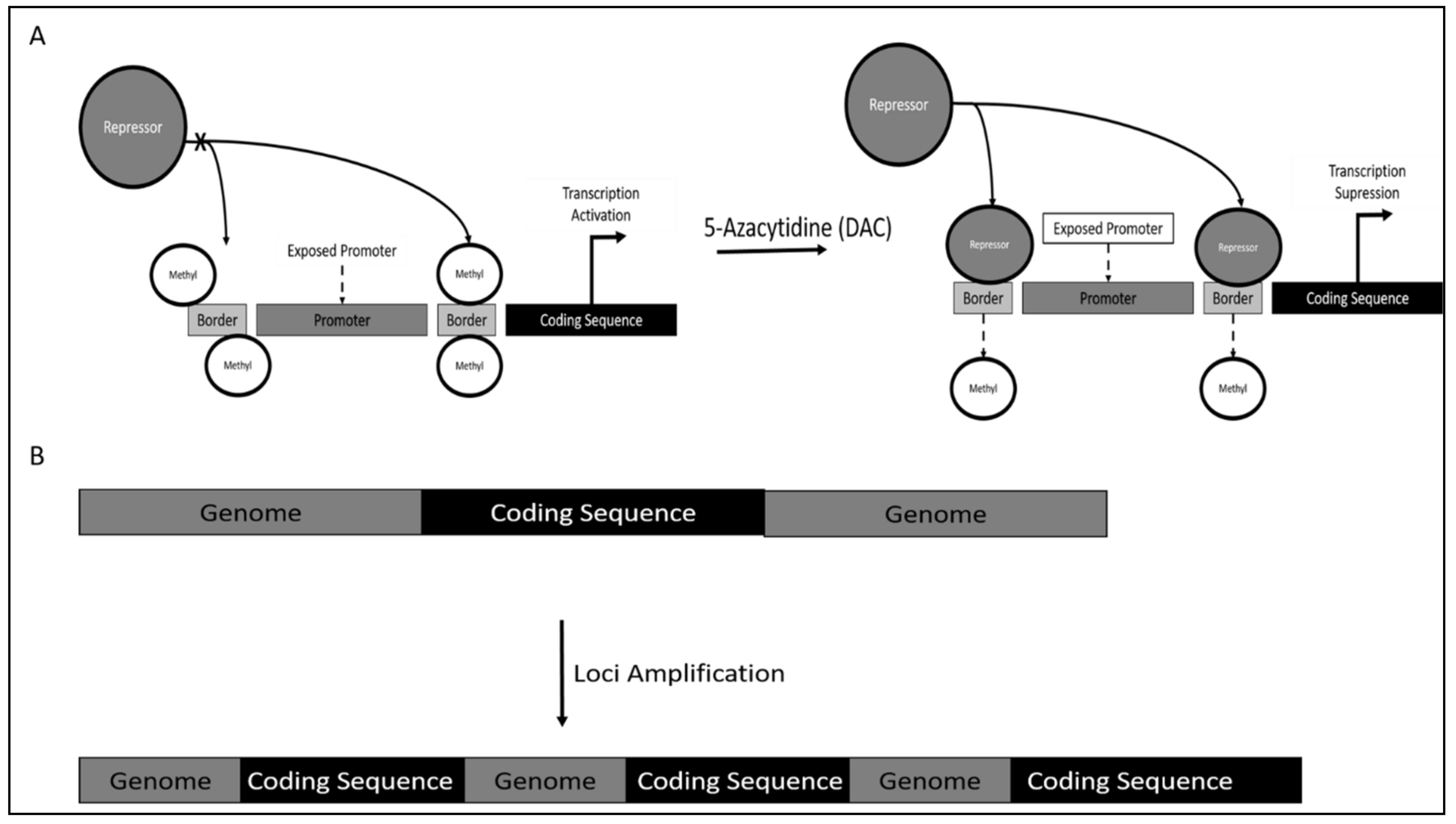
Figure 1. (A) Epigenetic regulation of TERT via promoter border region methylation. (B) Loci amplification of TERT gene resulting in an increase in copy number.
Genes 12 01188 g001
Figure 2. BCR-ABL control of TERT activity and expression generally targeted via Imatinib Mesylate or Tyrosine Kinase Inhibitor treatment.
Genes 12 01188 g002
Figure 3. Control of TERT activity and expression occurring in CLL. (A) Promoter methylation status; (B) chromosomal deletion containing loci encoding for transcription factor suppressors; (C) chromosomal aberrations resulting in fusion of critical cis-regulatory elements with TERT coding sequence.
Genes 12 01188 g003
Table 1. Combinatorial effects of co-administering TERT targeted treatment with other anti-cancer agents. Potential benefits of the combined therapies indicate synergistic effects leading to tumor regression via multiple pathways.
NoType of TERT TreatmentCombination TherapyPotential BenefitsReference
1hTERT DNA vaccinesImmune checkpoint blockade (CTLA-4 and PD-1)Immune checkpoint inhibitor potentially modifies the immune regulatory environment of tumors (including expansion of T cells) to synergize with TERT DNA vaccines, leading to synergistic anti-cancer activityDuperret et al., 2018 [148]
2hTERT epitope peptide vaccine Metronomic chemotherapyCombinatorial approach increases specific T cell response and decreased Treg frequencyTagliamonte et al., 2015 [165]
3Oncolytic adenovirus (Telomelysin)Immune checkpoint blockade (anti-PD-1/anti-PD-L1)Prior treatment with telomelysin sensitizes cancer cells to subsequent immune checkpoint inhibitors administration, which synergises increased tumor regressionKanaya et al., 2018 [151]
4Retroviral-delivered hTERT-specific siRNAChemotherapy (topoisomerase inhibitors or bleomycin) and ionizing radiation (IR)siRNA treatment induced replicative senescence, decreased telomerase activity, attenuated tumorigenicity, and sensitized cancer cells to chemotherapeutic agents and IRNakamura et al., 2005 [166]
5Lentiviral-delivered hTERT-specific shRNADoxorubicin (topo isomerase 2 inhibitor)hTERT shRNA sensitized cancer cells to doxorubicin through decreased proliferation rates and autophagyRomaniuk-Drapała et al., 2021 [167]
6Nucleoside analogue (6-thio-dG)Mitochondrial Hsp90 inhibitor (Gamitrinib)Nucleoside analogue treatment with 6-thio-dG triggers a mitochondrial antioxidant adaptive response, which can be counteracted with mitochondrial Hsp90 inhibitor for higher apoptotic effect in cancer cells Reyes-Uribe et al., 2018 [164]
7Telomere Homolog Oligonucleotide (T-Oligos)Chemotherapy (Vincristine, Cyclophosphamide, Adriamycin, Prednisone)Reduced combinational doses of T-oligos and chemotherapy stimulate cell cycle arrest, senescence, and apoptosis via caspase-3 and p53 activation. Longe et al., 2009 [168]
8Small molecule hTERT inhibitor (BIBR1532)Imatinib mesylate (IM) (tyrosine kinase inhibitor)IM has been determined as an effective drug against CML but potentiates leukemogenesis via STAT5 phosphorylation leading to TERT activation. Co-administration of TERT inhibitor such as BIBR1532 could ameliorate this side effect.Celeghin et al., 2016 & Yamada et al., 2011 [91,94]
9Small molecule hTERT inhibitor (BIBR1532)Doxorubicin (topo isomerase 2 inhibitor)Induction of synergistic anti-cancer effect via p73 activation, repression of proliferation by p21-mediated G1 arrest, downregulation of c-Myc, TERT and survivin, increase ROS leading to caspase-dependent apoptosis, and enhancing pro-oxidant effect of doxorubicin.Bashash et al., 2017 [141]
10Small molecule hTERT inhibitor (BIBR1532)Ionizing radiation (IR)BIBR1532 increased radiosensitivity of cancers by enhancing IR-mediated mitotic catastrophe, senescence, and apoptosis via induction of telomere dysfunction and inhibit the ATM/CHK1 pathwayDing et al., 2019 [169]
Publisher’s Note: MDPI stays neutral with regard to jurisdictional claims in published maps and institutional affiliations.