Case Report

Non-Syndromic Autosomal Dominant Hearing Loss: The First Italian Family Carrying a Mutation in the NCOA3 Gene

1
Department of Medicine, Surgery and Health Sciences, University of Trieste, 34127 Trieste, Italy
2
Institute for Maternal and Child Health—I.R.C.C.S. “Burlo Garofolo”, 34137 Trieste, Italy
3
Department of Cellular, Computational and Integrative Biology (CIBIO), University of Trento, 38123 Trento, Italy
4
UOC Otolaryngology, Institute I.R.C.C.S. “Casa Sollievo della Sofferenza”, 71013 San Giovanni Rotondo, Italy
5
UOC Medical Genetics, Institute I.R.C.C.S. “Casa Sollievo della Sofferenza”, 71013 San Giovanni Rotondo, Italy
*
Author to whom correspondence should be addressed.
Genes 2021, 12(7), 1043; https://doi.org/10.3390/genes12071043
Submission received: 8 June 2021 / Revised: 1 July 2021 / Accepted: 2 July 2021 / Published: 6 July 2021
(This article belongs to the Special Issue Genetics and Epigenetics of Hearing Loss)

Abstract

:
Hearing loss (HL) is the most frequent sensory disorder, affecting about 1–3 per 1000 live births, with more than half of the cases attributable to genetic causes. Despite the fact that many HL causative genes have already been identified, current genetic tests fail to provide a diagnosis for about 40% of the patients, suggesting that other causes still need to be discovered. Here, we describe a four-generation Italian family affected by autosomal dominant non-syndromic hearing loss (ADNSHL), in which exome sequencing revealed a likely pathogenic variant in NCOA3 (NM_181659.3, c.2909G>C, p.(Gly970Ala)), a gene recently described as a novel candidate for ADNSHL in a Brazilian family. A comparison between the two families highlighted a series of similarities: both the identified variants are missense, localized in exon 15 of the NCOA3 gene and lead to a similar clinical phenotype, with non-syndromic, sensorineural, bilateral, moderate to profound hearing loss, with a variable age of onset. Our findings (i.e., the identification of the second family reported globally with HL caused by a variant in NCOA3) further support the involvement of NCOA3 in the etiopathogenesis of ADNSHL, which should, thus, be considered as a new gene for autosomal dominant non-syndromic hearing loss.

1. Introduction

Hearing loss (HL) is the most frequent sensory disorder, affecting about 1–3 per 1000 live births [1]. In developed countries, up to 80% of congenital cases are attributable to genetic defects, which can lead to syndromic hearing loss with other organ abnormalities, or non-syndromic HL, which represents the majority of the cases (i.e., 70%, although recent findings suggest that this percentage might be lower) [2].
Non-syndromic hearing loss (NSHL) can be inherited as an autosomal recessive disease (i.e., 80%), autosomal dominant (i.e., 15–20%) or X-linked, mitochondrial or Y-linked, which, however, account for less than 1% of the total cases [1]. Definition of the molecular cause of NSHL is hampered by the high clinical and genetic heterogeneity of the disease, with more than 123 genes and 170 loci described thus far (Hereditary Hearing Loss Homepage, https://hereditaryhearingloss.org/, accessed on 25 May 2021). Despite technological advances, the current genetic tests fail to provide a diagnosis for about 40% of patients, suggesting that novel genes and mutations still need to be identified [3]. In particular, several studies demonstrated a lower detection rate for autosomal dominant NSHL families (ADNSHL) [4,5], which might be due to the phenotype variability among patients that leads to difficulties in defining a correct genotype–phenotype correlation and, therefore, a proper molecular diagnosis [6].
In light of this, the application of next-generation sequencing techniques, such as exome sequencing (ES), offers a powerful approach to identify and characterize new causative genes. Nevertheless, only the identification of additional families carrying mutations in the same genes provides definitive proof of their pathogenic role.
Here, we describe an Italian family affected by ADNSHL carrying a likely pathogenic variant in NCOA3, a gene recently described as a novel candidate for autosomal dominant progressive hearing loss in a Brazilian family [7].

2. Materials and Methods

The Italian family was recruited at the Institute for Maternal and Child Health—I.R.C.C.S. “Burlo Garofolo” (Trieste, Italy). All the patients provided written informed consent, and all research was conducted according to the ethical standard defined by the Helsinki Declaration.
The individuals of the family underwent a detailed clinical evaluation, including pure tone audiometric testing.
Subsequently, genomic DNA was extracted from peripheral whole blood samples using the QIAsymphony® SP instrument with QIAsymphony® Certal Kits (Qiagen, Venlo, The Netherlands), and DNA concentration was measured using a Nanodrop ND 1000 spectrophotometer (NanoDrop Technologies Inc., Wilmington, DE, USA).
A first round of genetic testing was carried out, including Sanger sequencing to analyze the entire coding region of the GJB2 gene, multiplex PCR to search for GJB6 deletions and Multiplex Ligation-dependent Probe Amplification (MLPA) analysis to identify deletion/duplication in the STRC, CATSPER2 and OTOA genes with SALSA® MLPA® probe mixes P461-A1 DIS (MRC-Holland, Amsterdam, The Netherlands) according to the manufacturer’s protocol.
Finally, ES was carried out on an Illumina NextSeq 550 instrument (Illumina Inc., San Diego, CA, USA). According to the manufacturer’s instructions, genomic libraries were prepared using the Twist Human Core Exome + Human RefSeq Panel kit (Twist Bioscience, South San Francisco, CA, USA). The process leads to the creation of FASTQ files, the standard file format for DNA sequencing data. The FASTQ files were processed through a custom pipeline (Germline-Pipeline), developed by enGenome srl. This workflow allows the generation of a final VCF file containing information regarding variants such as single-nucleotide variants (SNVs), short insertion/deletions (INDELs) and exon-level copy number variations (CNVs). Thus, the VCF files were analyzed on enGenome Expert Variant Interpreter (eVai) software (evai.engenome.com). All the variants of interest were confirmed by Sanger sequencing. Finally, a linkage analysis was performed using Merlin software (version 1.1.2) [8], applying a dominant model with a disease allele frequency of 0.01% and penetrance 0,1.0,1.0.

3. Results

A four-generation Italian family affected by ADNSHL was recruited at our institute (Figure 1).
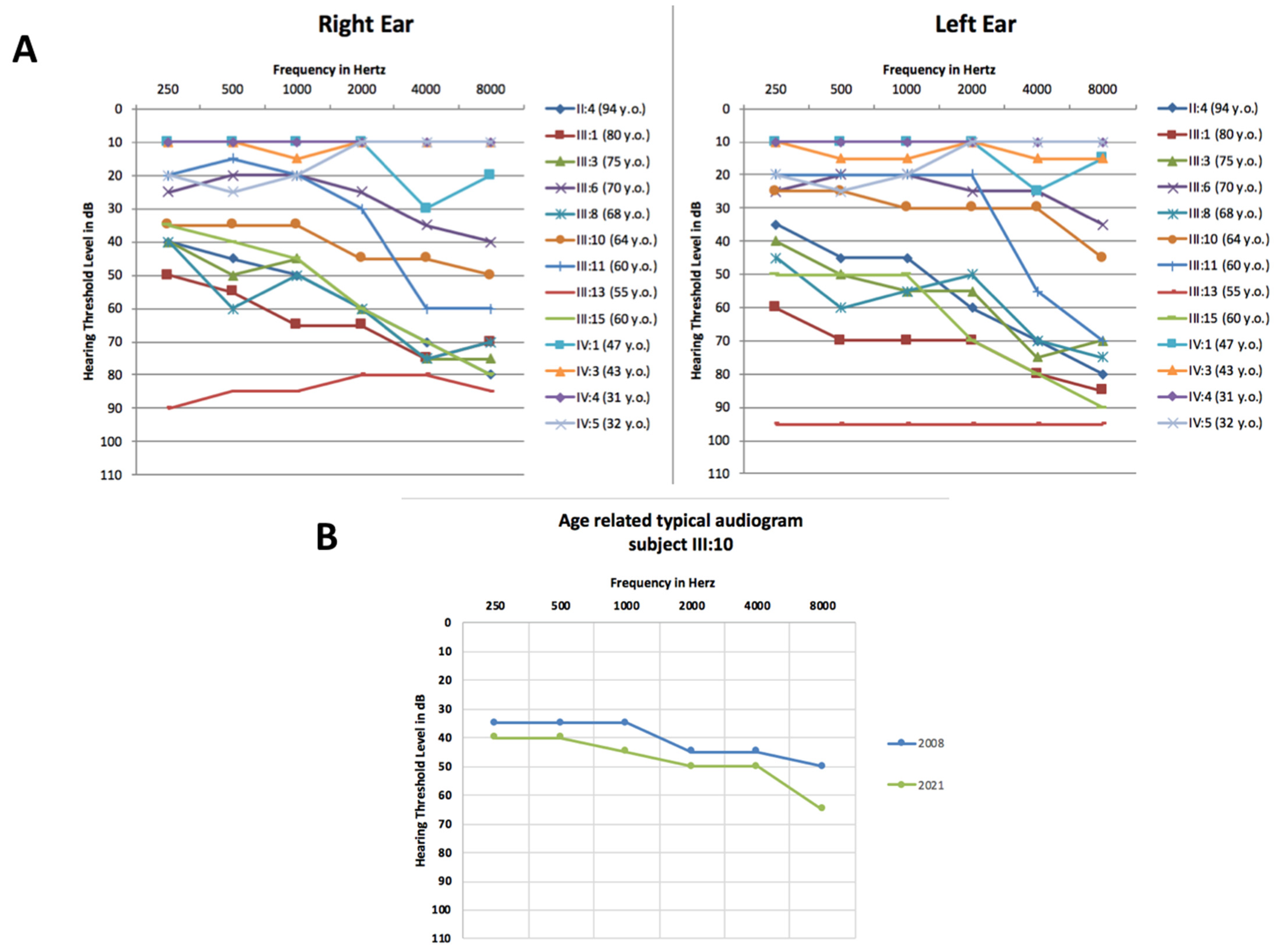
Individuals underwent a careful clinical examination and dysmorphology assessment, which did not highlight any significant anomaly. Afterwards, a hearing evaluation by pure tone audiometry was performed on selected members of the family (II:4, III:1, III:3, III:6, III:8, III:10, III:11, III:13 and III:15), showing a bilateral, symmetric and moderate to severe hearing loss with a medium and high frequencies drop threshold profile (Figure 2A). The age of onset of the hearing impairment ranged from the fourth to the fifth decade of life. Moreover, patients complained of a worsening of their hearing performance over time, a feature objectively verified only in patient III:10 (Figure 2B). They also did not refer to any other comorbidities.
Fourteen family members underwent a first round of genetic tests, including analysis of mutations in the GJB2 and GJB6 genes, followed by MLPA analysis for the STRC gene, being one of the most common causes of hearing loss, second only to GJB2. All the tests resulted negative. ES was performed on all of the fourteen available family members.
In particular, the analysis included two healthy individuals (II:3 and III:9) and members with severe and mild HL (II:4, II:5, III:1, III:3, III:5, III:7, III:8, III:10, III:13, III:15; III:6 and III:11) (Figure 2A).
The overall mean depth base coverage was 77.8X, while on average, 97% of the targeted region was covered at least 10-fold. Further details about the sequencing data are reported in Table 1.
ES analysis led to the identification of a few possible causative variants. Considering the high number of tested individuals (fourteen subjects), after filtering (e.g., minor allele frequency lower than 0.01%, pathogenicity score, pattern of inheritance), the ES analysis led to the identification of only six missense variants in six different genes of particular interest due to their frequency, pathogenicity score given by the in silico tools and co-segregation with HL in most of the individuals of the family (see Table 2). Two of these missense variants are associated with phenotypes not present in any of our patients, such as autism spectrum disorder (MBD2 and SMC2) [9,10]. In addition, one variant was located within a cancer driver candidate gene (ALDH1B1 [11]) and another (KCNH6) resulted false positive after Sanger confirmation. Moreover, the co-segregation of these variants with the HL phenotype was not strictly consistent with the phenotype (see Table 2).
Finally, the last two variants (c.2909G>C in the NCOA3 gene and c.9722T>A in the PKHD1L1 gene) were located in genes encoding for proteins described as being involved in normal hearing function [7,12]. The variant located in PKHD1L1 co-segregated with HL only in half of the patients, while the one within the NCOA3 gene was in all the affected members of the family (Figure 3A), except for patient III:8 (Table 2).
Moreover, linkage analysis for the NCOA3 variant (rs765371222) showed an LOD score of 3.262. Thus, taking into account all these data and considering the high phenotype similarity between this family and the Brazilian family, the NCOA3 variant was considered the likely causative one.
Many in silico predictors indicate the missense variant in the NCOA3 gene (NM_181659.3, c.2909G>C, p.(Gly970Ala), rs765371222) as damaging; in particular, Polyphen2 showed a damage score of 0.82, MutationTaster a score of 0.999, PaPI algorithm a score of 0.908 and a DANN a score of 0.996 [13,14,15,16]. The application of the ACMG criteria for HL [17] allowed us to classify this variant as likely pathogenic (PM2, PP1_Strong). The search of this specific allele in an internal database of 892 Whole Genome Sequencing data highlighted its total absence in healthy individuals, supporting its possible pathogenic effect. The variant co-segregated with HL in all the affected individuals of the family, with the exception of patient III:8, who is probably affected by a different type of HL, likely due to noise exposure or other environmental factors (Figure 3A).
Subsequently, other individuals of the fourth generation (IV:1, IV:2, IV:3, IV:4, IV:5, IV:6) were included in the study. Sanger sequencing was performed in the six subjects, confirming the presence of the variant in the individuals IV:1 and IV:5. They both underwent a hearing evaluation, which highlighted a mild hearing impairment in IV:1 and normal hearing in IV:5 (Figure 2A).

4. Discussion

The Nuclear Receptor Coactivator 3 (NCOA3) gene comprises 23 exons and encodes a protein of 1420 amino acids involved in the regulation of gene transcription [7]. Previous studies suggested a link between NCOA3 variants and different pathologies, such as hypertriglyceridemia, obesity and dyslipidemia [18], or an association with the progression of post-traumatic osteoarthritis, bone mass, abnormal cartilage behavior and notch signaling pathway [7,19]. Recently, da Silva et al. described a clear correlation between the NCOA3 gene and HL, showing the involvement of the protein in the development and physiology of the mouse auditory system and describing a family affected by ADNSHL carrying a missense variant in this gene [7]. In the present work, we reported the second family worldwide with a mutation within the NCOA3 gene and a HL phenotype. Interestingly, both the Italian and the Brazilian missense variants (c.2909G>C (p.Gly970Ala), c.2810C>G (p.Ser937Cys)) are predicted as pathogenic by several in silico tools, and they are closely located (exon 15). Although this region corresponds to an unknown region domain (Figure 3B), the c.2909G>C variant causes the substitution of a highly conserved glycine with an alanine, both polar amino acids, but alanine contains an additional -CH3 group. The tetrahedral structure of the methyl group increases the steric hindrance of the amino acid residue, which might eventually influence the correct folding processes of the protein, compromising its stability or function (Figure 3C). Unfortunately, it has not been possible to generate a reliable in silico protein model due to the low characterization of the protein region carrying the variant. Nevertheless, its involvement has been further confirmed with linkage analysis. Moreover, the audiometric pattern observed in our family resulted to be consistent with the one of the Brazilian family; patients displayed a non-syndromic, progressive, sensorineural, bilateral, moderate to profound HL mainly involving the high and medium frequencies, reaching a severe to profound severity in some of the affected subjects (Figure 2A). The family is characterized by a progressive and late-onset HL, suggesting that the phenotype might be due to the slow but constant misfolded protein accumulation, leading to long-term damage of the ear structures.
We investigated the co-segregation of the variant with HL in all the available family members, and it was found to be present in heterozygosis in all the affected individuals, except for patient III:8. Considering the high incidence of HL in the general population (~1–3:1000), it is not uncommon for large families to have at least one phenocopy (i.e., a phenotypic trait that resembles the trait expressed by a specific genotype, but in a subject who is not a carrier of that genotype). Given the high similarity of the studied family with the Brazilian one, it is likely that the HL phenotype of patient III:8 is due to other undermined causes. One possible explanation is her professional activity, since she is a factory worker constantly exposed to noise. Her audiometric profile is not exactly the one expected from noise-induced hearing loss; however, exceptions to this rule have already been described [20]. In addition, the absence of any other genetic cause leads us to consider her professional work as the most reasonable cause.
The genetic co-segregation with HL was further extended to the fourth generation, allowing the early identification of HL in subject IV:1. Since the genetic result, the individual, who is 47 years old, underwent an audiometric exam confirming his clinical status. As regards individual IV:5, her hearing thresholds are in the range of normality, with a slightly worse performance at the middle and low frequencies. It is worth mentioning that she is only 32 years old and might develop HL in the following decades as her relatives did. Indeed, none of the affected individuals of the family reported having hearing problems before 40 years of age.

5. Conclusions

The identification of new genes and variants associated with monogenic forms of HL is extremely valuable since it allows for extending the knowledge of the molecular basis of this disease and providing patients with a recurrence risk estimation. When a new candidate gene is discovered, functional studies are essential to support genetic findings; however, an independent confirmation in other families (better if in a completely different ethnic group) is fundamental for definitely validating the result. In light of this, our results further confirm the involvement of NCOA3 in the etiopathogenesis of ADNSHL, which should thus be considered a new gene for ADNSHL.

Author Contributions

Conceptualization, P.T., A.N. and G.G.; Methodology, R.P.O., A.N., M.N., M.P.C. and G.G.; Investigation P.T. and A.M.; Project administration, A.N. and G.G.; Writing—original draft, P.T.; writing—review and editing, R.P.O., M.N., A.N., A.M., M.P.C. and G.G.; Funding acquisition, G.G. All authors have read and agreed to the published version of the manuscript.

Funding

This research was funded by GIROTTO GIORGIA, grant number D70-RESRICGIROTTO to G.G. and BENEFICENTIA Stiftung to G.G.

Institutional Review Board Statement

The study was conducted according to the Declaration of Helsinki guidelines and approved by the Ethics Committee of the Institute for Maternal and Child Health—I.R.C.C.S. “Burlo Garofolo” of Trieste (Italy) (2007 242/07).

Informed Consent Statement

Informed consent was obtained from all subjects involved in the study.

Data Availability Statement

The data presented in this study are available on request from the corresponding author. The data are not publicly available due to privacy restrictions.

Acknowledgments

We would like to acknowledge the family whose case is herein discussed for their participation in the study. The authors also wish to thank Massimiliano Cocca for checking the presence of the variant in the available Whole Genome Sequencing data.

Conflicts of Interest

The authors declare no conflict of interest.

References

  1. Korver, A.M.; Smith, R.J.; Van Camp, G.; Schleiss, M.R.; Bitner-Glindzicz, M.A.; Lustig, L.R.; Usami, S.I.; Boudewyns, A.N. Congenital hearing loss. Nat. Rev. Dis. Primers 2017, 3, 16094. [Google Scholar] [CrossRef] [PubMed] [Green Version]
  2. Vona, B.; Doll, J.; Hofrichter, M.; Haaf, T. Non-syndromic hearing loss: Clinical and diagnostic challenges. Med. Genet. 2020, 32, 117–129. [Google Scholar] [CrossRef]
  3. Morgan, A.; Koboldt, D.C.; Barrie, E.S.; Crist, E.R.; García García, G.; Mezzavilla, M.; Faletra, F.; Mihalic Mosher, T.; Wilson, R.K.; Blanchet, C.; et al. Mutations in PLS1, encoding fimbrin, cause autosomal dominant nonsyndromic hearing loss. Hum. Mutat. 2019, 40, 2286–2295. [Google Scholar] [CrossRef]
  4. Sakuma, N.; Moteki, H.; Takahashi, M.; Nishio, S.Y.; Arai, Y.; Yamashita, Y.; Oridate, N.; Usami, S. An effective screening strategy for deafness in combination with a next-generation sequencing platform: A consecutive analysis. J. Hum. Genet. 2016, 61, 253–261. [Google Scholar] [CrossRef] [PubMed]
  5. Sloan-Heggen, C.M.; Smith, R.J. Navigating genetic diagnostics in patients with hearing loss. Curr. Opin. Pediatr. 2016, 28, 705–712. [Google Scholar] [CrossRef] [PubMed] [Green Version]
  6. Vona, B.; Nanda, I.; Hofrichter, M.A.; Shehata-Dieler, W.; Haaf, T. Non-syndromic hearing loss gene identification: A brief history and glimpse into the future. Mol. Cell. Probes 2015, 29, 260–270. [Google Scholar] [CrossRef] [PubMed]
  7. da Silva, R.S.; Dantas, V.; Alves, L.U.; Batissoco, A.C.; Oiticica, J.; Lawrence, E.A.; Kawafi, A.; Yang, Y.; Nicastro, F.S.; Novaes, B.C.; et al. NCOA3 identified as a new candidate to explain autosomal dominant progressive hearing loss. Hum. Mol. Genet. 2021, 29, 3691–3705. [Google Scholar] [CrossRef] [PubMed]
  8. Abecasis, G.R.; Cherny, S.S.; Cookson, W.O.; Cardon, L.R. Merlin--rapid analysis of dense genetic maps using sparse gene flow trees. Nat. Genet. 2002, 30, 97–101. [Google Scholar] [CrossRef] [PubMed]
  9. Iossifov, I.; O’Roak, B.J.; Sanders, S.J.; Ronemus, M.; Krumm, N.; Levy, D.; Stessman, H.A.; Witherspoon, K.T.; Vives, L.; Patterson, K.E.; et al. The contribution of de novo coding mutations to autism spectrum disorder. Nature 2014, 515, 216–221. [Google Scholar] [CrossRef] [PubMed] [Green Version]
  10. Kosmicki, J.A.; Samocha, K.E.; Howrigan, D.P.; Sanders, S.J.; Slowikowski, K.; Lek, M.; Karczewski, K.J.; Cutler, D.J.; Devlin, B.; Roeder, K.; et al. Refining the role of de novo protein-truncating variants in neurodevelopmental disorders by using population reference samples. Nat. Genet. 2017, 49, 504–510. [Google Scholar] [CrossRef] [PubMed] [Green Version]
  11. Nickerson, M.L.; Dancik, G.M.; Im, K.M.; Edwards, M.G.; Turan, S.; Brown, J.; Ruiz-Rodriguez, C.; Owens, C.; Costello, J.C.; Guo, G.; et al. Concurrent alterations in TERT, KDM6A, and the BRCA pathway in bladder cancer. Clin. Cancer Res. Off. J. Am. Assoc. Cancer Res. 2014, 20, 4935–4948. [Google Scholar] [CrossRef] [PubMed] [Green Version]
  12. Wu, X.; Ivanchenko, M.V.; Al Jandal, H.; Cicconet, M.; Indzhykulian, A.A.; Corey, D.P. PKHD1L1 is a coat protein of hair-cell stereocilia and is required for normal hearing. Nat. Commun. 2019, 10, 3801. [Google Scholar] [CrossRef] [PubMed] [Green Version]
  13. Adzhubei, I.; Jordan, D.M.; Sunyaev, S.R. Predicting functional effect of human missense mutations using PolyPhen-2. Curr. Protoc. Hum. Genet. 2013, 76, 7–20. [Google Scholar] [CrossRef] [PubMed] [Green Version]
  14. Schwarz, J.M.; Rödelsperger, C.; Schuelke, M.; Seelow, D. MutationTaster evaluates disease-causing potential of sequence alterations. Nat. Methods 2010, 7, 575–576. [Google Scholar] [CrossRef]
  15. Limongelli, I.; Marini, S.; Bellazzi, R. PaPI: Pseudo amino acid composition to score human protein-coding variants. BMC Bioinform. 2015, 16, 123. [Google Scholar] [CrossRef] [Green Version]
  16. Quang, D.; Chen, Y.; Xie, X. DANN: A deep learning approach for annotating the pathogenicity of genetic variants. Bioinformatics 2015, 31, 761–763. [Google Scholar] [CrossRef] [Green Version]
  17. Oza, A.M.; DiStefano, M.T.; Hemphill, S.E.; Cushman, B.J.; Grant, A.R.; Siegert, R.K.; Shen, J.; Chapin, A.; Boczek, N.J.; Schimmenti, L.A.; et al. ClinGen Hearing Loss Clinical Domain Working Group Expert specification of the ACMG/AMP variant interpretation guidelines for genetic hearing loss. Hum. Mutat. 2018, 39, 1593–1613. [Google Scholar] [CrossRef]
  18. Yu, M.; Gilbert, S.; Li, Y.; Zhang, H.; Qiao, Y.; Lu, Y.; Tang, Y.; Zhen, Q.; Cheng, Y.; Liu, Y. Association of NCOA3 polymorphisms with Dyslipidemia in the Chinese Han population. Lipids Health Dis. 2015, 14, 124. [Google Scholar] [CrossRef] [PubMed] [Green Version]
  19. Zhang, B.; Li, Z.; Wang, W.; Guo, J.; Kang, S.; Liu, S.; Li, H.; Wang, D.; Qi, X. NCOA3 Loss Disrupts Molecular Signature of Chondrocytes and Promotes Posttraumatic Osteoarthritis Progression. Cell. Physiol. Biochem. Int. J. Exp. Cell. Physiol. Biochem. Pharmacol. 2018, 49, 2396–2413. [Google Scholar] [CrossRef]
  20. Sliwinska-Kowalska, M. Hearing. Handb. Clin. Neurol. 2015, 131, 341–363. [Google Scholar] [CrossRef] [PubMed]
Figure 1. Pedigree of the Italian family. Pedigree of the four-generation family investigated in the present study. Filled symbols represent affected individuals. The age of each individual and the co-segregation of the NCOA3 variant with HL (NM_181659.3, c.2909G>C, p.(Gly970Ala)) are reported. Their ages are indicated followed by the terms y.o., meaning years old. The labels used to report the subjects’ genotypes are G/G for homozygous wild type and G/C for the individuals carrying the variant herein discussed in heterozygosis. Individuals with Roman numeric labels were analyzed in this study.
Genes 12 01043 g001
Figure 2. Audiograms of the Italian family. (A) Audiometric features, displayed as audiograms, were available for twelve members of the family. The thresholds of the right and left ears are shown for each patient. The audiometric patterns for patients IV:3 and IV:4 partially overlap. (B) Example of the progression of the hearing loss for patient III:10. In blue is displayed an audiogram performed in 2008, and in green, one of 2021 (Age-Related Typical Audiogram (ARTA)). Only the data for the best ear are reported.
Genes 12 01043 g002
Figure 3. DNA chromatograms, schematic representation of the protein domains and protein alignment. (A) Chromatograms displaying part of the NCOA3 sequence; on top, the wild-type sequence is shown, and on the bottom, that of an affected individual carrying the variant at the heterozygous state. The red box indicates the position of the variant. (B) Schematic representation of NCOA3 protein domains and the localization of the two mutations associated with ADNSHL. The helix loop helix domain is displayed as a green box; in orange, the PAS motif; in blue, low-complexity regions; in red, the coiled coil region; and in yellow, the SMART DUF1518 domain, commonly found in receptor co-activating proteins. Both the variants detected in the NCOA3 gene and associated with ADNSHL are located in the same undefined region of the protein; the one found in the Italian family is indicated with a purple star. (C) Protein alignment showing conservation of residue G970 across species.
Genes 12 01043 g003
Table 1. Next-generation sequencing data. Data relative to ES for each patient. The bold character highlights to which patient the data presented refers to.
Mean Target Coverage
(Mean Base Coverage Depth)
Coverage Uniformity
(Percentage of Bases with a Coverage Depth > 0.2 × Mean_Coverage)
% Target Bases 10X
(Percentage of Target Bases with Coverage above 10X)
II:378.46982497.547697.4332
II:477.62920195.236098.0183
II:579.01345896.120997.0431
III:177.01239798.012796.0126
III:378.72376897.542198.3218
III:577.62907196.930197.9854
III:677.20472095.918397.4510
III:775.92749296.533396.9981
III:876.63820698.142695.4832
III:978.65632995.672196.9453
III:1076.73926098.231097.0034
III:1178.73927996.923195.8741
III:1378.31742997.834296.7632
III:1579.12582096.763197.1632
Table 2. Genotype–phenotype associations. Co-segregation of the six variants with HL in the fourteen members of the family for whom ES was performed. Each patient’s age and gender are indicated. The underlined genotypes are not consistent with the phenotypes of the patients, while the ones reported in bold highlight the segregation of the most interesting variant (NM_181659.3, c.2909G>C, p.(Gly970Ala), rs765371222) in the affected individuals.
SubjectAffectionVariants
ALDH1B1
NM_000692.5
SMC2
NM_001042550.2
NCO3
NM_181659.3
MBD2
NM_015832.6
KCNH6
NM_030779.4
PKHD1L1
NM_177531.4
c.G1087Ac.A1514Gc.G2909Cc.G707Tc.G263Tc.T9722A
II-3
(male, 92 y.o)
healthyG/GA/AG/GG/Gfalse positiveT/T
II-4
(female, 94 y.o.)
affectedG/AA/GG/CG/Tfalse positiveT/A
II-5
(male, 88 y.o.)
affectedG/AA/GG/CG/Tfalse positiveT/A
III-1
(male, 80 y.o.)
affectedG/AA/GG/CG/Tfalse positiveT/T
III-3
(female, 75 y.o.)
affectedG/AA/GG/CG/Tfalse positiveT/A
III-5
(male, 71 y.o.)
affectedG/GA/AG/CG/Gfalse positiveT/T
III-6
(male, 70 y.o.)
affectedG/GA/GG/CG/Gfalse positiveT/T
III-7
(female, 68 y.o.)
affectedG/AA/GG/CG/Gfalse positiveT/T
III-8
(female, 70 y.o.)
affectedG/GA/GG/GG/Gfalse positiveT/T
III-9
(male, 68 y.o.)
healthyG/GA/GG/GG/Tfalse positiveT/T
III-10
(male, 64 y.o.)
affectedG/AA/GG/CG/Tfalse positiveT/A
III-11
(male, 60 y.o.)
affectedG/GA/GG/CG/Gfalse positiveT/T
III-13
(female, 56 y.o.)
affectedG/AA/GG/CG/Tfalse positiveT/A
III-15
(male, 60 y.o.)
affectedG/AA/GG/CG/Tfalse positiveT/A
Publisher’s Note: MDPI stays neutral with regard to jurisdictional claims in published maps and institutional affiliations.