Article

A Comprehensive, Targeted NGS Approach to Assessing Molecular Diagnosis of Lysosomal Storage Diseases

by
Valentina La Cognata
and
Sebastiano Cavallaro
*
Institute for Biomedical Research and Innovation (IRIB), National Research Council (CNR), 95126 Catania, Italy
*
Author to whom correspondence should be addressed.
Genes 2021, 12(11), 1750; https://doi.org/10.3390/genes12111750
Submission received: 6 September 2021 / Revised: 25 October 2021 / Accepted: 27 October 2021 / Published: 30 October 2021
(This article belongs to the Section Molecular Genetics and Genomics)

Abstract

:
With over 60 different disorders and a combined incidence occurring in 1:5000–7000 live births, lysosomal storage diseases (LSDs) represent a major public health problem and constitute an enormous burden for affected individuals and their families. Several reasons make the diagnosis of LSDs an arduous task for clinicians, including the phenotype and penetrance variability, the shared signs and symptoms, and the uncertainties related to biochemical enzymatic assay results. Developing a powerful diagnostic tool based on next generation sequencing (NGS) technology may help reduce the delayed diagnostic process for these families, leading to better outcomes for current therapies and providing the basis for more appropriate genetic counseling. Herein, we employed a targeted NGS-based panel to scan the coding regions of 65 LSD-causative genes. A reference group sample (n = 26) with previously known genetic mutations was used to test and validate the entire workflow. Our approach demonstrated elevated analytical accuracy, sensitivity, and specificity. We believe the adoption of comprehensive targeted sequencing strategies into a routine diagnostic route may accelerate both the identification and management of LSDs with overlapping clinical profiles, producing a significant reduction in delayed diagnostic response with beneficial results in the treatment outcome.

1. Introduction

Lysosomal storage disorders (LSDs) are rare inherited diseases characterized by the accumulation of specific undegraded metabolites inside the lysosomes [1,2,3]. This over-storage is commonly caused by a deficiency or absent activity of lysosomal hydrolases or, in a few cases, by the deficit of further non-enzymatic lysosomal proteins (such as integral membrane proteins) [3]. With a combined incidence of 1 in 1500 to 7000 live births, this group of monogenic inborn errors of metabolism encompasses ~70 different entities, including sphingolipidoses, mucopolysaccharidoses, glycoproteinoses, lipid storage diseases, lipofuscinosis, lysosomal integral membrane proteins diseases, and post-translational modifications dysfunctions [4,5]. Clinical signs and symptoms may occur from the prenatal period to adulthood and may develop progressively over time, leading to a wide spectrum of disease phenotypes from mild to extremely severe forms that involve neuropathological effects, psychomotor development delay, cognitive decline, musculoskeletal abnormalities, dysmorphia, organomegaly, and seizures [6]. Both the considerable clinical variability within each disease phenotype and the overlapping symptomatology among single LSDs hamper the path for a precise diagnosis, which often involves a delay in treatment and severe consequences on patients’ quality of life and their families [4].
Current diagnostic workflows include an accurate evaluation of both medical history and clinical presentations, which lead to the formulation of suspicion of one or more LSDs, followed by biochemical analysis to quantify either the accumulated storage product or the enzymatic activity in leukocytes, fibroblasts, urine, or rehydrated dried blood spots (DBS) for newborns [7,8]. Finally, if deficient enzyme activity is detected, second-tier confirmatory biomarker tests or Sanger sequencing are performed for the suspected gene. Although this diagnostic route represents the current gold standard, it presents several limitations. First, it requires deep clinical expertise to discriminate phenotypic overlapping manifestations and, thus, to reduce the number of biochemical tests used for each LSD-suspected patient. Second, the execution of multiple biochemical enzymatic assays may be expensive, time-consuming, and subject to high variability, and enzymatic tests may not be available for all diseases. Therefore, reaching a definitive molecular diagnosis for LSDs with traditional techniques is still challenging, can take several years, or may be unsuccessful.
In the past decade, the emergence of next generation sequencing (NGS) technologies has proven to be an effective alternative to conventional techniques, in both research and clinical settings, allowing for the simultaneous interrogation of several genes in one single reaction and reducing, considerably, the time and costs for Sanger sequencing of a single gene [9,10]. The introduction of ad hoc designed genetic tests (targeted NGS panels) into diagnostic workflows offers the opportunity for easier identification of LSDs, timely diagnosis, and optimized clinical management, reducing the psychological burden and providing appropriate genetic counseling to parents [4].
In this study, we aimed to design and evaluate both the diagnostic utility of a semi-automated and comprehensive sequencing assay based on a targeted NGS (tNGS) panel (hereafter referred to as LSDs_panel) developed to detect pathogenic variants in 65 LSD-related genes. We describe the panel performance, strengths, and limitations and propose it as a useful second-tier diagnostic test for specialists in everyday clinical management who might suspect an LSD, given its ability to provide accurate and timely information.

2. Materials and Methods

2.1. Sample Collection and Dosage

A reference group of DNA samples isolated from clinically diagnosed donor subjects (n = 26) were obtained from the NIGMS Human Genetic Cell Repository at the Coriell Institute for Medical Research (https://www.coriell.org/, accessed on 26 October 2021). The purchased samples were chosen for known variants localized in targeted genes and selected in order to ensure an adequate representation for most LSDs. Quantification of the genomic DNA was assessed by measuring the genomic copies of the human RNase P gene using the TaqMan® RNase P Detection Reagents Kit (Thermo Fisher Scientific, Waltham, MA, USA) and the Aria Dx Real-Time PCR System (Agilent Technologies, Santa Clara, CA, USA).

2.2. Panel Design and Library Preparation

For the selection of genes (n = 65) included in the panel, we relied on updated literature data [2] and a previous gene-set used for targeted strategies (Table 1). An on-demand panel (IAD199901) and a compatible made-to-order spike-in panel (IAD199905 including TPP1 and BLOC1S3 genes) were designed using the Ion AmpliSeq Designer software (https://ampliseq.com, accessed on 1 May 2020, Thermo Fisher Scientific, Waltham, MA, USA). The advantage of using Ion AmpliSeq on-demand panel customization is that primer pairs are pre-tested and optimized for high performance, whereas spike-ins are high concentrated made-to-order panels used to extend panels for genes not available on-demand.
The complete panel design (called LSDs_panel) covers 237.782 kb and includes 1241 amplicons with a size range of 125–275 bp distributed across two primer pools (625 primer pool 1 and 616 primer pool 2). The in silico coverage consisted of 99% for the on-demand panel and 99.18% for the spike-ins. The complete design of the LSDs_panel is available in Supplementary Table S1.
Library preparation was carried out using the Ion AmpliSeq™ Kit for Chef DL8 (DNA to Library, 8 samples/run) used for automated library preparation of the Ion AmpliSeq™ libraries on the Ion Chef™ System (Thermo Fisher Scientific, Waltham, MA, USA). According to the recommended number of amplification cycles in the standard protocol, the amplification conditions were set out to 16 cycles and four minutes of annealing/extension time. The library quality and molarity were assessed using the Ion Library TaqMan® Quantitation Kit (Thermo Fisher Scientific, Waltham, MA, USA) on the Aria Dx Real-Time PCR System (Agilent Technologies, Santa Clara, CA, USA). Serial dilutions of the E. coli DH10B Control Library (Thermo Fisher Scientific, Waltham, MA, USA) were prepared and run in triplicate to generate a standard curve. The molar concentration of libraries was determined using the Delta R—baseline-corrected raw fluorescence calculated with Aria DX Real-Time PCR Software (Agilent Technologies, Santa Clara, CA, USA). Barcoded libraries (up to 4-Chef runs corresponding to 32 libraries) were super-pooled in equimolar concentration using the strategies suggested for combining libraries prepared with different panels for equal coverage in order to obtain a final molarity of 40 pM each.

2.3. Chip Loading and Sequencing

Loading of the Ion 510 and the 540 Chips was carried out using the Ion 510, 520, 530, and 540 Kit-Chef (Thermo Fisher Scientific, Waltham, MA, USA) following manufacturer instructions. High throughput sequencing runs were carried out on the Ion Gene Studio S5 system (Thermo Fisher Scientific, Waltham, MA, USA). A run planned in the S5 Torrent Suite (v. 5.12.2) had the following parameters: analysis parameters, default; reference library, hg19; target regions, LSDs_panel; read length, 200 bp; flows, 550; and base calibration mode, default. The plugins used were Coverage Analysis, Ion Reporter Uploader, and Variant Caller (default settings).

2.4. Variant Calling and Prioritization

Read mapping was performed automatically in Torrent Suite (v. 5.12.2, Thermo Fisher Scientific, Waltham, MA, USA) by using the variant Caller plugin (v5.12.0.4) with default settings (germline_low_stringency). The called variants were automatically uploaded on Ion Reporter (Thermo Fisher Scientific, Waltham, MA, USA). The Copy Number Variation (CNV) performance was not assessed. The pipeline analysis for variant filtering was based on multiple adjusted steps including coverage min 30×, Homopolymer length ≤ 3, p-value < 0.001, ClinVar ≠ benign or likely benign, MAF < 0.001 or n.a., frequency 30–60% for heterozygous variants and >70% for homozygous variants, intronic variants included if the distance from exon is < 10 bp, SIFT score < 0.05/PolyPhen score > 0.85 or n.a., and variants effect ≠ synonymous unless they are pathogenic/likely pathogenic or with conflicting interpretation of pathogenicity. A comparison of the Torrent Variant Caller (TVC) prioritized variants with their respective genetic information from Coriell biobank was performed post-analysis. True-positive (TP), true-negatives (TN), false-positive (FP), and false-negative (FN) variant calls were defined by considering available data from the single causative gene in the Coriell repository. True positives (TPs) were defined as variants both detected by our filtering pipeline as well as expected from the Coriell collected data. True negatives (TNs) were considered additional variants detected in the causative gene but excluded by our prioritization pipeline and not reported in the repository data. False positives (FPs) were considered variants detected by our pipeline but not expected from the data. False negatives (FNs) were considered variants expected from the Coriell data but missed by our pipeline. Accuracy was calculated as follows: (TP + TN)/(TP + FP + TN + FN); sensitivity was calculated as follows: TP/(TP + FN); and specificity was calculated as follows: TN/(TN + FP). The Matthews correlation coefficient (MCC) (which measures the correlation between the predicted and observed binary classification of a sample) was calculated as follows: MCC = [(TP × TN) − (FP × FN)]/√[(TP + FP)(TP + FN)(TN + FP)(TN + FN)].

3. Results

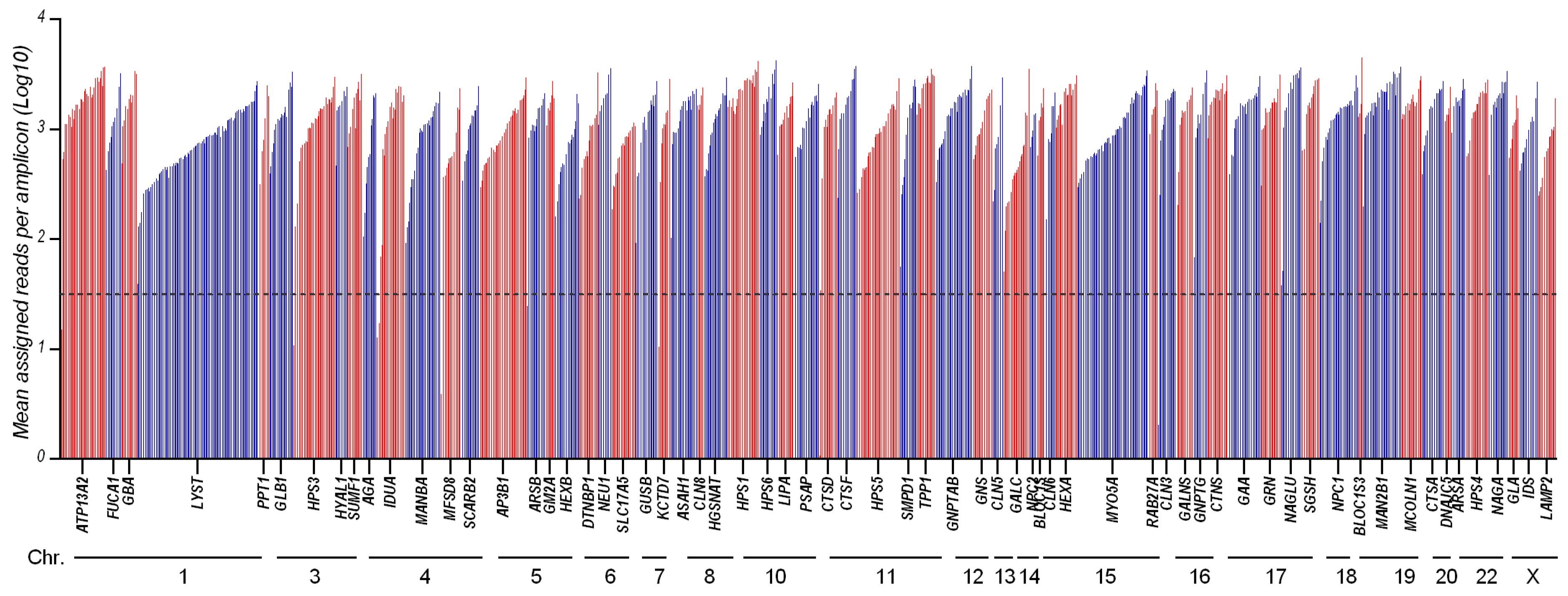
3.1. Panel Design and Performance

The LSDs_panel was designed to target the entire coding regions of 65 LSD-related genes (Table 1), which were previously reported to be a direct cause of an LSD when mutated in both alleles, in order to use it for diagnostic testing in patients with a high a priori probability of LSD based on the clinical phenotype. The LSDs_panel included 1241 amplicons (with a length of 125–275 bp) distributed between two primer pools (625 + 616 primer pairs) and covering a size of 237.782 kb, with an in silico coverage of 99% (the complete design of LSDs_panel is available in Supplementary Table S1). No additional intronic regions were targeted to maximize the coverage of exonic regions and to facilitate rapid and unambiguous interpretation in the context of diagnosis.
Before investigating the clinical utility of the gene panel, we sought to determine the analytical performance of our method in terms of depth of coverage across all targeted genes. Therefore, we used a reference group of DNA samples (n = 26, Table 2), isolated from clinically diagnosed donors from the NIGMS Human Genetic Cell Repository at the Coriell Institute for Medical Research and previously Sanger-sequenced for the LSD-suspected genes.
From the run metrics results, all samples were uniformly covered at depths that exceed the minimum coverage required (30×) for the accurate calling of variants. Coverage analysis shows that 1225/1241 of the amplicons (98.7%) had a sufficient amplification efficiency (mean assigned reads per amplicon Log10 ranging from 1.5 to 3.8), while 16 amplicons were suboptimal (Figure 1 and Supplementary Table S2).
Filtering the pipeline on the TVC (Torrent Variant Caller, Thermo Fisher Scientific, Waltham, MA, USA) was based on a stepwise-adjusted strategy to highlight relevant variants (i.e., coverage min 30×, homopolymer length ≤ 3, p-value < 0.001, ClinVar ≠ benign or likely benign, MAF < 0.001 or none, frequency 30–60% for heterozygous variants and >70% for homozygous variants, include intronic variants if the distance from exon is <10 bp, SIFT score < 0.05 or none, PolyPhen score > 0.85 or none, and variants effect ≠ synonymous unless they are pathogenic/likely pathogenic/uncertain significance or with conflicting interpretation of pathogenicity). A comparison with the previously known variants reported in Coriell biobank was performed by post-filtering analysis. True-positive (TP), true-negative (TN), false-positive (FP), and false-negative (FN) variant calls were defined by considering the available data from a single causative gene in the Coriell repository (see Section 2).
The overall accuracy of the panel was 98.4%, analytical sensitivity was 95.2%, while specificity was 97.6%. There were 40 correctly called true-positive variants, 83 true-negative reference calls, and 2 false-negative (missed) calls when comparing our results with the expected variants (Table 2). The MCC was 0.964 (MCC = +1 describes a perfect prediction, =0 means unable to return any valid information, and =−1 describes complete inconsistency between prediction and observation).

3.2. Control Samples Analysis

The majority of detected pathogenic mutations and polymorphisms are consistent with the data reported in the Coriell biobank. Interestingly, some additional observations in single causative genes emerged that are worthy to be mentioned in order to update data in the repository, as we describe below.
The NA06110 sample, acquired from Coriell biobank, derives from a female donor subject described as a compound heterozygote, with one allele carrying a G>A transition in the SGSH gene causing the Arg245His (R245H) aminoacidic variation and “no changes detected in the other allele”. The LSDs_panel was able to successfully detect the R245H change, identifying a second heterozygous mutation (i.e., the c.629G>A, causing the nonsense aminoacidic change—p.Trp210Ter) reported as pathogenic/likely pathogenic in ClinVar (Table 2). Thus, in addition to confirming the previously detected variant, our analysis indicated the presence of another, extending the genotypic portrait of the sample.
An additional observation is with regard to the NA02057 DNA sample, which carries a pathogenic homozygous G-to-C transversion in the AGA gene, resulting in a substitution of serine for cysteine at codon 163 (Cys163Ser (C163S)). The Coriell biobank reports also a heterozygous G-to-A transition (Arg161Gln (R161Q)) in the same gene, which was detected by the LSDs_panel, but classified as benign in ClinVar.
The two false negative variants were detected in the NA00879 and NA01256 samples (Table 2). The first (c.746G>A (Arg245His [R245H])) was completely missed by sequencing, whereas the second (c.1293TGG>TAG (Trp402Ter [W402X])) was detected by the panel but excluded due to very low coverage (below the threshold of 30×). We cannot rule out that missed genetic modifications are the result of high culture passages.
The LSDs_panel detected additional non-pathogenic variants in the analyzed samples (Table 2, in non-bold text) that may reduce enzymatic activity and may contribute to phenotypic manifestations. Given the variability of symptom manifestations as well as the phenotypic overlapping between genetically different disorders, the presence of additional secondary variants or genetic modifiers involved in lysosomal regulation and metabolism should be considered and may help to refine genotype–phenotype correlations.

4. Discussion

As outlined earlier, there are many factors hampering the diagnosis of LSDs, including the phenotypic and penetrance variability, the common signs and symptoms between certain disease groups, the genetic heterogeneity, and the difficulties of biochemical diagnostics. Developing a powerful diagnostic tool could mitigate the delayed diagnostic process for affected families, leading to better outcomes for current therapies and providing the basis for more appropriate genetic counseling. Many recent reports have emphasized the high clinical utility of NGS technologies and targeted gene panels in the diagnosis of suspected LSDs and their potential to reduce diagnostic delay [11,12,13,14,15,16,17].
Herein, we proposed a tNGS panel (LSDs_panel) based on AmpliSeq technologies to simultaneously screen the coding regions of 65 genes responsible for a heterogeneous group of LSDs and aimed at evaluating its clinical utility in suspected patients. By using a set (n = 26) of standard samples from Coriell Institute biobank (https://www.coriell.org/, accessed on 26 October 2021), we assessed the overall accuracy of the panel (98.4%), the analytical sensitivity (95.2%), and the specificity (97.6%) of the NGS workflow. Known pathogenic mutations in the reference samples were identified with the correct homozygous/heterozygous state.
Several published papers have shown the possibility of carrying out successful NGS sequencing studies from DNA extracted from Guthrie card (DBS) fingerprints, thus taking advantage of the possibility of using the same non-invasive sampling from newborns for both biochemical and sequencing tests [18,19]. Preliminary experiments in our lab starting from DBS-isolated DNA and sequenced with the LSD panel showed adequate amplicon coverage, revealing the feasibility of the NGS approach when starting from dried samples.
A second-tier application of the comprehensive LSDs_panel may be in the field of modifier genes, complex disorders, and polygenic inheritance [15,20,21]. It is well known that patients who share the same mutations may have a different phenotypic spectrum. Thus, the effect of the primary molecular defects may be modified by the presence of additional cumulative mutations located in other genes that encode proteins involved in lysosomal pathways (Table 2). The possibility of detecting variants with uncertain significance and/or secondary findings should be, however, carefully considered in reporting the results, clearing the (probable) non-causality role of the mutation. The decision to report such mutations should always be in accordance with informed consent signed by patients.
A strong limitation of the panel is the poor ability to detect complex rearrangements and recombined genomic regions, which may all require other techniques for elucidation. CNVs, including both deletions and amplifications, may be visualized starting from NGS data by manually checking the coverage of the suspected gene: the degree of coverage of the examined region with respect to the same region in other samples of the same run could suggest the presence of a CNV in heterozygous or homozygous state. However, in both cases, different molecular techniques should be used to confirm the suspected alterations as well as to exclude potential allelic dropout events.
Taken together, we demonstrated here that an NGS-based approach for the detection of LSDs may be a valuable adjunct test along with the well-established biochemical assays. Indeed, while enzyme analysis is still the gold standard for many LSDs (characterized by enzymatic deficiency), it may not accurately identify all obligate carriers and cannot be applied to disorders caused by alterations in transport or transmembrane (non-catalytic) proteins. That a broader spectrum of diseases can be monitored in one single test significantly shortens the analysis time for complex phenotypes or when a biochemical test cannot be offered. Finally, genotype–phenotype correlations may be carefully analyzed since they may be discordant, and clinicians should be cautious when counseling families regarding prognosis.

5. Conclusions

NGS technology is currently offering the opportunity to improve the LSD diagnostic workflow, given its low cost, semi-automated pipeline, short processing time, and ability to simultaneously detect multiple nucleotide variants on several genes. A broader adoption of targeted NGS-based tests, such as the assessment described here, should be taken into consideration to optimize clinical management of LSDs characterized by high levels of clinical and biochemical heterogeneity.
The use of targeted NGS may represent a real and valuable strategy for providing timely and correct diagnoses, for detecting carriership status, and for ensuring genetic counseling for family planning. Moreover, molecular profiling and genomic sequencing information may prompt the design of novel therapeutic drugs targeting specific mutations, thus opening the possibility for personalized medicine. Efforts in this sense may prompt patient-oriented outcomes, may improve the quality of life of patients and their families, and may reduce both direct and indirect costs (e.g., caregivers’ services) to national health services and families.

Supplementary Materials

The following are available online at https://www.mdpi.com/article/10.3390/genes12111750/s1, Table S1: Design (bed file) of the LSD panel; Table S2: Mean reads per amplicon.

Author Contributions

Conceptualization, V.L.C. and S.C.; data curation, V.L.C.; formal analysis, V.L.C.; funding acquisition, S.C.; investigation, V.L.C.; methodology, V.L.C.; resources, S.C.; supervision, S.C.; writing—original draft, V.L.C.; writing—review and editing, V.L.C. and S.C. All authors have read and agreed to the published version of the manuscript.

Funding

This work was supported by the joint project between IRIB-CNR and SANOFI “Early diagnosis of some lysosomal diseases: analysis of the clinical utility and diagnostic validity of genomic techniques for their molecular diagnosis. Assessments of the implications of the inclusion of lysosomal diseases in the context of a national neonatal screening program” (project n. 2018/9848).

Institutional Review Board Statement

Not applicable.

Informed Consent Statement

Not applicable.

Data Availability Statement

Details of the reference samples selected for the present validation can be found at https://www.coriell.org/ (accessed on 26 October 2021).

Acknowledgments

The authors gratefully acknowledge Cristina Calì, Alfia Corsino, Maria Patrizia D’Angelo, and Francesco Marino for administrative and technical support.

Conflicts of Interest

The authors declare no conflict of interest. The funders had no role in the design of the study, in writing the manuscript, or in the decision to publish the results.

References

  1. Platt, F.M. Emptying the stores: Lysosomal diseases and therapeutic strategies. Nat. Rev. Drug Discov. 2018, 17, 133–150. [Google Scholar] [CrossRef] [PubMed]
  2. Platt, F.M.; d’Azzo, A.; Davidson, B.L.; Neufeld, E.F.; Tifft, C.J. Lysosomal storage diseases. Nat. Rev. Dis. Primers 2018, 4, 27. [Google Scholar] [CrossRef]
  3. Ferreira, C.R.; Gahl, W.A. Lysosomal storage diseases. Transl. Sci. Rare Dis. 2017, 2, 1–71. [Google Scholar] [CrossRef] [Green Version]
  4. La Cognata, V.; Guarnaccia, M.; Polizzi, A.; Ruggieri, M.; Cavallaro, S. Highlights on Genomics Applications for Lysosomal Storage Diseases. Cells 2020, 9, 1902. [Google Scholar] [CrossRef]
  5. Kingma, S.D.; Bodamer, O.A.; Wijburg, F.A. Epidemiology and diagnosis of lysosomal storage disorders; challenges of screening. Best Pract. Res. Clin. Endocrinol. Metab. 2015, 29, 145–157. [Google Scholar] [CrossRef]
  6. Pinto, E.V.F.; Rojas Malaga, D.; Kubaski, F.; Fischinger Moura de Souza, C.; de Oliveira Poswar, F.; Baldo, G.; Giugliani, R. Precision Medicine for Lysosomal Disorders. Biomolecules 2020, 10, 1110. [Google Scholar] [CrossRef] [PubMed]
  7. Mokhtariye, A.; Hagh-Nazari, L.; Varasteh, A.R.; Keyfi, F. Diagnostic methods for Lysosomal Storage Disease. Rep. Biochem. Mol. Biol. 2019, 7, 119–128. [Google Scholar]
  8. Waggoner, D.J.; Tan, C.A. Expanding newborn screening for lysosomal disorders: Opportunities and challenges. Dev. Disabil. Res. Rev. 2011, 17, 9–14. [Google Scholar] [CrossRef]
  9. Komlosi, K.; Sólyom, A.; Beck, M. The Role of Next-Generation Sequencing in the Diagnosis of Lysosomal Storage Disorders. J. Inborn Errors Metab. Screen. 2016, 4, 4. [Google Scholar] [CrossRef]
  10. Encarnação, M.; Coutinho, M.F.; Silva, L.; Ribeiro, D.; Ouesleti, S.; Campos, T.; Santos, H.; Martins, E.; Cardoso, M.T.; Vilarinho, L.; et al. Assessing Lysosomal Disorders in the NGS Era: Identification of Novel Rare Variants. Int. J. Mol. Sci. 2020, 21, 6355. [Google Scholar] [CrossRef]
  11. Fernandez-Marmiesse, A.; Morey, M.; Pineda, M.; Eiris, J.; Couce, M.L.; Castro-Gago, M.; Fraga, J.M.; Lacerda, L.; Gouveia, S.; Perez-Poyato, M.S.; et al. Assessment of a targeted resequencing assay as a support tool in the diagnosis of lysosomal storage disorders. Orphanet J. Rare Dis. 2014, 9, 59. [Google Scholar] [CrossRef]
  12. Zanetti, A.; D’Avanzo, F.; Bertoldi, L.; Zampieri, G.; Feltrin, E.; De Pascale, F.; Rampazzo, A.; Forzan, M.; Valle, G.; Tomanin, R. Setup and Validation of a Targeted Next-Generation Sequencing Approach for the Diagnosis of Lysosomal Storage Disorders. J. Mol. Diagn. 2020, 22, 488–502. [Google Scholar] [CrossRef] [PubMed]
  13. Gheldof, A.; Seneca, S.; Stouffs, K.; Lissens, W.; Jansen, A.; Laeremans, H.; Verloo, P.; Schoonjans, A.S.; Meuwissen, M.; Barca, D.; et al. Clinical implementation of gene panel testing for lysosomal storage diseases. Mol. Genet. Genom. Med. 2019, 7, e00527. [Google Scholar] [CrossRef]
  14. Levesque, S.; Auray-Blais, C.; Gravel, E.; Boutin, M.; Dempsey-Nunez, L.; Jacques, P.E.; Chenier, S.; Larue, S.; Rioux, M.F.; Al-Hertani, W.; et al. Diagnosis of late-onset Pompe disease and other muscle disorders by next-generation sequencing. Orphanet J. Rare Dis. 2016, 11, 8. [Google Scholar] [CrossRef]
  15. Di Fruscio, G.; Schulz, A.; De Cegli, R.; Savarese, M.; Mutarelli, M.; Parenti, G.; Banfi, S.; Braulke, T.; Nigro, V.; Ballabio, A. Lysoplex: An efficient toolkit to detect DNA sequence variations in the autophagy-lysosomal pathway. Autophagy 2015, 11, 928–938. [Google Scholar] [CrossRef] [PubMed]
  16. Malaga, D.R.; Brusius-Facchin, A.C.; Siebert, M.; Pasqualim, G.; Saraiva-Pereira, M.L.; Souza, C.F.M.; Schwartz, I.V.D.; Matte, U.; Giugliani, R. Sensitivity, advantages, limitations, and clinical utility of targeted next-generation sequencing panels for the diagnosis of selected lysosomal storage disorders. Genet. Mol. Biol. 2019, 42, 197–206. [Google Scholar] [CrossRef] [PubMed] [Green Version]
  17. La Cognata, V.; Guarnaccia, M.; Morello, G.; Ruggieri, M.; Polizzi, A.; Cavallaro, S. Design and Validation of a Custom NGS Panel Targeting a Set of Lysosomal Storage Diseases Candidate for NBS Applications. Int. J. Mol. Sci. 2021, 22, 64. [Google Scholar] [CrossRef] [PubMed]
  18. Hendrix, M.M.; Cuthbert, C.D.; Cordovado, S.K. Assessing the Performance of Dried-Blood-Spot DNA Extraction Methods in Next Generation Sequencing. Int. J. Neonatal Screen. 2020, 6, 36. [Google Scholar] [CrossRef]
  19. Boemer, F.; Fasquelle, C.; d’Otreppe, S.; Josse, C.; Dideberg, V.; Segers, K.; Guissard, V.; Capraro, V.; Debray, F.G.; Bours, V. A next-generation newborn screening pilot study: NGS on dried blood spots detects causal mutations in patients with inherited metabolic diseases. Sci. Rep. 2017, 7, 17641. [Google Scholar] [CrossRef]
  20. Davidson, B.A.; Hassan, S.; Garcia, E.J.; Tayebi, N.; Sidransky, E. Exploring genetic modifiers of Gaucher disease: The next horizon. Hum. Mutat. 2018, 39, 1739–1751. [Google Scholar] [CrossRef]
  21. Parenti, G.; Medina, D.L.; Ballabio, A. The rapidly evolving view of lysosomal storage diseases. EMBO Mol. Med. 2021, 13, e12836. [Google Scholar] [CrossRef] [PubMed]
Figure 1. Amplicon coverage of the 65 targeted genes: 1241 amplicons distributed across 65 genes were amplified and sequenced with LSDs_panel. This chart shows the mean coverage of individual targeted amplicons across each gene for 26 analyzed samples. Amplicons with zero reads were arbitrarily represented as 0.
Genes 12 01750 g001
Table 1. LSD-related genes included in the panel and their associated disorders.
GeneCytogenetic LocationPathologyPhenotype
OMIM No.
AGA4q34.3Aspartylglucosaminuria208400
AP3B15q14.1Hermansky–Pudlak disease type 2608233
ARSA22q13.33Metachromatic leukodystrophy250100
ARSB5q14.1MPS VI, also known as Maroteaux–Lamy syndrome253200
ASAH18p22Farber lipogranulomatosis228000
ATP13A21p36.13CLN12b: Kufor–Rakeb syndrome or PARK9606693
BLOC1S615q21.1Hermansky–Pudlak disease type 9614171
BLOCS1319q13.32Hermansky–Pudlak disease type 8614077
CLN316p12.1CLN3: Batten–Spielmeyer–Sjogren disease204200
CLN513q22.3CLN5: Finnish variant late infantile256731
CLN615q23CLN6: Lake–Cavanagh or Indian variant601780
CLN88p23.3CLN8: northern epilepsy, epilepsy mental retardation600143
610003
CTNS17p13.2Cystinosis219800
CTSA20q13.12Galactosialidosis256540
CTSD11p15.5CLN10610127
CTSF11q13.2CLN13615362
DNAJC520q13.33CLN4: Parry disease and Kufs type A and B162350
DTNBP16p22.3Hermansky–Pudlak disease type 7614076
FUCA11p36.11Fucosidosis230000
GAA17q25.3Pompe disease232300
GALC14q31.3Globoid cell leukodystrophy, Krabbe disease245200
GALNS16q24.3MPS IVA, also known as Morquio syndrome A253000
GBA1q22Gaucher disease230800
GLAXq22.1Fabry disease301500
GLB13p22.3GM1 gangliosidosis; MPS IVB, also known as Morquio syndrome B253010
GM2A5q33.1GM2 gangliosidosis, GM2 activator deficiency272750
GNPTAB12q23.2Mucolipidosis II α/β, I-cell disease; mucolipidosis III α/β, pseudo-Hurler polydystrophy252500
252600
GNPTG16p13.3Mucolipidosis III γ, variant pseudo-Hurler polydystrophy252605
GNS12q14.3MPS IIID, also known as Sanfilippo syndrome D252940
GRN17q21.31CLN11614706
GUSB7q11.21MPS VII, also known as Sly disease253220
HEXA15q23GM2 gangliosidosis, Tay–Sachs disease272800
HEXB5q13.3GM2 gangliosidosis, Sandhoff diseaseb268800
HGSNAT8p11.2-p11.1MPS IIIC, also known as Sanfilippo syndrome C252930
HPS110q24.2Hermansky–Pudlak disease type 1203300
HPS33q24Hermansky–Pudlak disease type 3614072
HPS422q12.1Hermansky–Pudlak disease type 4614073
HPS511p15.1Hermansky–Pudlak disease type 5614074
HPS610q24.32Hermansky–Pudlak disease type 6614075
HYAL13p21.31MPS IX601492
IDSXq28MPS II, also known as Hunter syndrome309900
IDUA4p16.3MPS I: Hurler syndrome607014
607015
607016
KCTD77q11.21CLN14611726
LAMP2Xq24Danon disease300257
LIPA10q23.31Acid lipase deficiency: Wolman disease and cholesterol ester storage disease278000
LYST1q42.3Chédiak–Higashi disease214500
MAN2B119p13.13α-Mannosidosis248500
MANBA4q24β-Mannosidosis248510
MCOLN119p13.2Mucolipidosis IV252650
MFSD84q28.2CLN7: Turkish variant610951
MYO5A15q21.2Griscelli syndrome 1, also known as Elejalde syndrome214450
NAGA22q13.2Schindler disease: type Ib, also known as infantile-onset neuroaxonal dystrophy, type IIb also known as Kanzaki disease, and type IIIb, intermediate severity609241
609242
NAGLU17q21.2MPS IIIB, also known as Sanfilippo syndrome B252920
NEU16p21.33Sialidosis type I, Sialidosis type II256550
NPC118q11.2Niemann–Pick disease types C1257220
NPC214q24.3Niemann–Pick disease types C1 and C2607625
PPT11p34.2CLN1: Haltia–Santavuori disease and INCL256730
PSAP10q22.1Metachromatic leukodystrophy249900
RAB27A15q21.3Griscelli syndrome 2607624
SCARB24q21.1Action myoclonus-renal failure syndrome254900
SGSH17q25.3MPS IIIA, also known as Sanfilippo syndrome A252900
SLC17A56q13Sialic acid storage disease269920
SMPD111p15.4Niemann–Pick disease types A and B257200
607616
SUMF13p26.1Multiple sulfatase deficiency272200
TPP111p15.4CLN2, also known as Jansky–Bielschowsky disease204500
Table 2. Detected and missed pathogenic variants in reference samples from Coriell repository.
ID Coriell SampleGenesZigosityTranscriptCoding Amino Acid ChangeVariant EffectdbSNPClinVar
NA03392GNPTGHomNM_032520.5c.445delG
p.Ala149ProfsTer13
frameshiftDeletionrs1555451874P
NA03461HEXAHetNM_000520.6c.1421+1G>C
p.?
unknownrs147324677P
c.805G>A
p.Gly269Ser
missensers121907954P/LP
NA05093GNSHomNM_002076.4c.1063C>T
p.Arg355Ter
nonsensers119461974P
NA00654GLB1HetNM_000404.4c.1032T>C
p.Thr344=
synonymousrs199927127CIP
MAN2B1HetNM_000528.4c.2248C>T
p.Arg750Trp
missensers80338680P
c.1915C>T
p.Gln639Ter
nonsensers121434332P
NA02528AP3B1HetNM_003664.5c.1168-9C>T
p.?
unknownrs367648410CIP
MCOLN1HomNM_020533.3c.406-2A>G
p.?
unknownrs104886461P
NA01675MFSD8HetNM_152778.3c.590G>A
p.Gly197Asp
missensers28544073CIP
GM2AHomNM_000405.5c.412T>C
p.Cys138Arg
missensers137852797P
NA02455GLB1HetNM_000404.4c.1445G>A
p.Arg482His
missensers72555391P
c.817_818delTGinsCT
p.Trp273Leu
missensers1559401428P/LP
CLN6HetNM_017882.3c.821C>T
p.Ala274Val
missensers202012876US
NA02013GNPTABHetNM_024312.5c.3501_3502delTC
p.Leu1168GlnfsTer5
frameshiftDeletionrs34002892P
c.3233_3234insCCTA
p.Tyr1079LeufsTer3
frameshiftInsertion-n.a.
GNPTGHetNM_032520.5c.574G>C
p.Glu192Gln
missensers749314645US
NA02552GLB1HetNM_000404.4c.602G>A
p.Arg201His
missensers189115557P
HPS1HetNM_000195.5c.29G>T
p.Gly10Val
missensers759539605n.a.
NAGLUHetNM_000263.4c.889C>T
p.Arg297Ter
nonsensers104894592P/LP
c.1928G>A
p.Arg643His
missensers104894593US
NA17881HPS6HomNM_024747.6c.1714_1717delCTGT
p.Leu572AlafsTer40
frameshiftDeletionrs281865113P
NA17890LYSTHetNM_000081.4c.149G>A
p.Arg50Gln
missensers368095341n.a.
AP3B1HetNM_003664.5c.1975G>T
p.Glu659Ter
nonsensers121908907P
c.1525C>T
p.Arg509Ter
nonsensers121908906P
NA17721SLC17A5HomNM_012434.5c.115C>T
p.Arg39Cys
missensers80338794P
NA16081PPT1HetNM_000310.4c.451C>T
p.Arg151Ter
nonsensers137852700P/LP
c.236A>G
p.Asp79Gly
missensers137852697P
NA13204DTNBP1HetNM_032122.5c.489_490insT
p.Lys164Ter
nonsense-n.a.
HEXAHetNM_000520.6c.1277_1278insTATC
p.Tyr427IlefsTer5
frameshiftInsertionrs387906309P
c.805G>A
p.Gly269Ser
missensers121907954P/LP
NA18455MANBAHetNM_005908.4c.1442A>C
p.Tyr481Ser
missensers764041854n.a.
NPC2HetNM_006432.5c.140G>T
p.Cys47Phe
missensers1555345993US
c.58G>T
p.Glu20Ter
nonsensers80358260P
NA20387TPP1HetNM_000391.4c.622C>T
p.Arg208Ter
nonsensers119455955P
c.509-1G>C
p.?
unknownrs56144125P
GALNSHetNM_000512.5c.858G>A
p.Thr286=
synonymousrs140299014CIP
NA20019ASAH1HetNM_004315.6c.1039G>A
p.Asp347Asn
missensers1354060089US
c.460G>T
p.Glu154Ter
nonsensers1588982399LP
GNPTABHetNM_024312.5c.2708_2710delTTC
p.Leu904del
nonframeshiftDeletionrs774128798US
NA10866IDUAHetNM_000203.5c.785A>G
p.His262Arg
missensers1031451164n.a.
IDSHomNM_000202.8c.1403G>C
p.Arg468Pro
missensers113993946P
NA12928HPS1HomNM_000195.5c.1484_1485insCCCCCAGCAGGGGAGG
p.His497GlnfsTer90
frameshiftInsertion-n.a.
HPS6HetNM_024747.6c.2250G>A
p.Ser750=
synonymousrs139161525CIP
MYO5AHetNM_000259.3c.3567+4C>T
p.?
unknownrs186277072n.a.
NA06110SGSHHetNM_000199.5c.734G>A
p.Arg245His
missensers104894635P
Hetc.629G>A
p.Trp210Ter
nonsensers886041370P/LP
NA20379PPT1HetNM_000310.4c.364A>T
p.Arg122Trp
missensers137852695P
c.125G>A
p.Gly42Glu
missensers386833631LP
GAAHetNM_001079804.3c.525delT
p.Glu176ArgfsTer45
frameshiftDeletionrs386834235P
NA03124GUSBHetNM_000181.4c.454G>A
p.Asp152Asn
missensers149606212US
NPC1HetNM_000271.5c.3182T>C
p.Ile1061Thr
missensers80358259P
c.1947+5G>C
p.?
unknownrs770321568CIP
ARSAHetNM_001085425.3c.698_699insC
p.Gln234SerfsTer41
frameshiftInsertion-n.a.
NA03111LIPAHetNM_001127605.3c.967_968delAG
p.Ser323LeufsTer44
frameshiftDeletionrs917089035n.a.
c.894G>A
p.Gln298=
synonymousrs116928232P/LP
GALNSHetNM_000512.5c.499T>G
p.Phe167Val
missensers148565559US
NA02057AGAHetNM_000027.4c.488G>C
p.Cys163Ser
missensers121964904P
NA00879BLOC1S6HetNM_012388.4c.225-2_225-1insT
p.?
unknown-n.a.
SGSHHetNM_000199.5c.1339G>A
p.Glu447Lys
missensers104894639P/LP
SGSHSecond Variant not detected c.746G>A (Arg245His (R245H))
CTSAHetNM_000308.4c.263_264insG
p.Cys88TrpfsTer52
frameshiftInsertion-n.a.
NA01256IDUAHetNM_000203.5c.590-7G>A
p.?
unknownrs762411583P
Second Variant excluded because of very low coverage c.1293TGG>TAG (Trp402Ter (W402X))
P = pathogenic; LP = likely pathogenic; US = uncertain significance; CIP = conflicting interpretation of pathogenicity; n.a. = not available. True-positive variants are reported in bold, and new observed findings are reported in non-bold text.
Publisher’s Note: MDPI stays neutral with regard to jurisdictional claims in published maps and institutional affiliations.

