Article

RNF213 c.14576G>A Is Associated with Intracranial Internal Carotid Artery Saccular Aneurysms

1
Department of Neurological Surgery, Nippon Medical School, Bunkyo-ku, Tokyo 1138603, Japan
2
Division of Clinical Genetics, Kanazawa University Hospital, Kanazawa 9208640, Japan
3
Support Center for Genetic Medicine, Kanazawa University Hospital, Kanazawa 9208640, Japan
4
Department of Radiology, Nippon Medical School Musashi-Kosugi Hospital, Kanagawa 2118533, Japan
5
Department of Emergency and Critical Care Medicine, Nippon Medical School Hospital, Tokyo 1138603, Japan
6
Department of Neurosurgery, Nippon Medical School Tama Nagayama Hospital, Tama, Tokyo 2068512, Japan
*
Author to whom correspondence should be addressed.
Genes 2021, 12(10), 1468; https://doi.org/10.3390/genes12101468
Submission received: 28 August 2021 / Revised: 15 September 2021 / Accepted: 18 September 2021 / Published: 23 September 2021
(This article belongs to the Special Issue Molecular Mechanisms of Vascular Disease)

Abstract

:
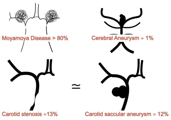
A mutation in RNF213 (c.14576G>A), a gene associated with moyamoya disease (>80%), plays a role in terminal internal carotid artery (ICA) stenosis (>15%) (ICS). Studies on RNF213 and cerebral aneurysms (AN), which did not focus on the site of origin or morphology, could not elucidate the relationship between the two. However, a report suggested a relationship between RNF213 and AN in French-Canadians. Here, we investigated the relationship between ICA saccular aneurysm (ICA-AN) and RNF213. We analyzed RNF213 expression in subjects with ICA-AN and atherosclerotic ICS. Cases with a family history of moyamoya disease were excluded. AN smaller than 4 mm were confirmed as AN only by surgical or angiographic findings. RNF213 was detected in 12.2% of patients with ICA-AN and 13.6% of patients with ICS; patients with ICA-AN and ICS had a similar risk of RNF213 mutation expression (odds ratio, 0.884; 95% confidence interval, 0.199–3.91; p = 0.871). The relationship between ICA-AN and RNF213 (c.14576G>A) was not correlated with the location of the ICA and bifurcation, presence of rupture, or multiplicity. When the etiology and location of AN were more restricted, the incidence of RNF213 mutations in ICA-AN was higher than that reported in previous studies. Our results suggest that strict maternal vessel selection and pathological selection of AN morphology may reveal an association between genetic mutations and ICA-AN development. The results of this study may form a basis for further research on systemic vascular diseases, in which the RNF213 (c.14576G>A) mutation has been implicated.

Graphical Abstract

1. Introduction

The ring finger protein (RNF)213 (c.14576G>A) mutation has been reported in 69.9–85.4% of cases of moyamoya disease (MMD) [1,2,3], an occlusive disease of the terminal portion of the internal carotid artery (ICA) [4]. Additionally, RNF213 (c.14576G>A) has been implicated in various systemic vascular diseases, including pulmonary hypertension, peripheral pulmonary artery stenosis, and coronary stenosis [5]. In East Asian patients, RNF213 (c.14576G>A) is also involved in 21–23.2% of cases of intracranial ICA stenosis (ICS); however, it is not observed in patients with vertebral artery stenosis [2,6].
The relationship between RNF213 (c.14576G>A) and the site of intracranial vascular disease has attracted attention lately. In a study on control subjects with no intracranial vascular diseases, RNF213 (c.14576G>A) was reported to have a high detection rate in East Asians (Japanese, 2.8%; Koreans, 2.5%; Chinese, 1.1%), but a negligible expression in Western Caucasians [2,7]. Furthermore, the frequency of RNF213 (c.14576G>A) in Japanese patients with cerebral aneurysms (CAN) was lower than that in patients with ICS and comparable (ranging from 0 to 2.1%) to that in control subjects with no vascular lesions [1]. Notably, RNF213 variants (p.Arg2438Cys and p.Ala2826Thr) were detected in French-Canadian patients with familial CAN [8].
Few studies have assessed the link between CAN and RNF213. Moreover, these reports failed to elucidate the details of CAN, including the site, morphology (saccular or fusiform shaped or dissected), and clinical characteristics of CAN [9]. CAN is known to exhibit different etiologies depending on the site of origin of the intracranial vessels, with different frequencies in men and women as well as different rupture rates, and thus should not be assessed in a similar way [10,11]. Hence, we aimed to investigate the role of RNF213 in patients with non-traumatic, non-infectious, non-neoplastic ICA “saccular” aneurysms (ICAN), diagnosed as ‘intracranial ICANs’ based on surgical findings in addition to multiple imaging methods.

2. Materials and Methods

2.1. Patient Information

This study was performed in accordance with the criteria of the STROBE (Strengthening the Reporting of Observational Studies in Epidemiology) statement. This study was a case-control study conducted at hospitals affiliated with our hospital and was approved by the Ethics Committee at the institution (Approved ID; H30-26-02, 1 July 2016). Written informed consent was obtained from the study participants for blood sampling, DNA storage, and genetic analysis. As the frequency of the RNF213 (c.14576G>A) mutation in Japanese patients with MMD has been established [1,2,6], we performed genetic analysis of the RNF213 (c.14576G>A) mutation in cases of ICAN and atherosclerotic intracranial ICA or middle cerebral artery stenosis or occlusion (ICS). Genetic analysis of the RNF213 (c.14576G>A) mutation was performed in patients who visited hospitals between August 2014 and August 2018 and agreed to participate in the study. A complete medical history was obtained from all patients.

2.2. Diagnosis

The ICAN cases included only non-traumatic, non-neoplastic, and non-infectious cases determined based on pre-existing disease and physiochemical and radiological findings. The diagnosis of ICAN was made by at least three physicians (two neurosurgeons and one radiologist), and both magnetic resonance angiography (MRA) and three-dimensional computed tomographic angiography (3DCTA) were mandatory. Infundibular dilatation [12] in the ICA was difficult to diagnose by MRA and 3DCTA alone when the size was < 4 mm. Hence, cerebral angiography or surgical findings by craniotomy were mandatory for the diagnosis of ICAN in this study. For the included cases, the location and multiplicity of ICAN, presence of rupture, and family history of CAN were noted along with other details, as previously mentioned. Patients with a family or personal history of diseases implicated in the development of CAN (including polycystic kidney disease, Ehlers–Danlos syndrome, and Marfan syndrome) were also excluded based on clinical findings. For ICS, at least two imaging studies from MRA/CTA/cerebral angiography were mandatory. Only patients with severe stenosis (>80%) from the petrous to the middle cerebral artery were included. Patients with MMD, quasi-MMD, congenital ICA deficiency, twig-like middle cerebral artery (MCA), or MCA dysplasia were excluded. Cases with a family history of MMD were excluded. Patients with a history or family history of diseases that have been indicated to be associated with MMD, including systemic lupus erythematosus, antiphospholipid antibody syndrome, periarteritis nodosa, and Sjogren’s syndrome, were also excluded from the study [4]. However, patients with atherosclerosis and diabetes mellitus were retained, despite their association with MMD [4]. Patients with a history or family history of other diseases associated with RNF213, including pulmonary hypertension, peripheral pulmonary artery stenosis, or coronary artery disease, were also excluded [5].

2.3. DNA Extraction and RNF213 Genotyping

Peripheral blood (5 cc) was collected from the patients and genomic DNA extraction was performed using the GENOMIX Kit (Talent, Trieste, Italy). Screening for the genotype of RNF213 (c.14576G>A) (exon 61) was performed by small amplicon genotyping based on high resolution melting curve analysis and verified by Sanger sequencing [13]. The polymerase chain reaction (PCR) primers for c.14576G>A were as follows: forward primer, 5′-GCAAGTTGAATACAGCTCCATCA-3′, and reverse primer, 5′- TGTGCTTGCTGAGGAAGCCT-3′. The PCR conditions included an initial denaturation step at 95 °C for 2 min, 45 cycles at 94 °C for 30 s, and an annealing step at 67 °C for 30 s. After the PCR, data were collected from 55 °C–97 °C in 96-well plates by LightScanner (Idaho Technology, Salt Lake City, UT, USA) at a ramp rate of 0.101 °C/s for high-resolution melting curve analysis.

2.4. Data Analyses

We performed comparative analyses between the ICAN and ICS cohorts for age at diagnosis, gender, RNF213 (c.14576G>A) mutation, and frequency of pre-existing disease. Continuous variables showing a non-normal distribution were compared between groups using the Mann–Whitney U test. Categorical variables, such as the proportion of patients with the RNF213 (c.14576G>A) mutation, were compared using Fisher’s exact test. A p-value < 0.05 was considered to indicate statistical significance. All analyses were performed using JMP 14.0 software (SAS Institute Inc., Cary, NC, USA).

3. Results

We assessed 47 cases of ICAN (total 69 CAN) and 22 cases of ICS during the study period. Of the 22 patients with ICS, two patients had ICAN. Hence, these two cases were included in both groups. Accordingly, 49 patients with ICAN and 22 patients with ICS were considered in the final study. The characteristics of patients in the ICAN and ICS groups are summarized in Table 1.
The c.14576G>A mutation in RNF213 (c.14576G>A) was found in six patients with ICAN (12.2%) and three patients with ICS (13.6%). No significant difference was noted in the frequency of RNF213 (c.14576G>A) in the assessed groups. Patients with ICAN exhibited a similar risk of RNF213 (c.14576G>A) mutation expression when compared with patients with ICS (odds ratio, 0.884; 95% confidence interval, 0.199–3.91; p = 0.871). Significantly more males (p = 0.0033) and cases of hypertension (p = 0.0038) and diabetes (p = 0.0046) were observed in the ICS group than in the ICAN group. The results from the analysis of characteristics in all patients with and without the RNF213 (c.14576G>A) are summarized in Table 2. None of the assessed variables differed significantly. As MMD is a bilateral carotid artery disease, we also assessed the frequency of bilateral lesions. No significant differences were observed in the occurrence of bilateral lesions in both groups.
The results from the analysis of patients with ICAN and ICS, with or without the RNF213 c.14576G>A mutation, are summarized in Table 3 and Table 4. With respect to the ICAN group, no significant differences were observed between patients with and without the RNF213 (c.14576G>A) mutation in all respects, including age, sex, past medical history, smoking history, family history of CAN, ruptured or unruptured ICAN, bilaterality, multiplicity, and location. With respect to the ICS group, no significant differences in age, gender, preexisting disease, or frequency of bilateral stenosis were noted. RNF213 (c.14576G>A) mutation was not detected in the two cases with both ICAN and ICS.

4. Discussion

Our results showed that the frequency of the RNF213 (c.14576G>A) mutation in Japanese patients with ICAN was approximately 12.5%. The frequency of RNF213 expression was not significantly different from the frequency of ICS in patients with non-MMD, whose family history of MMD was also investigated. In contrast, a link between the site of occurrence, gender, rupture, multiplicity, or familial nature of ICAN was not observed.
In this study, we constructed a study cohort with an emphasis on selecting patients with a reliable clinical phenotype of ICAN, no exogenous risk factors for CAN, and no family history of MMD and related disorders. The frequency of RNF213 expression has been reported to be significantly different in the ICA and vertebrobasilar artery systems, both in stenotic and dissecting lesions of ICA (21–28%) and VBAS (0–1%) [1,2,6,14]. However, there have been no studies on RNF213 that focus on the site of aneurysm development [8,9]. Furthermore, imaging and etiological diagnosis of small aneurysms is difficult [15]. Additionally, various causes and morphologies (e.g., trauma, dissection, and fusiform) must be reliably excluded when investigating the relationship between aneurysm development and genetic mutations. We suggested that RNF213 mutation should be examined by strictly classifying the location and initiation factors of the CAN.
We also examined our results for consistency with previous reports. In a survey of CAN in Japan, including CAN < 3 mm and fusiform type aneurysms, ICAN accounted for approximately 34% of all CAN [10]. Assuming that RNF213 was involved in 12.5% of ICAN only, it would comprise less than 4.2% of all CAN cases. Our study exhibited a frequency similar to that observed previously in Japanese subjects without intracranial vascular lesions [1,2]. The investigation of the relationship between RNF213 and CAN without focusing on the cause and branching site of CAN may be a factor in the failure to detect a relationship between CAN and RNF213 in previous studies.
The results of this study are not limited to the pathogenesis of cerebral aneurysms but may also have an impact on the search for causes of numerous diseases of systemic blood vessels. Cerebral aneurysms have been known to have characteristics, such as rupture rate differences, gender differences, and age differences depending on location and morphology, but no study has shown that there are differences in the expression of genetic mutations depending on location. In addition, there was a negative view of the relationship between aneurysms and RNF213 itself. We clarified the relationship between aneurysms and RNF213 by separating the aneurysm site and morphology. These results indicate that there is vascular site specificity in the functional expression of RNF213, which may have a ripple effect on the study of RNF213 in other sites, where its relationship with systemic vascular diseases is also clear. If RNF213 is detected, it may suggest which locations of blood vessels need to be watched in a systemic search for vascular diseases.
The relationship between genetic mutations and bias towards anterior circulation has been discussed. Moyamoya disease, for which RNF213 was discovered in 2011 [16,17] to be a disease susceptibility gene, is an occlusive disease of the bilateral ICA terminations occurring from childhood. Similarly, stenosis of pulmonary arteries [18] and coronary arteries [19] is among the pathologies of blood vessels wherein RNF213 has been implicated [5]. Of the internal carotid arteries that begin at the carotid bifurcation, the smooth muscle from the pyramidal bone to the end of the internal carotid artery is of neural crest origin [20], but the vertebral artery is not. For this reason, moyamoya disease or congenital carotid agenesis has been proposed as one of the neural crestopathies [20]. In addition, the smooth muscle of the pulmonary and coronary arteries is also of neural crest origin. Thus, it has recently become clear that the neural crest origin and the involvement of RNF213 are the two common features of these vessels. The SOX group has been reported [21] as a transcription factor that is specific to the neural crest, and RNF213 may play a similar role. In other words, RNF213 may affect the formation and function of neural crest cell blood vessels in the same way that SOX10 mutations cause the loss or dysfunction of neural crest cell-derived tissues [21]. In fact, it has been shown that the RNF213-encoded mysterin protein is involved in the regulation of functional vascular nerve formation as a cytoplasmic AAA+ ATPase and ubiquitinating enzyme [22].
This study had some limitations. First, the number of cases assessed was small. Thus, studies with larger sample sizes and assessing posterior circulation and analyzing various clinical phenotypes, such as aneurysm morphology, age of onset, and frequency of rupture, are needed. Second, we excluded cerebral aneurysms in all intracranial sites due to ethical reasons. As the detection rate of cerebral aneurysms and intracranial stenotic lesions increases with age, future studies should be conducted in elderly patients.

5. Conclusions

When the etiology and location of AN were more restricted, the incidence of RNF213 (c.14576G>A) mutations in ICA-AN was higher than that reported in previous studies. Our results suggest that strict maternal vessel selection and pathological selection of AN morphology may reveal an association between genetic mutations and ICA-AN development. The results of this study may form a basis for further research on systemic vascular diseases, in which the RNF213 (c.14576G>A) mutation has been implicated. Furthermore, these mutations may serve as predictive markers for patients with ICA-AN.

Author Contributions

Conceptualization, Y.M., A.M., T.T., A.W., E.I. and F.M.; methodology, A.W.; validation, Y.M., A.M., A.W., E.I. and F.M.; formal analysis, T.S., T.T., E.I., F.M. and R.N.; investigation, F.M. and K.S.; resources, K.K. and K.S.; data curation, Y.M., A.W., T.S., T.T., E.I., F.M. and R.N.; writing—original draft preparation, Y.M.; writing—review and editing, Y.M., A.M., A.W. and E.I.; supervision, T.S.; project administration, Y.M. and A.M. All authors have read and agreed to the published version of the manuscript.

Funding

This research received no external funding.

Institutional Review Board Statement

The study was conducted according to the guidelines of the Declaration of Helsinki and approved by the Ethics Committee of Nippon Medical School Hospital (H30-26-02, 1 July 2016).

Informed Consent Statement

Informed consent was obtained from all subjects involved in the study. Written informed consent has been obtained from the patients to publish this paper.

Data Availability Statement

The data presented in this study are available in the article.

Acknowledgments

This work was supported by the Japan Society for the Promotion of Science (JSPS) KAKENHI Grant-in-Aid for Scientific Research (C) Number 18K09008. We are grateful for the guidance of Yohei Kawasaki from Academic Statistical Support (https://statistical-support.com), an expert in statistical analysis.

Conflicts of Interest

The authors declare no conflict of interest.

References

  1. Miyawaki, S.; Imai, H.; Shimizu, M.; Yagi, S.; Ono, H.; Mukasa, A.; Nakatomi, H.; Shimizu, T.; Saito, N. Genetic variant RNF213 c.14576G>A in various phenotypes of intracranial major artery stenosis/occlusion. Stroke 2013, 44, 2894–2897. [Google Scholar] [CrossRef] [Green Version]
  2. Liao, X.; Deng, J.; Dai, W.; Zhang, T.; Yan, J. Rare variants of RNF213 and moyamoya/non-moyamoya intracranial artery stenosis/occlusion disease risk: A meta-analysis and systematic review. Environ. Health Prev. Med. 2017, 22, 75. [Google Scholar] [CrossRef] [PubMed] [Green Version]
  3. Ishisaka, E.; Watanabe, A.; Murai, Y.; Shirokane, K.; Matano, F.; Tsukiyama, A.; Baba, E.; Nakagawa, S.; Tamaki, T.; Mizunari, T.; et al. Role of RNF213 polymorphism in defining quasi-moyamoya disease and definitive moyamoya disease. Neurosurg. Focus 2021, 51, E2. [Google Scholar] [CrossRef] [PubMed]
  4. Hashimoto, N.; Tominaga, T.; Miyamoto, S.; Nagata, I.; Houkin, K.; Suzuki, N.; Takagi, Y. Research Committee on the Pathology and Treatment of Spontaneous Occlusion of the Circle of Willis; Health Labour Sciences Research Grant for Research on Measures for Infractable Diseases. Guidelines for diagnosis and treatment of moyamoya disease (spontaneous occlusion of the circle of Willis). Neurol. Med. Chir. 2012, 52, 245–266. [Google Scholar] [CrossRef] [Green Version]
  5. Bang, O.Y.; Chung, J.W.; Kim, D.H.; Won, H.H.; Yeon, J.Y.; Ki, C.S.; Shin, H.J.; Kim, J.S.; Hong, S.C.; Kim, D.K.; et al. Moyamoya Disease and Spectrums of RNF213 Vasculopathy. Transl. Stroke Res. 2020, 11, 580–589. [Google Scholar] [CrossRef] [PubMed]
  6. Shinya, Y.; Miyawaki, S.; Imai, H.; Hongo, H.; Ono, H.; Takenobu, A.; Nakatomi, H.; Teraoka, A.; Saito, N. Genetic Analysis of Ring Finger Protein 213 (RNF213) c.14576G>A in Intracranial Atherosclerosis of the Anterior and Posterior Circulations. J. Stroke Cerebrovasc. Dis. 2017, 26, 2638–2644. [Google Scholar] [CrossRef] [PubMed]
  7. Wang, X.; Wang, Y.; Nie, F.; Li, Q.; Zhang, K.; Liu, M.; Yang, L.; Zhang, Q.; Liu, S.; Zeng, F.; et al. Association of Genetic Variants with Moyamoya Disease in 13,000 Individuals: A Meta-Analysis. Stroke 2020, 51, 1647–1655. [Google Scholar] [CrossRef]
  8. Zhou, S.; Ambalavanan, A.; Rochefort, D.; Xie, P.; Bourassa, C.V.; Hince, P.; Dionne-Laporte, A.; Spiegelman, D.; Gan-Or, Z.; Mirarchi, C.; et al. RNF213 Is Associated with Intracranial Aneurysms in the French-Canadian Population. Am. J. Hum. Genet. 2016, 99, 1072–1085. [Google Scholar] [CrossRef] [Green Version]
  9. Miyawaki, S.; Imai, H.; Takayanagi, S.; Mukasa, A.; Nakatomi, H.; Saito, N. Identification of a genetic variant common to moyamoya disease and intracranial major artery stenosis/occlusion. Stroke 2012, 43, 3371–3374. [Google Scholar] [CrossRef] [Green Version]
  10. The Natural Course of Unruptured Cerebral Aneurysms in a Japanese Cohort. N. Engl. J. Med. 2012, 366, 2474–2482. [CrossRef] [PubMed] [Green Version]
  11. Zheng, J.; Zhang, J. Neoplastic cerebral aneurysm from metastatic tumor: A systematic review of clinical and treatment characteristics. Clin. Neurol. Neurosurg. 2015, 128, 107–111. [Google Scholar] [CrossRef] [PubMed]
  12. Kim, Y.J.; Yoon, D.Y.; Kim, E.S.; Yun, E.J.; Jeon, H.J.; Lee, J.Y.; Cho, B.M. Geometric parameters on MRA source images to differentiate small Proximal Posterior communicating artery aneurysms from Infundibular dilation. J. Neuroimaging 2021, 31, 532–540. [Google Scholar] [CrossRef]
  13. Watanabe, A.; Karasugi, T.; Sawai, H.; Naing, B.T.; Ikegawa, S.; Orimo, H.; Shimada, T. Prevalence of c.1559delT in ALPL, a common mutation resulting in the perinatal (lethal) form of hypophosphatasia in Japanese and effects of the mutation on heterozygous carriers. J. Hum. Genet. 2011, 56, 166–168. [Google Scholar] [CrossRef] [PubMed] [Green Version]
  14. Tashiro, R.; Fujimura, M.; Sakata, H.; Endo, H.; Tomata, Y.; Sato-Maeda, M.; Niizuma, K.; Tominaga, T. Genetic analysis of ring finger protein 213 (RNF213) c.14576G>A polymorphism in patients with vertebral artery dissection: A comparative study with moyamoya disease. Neurol. Res. 2019, 41, 811–816. [Google Scholar] [CrossRef] [PubMed]
  15. Walkoff, L.; Brinjikji, W.; Rouchaud, A.; Caroff, J.; Kallmes, D.F. Comparing magnetic resonance angiography (MRA) and computed tomography angiography (CTA) with conventional angiography in the detection of distal territory cerebral mycotic and oncotic aneurysms. Interv. Neuroradiol. 2016, 22, 524–528. [Google Scholar] [CrossRef]
  16. Kamada, F.; Aoki, Y.; Narisawa, A.; Abe, Y.; Komatsuzaki, S.; Kikuchi, A.; Kanno, J.; Niihori, T.; Ono, M.; Ishii, N.; et al. A genome-wide association study identifies RNF213 as the first Moyamoya disease gene. J. Hum. Genet. 2011, 56, 34–40. [Google Scholar] [CrossRef] [Green Version]
  17. Liu, W.; Morito, D.; Takashima, S.; Mineharu, Y.; Kobayashi, H.; Hitomi, T.; Hashikata, H.; Matsuura, N.; Yamazaki, S.; Toyoda, A.; et al. Identification of RNF213 as a susceptibility gene for moyamoya disease and its possible role in vascular development. PLoS ONE 2011, 6, e22542. [Google Scholar] [CrossRef] [PubMed] [Green Version]
  18. Kobayashi, H.; Kabata, R.; Kinoshita, H.; Morimoto, T.; Ono, K.; Takeda, M.; Choi, J.; Okuda, H.; Liu, W.; Harada, K.H.; et al. Rare variants in RNF213, a susceptibility gene for moyamoya disease, are found in patients with pulmonary hypertension and aggravate hypoxia-induced pulmonary hypertension in mice. Pulm. Circ. 2018, 8, 2045894018778155. [Google Scholar] [CrossRef] [PubMed] [Green Version]
  19. Morimoto, T.; Mineharu, Y.; Ono, K.; Nakatochi, M.; Ichihara, S.; Kabata, R.; Takagi, Y.; Cao, Y.; Zhao, L.; Kobayashi, H.; et al. Significant association of RNF213 p.R4810K, a moyamoya susceptibility variant, with coronary artery disease. PLoS ONE 2017, 12, e175649. [Google Scholar] [CrossRef] [Green Version]
  20. Komiyama, M. Cardio-cephalic neural crest syndrome: A novel hypothesis of vascular neurocristopathy. Interv. Neuroradiol. 2017, 23, 572–576. [Google Scholar] [CrossRef] [PubMed]
  21. Haldin, C.E.; LaBonne, C. SoxE factors as multifunctional neural crest regulatory factors. Int. J. Biochem. Cell Biol. 2010, 42, 441–444. [Google Scholar] [CrossRef] [Green Version]
  22. Key, J.; Maletzko, A.; Kohli, A.; Gispert, S.; Torres-Odio, S.; Wittig, I.; Heidler, J.; Bárcena, C.; López-Otín, C.; Lei, Y.; et al. Loss of mitochondrial ClpP, Lonp1, and Tfam triggers transcriptional induction of Rnf213, a susceptibility factor for moyamoya disease. Neurogenetics 2020, 21, 187–203. [Google Scholar] [CrossRef]
Table 1. Patient characteristics based on the disease group.
ICANICSp Value
Sample size (N)4922
RNF213 +630.573
Male: Female11:3813:90.0033
Mean Age62.1862.540.833
Standard deviation13.8211.18
Standard error1.9752.384
Median6565
Interquartile range20.519.5
Hypertension28200.0038
Diabetes mellitus590.0046
Dyslipidemia20140.0636
Smoking24140.1876
ICAN, internal carotid artery saccular aneurysm; ICMS, internal carotid artery or middle cerebral artery stenosis; N, number; RNF213, ring finger protein 213 (c.14576G>A); RNF213 +, RNF213 (c.14576G>A) carriers.
Table 2. Characteristics of cases with and without RNF213 c.14576G>A.
ICAN and ICSRNF +RNFp Value
N962
Male: Female3:621:410.647
Mean Age59.3362.730.489
Standard deviation14.8712.77
Standard error4.961.62
Median6565
Interquartile range29.519.5
Bilateral lesions2120.755
Hypertension7410.389
Diabetes mellitus1130.431
Dyslipidemia2320.097
Smoking4340.409
ICAN, internal carotid artery saccular aneurysm; ICS, internal carotid artery or middle cerebral artery stenosis; RNF, ring finger protein 213 (c.14576G>A); RNF213 +, RNF213 (c.14576G>A) carriers; RNF213 −, RNF213 (c.14576G>A) non-carriers.
Table 3. Characteristics of cases with and without RNF213 (c.14576G>A) in internal carotid artery aneurysm.
ICANRNF +RNFp Value
Number643
Mean Age60.562.420.819
Standard deviation17.2413.51
Standard error7.042.06
Median69.565
Interquartile range33.520
Male1100.592
Proximal to Paraclinoid1100.592
Proximal to SHA1190.204
Bilateral ICAN060.436
Bilateral CAN090.275
Multiple ICAN2100.46
Multiple CAN2140.649
Family history of CAN120.33
Ruptured CAN1120.49
Hypertension4230.482
Diabetes mellitus040.505
Dyslipidemia1180.204
Smoking2210.354
CAN, cerebral aneurysm; ICA, internal carotid artery; ICAN, internal carotid artery aneurysm; RNF, ring finger protein 213 (c.14576G>A); SHA, superior hypophyseal artery; RNF213 +, RNF213 (c.14576G>A) carriers; RNF213 −, RNF213 (c.14576G>A) non-carriers.
Table 4. Characteristics of cases with and without RNF213 (c.14576G>A) in the internal carotid artery stenosis group.
ICSRNF +RNFp Value
Number319
Mean Age5763.420.212
Standard deviation11.3611.21
Standard error6.562.57
Median6266
Interquartile range2121
Male2110.642
Bilateral stenosis230.117
Hypertension3170.74
Diabetes mellitus180.642
Dyslipidemia1130.291
Smoking2120.764
ICS, internal carotid artery or middle cerebral artery stenosis; RNF, ring finger protein 213 (c.14576G>A); RNF213 +, RNF213 (c.14576G>A) carriers; RNF213 −, RNF213 (c.14576G>A) non-carriers.
Publisher’s Note: MDPI stays neutral with regard to jurisdictional claims in published maps and institutional affiliations.