Article

The Potential Effect of Aberrant Testosterone Levels on Common Diseases: A Mendelian Randomization Study

1
Bio-X Institutes, Key Laboratory for the Genetics of Developmental and Neuropsychiatric Disorders (Ministry of Education), Shanghai Jiao Tong University, 1954 Huashan Road, Shanghai 200030, China
2
Shanghai Center for Women and Children’s Health, 339 Luding Road, Shanghai 200062, China
*
Author to whom correspondence should be addressed.
Genes 2020, 11(7), 721; https://doi.org/10.3390/genes11070721
Submission received: 8 June 2020 / Revised: 21 June 2020 / Accepted: 26 June 2020 / Published: 29 June 2020
(This article belongs to the Section Human Genomics and Genetic Diseases)

Abstract

:
Testosterone has historically been linked to sexual dysfunction; however, it has recently been shown to affect other physical and mental attributes. We attempted to determine whether changes in serum testosterone could play a role in chronic or degenerative diseases. We used two separate genetic instruments comprising of variants from JMJD1C and SHBG regions and conducted a two-sample Mendelian randomization for type II diabetes (T2D), gout, rheumatoid arthritis (RA), schizophrenia, bipolar disorder, Alzheimer’s disease and depression. For the JMJD1C locus, one unit increase in log transformed testosterone was significantly associated with RA (OR = 1.69, p = 0.02), gout (OR = 0.469, p = 0.001) and T2D (OR = 0.769, p = 0.048). Similarly, one unit increase in log transformed testosterone using variants from the SHBG locus was associated with depression (OR = 1.02, p = 0.001), RA (OR = 1.32, p < 0.001) and T2D (OR = 0.88, p = 0.003). Our results show that low levels of serum testosterone levels may cause gout and T2D, while higher than normal levels of testosterone may result in RA and depression. Our findings suggest that fluctuations in testosterone levels may have severe consequences that warrant further investigation.

1. Introduction

Testosterone is a universally known hormone which is responsible for the development of primary, as well as secondary male characteristics, such as genitalia, beards and muscle mass [1]. Historically, the role of testosterone was thought to be limited to the development of male characteristics and sexual function; however, recently it has come to light that the role of testosterone may be multifarious. Research has reported testosterone to be essential to male health, with some studies showing mortality rates to be as much as 40% higher in individuals with extremely low testosterone levels [2,3]. Studies conducted over the previous decades have discovered that testosterone has the potential to influence men both physically and psychologically, affecting the overall health of the male population.
Male hypogonadism is defined by The Endocrine Society as the inability to produce physiological levels of testosterone and is clinically defined as an early morning testosterone concentration of 250–300 ng/dL or below [4]. Hypogonadism can be present at any phase of life, from fetal development to old age; however, it is known that serum testosterone levels steadily decrease as men age [5,6] and is symptomized by decreased libido, erectile dysfunction, decreased muscle mass and bone mineral density, all of which were previously dismissed as a natural consequence of aging. Testosterone levels begin to naturally decrease between the ages of 20 and 30 in males, at a rate of about 1.3% per year [7], and by the age of 45, about 39% of men have serum testosterone levels below 300 ng/dL [8]. An investigation into testosterone tests undertaken by individuals in United Kingdom and the United States, from the years 2000 to 2011, shows that male hypogonadism is quite common, with an excess of 20% of tests displaying a low level of serum testosterone (< 300 mg/dL). This phenomenon is not only restricted to the elderly, as up to 76% of participants were between the ages of 40 and 64 [9].
Increasing awareness of hypogonadism and its association with common conditions such as type II diabetes mellitus, obesity and metabolic syndrome, ultimately gave rise to testosterone replacement therapy (TRT) in an attempt to reverse the effects of hypogonadism. A recent meta-analysis randomized control trial (RCT) reports that TRT can improve quality of life, libido, depression and erectile dysfunction [10]; further studies report the positive effects of TRT on obesity, lipid profile and cardiovascular risk factors [11]. The safety of testosterone supplement usage has been called into question, with increased incidents of cardiovascular events observed in patients undergoing therapy [12]. Two large-scale observational studies have since confirmed increased occurrence of cardiovascular events as a result of TRT [13,14], leading Health Canada and the US Food and Drug Administration (FDA) to place labels on TRT products in an attempt to warn the public of the potentially adverse cardiological effects of TRT.
Testosterone has also been shown to greatly influence male behavior, particularly aggressive behaviors which can termed as antisocial, while also stimulating generosity when it can serve to increase the male’s dominance [15,16]. Over the past few decades, testosterone has been consistently linked to cognitive decline, particularly in the elderly [17]. Results from several epidemiological studies have suggested that testosterone acts as a metabolic hormone in males, as there is a negative correlation between testosterone levels (both free as well as SHBG bound) and obesity across all age groups [18,19,20,21].
Mendelian randomization provides us with a technique that can uncover causal relationships without the extensive costs incurred by randomized control trials, and is becoming increasingly more viable as data from numerous large genome wide association studies (GWAS) investigating a diverse range of conditions over the past decade are now publicly available. Early Mendelian randomization studies involving testosterone were performed in small samples and were subsequently met with little success [22,23,24]; recent studies that shifted to the use of large-scale GWAS to source associations of variant with exposure and outcome have reported significant findings regarding testosterone levels and the risk of stroke, cardiovascular disease, polycystic ovary syndrome, as well as breast, endometrial and prostate cancer [25,26].
It is now obvious that the functions of testosterone extend well beyond the sexual domain, especially in men, affecting both physiological and mental aspects of life. Hypogonadism is widespread and inevitable; however, it is difficult to conduct studies to ascertain the full extent of testosterone influence, which has been hypothesized to play a role in a range of common metabolic, immune and psychiatric diseases [27,28,29,30,31,32,33].
In this study, we aim to determine the effect of testosterone on previously associated common diseases, namely, type II diabetes (T2D), rheumatoid arthritis (RA), gout, depression, bipolar disorder (BP), schizophrenia (SCZ) and Alzheimer’s disorder (AD) [27,28,29,30,31,32,33], by conducting a two-sample Mendelian randomization study in an effort to determine whether testosterone could potentially play a causative role in these disorders. To our knowledge, this is the first Mendelian randomization study looking into the relationship between testosterone and common diseases.

2. Materials and Methods

2.1. Genetic Instruments (Genetic Associations with Testosterone)

The genetic instruments for testosterone were created using data from the REDUCE study, where 3225 men of European descent between the ages of 50 and 75 years had consented to additional genetic testing [34]. The only variants significantly associated with serum testosterone at a genome-wide association level were present at 10q21, 17p13, Xp22, and were henceforth referred to as the JMJD1C, SHBG and FAM9B loci, respectively. Variants from the FAM9B locus were not included in our analysis since genetic variants in outcome data did not include the sex chromosomes. The SHBG, also known as sex hormone binding globulin, has a mechanistic link to all sex hormones and testosterone is no different, while the JMJD1C locus is associated with testosterone levels independent of SHBG.
Genetic instruments were created for both JMJD1C and SHBG loci separately, and a total of 661 and 325 variants were reported for the JMJD1C and SHBG loci, respectively. The genetic instrument for the JMJD1C locus was created based on a single genome-wide significant variant while the instrument for the SHBG locus was created using a step-wise selection procedure similar to the one used in a previous Mendelian randomization study [26]. In brief, for the SHBG locus, from the list of 325 variants, the strongest variant in terms of p-value was selected, and all other variants that were in linkage disequilibrium (defined as having an r2 value of > 0.40) were removed, after which the process was repeated for the next most significant variant, resulting in a set of variants with low pair-wise correlations.

2.2. Genetic Association with Outcomes

Genetic association data for the selected outcomes were sourced from the GWAS catalogue. In this study, we investigated the association of the genetic instrument with various outcomes, namely, type 2 diabetes (T2D), gout, rheumatoid arthritis (RA), depression, schizophrenia, bipolar disorder and Alzheimer’s disorder. The genetic associations for T2D were extracted from a recently published meta-analysis of GWA studies that comprised approximately 16 million variants and included 62,892 cases along with 596,424 controls of European descent [35]. This meta-analysis was comprised of data from DIAGRAM, GERA and UK Biobank data sets.
The second outcome was derived from a study by Tin et al. [36], who performed a transancestry meta-analysis of serum urate levels (457,690 individuals) and made a subsequent prediction of gout in a separate cohort of 334,880 individuals from the UK Biobank. For RA, the genetic associations were extracted from a meta-analysis of GWA studies that evaluated approximately 10 million single nucleotide polymorphisms (SNPs), with total sample size >100,000 individuals (of European and Asian descent) comprising 29,880 and 73,758 cases and controls, respectively [37].
For depression, we used the genetic associations from the UK biobank [38], which has divided depression into three distinct phenotypes, mainly broad depression, probable major depressive disorder (MDD) and MDD as defined by the International Classification of Diseases (ICD). Here we selected genetic associations for broad depression as the outcome measure, as it most closely related to general depression experienced by most individuals. The UK Biobank depression GWAS was comprised of 322,580 participants. Genetic associations for schizophrenia were extracted from the GWAS published by the schizophrenia working group of the Psychiatric Genomics Consortium (PGC) [39], which is the largest GWAS performed on a European population today, comprising 36,989 cases and 113,075 controls.
The genetic associations for bipolar disorder were extracted from a recent GWAS that was published in 2019, comprising 20,352 and 31,358 cases and controls, which was followed up with a separate group of an additional 9412 cases and 137,760 controls. A recently published meta-analysis of Alzheimer’s disorder studies comprising 67,614 cases was used to extract genetic associations with Alzheimer’s disorder. This meta-analysis combined data from the UK Biobank and International Genomics of Alzheimer’s Project (IGAP)

2.3. Statistical Analysis

Mendelian randomization is a technique that can be used to determine causal exposure–outcome relationships. Since alleles are randomly assigned at birth, genetic variants that are associated with an increase in exposure can be used to determine whether the said exposure could cause disease, while being completely randomized for confounders such as smoking or education, similar to a randomized control trial.
The genetic associations and outcomes were harmonized before the Mendelian randomization analysis, which was performed separately from the two loci (SHBG and JMJD1C). The Mendelian randomization analysis was performed using the inverse variance (IVW) method: if the genetic instrument is comprised of a single variant then the resulting causal estimate is simply the ratio of association of variant with outcome and exposure. If the genetic instrument consists of more than one variant, then the causal estimate is calculated by pooling the ratio estimates using an inverse variance meta-analysis.
Mendelian randomization analysis requires that genetic instruments utilized are associated with the outcome of interest only via the exposure, and is not associated with other exposures or confounders. In simple terms, Mendelian randomization requires that the genetic instrument used does not display pleiotropy; a test directional pleiotropy can be performed using the MR-Egger intercept estimate [40]. Two-sided p-values were reported for all analyses and significant casual estimates were defined as estimates with a p-value of < 0.05. Additional sensitivity analyses were performed using the weighted median method, which is capable of providing a consistent estimate even if up to 50% of the genetic instrument is invalid. All analyses were performed using the Mendelian randomization package in R.

3. Results

3.1. Genetic Instruments

After accounting for linkage disequilibrium, the genetic instrument for the JMJD1C locus was comprised of a single SNP (rs10822184), while the SHBG locus comprised of 20 variants (Table 1); however, a different set of variants was used depending on availability in the outcome data set. The exact composition of the genetic instruments and the pairwise correlations of the included variants can be found in the supplementary files (Figures S1–S3 and Tables S1–S3).

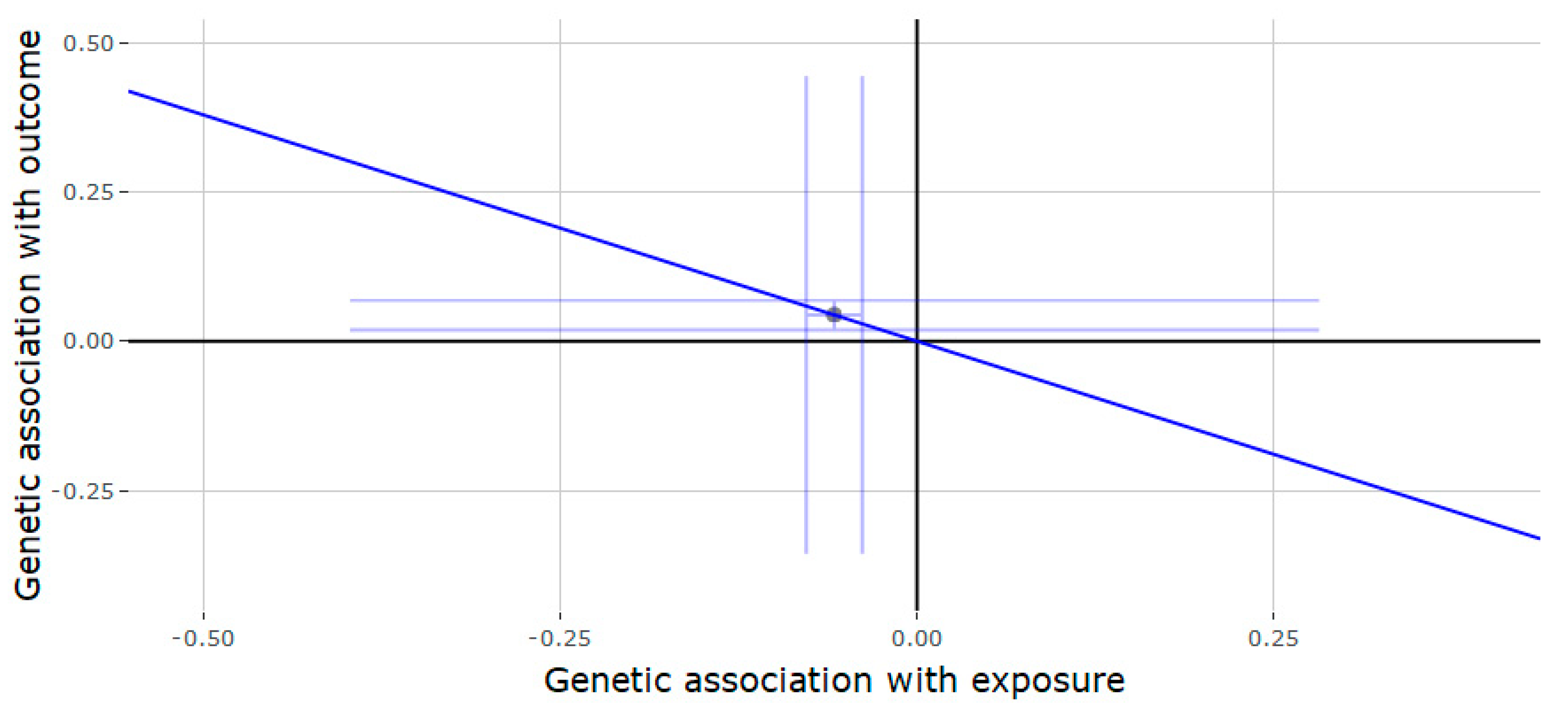
3.2. Mendelian Randomization of the JMJD1C Locus

The results of the Mendelian randomization or the JMJD1C locus (Table 2) showed that genetically predicted testosterone was negatively associated with gout (estimate = −0.757, 95% CI = −1.189, −0.324, p = 0.001) (Figure 1) and T2D (estimate = −0.262, 95% CI = −0.522, −0.002, p = 0.048) (Figure 2). Each increase in SD for genetically determined serum testosterone levels was positively associated with RA (estimate = 0.525, 95% CI = 0.083, 0.967, p = 0.020) (Figure 3). There was no association between genetically predicted testosterone and the following outcomes: Alzheimer disorder, bipolar disorder, schizophrenia and depression. Since the genetic instrument for the JMJD1C locus was comprised of only a single variant, it was not possible to conduct the sensitivity analyses (weighted median and MR-Egger methods).

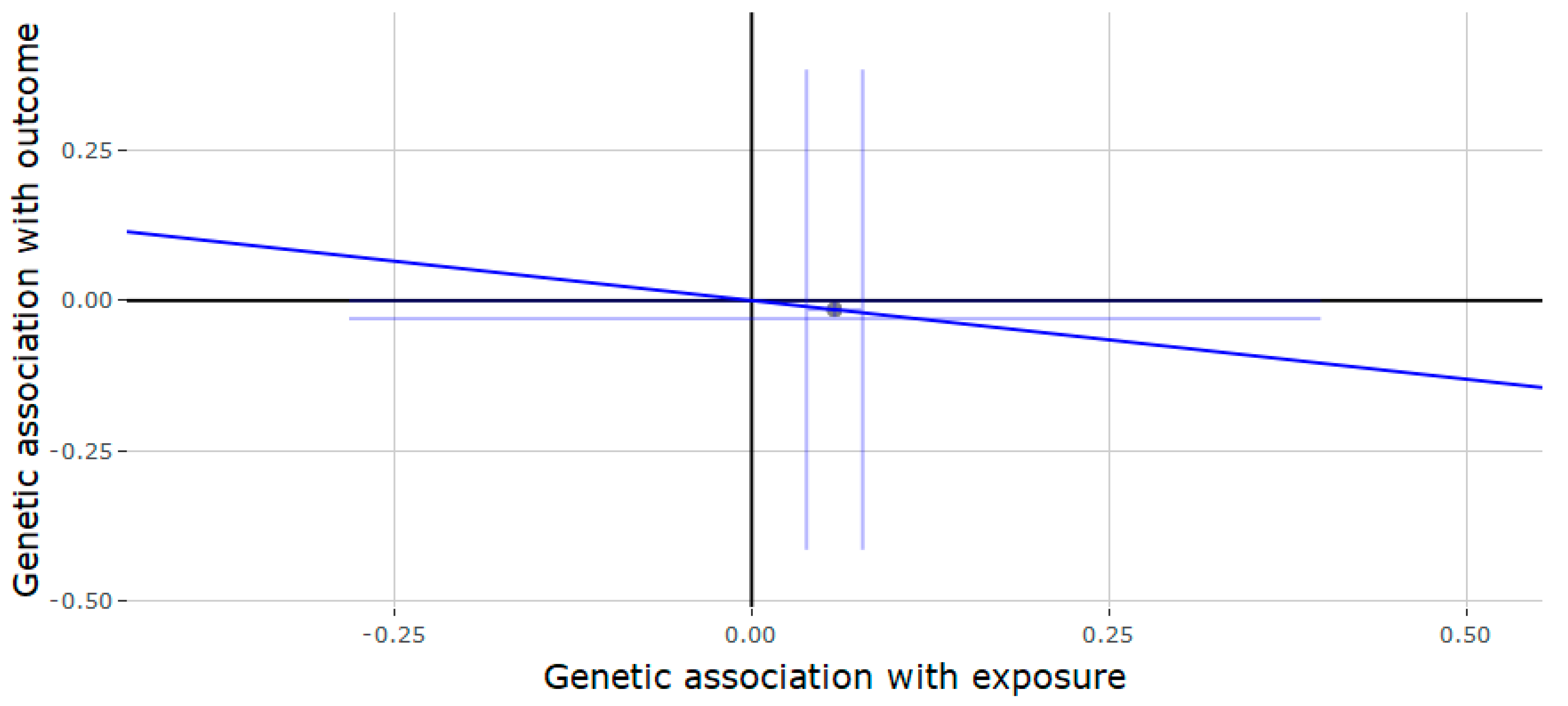
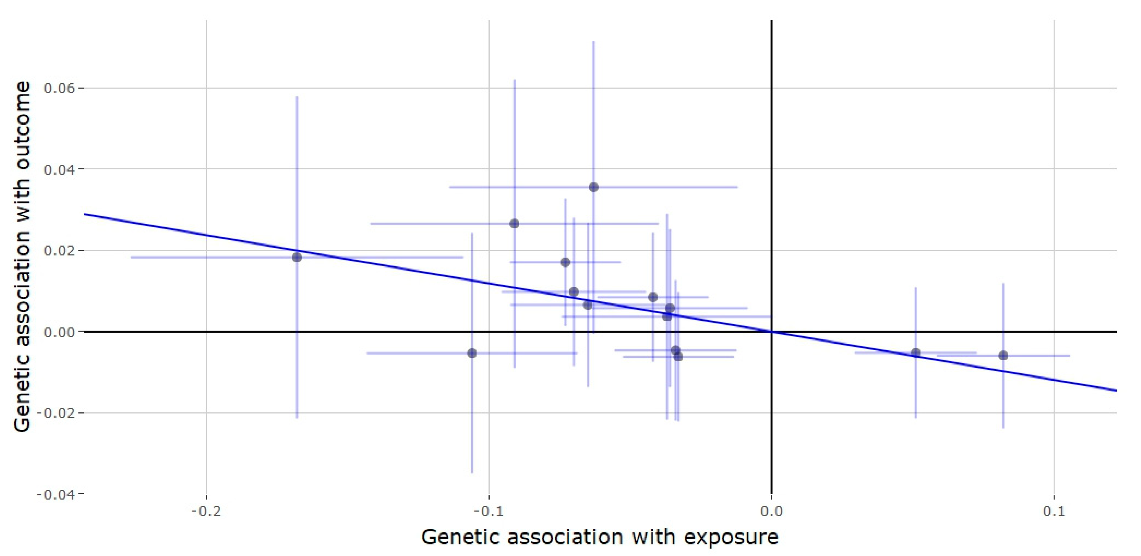
3.3. Mendelian Randomization of the SHBG Locus

Results of the Mendelian randomization analysis of predictors of testosterone from the SHBG region (Table 3) showed that genetically predicted testosterone was positively associated with depression and RA, while negatively associated with T2D. Each SD increase in genetically predicted testosterone was associated with a 1.02-fold increased risk of depression (estimate = 0.021, 95% CI = 0.008, 0.031, p = 0.001) (Figure 4). During the sensitivity analysis, the weighted median method showed similar results to the IVW analysis (estimate = 0.016, p = 0.040), while there was no evidence for pleiotropy (MR-Egger intercept = 0.002, p = 0.092). For RA, each SD increase in testosterone resulted in a 1.33-fold increase in the risk of RA (estimate = 0.285, 95% CI = 0.141, 0.429, p < 0.001) (Figure 5), which was similar to the estimates from the weighted median method sensitivity analysis (estimate = 0.368, p < 0.001) without any evidence of horizontal pleiotropy (MR-Egger intercept = 0.022, 95% CI = −0.006, 0.049, p = 0.120). Each SD increase in testosterone correlated to a 0.887-fold increase in the risk of T2D (estimate = −0.119, 95% CI = −0.203, −0.035, p = 0.005) (Figure 6), which was mirrored by the weighted median method analysis (estimate = −0.107, 95% CI = −0.219, −0.005, p = 0.062), without any horizontal pleiotropy detected (MR-Egger intercept estimate = 0.003, p = 0.219). Genetic association of genetically predicted testosterone with SCZ, BP, T2D and AD were null; however, the direction of effect for T2D was in agreement with that of the significant effect found in the JMJD1C analysis.

4. Discussion

Serum testosterone levels naturally decrease over the course of a man’s life or could be reduced as a result of hypogonadism. In this study, we aimed to explore the possible effects of this decrease on common diseases by Mendelian randomization, using genetically predicted testosterone levels derived from two gene regions significantly associated with serum testosterone levels. Mendelian randomization analyses were performed separately on the two instruments; results from the JMJD1C and SHBG loci showed significant associations with three outcomes each. We investigated a total of seven diseases, namely, T2D, SCZ, BP, AD, gout, RA and depression, of which four outcomes displayed significant associations with genetically predicted testosterone levels. Analyses of both loci supported significant associations of genetically predicted testosterone with RA and T2D, while also displaying the same direction of effect.
The results of our study show that an increase in serum testosterone has a protective effect on the risk of developing T2D, with both loci displaying significant results with consistent direction of effect. Existing literature has extensively studied the relationship between serum testosterone and T2D, and found testosterone can directly regulate glucose metabolism [41]; additionally, low levels of serum testosterone were also associated with reduced insulin sensitivity [42]. Testosterone plays an important role in glucose metabolism as it has been shown to increase expression of insulin receptors as well modify the glucose transporter (GLUT4) [43]. Testosterone could also regulate glucose homeostasis and glucose uptake via androgen receptor (AR)-dependent pathways in brain, adipose and pancreatic tissues, as well as AR-independent pathways, for example, by activating the LKB1/AMPK signaling pathway in adipocytes [44]. Impairment of the AMPK pathway has been observed in T2D patients, activation of which results in improved uptake of glucose [45]. Interestingly, activation of the LKB1/AMPK pathway has been shown to result in greater amyloid-β generation [46]. A recent meta-analysis investigating the relationship between low serum testosterone and risk of AD concluded that low serum testosterone was significantly associated with increased risk of AD [33]; however, this may be a characteristic of disease progression rather than a causal relationship. Alternatively, LKB1/AMPK activation via testosterone earlier in life could potentially cause the build-up of amyloid-β plaques and cause AD. T2D and AD share a common pathology to some extent, as both diseases display insulin signaling defects and resistance [47], while testosterone may play a crucial role in the pathogenesis of both these disorders. Additionally, hyperglycaemia, which could result, has also been shown to reduce serum testosterone levels [48,49], which indicates that these relationships are not as straight forward as one may think, as elevated glucose levels result in an increase in adipose tissue, which in turn result in a reduction in testosterone production [50].
Historically low levels of serum testosterone have been regularly associated with RA [51,52], and the hypothalamic–pituitary–adrenal axis has been shown to be affected by RA [53], potentially leading to patients affected by RA to display aberrant testosterone levels [54]. In our analysis, we show that increased serum testosterone increases the risk of developing RA, and a recent study investigating the future risk of developing RA in men concluded that low serum testosterone is associated with a rheumatoid factor (RF) negative prediction [29], suggesting the low levels of testosterone may provide protection against development of RA.
Hypogonadism has been linked to depression in males [55,56], while testosterone has potential antidepressive properties [57]; our results surprisingly suggest that increased testosterone may actually slightly increase the risk of depression.

5. Conclusions

In conclusion, our Mendelian randomization study assessed the effect of testosterone on the risk of several common diseases. We report that testosterone has a protective effect on the risk of T2D and gout, while having adverse effects on depression and RA. There were, however, a few limitations of our study, first and foremost being that the genetic instruments used in our Mendelian randomization were sourced from variant testosterone associations measured in men; therefore, our findings are largely relevant to common diseases in men. Another limitation of our study was the use of variants in the SHBG region to genetically predict testosterone. SHBG can bind to free testosterone, thereby affecting the concentrations of testosterone in the blood; though SHBG can be linked to testosterone levels mechanistically, it is difficult to distinguish between the effects of testosterone from that of SHBG.
Our findings, along with previous Mendelian randomization studies on the effect of testosterone on obesity and adverse cardiac events, highlight the importance of testosterone in overall health and well-being, particularly in males. The results of our study show that further research into the effects of testosterone is required, especially in light of the increasing utilization of testosterone therapy.

Supplementary Materials

The following are available online at https://www.mdpi.com/2073-4425/11/7/721/s1. Figure S1: LD matrix—depression, SHBG instrument locus; Figure S2: LD matrix—RA instrument, SHBG locus; Figure S3: LD matrix—T2D instrument, SHBG locus; Table S1: Depression, SHBG instrument locus; Table S2: RA instrument, SHBG locus; Table S3: T2D instrument, SHBG locus.

Author Contributions

Conceptualization, A.A.S.S.; methodology, A.A.S.S.; software, A.A.S.S.; validation, A.A.S.S. and Y.S.; formal analysis, A.A.S.S.; investigation, A.A.S.S.; resources, L.H.; data curation, A.A.S.S.; writing—original draft preparation, A.A.S.S.; writing—review and editing, Y.S.; visualization, A.A.S.S.; supervision, L.H.; project administration, Y.S.; funding acquisition, L.H. and Y.S. All authors have read and agreed to the published version of the manuscript.

Funding

This research and the Article Processing Charges (APC) were supported by the National Key R&D Program of China (2017YFC0908105), National Key R&D Program of China (2019YFA0905400), Shanghai Municipal Science and Technology Major Project (2017SHZDZX01), the Natural Science Foundation of China (U1804284, 81421061, 81701321, 31571012, 81501154), the Program of Shanghai Subject Chief Scientist (15XD1502200), the National Program for Support of Top-Notch Young Professionals, Shanghai Key Laboratory of Psychotic Disorders (13dz2260500), the National Program for Support of Top-Notch Young Professionals to Y.S., Shanghai Hospital Development Center (SHDC12016115), Shanghai Municipal Commission of Science and Technology (17JC1402900, 17490712200, 18DZ2260200) and shanghai municipal health commission (ZK2015B01, 201540114), Scientific Research and Development Fund of Shanghai Jiao Tong University (19X150010012).

Conflicts of Interest

The authors declare no conflict of interest.

References

  1. Mooradian, A.D.; Morley, J.E.; Korenman, S.G. Biological Actions of Androgens. Endocr. Rev. 1987, 8, 1–28. [Google Scholar] [CrossRef] [PubMed]
  2. Laughlin, G.A.; Barrett-Connor, E.; Bergstrom, J. Low Serum Testosterone and Mortality in Older Men. J. Clin. Endocrinol. Metab. 2008, 93, 68–75. [Google Scholar] [CrossRef] [PubMed]
  3. Shores, M.M.; Matsumoto, A.M.; Sloan, K.L.; Kivlahan, D.R. Low Serum Testosterone and Mortality in Male Veterans. Arch. Intern. Med. 2006, 166, 1660–1665. [Google Scholar] [CrossRef] [PubMed] [Green Version]
  4. Bhasin, S.; Cunningham, G.R.; Hayes, F.J.; Matsumoto, A.M.; Snyder, P.J.; Swerdloff, R.S.; Montori, V.M. Testosterone Therapy in Men with Androgen Deficiency Syndromes: An Endocrine Society Clinical Practice Guideline. J. Clin. Endocrinol. Metab. 2010, 95, 2536–2559. [Google Scholar] [CrossRef] [PubMed]
  5. Feldman, H.A.; Longcope, C.; Derby, C.A.; Johannes, C.B.; Araujo, A.B.; Cobiello, A.D.; Bremner, W.J.; McKinlay, J.B. Age trends in the level of serum testosterone and other hormones in middle-aged men: Longitudinal results from the Massachusetts male aging study. J. Clin. Endocrinol. Metab. 2002, 87, 589–598. [Google Scholar] [CrossRef] [PubMed]
  6. Harman, S.M.; Meteer, E.J.; Tobin, J.D.; Pearson, J.; Blackman, M.R. Longitudinal effects of aging on serum total and free testosterone levels in healthy men. Baltimore Longitudinal Study of Aging. J. Clin. Endocrinol. Metab. 2001, 86, 724–731. [Google Scholar] [CrossRef]
  7. Wu, F.; Tajar, A.; Pye, S.; Silman, A.J.; Finn, J.D.; O’Neill, T.W.; Bartfai, G.; Casanueva, F.; Forti, G.; Giwercman, A.; et al. Hypothalamic-Pituitary-Testicular Axis Disruptions in Older Men Are Differentially Linked to Age and Modifiable Risk Factors: The European Male Aging Study. J. Clin. Endocrinol. Metab. 2008, 93, 2737–2745. [Google Scholar] [CrossRef] [Green Version]
  8. Mulligan, T.; Frick, M.F.; Zuraw, Q.C.; Stemhagen, A.; McWhirter, C. Prevalence of hypogonadism in males aged at least 45 years: The HIM study. Int. J. Clin. Pract. 2008, 60, 762–769. [Google Scholar] [CrossRef] [Green Version]
  9. Layton, J.B.; Li, N.; Meier, C.R.; Sharpless, J.L.; Stürmer, T.; Jick, S.; Brookhart, M.A. Testosterone lab testing and initiation in the United Kingdom and the United States, 2000 to 2011. J. Clin. Endocrinol. Metab. 2014, 99, 835–842. [Google Scholar] [CrossRef]
  10. Elliott, J.; E Kelly, S.; Millar, A.C.; Peterson, J.; Chen, L.; Johnston, A.; Kotb, A.; Skidmore, B.; Bai, Z.; Mamdani, M.; et al. Testosterone therapy in hypogonadal men: A systematic review and network meta-analysis. BMJ Open 2017, 7, e015284. [Google Scholar]
  11. Canguven, O.; Talib, R.A.; El Ansari, W.; Yassin, D.; Salman, M.; Al-Ansari, A. Testosterone therapy has positive effects on anthropometric measures, metabolic syndrome components (obesity, lipid profile, Diabetes Mellitus control), blood indices, liver enzymes, and prostate health indicators in elderly hypogonadal men. Andrologia 2017, 49, e12768. [Google Scholar] [CrossRef]
  12. Gagliano-Jucá, T.; Basaria, S. Testosterone replacement therapy and cardiovascular risk. Nat. Rev. Cardiol. 2019, 16, 555–574. [Google Scholar] [CrossRef] [PubMed]
  13. Finkle, W.D.; Greenland, S.; Ridgeway, G.; Adams, J.L.; Frasco, M.; Cook, M.B.; Fraumeni, J.F.; Hoover, R.N. Increased Risk of Non-Fatal Myocardial Infarction Following Testosterone Therapy Prescription in Men. PLoS ONE 2014, 9, e85805. [Google Scholar] [CrossRef] [PubMed] [Green Version]
  14. Vigen, R.; O’Donnell, C.I.; Barón, A.E.; Grunwald, G.K.; Maddox, T.M.; Bradley, S.M.; Barqawi, A.; Woning, G.; Wierman, M.E.; Plomondon, M.E.; et al. Association of Testosterone Therapy with Mortality, Myocardial Infarction, and Stroke in Men With Low Testosterone Levels. JAMA 2013, 310, 1829–1836. [Google Scholar] [CrossRef] [PubMed] [Green Version]
  15. Carré, J.M.; Archer, J. Testosterone and human behavior: The role of individual and contextual variables. Curr. Opin. Psychol. 2018, 19, 149–153. [Google Scholar] [CrossRef] [PubMed] [Green Version]
  16. Dreher, J.-C.; Dunne, S.; Pazderska, A.; Frodl, T.; Nolan, J.J.; O’Doherty, J.P. Testosterone causes both prosocial and antisocial status-enhancing behaviors in human males. Proc. Natl. Acad. Sci. USA 2016, 113, 11633–11638. [Google Scholar] [CrossRef] [PubMed] [Green Version]
  17. Hua, J.T.; Hildreth, K.L.; Pelak, V.S. Effects of Testosterone Therapy on Cognitive Function in Aging. Cogn. Behav. Neurol. 2016, 29, 122–138. [Google Scholar] [CrossRef] [PubMed]
  18. Corona, G.; Mannucci, E.; Ricca, V.; Lotti, F.; Boddi, V.; Bandini, E.; Balercia, G.; Forti, G.; Maggi, M. The age-related decline of testosterone is associated with different specific symptoms and signs in patients with sexual dysfunction. Int. J. Androl. 2009, 32, 720–728. [Google Scholar] [CrossRef]
  19. Gapstur, S.M.; Gann, P.H.; Kopp, P.; Colangelo, L.; Longcope, C.; Liu, K. Serum androgen concentrations in young men: A longitudinal analysis of associations with age, obesity, and race. The CARDIA male hormone study. Cancer Epidemiol. Biomark. Prev. 2002, 11, 1041–1047. [Google Scholar]
  20. Jensen, M.B.; Andersson, A.-M.; Jørgensen, N.; Andersen, A.-G.; Carlsen, E.; Petersen, J.H.; Skakkebæk, N.E. Body mass index in relation to semen quality and reproductive hormonesamong 1558 Danish men. Fertil. Steril. 2004, 82, 863–870. [Google Scholar] [CrossRef]
  21. Svartberg, J.; Von Mühlen, D.; Sundsfjord, J.; Jorde, R. Waist circumference and testosterone levels in community dwelling men. The Tromsø study. Eur. J. Epidemiol. 2004, 19, 657–663. [Google Scholar] [CrossRef] [PubMed]
  22. Zhao, J.V.; Jiang, C.; Lam, T.H.; Liu, B.; Cheng, K.; Xu, L.; Yeung, S.L.A.; Zhang, W.; Leung, G.; Schooling, M. Genetically predicted testosterone and cardiovascular risk factors in men: A Mendelian randomization analysis in the Guangzhou Biobank Cohort Study. Int. J. Epidemiol. 2013, 43, 140–148. [Google Scholar] [CrossRef] [PubMed] [Green Version]
  23. Zhao, J.V.; Jiang, C.; Lam, T.H.; Liu, B.; Cheng, K.; Xu, L.; Yeung, S.L.A.; Zhang, W.; Leung, G.; Schooling, M. Genetically Predicted Testosterone and Systemic Inflammation in Men: A Separate-Sample Mendelian Randomization Analysis in Older Chinese Men. PLoS ONE 2015, 10, e0126442. [Google Scholar] [CrossRef] [PubMed] [Green Version]
  24. Zhao, J.V.; Lam, T.H.; Jiang, C.; Cherny, S.S.; Liu, B.; Cheng, K.K.; Zhang, W.; Leung, G.; Schooling, M. A Mendelian randomization study of testosterone and cognition in men. Sci. Rep. 2016, 6, 1–8. [Google Scholar] [CrossRef] [Green Version]
  25. Ruth, K.S.; Day, F.R.; Tyrrell, J.; Thompson, D.J.; Wood, A.R.; Mahajan, A.; Beaumont, R.N.; Wittemans, L.; Martin, S.; Busch, A.S.; et al. Using human genetics to understand the disease impacts of testosterone in men and women. Nat. Med. 2020, 26, 252–258. [Google Scholar] [CrossRef]
  26. Schooling, M.; Luo, S.; Yeung, S.L.A.; Thompson, D.J.; Karthikeyan, S.; Bolton, T.R.; Mason, A.M.; Ingelsson, E.; Burgess, S. Genetic predictors of testosterone and their associations with cardiovascular disease and risk factors: A Mendelian randomization investigation. Int. J. Cardiol. 2018, 267, 171–176. [Google Scholar] [CrossRef]
  27. Paruk, I.M.; Pirie, F.J.; Nkwanyana, N.M.; Motala, A.A. Prevalence of low serum testosterone levels among men with type 2 diabetes mellitus attending two outpatient diabetes clinics in KwaZulu-Natal Province, South Africa. S. Afr. Med. J. 2019, 109, 963–970. [Google Scholar] [CrossRef] [Green Version]
  28. Pui, K.; Waddell, C.; Dalbeth, N. Early onset of hyperuricaemia and gout following treatment for female to male gender reassignment. Rheumatology 2008, 47, 1840–1841. [Google Scholar] [CrossRef] [Green Version]
  29. Pikwer, M.; Giwercman, A.; Bergström, U.; Nilsson, J.; Jacobsson, L.T.H.; Turesson, C. Association between testosterone levels and risk of future rheumatoid arthritis in men: A population-based case–control study. Ann. Rheum. Dis. 2013, 73, 573–579. [Google Scholar] [CrossRef]
  30. Amiaz, R.; Seidman, S.N. Testosterone and depression in men. Curr. Opin. Endocrinol. Diabetes Obes. 2008, 15, 278–283. [Google Scholar] [CrossRef] [PubMed]
  31. Nead, K.T. Androgens and depression. Curr. Opin. Endocrinol. Diabetes Obes. 2019, 26, 175–179. [Google Scholar] [CrossRef] [PubMed]
  32. Wooderson, S.C.; Gallagher, P.; Watson, S.; Young, A.H. An exploration of testosterone levels in patients with bipolar disorder. BJPsych Open 2015, 1, 136–138. [Google Scholar] [CrossRef] [PubMed] [Green Version]
  33. Lv, W.; Du, N.; Liu, Y.; Fan, X.; Wang, Y.; Jia, X.; Hou, X.; Wang, B. Low Testosterone Level and Risk of Alzheimer’s Disease in the Elderly Men: A Systematic Review and Meta-Analysis. Mol. Neurobiol. 2015, 53, 2679–2684. [Google Scholar] [CrossRef] [PubMed]
  34. Jin, G.; Sun, J.; Kim, S.-T.; Feng, J.; Wang, Z.; Tao, S.; Chen, Z.; Purcell, L.; Smith, S.; Isaacs, W.B.; et al. Genome-wide association study identifies a new locus JMJD1C at 10q21 that may influence serum androgen levels in men. Hum. Mol. Genet. 2012, 21, 5222–5228. [Google Scholar] [CrossRef] [Green Version]
  35. Xue, A.; Wu, Y.; Zhu, Z.; Zhang, F.; Kemper, K.E.; Zheng, Z.; Yengo, L.; Lloyd-Jones, L.R.; Sidorenko, J.; Wu, Y.; et al. Genome-wide association analyses identify 143 risk variants and putative regulatory mechanisms for type 2 diabetes. Nat. Commun. 2018, 9, 1–14. [Google Scholar] [CrossRef] [Green Version]
  36. Tin, A.; Marten, J.; Halperin Kuhns, V.L.; Li, Y.; Wuttke, M.; Kirsten, H.; Sieber, K.B.; Qiu, C.; Gorski, M.; Yu, Z.; et al. Target genes, variants, tissues and transcriptional pathways influencing human serum urate levels. Nat. Genet. 2019, 51, 1459–1474. [Google Scholar] [CrossRef] [Green Version]
  37. Okada, Y.; Wu, D.; Trynka, G.; Raj, T.; Terao, C.; Ikari, K.; Kochi, Y.; Ohmura, K.; Suzuki, A.; Yoshida, S.; et al. Genetics of rheumatoid arthritis contributes to biology and drug discovery. Nature 2013, 506, 376–381. [Google Scholar] [CrossRef]
  38. Howard, D.M.; Adams, M.J.; Shirali, M.; Clarke, T.-K.; Marioni, R.E.; Davies, G.; Coleman, J.R.I.; Alloza, C.; Shen, X.; Barbu, M.C.; et al. Genome-wide association study of depression phenotypes in UK Biobank identifies variants in excitatory synaptic pathways. Nat. Commun. 2018, 9, 1–10. [Google Scholar]
  39. Schizophrenia Working Group of the Psychiatric Genomics Consortium. Biological insights from 108 schizophrenia-associated genetic loci. Nature 2014, 511, 421–427. [Google Scholar] [CrossRef] [Green Version]
  40. Burgess, S.; Thompson, S.G. Interpreting findings from Mendelian randomization using the MR-Egger method. Eur. J. Epidemiol. 2017, 32, 377–389. [Google Scholar] [CrossRef] [Green Version]
  41. Kim, C.; Halter, J.B. Endogenous sex hormones, metabolic syndrome, and diabetes in men and women. Curr. Cardiol. Rep. 2014, 16, 467. [Google Scholar] [CrossRef] [PubMed] [Green Version]
  42. Yialamas, M.A.; Dwyer, A.A.; Hanley, E.; Lee, H.; Pitteloud, N.; Hayes, F.J. Acute Sex Steroid Withdrawal Reduces Insulin Sensitivity in Healthy Men with Idiopathic Hypogonadotropic Hypogonadism. J. Clin. Endocrinol. Metab. 2007, 92, 4254–4259. [Google Scholar] [CrossRef] [PubMed] [Green Version]
  43. Rao, P.M.; Kelly, D.; Jones, T.H. Testosterone and insulin resistance in the metabolic syndrome and T2DM in men. Nat. Rev. Endocrinol. 2013, 9, 479–493. [Google Scholar] [CrossRef]
  44. Mitsuhashi, K.; Senmaru, T.; Fukuda, T.; Yamazaki, M.; Shinomiya, K.; Ueno, M.; Kinoshita, S.; Kitawaki, J.; Katsuyama, M.; Tsujikawa, M.; et al. Testosterone stimulates glucose uptake and GLUT4 translocation through LKB1/AMPK signaling in 3T3-L1 adipocytes. Endocrine 2016, 51, 174–184. [Google Scholar] [CrossRef] [PubMed]
  45. Coughlan, K.A.; Valentine, R.J.; Ruderman, N.B.; Saha, A.K. AMPK activation: A therapeutic target for type 2 diabetes? Diabetes Metab. Syndr. Obes. Targets Ther. 2014, 7, 241–253. [Google Scholar]
  46. Cai, Z.; Yan, L.-J.; Li, K.; Quazi, S.H.; Zhao, B. Roles of AMP-activated Protein Kinase in Alzheimer’s Disease. Neuromol. Med. 2012, 14, 1–14. [Google Scholar] [CrossRef]
  47. Gabbouj, S.; Ryhänen, S.; Marttinen, M.; Wittrahm, R.; Takalo, M.; Kemppainen, S.; Martiskainen, H.; Tanila, H.; Haapasalo, A.; Hiltunen, M.; et al. Altered Insulin Signaling in Alzheimer’s Disease Brain—Special Emphasis on PI3K-Akt Pathway. Front. Mol. Neurosci. 2019, 13, 629. [Google Scholar] [CrossRef]
  48. Caronia, L.M.; Dwyer, A.A.; Hayden, U.; Amati, F.; Pitteloud, N.; Hayes, F.J. Abrupt decrease in serum testosterone levels after an oral glucose load in men: Implications for screening for hypogonadism. Clin. Endocrinol. 2013, 78, 291–296. [Google Scholar] [CrossRef] [Green Version]
  49. Seidell, J.C.; Björntorp, P.; Sjöström, L.; Kvist, H.; Sannerstedt, R. Visceral fat accumulation in men is positively associated with insulin, glucose, and C-peptide levels, but negatively with testosterone levels. Metabolism 1990, 39, 897–901. [Google Scholar] [CrossRef]
  50. Grossmann, M. Testosterone and glucose metabolism in men: Current concepts and controversies. J. Endocrinol. 2013, 220, R37–R55. [Google Scholar] [CrossRef] [Green Version]
  51. Martens, H.F.; Sheets, P.K.; Tenover, J.S.; E Dugowson, C.; Bremner, W.J.; Starkebaum, G. Decreased testosterone levels in men with rheumatoid arthritis: Effect of low dose prednisone therapy. J. Rheumatol. 1994, 21, 1427–1431. [Google Scholar] [PubMed]
  52. Spector, T.D.; A Perry, L.; Tubb, G.; Silman, A.J.; Huskisson, E.C. Low free testosterone levels in rheumatoid arthritis. Ann. Rheum. Dis. 1988, 47, 65–68. [Google Scholar] [CrossRef] [PubMed]
  53. Geenen, R.; Van Middendorp, H.; Bijlsma, J.W. The Impact of Stressors on Health Status and Hypothalamic-Pituitary-Adrenal Axis and Autonomic Nervous System Responsiveness in Rheumatoid Arthritis. Ann. N. Y. Acad. Sci. 2006, 1069, 77–97. [Google Scholar] [CrossRef] [PubMed]
  54. Lashkari, M.; Noori, A.; Oveisi, S.; Kheirkhah, M. Association of serum testosterone and dehydroepiandrosterone sulfate with rheumatoid arthritis: A case control study. Electron. Physician 2018, 10, 6500–6505. [Google Scholar] [CrossRef] [Green Version]
  55. Seidman, S.N.; Walsh, B.T. Testosterone and depression in aging men. Am. J. Geriatr. Psychiatry 1999, 7, 18–33. [Google Scholar] [CrossRef]
  56. McHenry, J.; Carrier, N.; Hull, E.; Kabbaj, M. Sex differences in anxiety and depression: Role of testosterone. Front. Neuroendocrinol. 2013, 35, 42–57. [Google Scholar] [CrossRef] [Green Version]
  57. Zarrouf, F.A.; Artz, S.; Griffith, J.; Sirbu, C.; Kommor, M. Testosterone and Depression. Am. J. Geriatr. Psychiatry 2009, 15, 289–305. [Google Scholar] [CrossRef]
Figure 1. Genetic association of gout against genetic association of testosterone (JMJD1C locus). The slope (dark blue line) represents the causal association of testosterone with gout (generated via inverse variance weighted Mendelian randomization), while the point on the plot and the lights around it (light blue lines) represent the SNP and its confidence intervals.
Genes 11 00721 g001
Figure 2. Genetic association of type II diabetes (T2D) against genetic association of testosterone (JMJD1C locus). The slope (dark blue line) represents the causal association of testosterone with T2D (generated via inverse variance weighted Mendelian randomization), while the point on the plot and the lights around it (light blue lines) represent the SNP and its confidence intervals.
Genes 11 00721 g002
Figure 3. Genetic association of rheumatoid arthritis (RA) against genetic association of testosterone (JMJD1C locus). The slope (dark blue line) represents the causal association of testosterone with RA (generated via inverse variance weighted Mendelian randomization), while the point on the plot and the lights around it (light blue lines) represent the SNP and its confidence intervals.
Genes 11 00721 g003
Figure 4. Genetic association of depression against genetic association of testosterone (SHBG locus). The slope (dark blue line) represents the causal association of testosterone with depression (generated via inverse variance weighted Mendelian randomization), while the points on the plot and the lights around them (light blue lines) represent the SNP and its confidence intervals.
Genes 11 00721 g004
Figure 5. Genetic association of rheumatoid arthritis (RA) against genetic association of testosterone (SHBG locus). The slope (dark blue line) represents the causal association of testosterone with RA (generated via inverse variance weighted Mendelian randomization), while the points on the plot and the lights around them (light blue lines) represent the SNP and its confidence intervals.
Genes 11 00721 g005
Figure 6. Genetic association of type II diabetes (T2D) against genetic association of testosterone (SHBG locus). The slope (dark blue line) represents the causal association of testosterone with T2D (generated via inverse variance weighted Mendelian randomization), while the points on the plot and the lights around them (light blue lines) represent the SNP and its confidence intervals.
Genes 11 00721 g006
Table 1. Detailed composition of the genetic variants used in the Mendelian randomization analysis.
JMJD1CLocus
SNPPositionBetaBeta (se)p-ValueEAOA
rs1082218465007159−0.0580.011.12 × 10−8TC
SHBGLocus
SNPPositionBetaBeta (se)p-ValueEAOA
rs7274287478517−0.0730.011.26 × 10−12TC
rs179994174741480.0820.0121.39 × 10−12AG
rs178065667292887−0.1680.032.61 × 10−8CT
rs99137787474626−0.1060.0193.05 × 10−8TC
rs99001627387788−0.070.0136.13 × 10−8GA
rs3589406973359000.0540.0115.74 × 10−7AG
rs99082757367048−0.0650.0141.59 × 10−6TC
rs451159373962600.0510.0111.73 × 10−6CT
rs557848047477185−0.0750.0182.25 × 10−5TG
rs38538187287026−0.0440.012.36 × 10−5TC
rs129449547425855−0.1810.0432.88 × 10−5GA
rs558941907323962−0.0420.016.90 × 10−5CT
rs80695017335692−0.0870.0231.52 × 10−4GA
rs8585177474996−0.0910.0264.44 × 10−4CT
rs29556117490299−0.0530.0169.08 × 10−4CA
rs129420887423503−0.0330.011.59 × 10−3CT
rs23027627299585−0.0340.0113.06 × 10−3TC
rs129369347441490−0.0360.0149.74 × 10−3TC
rs49682117399786−0.0630.0261.4 × 10−2AG
rs47963057276779−0.0370.0194.6 × 10−2GT
Table 2. Mendelian Randomization (MR) using variants from the JMJD1C Locus—Inverse Variance Weighted (IVW) Method.
OutcomeOREstimate (se)p-Value
Alzheimer’s Disorder0.875−0.133 (0.145)0.359
Bipolar Disorder1.4020.338 (0.234)0.149
Schizophrenia1.1320.124 (0.182)0.496
Depression0.984−0.016 (0.020)0.437
Rheumatoid Arthritis1.6900.525 (0.226)0.020
Gout0.469−0.757 (0.221)0.001
Type 2 Diabetes0.769−0.262 (0.133)0.048
Table 3. Mendelian Randomization (MR) using variants from the SHBG Locus—Inverse Variance Weighted (IVW) Method.
OutcomeORBeta (se)p-ValueMR-Egger Intercept
Estimate (p-Value)
Alzheimer’s Disorder0.998−0.002 (0.029)0.9350.013 (0.015)
Bipolar Disorder0.991−0.009 (0.085)0.9200.007 (0.664)
Schizophrenia1.0380.038 (0.053)0.176−0.004 (0.672)
Depression1.020.020 (0.006)0.0010.002 (0.092)
Rheumatoid Arthritis1.3290.285 (0.060)<0.0010.022 (0.120)
Gout0.971−0.029 (0.064)0.6490.005 (0.683)
Type 2 Diabetes0.887−0.119 (0.030)0.003−0.027 (0.607)

