Article

Synthesis and Biological Evaluation of New Imine- and Amino-Chitosan Derivatives

by
Huda E. Abdelwahab
1,
Seham Y. Hassan
1,
Galila A. Yacout
2,
Mohamed A. Mostafa
1 and
Mohamed M. El Sadek
1,*
1
Chemistry Department, Faculty of Science, Alexandria University, Alexandria 21231, Egypt
2
Biochemistry Department, Faculty of Science, Alexandria University, Alexandria 21231, Egypt
*
Author to whom correspondence should be addressed.
Polymers 2015, 7(12), 2690-2700; https://doi.org/10.3390/polym7121532
Submission received: 17 September 2015 / Revised: 22 October 2015 / Accepted: 28 October 2015 / Published: 21 December 2015
(This article belongs to the Collection Polysaccharides)

Abstract

:
N-substituted chitosan derivatives were synthesized through condensation with a number of selected aryl and heteroaryl aldehydes. The synthesis of the amino-derivatives has been carried out by reductive amination with sodium borohydride as reducing agent. Their structures were characterized by (FT-IR, 1HNMR, and XRD). The antimicrobial activity of Chitosan Schiff’s base (CSB) derivatives were investigated against four types of bacteria and two crop-threatening pathogenic fungi, and the results indicated that the antibacterial and antifungal activities of the investigated derivatives are very promising. Additionally, different concentrations of the triazolo-Schiff’s base derivative 3c were used for cytotoxicity screening against Human Breast Adenocarcinoma Cells (MCF-7), Human Colon Carcinoma Cells (HCT-116), and Human Hepatocellular Liver Carcinoma Cells (HepG-2), and the obtained data revealed that the examined compounds have an excellent cell growth inhibitory effects on the cell lines as compared to standard.

Graphical Abstract

1. Introduction

Natural polysaccharides such as chitosan (CS) comprise a class of very important polymers that have been widely utilized in a variety of fields [1]. The most important feature of chitosan is its low toxicity compared with other natural polysaccharides. It is safety in terms of inertness, and low or no toxicity has been demonstrated by in vivo toxicity studies, in which it’s oral lethal dose 50 (LD50) in mice was found to be in excess of 16 g/day/kg body weight, which is higher than that of sucrose [2,3]. Additionally, chitosan is well tolerated by living tissues, including the skin, ocular membranes, as well as the nasal epithelium. For these reasons, chitosan is very valuable for a wide range of biomedical applications [4,5,6].
Chitosan has a variety of applications in pharmaceutical, medicinal, and agricultural fields as well as wastewater treatment, food, cosmetics, and so on [7,8,9,10]. Also, being a natural polymer, chitosan can be used in nucleic acid delivery and tissue engineering applications. Chitosan is a biocompatible material that interacts with living cells without being cytotoxic [11]. Chitosan has various biological properties including antimicrobial properties [12], antioxidant properties [13], and anti-inflammatory properties [14]. Chitosan is also mucoadhesive, making it highly suitable for gene delivery to epithelium including the lungs and gastrointestinal tract [15,16,17]. Chitosan has found use in novel applications such as vaccine and peptide delivery, in addition to its use in tissue engineering [2,6,18]. In fact, a number of commercial applications of chitosan benefit from its antimicrobial properties, including its use in food preservation [19,20], in dentistry and ophthalmology, in the manufacture of wound dressings, and antimicrobial finished textiles. Therefore, investigations of the. antimicrobial potential of chitosan and its derivatives has recently gained momentum. However, the unsatisfactory performance of naturally available polymers usually fails to meet the needs of different fields. In order to expand the range of applications, structure modification is considered to be the effective ways in improving the performance of natural polymers [21].
Accordingly, in this work we try to synthesize some new derivatives of chitosan by its reaction with a number of aromatic aldehydes and study their structures using different physical and chemical methods, as well as their antimicrobial and anticancer properties hoping to be more active.

2. Materials and Methods

2.1. Materials

Chitosan was purchased from Acros Organics, Morris Plains, NJ, USA. Its deacetylation degree is 88% and its average molecular weight is 100,000–300,000 Da. Acetic acid, methanol, were of analytical grade from Aldrich and were used as received. Dimethyl sulfoxide (DMSO), crystal violet and trypan blue dye were purchased from Sigma (St. Louis, MO, USA). Fetal Bovine serum, DMEM (Dulbecco Modified Eagle’s Medium), RPMI-1640, HEPES buffer solution, l-glutamine, gentamycin, and 0.25% Trypsin-EDAT were purchased from Lonza (Basel, Switzerland). Crystal violet (1%) was composed of 0.5% (w/v) crystal violet and 50% methanol, then made up to volume dd H2O and filtered through a whatmann No. 1 filter paper. Antimicrobial analysis and anti-cancer activity screening were done by the regional center for mycology and biotechnology, Al-Azhar University.

2.2. Characterization of Chitosan

Fourier transforms infrared spectroscopy (FT-IR) analysis: FT-IR spectra were recorded using KBr discs on Perkin Elmer- USA Spectrometer at room temperature within the wave number range of 4000–400 cm−1; Proton Nuclear Magnetic Resonance (1H NMR): 1H NMR spectra were recorded using a Gemini-300 MHz instrument in DMSO–d6 as a solvent at 25 °C. Chemical shifts (δ) are expressed in part per million (ppm) using tetramethylsilane as an internal standard; X-ray diffraction (XRD) analysis: In X-ray diffraction technique (XRD), X-ray diffraction profiles of chitosan and chitosan derivatives were recorded by Bruker, Germany powder X-ray diffractometer, model D8 Advance, source 2.2 kW Cu anode. The relative intensities were recorded within the range of 10°–90° (2θ) at a scanning rate of 5°·min−1.

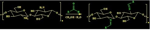
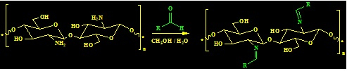
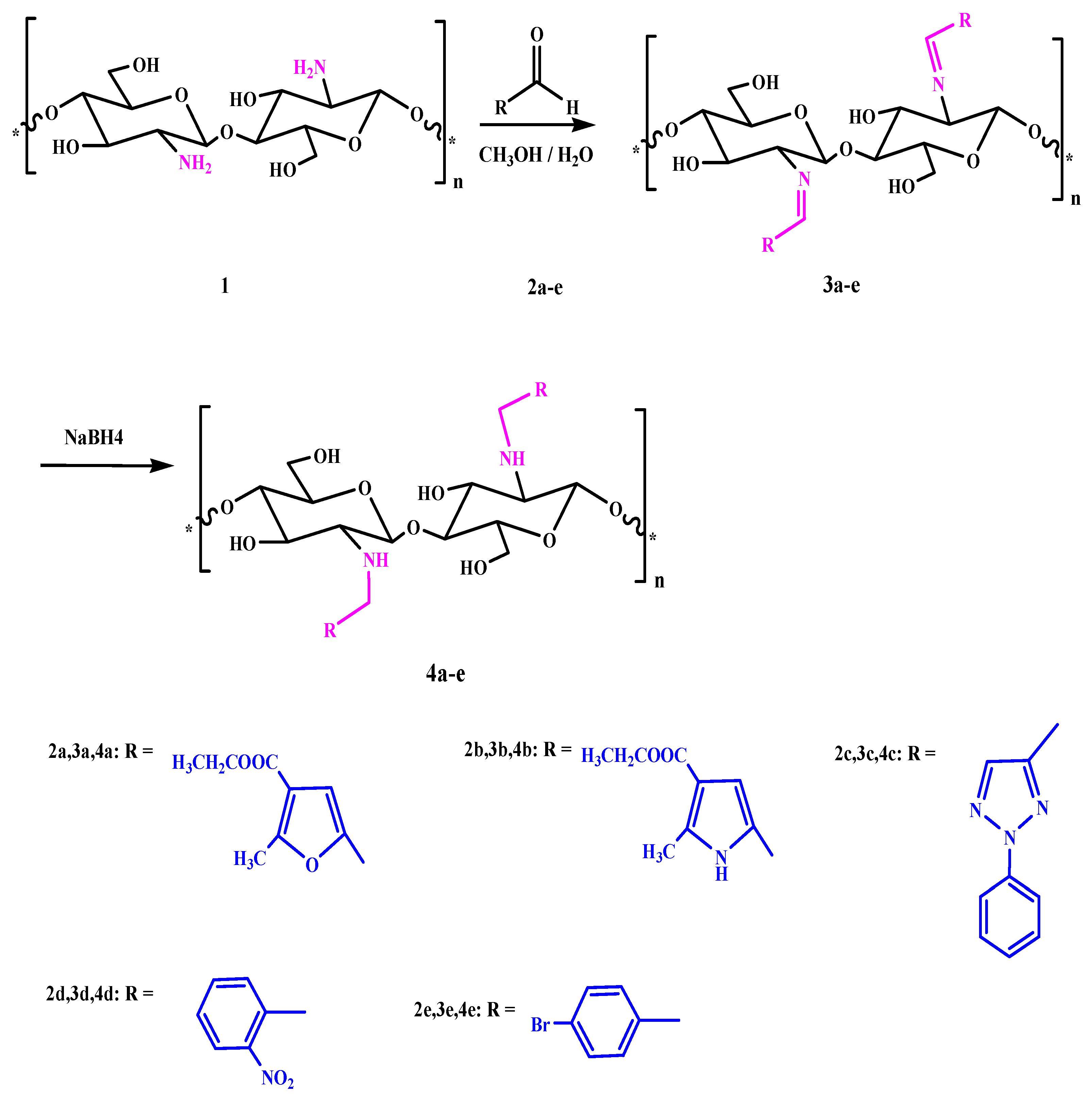
2.3. General Procedures for Chitosan Schiff-Base Synthesis

A solution of the aldehyde (20 mmol) in ethanol (20 mL) was added to chitosan (20 mmol) in 10% AcOH (50 mL). The mixture was stirred for 6–10 h at 70 °C, and then left overnight. After cooling, the homogenous hydrogels which formed were dried at 60 °C for dewatering to constant weight to give the product.
Chitosan furan Schiff-base (CFSB) 3a. Obtained from ethyl 5-formyl-2-methylfuran-3-carboxylate 2a in 91% yield as white powder; the mixture was stirred for 6 h; IR (KBr): 1642 (C=N), 1687 (COOEt), 3440 cm−1 (OH); Anal. Found: C, 52.18; H, 5.31; N, 4.13; O, 37.51.
Chitosan pyrrole Schiff-base (CPSB) 3b. Obtained from 5-formyl-2-methyl-1H-pyrrole-3-carboxylate 2b in 89.3% yield as white powder; The mixture was stirred for 8 h; IR (KBr): 1599 (C=N), 1655 (COOEt), 3322 cm−1 (OH); 1H NMR (300 MHz, DMSO): δ = 1.93 (q, 2H, CH2-ester; J1,2 = 2.3 Hz, J1,3 = 6.9 Hz), 2.38 (m, 1H, H-1′), 2.41(m, 2H, H-2′, H-3′), 2.61 (d, 2H, H-4′, H-5′; J1,2 = 2.3 Hz), 2.75 (m, 2H, H-6a′, H-6b′), 3.17 (t, 3H, CH3-ester; J1,2 = 2.3 Hz, J1,3 = 6.9 Hz), 3.25 (s, 3H, CH3-pyrrole), 4.53 (bs, 2H, 3′-OH, 6′-OH; exchangeable with D2O), 6.51 (s, 1H, CH=N), 6.55 (s, 1H, H-pyrrole), 10.33 (bs, 1H, NH; exchangeable with D2O), Anal. Found: C, 52.84; H, 5.94; N, 8.43; O, 32.86.
Chitosan triazole Schiff-base (CTSB) 3c. Obtained from 2-phenyl-2H-1,2,3-triazole-4-carbaldehyde 2c in 95.1% yield as faint gray powder; The mixture was stirred for 10 h; IR (KBr): 1639 (C=N), 3455 cm−1 (OH); Anal. Found: C, 59.58; H, 5.17; N, 18.34; O, 15.82.
Chitosan nitrophenyl Schiff-base (CNPSB) 3d. Obtained from o-nitrobenzaldehyde 2d in 94.3% yield as yellow powder; The mixture was stirred for 6 h; IR (KBr): 1638 (C=N), 3480 cm−1 (OH); 1H NMR (300 MHz, DMSO): δ = 3.56 (m, 1H, H-1′), 3.68 (m, 1H, H-2′), 3.83 (d, 2H, H-3′, H-4′; J1,2 = 2.4 Hz), 3.90 (m, 1H, H-5′), 4.07 (m, 2H, H-6a′, H-6b′), 5.44 (bs, 2H, 3′-OH, 6′-OH; exchangeable with D2O), 6.77 (s, 1H, CH=N), 6.98 (d, 2H, o-H; J1,2 = 2.9 Hz), 7.83 (d, 2H, m-H; J1,2 = 2.9 Hz), Anal. Found: C, 56.01; H, 5.11; N, 10.03; O, 28.65.
Chitosan bromophenyl Schiff-base (CBPSB) 3e. Obtained from p-bromobenzaldehyde 2e in 96.7% yield as white powder; The mixture was stirred for 7 h; IR (KBr): 1637 (C=N), 3466 cm−1 (OH); 1H NMR (300 MHz, DMSO): δ = 3.27 (m, 1H, H-1′), 3.38 (m, 1H, H-2′), 3.54 (d, 1H, H-3′; J1,2 = 1.8 Hz), 3.61 (m, 2H, H-4′, H-5′), 3.73 (m, 2H, H-6a’, H-6b′), 5.73 (bs, 2H, 3′-OH, 6′-OH; exchangeable with D2O), 7.42 (s, 1H, CH=N), 7.53 (d, 1H, o-H; J1,2 = 2.8 Hz), 7.84 (d, 2H, m-H; J1,2 = 2.8 Hz), Anal. Found: C, 45.03; H, 4.26; N, 4.05; O, 23.14.

2.4. General Procedures for Reduction of Imine by NaBH4

A solution of imine (20 mmol) in methanol (20 mL) was added to 10% AcOH (20 mL). The mixture was stirred for 10 min, and then 0.1 g of sodium borohydride was slowly added to the mixture with continuous stirring in ice bath for 24 h. After that the product was dried.
Chitosan-furan amine derivative 4a. Obtained from imine 3a in 84% yield as white powder; IR (KBr): 1647(COOEt), 3254, 3322 cm−1 (NH), (OH); Anal. Found: C, 61.07; H, 7.08; N, 4.64; O, 27.18.
Chitosan-pyrrole amine derivative 4b. Obtained from imine 3b in 86.2% yield as white powder; IR (KBr): 1659 (COOEt), 3334, 3387 cm−1 (NH), (OH); 1H NMR (300 MHz, DMSO): δ = 1.83 (q, 2H, CH2-ester; J1,2 = 2.3 Hz, J1,3 = 6.9 Hz), 2.41 (m, 1H, H-1′), 2.41 (m, 2H, H-2′, H-3′), 2.61 (d, 2H, H-4′, H-5′; J1,2 = 2.3 Hz), 2.73 (m, 2H, H-6a′, H-6b′), 3.27 (t, 3H, CH3-ester; J1,2 = 2.3 Hz, J1,3 = 6.9 Hz), 3.29 (s, 3H, CH3-pyrrole), 4.61 (bs, 2H, 3′-OH, 6′-OH; exchangeable with D2O), 6.51 (s, 2H, CH2), 6.53 (s, 1H, H-pyrrole), 9.53 (bs, 1H, NH; exchangeable with D2O), 10.35 (bs, 1H, NH; exchangeable with D2O), Anal. Found: C, 57.95; H, 7.05; N, 9.02; O, 25.68.
Chitosan-triazole derivative 4c. Obtained from imine 3c in 84% yield as faint green powder; IR (KBr): 3260, 3321 cm−1 (NH), (OH); Anal. Found: C, 58.79; H, 6.04; N, 18.33; O, 15.58.
Chitosan-nitrophenyl amine derivative 4d. Obtained from imine 3d in 91% yield as faint yellow powder; IR (KBr): 3331, 3340 cm−1 (NH), (OH); 1H NMR (300 MHz, DMSO): δ = 3.80 (d, 2H, H-1′, H-2′ J1,2 = 2.2 Hz), 3.90 (m, 2H, H-3′, H-4′), 4.14 (m, 3H, H-5′, H-6a′, H-6b′), 5.46 (bs, 2H, 3′-OH, 6′-OH; exchangeable with D2O), 6.97 (s, 2H, CH2), 7.01 (d, 2H, o-H; J1,2 = 2.4 Hz), 7.83 (d, 2H, m-H; J1,2 = 2.4 Hz), 9.14 (bs, 1H, NH; exchangeable with D2O), Anal. Found: C, 55.61; H, 5.47; N, 9.89; O, 28.14.
Chitosan-bromophenyl amine derivative 4e. Obtained from imine 3e in 91% yield as white powder; IR (KBr): 3264, 3398 cm−1 (NH), (OH); 1H NMR (300 MHz, DMSO): δ = 3.37 (m, 2H, H-2′, H-1′), 3.48 (m, 3H, H-3′, H-4′, H-5′), 3.83 (m, 2H, H-6a′, H-6b′), 5.81 (bs, 2H, 3′-OH, 6′-OH; exchangeable with D2O), 7.41 (s, 2H, CH2), 7.53 (d, 2H, o-H; J1,2 = 2.0 Hz), 7.83 (d, 2H, m-H; J1,2 = 2.0 Hz), 9.31 (bs, 1H, NH; exchangeable with D2O), Anal. Found: C, 44.76; H, 5.25; N, 4.12; O, 22.78.

2.5. Antimicrobial Activity

The antimicrobial activity of CSB derivatives were evaluated against Staphylococcus aureus (RCMBA 2004) and Bacillissubtilis (RCMBA 6005) as Gram-positive bacteria and against Pseudomonas aeruginosa and Escherichia coli (RCMBA 5003) as Gram-negative bacteria and against Aspergillus fumigates (RCMBA 06002), Syncephalastrum racemosum (RCMB 05098), as fungi. Agar disk diffusion method was used for the determination of the antibacterial and antifungal activity, the well diameter was 6 mm (100 µL was tested), and the concentration of the tested sample was 5 mg/mL.
The susceptibility tests were performed according to the NCCLS recommendations (National Committee For Clinical Laboratory Standards, 1993). Screening tests regarding the inhibition zone were carried out by the well diffusion method [22].
The inoculums suspension was prepared from colonies grown overnight on an agar plate, and inoculated into Mular Hinton broth (Merk, Darmstadt, Germany). A sterile swab was immersed in the bacterial suspension and used to inoculate Mueller-Hinton agar plates. Amphotericin B, Ampicillin and Gentamicin were used as references for anti-fungi, anti-Gram positive bacteria, and anti-Gram negative bacteria, respectively. The compounds were dissolved in dimethylsulfoxide (DMSO). The inhibition zone was measured around each well after 24 h incubation at 37 °C; controls using DMSO were adequately done.
MIC determinations were performed in the same way using agar disc diffusion method, but by using different concentrations from the testing compound.

2.6. Antiproliferative Activity Screening

Regarding cell line propagation, the cells were propagated in (DMEM) supplemented with 10% heat-inactivated fetal bovine serum, 1% l-glutamine, HEPES buffer and 50 μg/mL gentamycin. All cells were maintained at 37 °C in humidified atmosphere with 5% CO2 and were sub cultured two times a week. Cell toxicity was monitored by determining the effect of the examined compound on cell morphology and cell viability.
For cytotoxicity assay, the cells were seeded in 96-well plate at a cell concentration of 1 × 10⁴ cell per well in 100 μL of growth medium. Serial two-fold dilutions of the tested chemical compound were added to confluent cell monolayers that were then dispensed into 96-well flat-bottomed microtiter plates (Falcon, NJ, USA) using a multichannel pipette. The microtiter plates were incubated at 37 °C in a humidified incubator with 5% CO2 for a period of 48 h. Three wells were used for each concentration of each tested sample. Control cells were incubated without test samples and with or without DMSO. After incubation of the cells for 24 h at 37 °C, various concentrations of each sample (50, 25, 12.5, 6.25, 3.125 and 1.56 μg) were added separately. Then the incubation was continued for 48 h.
The viable cells yield was determined colorimetrically using MTTB (3,4,5-dimethylthiazol-2-yl-2,5-diphenyltetrazolium bromide). The water insoluble tetrazolium salt is converted to purple formazon by the mitochondrial dehydrogenase of viable cells. After the end of incubation period, media were aspirated, and the crystal violet solution (1%) was added to each well for at least 30 min. The stain was removed and plates were rinsed using tap water until all excess stain was removed. Glacial acetic acid (30%) was then added to all wells and mixed thoroughly, then the absorbance of the plates were measured after gently being shaken on Micro plate Reader (TECAN, Inc., Olympus Europa Holding GmbH, Männedorf, Switzerland), at 490 nm. All results were corrected for background absorbance detected in wells without added stain. Treated samples were compared with the cell control in the absence of the tested compound. All experiments were carried out in the triplicate. The cell cytotoxicity effect of the tested compound was calculated [16,17].

3. Results and Discussion

3.1. Synthesis of Chitosan Schiff’s Base (CSB) and Chitosan Amine Derivatives

Aryl and heteroaryl aldehydes were selectively grafted onto the primary amino groups of chitosan with formation of the corresponding Schiff bases 3ae. The imine group converted into the more stable amine with formation of the corresponding N-substituted amino-chitosan derivatives 4ae using sodium borohydride as a reducing agent.
Five different aldehydes were employed: (ethyl-2-formyl-5-methyl-4-furate 2a, ethyl-5-formyl-2-methyl-1H-pyrrole-3-carboxylate 2b, 1-(2-phenyl-2H-1,2,3-triazole-4-yl) ethanone 2c, 2-nitrobenzaldehyde 2d, and 4-bromobenzaldehyde 2e in this reaction (Scheme 1).
Scheme 1. Synthesis of chitosan Schiff’s base (CSB) and chitosan amine derivatives.
Polymers 07 01532 g003

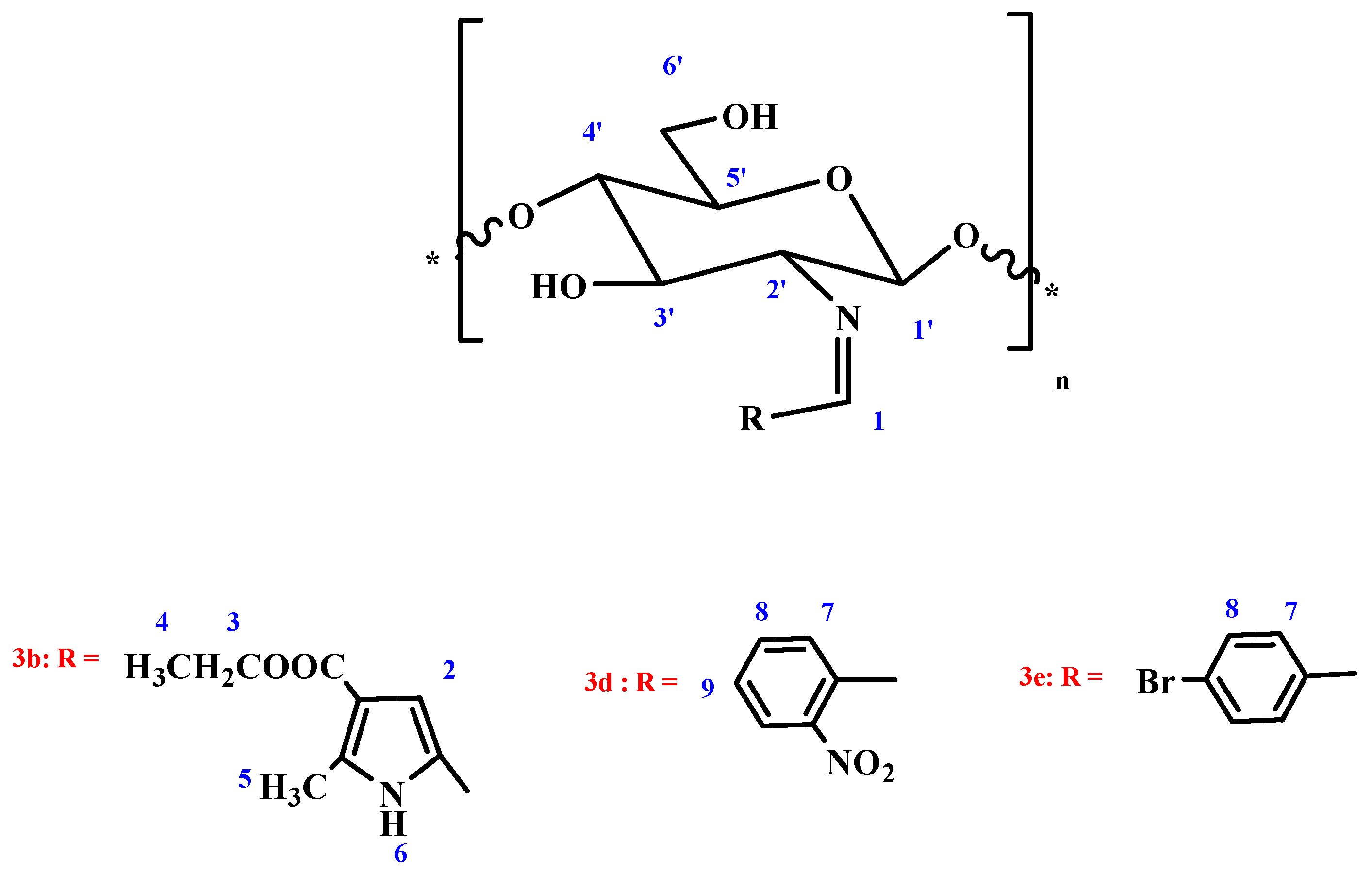
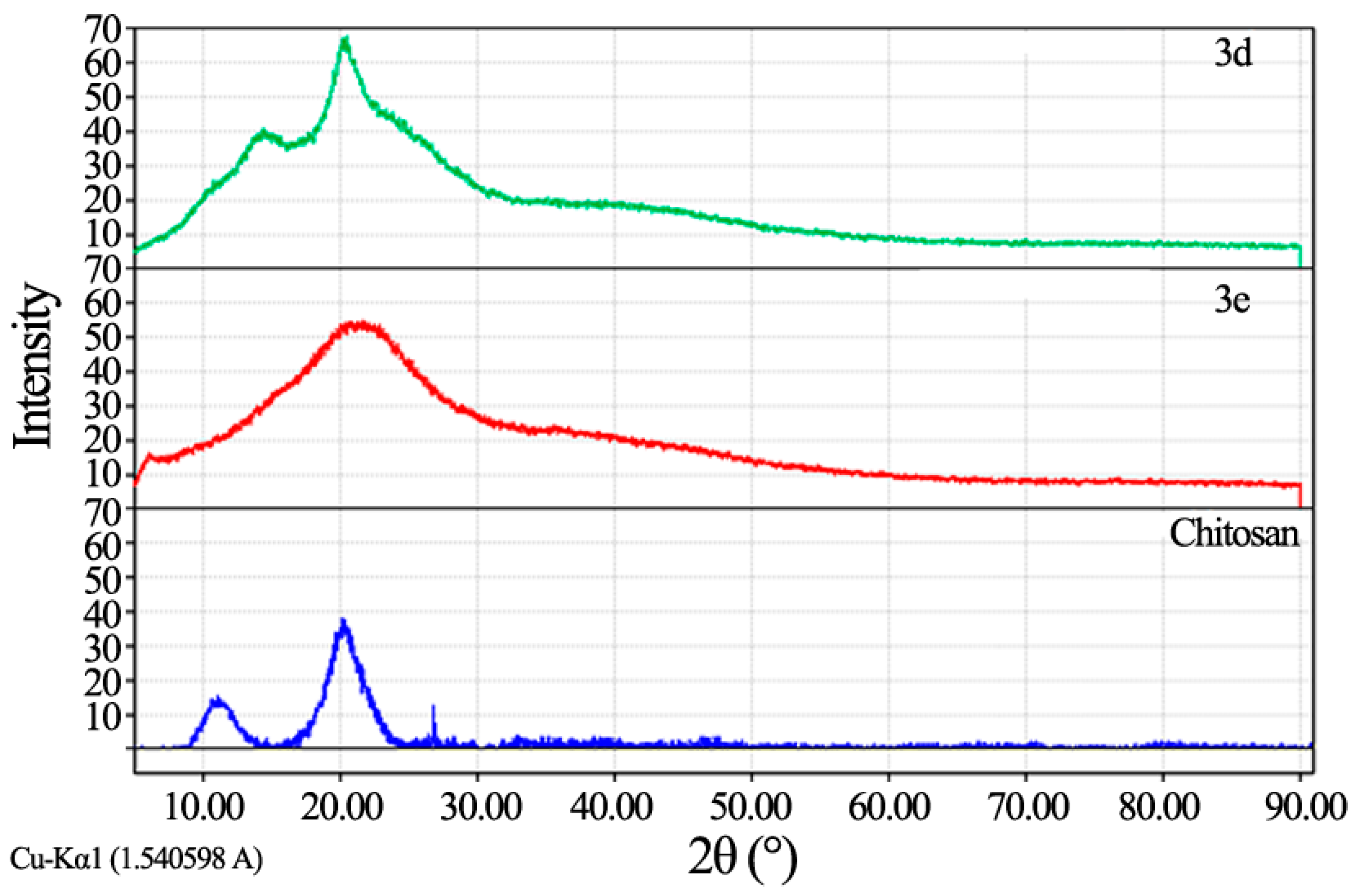
3.2. Characterization of Chitosan-Imine and Chitosan-Amine Derivatives

The infrared spectra of compounds 3ae showed the (OH) band at 3480–3322 cm−1, and (C=N) at 1599–1642 cm−1. On the other hand the infrared spectra of compounds 4ae showed the (OH) band at 3398–3321 cm−1, and (NH) at 3254–3334 cm−1 (Table 1).
Table 1. The infrared data of compounds 3ae, 4ae.
Compound Numnerγ KBr Max cm−1
C=NCOOEtNHOH
3a16421687-3440
3b1599165529763322
3c1639--3455
3d1638--3480
3e1637--3466
4a-164732543322
4b-165933343387
4c--32603321
4d--33313340
4e--32643398
The structure of imines 3ae is further proved by 1H NMR spectra, which showed the 1′-OH proton at 5.53–5.73 ppm. The rest of the sugar protons are at the range 2.61–4.07 ppm. The methyl protons at position-2 in the pyrrole ring appeared as a singlet at δ 3.25 ppm; as well as the disappearance of the two protons of (NH2), (Figure 1, Table 2). (OH) protons are D2O exchangeable in compounds 3ae. Additionally, the structure of amines 4ae was proved by 1HNMR spectra, which showed the 1′-OH proton at 5.06–5.05 ppm, the rest of the sugar protons at the range 4.58–4.28 ppm. As well as the appearance of the (NH) proton at 9.14–9.53 ppm, after shaking of compounds 4ae with D2O, their 1H NMR spectra, showed the disappearance of the (NH) proton, as well as (OH) protons (Table 3). In addition C13 NMR of compounds 3e and 4e showed the expected peaks.
Figure 1. Chitosan-imine structure.
Polymers 07 01532 g001
Table 2. The 1H NMR data of chitosan-imine derivatives.
Compound Numnerδ (ppm)
H-1H-2H-3H-4H-5H-6H-7H-8H-9
3b6.51 (s)6.55 (s)1.93 (s)3.17 (s)3.25 (d)10.33 (bs)---
3d6.77 (s)-----6.98 (d)7.83 (d)7.83 (d)
3e7.42 (s)-----7.53 (d)7.84 (d)-
Compound Numnerδ (ppm)
H-1′H-2′H-3′H-4′H-5′H-6a′, H-6b′3′-OH6′-OH
3b2.38 (m)2.41 (m)2.41 (m)2.61 (d)2.61 (d)2.75 (m)4.53 (bs)4.53 (bs)
3d3.56 (m)3.68 (m)3.83 (d)3.83 (d)3.90 (m)4.07 (m)5.44 (bs)5.44 (bs)
3e3.27 (m)3.38 (m)3.54 (d)3.61 (m)3.61 (m)3.73 (m)5.73 (bs)5.73 (bs)
Decreasing the crystal structure of chitosan after condensation with aldehydes was appearing in XRD patterns of chitosan Schiff’s base (CSB), which showed in case of compound 3e one broad peak around 2θ = 25°. On the other hand, XRD patterns of compound 3d showed two broad peaks around 2θ = 16° and 25° indicating a shift from the normal chitosan peaks a broad peak around 20° showing increasing in its amorphous nature (Figure 2).
Table 3. Inhibition indices of chitosan Schiff’s base (CSB) against S. aureus, B. subtilis, P. aeruginosa and E. coli.
SampleTested Microorganisms
Gram Positive BacteriaGram Negative Bacteria
S. aureusB. subtilisP. aeruginosaE. coli
3a22.2 ± 0.5824.6 ± 0.25NA21.4 ± 0.63
3b18.2 ± 0.6320.4 ± 0.58NA18.3 ± 0.72
3c16.7 ± 0.3619.2 ± 0.2713.3 ± 0.3613.6 ± 0.36
3d11.3 ± 0.6314.2 ± 0.58NA11.1 ± 0.63
3e21.4 ± 0.6322.3 ± 0.72NA21.2 ± 0.63
Ampicillin23.8 ± 0.232.4 ± 0.3NANA
GentamicinNANA17.3 ± 0.119.9 ± 0.3
NA: No Activity.
Figure 2. X-ray diffraction spectrum of chitosan derivatives.
Polymers 07 01532 g002

3.3. Antimicrobial Activity

All of the synthesized substituted derivatives under investigation showed in vitro antimicrobial activity against the tested microorganisms. The results of antibacterial activity of the chitosan Schiff’s base (CSB) derivatives using inhibition zone method are listed in Table 3. The obtained data revealed that all the tested compounds 3ae had no effect on P. aeruginosa (Gram positive bacteria), except the triazolo-derivative 3c, which showed excellent inhibitory effect on both Gram-positive and Gram-negative bacteria as compared to Ampicillin and Gentamicin (Table 3). Our data displayed that compound 3a is the most antimicrobially effective compound, which has an excellent anti-Gram positive bacteria, effect on both Straphylococcusaureus and Bacillissubtilis, and also on anti-Gram negative bacteria (E. coli). This effect may be due to the presence of the furan ring. On the other hand the bromo-derivative showed higher antibacterial effect than the nitro derivative on both Gram-positive and Gram-negative bacteria.
The antimicrobial activity of chitosan has been explained by several mechanisms. The first mechanism is mediated by the electrostatic forces between the protonated –NH3+ groups of chitosan and the electronegative charges on the microbial cell surface [23]. It suggests that the greater the number of cationized amines, the higher the antimicrobial activity.
Another proposed mechanism is the binding of chitosan with microbial DNA, which leads to the inhibition of the mRNA and protein synthesis via penetration of chitosan into the nuclei of the microorganisms [24]. The third mechanism is the chelating of metals, suppression of spore elements, and binding to essential nutrients to microbial growth.
The mean zone of inhibition in mm ± standard deviation beyond well diameter (6 mm) produced on a range of environmental and clinically pathogenic microorganisms using (5 mg/mL) concentration of tested samples. Results are depicted in the following table.
Generally, chitosan has been reported as being very effective in inhibiting spore germination, germ tube elongation, and radial growth [25]. The antifungal mechanism of chitosan involves cell wall morphogenesis with chitosan molecules interfering directly with fungal growth, similar to the effects observed in bacteria cells. The microscopic observation reported that chitosan molecules diffuse inside hyphae interfering on the enzymes activity responsible for the fungus growth [26].
The antifungal activities of CSB derivatives against A. fumigatus (RCMBA 06002), Syncephalastrum racemosum (RCMB 05098) are shown in Table 4, which shows that all the derivatives had effective activities against the Aspergillus fumigates, compared with that of the stander, with inhibitory indices ranging from 13.2 ± 0.72 to 21.3 ± 0.63 mm inhibition zone (Table 4). On the other hand, only one of the tested compounds showed an effect on Syncephalastrum racemosum, this activity of compound 3c may be due to the presence of the triazole ring. Again, the results also demonstrate how the antifungal activities are affected by the nature of the substituent in the aryl ring of the CSB derivatives, in which the bromo-derivative showed a greater effect compared to the nitro derivative (Table 4).
Table 4. Inhibition indices of CSB against A. fumigatus, Syncephalast rumracemosum.
SampleTested Microorganisms
A. fumigatesS. rumracemosum
3a21.3 ± 0.63NA
3b16.3 ± 0.72NA
3c16.8 ± 0.3913.4 ± 0.58
3d13.2 ± 0.72NA
3e19.6 ± 0.58NA
Amphotericin B23.7 ± 0.119.7 ± 0.2
NA: means No Activity.
The results obtained in Table 3 and Table 4 showed that all samples have promising results with mean zone of inhibition values less than the used standards. Amphotericin B as anti-fungi, Ampicillin as anti-Gram positive bacteria, and Gentamicin as anti-Gram negative bacteria.
Minimum inhibitory concentration (MIC) is the lowest concentration of an antimicrobial that will inhibit the visible growth of microorganisms after incubation for suitable time. The antimicrobial activity as MIC (µg/mL) of tested compounds 3a, 3b, 3c, 3d and 3e against tested microorganisms showed that all these compounds are effective for Aspergillus fumigates with MIC 1.95, 31.25, 62.5, 62.5 and 3.9, respectively, meanwhile compound 3a has the lowest MIC compared to Amphotericin B which used as standard with MIC 0.98.
In addition, these examined compounds showed MIC values against Staphylococcus aureus 1.95, 7.81, 125, 125 and 1.95 respectively, in which compound 3a and 3e are the most active compounds compared to the Ampicillin standard with MIC value 0.98 (Table 5).
Additionally, these compounds are effective against other Gram-positive bacteria Bacillus subtilis with MIC 0.98, 3.9, 62.5, 125 and 1.95 indicating that compound 3a is the most active one compared to Ampicillin MIC 0.49. Additionally, all these tested compounds are effective against Escherichia coli with MIC 1.95, 7.81, 125, 125 and 1.95 showed that compound 3a is the most effective one compared to Gentamicin with MIC equal to 3.9. The obtained data confirmed that compound 3a is the most active agent as antimicrobial, especially against Gram-negative bacteria (Table 5).
Table 5. Antimicrobial activity as MICS (µg/mL) of CSB against tested microorganisms.
Tested MicroorganismSamplesStandard
3a3b3c3d3e
FungiMIC (µg/mL)AmphotericinB
Aspergillus Fumigates1.9531.2562.562.53.90.98
Gram positive bacteria-Ampicillin
Staphilococcus aureus1.957.811251251.950.98
Bacillis subtilis0.983.962.51251.950.49
Gram negative bacteria-Gentamicin
Escherichia coli1.957.811251251.953.9

3.4. Antiproliferative Activity Screening

Cytotoxicity was tested against three cancer cell lines: HepG-2, Human Hepatocellular Liver Carcinoma Cells; HCT-116, Human Colon Carcinoma Cells; MCF-7, Human Breast Adenocarcinoma Cells.
Since triazole derivative showed strong reactivity against all the tested kinds of Gram-positive, and Gram-negative bacteria and fungi, it was chosen for cytotoxicity screening against cancer cells. The results show that the examined compound 3c had an excellent inhibitory effect on the cell lines growth compared to standard.
The reactivity of the examined compound 3c was tested against breast cancer (MCF-7), colon cancer (HCT-116) and hepatocellular cancer (HepG-2). The effect of this compound on cancer cell viability was tested using different concentrations (50–1.56 µg/mL) of the compound 3c. See Table 6.
Table 6. Inhibitory activity of compound 3c against HepG-2, MCF-7 and HCT-116 cell line compared to doxorubicin as reference drug.
Sample conc. (μg/mL)Viability %
3cDoxorubicin (std.)
HepG-2MCF-7HCT-116HepG-2MCF-7HCT-116
01.000001.000001.000001.000001.000001.00000
10.587160.6520780.833260.54510.42560.3902
2.50.1945603425100.544190.39030.29890.2900
50.254840.3304470.297400.25000.20000.1990
100.204690.3203880.250280.19590.14430.1510
IC501.211.542.891.20.440.47
The maximum cell growth inhibitory effect was obtained on HepG-2 with IC50 equal to 1.21 μg compared to IC50 of the used standard 1.2 μg.

4. Conclusions

Some of new Chitosan Schiff’s bases have been synthesized. Their structures were approved by standard methods. Evaluations of their anti-bacterial, anti-fungal, and cytotoxicity properties have been studied.

Acknowledgments

Department of chemistry, faculty of science, Alexandria University afforded the reagents, materials, and analysis tools.

Author Contributions

Mohamed M. El Sadek suggested and supervised the work and performed the article editing. Mohamed A. Mostafa and Seham Y. Hassan conceived and designed the experiments and analyzed the data. Galila A. Yacout is responsible for the biological part in the article. Huda E. Abdelwahab performed the experiments.

Conflicts of Interest

the authors declare no conflict of interest.

References

  1. Guo, M.; Ma, Y.; Wang, C.; Liu, H.; Li, Q.; Fei, M. Synthesis, anti-oxidant activity, and biodegradability of a novel recombinant polysaccharide derived from chitosan and lactose. Carbohydr. Polym. 2015, 118, 218–223. [Google Scholar] [CrossRef] [PubMed]
  2. Ando, A.; Noguchi, K.; Yanagi, M.; Shinoyama, H.; Kagawa, Y.; Hirata, H.; Yabuki, M.; Fujii, T. Primary structre of chitosanase produced by Bacillus circulans MH-K1. J. Gen. Appl. Microbiol. 1992, 38, 135–144. [Google Scholar] [CrossRef]
  3. Cheng, W.P.; Chi, F.H.; Yu, R.F.; Lee, Y.C. Using chitosan as coagulant in recovery of organic matters from the mash and lautar wastewater of brewery. J. Polym. Environ. 2005, 13, 383–388. [Google Scholar] [CrossRef]
  4. Bugg, T.D.; Walsh, C.T. Intracellular steps of bacterial cell wall peptidoglycan biosynthesis: Enzymology, antibiotics, and antibiotic-resistance. Nat. Prod. Rep. 1992, 9, 199–215. [Google Scholar] [CrossRef] [PubMed]
  5. Chen, R.H.; Tasaih, M.L. Effect of temperature on the intrinsic viscosity and conformation of chitosans in dilute HCl solution. Int. J. Biol. Macromol. 1998, 23, 135–141. [Google Scholar] [CrossRef]
  6. Kpping-Hggrd, M.; Tubulekas, I.; Guan, H.; Edwards, K.; Nilsson, M.; Varum, K.; Artursson, P. Chitosan as a nonviral gene delivery system. Structure–property relationships and characteristics compared with polyethylenimine in vitro and after lung administration in vivo. Gene Ther. 2001, 8, 1108–1121. [Google Scholar] [CrossRef] [PubMed]
  7. Khor, E.; Lim, L.Y. Implantable applications of chitin and chitosan. Biomaterials 2003, 24, 2339–2349. [Google Scholar] [CrossRef]
  8. Yuan, S.; Wei, T. New contact lens based on chitosan/gelatin composites. J. Bioact. Compat. Polym. 2004, 19, 467–479. [Google Scholar] [CrossRef]
  9. Crini, G. Non-conventional low-cost adsorbents for dye removal: A review. Bioresour. Technol. 2006, 97, 1061–1085. [Google Scholar] [CrossRef] [PubMed]
  10. Esam, A.E.; Mohamed, M.N.; Abdul, H.Y. Rheological and morphological studies of chitosan/agar/poly(vinyl alcohol) blends. J. Appl. Sci. Res. 2010, 6, 460–468. [Google Scholar]
  11. Chandy, T.; Sharma, C.P. Chitosan-as a biomaterial. Biomater. Artif. Cells Artif. Org. 1990, 18, 1–24. [Google Scholar] [CrossRef]
  12. Choi, B.K.; Kim, K.Y.; Yoo, Y.J.; Oh, S.J.; Choi, J.H.; Kim, C.Y. In vitro antimicrobial activity of a chitooligosaccharides mixture against Actinobacillus actinomycetemcomitans and Streptococcus mutans. Int. J. Antimicrob. Agents 2001, 18, 553–557. [Google Scholar] [CrossRef]
  13. Park, P.J.; Je, J.Y.; Kim, S.K. Free radical scavenging activity of chito-oligosaccharides by electron spin resonance spectrometry. J. Agric. Food Chem. 2003, 51, 4624–4627. [Google Scholar] [CrossRef] [PubMed]
  14. Chung, M.J.; Park, J.K.; Park, Y.I. Anti-inflammatory effects of low-molecular weight chitosan oligosaccharides in IgE-antigen complex-stimulated RBL-2H3 cells and asthma model mice. Int. Immunopharmacol. 2012, 12, 453–459. [Google Scholar] [CrossRef] [PubMed]
  15. Saranya, N.; Moorthi, A.; Saravanan, S.; Devi, M.P.; Selvamurugan, N. Chitosan and its derivatives for gene delivery. Int. J. Biol. Macromol. 2011, 48, 234–238. [Google Scholar] [CrossRef] [PubMed]
  16. Köping-Höggård, M.; Mel’nikova, Y.S.; Vårum, K.M.; Lindman, B.; Artursson, P. Relationship between the physical shape and the efficiency of oligomeric chitosan as a gene delivery system in vitro and in vivo. J. Gene Med. 2003, 5, 130–141. [Google Scholar] [CrossRef] [PubMed]
  17. Roy, K.; Mao, H.Q.; Huang, S.K.; Leong, K.W. Oral gene delivery with chitosan—DNA nanoparticles generates immunologic protection in a murine model of peanut allergy. Nat. Med. 1999, 5, 387–391. [Google Scholar] [PubMed]
  18. Brode, G.L.; Goddard, E.D.; Harris, W.C.; Salensky, G.A. Cationic polysaccharides for cosmetics and therapeutics. In Cosmetics and Pharmaceutical Applications of Polymers; Gebelein, C.G., Cheng, T.C., Yang, Y.C., Eds.; Plenum Press: New York, NY, USA, 1991; pp. 117–128. [Google Scholar]
  19. Akiyama, K.; Fujita, T.; Kuroshima, K.; Sanake, T.; Yokota, A.; Takata, R. Purification and gene cloning of chitosanase from Bacillus ehimensis EAG1. J. Biosci. Bioeng. 1999, 87, 383–385. [Google Scholar] [CrossRef]
  20. Masson, J.Y.; Bocher, I.; Neugebauer, W.A.; Ramotar, D.; Brzezinski, R. A new chitosanase gene from a Nocardioides sp. Is a third member of glycosyl hydrolase family 46. Microbiology 1995, 141, 2629–2635. [Google Scholar] [CrossRef] [PubMed]
  21. Beloshenko, V.A.; Askadskii, A.A.; Varyukhin, V.N. Promising ways of structural modification of polymers and polymer composites with the use of high pressure. Russ. Chem. Rev. 1998, 67, 951–973. [Google Scholar] [CrossRef]
  22. Jorgensen, J.H.; Swenson, J.M.; Tenover, F.C.; Ferraro, M.J.; Hindler, J.A.; Murray, P.R. Development of interpretive criteria and quality control limits for broth micro dilution and disk diffusion antimicrobial susceptibility testing of Streptococcus pneumonia. J. Clin. Microbiol. 1994, 32, 2448–2459. [Google Scholar] [PubMed]
  23. Feng, Q.L.; Wu, J.; Chen, G.Q.; Cui, F.Z.; Kim, T.N.; Kim, J.O. A mechanistic study of antibacterial effect of silver ions on Escherichia coli and Straphylococcus aureus. J. Biomed. Mater. Res. 2000, 52, 662–668. [Google Scholar] [CrossRef]
  24. Dina, R.; Kristine, V.; Albert, H.; Hans-Georg, S. Insights into the mode of action of chitosan as an antibacterial compound. Appl. Environ. Microbiol. 2008, 74, 3764–3773. [Google Scholar]
  25. El-Ghaouth, A.; Arul, J.; Grenier, J.; Asselin, A. Antifungal activity of chitosan on two postharvest pathogens of strawberry fruits. Phytopathology 1992, 82, 398–402. [Google Scholar] [CrossRef]
  26. Eweis, M.; Elkholy, S.S.; Elsabee, M.Z. Antifungal efficacy of chitosan and its thiourea derivatives upon the growth of some sugar-beet pathogens. Int. J. Biol. Macromol. 2006, 38, 1–8. [Google Scholar] [CrossRef] [PubMed]

