Article

Synthesis and Characterization of Maghemite Nanoparticles Functionalized with Poly(Sodium 4-Styrene Sulfonate) Saloplastic and Its Acute Ecotoxicological Impact on the Cladoceran Daphnia magna

by
Juan A. Ramos-Guivar
1,
Renzo Rueda-Vellasmin
1,2,
Erich V. Manrique-Castillo
1,*,
F. Mendoza-Villa
1,
Noemi-Raquel Checca-Huaman
3 and
Edson C. Passamani
2
1
Grupo de Investigación de Nanotecnología Aplicada para Biorremediación Ambiental, Energía, Biomedicina y Agricultura (NANOTECH), Facultad de Ciencias Físicas, Universidad Nacional Mayor de San Marcos, Av. Venezuela Cdra 34 S/N, Ciudad Universitaria, Lima 15081, Peru
2
Departamento de Física, Universidade Federal do Espírito Santo, Vitória 29075-910, Brazil
3
Centro Brasileiro de Pesquisas Físicas (CBPF), R. Xavier Sigaud, 150, Urca, Rio de Janeiro 22290-180, Brazil
*
Author to whom correspondence should be addressed.
Polymers 2024, 16(11), 1581; https://doi.org/10.3390/polym16111581
Submission received: 13 April 2024 / Revised: 22 May 2024 / Accepted: 28 May 2024 / Published: 3 June 2024
(This article belongs to the Section Polymer Composites and Nanocomposites)

Abstract

:
Using a modified co-precipitation method, 11(2) nm γ-Fe2O3 nanoparticles functionalized with PSSNa [Poly(sodium 4-styrenesulfonate)] saloplastic polymer were successfully synthesized, and their structural, vibrational, electronic, thermal, colloidal, hyperfine, and magnetic properties were systematically studied using various analytic techniques. The results showed that the functionalized γ-Fe2O3/PSSNa nanohybrid has physicochemical properties that allow it to be applied in the magnetic remediation process of water. Before being applied as a nanoadsorbent in real water treatment, a short-term acute assay was developed and standardized using a Daphnia magna biomarker. The ecotoxicological tests indicated that the different concentrations of the functionalized nanohybrid may affect the mortality of the Daphnia magna population during the first 24 h of exposure. A lethal concentration of 533(5) mg L−1 was found. At high concentrations, morphological changes were also seen in the body, heart, and antenna. Therefore, these results suggested the presence of alterations in normal growth and swimming skills. The main changes observed in the D. magna features were basically caused by the PSSNa polymer due to its highly stable colloidal properties (zeta potential > −30 mV) that permit a direct and constant interaction with the Daphnia magna neonates.

1. Introduction

The presence of toxic metals exceeding the permissible level in water is a hot topic in environmental sciences due to all the short- and long-term human health problems that these substances can cause to plants and living organisms [1]. For instance, arsenic (arsenite and arsenaite, and organic species) and lead (Pb) are two of the most harmful toxic metals. They are frequently found in ionic states in an aqueous media and are reported to cause intoxication of the central nervous system and male infertility, kidney failure, and cancer [2,3,4]. In this regard, novel metamaterials (nanoadsorbents) that are able to efficiently uptake the toxic metals in a short time-frame must be developed, studied, and tested in laboratories. In this sense, magnetic adsorption method has gained popularity in water remediation due to its excellent adsorption results and its easy removal from the effluent by an external magnetic field [5,6]. The synthesis of nanoabsorbents is often conducted following a co-precipitation chemical route, involving low budgets and huge amounts of materials obtained in short synthesis time (min) [5].
Among the adsorbents, magnetic composites, specifically those combined with polymers, are generally synthesized following a core@shell arrangement, where the material surface charge can be suitable for electrostatic attraction. In particular, these magnetic-polymer composites have revealed a considerable removal capacity for Cr(VI), Cu(II), Pb(II), and dyes in real short exposure times [7,8]. However, the use of magnetic@polymer nanocomposites for water remediation raises an additional concern, namely, its ecotoxicological impact, since, as an organic/inorganic external agent, the nanocomposites can affect the biocenosis of living organisms present in water, and consequently alter the ecological balance [9,10]. Thus, considering their promising adsorbent, colloidal, and magnetic properties, an ecotoxicity study must be developed first to understand if there is a negligible or negative impact on ecosystems caused by the magnetic remediation with any nanocomposite. It should be pointed out that each magnetic nanohybrid exhibits intrinsic physicochemical properties depending on their synthesis route, and the ecotoxicity test should be always performed before their technological applications.
Regarding the polymer, it is important to stress that poly(sodium 4-styrene sulfonate) (PSSNa) is a synthetic polymer categorized as a primary microplastic, intentionally manufactured for various applications, i.e., to prevent sexually transmitted diseases or for in vitro inhibition of Zika virus replication [11,12]. As a water-soluble anionic polyelectrolyte, it can be used in emulsion polymerization [13] and in the fabrication of advanced materials [14,15]. There is a poor literature reporting PSSNa ecotoxicity, hence its release into the environment contributes to an enhancement of microplastic pollution, now seen as a big problem that needs to be treated urgently. Therefore, to assess the potential environmental impact of PSSNa, it is essential to follow the recommended usage guidelines and updated regulatory tests [16,17]. In real effluents, one of the most often living organisms found is the Daphnia magna (D. magna). It is a species commonly used in toxicological studies due to its high sensitivity to external agents and the fact that it ideally represents zooplankton [18]. Based on this assumption, preliminary nanoecotoxicity properties of a magnetic@polymer nanocomposite must be evaluated to know its lethal concentration (LC50) that allows a controlled water magnetic treatment, i.e., controlling the reasonable increase in the adsorbent dose without creating a substantial impact on the effluent environment. Consequently, this procedure limits an excess of spreading nanohybrids into the aqueous environment, hence mitigating their toxicity effects in water bodies.
Thus, in this work, for the first time, the synthesis and physicochemical characterization of γ-Fe2O3-PSSNa nanohybrids are being reported. In addition, their ecotoxicity properties were evaluated for 24 h using D. magna biomarkers to establish the lethal concentration (24h-LC50) of interest for environmental aquatic release, i.e., to show the permissible concentration that the presented nanohybrid may have in real adsorption applications during short exposure periods.

2. Materials and Methods

2.1. Synthesis of γ-Fe2O3@PSSNa Core–Shell Arrangement

Iron(II) sulfate heptahydrate (FeSO4•7H2O), Iron(III) chloride anhydrous (FeCl3), and the polymer Poly(sodium 4-styrenesulfonate), linear formula (C8H7NaO3S)n, PSSNa, reactives were purchased from Sigma Aldrich and used without additional purification. The co-precipitation route was chosen for the synthesis of γ-Fe2O3 NPs and functionalization with PSSNa. A molar ratio of 0.5 between Fe2+ and Fe3+ salts were added in distilled water [19]. In total, 500 mg of PSSNa were used as stabilizing agent. The PSSNa powder was poured into a flat-bottom flask with 250 mL of distilled water, previously heated at about 80° C in the magnetic stirrer (the dispersion was stirred vigorously at 550 rpm for 30 min). The iron salts dispersion was subsequently added to the flask. The final dispersion composed of iron salts plus PSSNa turned to a reddish tone after adding the iron salts. Immediately after that, 25 mL of 28% NH4OH were dropped and kept under the same temperature and time, yielding a black color, indicating the presence of Fe3O4 NPs. Thereafter, the stirrer was turned off and left to 300 K. The suspended solid was magnetically separated and washed several times to remove supernatant residues. The sample was dried at 70 °C in an oven. After that, the sample was pulverized, and the color slightly changed to a reddish color, an effect often found in our synthesis, and that is characteristic of the Fe3O4 to γ-Fe2O3 transition due to a rich oxidant atmosphere.

2.2. Characterization

X-ray data were obtained from an RIGAKU Ultima IV diffractometer operating with a Cu tube at 40 kV and 30 mA. In addition, the X-ray data were collected with a CuKα radiation (λ = 1.5418 Å) using the Bragg–Brentano configuration. Identification of the Bragg diffraction peak was carried out using the software Match! v3 [20]. The Rietveld refinement was performed by employing the FullProf Suite. The instrument resolution function (IRF) was taken from the Al2O3 diffraction pattern. The diffractogram matches with the PDF 9006316 available on the Match database, which corresponds to the γ-Fe2O3 phase, with a lattice parameter a = 8.30 Å (initial value for refinement) and is described by the spatial group Fd 3 ¯ m.
Average particle size, particle size distribution (PSD), and their morphological characteristics were conducted using a 200 kV JEOL 2100F imaging microscope instrument from Tokyo, Japan. The equipment was operated in both scanning and transmission modes, and also with a high resolution. The PSD curve was obtained using a total of 800 to 1000 particles from 30 to 35 pictures. A log-normal distribution was applied, based on Ref. [21], to interpret the histograms. The polydispersity values were determined by calculating the standard deviation of the log-normal distribution.
The Raman spectra were obtained with micro-Raman Renishaw in a Via Raman spectrometer. The measurements were performed in a wide range up to 3000 cm1 using an objective × 50. Two lasers of 633 nm and 785 nm were employed due to the high fluorescence caused by the polymer PSS. The initial power of 7.17 mW (633 nm) was set for the γ-Fe2O3@PSSNa spectra collection, and subsequent measurements were performed at 0.05%, 0.1%, 0.5%, 1%, 5%, 10%, and 50% of the initial laser power. A total exposition time per sample of 20 s was considered, consisting in two accumulations of 10 s each. Transmission-mode Fourier Transform Infrared Spectroscopy (FTIR, Thermo Scientific Nicolet iS50, Waltham, MA, USA) analyses were performed on both samples, utilizing a 4 cm−1 resolution instrument, within the wavenumber of 4000 to 400 cm−1. The absorbance UV-Vis spectrum was obtained at 300 K in the range of 200–300 nm using an AVANTES spectrometer (Apeldoorn, The Netherlands). The integration time was 30 ms, and an average of 100 repetitions were performed to compile the spectrum. Subsequently, the acquired data were exported for processing using the AvaSoft8 software version. The TG measurements were performed using a Shimadzu equipment (Kyoto, Japan). The samples were subjected to heating in a synthetic air atmosphere with a flux rate of 50 mL min−1 at a rate of 10 °C/min, from 28 °C to 500 °C.
At 77 K, N2 adsorption/desorption isotherms were measured with a Tristar 3000 sorptometer from Micrometrics. A degasification process was conducted on the samples at 300 °C for a duration of 5 h beforehand. Using the BET and BJH models, the textural properties, including the specific surface area and pore size distribution, were ascertained. The volume of pores was evaluated at P/P0 = 0.98. Transmission 57Fe Mössbauer spectra were obtained in a Janis Corporation Inc. (San Francisco, CA, USA). He-closed cycle setup at 300 K and 15 K. The Mössbauer spectrometer and the sample holder were synchronized by being placed in an antivibration system and securely attached. 57Cobalt (Co) embedded in a Rhodium (Rh) matrix, with an activity of 25 milliCuries (mCi), was adapted in a drive that operates with a sinusoidal wave form. While the source always remains at 300 K, the absorber could be cooled down to 15 K (the lowest temperature of our Mössbauer setup). The powder absorbers were enclosed in nylon sample containers, with their thicknesses carefully selected to match 0.1 mg of 57Fe per cm2. The Mosswinn 4.0i program [22] was used to fit the 57Fe Mössbauer spectra. Magnetic properties were obtained in a Physical Property Measurement System, with a vibrating sample magnetometer option. Zero-field-cooling (ZFC) and field-cooling (FC) magnetic hysteresis loops (ZFC or FC M(H) loops) were obtained at 300 K and 5 K for a maximum scan field 7 T (i.e., ±7 T). The FC M(H) loop was conducted, cooling down the sample from 300 K to 5 K under an applied field of 1 T. The FC M(H) experiments were carried out to investigate an existence of the exchange bias effect.

2.3. D. magna Culture

The culture was maintained under favorable environmental conditions to favor the permanence of the asexual reproductive phase [23]. The temperature of the culture remains at (20 ± 2) °C, with a photoperiod ratio of 8 h of light and 16 h of darkness. The pH was kept at (7.5 ± 0.5). The individuals were fed daily, and the aquariums were cleaned periodically. In order to obtain embryos and maintain their culture, the medium was maintained in optimal conditions, with continual monitoring of physicochemical parameters and weekly replacement of the live water. To ensure enough space in the culture media for subsequent generations, the older organisms were removed, and the sediments were cleaned two to three times per week.

2.4. D. magna Exposure Protocol and LC50 Acute Toxicity Determination

On the day prior to the nanohybrid exposition, D. magna with embryos were chosen to guarantee that the organisms were younger than 24 h during the experiments. A total of thirty neonates were chosen for each concentration of the γ-Fe2O3@PSSNa nanohybrid. Prior to that, the γ-Fe2O3@PSSNa nanohybrid was stirred for 10 min in separate glass beakers containing 200 mL of the culture media. The following nine different concentrations were used for the ecotoxicological experiments: 25, 50, 100, 200, 400, 600, 800, 1000, and 1200 mg L−1. The neonates were exposed to a 24 h exposure period in the laboratory, following the conditions described in the previous section. The organisms that survived were moved to a culture medium that did not include any γ-Fe2O3@PSSNa. They were then kept for the next 13 days, receiving a daily feeding of 2 mL of microalgae Chlorella vulgaris. The LC50 values of the exposed D. magna to γ-Fe2O3@PSSNa were calculated using a sigmoidal nonlinear fit, as has been suggested with the experimental data behavior for the mortality (%) vs. logarithm of the concentration plot.

2.5. Morphological Evaluation in D. magna

The morphological alterations in the D. magna population exposed to γ-Fe2O3@PSSNa were studied using a Greetmed model DN117M optical microscope equipped with a camera and ScopeImage 9.0 software. Following a 13-day period of exposure, the dimensions of the eye, antenna, heart, body, and tail structures were evaluated for all tested concentrations in order to compare the findings with the corresponding negative controls [24]. Furthermore, the statistical analysis employed the Student’s t-test, and the outcomes were visually depicted using box plot diagrams. Significant results were statistically defined as having p-values less than 0.05, using SPSS statistical software v27, and with a confidence interval percentage equal to 95%. For this morphological analysis, the concentrations used for the γ-Fe2O3@PSSNa were gradually increased by the following: 50 mg L−1, 100 mg L−1, 200 mg L−1, 400 mg L−1, and 800 mg L−1. The number of individuals counted for each concentration were 6, 13, 16, 5, and 2 daphnids, respectively. For the negative control (N.C.), the number of individuals counted were 26.

3. Results and Discussion

3.1. Rietveld Refinement and TEM Analysis

Figure 1 shows the Rietveld refined X-ray diffractogram, where the featured crystallographic planes (311), (111), (202), (400), (333), and (404) for the γ-Fe2O3 phase are clearly depicted. Table S1 resumes the refined parameters. The lattice parameter was found to be equal to a = 8.30 Å, a value that agrees with that reported in the literature for this phase [25]. The crystallite size has a value of 12 nm, indicating a nanometric regime of the γ-Fe2O3 phase. On the other hand, considering the polymer character of the PSSNa, no Bragg peak has been detected in X-ray experiments, i.e., this phase may behave amorphously from the X-ray point of view (absence of a long-range atomic order).
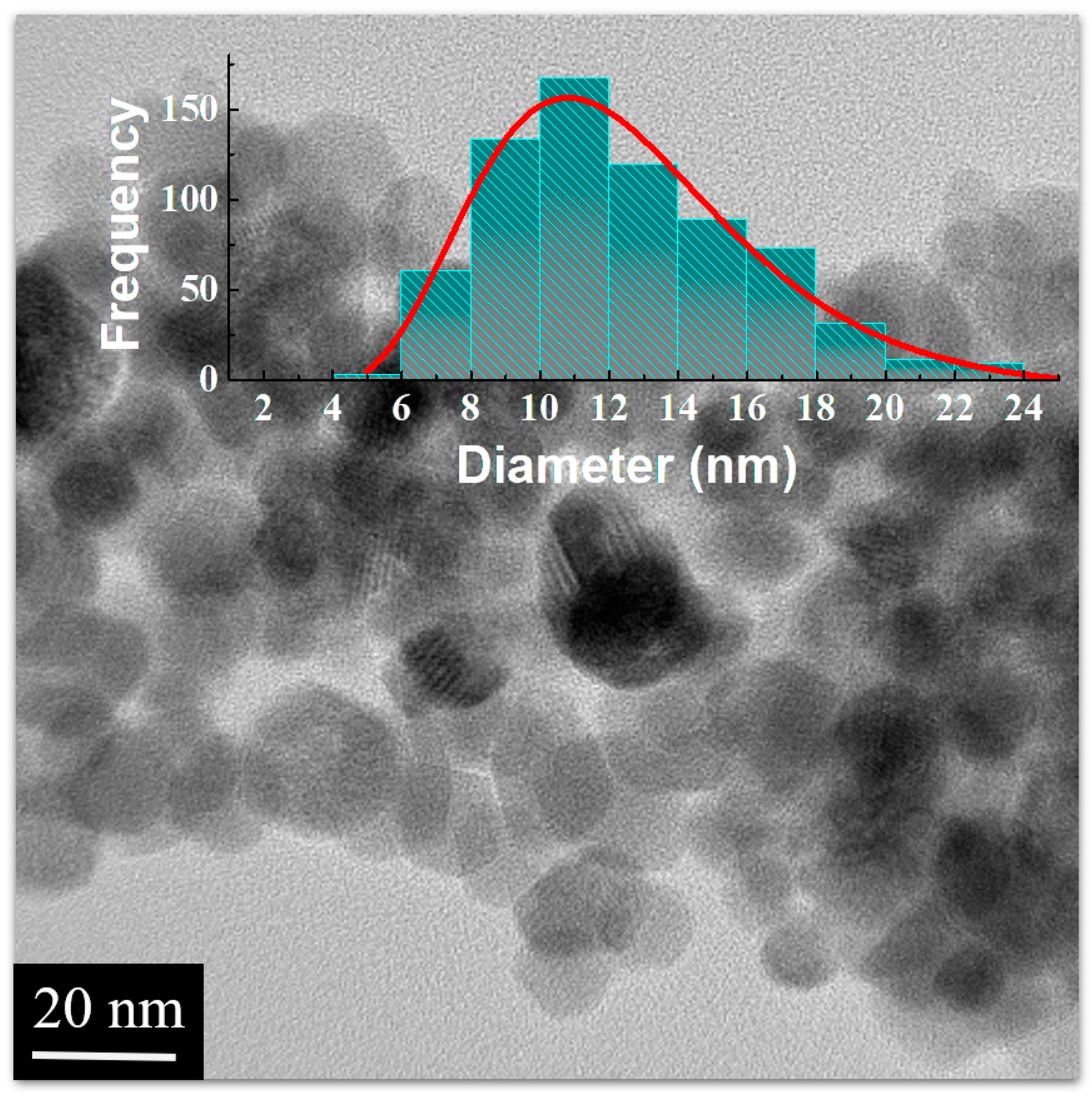
Figure 2 displays the TEM image of the γ-Fe2O3@PSSNa nanohybrid, where the NPs depict their polydisperse behavior. The average particle has a value of 11(2) nm, which coincides with the estimated crystallite size from the X-ray data. The TEM image also suggests that PSSNa covers the γ-Fe2O3 NPs, and consequently it may be responsible for preventing γ-Fe2O3 agglomeration. This finding favors for a chemical stability of γ-Fe2O3 NPs as also reported in the literature [14,21].

3.2. Raman Analysis

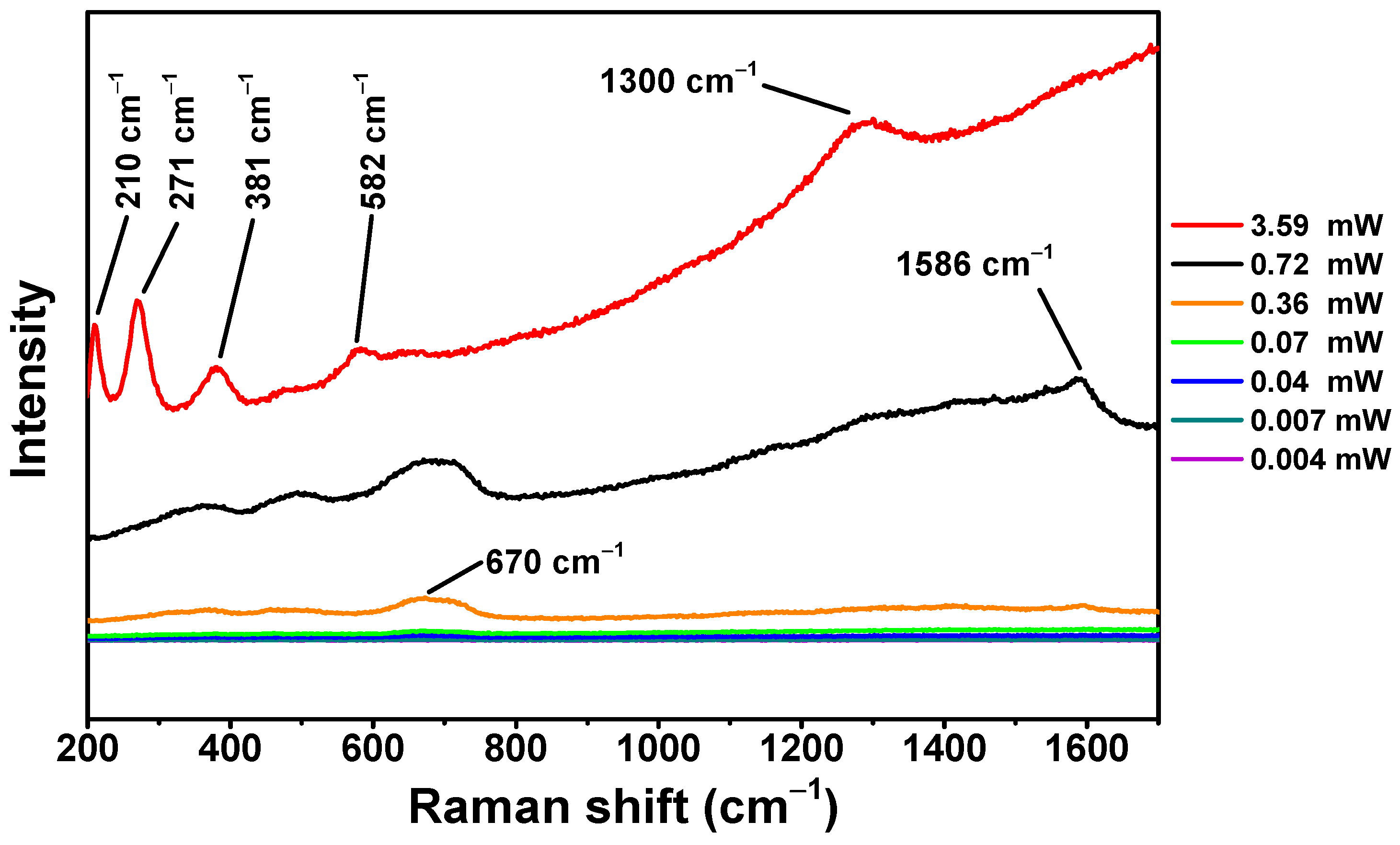
For the Raman analysis of the γ-Fe2O3@PSSNa nanohybrid, the optical range of 200 to 1700 cm−1 was chosen at several laser powers. In Figure 3, the spectrum corresponding to 3.59 mW (red line) presents two optical active modes at 210 cm−1 (A1g) and 271 cm−1 (Eg) related to the polymorphic phase of low crystallized α-Fe2O3 [26]. The Raman modes at 381 and 582 cm−1 assign the contribution of the mixture of γ-Fe2O3 and low crystallized α-Fe2O3. The γ-Fe2O3 band can also be related to the additional peak found at 1300 cm−1, which appears close to that found by Hanesch at 1330 cm−1 [27]. In the literature, a threshold value for laser power has been reported depending on the γ-Fe2O3 magnetic structure and its combination with other nonmagnetic matrices [28,29,30]. Hence, we would like to point out that a bad interpretation of Raman spectra depends on this threshold value and must be individually determined for different magnetic nanohybrids, specifically for composites with γ-Fe2O3. More importantly, an intense laser power can result in the sample burning (carbon), hence not allowing us to correctly analyze the organic contribution.
In the signal that uses 0.72 mW (black line), the peak of 1586 cm−1 appears isolated, which could be attributed to the presence of the PSSNa polymer as previously reported at 1598 cm−1 [31]. The Raman modes located at 370, 460, 490, 670, and 1596 cm−1 were found for the 0.36 mW spectrum (orange line). The Raman mode that appears at 370 cm−1 is assigned to γ-Fe2O3 [26]. The contribution of the 460 and 490 cm−1 active modes produces a broad signal that can be attributed to γ-Fe2O3 [28,32]. The broad peak that appears between 670 cm−1 indicates the presence of γ-Fe2O3 [27]. Moreover, the isolated peak of 1596 cm−1 was attributed to PSSNa [27]. Below this laser power, it was not possible to identify the polymer contribution (this is explained due to the predominance of the iron-oxide phase in the sample). Hence, the 0.36 mW laser power value was stablished as the threshold laser power for this sample. Above this value, the polymorphic α-Fe2O3 phase has its onset ( γ α transformation), and total crystallization can be achieved by tuning the laser power [23,28,29,30].

3.3. FTIR and Optical UV Vis Analysis

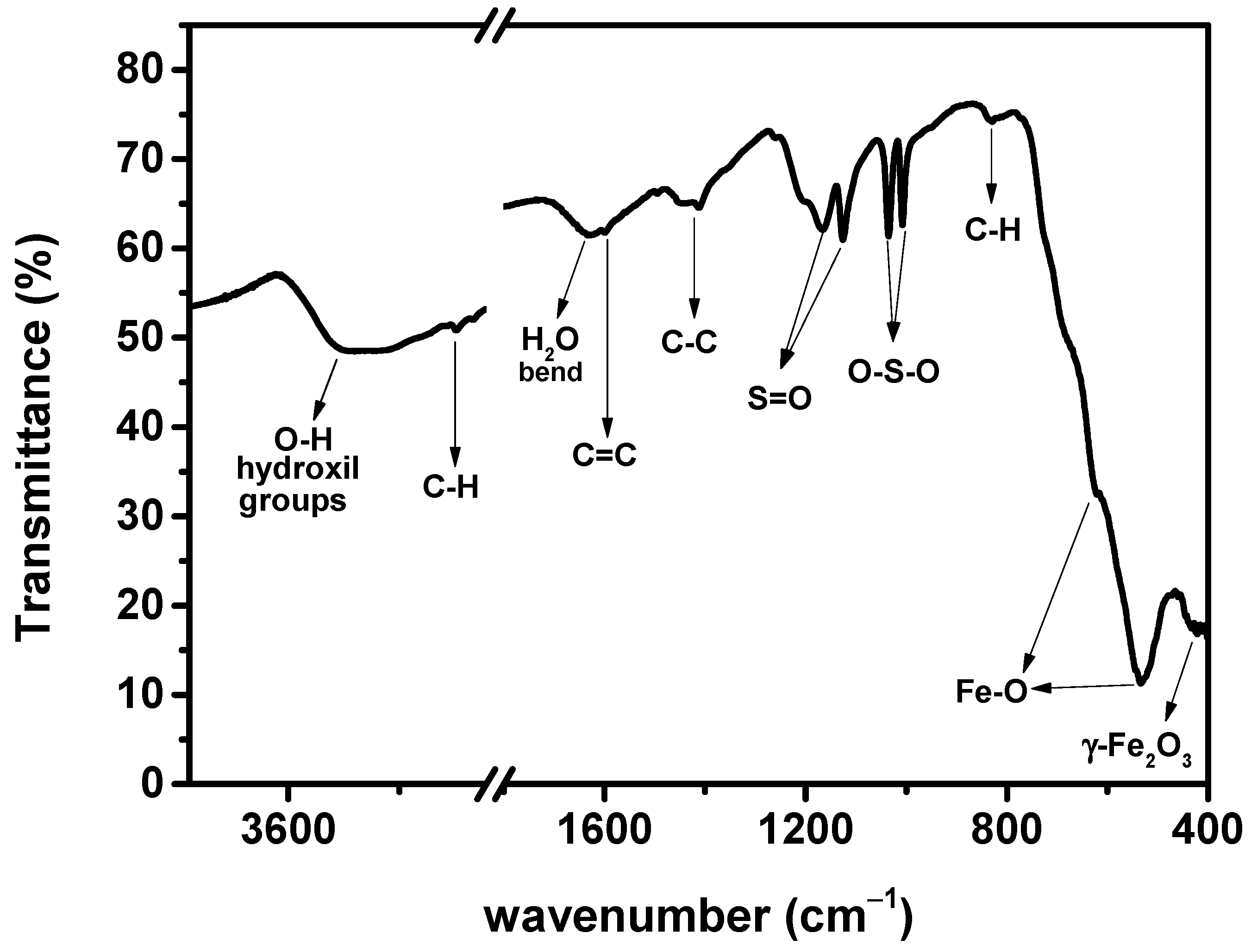
The IR spectra of pure PSSNa and γ-Fe2O3@PSSNa are shown in Figure 4. IR peaks ca. 434, 534, and 619 cm−1 indicate the metal–oxygen bonding found in the tetrahedral sites, where the first peak could correspond to the only deformation of octahedral sites of the γ-Fe2O3 phase [33,34,35,36]. Bali et al. [34] have identified an IR peak at about 2925 cm−1 and associating it with the stretching vibration of the C-H bond. This IR peak value is close to that at 2920 cm−1 found in our IR spectrum of the γ-Fe2O3@PSSNa nanohybrid. In addition, the IR peak at 830 cm−1 corresponds to an out-of-plane vibration of the C-H groups in the benzene rings [37]. At about 3400 and 1630 cm−1, the broad IR peaks correspond to the stretching and bending vibrations for the O-H bond in the retained physiosorbed water [33,35].
The presence of benzene rings is expressed by the stretching of the C=C and C-C bonds at 1590 and 1430 cm−1, respectively. Funda et al. [38] associates these effects with the presence of PSSNa. The S O 3 groups present in the PSSNa polymer appear around the peaks at 1124 and 1167 cm−1, while the typical symmetric stretching of O–S–O is located at 1006 and 1034 cm−1 [36,39]. This information confirms the presence of a PSSNa polymer functionalizing the γ-Fe2O3 phase.
Figure S1a shows the optical absorption spectrum of the γ-Fe2O3@PSSNa nanohybrid in the range of 250–550 nm. A pronounced decay and a concomitant rise in the optical spectrum at 350 nm may indicate the presence of a new peak at about 367 nm. Indeed, the presence of PSSNa occurs with a shift towards longer wavelengths in the range of 350 to 600 nm due to the presence of PSSNa aromatic groups by the π→π* transition [40]. Also, the position close to 370 nm exhibits the 6A1→4E transition for the γ-Fe2O3 phase [41], indicating a degeneracy in the energy region for these two phases of the sample (lack of energy resolution to solve these phases in the optical spectrum).
The forbidden band, Eg, of the γ-Fe2O3@PSSNa nanohybrid, obtained with the UV-VIS absorption data, was experimentally determined from the absorption coefficient α, measured through the Tauc formula [42]:
α h ν = A h ν E g n
where A is a constant, (h ν ) is the photon energy, and n can be either 1/2 for direct transmission or 2 for indirect transmission.
Figure S1b belongs to the indirect transmission graph with n = 1/2. This quantity was obtained by extrapolating a straight line over a straight portion of the curve, chosen between the range of 3.44 and 3.52 eV. The intersection of this straight line with the x-axis gives a value of Eg = 1.42 eV. It is less than the 2.0 eV reported for bulk γ-Fe2O3 [43]. Consequently, this reduction could be attributed to size-finite effects that directly influence the Eg value, as reported in the literature [43].

3.4. Colloidal, Thermal, and Textural Properties

PSSNa is commonly used to improve the dispersion stability of ceramic slurry, such as dispersants of alumina, titanium dioxide, zirconia, and other metal-oxide ceramics [44]. Regarding the value of the hydrodynamic diameter of PSSNa, different values have been reported. For a PLL/PSSNa complex, a value of 2.1 μm at 300 K and pH = 5 was reported [45], while a hydrodynamic radius of a bare molecule equal to 2.5 nm is found in Ref. [46]. In this study, the commercial polymer PSSNa has a hydrodynamic diameter value of 5.2 μm, which is twice the value found in Ref. [45]. On the other hand, the γ-Fe2O3@PSSNa nanohybrid shows a value of 210 nm (or 0.21 μm), which is ten times smaller than 2.1 μm found for the PLL/PSSNa complex. Also, it is important to mention that no substantial agglomeration of the functionalized γ-Fe2O3 has been occurred after a long time [47], suggesting that the polymer plays an important role on the NP agglomeration process.
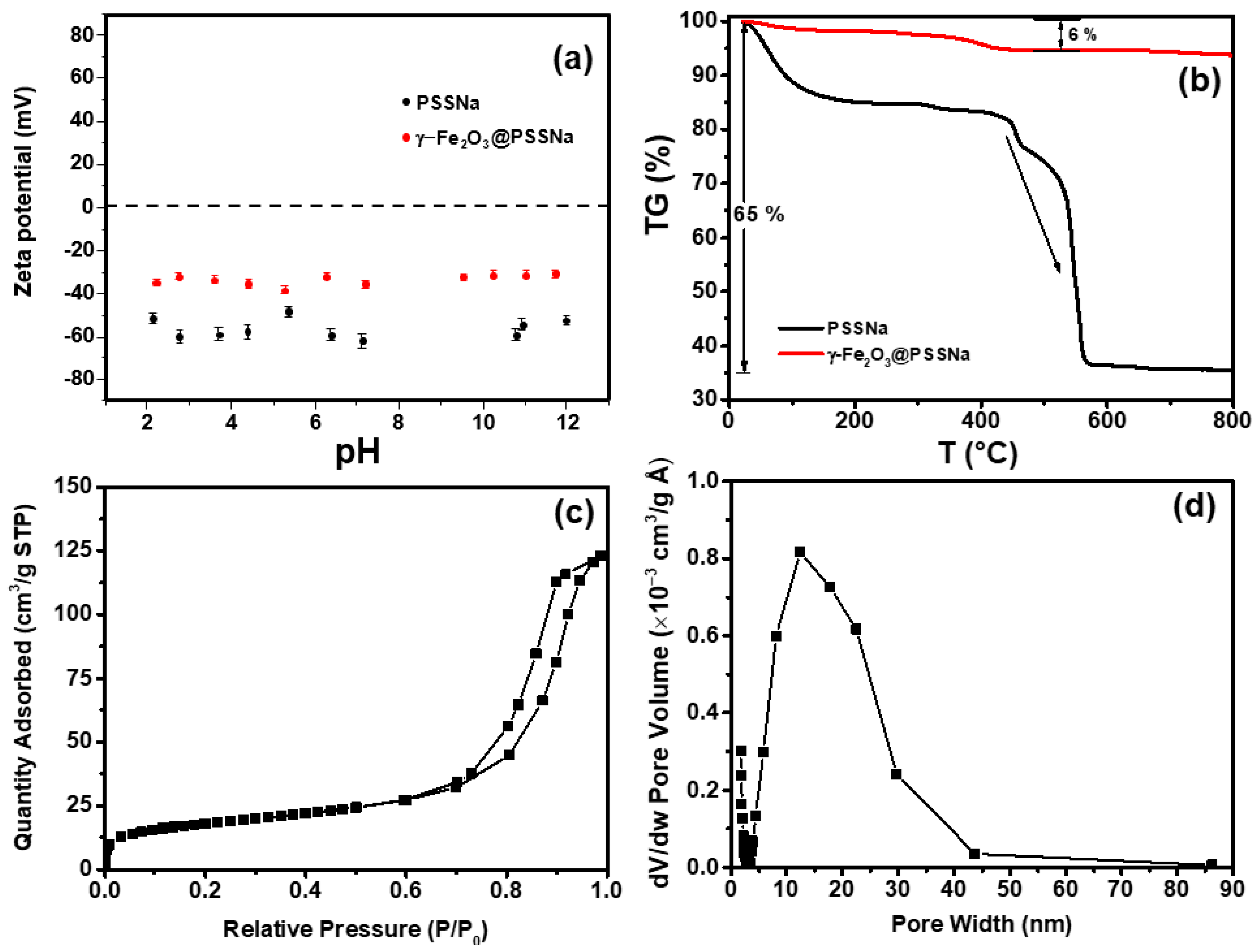
Colloidal stability was evaluated by analyzing the Zeta potential vs. pH plot, shown in Figure 5a. For the bare PSSNa saloplastic, a negative charge was manifested in the entire pH range, with high stability values varying between −40 and −70 mV. Similar negative values of −41 to −52 mV have been reported for the PSSNa/MWNT in the KCl solution, which has a pH close to neutral [48]. Furthermore, a high Zeta potential for the PSSNa-coated Au of (−47.9 ± 3.2) mV was reported [49]. Thus, regarding the γ-Fe2O3@PSSNa nanohybrid, a negative Zeta potential was detected throughout the measured pH range, with values ranging between −30 and −40 mV. This phenomenon is mainly attributed to the effect of PSSNa on colloidal stability, since the γ-Fe2O3 NPs generally have isoelectric points greater than 6. For example, a p.z.c. value of 6.3 for the 12 nm MZ0 composite [5] and 8.3 for the 6 nm γ-Fe2O3 NPs have been reported [50]. These results suggest that both γ-Fe2O3-based NP materials demonstrated a remarkable colloidal stability with a marked predominance of negative surface potential, similar to the results obtained from the 12 nm γ-Fe2O3@PSSNa nanohybrid, here studied.
Thermogravimetric measurements for the PSSNa and γ-Fe2O3@PSSNa samples are shown in Figure 5b. For pure PSSNa, a total weight loss of 65% was observed. Sulfonic-acid group (SO3H) desulfonation and polystyrene degradation in the 200–550 °C range were related to the abrupt decrease after 400 °C. Up to roughly 800 °C, the primary carbon-chain skeleton was observed [51]. For the γ-Fe2O3@PSSNa nanohybrid, the quantified weight loss of 6% has the same change as that of pure PSSNa, and the small loss is because the PSSNa makes up a small percentage of the mass of γ-Fe2O3@PSSNa.
Figure 5c shows the hysteresis adsorption–desorption isotherm obtained at 77 K for the γ-Fe2O3@PSSNa nanohybrid. The found textural properties were BET area of 66 m2/g, pore volume of 0.186 cm3/g, and pore diameter equal to 20 nm. The pore distribution, which was estimated using the BJH approach, showed a broad range of pores, as displayed in Figure 5d. The isotherm curve exhibited the distinctive shape of an IV-type isotherm, which is associated with mesoporous materials [52].

3.5. Magnetic Studies

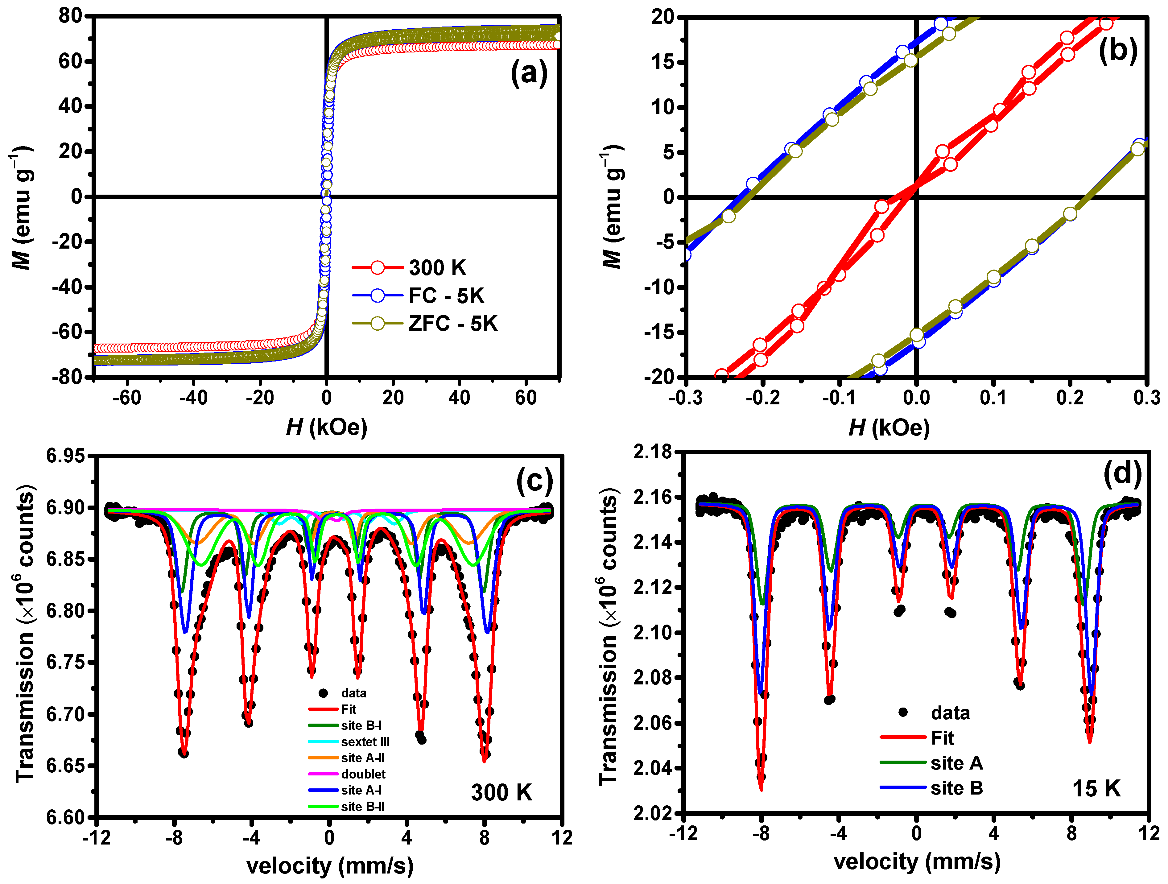
Figure 6a shows the ZFC and FC M(H) curve recorded at 300 and 5 K for the γ-Fe2O3@PSSNa nanohybrid. The zoomed region in Figure 6b shows the magnetic behavior of γ-Fe2O3@PSSNa at 5 K. 300 K M(H) curve shows a low coercive field (HC) of 100 Oe; see also Table S2. It can be explained assuming that the distribution of magnetic particles is weakly interacting and/or a result of ferrimagnetic-like character of the of γ-Fe2O3 NPs. The Law of Approach to Saturation (LAS), given by Equation (2) [24], was used to fit the M(H) curves in the range of +20 to +70 kOe:
M = M S 1 b H 2 + χ H .
where Ms is the saturation magnetization, χ is the high-field susceptibility, and the effective anisotropy constant, K e f f , can be obtained by replacing the value of Ms in b = 4 15 K e f f 2 M S 2 .
Table S2 summarizes the magnetic parameters obtained with the LAS. In the ZFC protocol, a Hc field of 0.22 kOe and a remnant magnetization Mr of =15.45 emu g−1 were calculated. The fitting of FC M(H) data recorded at 5 K displays similar values, i.e., Hc = 0.22 kOe and Mr = 16.73 emu g−1, indicating the absence of magnetic interactions that could result in the exchange of bias effect sometimes found in an ensemble of nanoparticles (it should favor a shift of the M(H) loop towards the negative field axis [19]). The Ms showed value of 66.0(1) emu g−1 for 300 K and 71.0(1) emu g−1 at 5 K for both ZFC and FC M(H) loops. The obtained Ms values agree with bulk γ-Fe2O3 [53]. The values for χ, obtained from the fittings and shown in Table S2, indicate that the saturation regime was roughly reached, and the K e f f values are like those reported in the literature for nanometric Fe-oxide based samples [19,24].
Figure 6c,d presents the 300 K and 15 K 57Fe Mössbauer spectra of the γ-Fe2O3@PSSNa nanohybrid. Table S3 reports the fitted hyperfine parameters. The 300 K spectrum was fitted with six components. Four sextets were used to represent the distribution of particle sizes of the γ-Fe2O3 NPs, also seen in TEM images. Indeed, the bulk cubic spinel γ-Fe2O3 phase only shows two well-defined sites: tetrahedral (A) and octahedral (B) sites of the ion Fe3+ spins. However, considering the large distribution particle sizes of the γ-Fe2O3 NPs, part of these NPs (<10 nm) starts to enter in a spin relaxation regime at 300 K, broadening the absorption line. Thus, two additional sextets were added to represent this fraction of NPs. The five (doublet) and six (III) subspectra, added to fit the 300 K spectrum, can, respectively, be associated with very small γ-Fe2O3 NPs in the superamagnetic regime and uncompensated surface Fe3+, as proposed by Zakharova et al. [54]. To estimate the thickness of this shell, spherical particles were considered, such as the surface to volume ratio, which can be assumed to be V e / V = 3 Δ r / r , where V e = 4 π r 2 Δ r and V = 4 / 3 π r 3 . Thus, the thickness of Fe3+ shell spins, defined by Equation (3) [54], can be estimated:
Δ r = t o t a l   a r e a   f r a c t i o n 6 × D T E M
The total area fraction is indicated in Table S3, i.e., the contribution of the surface site area A to B was added, giving a total of 43% and D T E M = 11.4   n m , resulting in an average thickness of Δ r = 0.91   n m .
The 15 K spectrum in Figure 6d only displays the two static magnetic sextets that agree with a highly stoichiometric γ-Fe2O3, with remarkable asymmetry between lines 1:6. As expected, in pure samples, the values for CS and Bhf are close to each other. In addition, the relative fraction (R.A.A.) for the A and B sites (37% and 63%, respectively) are close to the stoichiometric formula (1.6) [50].

3.6. Ecotoxicological Analysis in D. magna

3.6.1. LC50 of γ-Fe2O3@PSSNa Nanohybrid

The LC50 value found using the sigmoidal non-linear fit was 533(5) mg L−1, as seen in Figure 7. This value is close to 550 mg L−1 found for the ternary nanohybrid reported in the literature [24]. Table 1 presents a brief data collection of the parent systems used for comparison of the ecotoxicological profile.
In short, the ecotoxicological impact will depend on NPs size, as shown in Table 1. On the other hand, it should be said that the colloidal stability and hydrodynamic diameter are parameters not often reported in various studies with NPs. However, in the present study, the γ-Fe2O3@PSSNa nanohybrid depicted zero p.z.c. and highly stable Zeta potential values. These results indicate that the nanohybrid will interact for more time with the neonates, affecting their mortality. Indeed, γ-Fe2O3 (bulk or nano) by itself depicts low colloidal stability [23], tending to quick sedimentation, therefore, its colloidal stability will certainly affect the LC50 value, as demonstrated in this work.
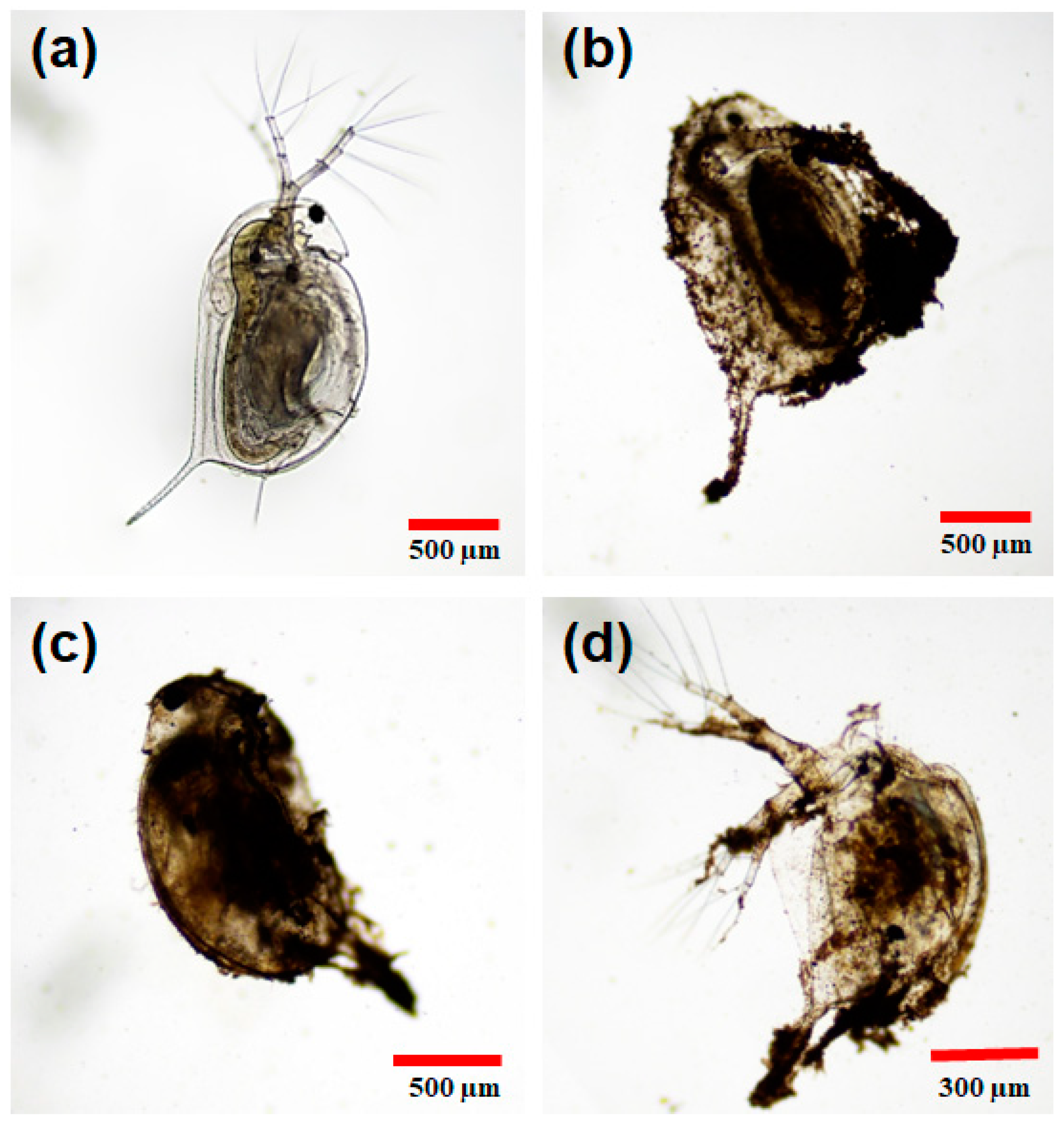
Figure 8 shows the visual effects of the γ-Fe2O3@PSSNa nanohybrid after 24 h of exposure. The N.C., without exposure to γ-Fe2O3@PSSNa, is shown in Figure 8a. It can be noted that the limbs are free and there is no obstruction in the digestive tract. This observation differs from what is shown in the following images, where it is seen that the NPs obstruct the limbs and adhere to the chitinous exoskeleton blocking the ability to move. Figure 8b displays the exoskeleton with the NPs with a concentration of γ-Fe2O3@PSSNa of 800 mg L−1. Figure 8c depicts D. magna immersed in a medium with a concentration of 600 mg L−1, whereas Figure 8d presents the residues left in the exoskeleton at a concentration of γ-Fe2O3@PSSNa of 400 mg L−1.

3.6.2. Morphological Analysis in D. magna

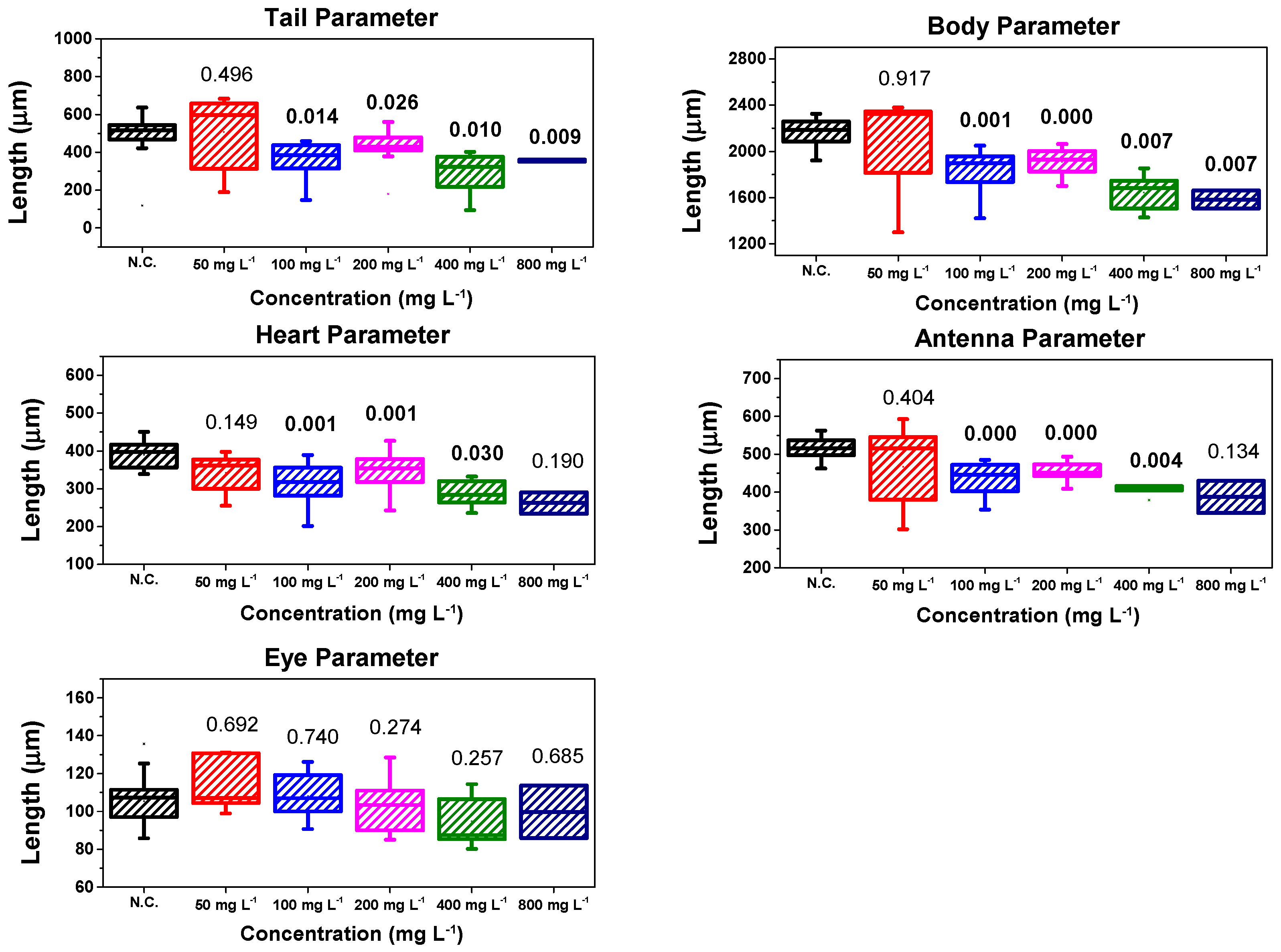
After the control period of the surviving D. magna, morphological analysis was performed. Once it was confirmed that the morphological parameter data followed a normal distribution, Student’s t-test was applied. The results of the morphological analysis are shown in a box plot, as in previous ecotoxicity research [23,24,58,63]. Figure 9 shows the results of morphological analysis of the γ-Fe2O3@PSSNa nanohybrid for the concentrations of 50 mg L−1, 100 mg L−1, 200 mg L−1, 400 mg L−1, and 800 mg L−1, after the established control time, which was 13-days after exposure of D. magna to γ-Fe2O3@PSSNa.
The results suggest that only the concentration of 50 mg L−1 yields no significant change for any of their morphological parameters, while the eye parameter has no significant change regardless of the concentrations applied. Thus, the latter indicates that D. magna can maintain orientation after exposure to γ-Fe2O3@PSSNa.
On the other hand, the morphological parameters (heart, tail, body, and antenna) showed a significant change with respect to the concentrations, except for the 800 mg L−1 concentration, in which the antenna and heart parameters did not present a substantial modification. It is important to mention that these changes in morphological parameters may be related to genotoxicological damage occurring in D. magna, as also observed in Chironomus riparius using graphene oxide, in which there was an alteration in genes involved in the metabolism [64]. Thus, it can be inferred that it probably occurred after D. magna ingested γ-Fe2O3@PSSNa, which has an intermediate size between 20 nm and 70 μm, as suggested by Matson [62]. This effect would also explain why D. magna was reduced in size at concentrations of 100 mg L−1, 200 mg L−1, 400 mg L−1, and 800 mg L−1 of γ-Fe2O3@PSSNa with respect to the negative control.
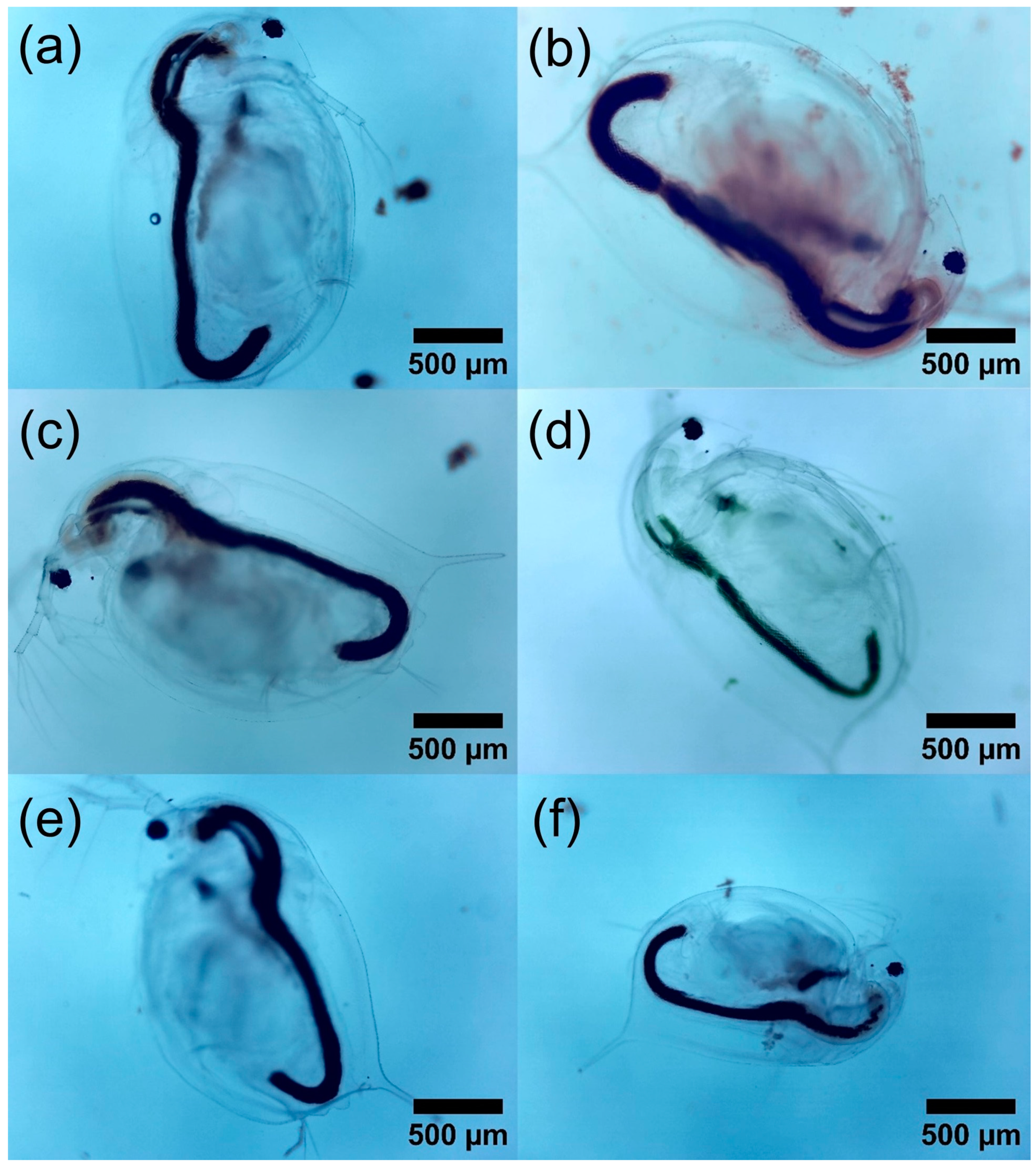
Therefore, it can be said that the PSSNa polymer is likely the main cause of a significant change in the other morphological parameters, if one considers the results reported for γ-Fe2O3 NPs functionalizing graphene oxide that only show significant changes at high γ-Fe2O3 concentrations [58]. These results in the morphological parameters of D. magna are interesting, since these significant changes in the morphological parameters of the tail and antenna would affect the swimming of D. magna, while the reduced size of the heart may be due to the acute exposure to γ-Fe2O3@PSSNa, as shown in previous research that reported that anomalies in embryonic development can affect the morphology of D. magna [65]. Moreover, these changes are more sensitive in embryos than in juveniles of D. magna [66]. Although this study used neonates and not embryos or young individuals of D. magna, it is possible that the exposure of D. magna to γ-Fe2O3@PSSNa caused these significant changes in their morphological parameters, as it developed in the days following γ-Fe2O3@PSSNa exposure, whose significant p-values are shown in the box plots in Figure 9. Finally, a qualitative comparison is made using the images of D. magna on the last-control day, as shown in Figure 10a–f.

4. Conclusions

γ-Fe2O3 NPs functionalized with PSSNa polymer were successfully synthesized using a modified co-precipitation route. Different experimental methods were initially applied to characterize their structural, vibrational, electronic, thermal, colloidal, hyperfine, and magnetic properties. Magnetization and 57Fe Mössbauer data suggested that the γ-Fe2O3@PSSNa composite shows ferrimagnetic-like features that allow for magnetic remediation. Infrared and Raman indicated that the polymer covered the γ-Fe2O3 NPs, yielding chemical stability and reducing agglomeration. This material was tested for its ecotoxicological impact on D. magna. The results of the morphological analysis revealed that the γ-Fe2O3@PSSNa nanohybrid, although not totally toxic since its 24 h LC50 is 533(5) mg L−1, undergoes significant changes in the morphological parameters of the D. magna at concentrations below 800 mg L−1 after 13-days of exposure. It is relevant to note that, at all concentrations examined, no noticeable change in the ocular parameter was evident. Furthermore, at a concentration of 50 mg L−1, no relevant alterations were observed in any of the morphological parameters here evaluated. These findings indicate the optimal concentration of the γ-Fe2O3@PSSNa composite for the magnetic remediation of contaminated water, i.e., a permissible value that can be applied with a big impact on living organisms found in ordinary effluents.

Supplementary Materials

The following supporting information can be downloaded at: https://www.mdpi.com/2073-4360/16/11/1581/s1, Table S1: Rietveld refined parameters for the γ-Fe2O3@PSSNa nanohybrid. Refinement and statistical parameters, Rp(%) as profile refinement, Rwp (%) as weighted profile residual, Rexp(%) as the expected profile residual, and goodness of the fit, χ 2 . B is the temperature factor and Occ. represents the fraction occupancies. Table S2. Magnetic parameters obtained from LAS equation. n.d. not defined. Table S3. Hyperfine parameters obtained from the fits of the 300 K and 15 K 57Fe Mössbauer spectra of the γ-Fe2O3@PSSNa nanohybrid. R.A.A. is the relative absorption area, Bhf the hyperfine magnetic field, CS (mm/s) the center shifts respective vs. Fe at 300 K (0.114 mm/s), Q (mm/s) is the quadrupolar shifting, W is the Lorentzian line width, and σ the width of Gaussian distribution. Figure S1: (a) Absorbance UV Vis spectrum, and (b) forbidden band determination from the intersection of photon energy (eV).

Author Contributions

Conceptualization, J.A.R.-G., R.R.-V., E.V.M.-C., F.M.-V. and E.C.P.; methodology, J.A.R.-G., R.R.-V., E.V.M.-C., F.M.-V. and E.C.P.; software, J.A.R.-G., R.R.-V., E.V.M.-C., F.M.-V. and N.-R.C.-H.; validation, J.A.R.-G., R.R.-V., E.V.M.-C., F.M.-V. and E.C.P.; formal analysis, J.A.R.-G., R.R.-V., E.V.M.-C., F.M.-V., N.-R.C.-H. and E.C.P.; investigation, J.A.R.-G., R.R.-V., E.V.M.-C., F.M.-V., N.-R.C.-H. and E.C.P.; resources, J.A.R.-G., R.R.-V., E.V.M.-C., F.M.-V., N.-R.C.-H. and E.C.P.; data curation, J.A.R.-G., R.R.-V., F.M.-V. and N.-R.C.-H.; writing—original draft preparation, J.A.R.-G. and R.R.-V.; writing—review and editing, J.A.R.-G., R.R.-V., E.V.M.-C., F.M.-V., N.-R.C.-H. and E.C.P.; visualization, J.A.R.-G., R.R.-V., E.V.M.-C., F.M.-V., N.-R.C.-H. and E.C.P.; supervision, J.A.R.-G., R.R.-V., E.V.M.-C., F.M.-V., N.-R.C.-H. and E.C.P.; project administration, J.A.R.-G., E.V.M.-C. and E.C.P.; funding acquisition, J.A.R.-G. and E.V.M.-C. All authors have read and agreed to the published version of the manuscript.

Funding

This research was financed by the Vicerrectorado de Investigación y Posgrado (VRIP) from Universidad Nacional Mayor de San Marcos (UNMSM) through the Project PCONFIGI B23131591 approved by Rectoral Resolution 006081-R-23.

Institutional Review Board Statement

Not applicable.

Data Availability Statement

The original data of this research can be requested any time to the corresponding author’s email address: [email protected].

Acknowledgments

Edson C. Passamani is grateful for Brazilian financial support from Fundação de Amparo à Pesquisa e Inovação do Espírito Santo (FAPES: TO-975/2022 and TO-640/2022) and Conselho Nacional de Desenvolvimento Científico e Tecnológico (CNPq). The authors are thankful to Amaya Arencibia, from Rey Juan Carlos University, for supporting the experimental data of adsorption–desorption isotherms at 77 K.

Conflicts of Interest

The authors of this manuscript declare no conflicts of interest or competing interests if published.

Correction Statement

This article has been republished with a minor correction to the existing affiliation information. This change does not affect the scientific content of the article.

References

  1. Fu, Z.; Xi, S. The Effects of Heavy Metals on Human Metabolism. Toxicol. Mech. Methods 2020, 30, 167–176. [Google Scholar] [CrossRef] [PubMed]
  2. Lawal, K.K.; Ekeleme, I.K.; Onuigbo, C.M.; Ikpeazu, V.O.; Obiekezie, S.O. A Review on the Public Health Implications of Heavy Metals. WJARR 2021, 10, 255–265. [Google Scholar] [CrossRef]
  3. Jalili, C.; Kazemi, M.; Cheng, H.; Mohammadi, H.; Babaei, A.; Taheri, E.; Moradi, S. Associations between Exposure to Heavy Metals and the Risk of Chronic Kidney Disease: A Systematic Review and Meta-Analysis. Crit. Rev. Toxicol. 2021, 51, 165–182. [Google Scholar] [CrossRef] [PubMed]
  4. Kaur, G.; Kumar, R.; Mittal, S.; Sahoo, P.K.; Vaid, U. Ground/Drinking Water Contaminants and Cancer Incidence: A Case Study of Rural Areas of South West Punjab, India. Hum. Ecol. Risk Assess. 2021, 27, 205–226. [Google Scholar] [CrossRef]
  5. Ramos-Guivar, J.A.; Checca-Huaman, N.R.; Litterst, F.J.; Passamani, E.C. Synergetic Effect between Zeolite 5 A and Maghemite Nanoparticles for Fast Lead Uptake from the Peruvian River Cumbaza: Study of Surface Adsorption Mechanism Using X-ray Photoelectron Spectroscopy. Appl. Surf. Sci. 2023, 18, 100489. [Google Scholar] [CrossRef]
  6. Pinotti, C.N.; Ramos-Guivar, J.A.; Proveti, J.R.; Canchanya-Huaman, Y.; Arias-Contreras, M.A.; Checca-Huaman, N.R.; Passamani, E.C. Fractal-like Kinetics for Enhanced Boron Adsorption on Heterogeneous Magnetic Composite Surfaces. Mater. Chem. Phys. 2023, 308, 128313. [Google Scholar] [CrossRef]
  7. Peng, Q.; Zhao, H.; Chen, G.; Yang, Q.; Cao, X.; Xiong, S.; Liu, Q. Synthesis of Novel Magnetic Pitch-based Hypercrosslinked Polymers as Adsorbents for Effective Recovery of Ag+ with High Selectivity. J. Environ. Manag. 2023, 339, 117763. [Google Scholar] [CrossRef] [PubMed]
  8. Hu, X.; Hu, Y.; Xu, G.; Li, M.; Zhu, Y.; Jiang, L.; Tu, Y.; Zhu, X.; Xie, X.; Li, A. Green Synthesis of a Magnetic β-cyclodextrin Polymer for Rapid Removal of Organic Micro-pollutants and Heavy Metals from Dyeing Wastewater. Environ. Res. 2020, 180, 108796. [Google Scholar] [CrossRef] [PubMed]
  9. Yuan, W.; Zhou, Y.; Chen, Y.; Liu, X.; Wang, J. Toxicological Effects of Microplastics and Heavy Metals on the Daphnia magna. Sci. Total Environ. 2020, 746, 141254. [Google Scholar] [CrossRef] [PubMed]
  10. Tatarazako, N.; Oda, S. The Water Flea Daphnia magna (Crustacea, Cladocera) as a Test Species for Screening and Evaluation of Chemicals with Endocrine Disrupting Effects on Crustaceans. Ecotoxicology 2007, 16, 197–203. [Google Scholar] [CrossRef] [PubMed]
  11. Herold, B.C.; Bourne, N.; Marcellino, D.; Kirkpatrick, R.; Strauss, D.M.; Zaneveld, L.J.; Waller, D.P.; Anderson, R.A.; Chany, C.J.; Barham, B.J.; et al. Poly (Sodium 4-Styrene Sulfonate): An Effective Candidate Topical Antimicrobial for the Prevention of Sexually Transmitted Diseases. J. Infect. Dis. 2000, 181, 770–773. [Google Scholar] [CrossRef] [PubMed]
  12. Botwina, P.; Obłoza, M.; Szczepański, A.; Szczubiałka, K.; Nowakowska, M.; Pyrć, K. In Vitro Inhibition of Zika Virus Replication with Poly (Sodium 4-Styrenesulfonate). Viruses 2020, 12, 926. [Google Scholar] [CrossRef] [PubMed]
  13. Farias-Cepeda, L.; Herrera-Ordonez, J.; Estevez, M.; Luna-Barcenas, G.; Rosales-Marines, L. New Insights on Surfactant-Free Styrene Emulsion Polymerization in the Presence of Sodium Styrene Sulfonate. Colloid Polym. Sci. 2016, 294, 1571–1576. [Google Scholar] [CrossRef]
  14. Corr, S.A.; Gun’ko, Y.K.; Tekoriute, R.; Meledandri, C.J.; Brougham, D.F. Poly (Sodium-4-Styrene) Sulfonate− Iron Oxide Nanocomposite Dispersions with Controlled Magnetic Resonance Properties. J. Phys. Chem. C 2008, 112, 13324–13327. [Google Scholar] [CrossRef]
  15. Willott, J.D.; Lindhoud, S.; de Vos, W.M. Hot-pressing Polyelectrolyte Complexes into Tunable Dense Saloplastics. Polymer 2022, 242, 124583. [Google Scholar] [CrossRef]
  16. Nasser, F.; Lynch, I. Updating Traditional Regulatory Tests for Use with Novel Materials: Nanomaterial Toxicity Testing with Daphnia magna. Saf. Sci. 2019, 118, 497–504. [Google Scholar] [CrossRef]
  17. Shin, H.K.; Seo, M.; Shin, S.E.; Kim, K.Y.; Park, J.W.; No, K.T. Meta-analysis of Daphnia magna Nanotoxicity Experiments in Accordance with Test Guidelines. Environ. Sci. Nano 2018, 5, 765–775. [Google Scholar] [CrossRef]
  18. Tkaczyk, A.; Bownik, A.; Dudka, J.; Kowal, K.; Ślaska, B. Daphnia magna Model in the Toxicity Assessment of Pharmaceuticals: A Review. Sci. Total Environ. 2021, 763, 143038. [Google Scholar] [CrossRef] [PubMed]
  19. Ramos-Guivar, J.A.; Morales, M.A.; Litterst, F.J. Suppression of Exchange Bias Effect in Maghemite Nanoparticles Functionalized with H2Y. J. Magn. Magn. Mater. 2016, 420, 324–335. [Google Scholar] [CrossRef]
  20. Putz, H.; Brandeburg, K. Match!—Phase Identification from Powder Diffraction. Available online: https://www.crystalimpact.de/match (accessed on 27 May 2024).
  21. Rueda-Vellasmin, R.; Checca-Huaman, N.R.; Passamani, E.C.; Litterst, F.J.; Ramos-Guivar, J.A. Mössbauer Studies of Core-Single-Shell and Core-Double-Shell Polymer Functionalized Magnetic Nanoparticles. Hyperfine Interact. 2022, 243, 27. [Google Scholar] [CrossRef]
  22. Klencsár, Z. Mosswinn 4.0i, Revision, 13 January 2024. Available online: http://www.mosswinn.hu/ (accessed on 27 May 2024).
  23. Moyano-Arocutipa, M.F.; Zarria-Romero, J.Y.; Huertas-Chambilla, M.Y.; Checca-Huaman, N.R.; Pino, J.; Passamani, E.C.; Ramos-Guivar, J.A. In Situ and after Synthesis of Magnetic Nanoarchitectures Grown onto Zeolite Type 5A/CTAB Frameworks and Their Ecotoxicological Properties. Cryst. Growth Des. 2023, 23, 2951–2970. [Google Scholar] [CrossRef]
  24. Tamanaha-Vegas, C.A.; Zarria-Romero, J.Y.; Greneche, J.M.; Passamani, E.C.; Ramos-Guivar, J.A. Surface Magnetic Properties of a Ternary Nanocomposite and Its Ecotoxicological Properties in Daphnia magna. Adv. Powder Technol. 2022, 33, 103395. [Google Scholar] [CrossRef]
  25. Ramos-Guivar, J.A.; Flores-Cano, D.A.; Passamani, E.C. Differentiating Nanomaghemite and Nanomagnetite and Discussing Their Importance in Arsenic and Lead Removal from Contaminated Effluents: A Critical Review. Nanomaterials 2021, 11, 2310. [Google Scholar] [CrossRef] [PubMed]
  26. Jubb, A.M.; Allen, H.C. Vibrational Spectroscopic Characterization of Hematite, Maghemite, and Magnetite Thin Films Produced by Vapor Deposition. ACS Appl. Mater. Interface 2010, 2, 2804–2812. [Google Scholar] [CrossRef]
  27. Hanesch, M. Raman Spectroscopy of Iron Oxides and (Oxy) Hydroxides at Low Laser Power and Possible Applications in Environmental Magnetic Studies. Geophys. J. Int. 2009, 177, 941–948. [Google Scholar] [CrossRef]
  28. Ramos-Guivar, J.A.; Zarria-Romero, J.Y.; Castro-Merino, I.L.; Greneche, J.M.; Passamani, E.C. Improvement of the Thermal Stability of Nanomaghemite by Functionalization with Type 5A Zeolite and Magnetic Properties Studied by In-Field 57Fe Mössbauer Measurements. J. Magn. Magn. Mater. 2022, 552, 169241. [Google Scholar] [CrossRef]
  29. Mendili, Y.E.; Grasset, F.; Randrianantoandro, N.; Nerambourg, N.; Greneche, J.M.; Bardeau, J.F. Improvement of Thermal Stability of Maghemite Nanoparticles Coated with Oleic Acid and Oleylamine Molecules: Investigations under Laser Irradiation. J. Phys. Chem. C 2015, 119, 10662–10668. [Google Scholar] [CrossRef]
  30. Ramos-Guivar, J.A.; Gonzalez-Gonzalez, J.C.; Litterst, F.J.; Passamani, E.C. Rietveld Refinement, μ-Raman, X-ray Photoelectron, and Mossbauer Studies of Metal Oxide Nanoparticles Growth on Multiwall Carbon Nanotubes and Graphene Oxide. Cryst. Growth Des. 2021, 21, 2128–2141. [Google Scholar] [CrossRef]
  31. Edwards, H.G.M.; Brown, D.R.; Dale, J.A.; Plant, S. Raman Spectroscopy of Sulfonated Polystyrene Resins. Vib. Spectros. 2000, 24, 213–224. [Google Scholar] [CrossRef]
  32. De Faria, D.L.; Silva, S.V.; De Oliveira, M.T. Raman Microspectroscopy of Some Iron Oxides and Oxyhydroxides. J. Raman Spectros. 1997, 28, 873–878. [Google Scholar] [CrossRef]
  33. Ramos-Guivar, J.A.; Passamani, E.C.; Litterst, J. Superspin Glass State in Functionalized Zeolite 5A-Maghemite Nanoparticles. AIP Adv. 2021, 11, 035223. [Google Scholar] [CrossRef]
  34. Ogholbeyg, A.B.; Kianvash, A.; Hajalilou, A.; Abouzari-Lotf, E.; Zarebkohan, A. Cytotoxicity Characteristics of Green Assisted-Synthesized Superparamagnetic Maghemite (γ-Fe2O3) Nanoparticles. J. Mater. Sci. Mater. Electron. 2018, 29, 12135–12143. [Google Scholar] [CrossRef]
  35. Rodriguez, A.F.R.; Rocha, C.O.; Piazza, R.D.; Dos Santos, C.C.; Morales, M.A.; Faria, F.S.E.D.V.; Iqbal, M.Z.; Barbosa, L.; Chaves, Y.O.; Mariuba, L.A.; et al. Synthesis, Characterization and Applications of Maghemite Beads Functionalized with Rabbit Antibodies. Nanotechnology 2018, 29, 365701. [Google Scholar] [CrossRef] [PubMed]
  36. Daou, T.J.; Greneche, J.-M.; Lee, S.-J.; Lee, S.; Lefevre, C.; Bégin-Colin, S.; Pourroy, G. Spin Canting of Maghemite Studied by NMR and In-Field Mössbauer Spectrometr. J. Phys. Chem. 2010, 114, 8794–8799. [Google Scholar] [CrossRef]
  37. Brijmojan, S.B.; Swier, S.; Weiss, R.A.; Shaw, M.T. Synthesis and Characterization of Cross-linked Sulfonated Polystyrene Nanoparticles. Ind. Eng. Chem. Res. 2005, 44, 8039–8045. [Google Scholar] [CrossRef]
  38. Funda, S.; Ohki, T.; Liu, Q.; Hossain, J.; Ishimaru, Y.; Ueno, K.; Shirai, H. Correlation between the Fine Structure of Spin-Coated PEDOT: PSS and the Photovoltaic Performance of Organic/Crystalline-Silicon Heterojunction Solar Cells. J. Appl. Sci. 2016, 120, 033103. [Google Scholar] [CrossRef]
  39. Karri, S.N.; Srinivasan, P. Synthesis of PEDOT:PSS using benzoyl peroxide as an alternative oxidizing agent for ESD coating and electro-active material in supercapacitor. Mater. Sci. Energy Technol. 2019, 2, 208–2015. [Google Scholar] [CrossRef]
  40. Cristovan, F.H.; Nascimento, C.M.; Bell, M.J.V.; Laureto, E.; Duarte, J.L.; Dias, I.F.L.; Cruz, W.O.; Marletta, A. Synthesis and Optical Characterization of Polystyrene Sulfonate Films Doped with Nd(III). Chem. Phys. 2006, 326, 514–520. [Google Scholar] [CrossRef]
  41. Cornell, R.M.; Schwertmann, U. The Iron Oxides: Structure, Properties, Reactions, Occurrences, and Uses; Wiley-vch: Weinheim, Germany, 2003. [Google Scholar] [CrossRef]
  42. Barakat, N.A. Synthesis and Characterization of Maghemite Iron Oxide (γ-Fe2O3) Nanofibers: Novel Semiconductor with Magnetic Feature. J. Mater. Sci. 2012, 47, 6237–6245. [Google Scholar] [CrossRef]
  43. Mirza, I.M.; Ali, K.; Sarfraz, A.K.; Ali, A.; Ul Haq, A. A Study of Dielectric, Optical and Magnetic Characteristics of Maghemite Nanocrystallites. Mater. Chem. Phys. 2015, 164, 183–187. [Google Scholar] [CrossRef]
  44. Zhai, C.; Niu, Y.; Liu, J.; Yang, T. Effect of Octadecylamine Polyoxyethylene Ether on the Adsorption Feature of Sodium Polystyrene Sulfonate on the SiC Surface and the Relevant Dispersion Stability of Slurry. Coll. Surf. A 2022, 633, 127799. [Google Scholar] [CrossRef]
  45. Naassaoui, I.; Aschi, A. Influence of Temperature and Salt on Coacervation in an Aqueous Mixture of Poly-L-Lysine (PLL) and Poly-(Sodium Styrene Sulfonate) (PSSNA). Eur. Biophys. J. 2021, 50, 877–887. [Google Scholar] [CrossRef] [PubMed]
  46. Adamczyk, Z.; Jachimska, B.; Jasiński, T.; Warszyński, P.; Wasilewska, M. Structure of Poly (Sodium 4-Styrenesulfonate)(PSS) in Electrolyte Solutions: Theoretical Modeling and Measurements. Coll. Surf. A 2009, 343, 96–103. [Google Scholar] [CrossRef]
  47. Nurdin, I.; Ridwan; Satriananda. The Effect of pH and Time on the Stability of Superparamagnetic Maghemite Nanoparticle Suspensions. MATEC Web Conf. 2016, 39, 01001. [Google Scholar] [CrossRef]
  48. Ma, L.; He, Y.; Luo, P.; Zhang, L.; Yu, Y. Automatic Dispersion, Long-Term Stability of Multi-Walled Carbon Nanotubes in High Concentration Electrolytes. J. Nanopart. Res. 2018, 20, 45. [Google Scholar] [CrossRef]
  49. Harris, C.M.; Miller, S.G.; Andresen, K.; Thompson, L.J. Quantitative Measurement of Sodium Polystyrene Sulfonate Adsorption onto CTAB Capped Gold Nanoparticles Reveals Hard and Soft Coronas. J. Colloid Interface Sci. 2018, 510, 39–44. [Google Scholar] [CrossRef] [PubMed]
  50. Jordan, N.; Ritter, A.; Foerstendorf, H.; Scheinost, A.C.; Weiß, S.; Heim, K.; Grenzer, J.; Mücklich, A.; Reuther, H. Adsorption Mechanism of Selenium(VI) onto Maghemite. GCA 2013, 103, 63–75. [Google Scholar] [CrossRef]
  51. Chen, B.W.; He, Y.C.; Sung, S.Y.; Le, T.T.H.; Hsieh, C.L.; Chen, J.Y.; Wei, Z.H.; Yao, D.J. Synthesis and Characterization of Magnetic Nanoparticles Coated with Polystyrene Sulfonic Acid for Biomedical Applications. Sci. Technol. Adv. Mater. 2020, 21, 471–481. [Google Scholar] [CrossRef] [PubMed]
  52. Thommes, M.; Kaneko, K.; Niemark, A.V.; Olivier, J.P.; Rodríguez-Reinoso, F.; Rouquerol, J.; Sing, K.S.W. Physisorption of Gases, with Special Reference to the Evaluation of Surface Area and Pore Size Distribution (IUPAC Technical Report). Pure Appl. Chem. 2015, 87, 1051–1069. [Google Scholar] [CrossRef]
  53. Mejía, M.E.; Pariona, N.; Bravo, J.A.; Ramos-Guivar, J.A.; Mtz-Enriquez, A.I. Synthesis and Characterization of Maghemite Submicron Particles: Novel Adsorbent for Arsenic Removal. Hyperfine Interact. 2023, 244, 8. [Google Scholar] [CrossRef]
  54. Zakharova, I.N.; Shipilin, M.A.; Alekseev, V.P.; Shipilin, A.M. Mössbauer Study of Maghemite Nanoparticles. Technol. Phys. Lett. 2012, 38, 55–58. [Google Scholar] [CrossRef]
  55. García, A.; Espinosa, R.; Delgado, L.; Casals, E.; González, E.; Puntes, V.; Barata, C.; Font, X.; Sánchez, A. Acute Toxicity of Cerium Oxide, Titanium Oxide and Iron Oxide Nanoparticles Using Standardized Tests. Desalination 2011, 269, 136–141. [Google Scholar] [CrossRef]
  56. Magro, M.; De Liguoro, M.; Franzago, E.; Baratella, D.; Vianello, F. The Surface Reactivity of Iron Oxide Nanoparticles as a Potential Hazard for Aquatic Environments: A Study on Daphnia magna Adults and Embryos. Sci. Rep. 2018, 8, 13017. [Google Scholar] [CrossRef] [PubMed]
  57. Baumann, J.; Köser, J.; Arndt, D.; Filser, J. The Coating Makes the Difference: Acute Effects of Iron Oxide Nanoparticles on Daphnia magna. Sci. Total Environ. 2014, 484, 176–184. [Google Scholar] [CrossRef] [PubMed]
  58. Zarria-Romero, J.Y.; Ocampo-Anticona, J.A.; Pinotti, C.N.; Passamani, E.C.; Checca-Huaman, N.R.; Castro-Merino, I.L.; Pino, J.; Shiga, B.; Ramos-Guivar, J.A. Ecotoxicological Properties of Functionalized Magnetic Graphene Oxide and Multiwall Carbon Nanotubes in Daphnia magna. Ceram. Int. 2023, 49, 15200–15212. [Google Scholar] [CrossRef]
  59. Farsi, L.; Sabzalipour, S.; Khodadadi, M.; Fard, N.J.H.; Jamali-Sheini, F. The Ecotoxicity of Nanoparticles Co2O3 and Fe2O3 on Daphnia magna in Freshwater. J. Water Chem. Technol. 2021, 43, 509–516. [Google Scholar] [CrossRef]
  60. Shariati, F.; Poordeljoo, T.; Zanjanchi, P. The Acute Toxicity of SiO2 and Fe3O4 Nanoparticles on Daphnia magna. Silicon 2020, 12, 2941–2946. [Google Scholar] [CrossRef]
  61. Nikitin, O.V.; Nasyrova, E.I.; Kuzmin, R.S.; Minnegulova, L.M.; Latypova, V.Z. Effects of Polystyrene Microparticles on the Morphofunctional Parameters of Daphnia magna. ББК 2013, 6, 117. [Google Scholar]
  62. Mattsson, K.; Johnson, E.V.; Malmendal, A.; Linse, S.; Hansson, L.A.; Cedervall, T. Brain Damage and Behavioural Disorders in Fish Induced by Plastic Nanoparticles Delivered through the Food Chain. Sci. Rep. 2017, 7, 11452. [Google Scholar] [CrossRef] [PubMed]
  63. Mendoza-Villa, F.; Checca-Huaman, N.R.; Ramos-Guivar, J.A. Ecotoxicological Properties of Titanium Dioxide Nanomorphologies in Daphnia magna. Nanomaterials 2023, 13, 927. [Google Scholar] [CrossRef] [PubMed]
  64. Martin-Folgar, R.; Esteban-Arranz, A.; Negri, V.; Morales, M. Graphene Oxides (GOs) with Different Lateral Dimensions and Thicknesses Affect the Molecular Response in Chironomus riparius. Nanomaterials 2023, 13, 967. [Google Scholar] [CrossRef] [PubMed]
  65. Wang, K.S.; Lu, C.Y.; Chang, S.H. Evaluation of Acute Toxicity and Teratogenic Effects of Plant Growth Regulators by Daphnia magna Embryo Assay. J. Hazard. Mater. 2011, 190, 520–528. [Google Scholar] [CrossRef] [PubMed]
  66. Abe, T.; Saito, H.; Niikura, Y.; Shigeoka, T.; Nakano, Y. Embryonic Development Assay with Daphnia magna: Application to Toxicity of Aniline Derivatives. Chemosphere 2001, 45, 487–495. [Google Scholar] [CrossRef] [PubMed]
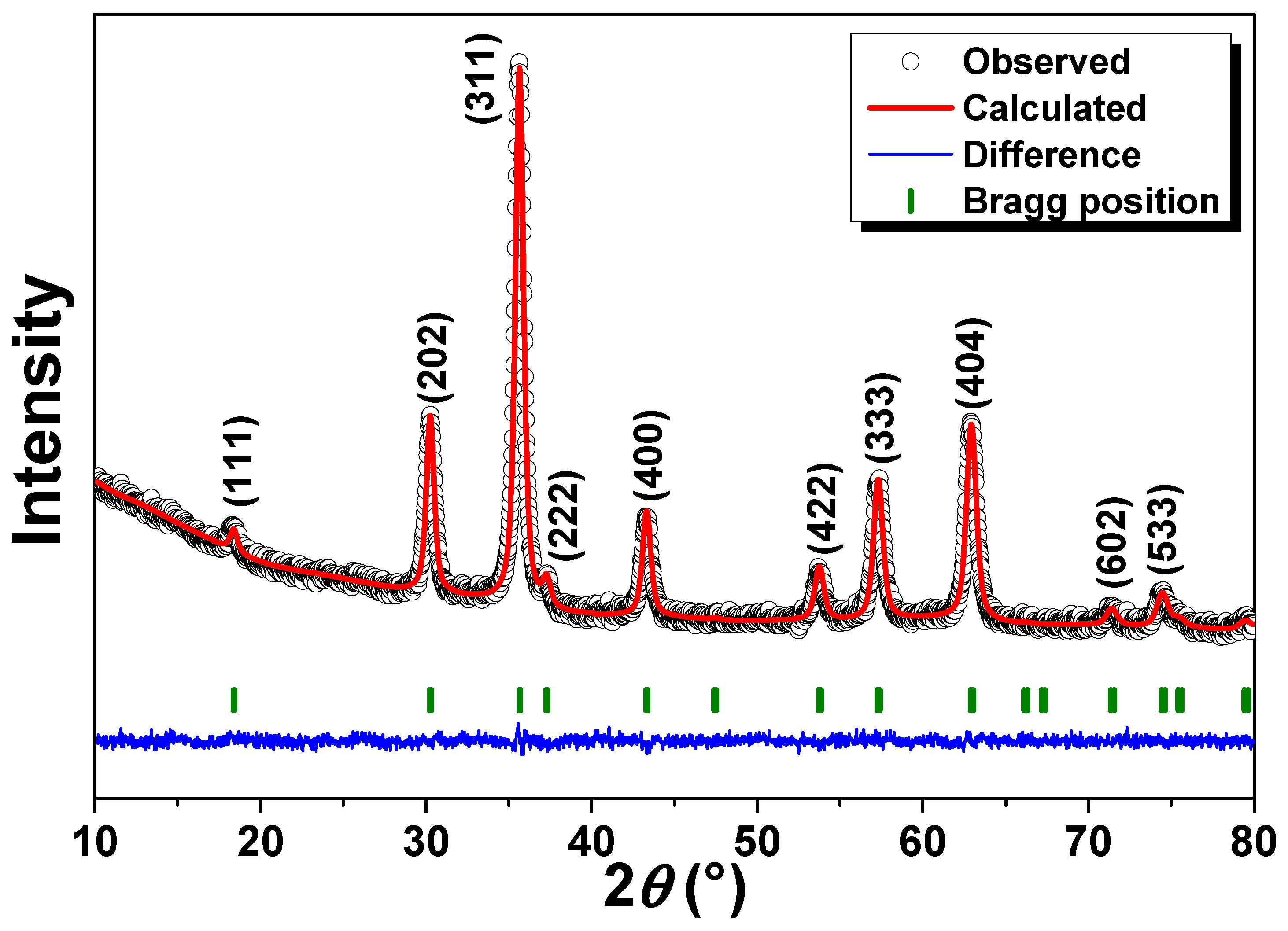
Figure 1. Refined X-ray diffractogram of the γ-Fe2O3@PSSNa nanohybrid. The Miller indexes of diffraction peaks are given in the top of each Bragg peak.
Polymers 16 01581 g001
Figure 2. TEM image of the γ-Fe2O3@PSSNa nanohybrid. Inset shows the bar graph correlated to the particle size frequency.
Polymers 16 01581 g002
Figure 3. Raman spectra of the γ-Fe2O3@PSSNa nanohybrid measured at selected laser powers.
Polymers 16 01581 g003
Figure 4. FTIR spectrum of the γ-Fe2O3@PSSNa nanohybrid recorded over the full range from 4000 to 400 cm−1.
Polymers 16 01581 g004
Figure 5. (a) Zeta potential and hydrodynamic size distribution of the PSSNa and γ-Fe2O3@PSSNa samples. Error < 5%. (b) TG measurement for the PSSNa and γ-Fe2O3@PSSNa samples. The arrow indicates the abrupt weight-loss decay after 400 °C. (c) N2 adsorption–desorption isotherm for the γ-Fe2O3@PSSNa nanohybrid, and (d) pore size distribution for the γ-Fe2O3@PSSNa nanohybrid.
Polymers 16 01581 g005
Figure 6. (a) FC and ZFC M(H) curves for the γ-Fe2O3@PSSNa nanohybrid recorded at 300 K and 5 K. (b) Zoomed region for the M(H) curves displayed in (a). (c) 57Fe Mössbauer spectra at 300 K and (d) 15 K for the γ-Fe2O3@PSSNa nanohybrid.
Polymers 16 01581 g006
Figure 7. Sigmoidal fit for the mortality of D. magna exposed to the γ-Fe2O3@PSSNa nanohybrid.
Polymers 16 01581 g007
Figure 8. Optical Images of the direct interaction of γ-Fe2O3@PSSNa on D. magna. (a) An individual of the N.C. without exposure to γ-Fe2O3@PSSNa. In (b,c), two dead individuals exposed to γ-Fe2O3@PSSNa at 800 and 600 mgL−1, respectively. In (d), a molt is observed (only exoskeleton), evidencing the affinity of γ-Fe2O3@PSSNa to the chitinous exoskeleton; the applied concentration was 400 mg L−1. Photographs were taken with the 4× objective under a conventional bright-field optical microscope.
Polymers 16 01581 g008
Figure 9. Box plots for the morphological parameters after exposing D. magna to γ-Fe2O3@PSSNa. N.C. stands for negative control.
Polymers 16 01581 g009
Figure 10. D. magna after 13-days of exposure to (a) N.C., (b) 50 mg L−1, (c) 100 mg L−1, (d) 200 mg L−1, (e) 400 mg L−1, and (f) 800 mg L−1.
Polymers 16 01581 g010
Table 1. LC50 values for similar nanosystems and polymers to the γ-Fe2O3@PSSNa nanohybrid exposed to D. magna. (n.d. = not determined). * SAMN: Surface Active Maghemite Nanoparticle.
Parent SystemMean Particle Size in Aqueous Media (nm)NPs SourceExposition Time (h)LC50 (mg L−1)Reference
Fe3O4 NPs (6 nm)n.d.Synthesized482.3[55]
SAMN * (11 (2) nm)5–20Synthesized481.25–40[56]
PVP-IONP (6.1 (6) nm)82.3Synthesized489750[57]
MWCNTs-γ-Fe2O3 (13.8 (6) nm for γ-Fe2O3)n.d.Synthesized24381.8[58]
GO-γ-Fe2O3 (10.4 (2) nm for γ-Fe2O3) n.d.Synthesized240.9[58]
Fe2O3 (20–40 nm) n.d.Commercial96163.21[59]
NP-Fe3O4 (<20 nm) n.d.Synthesized48977.24[60]
PS (<75 μm) n.d.Commercial4878.94[61]
PAO2N (52 nm) 56Commercial<24<75[62]
γ-Fe2O3@PSSNa (11(2) nm) 232.2Synthesized24533(5)This work
Disclaimer/Publisher’s Note: The statements, opinions and data contained in all publications are solely those of the individual author(s) and contributor(s) and not of MDPI and/or the editor(s). MDPI and/or the editor(s) disclaim responsibility for any injury to people or property resulting from any ideas, methods, instructions or products referred to in the content.