


Mevacor/Poly(vinyl acetate/2-hydroxyethyl methacrylate) as Solid Solution: Preparation, Solubility Enhancement and Drug Delivery

 ,
,  ,
,  ,
,  and
and
Abstract
1. Introduction
2. Materials and Methods
2.1. Chemicals
2.2. Preparation of PHEMA and PVAc Homopolymers
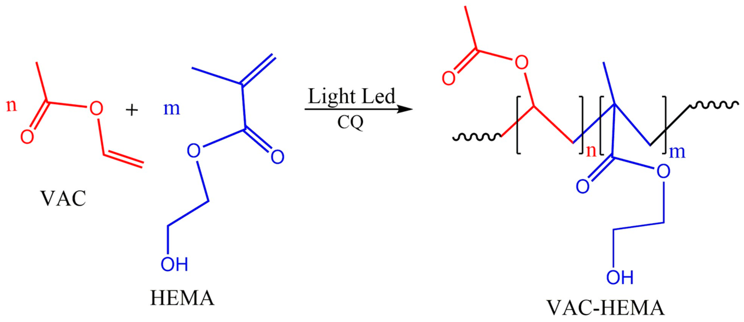
2.3. Preparation of MVR/VAC-HEMA Solid Solution
2.4. Characterization
2.4.1. FTIR Analysis
2.4.2. HNMR Analysis
2.4.3. SEC Analysis
2.4.4. DSC Analysis
2.4.5. XR-Diffraction Analysis
2.4.6. SEM Analysis
2.4.7. Cell Toxicity and Cell Adhesion
2.4.8. Swellability
2.4.9. In Vitro Release Dynamic
3. Results and Discussion
3.1. Characterization
3.1.1. FTIR Analysis
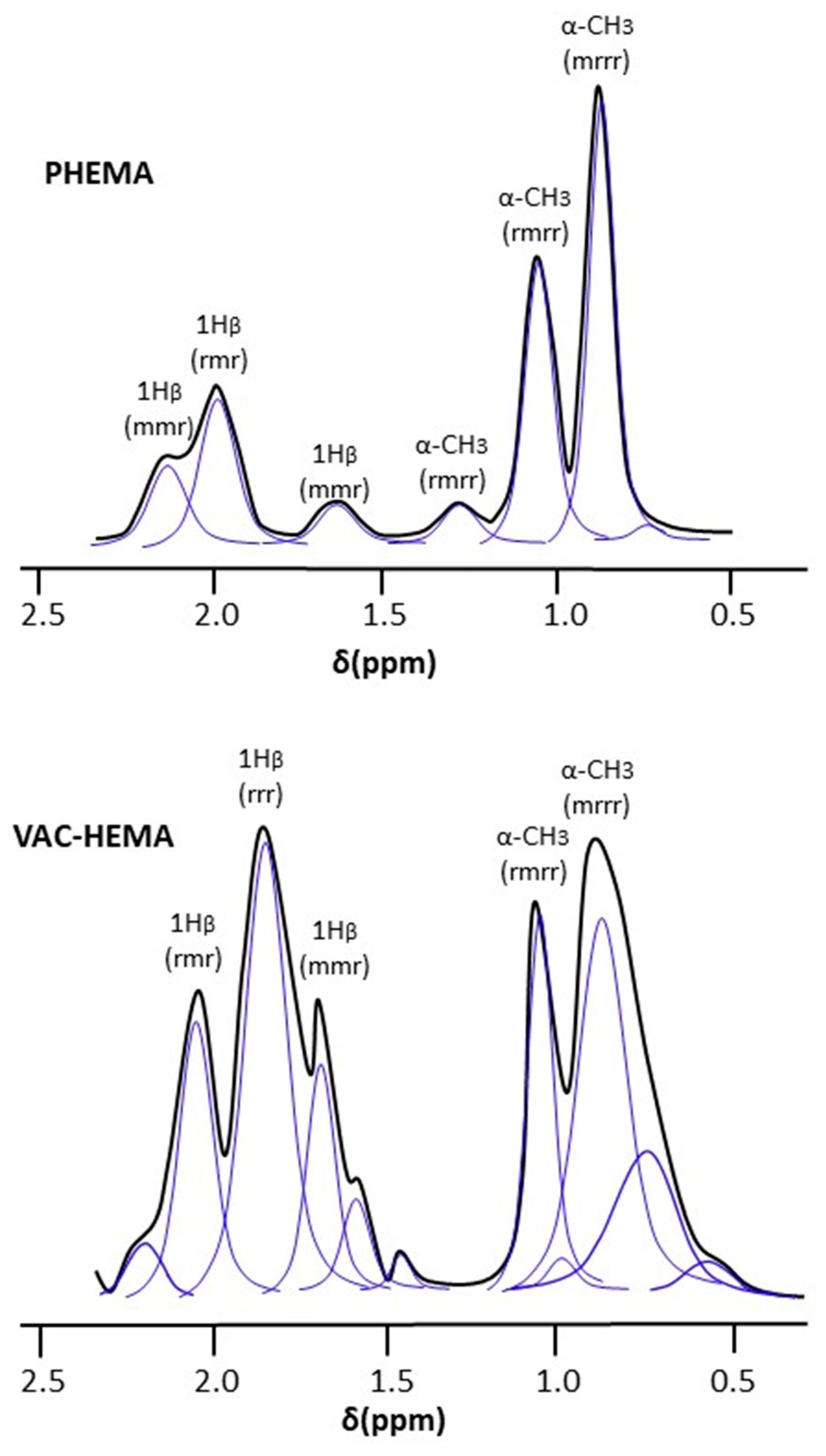
3.1.2. NMR Analysis
3.1.3. XR-Diffraction
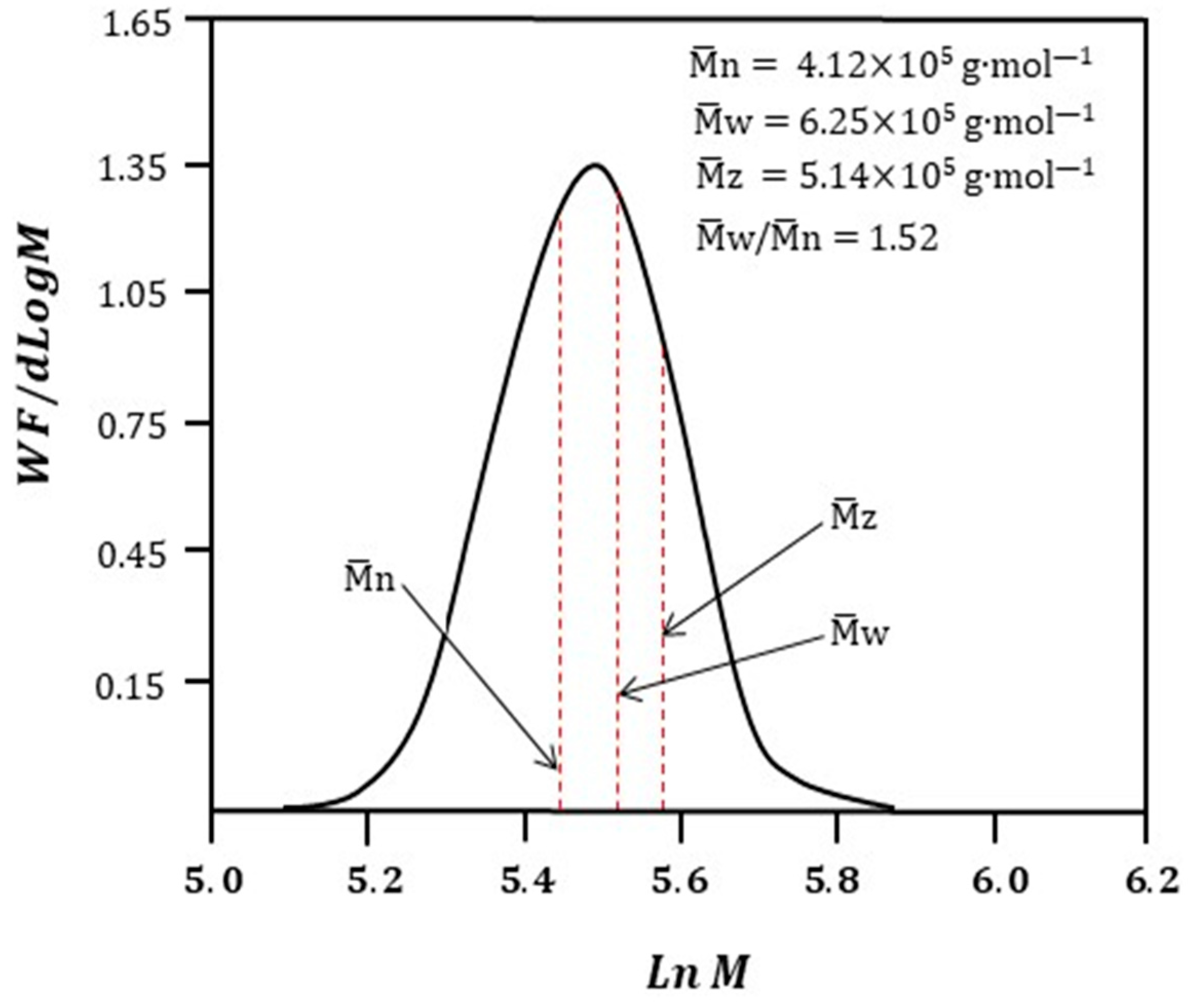
3.1.4. SEC Analysis
3.1.5. DSC Analysis
3.1.6. SEM Analysis
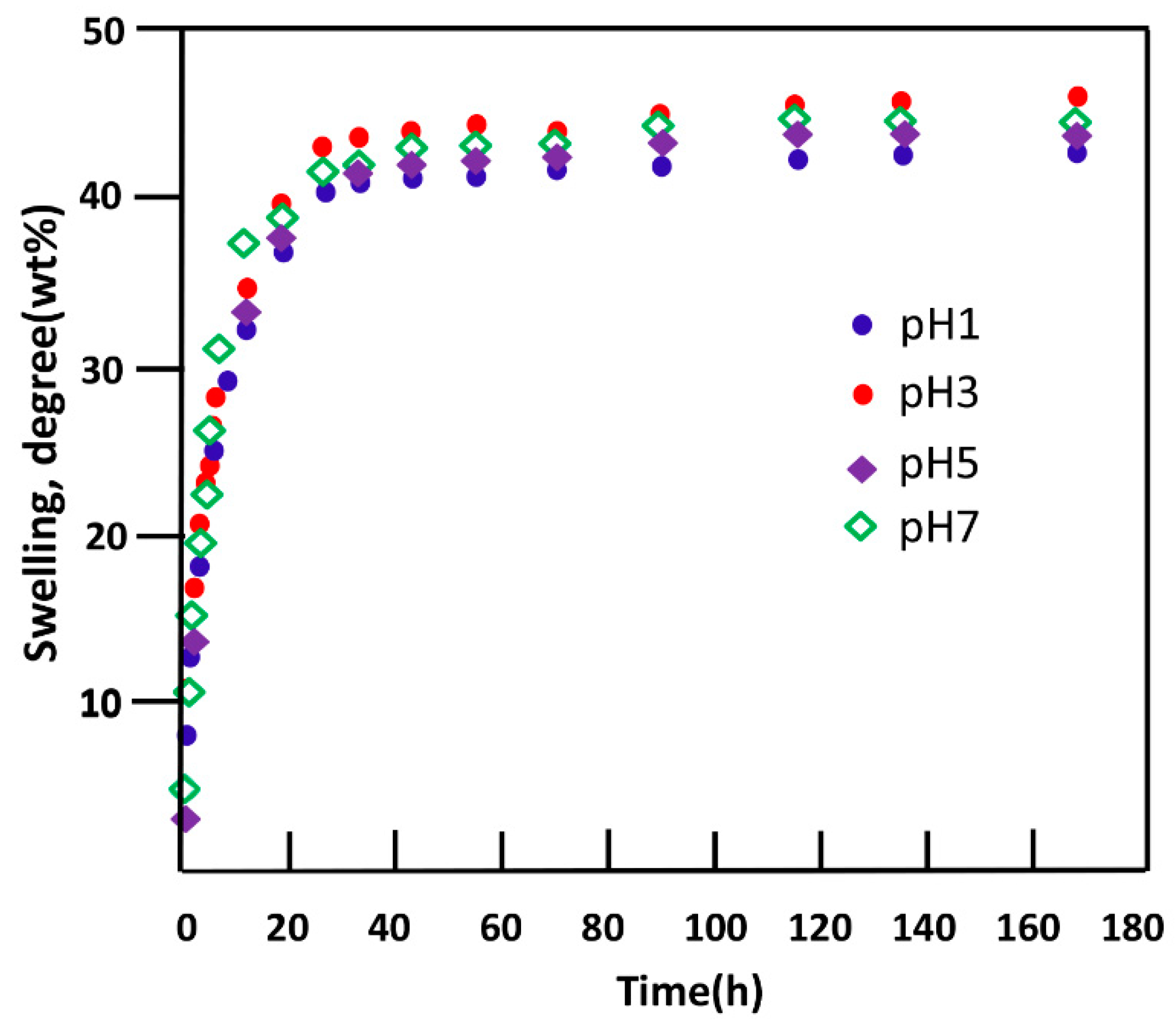
3.1.7. Swellability
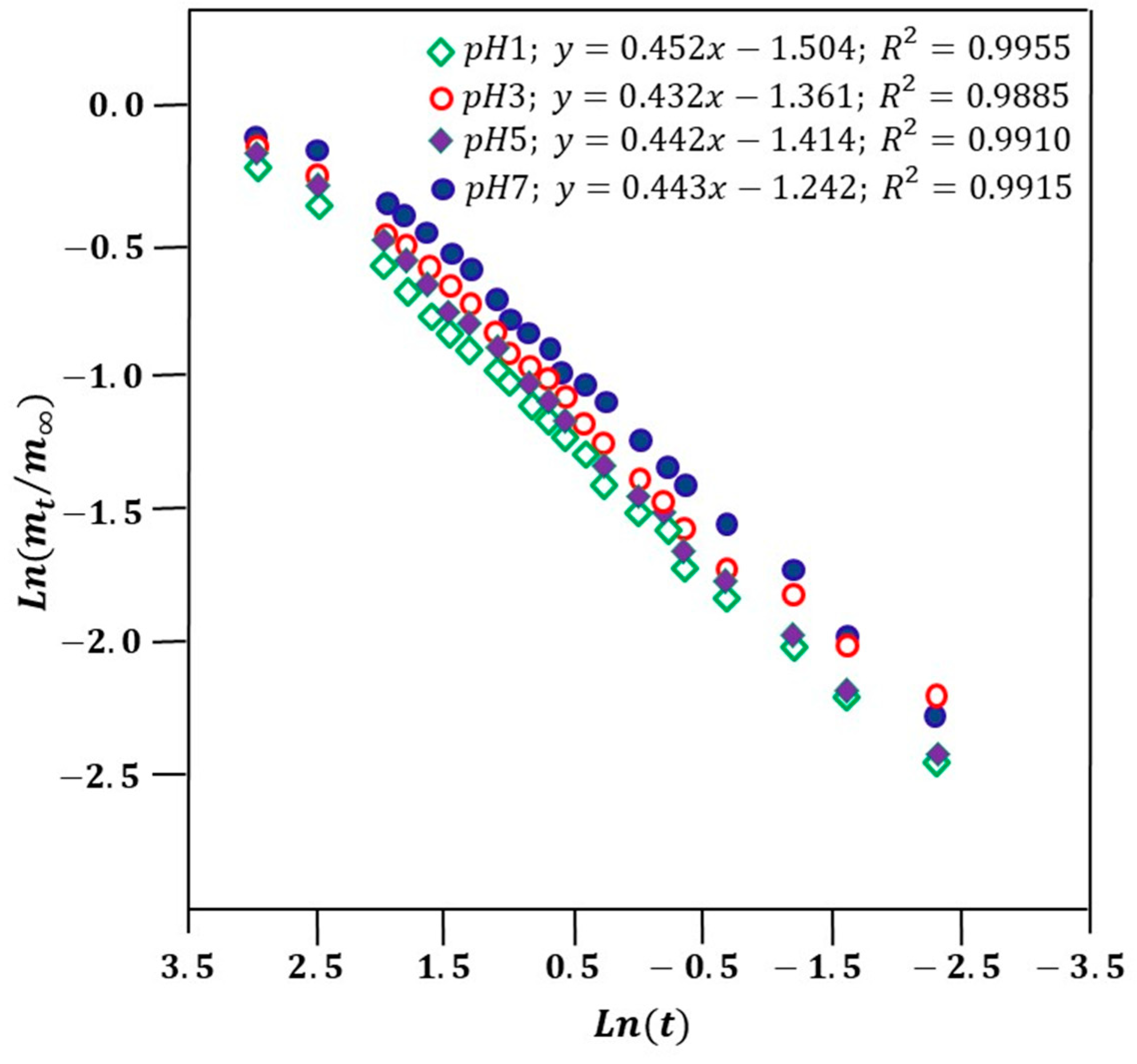
3.1.8. Water Diffusion
3.1.9. MVR Solubility Enhancement
3.1.10. Cytotoxicity
3.1.11. Cell Adhesion
3.2. In Vitro Release Dynamic
3.2.1. Diffusion Behavior of MVR
3.2.2. Effect of the Initial Amount of MVR
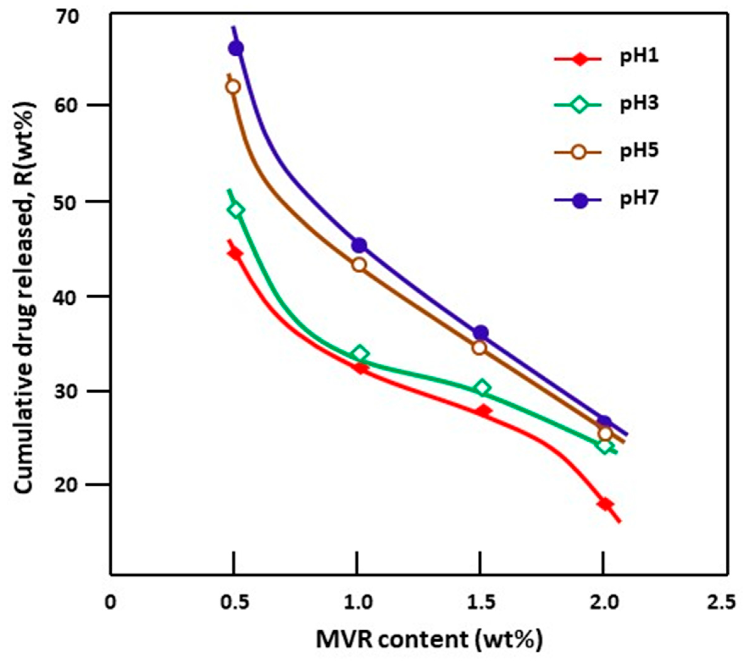
3.3. Effect of pH Medium
3.4. Performance of the MVR/VAC-HEMA Drug Carrier System
4. Conclusions
Author Contributions
Funding
Institutional Review Board Statement
Data Availability Statement
Acknowledgments
Conflicts of Interest
References
- Akelah, A.; Rehab, A. Controlled release systems based on polyacrylates and polyacrylates/clay nanocomposites of atenolol drug. SPE Polym. 2022, 3, 118–127. [Google Scholar] [CrossRef]
- Lee, S.; Na, K. Oleic acid conjugated polymeric photosensitizer for metastatic cancer targeting in photodynamic therapy. Biomater. Res. 2020, 24, 1. [Google Scholar] [CrossRef] [PubMed]
- Tiwari, S.; Marathe, S.; Chandola, M.; Thakur, N.; Rathore, R.; Garg, S. Synthesis and characterization of nanocomposites for drug delivery applications. J. Polym. Texfile Eng. 2016, 3, 1–7. [Google Scholar]
- Vigata, M.; Meinert, C.; Hutmacher, D.W.; Bock, N. Hydrogels as drug delivery systems: A review of current characterization and evaluation techniques. Pharmaceutics 2020, 12, 1188. [Google Scholar] [CrossRef] [PubMed]
- Naahidi, S.; Jafari, M.; Logan, M.; Wang, Y.; Yuan, Y.; Bae, H.; Dixon, B.; Chen, P. Biocompatibility of hydrogel-based scaffolds for tissue engineering applications. Biotechnol. Adv. 2017, 35, 530–544. [Google Scholar] [CrossRef] [PubMed]
- Loessner, D.; Meinert, C.; Kaemmerer, E.; Martine, L.C.; Yue, K.; Levett, P.A.; Klein, T.J.; Melchels, F.P.; Khademhosseini, A.; Hutmacher, D.W. Functionalization, preparation and use of cell-laden gelatin methacryloyl–based hydrogels as modular tissue culture platforms. Nat. Protoc. 2016, 11, 727–746. [Google Scholar] [CrossRef] [PubMed]
- Gotto, A.M., Jr. Statins: Powerful drugs for lowering cholesterol: Advice for patients. Circulation 2002, 105, 1514–1516. [Google Scholar] [CrossRef] [PubMed]
- Tobert, J.A. Efficacy and long-term adverse effect pattern of lovastatin. Am. J. Cardiol. 1988, 62, J28–J34. [Google Scholar] [CrossRef]
- Lovastatin Monograph for Professionals. Available online: https://www.drugs.com/monograph/lovastatin.html (accessed on 1 January 2022).
- Fink, C.; Sun, D.; Wagner, K.; Schneider, M.; Bauer, H.; Dolgos, H.; Mäder, K.; Peters, S.A. Evaluating the role of solubility in oral absorption of poorly water-soluble drugs using physiologically-based pharmacokinetic modeling. Clin. Pharmacol. Ther. 2020, 107, 650–661. [Google Scholar] [CrossRef]
- Górniak, A.; Gajda, M.; Pluta, J.; Czapor-Irzabek, H.; Karolewicz, B. Thermal, spectroscopic and dissolution studies of lovastatin solid dispersions with acetylsalicylic acid. J. Therm. Anal. Calorim. 2016, 125, 777–784. [Google Scholar] [CrossRef]
- UmakantVerma, J.; Mokale, V. Preparation of freeze-dried solid dispersion powder using mannitol to enhance solubility of lovastatin and development of sustained release tablet dosage form. Am. J. Pharm. Sci. Nanotechnol. 2014, 1, 11–26. [Google Scholar]
- Patel, R.P.; Patel, D.J.; Bhimani, D.B.; Patel, J.K. Physicochemical characterization and dissolution study of solid dispersions of furosemide with polyethylene glycol 6000 and polyvinylpyrrolidone K30. Dissolut. Technol. 2008, 15, 17–25. [Google Scholar] [CrossRef]
- Mochizuki, A.; Ogawa, H.; Nishimori, Y. Water structure in poly (2-hydroxyethyl methacrylate): Effect of molecular weight of poly (2-hydroxyethyl methacrylate) on its property related to water. J. Appl. Polym. Sci. 2012, 125, 53–60. [Google Scholar] [CrossRef]
- Weaver, J.; Bannister, I.; Robinson, K.; Bories-Azeau, X.; Armes, S.; Smallridge, M.; McKenna, P. Stimulus-responsive water-soluble polymers based on 2-hydroxyethyl methacrylate. Macromolecules 2004, 37, 2395–2403. [Google Scholar] [CrossRef]
- Macret, M.; Hild, G. Hydroxyalkyl methacrylates: Hydrogel formation based on the radical copolymerization of 2-hydroxyethylmethacrylate and 2, 3-dihydroxypropylmethacrylate. Polymer 1982, 23, 748–753. [Google Scholar] [CrossRef]
- Wichterle, O.; Lim, D. Hydrophilic gels for biological use. Nature 1960, 185, 117–118. [Google Scholar] [CrossRef]
- Jeyanthi, R.; Rao, K.P. In vivo biocompatibility of collagenpoly (hydroxyethyl methacrylate) hydrogels. Biomaterials 1990, 11, 238–243. [Google Scholar] [CrossRef] [PubMed]
- Denizli, A.; Kiremitci, M.; Pişkin, E. Subcutaneous polymeric matrix system p (HEMA-BGA) for controlled release of an anticancer drug (5-fluorouracil): II: Release kinetics. Biomaterials 1988, 9, 363–366. [Google Scholar] [CrossRef]
- Kiremitçi-Gümüşderelioglu, M.; Gökçe, M.; Fikret Akata, R. A novel MMC-loaded pHEMA drainage device for the treatment of glaucoma: In vitro and in vivo studies. J. Biomater. Sci. Polym. Ed. 1996, 7, 857–869. [Google Scholar] [CrossRef]
- Trigo, R.; Blanco, M.; Teijon, J.; Sastre, R. Anticancer drug, ara-C, release from pHEMA hydrogels. Biomaterials 1994, 15, 1181–1186. [Google Scholar] [CrossRef]
- Vinyl acetate, polyvinyl acetate and polyvinyl alcohol. IARC Monogr. Eval. Carcinog. Risk Chem. Hum. 1979, 19, 341–366.
- Sivalingam, G.; Chattopadhyay, S.; Madras, G. Enzymatic degradation of poly (ε-caprolactone), poly (vinyl acetate) and their blends by lipases. Chem. Eng. Sci. 2003, 58, 2911–2919. [Google Scholar] [CrossRef]
- Novoa, G.A.G.; Heinämäki, J.; Mirza, S.; Antikainen, O.; Colarte, A.I.; Paz, A.S.; Yliruusi, J. Physical solid-state properties and dissolution of sustained-release matrices of polyvinylacetate. Eur. J. Pharm. Biopharm. 2005, 59, 343–350. [Google Scholar] [CrossRef] [PubMed]
- Babaahmadi, M.; Sabzi, M.; Ghasemi, I. Mechanical and Shape Memory Properties of Poly (vinyl acetate)/Poly (lactic acid) Blends. Iran. J. Polym. Sci. Technol. 2018, 31, 57–68. [Google Scholar]
- Jannesari, M.; Varshosaz, J.; Morshed, M.; Zamani, M. Composite poly (vinyl alcohol)/poly (vinyl acetate) electrospun nanofibrous mats as a novel wound dressing matrix for controlled release of drugs. Int. J. Nanomed. 2011, 6, 993–1003. [Google Scholar]
- Sivalingam, G.; Karthik, R.; Madras, G. Blends of poly (ε-caprolactone) and poly (vinyl acetate): Mechanical properties and thermal degradation. Polym. Degrad. Stab. 2004, 84, 345–351. [Google Scholar] [CrossRef]
- Bailly, N.; Thomas, M.; Klumperman, B. Poly (N-vinylpyrrolidone)-block-poly (vinyl acetate) as a drug delivery vehicle for hydrophobic drugs. Biomacromolecules 2012, 13, 4109–4117. [Google Scholar] [CrossRef]
- Osman, A.F.; Hamid, A.R.A.; Fitri, T.F.M.; Fauzi, A.A.A.; Halim, K.A.A. Poly (ethylene-co-vinylacetate) copolymer based nanocomposites: A review. IOP Conf. Ser. Mater. Sci. Eng. 2020, 864, 012121. [Google Scholar] [CrossRef]
- Akl, M.; Abd Elrazek, H.; Abdel Bary, E. Poly (Ethylene-Co-Vinyl Acetate) Blends for Controlled Drug Release. Am. J. Adv. Drug Deliv. 2018, 6, 052–060. [Google Scholar] [CrossRef]
- Lin, H.-P.; Tu, H.-P.; Hsieh, Y.-P.; Lee, B.-S. Controlled release of lovastatin from poly (lactic-co-glycolic acid) nanoparticles for direct pulp capping in rat teeth. Int. J. Nanomed. 2017, 12, 5473. [Google Scholar] [CrossRef]
- Tarafder, S.; Nansen, K.; Bose, S. Lovastatin release from polycaprolactone coated β-tricalcium phosphate: Effects of pH, concentration and drug–polymer interactions. Mater. Sci. Eng. C 2013, 33, 3121–3128. [Google Scholar] [CrossRef] [PubMed]
- Madhavi, V. Formulation and evaluation of lovstatin controlled release tablets. IJPPA 2017, 1, 79–84. [Google Scholar]
- Belzer, C.; De Vos, W.M. Microbes inside—From diversity to function: The case of Akkermansia. ISME J. 2012, 6, 1449–1458. [Google Scholar] [CrossRef] [PubMed]
- Alqahtani, S.M.; Al Khulaifi, R.S.; Alassaf, M.; Saeed, W.S.; Bedja, I.; Aldarwesh, A.; Aljubailah, A.; Semlali, A.; Aouak, T. Preparation and Characterization of Poly (vinyl acetate-co-2-hydroxyethyl methacrylate) and In Vitro Application as Contact Lens for Acyclovir Delivery. Int. J. Mol. Sci. 2023, 24, 5483. [Google Scholar] [CrossRef] [PubMed]
- Chuong, M.C.; Choy, E.; Douk, K.; Duong, L.H.; Hoang, S.K.; Le, N.; Lim, M.; Poirier, B.; Prasad, D.; Radke, M. The development of delayed-then-extended-release lovastatin tablet. Int. J. Appl. Pharm. 2013, 5, 11–18. [Google Scholar]
- Semlali, A.; Jacques, E.; Rouabhia, M.; Milot, J.; Laviolette, M.; Chakir, J. Regulation of epithelial cell proliferation by bronchial fibroblasts obtained from mild asthmatic subjects. Allergy 2010, 65, 1438–1445. [Google Scholar] [CrossRef] [PubMed]
- Alghamdi, A.A.; Alsolami, A.; Saeed, W.S.; Al-Odayni, A.-B.M.; Semlali, A.; Aouak, T. Miscibility of poly (acrylic acid)/poly (methyl vinyl ketone) blend and in vitro application as drug carrier system. Des. Monomers Polym. 2018, 21, 145–162. [Google Scholar] [CrossRef]
- Semlali, A.; Papadakos, S.; Contant, C.; Zouaoui, I.; Rouabhia, M. Rapamycin inhibits oral cancer cell growth by promoting oxidative stress and suppressing ERK1/2, NF-κB and beta-catenin pathways. Front. Oncol. 2022, 12, 873447. [Google Scholar] [CrossRef]
- Hakiem, A.F.A.; Mohamed, N.A.; Ali, H.R. FTIR spectroscopic study of two isostructural statins: Simvastatin and Lovastatin as authentic and in pharmaceuticals. Spectrochim. Acta Part A Mol. Biomol. Spectrosc. 2021, 261, 120045. [Google Scholar]
- Pham, Q.T.; Petiaud, R.; Llauro-Darricades, M.-F.; Waton, H. Proton & Carbon NMR Spectra of Polymers; CRC Press: Boca Raton, FL, USA, 2019. [Google Scholar]
- Guan, J.; Liu, Q.; Zhang, X.; Zhang, Y.; Chokshi, R.; Wu, H.; Mao, S. Alginate as a potential diphase solid dispersion carrier with enhanced drug dissolution and improved storage stability. Eur. J. Pharm. Sci. 2018, 114, 346–355. [Google Scholar] [CrossRef]
- Khan, S.; Ranjha, N.M. Effect of degree of cross-linking on swelling and on drug release of low viscous chitosan/poly (vinyl alcohol) hydrogels. Polym. Bull. 2014, 71, 2133–2158. [Google Scholar] [CrossRef]
- Numata, I.; Cochrane, M.A.; Souza, C.M., Jr.; Sales, M.H. Carbon emissions from deforestation and forest fragmentation in the Brazilian Amazon. Environ. Res. Lett. 2011, 6, 044003. [Google Scholar] [CrossRef]
- Akula, P.; PK, L. Effect of pH on weakly acidic and basic model drugs and determination of their ex vivo transdermal permeation routes. Braz. J. Pharm. Sci. 2018, 54, 1–8. [Google Scholar] [CrossRef]
- Aljubailah, A.; Alharbi, W.N.O.; Haidyrah, A.S.; Al-Garni, T.S.; Saeed, W.S.; Semlali, A.; Alqahtani, S.; Al-Owais, A.A.; Karami, A.M.; Aouak, T. Copolymer Involving 2-Hydroxyethyl Methacrylate and 2-Chloroquinyl Methacrylate: Synthesis, Characterization and In Vitro 2-Hydroxychloroquine Delivery Application. Polymers 2021, 13, 4072. [Google Scholar] [CrossRef]
- Comyn, J. Introduction to polymer permeability and the mathematics of diffusion. In Polymer Permeability; Springer: Dordrecht, The Netherlands, 1985; pp. 1–10. [Google Scholar]
- Masaro, L.; Zhu, X. Physical models of diffusion for polymer solutions, gels and solids. Prog. Polym. Sci. 1999, 24, 731–775. [Google Scholar] [CrossRef]
- Glomme, A.; März, J.; Dressman, J.B. Comparison of a miniaturized shake-flask solubility method with automated potentiometric acid/base titrations and calculated solubilities. J. Pharm. Sci. 2005, 94, 1–16. [Google Scholar] [CrossRef] [PubMed]


| VAC-HEMA Hydrogel | ||||
|---|---|---|---|---|
| pH Medium | 1 | 3 | 5 | 7 |
| n | 0.488 ± 0.087 | 0.492 ± 0.086 | 0.502 ± 0.073 | 0.493 ± 0.072 |
| k(h)−1 | 0.222 ± 0.120 | 0.256 ± 0.111 | 0.243 ± 0.121 | 0.289 ± 0.118 |
| D·10−3(mm2·h−1) | 1.901 ± 0.054 | 2.273 ± 0.034 | 2.435 ± 0.028 | 2.792 ± 0.025 |
| MVR (mg·mL−1) | ||
|---|---|---|
| pH | 1 | 7 |
| Powder | 0.0060 ± 0.0013 | 0.0038 ± 0.0015 |
| MVR/VAC-HEMA1.5 | 0.1313 ± 0.0010 | 0.1407 ± 0.0012 |
| System | pH | k′·102 (h−0.5) | R2 | D′·10−3 (mm2·h−1) | System | k′·102 (h−0.5) | R2 | D′·10−3 (mm2·h−1) |
|---|---|---|---|---|---|---|---|---|
| I | 1 | 3.29 | 0.9966 | 3.394 | III | 2.00 | 0.9900 | 0.788 |
| 3 | 3.58 | 0.9454 | 4.473 | 2.59 | 0.9969 | 1.979 | ||
| 5 | 6.11 | 0.9874 | 9.221 | 2.64 | 0.9961 | 1.741 | ||
| 7 | 5.57 | 0.9818 | 7.793 | 2.81 | 0.9881 | 1.961 | ||
| II | 1 | 2.11 | 0.9883 | 1.348 | IV | 1.46 | 0.9839 | 0.629 |
| 3 | 2.79 | 0.9844 | 2.203 | 1.85 | 0.9785 | 0.984 | ||
| 5 | 3.65 | 0.9952 | 3.850 | 2.05 | 0.9909 | 1.266 | ||
| 7 | 3.79 | 0.9898 | 4.130 | 2.17 | 0.9913 | 1.298 |
| System | pH | Pseudo- Stable Zone (h) | Cumulative MVR Released (wt%) | Release Rate (wt%/h) | System | Pseudo- Stable Zone (h) | Cumulative MVR Released (wt%) | Release Rate (wt%/h) |
|---|---|---|---|---|---|---|---|---|
| I | 1 | 0–3 | 22.0 ± 0.3 | 7.33 ± 0.10 | III | 0–3 | 18.0 ± 0.5 | 06.0 ± 0.17 |
| 5–36 | 12.0 ± 0.3 | 0.41 ± 0.01 | 7–36 | 10.0 ± 0.4 | 0.34 ± 0.01 | |||
| 36–72 | 02.0 ± 0.2 | 0.07 ± 0.01 | 36–72 | 0.3 ± 0.2 | 0.01 ± 0.01 | |||
| 3 | 0–5 | 24.0 ± 0.3 | 4.80 ± 0.06 | 0–3 | 18.0 ± 0.5 | 06.0 ± 0.17 | ||
| 5–36 | 26.5 ± 0.6 | 0.85 ± 0.02 | 3–35 | 15.0 ± 0.5 | 0.47 ± 0.02 | |||
| 36–72 | 06.5 ± 0.3 | 0.18 ± 0.01 | 36–72 | 01.0 ± 0.2 | 0.03 ± 0.01 | |||
| 5 | 0–5 | 32.0 ± 0.4 | 6.40 ± 0.08 | 0–3 | 19.0 ± 0.5 | 6.33 ± 0.17 | ||
| 5–22 | 25.0 ± 0.3 | 1.47 ± 0.02 | 3–16 | 13.0 ± 0.4 | 01.0 ± 0.03 | |||
| 26–72 | 08.0 ± 0.2 | 0.17 ± 0.01 | 16–72 | 06.0 ± 0.3 | 0.11 ± 0.01 | |||
| 7 | 0–5 | 32.7 ± 0.5 | 6.54 ± 0.10 | 0–3 | 19.0 ± 0.6 | 6.33 ± 0.02 | ||
| 5–26 | 22.5 ± 0.3 | 1.07 ± 0.01 | 3–16 | 20.0 ± 0.6 | 1.54 ± 0.05 | |||
| 26–72 | 07.0 ± 0.3 | 0.15 ± 0.01 | 16–72 | 06.0 ± 0.4 | 0.11 ± 0.01 | |||
| II | 1 | 0–2 | 10.0 ± 0.3 | 5.00 ± 0.15 | IV | 0–2 | 05.0 ± 0.2 | 2.50 ± 0.10 |
| 2–12 | 11.3 ± 0.3 | 1.13 ± 0.03 | 2–10 | 08.0 ± 0.3 | 1.00 ± 0.04 | |||
| 17–72 | 4.0 ± 0.2 | 0.72 ± 0.01 | 10–72 | 08.0 ± 0.4 | 0.13 ± 0.01 | |||
| 3 | 0–2 | 11.0 ± 0.4 | 5.50 ± 0.20 | 0–2 | 08.0 ± 0.4 | 4.00 ± 0.20 | ||
| 2–14 | 15.0 ± 0.5 | 1.25 ± 0.04 | 2–16 | 10.0 ± 0.4 | 0.71 ±0.03 | |||
| 14–72 | 3.0 ± 0.2 | 0.05 ± 0.01 | 16–72 | 06.0 ± 0.4 | 0.11 ± 0.01 | |||
| 5 | 0–2 | 13.0 ± 0.3 | 06.5 ± 0.15 | 0–2 | 09.0 ± 0.2 | 4.50 ± 0.10 | ||
| 2–17 | 15.0 ± 0.4 | 01.0 ± 0.03 | 2–11 | 09.0 ± 0.4 | 1.00 ± 0.04 | |||
| 19–72 | 7.0 ± 0.3 | 0.13 ± 0.01 | 13–72 | 06.0 ± 0.3 | 0.10 ± 0.01 | |||
| 7 | 0–2 | 14.0 ± 0.2 | 07.0 ± 0.10 | 0–2 | 10.0 ± 0.2 | 5.00 ± 0.10 | ||
| 2–19 | 6.0 ± 0.4 | 0.35 ± 0.02 | 2–13 | 11.5 ± 0.3 | 1.06 ± 0.03 | |||
| 19–72 | 6.0 ± 0.3 | 0.11 ± 0.01 | 13–72 | 06.5 ± 0.3 | 0.11 ± 0.01 |
| MVR/VAC-HEMA System | Stomach Transit (wt%) | Small Intestine Transit (wt%) | Colon Transit (wt%) | SDO (wt%) | ||||
|---|---|---|---|---|---|---|---|---|
| Transit Time | Min (1 h) | Max (4 h) | Min (4 h) | Max (12 h) | Min (48 h) | Max (72) | Min (48 h) | Max (72) |
| MVR/VAC-HEMA0.5 | 6.07 | 13.37 | 26.16 | 40.19 | 33.06 | 36.66 | 10.86 | 14.81 |
| MVR/VAC-HEMA1.0 | 6.00 | 18.41 | 20.00 | 28.10 | 42.52 | 45.82 | 8.76 | 19.90 |
| MVR/VAC-HEMA1.5 | 5.25 | 10.13 | 14.70 | 17.50 | 23.19 | 25.83 | 12.17 | 18.95 |
| MVR/VAC-HEMA2.0 | 6.25 | 8.21 | 12.12 | 20.60 | 25.35 | 28.00 | 14.38 | 14.45 |
Disclaimer/Publisher’s Note: The statements, opinions and data contained in all publications are solely those of the individual author(s) and contributor(s) and not of MDPI and/or the editor(s). MDPI and/or the editor(s) disclaim responsibility for any injury to people or property resulting from any ideas, methods, instructions or products referred to in the content. |
© 2023 by the authors. Licensee MDPI, Basel, Switzerland. This article is an open access article distributed under the terms and conditions of the Creative Commons Attribution (CC BY) license (https://creativecommons.org/licenses/by/4.0/).
Share and Cite
Alassaf, M.; Alqahtani, S.M.; Al Khulaifi, R.S.; Saeed, W.S.; Alsubaie, F.S.; Semlali, A.; Aouak, T. Mevacor/Poly(vinyl acetate/2-hydroxyethyl methacrylate) as Solid Solution: Preparation, Solubility Enhancement and Drug Delivery. Polymers 2023, 15, 3927. https://doi.org/10.3390/polym15193927
Alassaf M, Alqahtani SM, Al Khulaifi RS, Saeed WS, Alsubaie FS, Semlali A, Aouak T. Mevacor/Poly(vinyl acetate/2-hydroxyethyl methacrylate) as Solid Solution: Preparation, Solubility Enhancement and Drug Delivery. Polymers. 2023; 15(19):3927. https://doi.org/10.3390/polym15193927
Chicago/Turabian StyleAlassaf, Mohammed, Saad Mohammed Alqahtani, Rana Salem Al Khulaifi, Waseem Sharaf Saeed, Faisal S. Alsubaie, Abdelhabib Semlali, and Taieb Aouak. 2023. "Mevacor/Poly(vinyl acetate/2-hydroxyethyl methacrylate) as Solid Solution: Preparation, Solubility Enhancement and Drug Delivery" Polymers 15, no. 19: 3927. https://doi.org/10.3390/polym15193927
APA StyleAlassaf, M., Alqahtani, S. M., Al Khulaifi, R. S., Saeed, W. S., Alsubaie, F. S., Semlali, A., & Aouak, T. (2023). Mevacor/Poly(vinyl acetate/2-hydroxyethyl methacrylate) as Solid Solution: Preparation, Solubility Enhancement and Drug Delivery. Polymers, 15(19), 3927. https://doi.org/10.3390/polym15193927