
A Review on Antibacterial Biomaterials in Biomedical Applications: From Materials Perspective to Bioinks Design

 , ,
, , 

 and
and 
Abstract
:1. Introduction
2. Why Antibacterial Features Are Necessary for Biomaterials?
3. Strategies for Achieving Antibacterial Biomaterials
3.1. Surface Treatment with Bacteria Repelling and Antiadhesive Substances
3.2. Materials with Antibacterial Properties
3.2.1. Antibacterial Activity of Copper
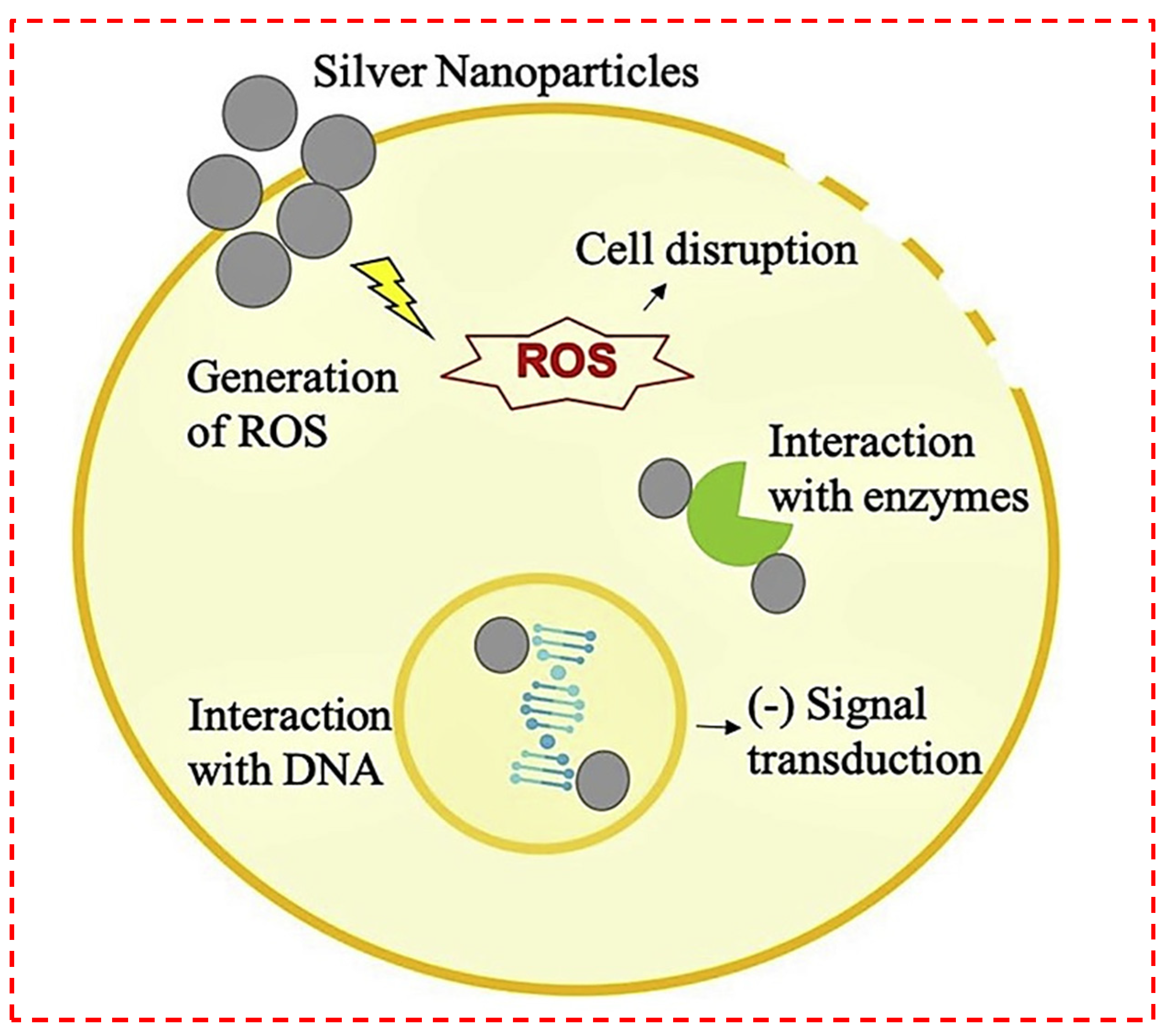
3.2.2. Antibacterial Activity of Silver
3.2.3. Antibacterial Activity of Gold
3.2.4. Antibacterial Activity of Zinc Oxide
3.2.5. Antibacterial Activity of Titanium Dioxide
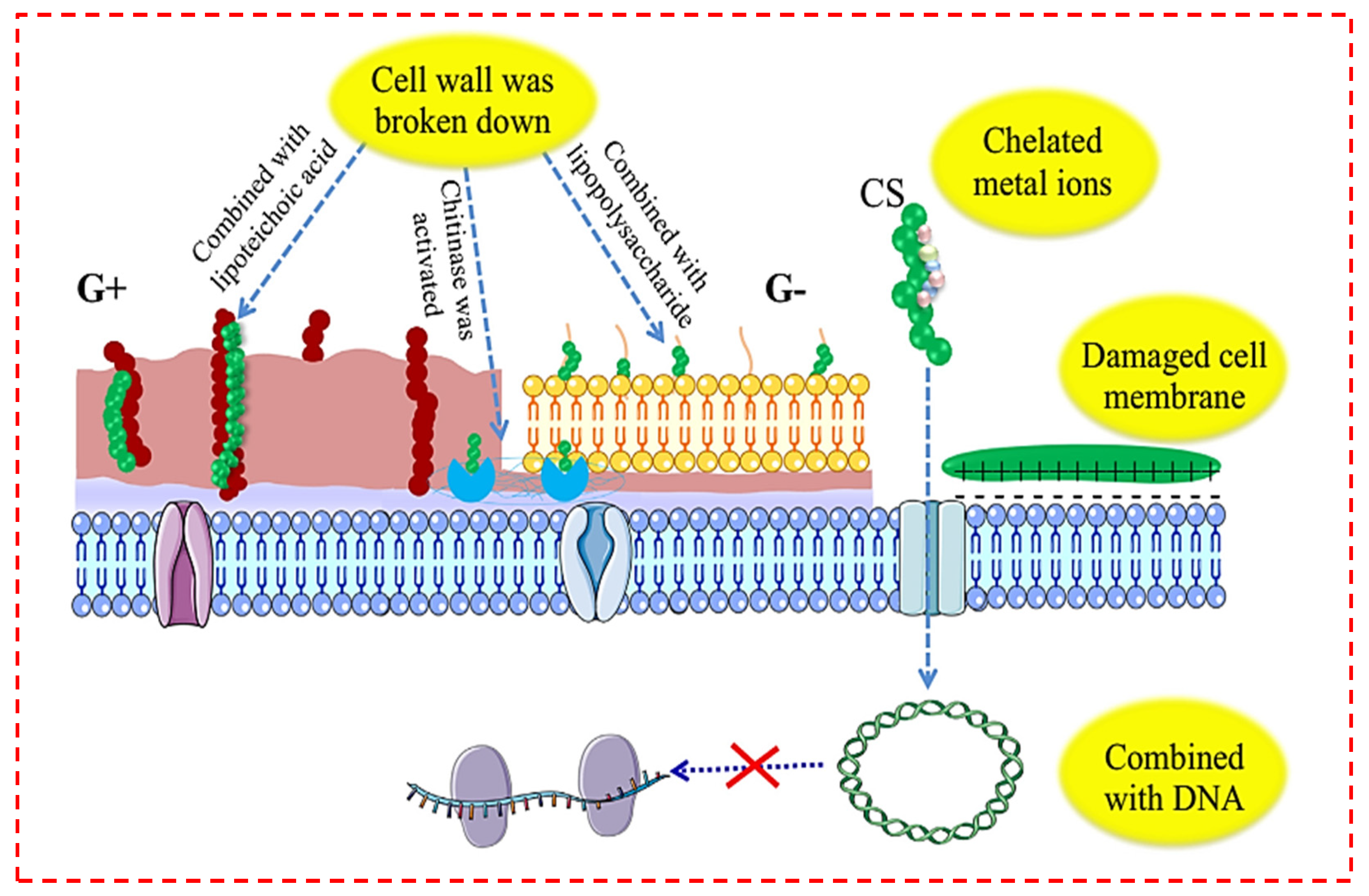
3.2.6. Antibacterial Activity of Chitosan
4. Printed Constructs from Antibacterial Inks
4.1. Metallic Ion-Containing Inks
4.2. Chitosan-Containing Inks
4.3. Other Antibacterial Inks
5. Conclusions and Future Perspectives
Author Contributions
Funding
Institutional Review Board Statement
Informed Consent Statement
Conflicts of Interest
References
- Chen, X.B. Extrusion Bioprinting of Scaffolds for Tissue Engineering Applications; Springer International Publishing AG: Cham, Switzerland, 2019. [Google Scholar]
- Sadeghianmaryan, A.; Naghieh, S.; Yazdanpanah, Z.; Sardroud, H.A.; Sharma, N.; Wilson, L.D.; Chen, X. Fabrication of chitosan/alginate/hydroxyapatite hybrid scaffolds using 3D printing and impregnating techniques for potential cartilage regeneration. Int. J. Biol. Macromol. 2022, 204, 62–75. [Google Scholar] [CrossRef]
- Zimmerling, A.; Zhou, Y.; Chen, X. Bioprinted constructs for respiratory tissue engineering. Bioprinting 2021, 24, e00177. [Google Scholar] [CrossRef]
- Delkash, Y.; Gouin, M.; Rimbeault, T.; Mohabatpour, F.; Papagerakis, P.; Maw, S.; Chen, X. Bioprinting and In Vitro Characterization of an Eggwhite-Based Cell-Laden Patch for Endothelialized Tissue Engineering Applications. J. Funct. Biomater. 2021, 12, 45. [Google Scholar] [CrossRef]
- Soleymani Eil Bakhtiari, S.; Bakhsheshi-Rad, H.R.; Karbasi, S.; Razzaghi, M.; Tavakoli, M.; Ismail, A.F.; Sharif, S.; Rama Krishna, S.; Chen, X.; Berto, F. 3-Dimensional Printing of Hydrogel-Based Nanocomposites: A Comprehensive Review on the Technology Description, Properties, and Applications. Adv. Eng. Mater. 2021, 23, 2100477. [Google Scholar] [CrossRef]
- Fu, Z.; Naghieh, S.; Xu, C.; Wang, C.; Sun, W.; Chen, D.X. Printability in extrusion bioprinting. Biofabrication 2021, 13, 033001. [Google Scholar] [CrossRef]
- Sadeghianmaryan, A.; Naghieh, S.; Sardroud, H.A.; Yazdanpanah, Z.; Soltani, Y.A.; Sernaglia, J.; Chen, X. Extrusion-based printing of chitosan scaffolds and their in vitro characterization for cartilage tissue engineering. Int. J. Biol. Macromol. 2020, 164, 3179–3192. [Google Scholar] [CrossRef]
- You, F.; Wu, X.; Kelly, M.; Chen, X. Bioprinting and in vitro characterization of alginate dialdehyde–gelatin hydrogel bio-ink. Bio-Des. Manuf. 2020, 3, 48–59. [Google Scholar] [CrossRef] [Green Version]
- Soleymani Eil Bakhtiari, S.; Bakhsheshi-Rad, H.R.; Karbasi, S.; Tavakoli, M.; Razzaghi, M.; Ismail, A.F.; RamaKrishna, S.; Berto, F. Polymethyl Methacrylate-Based Bone Cements Containing Carbon Nanotubes and Graphene Oxide: An Overview of Physical, Mechanical, and Biological Properties. Polymers 2020, 12, 1469. [Google Scholar] [CrossRef]
- You, F.; Chen, D.X.; Cooper, D.M.L.; Chang, T.; Eames, F.B. Homogeneous hydroxyapatite/alginate composite hydrogel promotes calcified cartilage matrix deposition with potential for three-dimensional bioprinting. Biofabrication 2018, 11, 015015. [Google Scholar] [CrossRef]
- Ning, L.; Sun, H.; Lelong, T.; Guilloteau, R.; Zhu, N.; Schreyer, D.J.; Chen, X. 3D bioprinting of scaffolds with living Schwann cells for potential nerve tissue engineering applications. Biofabrication 2018, 10, 035014. [Google Scholar] [CrossRef]
- Bakhsheshi-Rad, H.R.; Ismail, A.F.; Aziz, M.; Akbari, M.; Hadisi, Z.; Omidi, M.; Chen, X. Development of the PVA/CS nanofibers containing silk protein sericin as a wound dressing: In vitro and in vivo assessment. Int. J. Biol. Macromol. 2020, 149, 513–521. [Google Scholar] [CrossRef]
- Bakhsheshi-Rad, H.; Hadisi, Z.; Ismail, A.; Aziz, M.; Akbari, M.; Berto, F.; Chen, X. In vitro and in vivo evaluation of chitosan-alginate/gentamicin wound dressing nanofibrous with high antibacterial performance. Polym. Test. 2020, 82, 106298. [Google Scholar] [CrossRef]
- Jammalamadaka, U.; Tappa, K. Recent Advances in Biomaterials for 3D Printing and Tissue Engineering. J. Funct. Biomater. 2018, 9, 22. [Google Scholar] [CrossRef] [Green Version]
- Klebe, R.J. Cytoscribing: A method for micropositioning cells and the construction of two- and three-dimensional synthetic tissues. Exp. Cell Res. 1988, 179, 362–373. [Google Scholar] [CrossRef]
- Gu, Z.; Fu, J.; Lin, H.; He, Y. Development of 3D bioprinting: From printing methods to biomedical applications. Asian J. Pharm. Sci. 2020, 15, 529–557. [Google Scholar] [CrossRef] [PubMed]
- Muhammad, M.H.; Idris, A.L.; Fan, X.; Guo, Y.; Yu, Y.; Jin, X.; Qiu, J.; Guan, X.; Huang, T. Beyond Risk: Bacterial Biofilms and Their Regulating Approaches. Front. Microbiol. 2020, 11, 928. [Google Scholar] [CrossRef]
- Singh, A.; Dubey, A.K. Various Biomaterials and Techniques for Improving Antibacterial Response. ACS Appl. Biol. Mater. 2018, 1, 3–20. [Google Scholar] [CrossRef]
- Suresh, A.K.; Pelletier, D.A.; Wang, W.; Moon, J.-W.; Gu, B.; Mortensen, N.P.; Allison, D.P.; Joy, D.C.; Phelps, T.J.; Doktycz, M.J. Silver Nanocrystallites: Biofabrication using Shewanella oneidensis, and an Evaluation of Their Comparative Toxicity on Gram-negative and Gram-positive Bacteria. Environ. Sci. Technol. 2010, 44, 5210–5215. [Google Scholar] [CrossRef]
- Spieser, H.; Jardin, A.; Deganello, D.; Gethin, D.; Bras, J.; Denneulin, A. Rheology of cellulose nanofibrils and silver nanowires for the development of screen-printed antibacterial surfaces. J. Mater. Sci. 2021, 56, 12524–12538. [Google Scholar] [CrossRef]
- Hasan, K.M.; Pervez, M.; Talukder, M.; Sultana, M.; Mahmud, S.; Meraz, M.; Bansal, V.; Genyang, C. A novel coloration of polyester fabric through green silver nanoparticles (G-AgNPs@ PET). Nanomaterials 2019, 9, 569. [Google Scholar] [CrossRef] [Green Version]
- Setayeshmehr, M.; Hafeez, S.; van Blitterswijk, C.; Moroni, L.; Mota, C.; Baker, M. Bioprinting Via a Dual-Gel Bioink Based on Poly(Vinyl Alcohol) and Solubilized Extracellular Matrix towards Cartilage Engineering. Int. J. Mol. Sci. 2021, 22, 3901. [Google Scholar] [CrossRef] [PubMed]
- Valot, L.; Martinez, J.; Mehdi, A.; Subra, G. Chemical insights into bioinks for 3D printing. Chem. Soc. Rev. 2019, 48, 4049–4086. [Google Scholar] [CrossRef] [PubMed] [Green Version]
- Rastin, H.; Ramezanpour, M.; Hassan, K.; Mazinani, A.; Tung, T.T.; Vreugde, S.; Losic, D. 3D bioprinting of a cell-laden antibacterial polysaccharide hydrogel composite. Carbohydr. Polym. 2021, 264, 117989. [Google Scholar] [CrossRef] [PubMed]
- Guzmán-Soto, I.; McTiernan, C.; Gonzalez-Gomez, M.; Ross, A.; Gupta, K.; Suuronen, E.J.; Mah, T.-F.; Griffith, M.; Alarcon, E.I. Mimicking biofilm formation and development: Recent progress in in vitro and in vivo biofilm models. iScience 2021, 24, 102443. [Google Scholar] [CrossRef]
- Jacobs, A.; Renaudin, G.; Forestier, C.; Nedelec, J.-M.; Descamps, S. Biological properties of copper-doped biomaterials for orthopedic applications: A review of antibacterial, angiogenic and osteogenic aspects. Acta Biomater. 2020, 117, 21–39. [Google Scholar] [CrossRef]
- Dubinenko, G.; Zinoviev, A.; Bolbasov, E.; Kozelskaya, A.; Shesterikov, E.; Novikov, V.; Tverdokhlebov, S. Highly filled poly (l-lactic acid)/hydroxyapatite composite for 3D printing of personalized bone tissue engineering scaffolds. J. Appl. Polym. Sci. 2021, 138, 49662. [Google Scholar] [CrossRef]
- Messaoudi, O.; Henrionnet, C.; Bourge, K.; Loeuille, D.; Gillet, P.; Pinzano, A. Stem Cells and Extrusion 3D Printing for Hyaline Cartilage Engineering. Cells 2020, 10, 2. [Google Scholar] [CrossRef]
- Zhang, J.; Yun, S.; Karami, A.; Jing, B.; Zannettino, A.; Du, Y.; Zhang, H. 3D printing of a thermosensitive hydrogel for skin tissue engineering: A proof of concept study. Bioprinting 2020, 19, e00089. [Google Scholar] [CrossRef]
- Mondal, S.; Nguyen, T.P.; Pham, V.H.; Hoang, G.; Manivasagan, P.; Kim, M.H.; Nam, S.Y.; Oh, J. Hydroxyapatite nano bioceramics optimized 3D printed poly lactic acid scaffold for bone tissue engineering application. Ceram. Int. 2020, 46, 3443–3455. [Google Scholar] [CrossRef]
- Babilotte, J.; Martin, B.; Guduric, V.; Bareille, R.; Agniel, R.; Roques, S.; Héroguez, V.; Dussauze, M.; Gaudon, M.; Le Nihouannen, D.; et al. Development and characterization of a PLGA-HA composite material to fabricate 3D-printed scaffolds for bone tissue engineering. Mater. Sci. Eng. C 2021, 118, 111334. [Google Scholar] [CrossRef]
- Zhao, X.; Li, P.; Guo, B.; Ma, P.X. Antibacterial and conductive injectable hydrogels based on quaternized chitosan-graft-polyaniline/oxidized dextran for tissue engineering. Acta Biomater. 2015, 26, 236–248. [Google Scholar] [CrossRef] [PubMed]
- Cai, J.; Liu, R. Introduction to Antibacterial Biomaterials. Biomater. Sci. 2020, 8, 6812–6813. [Google Scholar] [CrossRef] [PubMed]
- Lu, H.; Liu, Y.; Guo, J.; Wu, H.; Wang, J.; Wu, G. Biomaterials with Antibacterial and Osteoinductive Properties to Repair Infected Bone Defects. Int. J. Mol. Sci. 2016, 17, 334. [Google Scholar] [CrossRef] [PubMed]
- Hasan, J.; Crawford, R.; Ivanova, E.P. Antibacterial surfaces: The quest for a new generation of biomaterials. Trends Biotechnol. 2013, 31, 295–304. [Google Scholar] [CrossRef] [PubMed]
- Afghah, F.; Ullah, M.; Zanjani, J.S.M.; Süt, P.A.; Sen, O.; Emanet, M.; Okan, B.S.; Culha, M.; Menceloglu, Y.; Yildiz, M.; et al. 3D printing of silver-doped polycaprolactone-poly propylene succinate composite scaffolds for skin tissue engineering. Biomed. Mater. 2020, 15, 035015. [Google Scholar] [CrossRef]
- Radhakrishnan, S.; Nagarajan, S.; Belaid, H.; Farha, C.; Iatsunskyi, I.; Coy, E.; Soussan, L.; Huon, V.; Bares, J.; Belkacemi, K.; et al. Fabrication of 3D printed antimicrobial polycaprolactone scaffolds for tissue engineering applications. Mater. Sci. Eng. C 2021, 118, 111525. [Google Scholar] [CrossRef]
- Zhu, T.; Zhu, M.; Zhu, Y. Fabrication of forsterite scaffolds with photothermal-induced antibacterial activity by 3D printing and polymer-derived ceramics strategy. Ceram. Int. 2020, 46, 13607–13614. [Google Scholar] [CrossRef]
- Tardajos, M.G.; Cama, G.; Dash, M.; Misseeuw, L.; Gheysens, T.; Gorzelanny, C.; Coenye, T.; Dubruel, P. Chitosan functionalized poly-ε-caprolactone electrospun fibers and 3D printed scaffolds as antibacterial materials for tissue engineering applications. Carbohydr. Polym. 2018, 191, 127–135. [Google Scholar] [CrossRef]
- Kumar, R.; Umar, A.; Kumar, G.; Nalwa, H.S. Antimicrobial properties of ZnO nanomaterials: A review. Ceram. Int. 2017, 43, 3940–3961. [Google Scholar] [CrossRef]
- Bazaka, K.; Jacob, M.; Crawford, R.J.; Ivanova, E.P. Efficient surface modification of biomaterial to prevent biofilm formation and the attachment of microorganisms. Appl. Microbiol. Biotechnol. 2012, 95, 299–311. [Google Scholar] [CrossRef]
- Arciola, C.R.; Campoccia, D.; Montanaro, L. Implant infections: Adhesion, biofilm formation and immune evasion. Nat. Rev. Microbiol. 2018, 16, 397–409. [Google Scholar] [CrossRef]
- Bakhsheshi-Rad, H.R.; Hamzah, E.; Ismail, A.F.; Aziz, M.; Kasiri-Asgarani, M.; Ghayour, H.; Razzaghi, M.; Hadisi, Z. In vitro corrosion behavior, bioactivity, and antibacterial performance of the silver-doped zinc oxide coating on magnesium alloy. Mater. Corros. 2017, 68, 1228–1236. [Google Scholar] [CrossRef]
- Campoccia, D.; Montanaro, L.; Arciola, C.R. A review of the biomaterials technologies for infection-resistant surfaces. Biomaterials 2013, 34, 8533–8554. [Google Scholar] [CrossRef] [PubMed]
- Lv, W.; Luo, J.; Deng, Y.; Sun, Y. Biomaterials immobilized with chitosan for rechargeable antimicrobial drug delivery. J. Biomed. Mater. Res. Part A 2013, 101A, 447–455. [Google Scholar] [CrossRef] [PubMed]
- Choudhury, H.; Pandey, M.; Lim, Y.Q.; Low, C.Y.; Lee, C.T.; Marilyn, T.C.L.; Loh, H.S.; Lim, Y.P.; Bhattamishra, S.K.; Kesharwani, P.; et al. Silver nanoparticles: Advanced and promising technology in diabetic wound therapy. Mater. Sci. Eng. C 2020, 112, 110925. [Google Scholar] [CrossRef]
- Cui, Y.; Zhao, Y.; Tian, Y.; Zhang, W.; Lü, X.; Jiang, X. The molecular mechanism of action of bactericidal gold nanoparticles on Escherichia coli. Biomaterials 2012, 33, 2327–2333. [Google Scholar] [CrossRef]
- Mitik-Dineva, N.; Wang, J.; Truong, V.K.; Stoddart, P.; Malherbe, F.; Crawford, R.; Ivanova, E.P. Escherichia coli, Pseudomonas aeruginosa, and Staphylococcus aureus Attachment Patterns on Glass Surfaces with Nanoscale Roughness. Curr. Microbiol. 2009, 58, 268–273. [Google Scholar] [CrossRef]
- Sahithi, K.; Swetha, M.; Prabaharan, M.; Moorthi, A.; Saranya, N.; Ramasamy, K.; Srinivasan, N.; Partridge, N.; Selvamurugan, N. Synthesis and Characterization of NanoscaleHydroxyapatite-Copper for Antimicrobial Activity Towards Bone Tissue Engineering Applications. J. Biomed. Nanotechnol. 2010, 6, 333–339. [Google Scholar] [CrossRef]
- Mishra, S.K.; Mary, D.S.; Kannan, S. Copper incorporated microporous chitosan-polyethylene glycol hydrogels loaded with naproxen for effective drug release and anti-infection wound dressing. Int. J. Biol. Macromol. 2017, 95, 928–937. [Google Scholar] [CrossRef]
- Baino, F. Copper-Doped Ordered Mesoporous Bioactive Glass: A Promising Multifunctional Platform for Bone Tissue Engineering. Bioengineering 2020, 7, 45. [Google Scholar] [CrossRef]
- Azeena, S.; Subhapradha, N.; Selvamurugan, N.; Narayan, S.; Srinivasan, N.; Murugesan, R.; Chung, T.; Moorthi, A. Antibacterial activity of agricultural waste derived wollastonite doped with copper for bone tissue engineering. Mater. Sci. Eng. C 2017, 71, 1156–1165. [Google Scholar] [CrossRef] [PubMed]
- Li, X.; Wang, Y.; Guo, M.; Wang, Z.; Shao, N.; Zhang, P.; Chen, X.; Huang, Y. Degradable Three Dimensional-Printed Polylactic Acid Scaffold with Long-Term Antibacterial Activity. ACS Sustain. Chem. Eng. 2018, 6, 2047–2054. [Google Scholar] [CrossRef]
- Wu, C.; Zhou, Y.; Xu, M.; Han, P.; Chen, L.; Chang, J.; Xiao, Y. Copper-containing mesoporous bioactive glass scaffolds with multifunctional properties of angiogenesis capacity, osteostimulation and antibacterial activity. Biomaterials 2013, 34, 422–433. [Google Scholar] [CrossRef] [PubMed]
- Abou Neel, E.A.; Ahmed, I.; Pratten, J.; Nazhat, S.N.; Knowles, J.C. Characterisation of antibacterial copper releasing degradable phosphate glass fibres. Biomaterials 2005, 26, 2247–2254. [Google Scholar] [CrossRef] [PubMed]
- Li, J.; Zhai, D.; Lv, F.; Yu, Q.; Ma, H.; Yin, J.; Yi, Z.; Liu, M.; Chang, J.; Wu, C. Preparation of copper-containing bioactive glass/eggshell membrane nanocomposites for improving angiogenesis, antibacterial activity and wound healing. Acta Biomater. 2016, 36, 254–266. [Google Scholar] [CrossRef]
- Yang, Y.; Zheng, K.; Liang, R.; Mainka, A.; Taccardi, N.; Roether, J.A.; Detsch, R.; Goldmann, W.H.; Virtanen, S.; Boccaccini, A.R. Cu-releasing bioactive glass/polycaprolactone coating on Mg with antibacterial and anticorrosive properties for bone tissue engineering. Biomed. Mater. 2017, 13, 015001. [Google Scholar] [CrossRef]
- Xu, S.; Wu, Q.; Guo, Y.; Ning, C.; Dai, K. Copper containing silicocarnotite bioceramic with improved mechanical strength and antibacterial activity. Mater. Sci. Eng. C 2021, 118, 111493. [Google Scholar] [CrossRef]
- Menazea, A.; Ahmed, M. Synthesis and antibacterial activity of graphene oxide decorated by silver and copper oxide nanoparticles. J. Mol. Struct. 2020, 1218, 128536. [Google Scholar] [CrossRef]
- Tripathi, A.; Saravanan, S.; Pattnaik, S.; Moorthi, A.; Partridge, N.; Selvamurugan, N. Bio-composite scaffolds containing chitosan/nano-hydroxyapatite/nano-copper–zinc for bone tissue engineering. Int. J. Biol. Macromol. 2012, 50, 294–299. [Google Scholar] [CrossRef]
- Augustine, R.; Kalarikkal, N.; Thomas, S. Electrospun PCL membranes incorporated with biosynthesized silver nanoparticles as antibacterial wound dressings. Appl. Nanosci. 2016, 6, 337–344. [Google Scholar] [CrossRef] [Green Version]
- Saini, R.K.; Bagri, L.P.; Bajpai, A.K. Nano-silver hydroxyapatite based antibacterial 3D scaffolds of gelatin/alginate/poly (vinyl alcohol) for bone tissue engineering applications. Colloids Surf. B Biointerfaces 2019, 177, 211–218. [Google Scholar] [CrossRef] [PubMed]
- Wang, X.; Cheng, F.; Gao, J.; Wang, L. Antibacterial wound dressing from chitosan/polyethylene oxide nanofibers mats embedded with silver nanoparticles. J. Biomater. Appl. 2015, 29, 1086–1095. [Google Scholar] [CrossRef] [PubMed]
- Shameli, K.; Ahmad, M.B.; Yunus, W.M.; Ibrahim, N.A.; Rahman, R.A.; Jokar, M.; Darroudi, M. Silver/poly (lactic acid) nanocomposites: Preparation, characterization, and antibacterial activity. Int. J. Nanomed. 2010, 5, 573. [Google Scholar] [CrossRef] [PubMed] [Green Version]
- Hosseini, H.; Zirakjou, A.; Goodarzi, V.; Mousavi, S.M.; Khonakdar, H.A.; Zamanlui, S. Lightweight aerogels based on bacterial cellulose/silver nanoparticles/polyaniline with tuning morphology of polyaniline and application in soft tissue engineering. Int. J. Biol. Macromol. 2020, 152, 57–67. [Google Scholar] [CrossRef]
- Vaidhyanathan, B.; Vincent, P.; Vadivel, S.; Karuppiah, P.; Al-Dhabi, N.A.; Sadhasivam, D.R.; Vimalraj, S.; Saravanan, S. Fabrication and Investigation of the Suitability of Chitosan-Silver Composite Scaffolds for Bone Tissue Engineering Applications. Process Biochem. 2021, 100, 178–187. [Google Scholar] [CrossRef]
- Yahyaei, B.; Peyvandi, N.; Akbari, H.; Arabzadeh, S.; Afsharnezhad, S.; Ajoudanifar, H.; Pourali, P. Production, assessment, and impregnation of hyaluronic acid with silver nanoparticles that were produced by Streptococcus pyogenes for tissue engineering applications. Appl. Biol. Chem. 2016, 59, 227–237. [Google Scholar] [CrossRef]
- Patil, S.; Singh, N. Antibacterial silk fibroin scaffolds with green synthesized silver nanoparticles for osteoblast proliferation and human mesenchymal stem cell differentiation. Colloids Surf. B Biointerfaces 2019, 176, 150–155. [Google Scholar] [CrossRef]
- Wadke, P.; Chhabra, R.; Jain, R.; Dandekar, P. Silver-embedded starch-based nanofibrous mats for soft tissue engineering. Surf. Interfaces 2017, 8, 137–146. [Google Scholar] [CrossRef]
- Stanić, V.; Janaćković, D.; Dimitrijević, S.; Tanasković, S.B.; Mitrić, M.; Pavlović, M.S.; Krstić, A.; Jovanović, D.; Raičević, S. Synthesis of antimicrobial monophase silver-doped hydroxyapatite nanopowders for bone tissue engineering. Appl. Surf. Sci. 2011, 257, 4510–4518. [Google Scholar] [CrossRef]
- Bakhsheshi-Rad, H.R.; Dayaghi, E.; Ismail, A.F.; Aziz, M.; Akhavan-Farid, A.; Chen, X. Synthesis and in-vitro characterization of biodegradable porous magnesium-based scaffolds containing silver for bone tissue engineering. Trans. Nonferrous Met. Soc. China 2019, 29, 984–996. [Google Scholar] [CrossRef]
- Bakhsheshi-Rad, H.R.; Ismail, A.F.; Aziz, M.; Akbari, M.; Hadisi, Z.; Khoshnava, S.M.; Pagan, E.; Chen, X. Co-incorporation of graphene oxide/silver nanoparticle into poly-L-lactic acid fibrous: A route toward the development of cytocompatible and antibacterial coating layer on magnesium implants. Mater. Sci. Eng. C 2020, 111, 110812. [Google Scholar] [CrossRef] [PubMed]
- Niu, X.; Wei, Y.; Liu, Q.; Yang, B.; Ma, N.; Li, Z.; Zhao, L.; Chen, W.; Huang, D. Silver-loaded microspheres reinforced chitosan scaffolds for skin tissue engineering. Eur. Polym. J. 2020, 134, 109861. [Google Scholar] [CrossRef]
- Marsich, E.; Bellomo, F.; Turco, G.; Travan, A.; Donati, I.; Paoletti, S. Nano-composite scaffolds for bone tissue engineering containing silver nanoparticles: Preparation, characterization and biological properties. J. Mater. Sci. Mater. Med. 2013, 24, 1799–1807. [Google Scholar] [CrossRef] [PubMed]
- Saravanan, S.; Nethala, S.; Pattnaik, S.; Tripathi, A.; Moorthi, A.; Selvamurugan, N. Preparation, characterization and antimicrobial activity of a bio-composite scaffold containing chitosan/nano-hydroxyapatite/nano-silver for bone tissue engineering. Int. J. Biol. Macromol. 2011, 49, 188–193. [Google Scholar] [CrossRef] [PubMed]
- Xing, Z.-C.; Chae, W.-P.; Baek, J.-Y.; Choi, M.-J.; Jung, Y.; Kang, I.-K. In Vitro Assessment of Antibacterial Activity and Cytocompatibility of Silver-Containing PHBV Nanofibrous Scaffolds for Tissue Engineering. Biomacromolecules 2010, 11, 1248–1253. [Google Scholar] [CrossRef]
- Lim, M.M.; Sultana, N. In vitro cytotoxicity and antibacterial activity of silver-coated electrospun polycaprolactone/gelatine nanofibrous scaffolds. 3 Biotech 2016, 6, 211. [Google Scholar] [CrossRef] [Green Version]
- Sumitha, M.S.; Shalumon, K.T.; Sreeja, V.N.; Jayakumar, R.; Nair, S.V.; Menon, D. Biocompatible and Antibacterial Nanofibrous Poly(ϵ-caprolactone)-Nanosilver Composite Scaffolds for Tissue Engineering Applications. J. Macromol. Sci. Part A 2012, 49, 131–138. [Google Scholar] [CrossRef]
- Ribeiro, M.; Ferraz, M.P.; Monteiro, F.; Fernandes, M.H.; Beppu, M.M.; Mantione, D.; Sardon, H. Antibacterial silk fibroin/nanohydroxyapatite hydrogels with silver and gold nanoparticles for bone regeneration. Nanomed. Nanotechnol. Biol. Med. 2017, 13, 231–239. [Google Scholar] [CrossRef]
- Chitra, G.; Franklin, D.; Sudarsan, S.; Sakthivel, M.; Guhanathan, S. Noncytotoxic silver and gold nanocomposite hydrogels with enhanced antibacterial and wound healing applications. Polym. Eng. Sci. 2018, 58, 2133–2142. [Google Scholar] [CrossRef]
- Li, Q.; Lu, F.; Zhou, G.; Yu, K.; Lu, B.; Xiao, Y.; Dai, F.; Wu, D.; Lan, G. Silver Inlaid with Gold Nanoparticle/Chitosan Wound Dressing Enhances Antibacterial Activity and Porosity, and Promotes Wound Healing. Biomacromolecules 2017, 18, 3766–3775. [Google Scholar] [CrossRef]
- Prakash, J.; Prema, D.; Venkataprasanna, K.; Balagangadharan, K.; Selvamurugan, N.; Venkatasubbu, G.D. Nanocomposite chitosan film containing graphene oxide/hydroxyapatite/gold for bone tissue engineering. Int. J. Biol. Macromol. 2020, 154, 62–71. [Google Scholar] [CrossRef] [PubMed]
- Russo, T.; Gloria, A.; De Santis, R.; D’Amora, U.; Balato, G.; Vollaro, A.; Oliviero, O.; Improta, G.; Triassi, M.; Ambrosio, L. Preliminary focus on the mechanical and antibacterial activity of a PMMA-based bone cement loaded with gold nanoparticles. Bioact. Mater. 2017, 2, 156–161. [Google Scholar] [CrossRef]
- Menazea, A.; Ahmed, M. Wound healing activity of Chitosan/Polyvinyl Alcohol embedded by gold nanoparticles prepared by nanosecond laser ablation. J. Mol. Struct. 2020, 1217, 128401. [Google Scholar] [CrossRef]
- Yang, X.; Yang, J.; Wang, L.; Ran, B.; Jia, Y.; Zhang, L.; Yang, G.; Shao, H.; Jiang, X. Pharmaceutical Intermediate-Modified Gold Nanoparticles: Against Multidrug-Resistant Bacteria and Wound-Healing Application via an Electrospun Scaffold. ACS Nano 2017, 11, 5737–5745. [Google Scholar] [CrossRef] [PubMed]
- Haidari, H.; Kopecki, Z.; Bright, R.; Cowin, A.J.; Garg, S.; Goswami, N.; Vasilev, K. Ultrasmall AgNP-impregnated biocompatible hydrogel with highly effective biofilm elimination properties. ACS Appl. Mater. Interfaces 2020, 12, 41011–41025. [Google Scholar] [CrossRef]
- Zangeneh, M.M.; Saneei, S.; Zangeneh, A.; Toushmalani, R.; Haddadi, A.; Almasi, M.; Amiri-Paryan, A. Preparation, characterization, and evaluation of cytotoxicity, antioxidant, cutaneous wound healing, antibacterial, and antifungal effects of gold nanoparticles using the aqueous extract of Falcaria vulgaris leaves. Appl. Organomet. Chem. 2019, 33, e5216. [Google Scholar] [CrossRef]
- Elbagory, A.M.; Meyer, M.; Cupido, C.N.; Hussein, A.A. Inhibition of Bacteria Associated with Wound Infection by Biocompatible Green Synthesized Gold Nanoparticles from South African Plant Extracts. Nanomaterials 2017, 7, 417. [Google Scholar] [CrossRef] [Green Version]
- Mahmoud, N.N.; Hikmat, S.; Abu Ghith, D.; Hajeer, M.; Hamadneh, L.; Qattan, D.; Khalil, E.A. Gold nanoparticles loaded into polymeric hydrogel for wound healing in rats: Effect of nanoparticles’ shape and surface modification. Int. J. Pharm. 2019, 565, 174–186. [Google Scholar] [CrossRef]
- Varaprasad, K.; Raghavendra, G.M.; Jayaramudu, T.; Seo, J. Nano zinc oxide–sodium alginate antibacterial cellulose fibres. Carbohydr. Polym. 2015, 135, 349–355. [Google Scholar] [CrossRef]
- Balaure, P.C.; Holban, A.M.; Grumezescu, A.M.; Mogoşanu, G.D.; Bălşeanu, T.A.; Stan, M.S.; Dinischiotu, A.; Volceanov, A.; Mogoantă, L. In vitro and in vivo studies of novel fabricated bioactive dressings based on collagen and zinc oxide 3D scaffolds. Int. J. Pharm. 2019, 557, 199–207. [Google Scholar] [CrossRef]
- Rakhshaei, R.; Namazi, H.; Hamishehkar, H.; Kafil, H.S.; Salehi, R. In situ synthesized chitosan–gelatin/ZnO nanocomposite scaffold with drug delivery properties: Higher antibacterial and lower cytotoxicity effects. J. Appl. Polym. Sci. 2019, 10, 47590. [Google Scholar] [CrossRef]
- Trandafilović, L.V.; Božanić, D.K.; Dimitrijević-Branković, S.; Luyt, A.S.; Djoković, V. Fabrication and antibacterial properties of ZnO–alginate nanocomposites. Carbohydr. Polym. 2012, 88, 263–269. [Google Scholar] [CrossRef]
- Chen, J.; Zhang, X.; Cai, H.; Chen, Z.; Wang, T.; Jia, L.; Wang, J.; Wan, Q.; Pei, X. Osteogenic activity and antibacterial effect of zinc oxide/carboxylated graphene oxide nanocomposites: Preparation and in vitro evaluation. Colloids Surf. B Biointerfaces 2016, 147, 397–407. [Google Scholar] [CrossRef] [PubMed]
- Shalumon, K.T.; Anulekha, K.H.; Nair, S.V.; Nair, S.V.; Chennazhi, K.P.; Jayakumar, R. Sodium alginate/poly (vinyl alcohol)/nano ZnO composite nanofibers for antibacterial wound dressings. Int. J. Biol. Macromol. 2011, 49, 247–254. [Google Scholar] [CrossRef]
- Farhoudian, S.; Yadollahi, M.; Namazi, H. Facile synthesis of antibacterial chitosan/CuO bio-nanocomposite hydrogel beads. Int. J. Biol. Macromol. 2016, 82, 837–843. [Google Scholar] [CrossRef]
- Shrestha, B.K.; Shrestha, S.; Tiwari, A.P.; Kim, J.-I.; Ko, S.W.; Kim, H.-J.; Park, C.H.; Kim, C.S. Bio-inspired hybrid scaffold of zinc oxide-functionalized multi-wall carbon nanotubes reinforced polyurethane nanofibers for bone tissue engineering. Mater. Des. 2017, 133, 69–81. [Google Scholar] [CrossRef]
- Saxena, V.; Pandey, L.M. Synthesis, characterization and antibacterial activity of aluminum doped zinc oxide. Mater. Today Proc. 2019, 18, 1388–1400. [Google Scholar] [CrossRef]
- Felice, B.; Sánchez, M.A.; Socci, M.C.; Sappia, L.D.; Gómez, M.I.; Cruz, M.K.; Felice, C.J.; Martí, M.; Pividori, M.I.; Simonelli, G.; et al. Controlled degradability of PCL-ZnO nanofibrous scaffolds for bone tissue engineering and their antibacterial activity. Mater. Sci. Eng. C 2018, 93, 724–738. [Google Scholar] [CrossRef] [Green Version]
- Augustine, R.; Malik, H.; Singhal, D.K.; Mukherjee, A.; Malakar, D.; Kalarikkal, N.; Thomas, S. Electrospun polycaprolactone/ZnO nanocomposite membranes as biomaterials with antibacterial and cell adhesion properties. J. Polym. Res. 2014, 21, 1–17. [Google Scholar] [CrossRef]
- Azimi, B.; Sorayani Bafqi, M.S.; Fusco, A.; Ricci, C.; Gallone, G.; Bagherzadeh, R.; Donnarumma, G.; Uddin, M.J.; Latifi, M.; Lazzeri, A.; et al. Electrospun ZnO/poly (vinylidene fluoride-trifluoroethylene) scaffolds for lung tissue engineering. Tissue Eng. Part A 2020, 26, 1312–1331. [Google Scholar] [CrossRef]
- Chen, L.; Pan, H.; Zhuang, C.; Peng, M.; Zhang, L. Joint wound healing using polymeric dressing of chitosan/strontium-doped titanium dioxide with high antibacterial activity. Mater. Lett. 2020, 268, 127555. [Google Scholar] [CrossRef]
- Son, B.; Yeom, B.-Y.; Song, S.H.; Lee, C.-S.; Hwang, T.S. Antibacterial electrospun chitosan/poly(vinyl alcohol) nanofibers containing silver nitrate and titanium dioxide. J. Appl. Polym. Sci. 2009, 111, 2892–2899. [Google Scholar] [CrossRef]
- Razali, M.H.; Ismail, N.A.; Amin, K.A.M. Titanium dioxide nanotubes incorporated gellan gum bio-nanocomposite film for wound healing: Effect of TiO2 nanotubes concentration. Int. J. Biol. Macromol. 2020, 153, 1117–1135. [Google Scholar] [CrossRef] [PubMed]
- El-Aassar, M.R.; El Fawal, G.F.; El-Deeb, N.M.; Hassan, H.S.; Mo, X.; El-Aassar, M. Electrospun Polyvinyl Alcohol/ Pluronic F127 Blended Nanofibers Containing Titanium Dioxide for Antibacterial Wound Dressing. Appl. Biochem. Biotechnol. 2016, 178, 1488–1502. [Google Scholar] [CrossRef]
- Mad Jin, R.; Sultana, N.; Baba, S.; Hamdan, S.; Ismail, A.F. Porous PCL/chitosan and nHA/PCL/ chitosan scaffolds for tissue engineering applications: Fabrication and evaluation. J. Nanomater. 2015, 2015, 357372. [Google Scholar] [CrossRef]
- Bhardwaj, N.; Kundu, S.C. Silk fibroin protein and chitosan polyelectrolyte complex porous scaffolds for tissue engineering applications. Carbohydr. Polym. 2011, 85, 325–333. [Google Scholar] [CrossRef]
- Sarasam, A.R.; Krishnaswamy, A.R.K.; Madihally, S.V. Blending Chitosan with Polycaprolactone: Effects on Physicochemical and Antibacterial Properties. Biomacromolecules 2006, 7, 1131–1138. [Google Scholar] [CrossRef]
- Kenawy, E.; Omer, A.M.; Tamer, T.M.; Elmeligy, M.A.; Eldin, M.S.M. Fabrication of biodegradable gelatin/chitosan/cinnamaldehyde crosslinked membranes for antibacterial wound dressing applications. Int. J. Biol. Macromol. 2019, 139, 440–448. [Google Scholar] [CrossRef]
- Wang, Y.; He, C.; Feng, Y.; Yang, Y.; Wei, Z.; Zhao, W.; Zhao, C. A chitosan modified asymmetric small-diameter vascular graft with anti-thrombotic and anti-bacterial functions for vascular tissue engineering. J. Mater. Chem. B 2020, 8, 568–577. [Google Scholar] [CrossRef]
- Kavitha, K.; Sutha, S.; Prabhu, M.; Rajendran, V.; Jayakumar, T. In situ synthesized novel biocompatible titania–chitosan nanocomposites with high surface area and antibacterial activity. Carbohydr. Polym. 2013, 93, 731–739. [Google Scholar] [CrossRef]
- Liu, Y.; Ji, P.; Lv, H.; Qin, Y.; Deng, L. Gentamicin modified chitosan film with improved antibacterial property and cell biocompatibility. Int. J. Biol. Macromol. 2017, 98, 550–556. [Google Scholar] [CrossRef] [PubMed] [Green Version]
- Doulabi, A.H.; Mirzadeh, H.; Imani, M.; Samadi, N. Chitosan/polyethylene glycol fumarate blend film: Physical and antibacterial properties. Carbohydr. Polym. 2013, 92, 48–56. [Google Scholar] [CrossRef] [PubMed]
- Kara, F.; Aksoy, E.A.; Yuksekdag, Z.; Hasirci, N.; Aksoy, S. Synthesis and surface modification of polyurethanes with chitosan for antibacterial properties. Carbohydr. Polym. 2014, 112, 39–47. [Google Scholar] [CrossRef] [PubMed]
- Ozkan, O.; Sasmazel, H.T. Antibacterial Performance of PCL-Chitosan Core–Shell Scaffolds. J. Nanosci. Nanotechnol. 2018, 18, 2415–2421. [Google Scholar] [CrossRef] [PubMed]
- Huang, L.; Zhu, Z.; Wu, D.; Gan, W.; Zhu, S.; Li, W.; Tian, J.; Li, L.; Zhou, C.; Lu, L. Antibacterial poly (ethylene glycol) diacrylate/chitosan hydrogels enhance mechanical adhesiveness and promote skin regeneration. Carbohydr. Polym. 2019, 225, 115110. [Google Scholar] [CrossRef] [PubMed]
- Chatterjee, A.K.; Chakraborty, R.; Basu, T. Mechanism of antibacterial activity of copper nanoparticles. Nanotechnology 2014, 25, 135101. [Google Scholar] [CrossRef]
- Raffi, M.; Mehrwan, S.; Bhatti, T.M.; Akhter, J.I.; Hameed, A.; Yawar, W.; ul Hasan, M.M. Investigations into the antibacterial behavior of copper nanoparticles against Escherichia coli. Ann. Microbiol. 2010, 60, 75–80. [Google Scholar] [CrossRef]
- Menazea, A.; Ahmed, M. Nanosecond laser ablation assisted the enhancement of antibacterial activity of copper oxide nano particles embedded though Polyethylene Oxide/Polyvinyl pyrrolidone blend matrix. Radiat. Phys. Chem. 2020, 174, 108911. [Google Scholar] [CrossRef]
- Lansdown, A. Silver I: Its antibacterial properties and mechanism of action. J. Wound Care 2002, 11, 125–130. [Google Scholar] [CrossRef]
- Patil, M.; Kim, G.-D. Eco-friendly approach for nanoparticles synthesis and mechanism behind antibacterial activity of silver and anticancer activity of gold nanoparticles. Appl. Microbiol. Biotechnol. 2017, 101, 79–92. [Google Scholar] [CrossRef]
- Choi, O.; Deng, K.K.; Kim, N.J.; Ross, L., Jr.; Surampalli, R.Y.; Hu, Z. The inhibitory effects of silver nanoparticles, silver ions, and silver chloride colloids on microbial growth. Water Res. 2008, 42, 3066–3074. [Google Scholar] [CrossRef]
- Ortiz-Benítez, E.A.; Velázquez-Guadarrama, N.; Figueroa, N.V.D.; Quezada, H.; Olivares-Trejo, J.D.J. Antibacterial mechanism of gold nanoparticles on Streptococcus pneumoniae. Metallomics 2019, 11, 1265–1276. [Google Scholar] [CrossRef] [PubMed]
- Gu, X.; Xu, Z.; Gu, L.; Xu, H.; Han, F.; Chen, B.; Pan, X. Preparation and antibacterial properties of gold nanoparticles: A review. Environ. Chem. Lett. 2020, 19, 167–187. [Google Scholar] [CrossRef]
- Sirelkhatim, A.; Mahmud, S.; Seeni, A.; Kaus, N.H.M.; Ann, L.C.; Bakhori, S.K.M.; Hasan, H.; Mohamad, D. Review on Zinc Oxide Nanoparticles: Antibacterial Activity and Toxicity Mechanism. Nano-Micro Lett. 2015, 7, 219–242. [Google Scholar] [CrossRef] [PubMed] [Green Version]
- Li, M.; Zhu, L.; Lin, D. Toxicity of ZnO Nanoparticles to Escherichia coli: Mechanism and the Influence of Medium Components. Environ. Sci. Technol. 2011, 45, 1977–1983. [Google Scholar] [CrossRef] [PubMed]
- Prakash, J.; Cho, J.; Mishra, Y.K. Photocatalytic TiO2 nanomaterials as potential antimicrobial and antiviral agents: Scope against blocking the SARS-COV-2 spread. Micro Nano Eng. 2021, 14, 100100. [Google Scholar] [CrossRef]
- Augustine, R.; Rehman, S.R.; Ahmed, R.; Zahid, A.A.; Sharifi, M.; Falahati, M.; Hasan, A. Electrospun chitosan membranes containing bioactive and therapeutic agents for enhanced wound healing. Int. J. Biol. Macromol. 2020, 156, 153–170. [Google Scholar] [CrossRef]
- Wang, W.; Xue, C.; Mao, X. Chitosan: Structural modification, biological activity and application. Int. J. Biol. Macromol. 2020, 164, 4532–4546. [Google Scholar] [CrossRef]
- Ramesh, S.; Kovelakuntla, V.; Meyer, A.S. Three-dimensional printing of stimuli-responsive hydrogel with antibacterial activity. Bioprinting 2021, 24, e00106. [Google Scholar] [CrossRef]
- Wang, S.; Li, R.; Qing, Y.; Wei, Y.; Wang, Q.; Zhang, T.; Sun, C.; Qin, Y.; Li, D.; Yu, J. Antibacterial activity of Ag-incorporated zincosilicate zeolite scaffolds fabricated by additive manufacturing. Inorg. Chem. Commun. 2019, 105, 31–35. [Google Scholar] [CrossRef]
- Zhang, Y.; Zhai, D.; Xu, M.; Yao, Q.; Zhu, H.; Chang, J.; Wu, C. 3D-printed bioceramic scaffolds with antibacterial and osteogenic activity. Biofabrication 2017, 9, 025037. [Google Scholar] [CrossRef] [PubMed]
- Li, J.; Li, L.; Zhou, J.; Zhou, Z.; Wu, X.-L.; Wang, L.; Yao, Q. 3D printed dual-functional biomaterial with self-assembly micro-nano surface and enriched nano argentum for antibacterial and bone regeneration. Appl. Mater. Today 2019, 17, 206–215. [Google Scholar] [CrossRef]
- Wu, Z.; Hong, Y. Combination of the Silver–Ethylene Interaction and 3D Printing to Develop Antibacterial Superporous Hydrogels for Wound Management. ACS Appl. Mater. Interfaces 2019, 11, 33734–33747. [Google Scholar] [CrossRef] [PubMed]
- Shi, G.; Wang, Y.; Derakhshanfar, S.; Xu, K.; Zhong, W.; Luo, G.; Liu, T.; Wang, Y.; Wu, J.; Xing, M. Biomimicry of oil infused layer on 3D printed poly (dimethylsiloxane): Non-fouling, antibacterial and promoting infected wound healing. Mater. Sci. Eng. C 2019, 100, 915–927. [Google Scholar] [CrossRef]
- Zhu, Y.; Liu, K.; Deng, J.; Ye, J.; Ai, F.; Ouyang, H.; Wu, T.; Jia, J.; Cheng, X.; Wang, X. 3D printed zirconia ceramic hip joint with precise structure and broad-spectrum antibacterial properties. Int. J. Nanomed. 2019, 14, 5977–5987. [Google Scholar] [CrossRef] [Green Version]
- Zou, F.; Jiang, J.; Lv, F.; Xia, X.; Ma, X. Preparation of antibacterial and osteoconductive 3D-printed PLGA/Cu(I)@ZIF-8 nanocomposite scaffolds for infected bone repair. J. Nanobiotechnol. 2020, 18, 1–14. [Google Scholar] [CrossRef] [Green Version]
- Shao, J.; Ma, J.; Lin, L.; Wang, B.; Jansen, J.A.; Walboomers, X.F.; Zuo, Y.; Yang, F. Three-Dimensional Printing of Drug-Loaded Scaffolds for Antibacterial and Analgesic Applications. Tissue Eng. Part C Methods 2019, 25, 222–231. [Google Scholar] [CrossRef]
- Lv, Y.; Li, L.; Yin, P.; Lei, T. Synthesis and evaluation of the structural and antibacterial properties of doped copper oxide. Dalton Trans. 2020, 49, 4699–4709. [Google Scholar] [CrossRef]
- Shams, S.; Ahmad, W.; Memon, A.H.; Shams, S.; Wei, Y.; Yuan, Q.; Liang, H. Cu/H 3 BTC MOF as a potential antibacterial therapeutic agent against Staphylococcus aureus and Escherichia coli. New J. Chem. 2020, 44, 17671–17678. [Google Scholar] [CrossRef]
- Dorj, B.; Park, J.-H.; Kim, H.-W. Robocasting chitosan/nanobioactive glass dual-pore structured scaffolds for bone engineering. Mater. Lett. 2012, 73, 119–122. [Google Scholar] [CrossRef]
- Zhang, D.; Liu, Y.; Liu, Z.; Wang, Q. Advances in Antibacterial Functionalized Coatings on Mg and Its Alloys for Medical Use—A Review. Coatings 2020, 10, 828. [Google Scholar] [CrossRef]
- Dorati, R.; DeTrizio, A.; Modena, T.; Conti, B.; Benazzo, F.; Gastaldi, G.; Genta, I. Biodegradable Scaffolds for Bone Regeneration Combined with Drug-Delivery Systems in Osteomyelitis Therapy. Pharmaceuticals 2017, 10, 96. [Google Scholar] [CrossRef] [PubMed] [Green Version]
- Barnes, R.J.; Molina, R.; Xu, J.; Dobson, P.J.; Thompson, I.P. Comparison of TiO2 and ZnO nanoparticles for photocatalytic degradation of methylene blue and the correlated inactivation of gram-positive and gram-negative bacteria. J. Nanopart. Res. 2013, 15, 1–11. [Google Scholar] [CrossRef]
- Bandyopadhyay, A.; Bose, S.; Das, S. 3D printing of biomaterials. MRS Bull. 2015, 40, 108–115. [Google Scholar] [CrossRef] [Green Version]
- Guler, S.; Ozseker, E.E.; Akkaya, A. Developing an antibacterial biomaterial. Eur. Polym. J. 2016, 84, 326–337. [Google Scholar] [CrossRef]
- Ahmed, W.; Zhai, Z.; Gao, C. Adaptive antibacterial biomaterial surfaces and their applications. Mater. Today Bio 2019, 2, 100017. [Google Scholar] [CrossRef]
- Saberi, A.; Bakhsheshi-Rad, H.R.; Ismail, A.F.; Sharif, S.; Razzaghi, M.; Ramakrishna, S.; Berto, F. The Effect of Co-Encapsulated GO-Cu Nanofillers on Mechanical Properties, Cell Response, and Antibacterial Activities of Mg-Zn Composite. Metals 2022, 12, 207. [Google Scholar] [CrossRef]
- Shih, Y.-T.; Chen, A.-P.; Lai, M.-F.; Lin, M.-C.; Shiu, B.-C.; Lou, C.-W.; Lin, J.-H. Hemostasis Evaluation of Antibacterial and Highly Absorbent Composite Wound Dressings in Animal Hemostasis Models. Polymers 2022, 14, 1764. [Google Scholar] [CrossRef]
- Zhang, H.; Liu, L.; Hou, P.; Pan, H.; Fu, S. Polyisocyanide Quaternary Ammonium Salts with Exceptionally Star-Shaped Structure for Enhanced Antibacterial Properties. Polymers 2022, 14, 1737. [Google Scholar] [CrossRef]
- Chaiwarit, T.; Sommano, S.R.; Rachtanapun, P.; Kantrong, N.; Ruksiriwanich, W.; Kumpugdee-Vollrath, M.; Jantrawut, P. Development of Carboxymethyl Chitosan Nanoparticles Prepared by Ultrasound-Assisted Technique for a Clindamycin HCl Carrier. Polymers 2022, 14, 1736. [Google Scholar] [CrossRef]
- Li, Y.; Chen, Y.; Wu, Q.; Huang, J.; Zhao, Y.; Li, Q.; Wang, S. Improved Hydrophobic, UV Barrier and Antibacterial Properties of Multifunctional PVA Nanocomposite Films Reinforced with Modified Lignin Contained Cellulose Nanofibers. Polymers 2022, 14, 1705. [Google Scholar] [CrossRef] [PubMed]
- Suvandee, W.; Teeranachaideekul, V.; Jeenduang, N.; Nooeaid, P.; Makarasen, A.; Chuenchom, L.; Techasakul, S.; Dechtrirat, D. One-Pot and Green Preparation of Phyllanthus emblica Extract/Silver Nanoparticles/Polyvinylpyrrolidone Spray-On Dressing. Polymers 2022, 14, 2205. [Google Scholar] [CrossRef]
- Tang, Y.; Cao, L.; Xu, L.; Wang, Z.; Shi, Q.; Zhang, Y.; Yu, L. Dependable Performance of Thin Film Composite Nanofiltration Membrane Tailored by Capsaicin-Derived Self-Polymer. Polymers 2022, 14, 1671. [Google Scholar] [CrossRef] [PubMed]
- McFarland, A.W., Jr.; Elumalai, A.; Miller, C.C.; Humayun, A.; Mills, D.K. Effectiveness and Applications of a Metal-Coated HNT/Polylactic Acid Antimicrobial Filtration System. Polymers 2022, 14, 1603. [Google Scholar] [CrossRef]
- Wang, F.; Wang, R.; Pan, Y.; Du, M.; Zhao, Y.; Liu, H. Gelatin/Chitosan Films Incorporated with Curcumin Based on Photodynamic Inactivation Technology for Antibacterial Food Packaging. Polymers 2022, 14, 1600. [Google Scholar] [CrossRef]
- Elessawy, N.A.; Gouda, M.H.; Elnouby, M.; Ali, S.M.; Salerno, M.; Youssef, M.E. Sustainable Microbial and Heavy Metal Reduction in Water Purification Systems Based on PVA/IC Nanofiber Membrane Doped with PANI/GO. Polymers 2022, 14, 1558. [Google Scholar] [CrossRef]
- Cocean, G.; Cocean, A.; Postolachi, C.; Garofalide, S.; Bulai, G.; Munteanu, B.S.; Cimpoesu, N.; Cocean, I.; Gurlui, S. High-Power Laser Deposition of Chitosan Polymers: Medical and Environmental Applications. Polymers 2022, 14, 1537. [Google Scholar] [CrossRef]
- Tsegay, F.; Elsherif, M.; Butt, H. Smart 3D Printed Hydrogel Skin Wound Bandages: A Review. Polymers 2022, 14, 1012. [Google Scholar] [CrossRef]
- Patti, A.; Acierno, D. Towards the Sustainability of the Plastic Industry through Biopolymers: Properties and Potential Applications to the Textiles World. Polymers 2022, 14, 692. [Google Scholar] [CrossRef]
- Maiz-Fernández, S.; Pérez-Álvarez, L.; Silván, U.; Vilas-Vilela, J.L.; Lanceros-Méndez, S. pH-Induced 3D Printable Chitosan Hydrogels for Soft Actuation. Polymers 2022, 14, 650. [Google Scholar] [CrossRef]
- Jamnongkan, T.; Jaroensuk, O.; Khankhuean, A.; Laobuthee, A.; Srisawat, N.; Pangon, A.; Mongkholrattanasit, R.; Phuengphai, P.; Wattanakornsiri, A.; Huang, C.-F. A Comprehensive Evaluation of Mechanical, Thermal, and Antibacterial Properties of PLA/ZnO Nanoflower Biocomposite Filaments for 3D Printing Application. Polymers 2022, 14, 600. [Google Scholar] [CrossRef] [PubMed]
- Gruber, P.; Hoppe, V.; Grochowska, E.; Paleczny, J.; Junka, A.; Smolina, I.; Kurzynowski, T. Material Extrusion-Based Additive Manufacturing of Poly(Lactic Acid) Antibacterial Filaments—A Case Study of Antimicrobial Properties. Polymers 2021, 13, 4337. [Google Scholar] [CrossRef] [PubMed]
- Idriss, H.; Elashnikov, R.; Rimpelová, S.; Vokatá, B.; Haušild, P.; Kolská, Z.; Lyukatov, O.; Švorčík, V. Printable Resin Modified by Grafted Silver Nanoparticles for Preparation of Antifouling Microstructures with Antibacterial Effect. Polymers 2021, 13, 3838. [Google Scholar] [CrossRef]
- Ahmed, W.; Siraj, S.; Al-Marzouqi, A. Embracing Additive Manufacturing Technology through Fused Filament Fabrication for Antimicrobial with Enhanced Formulated Materials. Polymers 2021, 13, 1523. [Google Scholar] [CrossRef]
- Rezić, I.; Majdak, M.; Bilić, V.L.; Pokrovac, I.; Martinaga, L.; Škoc, M.S.; Kosalec, I. Development of Antibacterial Protective Coatings Active against MSSA and MRSA on Biodegradable Polymers. Polymers 2021, 13, 659. [Google Scholar] [CrossRef] [PubMed]
- Cho, Y.S.; Kim, H.-K.; Ghim, M.-S.; Hong, M.W.; Kim, Y.Y.; Cho, Y.-S. Evaluation of the Antibacterial Activity and Cell Response for 3D-Printed Polycaprolactone/Nanohydroxyapatite Scaffold with Zinc Oxide Coating. Polymers 2020, 12, 2193. [Google Scholar] [CrossRef]
- Yang, F.; Zeng, J.; Long, H.; Xiao, J.; Luo, Y.; Gu, J.; Zhou, W.; Wei, Y.; Dong, X. Micrometer Copper-Zinc Alloy Particles-Reinforced Wood Plastic Composites with High Gloss and Antibacterial Properties for 3D Printing. Polymers 2020, 12, 621. [Google Scholar] [CrossRef] [Green Version]
- Hasan, K.F.; Kóczán, Z.; Horváth, P.G.; Bak, M.; Horváth, A.; Bejó, L.; Alpár, T. Green synthesis of nanosilver using Fomes fomentarius mushroom extract over aramid fabrics with improved coloration effects. Text. Res. J. 2022, 23, 00405175221086892. [Google Scholar] [CrossRef]
- Hasan, K.M.F.; Wang, H.; Mahmud, S.; Islam, A.; Habib, A.; Genyang, C. Enhancing mechanical and antibacterial performances of organic cotton materials with greenly synthesized colored silver nanoparticles. Int. J. Cloth. Sci. Technol. 2022, 34. [Google Scholar] [CrossRef]
- Hasan, K.M.F.; Horváth, P.G.; Alpar, T. Potential Natural Fiber Polymeric Nanobiocomposites: A Review. Polymers 2020, 12, 1072. [Google Scholar] [CrossRef]
- Sikorski, D.; Bauer, M.; Frączyk, J.; Draczyński, Z. Antibacterial and Antifungal Properties of Modified Chitosan Nonwovens. Polymers 2022, 14, 1690. [Google Scholar] [CrossRef] [PubMed]


| Materials | Antibacterial Nanoparticles | Structure | Application | Results | Ref. |
|---|---|---|---|---|---|
| HAp-PEG | Cu | NP | BTE | The antibacterial performance of nHA-Cu/PEG specimens was higher, and they were more effective toward Gram-positive pathogens than Gram-negative strains. | [49] |
| CS-PEG | Cu | Microporous hydrogels | Wound dressing | The addition of Cu2+ to the CS-PEG films escalated the films’ antibacterial performance. | [50] |
| Silicate MBG-Pluronic P123 | Cu | Powder | BTE | The concentration of Cu in the MBG composition influenced both structural and functional characteristics: as Cu levels grew, SSA dropped, but antibacterial performance towards S. aureus escalated. | [51] |
| Wollastonite | Cu | Particles | BTE | The incorporation of Cu to the wollastonite improves the inhibition zone against both S. aureus and E. coli strains; however, the growth inhibition towards Gram-positive bacteria strains was determined to be extremely effective. | [52] |
| HAp | Cu | Scaffold | BTE | Cu was added to the HA scaffolds, which escalated antimicrobial performance. On day 7, the cells on the 5Cu–HA scaffolds treated with a 5% CuSO4 curing solution showed good growth. | [53] |
| BG | Cu | Scaffold | BTE | The scaffolds escalated cell response, including cell viability and cell attachment, drug delivery and antibacterial behavior. | [54] |
| PGF | Cu | Fiber | Wound healing | The opportunistic bacterium S. epidermidis was killed most effectively by the Cu2+ ions produced by the 10% CuO glass fibers. | [55] |
| ESM-BG | Cu | Membrane | Wound healing | The 5Cu-BG/ESM films were able to generate Cu2+ ions for an extended period of time and effectively suppressed the survival of bacteria (E. coli). Cu2+ ions produced by Cu-BG/ESM nanocomposite films have a critical role in angiogenesis and antibacterial behavior. | [56] |
| BG, PCL | Cu | Coatings | Coating for Mg-based biomaterials | The generation of Cu2+ ions from Cu-BGN coatings inhibited the growth of S. carnosus and E. coli. | [57] |
| CPS | Cu | Powder | BTE | After sintering at 1200 °C, the bending strength of CPS increased from 29.2 MPa to 63.4 MPa with the addition of 3.0 wt. % CuO. Cu-CPS bioceramics outperformed S. aureus and E. coli strains in vitro, demonstrating greater antibacterial performance. | [58] |
| GO | Cu-Ag | Powder | Biomedical | GO/AgNPs and GO/CuONPs presented significant antibacterial performance. | [59] |
| CS-HAp | Cu-Zn | Scaffold | BTE | The incorporation of nCu-Zn to the CS/nHA scaffolds boosted swelling, reduced breakdown, escalated protein adsorption, and enhanced antibacterial behavior, while causing no toxicity in rat osteoprogenitor cells. | [60] |
| PCL | Ag | Membrane | Wound dressing | Up to 0.5 wt. % AgNPs concentration, tensile strength, elongation at break, and tensile modulus were substantially greater for PCL/Ag nanocomposite membranes. After incorporating 1 wt. % AgNPs, the PCL’s intrinsic elastic nature transformed to a brittle nature. The antibacterial performance of PCL/Ag toward S. aureus and E. coli was outstanding. | [61] |
| HAp/Gel/Alg/PVA | Ag | Scaffold | BTE | The nanocomposite scaffolds exhibit compressive strength in the range of 4.02 to 29.5 MPa and modulus in the range of 34 to 198 MPa, according to their mechanical characteristics. The scaffolds have a great antibacterial performance toward Bacillus and E. coli. | [62] |
| CS-PEO | Ag | Nanofibers | Wound dressing | The incorporation of Ag to the CS/PEO blend solutions improved the mechanical performance of the CS/PEO nanofiber mats. The antibacterial test revealed that Ag-CS/PEO nanofiber mats exhibited good bactericidal behavior toward both Gram-negative E. coli and Gram-positive S. aureus bacteria. | [63] |
| PLA | Ag | Nanocomposite | TE | With an escalation in the concentration of AgNPs in the PLA, Ag/PLA-NC films had a considerable antibacterial performance. | [64] |
| Cellulose/ PANI | Ag | Aerogels | STE | The antibacterial performance of BC/Ag/PANI aerogels toward E. coli and S. aureus bacteria was substantial. | [65] |
| CS | Ag | Scaffold | BTE | Antimicrobial performance, biocompatibility with mammalian cells, and enhancement of osteogenic differentiation were observed in the CS-Ag scaffold. | [66] |
| HA | Ag | Matrix | TE | AgNPs and HA/SNPs, unlike neat HA, displayed antimicrobial action | [67] |
| SF | Ag | Scaffold | BTE | The antibacterial performance of silk fibroin films encapsulated with AgNPs was tested toward both Gram-negative and antibiotic resistant bacteria, and it was observed to be successful in both cases. | [68] |
| Starch/PVA | Ag | Nanofibers | STE | The antimicrobial property was enhanced by coating the nanofibers with AgNPs | [69] |
| HAp | Ag | Nanopowders | BTE | In vitro antibacterial behavior of Ag-doped hydroxyapatite specimens toward S. aureus, E. coli, and Candida albicans pathogens has been reported in antimicrobial experiments. | [70] |
| Mg | Ag | Scaffold | BTE | The antimicrobial behavior of Mg-based scaffolds encapsulated with Ag was examined, and it was observed that escalating the content of incorporated Ag suppressed the development of E. coli and S. aureus in the IZ around the Mg-based scaffolds. | [71] |
| CS/PU | Ag | Membrane | DBM and TE | The AgNPs in the membrane were found to have an antimicrobial impact. A medical dressing membrane fabricated from a CS membrane incorporating a trace concentration of AgNPs can be employed. | [72] |
| CS | Ag | Scaffold | Skin TE | Ag was responsible for the Ag@CMs/CS scaffold’s good antibacterial behavior owing to its prolonged release of Ag@CMs. Nevertheless, all Ag@CMs/CS scaffolds demonstrated good cell growth and spread, as well as an escalation in antibacterial activity, owing to their sustained release features. | [73] |
| Alg/HAp | Ag | Scaffold | BTE | Silver has been shown to have no influence on the scaffolds’ ability to enhance osteoblast proliferation, while also having a significant bactericidal effect toward both Gram-positive and Gram-negative bacterial strains in in vitro biological studies. | [74] |
| CS/HAp | Ag | Scaffold | BTE | The IZ of the CS/nHAp/nAg scaffolds toward E. coli and S. aureus was determined to be 13.34 ± 2.75 mm and 12.78 ± 1.10 mm, respectively. | [75] |
| PHBV | Ag | Scaffold | TE | Only silver incorporating PHBV nanofibrous scaffolds had significant antibacterial performance and inhibited the growth of S. aureus and K. pneumoniae bacteria. | [76] |
| Gel/PCL | Ag | Scaffold | TE | Except for the Ag-coated PCL nanofibrous scaffold loaded with 1.25% AgNO3 solution, there was an obvious IZ around Ag-coated nanofibrous scaffolds for both Gram-positive and Gram-negative bacteria. Only 0.8% Ag was detected in this specimen. The bacteria tested were not destroyed by the low dose of Ag. Antimicrobial effects were detected when the Ag amount was escalated to 4.2%. | [77] |
| PCL | Ag | Scaffold | TE | AgNPs escalated the antibacterial behavior of PCL scaffolds, according to disc diffusion experiments. | [78] |
| SF/HAp | Au-Ag | Hydrogels | BTE | Both Gram-positive and Gram-negative bacteria were inhibited significantly by hydrogels containing AgNPs and AuNPs. Utilizing osteoblastic cells, cytocompatibility experiments demonstrated that the hydrogels can be employed as antimicrobial materials with up to 0.5 wt. % AgNPs and all amount of AuNPs, without impairing cell behavior. | [79] |
| DEG | Au-Ag | Hydrogel | Wound healing | Antibacterial activity of Ag encapsulated hydrogels has been found to be greater compared to Au encapsulated hydrogels. | [80] |
| CS | Au-Ag | Nanocomposites | Wound dressings | In vivo results exhibited that CS-Au-Ag enhanced wound healing significantly more than CS-Ag, indicating that CS-Au-Ag has considerable potential as a wound dressing. | [81] |
| CS/PVA/HAp | Au-GO | Film | BTE | In all experiments, the IZ for the CS/PVA/HA/Au composite film was greater than for the CS/PVA/HA film. Moreover, the Cs/PVA/GO/HA/Au film presented the highest antibacterial performance. | [82] |
| PMMA | Au | Bone cement | TKA, THR | In comparison to control specimens (without AuNPs), live bacterial cells were diminished by up to 54% and 56% for MRSA and Pseudomonas, respectively, on bone cements made by incorporating 1 wt. % AuNPs. | [83] |
| CS/PVA | Au | NP | Wound healing | For the lowest and highest encapsulation of AuNPs, the IZs grew from 4.2 ± 0.9 mm to 13.1 ± 1.3 mm versus E. coli and from 6.4 ± 1.2 mm to 24.8 ± 2.4 mm versus S. aureus, respectively. | [84] |
| PCL/Gel | APA-coated Au | Scaffold | Wound dressings | Even when exposed with MDR bacteria, APA-treated AuNPs (Au-APA) showed significant antibacterial performance. It also exhibited a remarkable capacity to treat MDR bacteria wound infections. | [85] |
| CS | Au Nanoclusters | Nanoaggregate | Wound healing | In contrast to their individual components, the synergetic combination fabricated by the Au and CS in the nanoaggregates led to a greater antibacterial action versus E. coli and S. aureus bacterial strains. | [86] |
| Gold | Au | NP | Wound healing | Many conventional antibiotics have lower antibacterial and antifungal activities than AuNPs@F. vulgaris. AuNPs@F. vulgaris also inhibited all bacteria from growing at 28 mg/mL concentrations and completely eradicated them at 216 mg/mL concentrations. | [87] |
| Gold | Au | NP | Wound healing | GNPs generated by H. hemerocallidea had an antibacterial effect versus all of the microorganisms examined; however, GNPs generated by G. africana had an antibacterial effect solely versus Pseudomonas aeruginosa. | [88] |
| PEG | Au | Hydrogel | Wound healing | PEG-AuNRs and PAH-AuNRs hydrogels showed significant antibacterial behavior in vitro versus S. aureus and P. aeruginosa, as well as great tissue regeneration characteristics when applied topically to wounds in an animal model. | [89] |
| SA/Cellulose | ZnO | Fibers | Biomedical | The antibacterial performance of the effectively manufactured ZnO-SA-cellulose nanofiber versus E. coli was outstanding. | [90] |
| Col | ZnO | Nanocomposites | Wound healing | In the existence of all Col-ZnO wound dressings, the development of S. aureus strains was suppressed. Nanostructured wound dressings have a 5 mm growth zone of inhibition. | [91] |
| CS/Gel | ZnO | Scaffold | STE | While CS has antibacterial characteristics, its antimicrobial effects are inhibited at neutral pH. The antibacterial behavior of the scaffolds was raised as the ZnO content was escalated. | [92] |
| Alg | ZnO | Nanocomposites | Medical | After 2 h of exposure, all of the ZnO–alginate nanocomposite specimens demonstrated fast and significant antibacterial action, with a 99.9% decrease for S. aureus and a 100% decrease for E. coli. | [93] |
| GO-COOH | ZnO | Nanocomposites | BTE | Against S. mutans, ZnO/GO-COOH nanocomposites demonstrated an antibacterial activity. | [94] |
| SA/PVA | ZnO | Nanofibers | Wound dressing | The antimicrobial effect of SA/PVA/ZnO mats was tested using two bacteria strains: S. aureus and E. coli, and it was revealed that SA/PVA/ZnO mats have an antibacterial effect owing to ZnO nanoparticles. | [95] |
| CMC | ZnO | Hydrogel | Biomedical | Antibacterial characteristics are better in hydrogels containing more ZnO nanoparticles. Gram-positive bacteria were more resistant to CMC/ZnO nanocomposite hydrogels compared to the Gram-negative bacteria. | [96] |
| PU | ZnO-fMWCNTs | Scaffold | BTE | Electrospun scaffolds comprising 0.2 wt. % ZnO and 0.4 wt. % fMWCNTs were shown to have an antibacterial effect and good biocompatibility, as well as unique bioactive characteristics and cell–biomaterial interaction. | [97] |
| Al-doped ZnO (AZO) | ZnO-Al | NP | Biomedical | Al-doped ZnO (AZO) zone of inhibition versus E. coli and E. hirae was reported to be 10.19 ± 0.04 mm and 10.20 ± 0.02 mm, respectively. Electrostatic interactions influenced the antibacterial behavior of AZO, which was reported to be escalated when compared to ZnO. | [98] |
| PCL/HAp | ZnO | Scaffold | BTE | An antibacterial effect was seen in all PCL:ZnO scaffolds versus S. aureus, which could be related to the generation of Zn2+ ions. | [99] |
| PCL | ZnO | Nanocomposites | TE | Pure PCL membranes and fiber mats with less than 5% ZnONPs exhibited less significant action toward the germs tested. The PCL membrane encapsulated with 5% ZnONPs demonstrated statistically significant antibacterial action versus E. coli and S. aureus, with IZ diameters of 8.76 ± 1.2 and 9.98 ± 0.6 mm, respectively. | [100] |
| P(VDF-TrFE) | ZnO | Scaffold | LTE | S. aureus and P. aeruginosa biofilm formation was inhibited by the ZnO/P(VDF-TrFE) electrospun fiber meshes, and the cell/scaffold structures were effective to hinder S. aureus adhesion, and P. aeruginosa invasiveness, regardless of the scaffold type. | [101] |
| CS | TiO2 | Scaffold | Wound healing | In nursing care, the produced CS/Sr-TiO2 nanocomposite coating exhibits increased antibacterial performance as well as superior joint wound healing characteristics. | [102] |
| CS/PVA | TiO2-Ag | Nanofibers | Biomedical | The nanofibers had antibacterial performance versus S. aureus and E. coli of 99 and 98 percent, respectively. | [103] |
| GG | TiO2 | Film | Wound healing | Antibacterial performance of GG+TiO2-NTs (20 w/w percent) was measured as a 16 ± 0.06, 16 ± 0.06, 14 ± 0.06, and 12 ± 0.25 mm IZ versus S. aureus, Streptococcus, E. coli, and P. aeruginosa, respectively. | [104] |
| PVA/Plur/PEI | TiO2 | Nanofibers | Wound healing | The antibacterial effects of the PVA-Plur-PEI/TiO2 nanofibers are more effective versus Gram-positive bacteria compared to the PVA-Plur-PEI nanofibers | [105] |
| PCL | CS-tetracycline HCL | Scaffold | TE | The PCL/CS and nHA/PCL/CS scaffolds were found to be ineffective against E. coli and Bacillus cereus. Because the CS level is low, blending PCL with it has no antibacterial characteristics. Tetracycline HCL encapsulated in the scaffold improved the blend’s antibacterial characteristics and demonstrated excellent results against both Gram-positive and Gram-negative bacteria. | [106] |
| SF | CS | Scaffold | TE | When CS was used in higher concentrations in the blends, it had an antimicrobial impact. In addition, as compared to blended scaffolds, CS was more effective at inhibiting S. aureus development. | [107] |
| PCL | CS | Membranes | Biomedical | S. mutans and A. actinomycetemcomitans bacteria were resistant to CS. The antibacterial properties of CS were affected by the addition of PCL. | [108] |
| Gel/CS | CS- cinnamaldehyde | Membranes | Wound dressing | The antibacterial behavior of CS/Gel was moderate, with a considerable rise in inhibitory potential as the cinnamaldehyde concentration was elevated. | [109] |
| PCL | CS-CMC | Scaffold | VTE | Both S. aureus and E. coli showed no bactericidal effects towards the PCL nanofibrous membrane. A smaller number of bacteria were destroyed by the PCL/CMC nanofibrous membranes. On the other hand, a large number of dead bacteria were found on the PCL/CS surface. | [110] |
| TiO2 | CS | Nanocomposites | TE | In the same amount, a neat nano-TiO2 impregnated disk exhibits no zone of inhibition; whereas a TiO2–CS nanocomposite reveals an inhibition. | [111] |
| CS | CS-Gentamicin | Film | Biomedical | In comparison with the neat CS film, the CFU of S. aureus and E. coli on Col-GT’s agar culture dish were substantially lower than CS specimens. Compared to the CS film, the CS-GT film has a markedly improved antimicrobial performance. The CFU on the agar culture dish of CS-GT are much lower than on the agar culture dish of the CS film. | [112] |
| PEGF | CS | Film | Wound dressing | The antibacterial behavior of the blend films versus P. aeruginosa and S. aureus was impressive (Kill percent > 99.76 ± 0.16%). | [113] |
| PU | CS | Film | Medical | S. aureus and P. aeruginosa bacteria had dramatically improved antimicrobial property after being treated with CS. After CS treatment of PU films, the number of bacterium colonies was reduced to around 102–105 CFU/mL, and the amount of connected live bacteria dropped considerably. | [114] |
| PCL | CS | Scaffold | Wound dressing | The antibacterial behavior of the PCL-CS scaffolds was remarkable, with obvious IZ values of 13.97 ± 0.12 mm and 12.11 ± 0.13 mm versus E. coli and S. aureus, respectively, that were comparable to the native CS. | [115] |
| PEGDA | CS-TCS-Trp-rich peptides | Hydrogels | Wound dressing | The specimen with the appropriate formula of 15% PEGDA and 2% CS or TCS had outstanding mechanical adhesiveness, maintained antibacterial peptide and plasmid DNA release, and dramatically enhanced in vivo wound healing. | [116] |
| Material | Antibacterial Agent | 3D Printing Method | Antibacterial Assay | Cellular Assay and Cell Type | App | Ref. |
|---|---|---|---|---|---|---|
| PCL | Silver, using 1% and 3% silver nitrate | FDM based | Scaffolds encapsulated with 3 wt. % Ag presented large IZ, while no clear IZ detected for PCL and 1wt. % Ag | Higher cell response for 1 wt. % Ag than PCL, while 3 wt. % Ag presented poor cell viability.Cell type: hFOB | BTE | [37] |
| PCL-PPSu | Ag | Extrusion-based | Copolymers encapsulated with AgNO3 presented antimicrobial performance toward E. coli, P. aeruginosa, S. aureus, and C. albicans | Encapsulation of a high amount of AgNO3 led to reduction in viability, owing to the release of a high amount of Ag+ ions from the scaffold to the surrounding environment. Cell type: HDF | STE | [36] |
| CS/PEO/GP | ZnO | BioX bioprinter | ZnONPs with a size of 90 nm treated with UV presented the greatest antibacterial performance | - | TE | [130] |
| PCL | CS | Extrusion-based | Lower bacteria growth rate was detected for CS-treated scaffolds, where the Mw of chitosan has a less significant effect on antibacterial performance | CS-treated scaffolds exhibited excellent cell attachment and cell viability. Cell type: L929 fibroblasts | TE | [39] |
| PLA | Ponericin | FDM | Both Gram-positive and negative bacteria were significantly inhibited up to 24 h and the IZ remained stable up to 72 h | The scaffolds presented excellent MC3T3-E1 cell attachment, spread, and growth. Cell type: MC3T3-E1 | BTE | [40] |
| 3DPZS | Ag | Extrusion-based | Ag-3DPZS presented excellent antibacterial behavior, owing to the generation of Ag into the surrounding environment | No significant difference between the Ag-3DPZS sample and the control sample was observed, implying the non-cytotoxicity of Ag encapsulated with a zeolite scaffold. Cell type: MC3T3-E1 | BTE | [131] |
| β-TCP | Ag | Printing machine with a sprayer | The scaffolds encapsulated with Ag@GO exhibited excellent antibacterial performance toward E. coli | The scaffolds encapsulated with Ag@GO escalated ALP and osteogenic differentiation Cell type: rBMSCs | BTE | [132] |
| PCL- PDA | Ag | FDM | PCL/PDA/AgNPs scaffolds could reduce bacterial attachment and regeneration, while increasing the diameter of the IZ | PCL/PDA/AgNPs scaffolds presented a suitable cell response. Cell type: BMSCs | BTE | [133] |
| PAM/ HPMC and CS | Ag | FDM | No IZs around the HPMC/CS-encapsulated hydrogel dressings were found, while the AgNP-crosslinked dressings presented obvious IZs toward S. aureus and E. coli | All hydrogel dressings presented good L929 cell viability, and the release of Ag from the crosslinked dressing did not induce cytotoxicity. Cell type: L929 | Wound dressing | [134] |
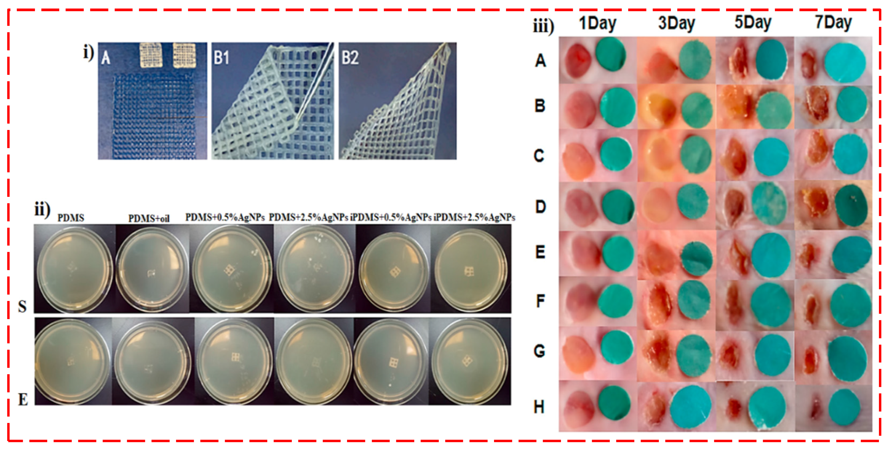
| iPDMS and silicone oil | Ag | Bioprinter | iPDMS/AgNPs could significantly prevent wound dressing infection | Excellent biocompatibility, promoting neo-epithelial and granulation tissue formation to accelerate wound healing in vivo. Cell type: Fibroblast | Wound dressing | [135] |
| ZrO2 | ZnO | 3D printer (Makerbot Z18, America) | The ZrO2-ZnO ceramics had a substantial antibacterial performance | ZrO2-ZnO ceramics presented high cell viability (around 80%). Cell type: MC3T3-E1 | Hip joint | [136] |
| PLGA | ZIF-8, Copper | Extrusion-based | PLGA/Cu(I)@ZIF-8 scaffolds destroyed S. aureus bacteria, and bacteria numbers were considerably diminished in infected rats after implantation with the scaffolds | The cells were well spread and attached with a high growth rate on PLGA/Cu(I)@ZIF-8 scaffolds. Cell type: mMSC | BTE | [137] |
| PCL/ Lidocaine | Ag | Extrusion-based | Scaffolds loaded with Ag presented excellent IZs towards S. aureus and E. coli in a dose-dependent manner | Ag-encapsulated scaffolds showed a toxic effect to MC3T3 cells, as a result of dual-released lidocaine and Ag, while no cytotoxicity effect was detected for the neat lidocaine- or Ag3PO4-loaded scaffolds. Cell type: HFFs and MC3T3 | Infection prevention and pain relief | [138] |
Publisher’s Note: MDPI stays neutral with regard to jurisdictional claims in published maps and institutional affiliations. |
© 2022 by the authors. Licensee MDPI, Basel, Switzerland. This article is an open access article distributed under the terms and conditions of the Creative Commons Attribution (CC BY) license (https://creativecommons.org/licenses/by/4.0/).
Share and Cite
Pahlevanzadeh, F.; Setayeshmehr, M.; Bakhsheshi-Rad, H.R.; Emadi, R.; Kharaziha, M.; Poursamar, S.A.; Ismail, A.F.; Sharif, S.; Chen, X.; Berto, F. A Review on Antibacterial Biomaterials in Biomedical Applications: From Materials Perspective to Bioinks Design. Polymers 2022, 14, 2238. https://doi.org/10.3390/polym14112238
Pahlevanzadeh F, Setayeshmehr M, Bakhsheshi-Rad HR, Emadi R, Kharaziha M, Poursamar SA, Ismail AF, Sharif S, Chen X, Berto F. A Review on Antibacterial Biomaterials in Biomedical Applications: From Materials Perspective to Bioinks Design. Polymers. 2022; 14(11):2238. https://doi.org/10.3390/polym14112238
Chicago/Turabian StylePahlevanzadeh, Farnoosh, Mohsen Setayeshmehr, Hamid Reza Bakhsheshi-Rad, Rahmatollah Emadi, Mahshid Kharaziha, S. Ali Poursamar, Ahmad Fauzi Ismail, Safian Sharif, Xiongbiao Chen, and Filippo Berto. 2022. "A Review on Antibacterial Biomaterials in Biomedical Applications: From Materials Perspective to Bioinks Design" Polymers 14, no. 11: 2238. https://doi.org/10.3390/polym14112238
APA StylePahlevanzadeh, F., Setayeshmehr, M., Bakhsheshi-Rad, H. R., Emadi, R., Kharaziha, M., Poursamar, S. A., Ismail, A. F., Sharif, S., Chen, X., & Berto, F. (2022). A Review on Antibacterial Biomaterials in Biomedical Applications: From Materials Perspective to Bioinks Design. Polymers, 14(11), 2238. https://doi.org/10.3390/polym14112238