
Outdoor Wood Mats-Based Engineering Composite: Influence of Process Parameters on Decay Resistance against Wood-Degrading Fungi Trametes versicolor and Gloeophyllum trabeum

 ,
,  , ,
, ,
Abstract
:1. Introduction
2. Materials and Methods
2.1. Materials
2.2. Preparation of Outdoor Wood Mats-Based Engineering Composites (OWMECs)
2.3. Decay Resistance
2.4. Chemical Analysis
2.5. Fourier Transform Infrared (FTIR) Analysis
2.6. X-ray Diffraction (XRD) Analysis
2.7. Scanning Electron Microscopy (SEM) Analysis
2.8. Statistical Analysis
3. Results and Discussion
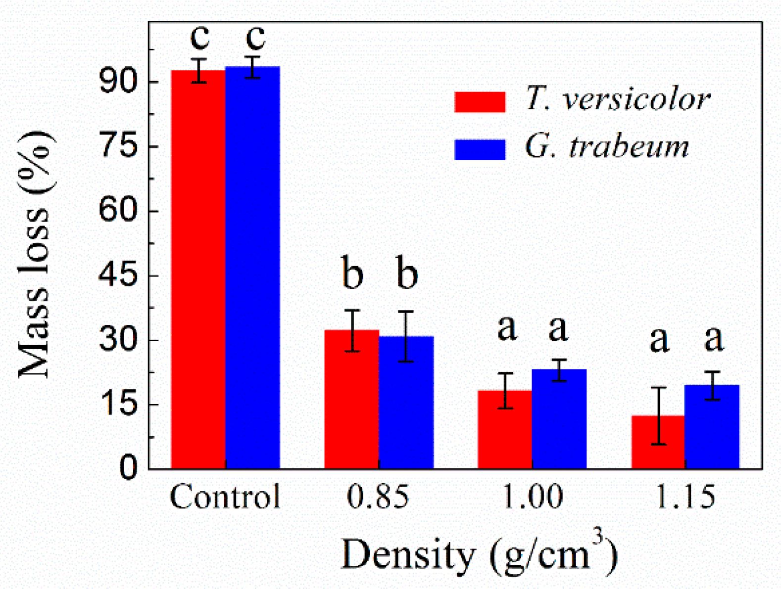
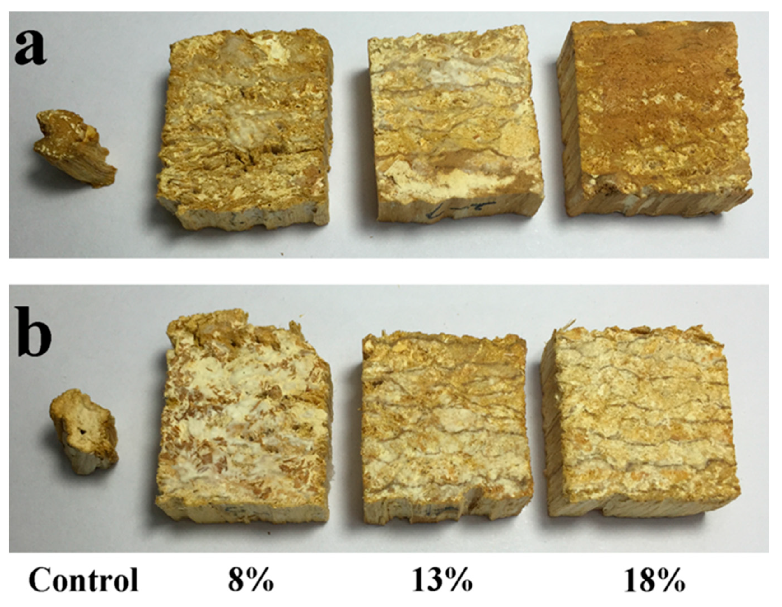
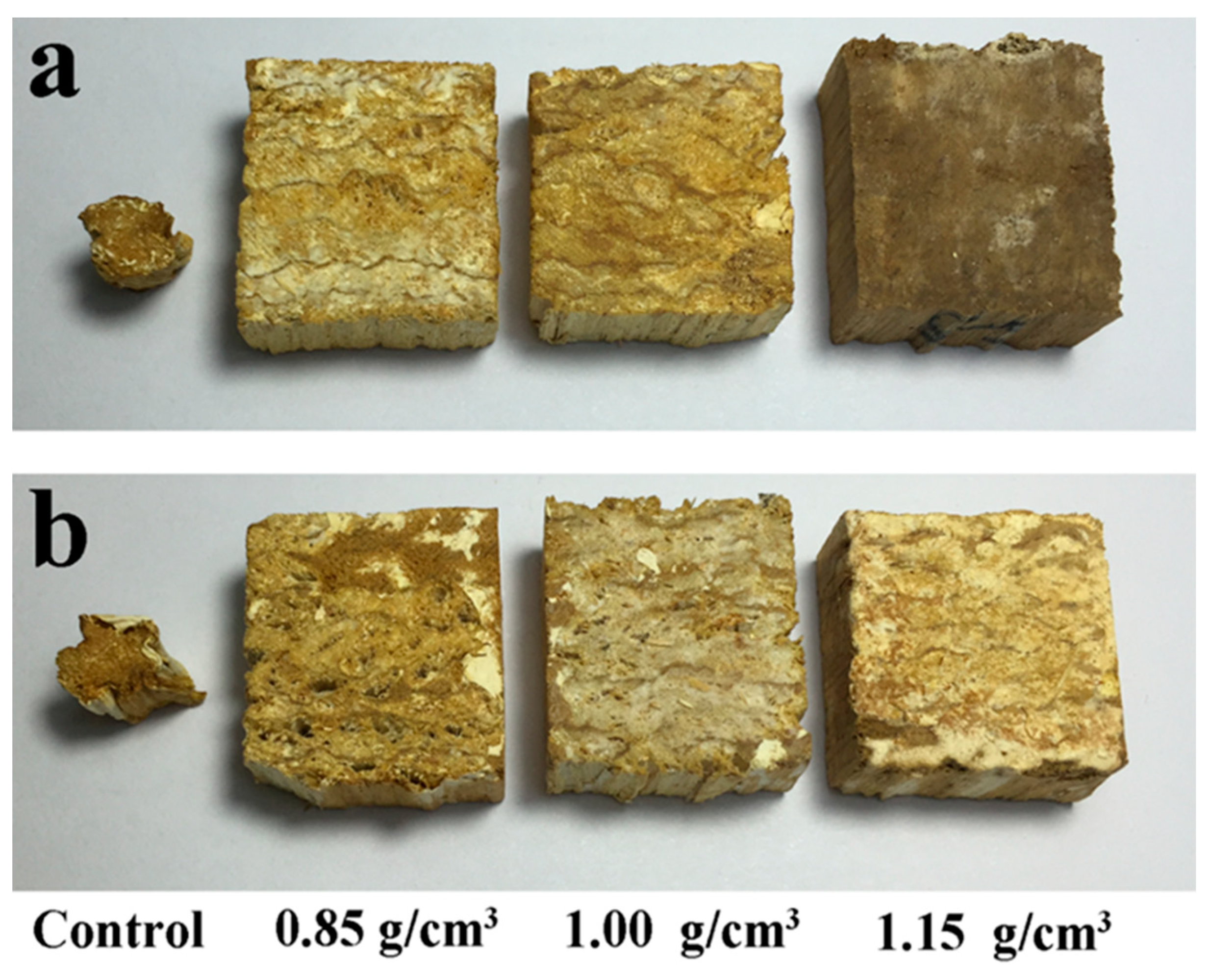
3.1. Mass Loss Analysis
3.2. Chemical Analysis
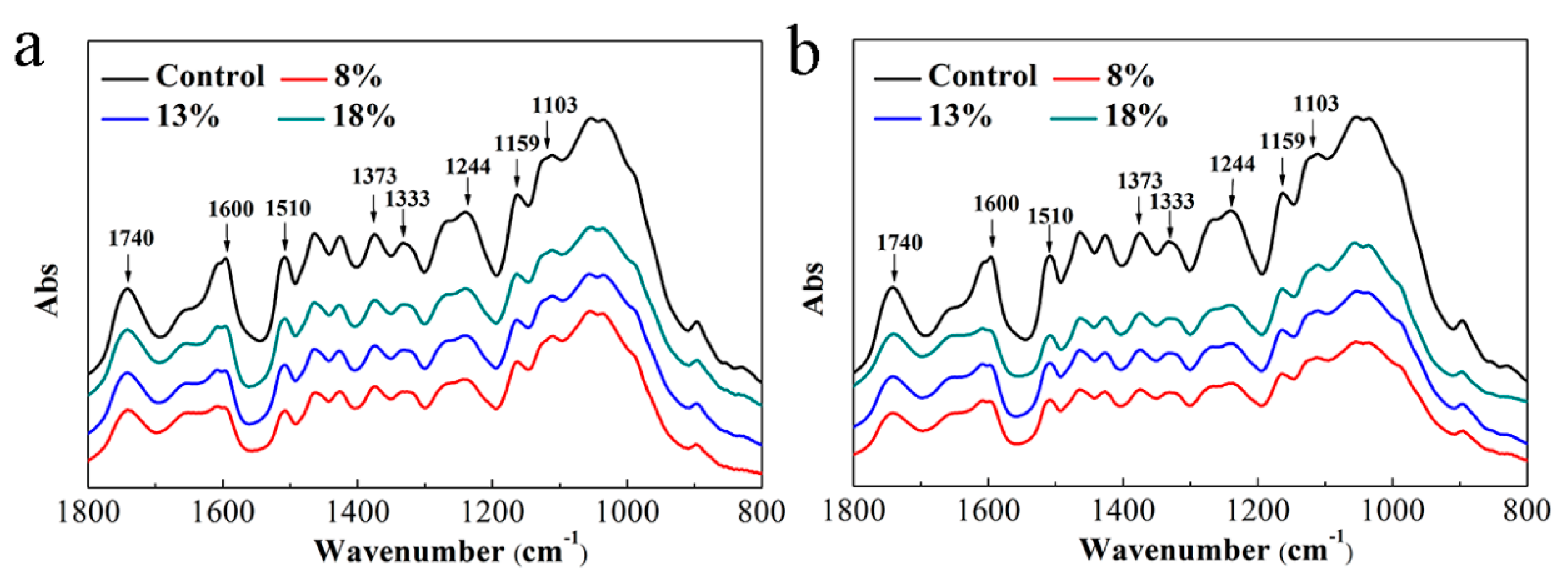
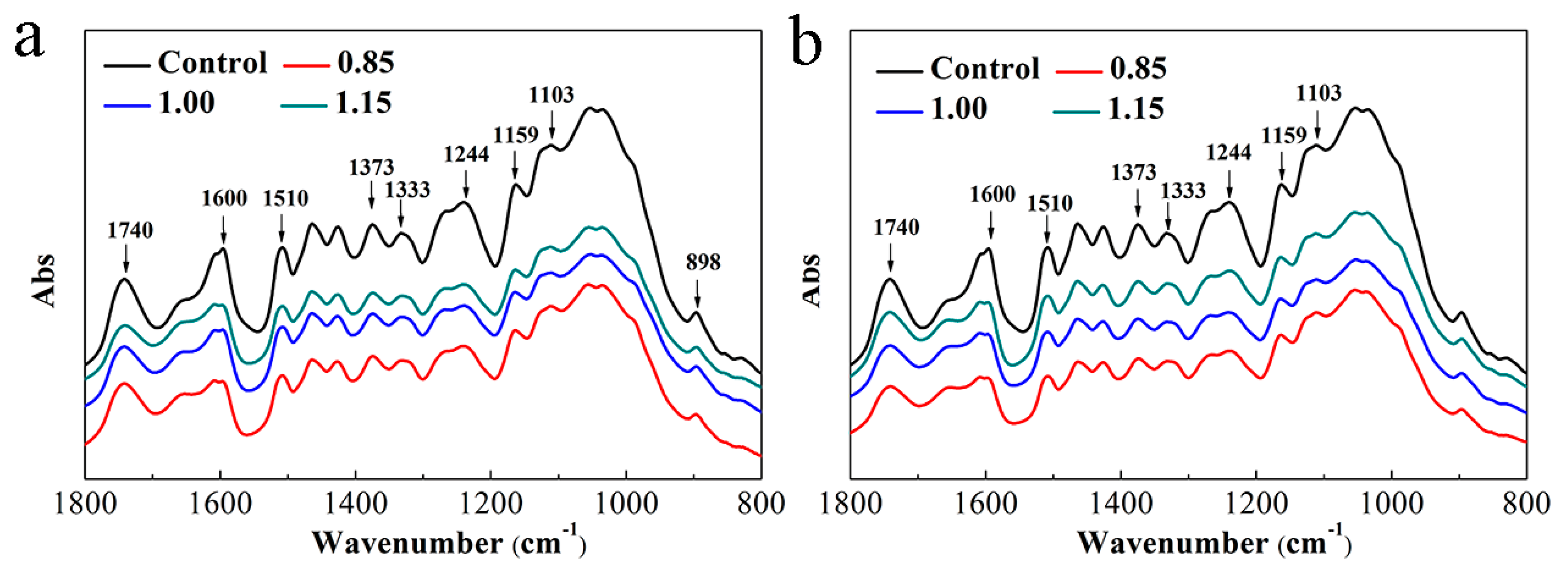
3.3. FTIR Analysis
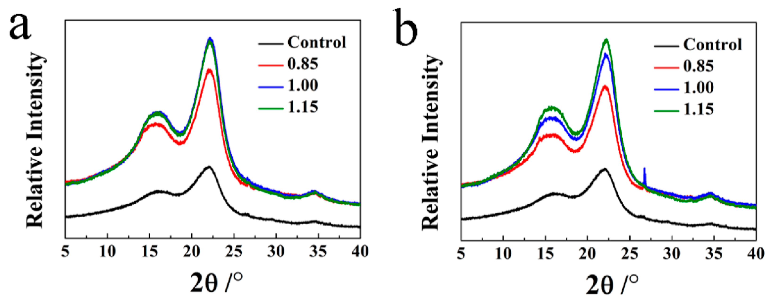
3.4. XRD Analysis
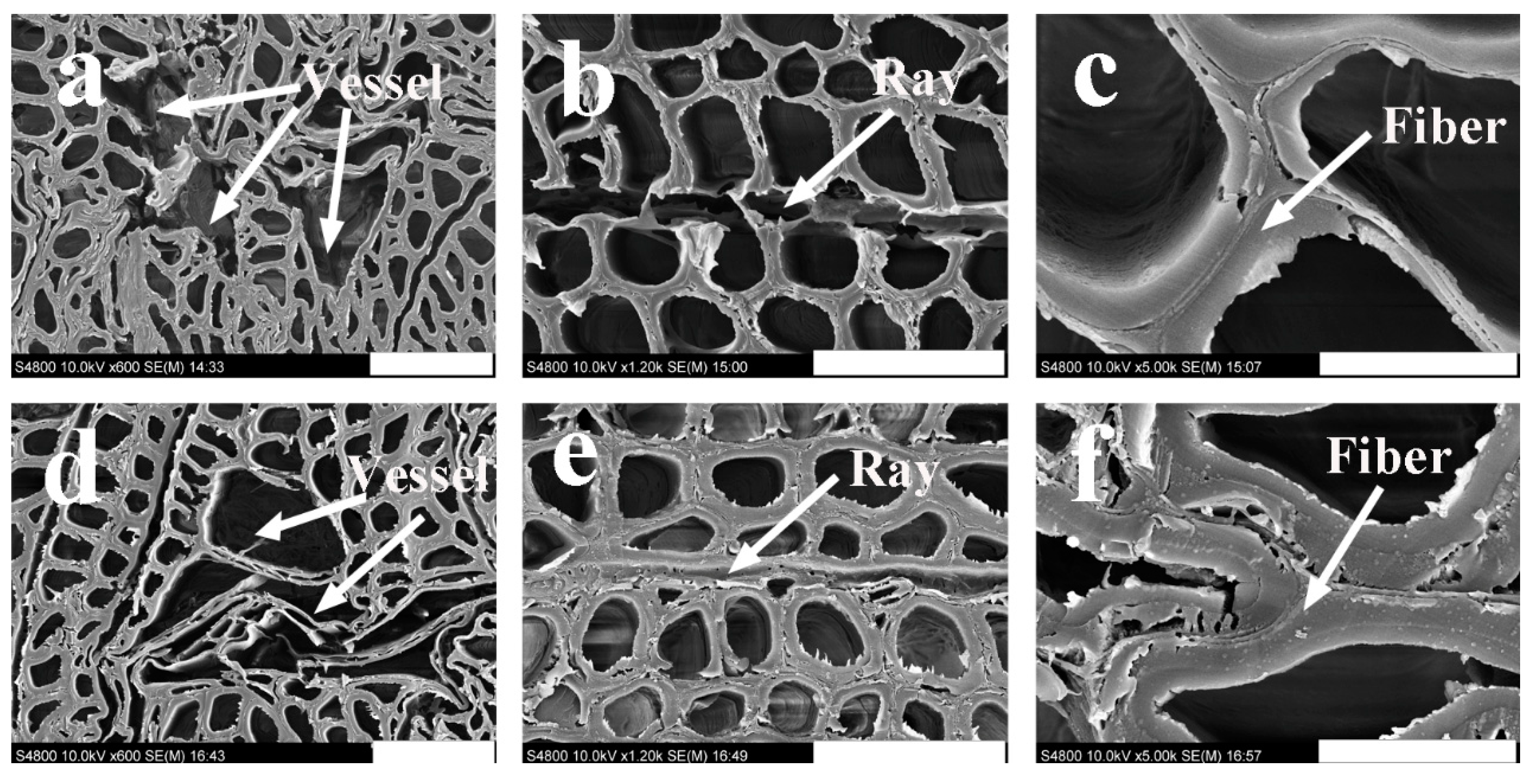
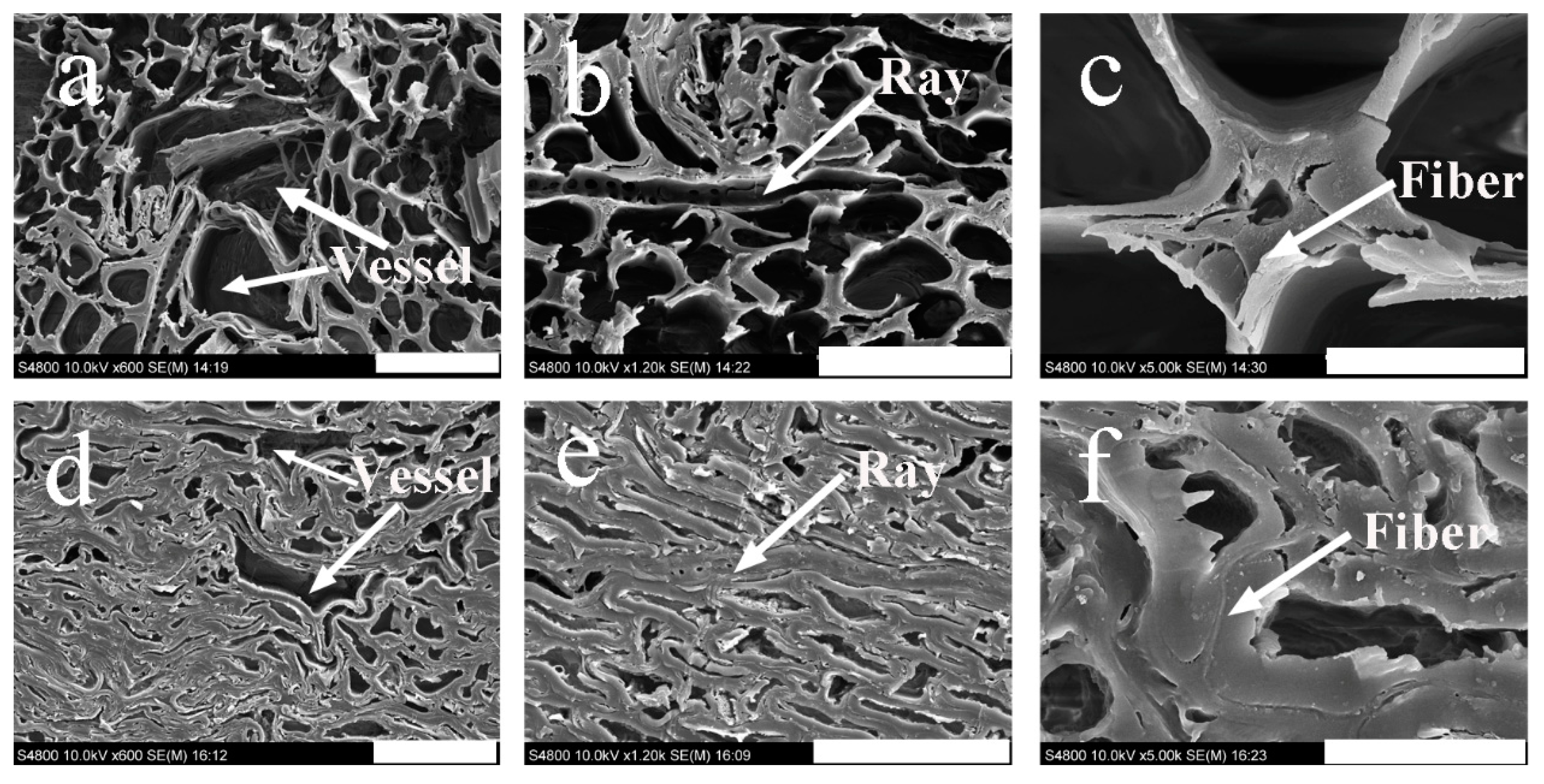
3.5. SEM Analysis
4. Conclusions
Author Contributions
Funding
Institutional Review Board Statement
Informed Consent Statement
Data Availability Statement
Conflicts of Interest
References
- Zhang, Y.H.; Huang, Y.X.; Qi, Y.; Yu, W.J. Novel engineered scrimber with outstanding dimensional stability from finely fluffed poplar veneers. Measurement 2018, 124, 318–321. [Google Scholar] [CrossRef]
- Bao, M.Z.; Li, N.; Huang, C.J.; Chen, Y.H.; Yu, W.J.; Yu, Y.L. Fabrication, physical–mechanical properties and morphological characterizations of novel scrimber composite. Eur. J. Wood Wood Prod. 2019, 77, 741–747. [Google Scholar] [CrossRef]
- He, M.J.; Zhang, J.; Li, Z.; Li, M.L. Production and mechanical performance of scrimber composite manufactured from poplar wood for structural applications. J. Wood Sci. 2016, 62, 429–440. [Google Scholar] [CrossRef] [Green Version]
- Zhang, Y.M.; Huang, X.A.; Zhang, Y.H.; Yu, Y.L.; Yu, W.J. Scrimber board (SB) manufacturing by a new method and characterization of SB’s mechanical properties and dimensional stability. Holzforschung 2017, 72, 283–289. [Google Scholar] [CrossRef]
- Kim, M.J.; Choi, Y.S.; Oh, J.J.; Kim, G.-H. Experimental investigation of the humidity effect on wood discoloration by selected mold and stain fungi for a proper conservation of wooden cultural heritages. J. Wood Sci. 2020, 66, 31. [Google Scholar] [CrossRef]
- Karim, M.; Daryaei, M.G.; Torkaman, J.; Oladi, R.; Ghanbary, M.A.T.; Bari, E. In Vivo investigation of chemical alteration in oak wood decayed by Pleurotus ostreatus. Int. Biodeterior. Biodegrad. 2016, 108, 127–132. [Google Scholar] [CrossRef]
- Witomski, P.; Olek, W.; Bonarski, J.T. Changes in strength of Scots pine wood (Pinus silvestris L.) decayed by brown rot (Coniophora puteana) and white rot (Trametes versicolor). Constr. Build. Mater. 2016, 102, 162–166. [Google Scholar] [CrossRef]
- Kataoka, Y.; Kiguchi, M.; Fujiwara, T.; Evans, P.D. The effects of within-species and between-species variation in wood density on the photodegradation depth profiles of sugi (Cryptomeria japonica) and hinoki (Chamaecyparis obtusa). J. Wood Sci. 2005, 51, 531–536. [Google Scholar] [CrossRef]
- Bao, M.Z.; Rao, F.; He, S.; Bao, Y.J.; Wu, Z.X.; Li, N.; Chen, Y.H. A note on the surface deterioration of scrimber composites exposed to artificial ageing. Coatings 2019, 9, 846. [Google Scholar] [CrossRef] [Green Version]
- Zhang, Y.H.; Qi, Y.; Huang, Y.X.; Yu, Y.L.; Liang, Y.J.; Yu, W.J. Influence of veneer thickness, mat formation and resin content on some properties of novel poplar scrimbers. Holzforschung 2018, 72, 673–680. [Google Scholar] [CrossRef]
- Evans, P.D.; Gibson, S.K.; Cullis, I.; Liu, C.; Sèbe, G. Photostabilization of wood using low molecular weight phenol formaldehyde resin and hindered amine light stabilizer. Polym. Degrad. Stab. 2013, 98, 158–168. [Google Scholar] [CrossRef]
- Meng, F.; Liu, R.; Zhang, Y.; Huang, Y.; Yu, Y.; Yu, W. Improvement of the water repellency, dimensional stability, and biological resistance of bamboo-based fiber reinforced composites. Polym. Compos. 2019, 40, 506–513. [Google Scholar] [CrossRef]
- Kumar, A.; Ryparovà, P.; Kasal, B.; Adamopoulos, S.; Hajek, P. Resistance of bamboo scrimber against white-rot and brown-rot fungi. Wood Mater. Sci. Eng. 2020, 15, 57–63. [Google Scholar] [CrossRef]
- Bakar, E.S.; Hao, J.; Ashaari, Z.; Choo Cheng Yong, A. Durability of phenolic-resin-treated oil palm wood against subterranean termites a white-rot fungus. Int. Biodeterior. Biodegrad. 2013, 85, 126–130. [Google Scholar] [CrossRef] [Green Version]
- Bao, M.Z.; Huang, X.A.; Zhang, Y.H.; Yu, W.J.; Yu, Y.L. Effect of density on the hygroscopicity and surface characteristics of hybrid poplar compreg. J. Wood Sci. 2016, 62, 441–454. [Google Scholar] [CrossRef] [Green Version]
- Durability of Wood—Part 1: Method for Laboratory Test of Natural Decay Resistance (GB/T13942.1-2009); Standard Administration of China: Beijing, China, 2009; pp. 1–5.
- Fibrous Raw Material—Determination of Acid-Insoluble Lignin (GB/T 2677.8-1994); Standard Administration of China: Beijing, China, 1994; pp. 213–215.
- Fibrous Raw Material—Determination of Holocellulose (GB/T 2677.10-1995); Standard Administration of China: Beijing, China, 1995; pp. 220–223.
- Pulps-Determination of A-Cellulose (GB/T 744-1989); Standard Administration of China: Beijing, China, 1989; pp. 156–158.
- Freitag, C.; Kamke, F.A.; Morrell, J.J. Resistance of resin-impregnated VTC processed hybrid-poplar to fungal attack. Int. Biodeterior. Biodegrad. 2015, 99, 174–176. [Google Scholar] [CrossRef]
- Bari, E.; Schmidt, O.; Oladi, R. A histological investigation of oriental beech wood decayed by Pleurotus ostreatus and Trametes versicolor. For. Pathol. 2015, 45, 349–357. [Google Scholar] [CrossRef]
- Bari, E.; Nazarnezhad, N.; Kazemi, S.M.; Ghanbary, M.A.T.; Mohebby, B.; Schmidt, O.; Clausen, C.A. Comparison between degradation capabilities of the white rot fungi Pleurotus ostreatus and Trametes versicolor in beech wood. Int. Biodeterior. Biodegrad. 2015, 104, 231–237. [Google Scholar] [CrossRef]
- Bari, E.; Taghiyari, H.R.; Naji, H.R.; Schmidt, O.; Ohno, K.M.; Clausen, C.A.; Bakar, E.S. Assessing the destructive behaviors of two white-rot fungi on beech wood. Int. Biodeterior. Biodegrad. 2016, 114, 129–140. [Google Scholar] [CrossRef]
- Kirk, T.; Highley, T. Quantitative changes in structural components of conifer woods during decay by white-and brown-rot fungi. Phytopathology 1973, 63, 1338–1342. [Google Scholar] [CrossRef]
- Irbe, I.; Andersons, B.; Chirkova, J.; Kallavus, U.; Andersone, I.; Faix, O. On the changes of pinewood (Pinus sylvestris L.) chemical composition and ultrastructure during the attack by brown-rot fungi Postia placenta and Coniophora puteana. Int. Biodeterior. Biodegrad. 2006, 57, 99–106. [Google Scholar] [CrossRef]
- Pandey, K. A study of chemical structure of soft and hardwood and wood polymers by FTIR spectroscopy. J. Appl. Polym. Sci. 1999, 71, 1969–1975. [Google Scholar] [CrossRef]
- Pandey, K.; Pitman, A. FTIR studies of the changes in wood chemistry following decay by brown-rot and white-rot fungi. Int. Biodeterior. Biodegrad. 2003, 52, 151–160. [Google Scholar] [CrossRef]
- Schwarze, F.W.M.R. Wood decay under the microscope. Fungal Biol. Rev. 2007, 21, 133–170. [Google Scholar] [CrossRef]
- Fackler, K.; Schwanninger, M. Polysaccharide degradation and lignin modification during brown rot of spruce wood: A polarised Fourier transform near infrared study. J. Near Infrared Spectrosc. 2010, 18, 403–416. [Google Scholar] [CrossRef]
- Nabil, F.; Zaidon, A.; Anwar, U.; Bakar, E.; Lee, S.; Paridah, M. Impregnation of sesenduk (Endospermum diadenum) wood with phenol formaldehyde and nanoclay admixture: Effect on fungal decay and termites attack. Sains Malays. 2016, 45, 255–262. [Google Scholar]


| Resin Content (%) | Fungus | Holocellulose (%) | α-Cellulose (%) | Acid Insoluble Lignin (%) |
|---|---|---|---|---|
| 8 | - | 68.47 (0.10) c | 40.58 (0.17) b | 25.09 (0.11) a |
| 13 | - | 64.19 (0.21) b | 38.61 (0.54) a | 28.12 (0.16) b |
| 18 | - | 62.64 (0.20) a | 37.11 (0.13) a | 31.26 (0.16) c |
| 8 | T. versicolor | 63.13 (0.11) c | 37.32 (0.08) b | 28.86 (0.10) a |
| 13 | 61.53 (0.16) b | 37.31 (0.14) b | 33.41 (0.11) b | |
| 18 | 60.46 (0.07) a | 36.82 (0.10) a | 34.44 (0.44) c | |
| 8 | G. trabeum | 60.25 (0.13) a | 34.98 (0.20) a | 31.16 (0.24) a |
| 13 | 60.39 (0.11) a | 36.89 (0.23) b | 33.75 (0.08) b | |
| 18 | 60.11 (0.06) a | 36.68 (0.10) b | 35.48 (0.18) c |
| Density (g/cm3) | Fungus | Holocellulose (%) | α-Cellulose (%) | Acid Insoluble Lignin (%) |
|---|---|---|---|---|
| 0.85 | - | 63.78 (0.18) a | 38.31 (0.13) a | 29.63 (0.13) c |
| 1.00 | - | 64.19 (0.21) a | 38.61 (0.17) a | 28.12 (0.16) b |
| 1.15 | - | 65.32 (0.11) b | 39.37 (0.08) b | 27.31 (0.21) a |
| 0.85 | T. versicolor | 60.29 (0.14) a | 35.92 (0.18) b | 32.48 (0.18) a |
| 1.00 | 63.55 (0.08) c | 38.99 (0.16) c | 32.01 (0.34) a | |
| 1.15 | 62.18 (0.16) b | 35.04 (0.11) a | 32.64 (0.17) a | |
| 0.85 | G. trabeum | 59.32 (0.11) a | 35.56 (0.13) a | 32.52 (0.16) a |
| 1.00 | 61.24 (0.10) c | 37.77 (0.13) b | 32.47 (0.13) a | |
| 1.15 | 60.90 (0.92) b | 37.45 (0.06) b | 33.18 (0.08) b |
| Resin Content (%) | Fungus | Cr (%) |
|---|---|---|
| Control | - | 16.34 |
| 8.0 | T. versicolor | 16.64 |
| 13.0 | 21.10 | |
| 18.0 | 21.81 | |
| 8.0 | G. trabeum | 18.65 |
| 13.0 | 18.89 | |
| 18.0 | 21.45 |
| Density (g/cm3) | Fungus | Cr (%) |
|---|---|---|
| Control | - | 16.34 |
| 0.85 | T. versicolor | 19.72 |
| 1.00 | 22.81 | |
| 1.15 | 23.27 | |
| 0.85 | G. trabeum | 19.21 |
| 1.00 | 21.78 | |
| 1.15 | 23.79 |
Publisher’s Note: MDPI stays neutral with regard to jurisdictional claims in published maps and institutional affiliations. |
© 2021 by the authors. Licensee MDPI, Basel, Switzerland. This article is an open access article distributed under the terms and conditions of the Creative Commons Attribution (CC BY) license (https://creativecommons.org/licenses/by/4.0/).
Share and Cite
Bao, M.; Li, N.; Bao, Y.; Li, J.; Zhong, H.; Chen, Y.; Yu, Y. Outdoor Wood Mats-Based Engineering Composite: Influence of Process Parameters on Decay Resistance against Wood-Degrading Fungi Trametes versicolor and Gloeophyllum trabeum. Polymers 2021, 13, 3173. https://doi.org/10.3390/polym13183173
Bao M, Li N, Bao Y, Li J, Zhong H, Chen Y, Yu Y. Outdoor Wood Mats-Based Engineering Composite: Influence of Process Parameters on Decay Resistance against Wood-Degrading Fungi Trametes versicolor and Gloeophyllum trabeum. Polymers. 2021; 13(18):3173. https://doi.org/10.3390/polym13183173
Chicago/Turabian StyleBao, Minzhen, Neng Li, Yongjie Bao, Jingpeng Li, Hao Zhong, Yuhe Chen, and Yanglun Yu. 2021. "Outdoor Wood Mats-Based Engineering Composite: Influence of Process Parameters on Decay Resistance against Wood-Degrading Fungi Trametes versicolor and Gloeophyllum trabeum" Polymers 13, no. 18: 3173. https://doi.org/10.3390/polym13183173
APA StyleBao, M., Li, N., Bao, Y., Li, J., Zhong, H., Chen, Y., & Yu, Y. (2021). Outdoor Wood Mats-Based Engineering Composite: Influence of Process Parameters on Decay Resistance against Wood-Degrading Fungi Trametes versicolor and Gloeophyllum trabeum. Polymers, 13(18), 3173. https://doi.org/10.3390/polym13183173