
Synthesis and Characterization of Porous, Electro-Conductive Chitosan–Gelatin–Agar-Based PEDOT: PSS Scaffolds for Potential Use in Tissue Engineering

 , , , , , and
Abstract
:1. Introduction
2. Materials and Methods
2.1. Materials
2.2. Synthesis of Conductive PEDOT: PSS
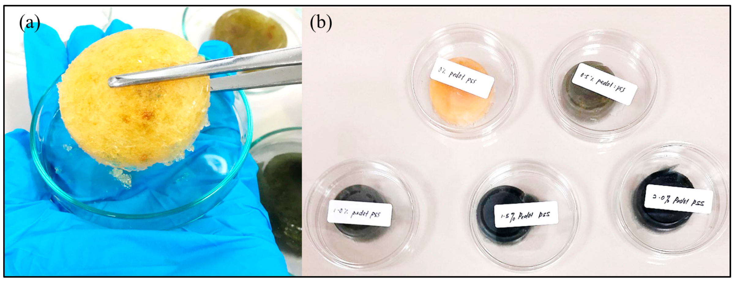
2.3. Fabrication of Porous Hydrogels
- (i)
- Solution mixing: Exactly 7.2 wt.% of gelatin powder was added into 7% acetic solution followed by the addition of 1.8 wt.% of chitosan. The mixture was stirred for 30 min at 80 °C. After the mixture was completely dissolved and formed a yellowish clear solution, exactly 0.05 wt.% agarose was added to regenerate pores and was left, stirring, for another 30 min at 90 °C. Consequently, a specific volume of DMSO–PEDOT: PSS was added into the solution, as shown in Table 1.
- (ii)
- Freezing process: Exactly 0.5% glutaraldehyde was added into the Cs–Gel–Agar–PEDOT: PSS solution, and the resulting mixture was left for 30 min at room temperature. This addition was done to establish the chemical crosslinking of the molecules present in the Cs–Gel–Agar-PEDOT: PSS solution. Next, the mixture underwent a freezing process at −20 °C for 180 min to form hydrogels.
- (iii)
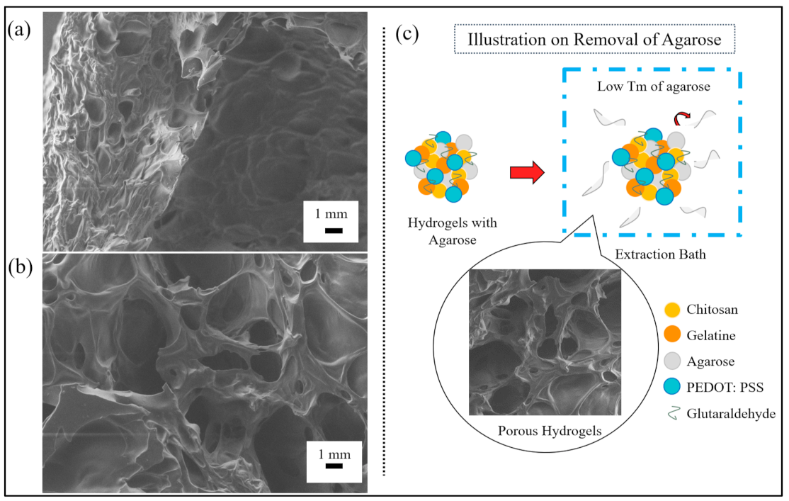
- Removal of agarose: The agarose was used as a sacrificial template for fabricating the porous-structured hydrogels. The removal of agarose was done by immersing the hydrogels in an extraction bath containing 10 v/v % glutaraldehyde at 90 °C. Figure 1 shows the fabricated porous hydrogels.
2.4. Characterization of Porous Hydrogels
2.4.1. Morphology and Pore Size Observations
2.4.2. Attenuated Total Reflectance–Fourier Transform Infrared (FTIR) Analysis
2.4.3. X-ray Diffraction (XRD) Analysis
2.4.4. Electrochemical Impedance Spectroscopy (EIS) Analysis
2.4.5. In Vitro Stability Studies
3. Results and Discussion
3.1. Porous Hydrogel Formation
- (a)
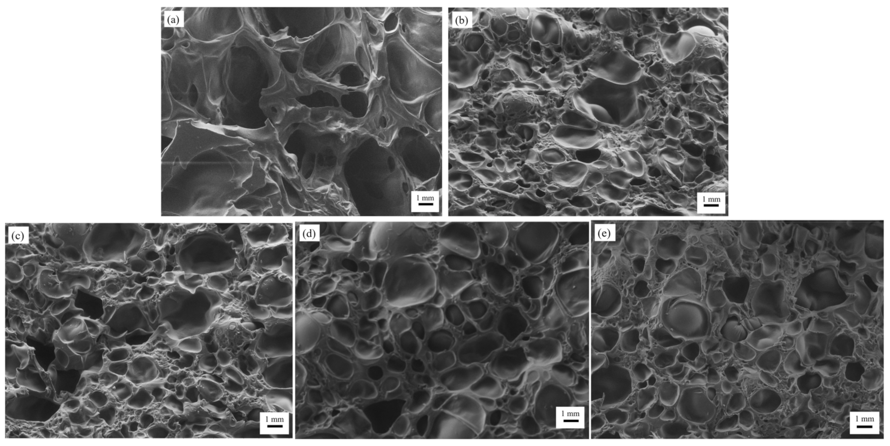
- Morphology of Porous Hydrogels formed from the Reversed Casting Method
- (b)
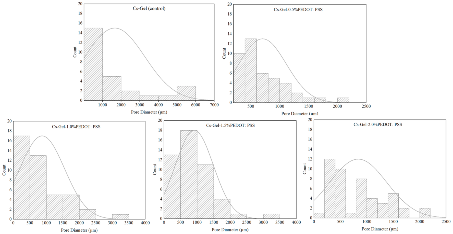
- Pore Size Observation of Porous Hydrogels
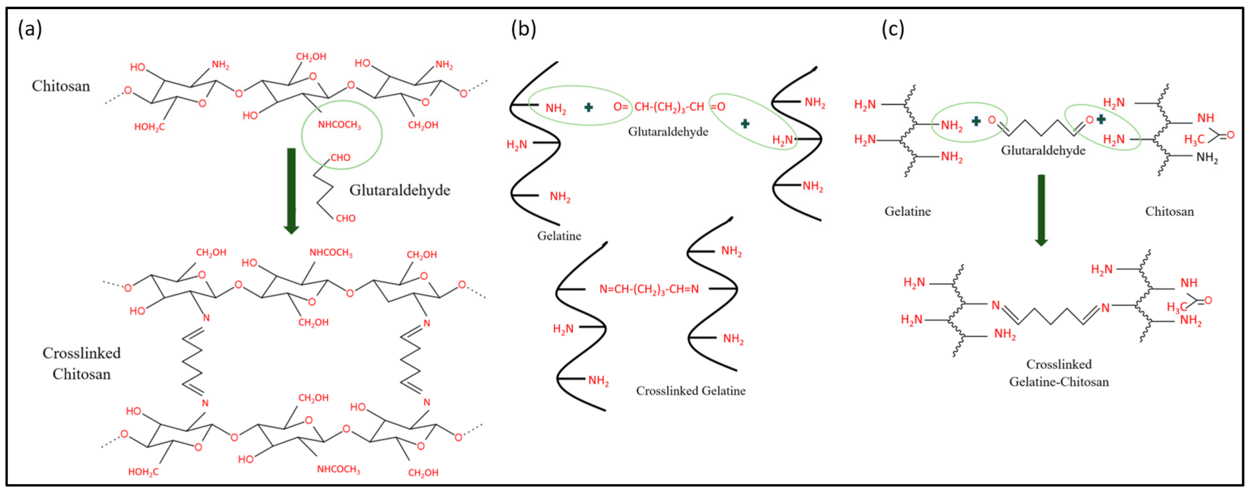
3.2. Proposed Mechanism for the Chemical Reaction of Porous Hydrogels
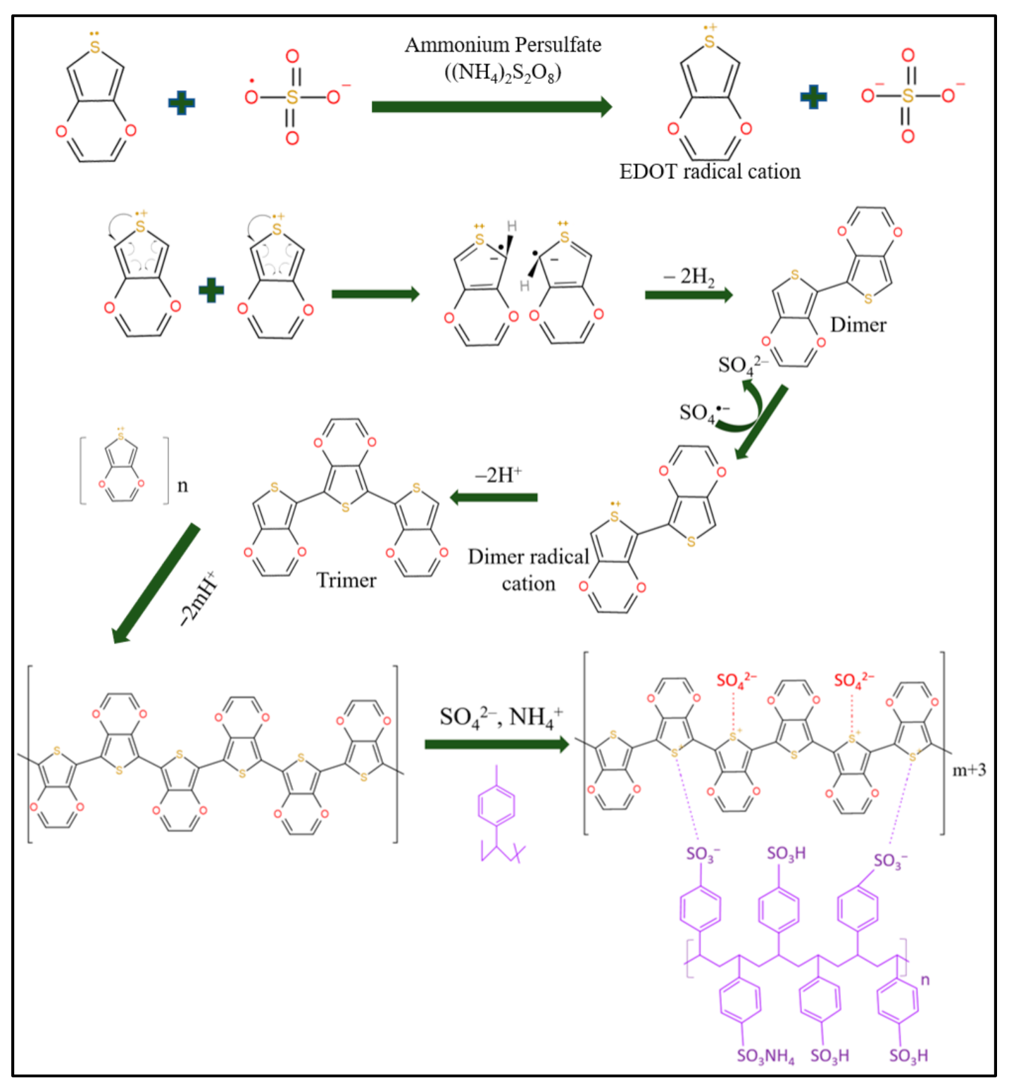
3.2.1. Synthesis of Conductive PEDOT: PSS-Doped DMSO Dispersion
S2O82− (aq) ⇌ 2SO4•− (aq)
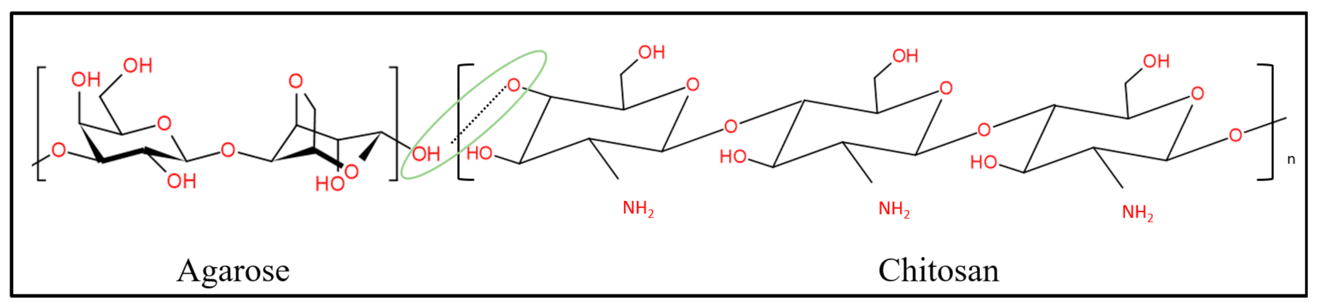
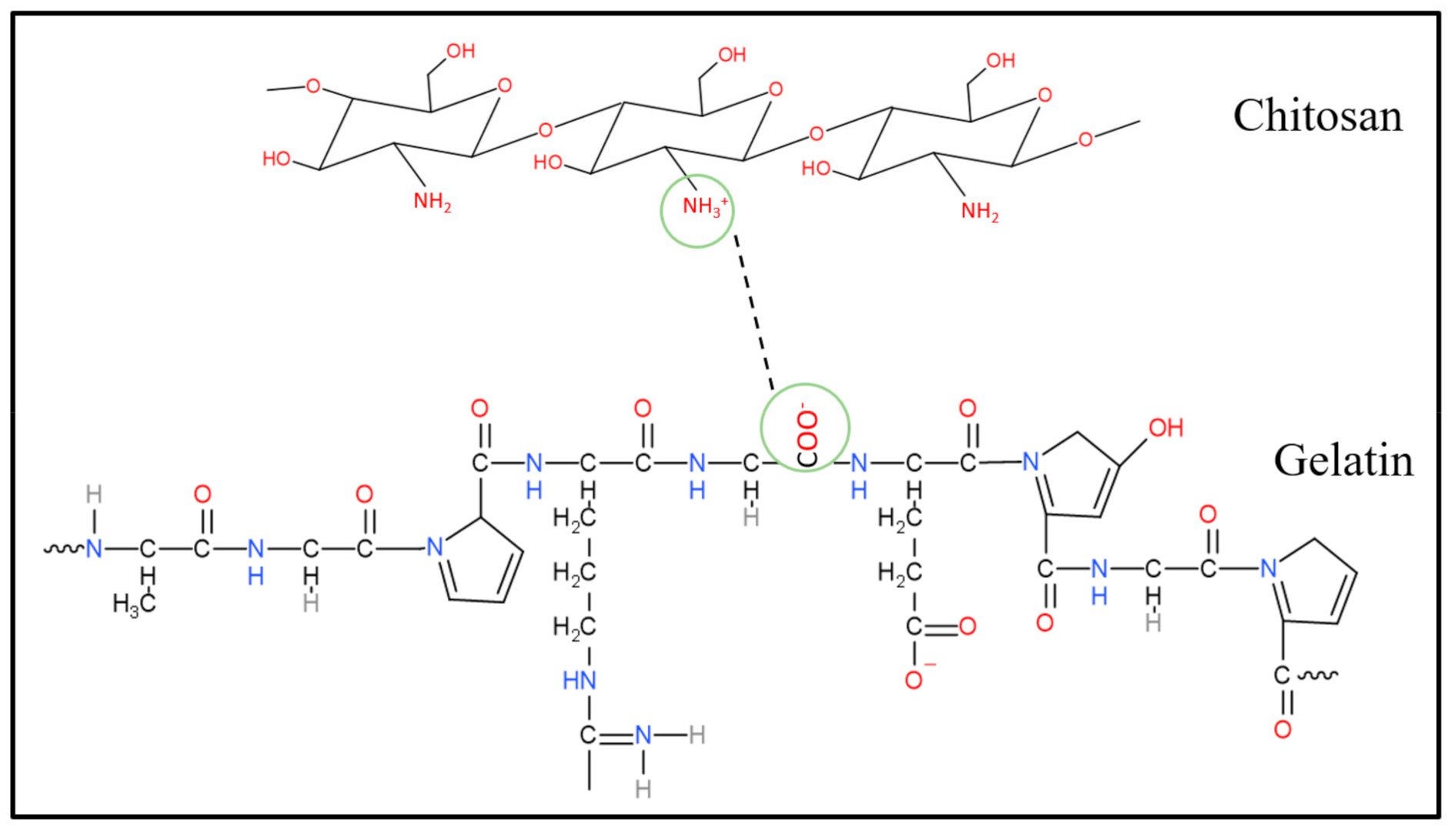
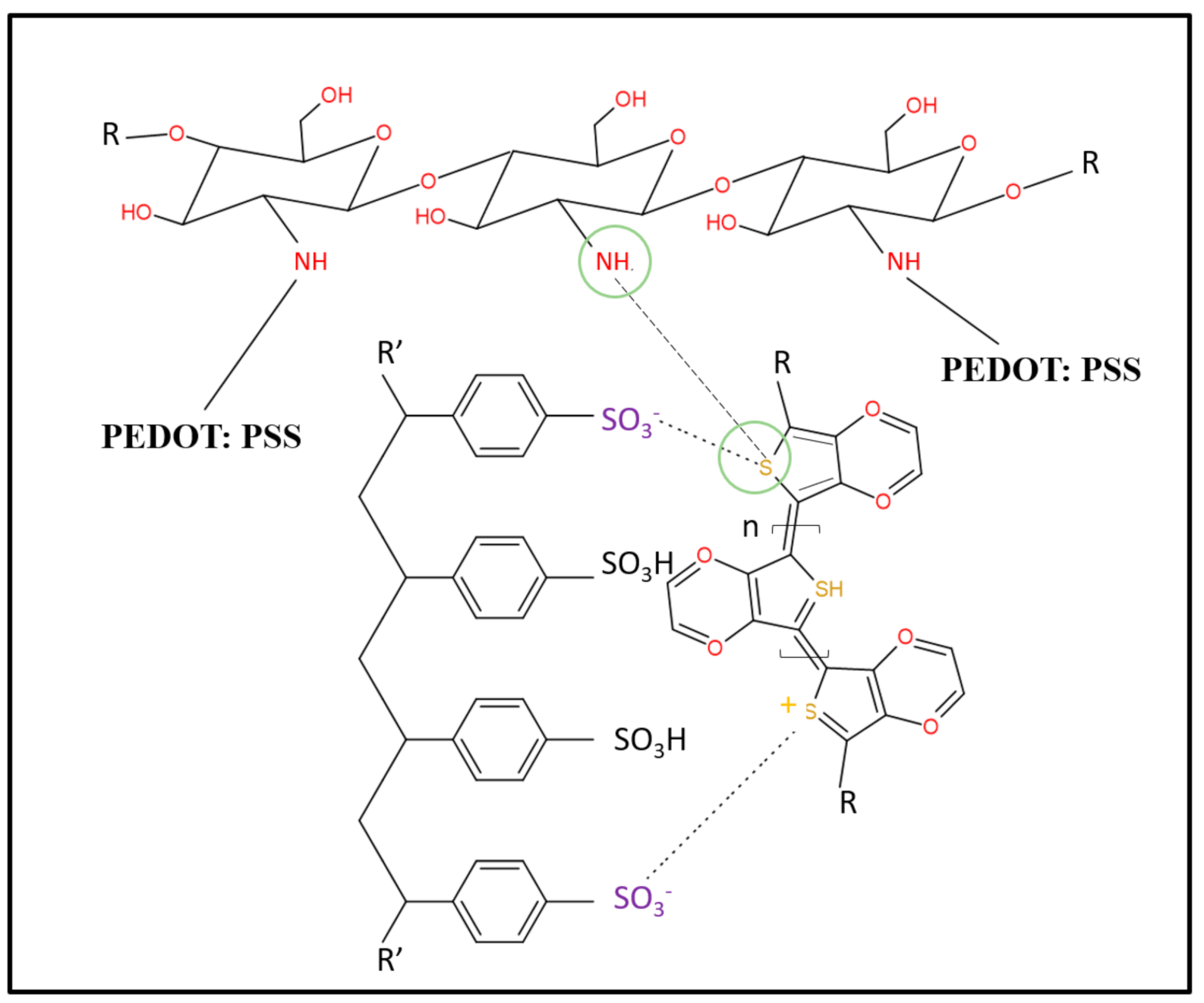
3.2.2. Possible Reactions of Chitosan–Gelatin–Agar-Based PEDOT: PSS Porous Hydrogel Scaffolds
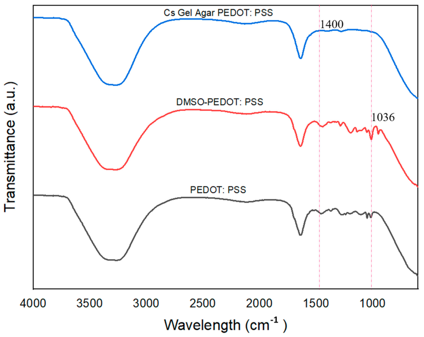
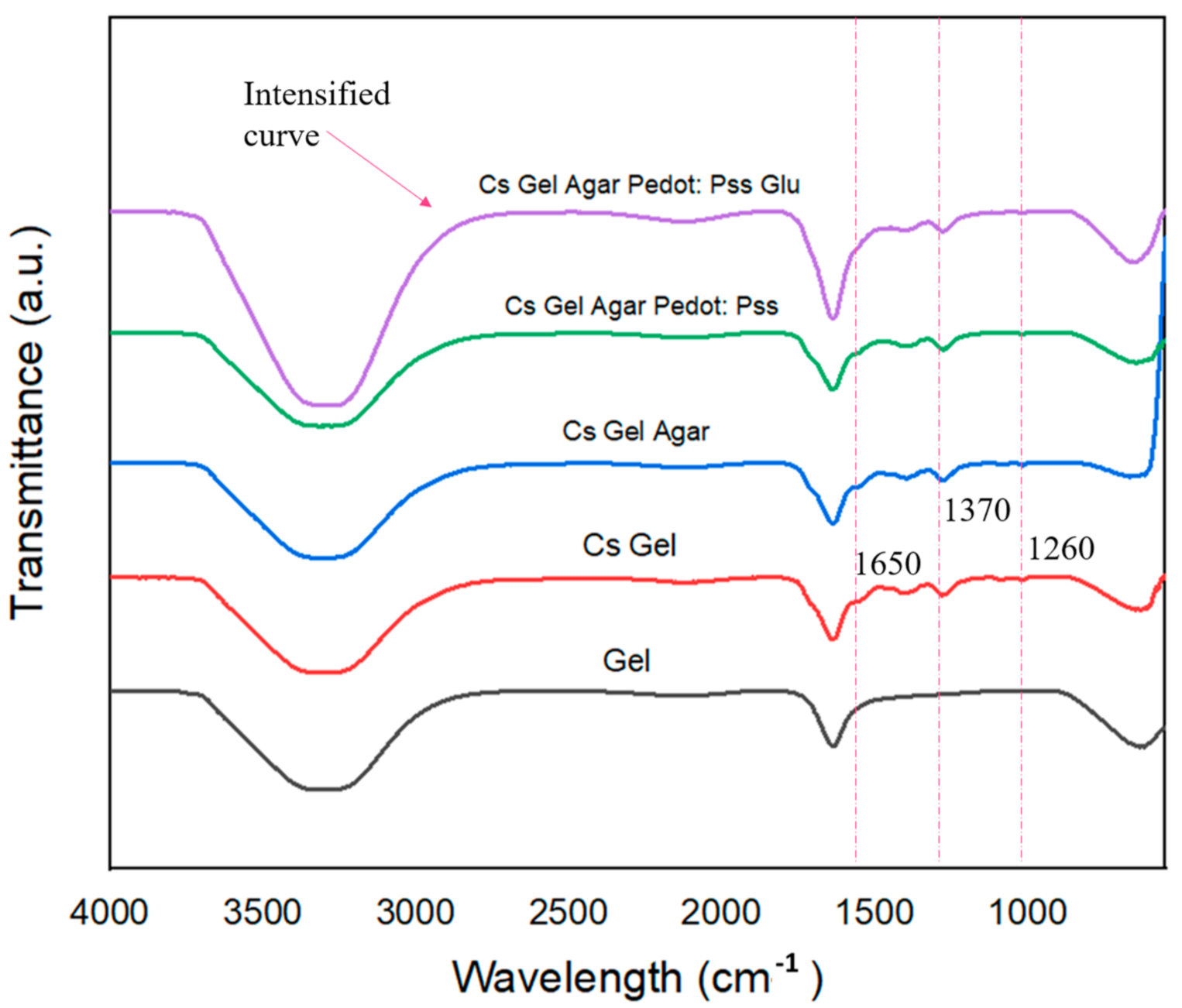
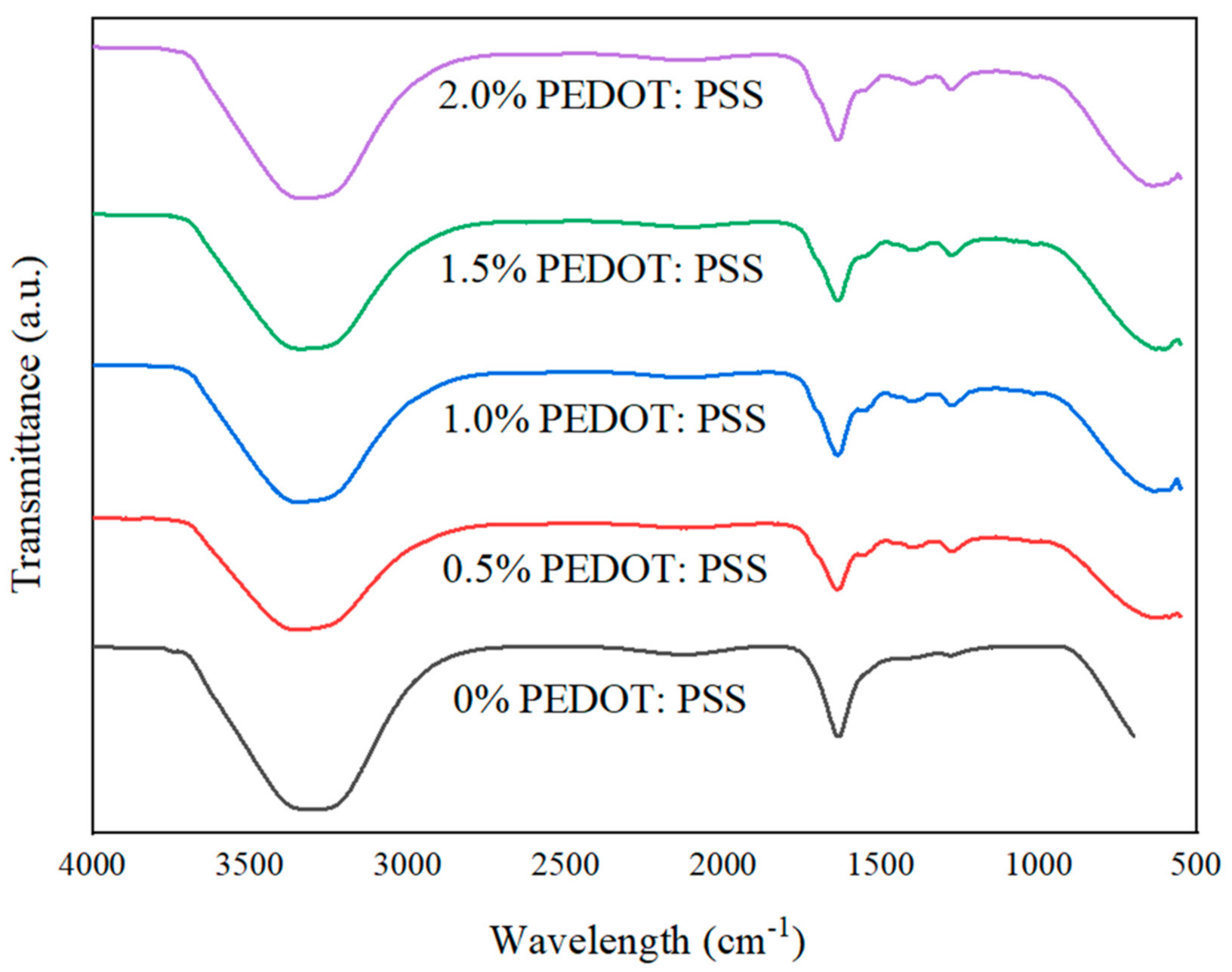
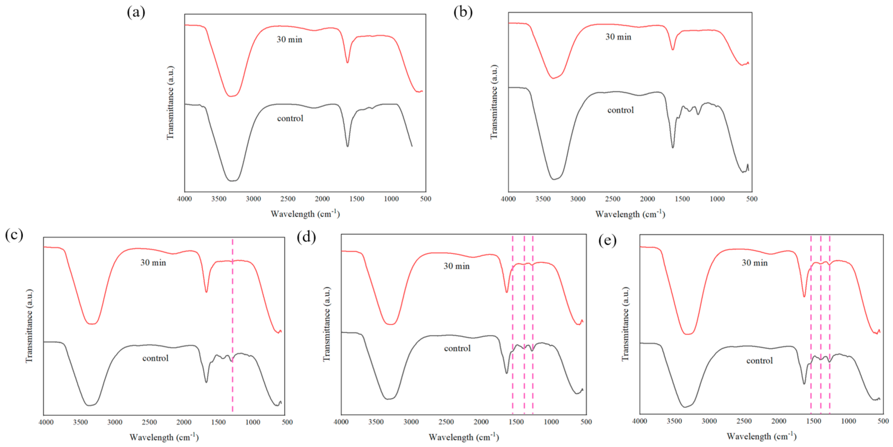
3.3. Attenuated Total Reflectance–Fourier Transform Infrared Spectroscopy (ATR–FTIR) of the Porous Hydrogels
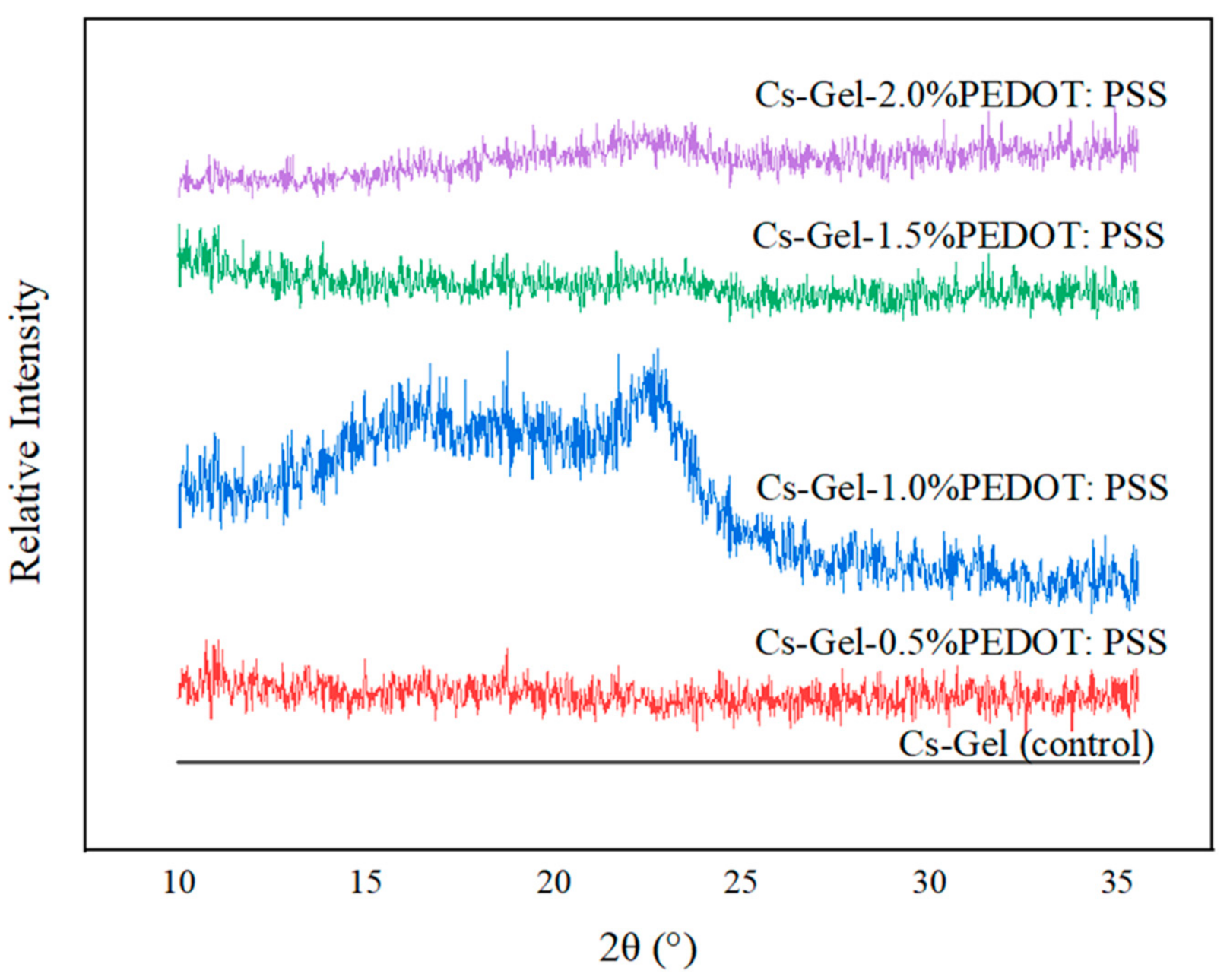
3.4. Phase and Crystallinity Studies of Porous Hydrogels
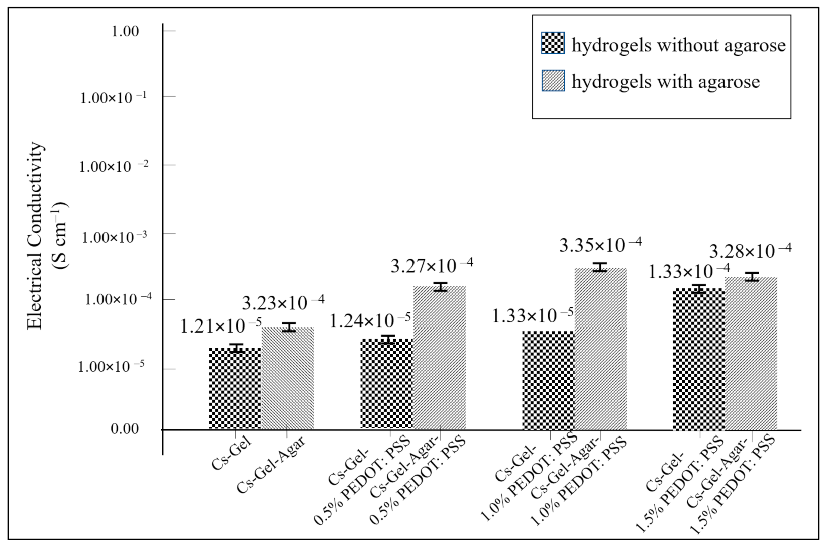
3.5. Electrical Conductivity of Porous Hydrogels
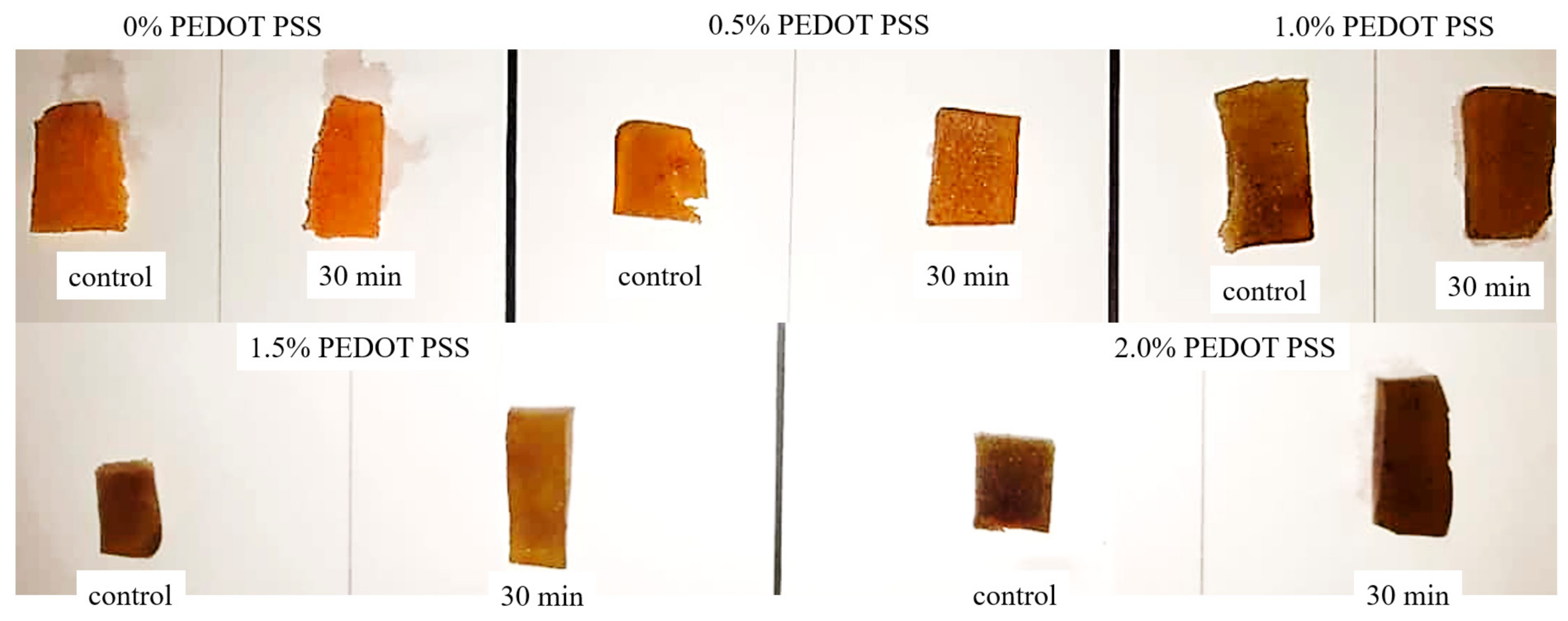
3.6. Stability of Porous Hydrogels under PBS Conditions
4. Conclusions
Author Contributions
Funding
Institutional Review Board Statement
Informed Consent Statement
Data Availability Statement
Acknowledgments
Conflicts of Interest
References
- Tong, Z.; Jin, L.; Oliveira, J.M.; Reis, R.L.; Zhong, Q.; Mao, Z.; Gao, C. Adaptable hydrogel with reversible linkages for regenerative medicine: Dynamic mechanical microenvironment for cells. Bioact. Mater. 2020, 6, 1375–1387. [Google Scholar] [CrossRef] [PubMed]
- Shi, Z.; Gao, X.; Ullah, M.W.; Li, S.; Wang, Q.; Yang, G. Electroconductive natural polymer-based hydrogels. Biomaterials 2016, 111, 40–54. [Google Scholar] [CrossRef] [PubMed]
- Dong, R.; Ma, P.X.; Guo, B. Conductive biomaterials for muscle tissue engineering. Biomaterials 2019, 229, 119584. [Google Scholar] [CrossRef] [PubMed]
- Yang, S.; Jang, L.; Kim, S.; Yang, J.; Yang, K.; Cho, S.-W.; Lee, J.Y. Polypyrrole/alginate hybrid hydrogels: Electrically conductive and soft biomaterials for human mesenchymal stem cell culture and potential neural tissue engineering applications. Macromol. Biosci. 2016, 16, 1653–1661. [Google Scholar] [CrossRef]
- Liu, X.; Liu, J.; Lin, S.; Zhao, X. Hydrogel machines. Mater. Today 2020, 36, 102–124. [Google Scholar] [CrossRef]
- Navaei, A.; Saini, H.; Christenson, W.; Sullivan, R.T.; Ros, R.; Nikkhah, M. Gold nanorod-incorporated gelatin-based conductive hydrogels for engineering cardiac tissue constructs. Acta Biomater. 2016, 41, 133–146. [Google Scholar] [CrossRef]
- Deng, Z.; Hu, T.; Lei, Q.; He, J.; Ma, P.X.; Guo, B. Stimuli-responsive conductive nanocomposite hydrogels with high stretchability, self-healing, adhesiveness, and 3D printability for human motion sensing. ACS Appl. Mater. Interfaces 2019, 11, 6796–6808. [Google Scholar] [CrossRef]
- Talikowska, M.; Fu, X.; Lisak, G. Application of conducting polymers to wound care and skin tissue engineering: A review. Biosens. Bioelectron. 2019, 135, 50–63. [Google Scholar] [CrossRef]
- Park, J.; Choi, J.H.; Kim, S.; Jang, I.; Jeong, S.; Lee, J.Y. Micropatterned conductive hydrogels as multifunctional muscle-mimicking biomaterials: Graphene-incorporated hydrogels directly patterned with femtosecond laser ablation. Acta Biomater. 2019, 97, 141–153. [Google Scholar] [CrossRef]
- Liang, Y.; Mitriashkin, A.; Lim, T.T.; Goh, J.C.-H. Conductive polypyrrole-encapsulated silk fibroin fibers for cardiac tissue engineering. Biomaterials 2021, 276, 121008. [Google Scholar] [CrossRef]
- Rinoldi, C.; Lanzi, M.; Fiorelli, R.; Nakielski, P.; Zembrzycki, K.; Kowalewski, T.; Urbanek, O.; Grippo, V.; Jezierska-Woźniak, K.; Maksymowicz, W.; et al. Three-dimensional printable conductive semi-interpenetrating polymer network hydrogel for neural tissue applications. Biomacromolecules 2021, 22, 3084–3098. [Google Scholar] [CrossRef]
- Guimard, N.K.; Gomez, N.; Schmidt, C.E. Conducting polymers in biomedical engineering. Prog. Polym. Sci. 2007, 32, 876–921. [Google Scholar] [CrossRef]
- Kisieliute, A.; Popov, A.; Apetrei, R.-M.; Cârâc, G.; Morkvenaite-Vilkonciene, I.; Ramanaviciene, A.; Ramanavicius, A. Towards microbial biofuel cells: Improvement of charge transfer by self-modification of microoganisms with conducting polymer—Polypyrrole. Chem. Eng. J. 2018, 356, 1014–1021. [Google Scholar] [CrossRef]
- Moutsatsou, P.; Coopman, K.; Georgiadou, S. Biocompatibility assessment of conducting PANI/chitosan nanofibers for wound healing applications. Polymers 2017, 9, 687. [Google Scholar] [CrossRef] [Green Version]
- Mantione, D.; del Agua, I.; Sanchez-Sanchez, A.; Mecerreyes, D. Poly(3,4-ethylenedioxythiophene) (PEDOT) derivatives: Innovative conductive polymers for bioelectronics. Polymers 2017, 9, 354. [Google Scholar] [CrossRef]
- Raina, D.B.; Qayoom, I.; Larsson, D.; Zheng, M.H.; Kumar, A.; Isaksson, H.; Lidgren, L.; Tägil, M. Guided tissue engineering for healing of cancellous and cortical bone using a combination of biomaterial based scaffolding and local bone active molecule delivery. Biomaterials 2018, 188, 38–49. [Google Scholar] [CrossRef]
- Raghvendrakumar, M.; Yadu, V.K.; Aswathy, V.; Parvathy, P.; Sunija, S.; Neelakandan, M.S.; Nitheesha, S.; Vishnu, K.A. Chitosan as promising materials for biomedical application: Review. Res. Dev. Mater. Sci. 2017, 2, 170–185. [Google Scholar]
- Dorati, R.; Pisani, S.; Maffeis, G.; Conti, B.; Modena, T.; Chiesa, E.; Bruni, G.; Musazzi, U.M.; Genta, I. Study on hydrophilicity and degradability of chitosan/polylactide-co-polycaprolactone nanofibre blend electrospun membrane. Carbohydr. Polym. 2018, 199, 150–160. [Google Scholar] [CrossRef]
- Qu, J.; Zhao, X.; Liang, Y.; Xu, Y.; Ma, P.X.; Guo, B. Degradable conductive injectable hydrogels as novel antibacterial, anti-oxidant wound dressings for wound healing. Chem. Eng. J. 2019, 362, 548–560. [Google Scholar] [CrossRef]
- Lončarević, A.; Ivanković, M.; Rogina, A. Lysozyme-induced degradation of chitosan: The characterisation of degraded chitosan scaffolds. J. Tissue Repair Regen. 2017, 1, 12–22. [Google Scholar] [CrossRef] [Green Version]
- Islam, N.; Dmour, I.; Taha, M. Degradability of chitosan micro/nanoparticles for pulmonary drug delivery. Heliyon 2019, 5, e01684. [Google Scholar] [CrossRef] [Green Version]
- Jennings, J. Controlling chitosan degradation properties in vitro and in vivo. In Chitosan Based Biomaterials; Woodhead Publishing: Sawston, UK, 2017; Volume 1, pp. 159–182. [Google Scholar]
- Raza, F.; Zafar, H.; Zhu, Y.; Ren, Y.; Ullah, A.; Khan, A.U.; He, X.; Han, H.; Aquib; Boakye-Yiadom, K.O.; et al. A review on recent advances in stabilizing peptides/proteins upon fabrication in hydrogels from biodegradable polymers. Pharmaceutics 2018, 10, 16. [Google Scholar] [CrossRef] [Green Version]
- Raza, F.; Zhu, Y.; Chen, L.; You, X.; Zhang, J.; Khan, A.; Khan, M.W.; Hasnat, M.; Zafar, H.; Wu, J.; et al. Paclitaxel-loaded pH responsive hydrogel based on self-assembled peptides for tumor targeting. Biomater. Sci. 2019, 7, 2023–2036. [Google Scholar] [CrossRef]
- Zhu, Y.; Wang, L.; Li, Y.; Huang, Z.; Luo, S.; He, Y.; Han, H.; Raza, F.; Wu, J.; Ge, L. Injectable pH and redox dual responsive hydrogels based on self-assembled peptides for anti-tumor drug delivery. Biomater. Sci. 2020, 8, 5415–5426. [Google Scholar] [CrossRef]
- Pal, R.K.; Turner, E.E.; Chalfant, B.H.; Yadavalli, V.K. Mechanically robust, photopatternable conductive hydrogel composites. React. Funct. Polym. 2017, 120, 66–73. [Google Scholar] [CrossRef]
- Mahmoudinezhad, M.H.; Karkhaneh, A.; Jadidi, K. Effect of PEDOT: PSS in tissue engineering composite scaffold on improvement and maintenance of endothelial cell function. J. Biosci. 2018, 43, 307–319. [Google Scholar] [CrossRef]
- Tomczykowa, M.; Plonska-Brzezinska, M.E. Conducting polymers, hydrogels and their composites: Preparation, properties and bioapplications. Polymers 2019, 11, 350. [Google Scholar] [CrossRef] [PubMed] [Green Version]
- Rogers, Z.J.; Zeevi, M.P.; Koppes, R.; Bencherif, S.A. Electroconductive hydrogels for tissue engineering: Current status and future perspectives. Bioelectricity 2020, 2, 279–292. [Google Scholar] [CrossRef]
- Szymczyk-Ziółkowska, P.; Łabowska, M.B.; Detyna, J.; Michalak, I.; Gruber, P. A review of fabrication polymer scaffolds for biomedical applications using additive manufacturing techniques. Biocybern. Biomed. Eng. 2020, 40, 624–638. [Google Scholar] [CrossRef]
- Dubey, N.; Kushwaha, C.S.; Shukla, S.K. A review on electrically conducting polymer bionanocomposites for biomedical and other applications. Int. J. Polym. Mater. Polym. Biomater. 2020, 69, 709–727. [Google Scholar] [CrossRef]
- Pourjavadi, A.; Doroudian, M.; Ahadpour, A.; Azari, S. Injectable chitosan/κ-carrageenan hydrogel designed with au nanoparticles: A conductive scaffold for tissue engineering demands. Int. J. Biol. Macromol. 2018, 126, 310–317. [Google Scholar] [CrossRef]
- Fang, Y.; Zhang, T.; Song, Y.; Sun, W. Assessment of various crosslinking agents on collagen/chitosan scaffolds for myocardial tissue engineering. Biomed. Mater. 2019, 15, 045003. [Google Scholar] [CrossRef]
- Palmese, L.L.; Thapa, R.K.; O Sullivan, M.; Kiick, K.L. Hybrid hydrogels for biomedical applications. Curr. Opin. Chem. Eng. 2019, 24, 143–157. [Google Scholar] [CrossRef]
- Khansari, M.M.; Sorokina, L.V.; Mukherjee, P.; Mukhtar, F.; Shirdar, M.R.; Shahidi, M.; Shokuhfar, T. Classification of hydrogels based on their source: A review and application in stem cell regulation. JOM 2017, 69, 1340–1347. [Google Scholar] [CrossRef]
- Vasile, C.; Pamfil, D.; Stoleru, E.; Baican, M. New developments in medical applications of hybrid hydrogels containing natural polymers. Molecules 2020, 25, 1539. [Google Scholar] [CrossRef] [Green Version]
- Asti, A.; Gioglio, L. Natural and synthetic biodegradable polymers: Different scaffolds for cell expansion and tissue formation. Int. J. Artif. Organs 2014, 37, 187–205. [Google Scholar] [CrossRef]
- Weiden, J.; Voerman, D.; Dölen, Y.; Das, R.K.; van Duffelen, A.; Hammink, R.; Eggermont, L.; Rowan, A.E.; Tel, J.; Figdor, C.G. Injectable biomimetic hydrogels as tools for efficient t cell expansion and delivery. Front. Immunol. 2018, 9, 2798. [Google Scholar] [CrossRef]
- Zhang, F.; King, M.W. Biodegradable polymers as the pivotal player in the design of tissue engineering scaffolds. Adv. Heal. Mater. 2020, 9, e1901358. [Google Scholar] [CrossRef]
- Ma, C.; Choi, J.-B.; Jang, Y.-S.; Kim, S.-Y.; Bae, T.-S.; Kim, Y.-K.; Park, J.-M.; Lee, M.-H. Mammalian and fish gelatin methacryloyl–alginate interpenetrating polymer network hydrogels for tissue engineering. ACS Omega 2021, 6, 17433–17444. [Google Scholar] [CrossRef]
- Janoušková, O. Synthetic polymer scaffolds for soft tissue engineering. Physiol. Res. 2018, 67, S335–S348. [Google Scholar] [CrossRef]
- Zhang, Y.; Liu, X.; Zeng, L.; Zhang, J.; Zuo, J.; Zou, J.; Ding, J.; Chen, X. Polymer fiber scaffolds for bone and cartilage tissue engineering. Adv. Func. Mat. 2019, 29, 1903279. [Google Scholar] [CrossRef]
- Chandika, P.; Heo, S.-Y.; Kim, T.-H.; Oh, G.-W.; Kim, G.-H.; Kim, M.-S.; Jung, W.-K. Recent advances in biological macromolecule based tissue-engineered composite scaffolds for cardiac tissue regeneration applications. Int. J. Biol. Macromol. 2020, 164, 2329–2357. [Google Scholar] [CrossRef]
- Mao, J.; Yu, Q.J.; Wang, S. Preparation of multifunctional hydrogels with pore channels using agarose sacrificial templates and its applications. Polym. Adv. Technol. 2021, 32, 1752–1762. [Google Scholar] [CrossRef]
- Salerno, A.; Borzacchiello, R.; Netti, P.A. Pore structure and swelling behavior of porous hydrogels prepared via a thermal reverse-casting technique. J. Appl. Polym. Sci. 2011, 122, 3651–3660. [Google Scholar] [CrossRef]
- Fani, N.; Hajinasrollah, M.; Asghari Vostikolaee, M.H.; Baghaban Eslaminejad, M.; Mashhadiabbas, F.; Tongas, N.; Rasoulianboroujeni, M.; Yadegari, A.; Ede, K.F.; Tahriri, M.; et al. Influence of conductive PEDOT: PSS in a hard tissue scaffold: In vitro and in vivo study. J. Bioact. Compat. Polym. 2019, 6, 436–441. [Google Scholar] [CrossRef]
- Sarvari, R.; Akbari-Alanjaraghi, M.; Massoumi, B.; Beygi-Khosrowshahi, Y.; Agbolaghi, S. Conductive and biodegradable scaffolds based on a five-arm and functionalized star-like polyaniline-polycaprolactone copolymer with a d-glucose core. New J. Chem. 2017, 41, 6371–6384. [Google Scholar] [CrossRef]
- Iandolo, D.; Sheard, J.; Karavitas Levy, G.; Pitsalidis, C.; Tan, E.; Dennis, A.; Kim, J.S.; Markaki, A.E.; Widera, D.; Owens, R.M. Biomimetic and electroactive 3D scaffolds for human neural crest-derived stem cell expansion and osteogenic differentiation. MRS Commun. 2020, 10, 179–187. [Google Scholar] [CrossRef]
- Chen, X.; Wu, Y.; Ranjan, V.D.; Zhang, Y. Three-dimensional electrical conductive scaffold from biomaterial-based carbon microfiber sponge with bioinspired coating for cell proliferation and differentiation. Carbon N. Y. 2018, 134, 174–182. [Google Scholar] [CrossRef]
- Abedi, A.; Hasanzadeh, M.; Tayebi, L. Conductive nanofibrous Chitosan/PEDOT: PSS tissue engineering scaffolds. Mater. Chem. Phys. 2019, 237, 121882. [Google Scholar]
- Sakunpongpitiporn, P.; Phasuksom, K.; Paradee, N.; Sirivat, A. Facile synthesis of highly conductive PEDOT:PSS: Via surfactant templates. RSC Adv. 2019, 9, 6363–6378. [Google Scholar] [CrossRef] [Green Version]
- Wen, Y.; Xu, J. Scientific importance of water-processable PEDOT–PSS and preparation, challenge and new application in sensors of its film electrode: A review. J. Polymer Scien. Pt. A Polymer Chem. 2017, 7, 1121–1150. [Google Scholar] [CrossRef] [Green Version]
- Inoue, H.; Shimogama, N.; Seike, M.; Oyama, K.; Mukai, S.; Higashimoto, S.; Hirai, T.; Nakamura, Y.; Fujii, S. Poly (3,4-ethylenedioxythiophene) grains synthesized by solvent-free chemical oxidative polymerization. Chem. Lett. 2019, 8, 968–970. [Google Scholar] [CrossRef]
- Yan, W.; Li, J.; Zhang, G.; Wang, L.; Ho, D. A synergistic self-assembled 3D PEDOT: PSS/graphene composite sponge for stretchable microsupercapacitors. J. Mat. Chem. A 2020, 2, 554–564. [Google Scholar] [CrossRef]
- Dhar, S.; Majumder, T.; Chakraborty, P.; Mondal, S.P. DMSO modified PEDOT: PSS polymer/ZnO nanorods Schottky junction ultraviolet photodetector: Photoresponse, external quantum efficiency, detectivity, and responsivity augmentation using N doped graphene quantum dots. Org. Electron 2018, 53, 101–110. [Google Scholar] [CrossRef]
- Lingstedt, L.V.; Ghittorelli, M.; Lu, H.; Koutsouras, D.A.; Marszalek, T.; Torricelli, F.; Crăciun, N.I.; Gkoupidenis, P.; Blom, P.W.M. Effect of DMSO Solvent Treatments on the Performance of PEDOT:vPSS Based Organic Electrochemical Transistors. Adv. Electron. Mater. 2019, 5, 1–8. [Google Scholar]
- Lee, I.; Kim, G.W.; Yang, M.; Kim, T.S. Simultaneously Enhancing the Cohesion and Electrical Conductivity of PEDOT: PSS Conductive Polymer Films using DMSO Additives. ACS Appl. Mater. Interfaces. 2016, 8, 302–310. [Google Scholar] [CrossRef] [PubMed]
- Yildirim, E.; Wu, G.; Yong, X.; Tan, T.L.; Zhu, Q.; Xu, J.; Ouyang, J.; Wang, J.S.; Yang, S.W. A theoretical mechanistic study on electrical conductivity enhancement of DMSO treated PEDOT: PSS. J. Mater. Chem. C. 2018, 6, 5122–5131. [Google Scholar] [CrossRef]
- Lu, B.; Yuk, H.; Lin, S.; Jian, N.; Qu, K.; Xu, J.; Zhao, X. Pure PEDOT: PSS hydrogels. Nat. Commun. 2019, 1, 1–10. [Google Scholar]
- Gasiorowski, J.; Menon, R.; Hingerl, K.; Dachev, M.; Sariciftci, N.S. Surface morphology, optical properties and conductivity changes of poly(3,4-ethylenedioxythiophene):poly(styrenesulfonate) by using additives. Thin Solid Films 2013, 536, 211–215. [Google Scholar] [CrossRef] [PubMed] [Green Version]
- Pasha, A.; Roy, A.S.; Murugendrappa, M.V.; Al-Hartomy, O.A.; Khasim, S. Conductivity and dielectric properties of PEDOT-PSS doped DMSO nano composite thin films. J. Mater. Sci. Mater. Electron 2016, 27, 8332–8339. [Google Scholar] [CrossRef]
- Tarmidzi, F.M.; Sasongko, S.B. Highly Conductive PEDOT: PSS Flexible Film with Secondary Doping and Spray Pyrolysis Method. Int. J. Appl. Eng. Res. 2018, 13, 10234–10239. [Google Scholar]
- Akakuru, O.U.; Isiuku, B.O. Chitosan Hydrogels and their Glutaraldehyde-Crosslinked Counterparts as Potential Drug Release and Tissue Engineering Systems - Synthesis, Characterization, Swelling Kinetics and Mechanism. J. Phys. Chem. Biophys. 2017, 7, 3. [Google Scholar]
- Rashedi, S.; Afshar, S.; Rostami, A.; Ghazalian, M.; Nazockdast, H. Co-electrospun poly(lactic acid)/gelatin nanofibrous scaffold prepared by a new solvent system: Morphological, mechanical and in vitro degradability properties. Int. J. Polym. Mater. Polym. Biomater. 2020, 0, 1–9. [Google Scholar] [CrossRef]
- Haugh, M.G.; Murphy, C.M.; McKiernan, R.C.; Altenbuchner, C.; O’Brien, F.J. Crosslinking and mechanical properties significantly influence cell attachment, proliferation, and migration within collagen glycosaminoglycan scaffolds. Tissue Eng. -Part A 2011, 17, 1201–1208. [Google Scholar] [CrossRef]
- Susanti, E.; Wulandari, P.; Herman. Effect of localized surface plasmon resonance from incorporated gold nanoparticles in PEDOT: PSS hole transport layer for hybrid solar cell applications. J. Phys. Conf. Ser. 2018, 1080, 012010. [Google Scholar] [CrossRef]
- Xu, S.; Liu, C.; Xiao, Z.; Zhong, W.; Luo, Y.; Ou, H.; Wiezorek, J. Cooperative effect of carbon black and dimethyl sulfoxide on PEDOT: PSS hole transport layer for inverted planar perovskite solar cells. Sol. Energy 2017, 157, 125–132. [Google Scholar] [CrossRef]
- Islam, N.; Wang, H.; Maqbool, F.; Ferro, V. In Vitro Enzymatic Digestibility of Glutaraldehyde-Crosslinked Chitosan Nanoparticles in Lysozyme Solution and Their Applicability in Pulmnary Drug Delivery. Molecules 2019, 24, 1–17. [Google Scholar] [CrossRef] [Green Version]
- Quijada, C. Special issue: Conductive polymers: Materials and applications. Materials 2020, 10, 2344. [Google Scholar] [CrossRef]
- Samimi Gharaie, S.; Habibi, S.; Nazockdast, H. Fabrication and characterization of chitosan/gelatin/thermoplastic polyurethane blend nanofibers. J. Text. Fibrous Mater. 2018, 1, 251522111876932. [Google Scholar] [CrossRef]
- Wang, S.; Sun, C.; Guan, S.; Li, W.; Xu, J.; Ge, D.; Zuang, M.; Liu, T.; Ma, X.W. Chitosan/gelatin porous scaffolds assembled with conductive poly(3,4-ethylenedioxythiophene) nanoparticles for neural tissue engineering. J. Mater. Chem. B 2017, 5, 4774–4788. [Google Scholar] [CrossRef]
- Peng, J.; Wang, X.; Lou, T. Preparation of chitosan/gelatin composite foam with ternary solvents of dioxane/acetic acid/water and its water absorption capacity. Poly. Bull. 2020, 10, 5227–5244. [Google Scholar] [CrossRef]
- Gautam, S.; Chou, C.-F.; Dinda, A.K.; Potdar, P.D.; Mishra, N.C. Fabrication and characterization of PCL/gelatin/chitosan ternary nanofibrous composite scaffold for tissue engineering applications. J. Mat. Sci. 2013, 3, 1076–1089. [Google Scholar] [CrossRef]
- Bhattacharjee, P.; Ahearne, M. Fabrication and Biocompatibility of Electroconductive Silk Fibroin/PEDOT: PSS Composites for Corneal Epithelial Regeneration. Polymers 2020, 12, 3028. [Google Scholar] [CrossRef] [PubMed]


| No. | Samples | Volume of PEDOT: PSS in 20 mL Cs–Gel–Agarose Solution |
|---|---|---|
| 1. | Cs–Gel–Agarose | 0.00 mL |
| 2. | Cs–Gel–Agarose–0.5% PEDOT: PSS | 0.15 mL |
| 3. | Cs–Gel–Agarose–1.0% PEDOT: PSS | 0.30 mL |
| 4. | Cs–Gel–Agarose–1.5% PEDOT: PSS | 0.45 mL |
| 5. | Cs–Gel–Agarose–2.0% PEDOT: PSS | 0.60 mL |
| No. | Samples | Average Pore Diameter (µm) |
|---|---|---|
| 1. | Cs–Gel (control) | 1658.90 |
| 2. | Cs–Gel–0.5% PEDOT: PSS | 703.40 |
| 3. | Cs–Gel–1.0% PEDOT: PSS | 872.30 |
| 4. | Cs–Gel–1.5% PEDOT: PSS | 916.21 |
| 5. | Cs–Gel–2.0% PEDOT: PSS | 837.68 |
| Samples | Conductivity (S cm−1) |
|---|---|
| Pure PEDOT: PSS | 1.71 × 10⁻⁵ ± 1.00 × 10⁻⁷ |
| DMSO–PEDOT: PSS | 3.75 × 10⁻¹ ± 5.55 × 10⁻³ |
| Cs–Gel | 1.21 × 10⁻⁵ ± 5.77 × 10⁻⁸ |
| Cs–Gel–0.5% PEDOT: PSS | 1.24 × 10⁻⁵ ± 0.00 |
| Cs–Gel–1.0% PEDOT: PSS | 1.33 × 10⁻⁵ ± 5.77 × 10⁻⁸ |
| Cs–Gel–1.5% PEDOT: PSS | 1.33 × 10⁻⁴ ± 1.53 × 10⁻⁷ |
| Samples | Conductivity (S cm−1) |
|---|---|
| Cs–Gel | 1.21 × 10⁻⁵ ± 5.77 × 10⁻⁸ |
| Cs–Gel–Agarose | 3.23 × 10⁻⁴ ± 5.51 × 10⁻⁶ |
| Cs–Gel–Agar–0.5% PEDOT: PSS | 3.27 × 10⁻⁴ ± 5.77 × 10⁻⁸ |
| Cs–Gel–Agar–1.0% PEDOT: PSS | 3.35 × 10⁻⁴ ± 1.53 × 10⁻⁶ |
| Cs–Gel–Agar–1.5% PEDOT: PSS | 3.28 × 10⁻⁴ ± 6.51 × 10⁻⁶ |
| Cs–Gel–Agar–2.0% PEDOT: PSS | 3.26 × 10⁻⁴ ± 1.00 × 10⁻⁶ |
Publisher’s Note: MDPI stays neutral with regard to jurisdictional claims in published maps and institutional affiliations. |
© 2021 by the authors. Licensee MDPI, Basel, Switzerland. This article is an open access article distributed under the terms and conditions of the Creative Commons Attribution (CC BY) license (https://creativecommons.org/licenses/by/4.0/).
Share and Cite
Ahmad Ruzaidi, D.A.; Mahat, M.M.; Mohamed Sofian, Z.; Nor Hashim, N.A.; Osman, H.; Nawawi, M.A.; Ramli, R.; Jantan, K.A.; Aizamddin, M.F.; Azman, H.H.; et al. Synthesis and Characterization of Porous, Electro-Conductive Chitosan–Gelatin–Agar-Based PEDOT: PSS Scaffolds for Potential Use in Tissue Engineering. Polymers 2021, 13, 2901. https://doi.org/10.3390/polym13172901
Ahmad Ruzaidi DA, Mahat MM, Mohamed Sofian Z, Nor Hashim NA, Osman H, Nawawi MA, Ramli R, Jantan KA, Aizamddin MF, Azman HH, et al. Synthesis and Characterization of Porous, Electro-Conductive Chitosan–Gelatin–Agar-Based PEDOT: PSS Scaffolds for Potential Use in Tissue Engineering. Polymers. 2021; 13(17):2901. https://doi.org/10.3390/polym13172901
Chicago/Turabian StyleAhmad Ruzaidi, Dania Adila, Mohd Muzamir Mahat, Zarif Mohamed Sofian, Nikman Adli Nor Hashim, Hazwanee Osman, Mohd Azizi Nawawi, Rosmamuhamadani Ramli, Khairil Anuar Jantan, Muhammad Faiz Aizamddin, Hazeeq Hazwan Azman, and et al. 2021. "Synthesis and Characterization of Porous, Electro-Conductive Chitosan–Gelatin–Agar-Based PEDOT: PSS Scaffolds for Potential Use in Tissue Engineering" Polymers 13, no. 17: 2901. https://doi.org/10.3390/polym13172901
APA StyleAhmad Ruzaidi, D. A., Mahat, M. M., Mohamed Sofian, Z., Nor Hashim, N. A., Osman, H., Nawawi, M. A., Ramli, R., Jantan, K. A., Aizamddin, M. F., Azman, H. H., Robin Chang, Y. H., & Hamzah, H. H. (2021). Synthesis and Characterization of Porous, Electro-Conductive Chitosan–Gelatin–Agar-Based PEDOT: PSS Scaffolds for Potential Use in Tissue Engineering. Polymers, 13(17), 2901. https://doi.org/10.3390/polym13172901