Article

Particle Size and Dispersity Control in High-Quality Mid-Wave Infrared HgSe Quantum Dots

1
School of Physics and Optoelectronic Engineering, Zhongyuan University of Technology, Zhengzhou 450007, China
2
Key Laboratory for Special Functional Materials of Ministry of Education, National & Local Joint Engineering Research Center for High-Efficiency Display and Lighting Technology, Henan University, Kaifeng 475004, China
*
Authors to whom correspondence should be addressed.
Crystals 2025, 15(10), 872; https://doi.org/10.3390/cryst15100872
Submission received: 17 September 2025 / Revised: 3 October 2025 / Accepted: 7 October 2025 / Published: 8 October 2025
(This article belongs to the Section Inorganic Crystalline Materials)

Abstract

Infrared HgSe quantum dots (QDs) enable mid-infrared and longer-wavelength infrared detection through intraband absorption, thereby expanding the selection range of traditional infrared detector materials, which holds promise for overcoming the challenges of complex fabrication processes and high costs. However, control of the size and distribution of HgSe QDs is a key factor limiting the performance enhancement of infrared detectors. Here, the reaction temperatures, growth periods, and reactant stoichiometries of the precursors were systematically regulated to achieve high-quality HgSe QDs with sizes ranging from 2.42 nm to 7.54 nm and excellent monodispersity. Further ligand exchange and film formation tests indicate that this HgSe QD film exhibits excellent flatness. Consequently, the high-quality mid-infrared HgSe QDs reported here are anticipated to facilitate subsequent advancements in associated domains.

1. Introduction

The mid-wave infrared spectrum, with wavelengths ranging from 3 to 5 μm, is of particular interest due to its relevance to both the human eye’s safe region and the critical window for infrared detection [1,2,3]. Conventional infrared detection materials achieve infrared sensing through interband absorption, a process that generally confines their choice to semimetals or small gap ternary alloys [4,5,6]. Furthermore, the predominant reliance on epitaxial growth techniques in their fabrication process gives rise to elevated material costs and intricate procedures, consequently impeding the feasibility of large-scale implementation [7]. Due to their discrete energy levels, quantum dots have the capacity to extend detection wavelengths into the mid-infrared and even long-infrared ranges through intraband absorption, offering promising new material options for infrared detection [6,8,9,10,11,12].
Compared to traditional materials, infrared QD photodetectors theoretically offer advantages such as higher operating temperatures, lower dark currents, faster response, simpler fabrication processes, and lower costs, making them a subject of widespread research interest [13,14,15,16,17,18]. Common infrared QDs primarily include lead-chalcogenides [19], mercury-chalcogenides [12,20,21,22,23], and silver-chalcogenides [18]. Among these, HgSe QDs have emerged as a research hotspot due to their unique doping characteristics [6], which can significantly reduce dark current and enhance detection performance. However, the ability to achieve precise size control and good monodispersity is a critical factor affecting the photocurrent extraction efficiency of these detectors [24,25]. Consequently, the preparation of high-quality HgSe QDs is imperative for the advancement of infrared-related research. However, there is a paucity of relevant reports.
In this study, we present a synthesis strategy for high-quality HgSe QDs. Initially, by modulating the activity of the Se precursor, it was determined that selenourea-oleylamine possesses the potential to be used to prepare high-quality HgSe QDs. Subsequent optimization of the reaction temperature and growth period yielded high-quality HgSe QDs with continuously tunable sizes ranging from 2.42 nm to 6.08 nm and excellent dispersion. Furthermore, by optimizing the stoichiometric ratio between Hg and Se, the QDs size was increased to 7.54 nm while maintaining outstanding monodispersity. Finally, ligand exchange and film formation tests demonstrated that this HgSe QD thin film exhibits excellent uniformity and flatness. Therefore, this synthesis strategy holds promise for addressing the challenge of producing high-quality HgSe QDs, thereby advancing the development of related detectors.

2. Materials and Methods

2.1. Materials

Mercuric chloride (HgCl2, 99.5%), selenourea (98%), dodecanethiol (DDT, 98%), tetrachloroethylene (TCE, 98%), oleylamine (OAM, 98%), anhydrous ammonium acetate (AA, 98%), N,N-dimethylformamide (DMF, 98%), lead iodide (PbI2, 98%), and tri-n-octylphosphine (TOP, 90%) were purchased from Sigma-Aldrich. Hexane (chromatography grade), methanol (analytical grade), ethanol (analytical grade), toluene (analytical grade), and n-octane (analytical grade) were purchased from Beijing Chemical Reagent Co., Ltd. (Beijing, China).

2.2. Synthesis of Precursors

TOP-Se: 0.1 mmol of Se powder and 1 mL of TOP should be meticulously placed into a single-neck flask. Subsequently, the mixture should be stirred at room temperature until the selenium powder is fully dissolved.
ODE-Se: 0.1 mmol Se powder and 1 mL ODE should be placed into a three-neck flask. Subsequently, the mixture should be exposed to a temperature of 270 °C accompanied by agitation, and this process must be continued until the complete dissolution of the Se powder has been achieved.
OAM-selenourea: Initially, 0.1 mmol of selenourea and 1 mL of oleylamine should be added to a single-neck flask. The mixture should be heated to 140 °C, with stirring continued for a period of two hours, in order to produce a clear, light brown solution. Finally, the solution should be cooled to room temperature for subsequent utilization.
Hg precursor: A precise molar quantity of mercury chloride, along with 4 mL OAM, is to be meticulously introduced into a three-neck flask. Subsequently, the mixture should be stirred while heating to 80 °C. The flask must undergo a process of evacuation in order to remove water and oxygen. Subsequent to this, nitrogen must be introduced for the purpose of inerting. The heating process should be continued until the temperature reaches 120 °C and a colorless, transparent solution is obtained.
All preparation procedures must be conducted in an inert atmosphere within a glove box.

2.3. Synthesis of HgSe QDs

The Hg precursor should be maintained at distinct reaction temperatures (80 °C, 100 °C, 120 °C) and subsequently should be rapidly injected, along with 1 mL of the Se precursor, into the system. To obtain HgSe QDs, it is necessary to maintain the reaction for varying durations (1 min, 4 min, 6 min). Subsequently, the reaction should be quenched through the addition of 0.4 mL TOP, 0.8 mL DDT, and 6.8 mL TCE to the reaction system. The reaction products are to be purified using a method that involves the use of methanol, toluene, and n-hexane. Initially, 5 mL of methanol should be added to 5 mL of the original HgSe QD solution. This mixture should then be subjected to centrifugation at 8000 rpm for a duration of 5 min. Subsequently, the upper layer should be decanted and the black precipitate at the bottom dispersed in 5 mL of n-hexane. The mixture should then be refrigerated for 20 min. Subsequently, 3 mL of toluene should be added and the mixture subjected to centrifugation at 6000 rpm for a period of 3 min. The superior portion of the mixture should then be discarded, after which the black precipitate at the base of the container should be dispersed using n-hexane. It is imperative that the HgSe QD solution undergoes a drying process for subsequent utilization.

2.4. Ligand Exchange

Initially, 0.5 mmol of PbI2 and 0.2 mmol of AA should be added to 5 mL of DMF, with stirring continuing until a yellow, transparent ligand solution has been formed. The subsequent step involves the addition of 5 mL of a n-octane solution containing 1 mg/mL HgSe QDs to the ligand solution. It is evident that DMF possesses a higher density than n-octane. Consequently, the HgSe QD solution in n-octane will occupy the upper layer, with DMF forming the lower layer. Furthermore, due to the considerably higher binding affinity of I ions towards HgSe in comparison to oleylamine groups, I ions will swiftly and thoroughly displace the oleylamine groups during the stirring process. This will facilitate the transfer of HgSe quantum dots from the non-polar n-octane phase to the polar DMF solvent phase. Finally, as the ligand exchange process continues, a transition will occur in the upper solution from a black to a colorless and transparent state, while the lower yellow transparent solution will undergo a change to a black state. This finding suggests that HgSe QDs will have been transferred from the n-octane solvent into the DMF solvent.

2.5. Film Preparation

The preparation of HgSe QD films is achieved through the utilization of the spin-coating method. The specific procedure is outlined as follows. Initially, the phase-transferred HgSe QDs should be dispersed in a mixed solvent comprising 5% DMF and 95% n-butylamine, yielding an HgSe QD solution with a concentration of 30 mg/mL. Subsequently, the solution should be subjected to a spin-coating process onto an ITO substrate at a rate of 1500 r/min. The coated substrate should then be subjected to heating at 80 °C for a period of 15 min, with the objective of forming the HgSe QD film for subsequent use.

2.6. Characterizaiton

The morphology and dimensions of HgSe QDs must be obtained by transmission electron microscopy JEOLJEM-2100. X-ray diffraction patterns should be measured with D8-ADVANCE at 20–80°. The intraband absorption should be measured using a Fourier transform infrared spectrometer (30 cm−1–8300 cm−1). Surface morphology information about the HgSe QD films should be obtained using a field emission scanning electron microscope (JEOL JSM-7610F Plus, Tokyo, Japan) and an atomic force microscope (AFM, Dimension Icon, Bruker, Billerica, MA, USA).

3. Results and Discussions

The growth mechanism of QDs was consistent with the Lamer and Ostwald ripening mechanism [26]. In order to obtain QDs that exhibit favorable size monodispersity, it was necessary to separate the nucleation and growth stages. Consequently, the hot-injection method was selected for the synthesis of QDs. The synthesis process of QDs is a complex series of reactions that can be controlled through the manipulation of various factors, including reaction temperature, duration, and quenching conditions. By meticulously regulating these parameters, it is possible to exert a high degree of control over the different stages of the process, thereby ensuring the production of high-quality QDs (Figure 1a). In this study, we obtained high-quality HgSe QDs by screening Se precursors, controlling reaction temperature and duration, adjusting the Hg:Se stoichiometric ratio, and confirming the results through transmission electron microscopy (TEM) (Figure 1b), X-ray diffraction (XRD) (Figure 1c), and Fourier transform infrared spectroscopy (FTIR) (Figure 1d) to control the characterization.
The chemical properties of the precursors directly influence key parameters such as reaction rate, product morphology, and particle size distribution. Consequently, the selection of an appropriate precursor was imperative for ensuring the successful preparation of high-quality QDs. In this study, we initiated our investigation by comparing HgSe QDs synthesized using different precursors, as illustrated in Figure S1. It has been observed that HgSe quantum dots synthesized using TOP-Se and ODE-Se as Se sources exhibit inferior quality. In contrast, HgSe QDs synthesized using selenourea-oleylamine demonstrate exceptional uniformity and crystallinity (Figure 1b). Figure 1c,d illustrate that the HgSe QDs display a distinct zinc-blended structure and intraband absorption characteristics. Consequently, selenourea-oleylamine was employed as the precursor, with the synthesis reaction parameters—including temperature, growth period, and reactant stoichiometric ratio—being meticulously controlled to achieve higher-quality HgSe QDs.

3.1. The Regulation of Reaction Temperature and the Growth Period

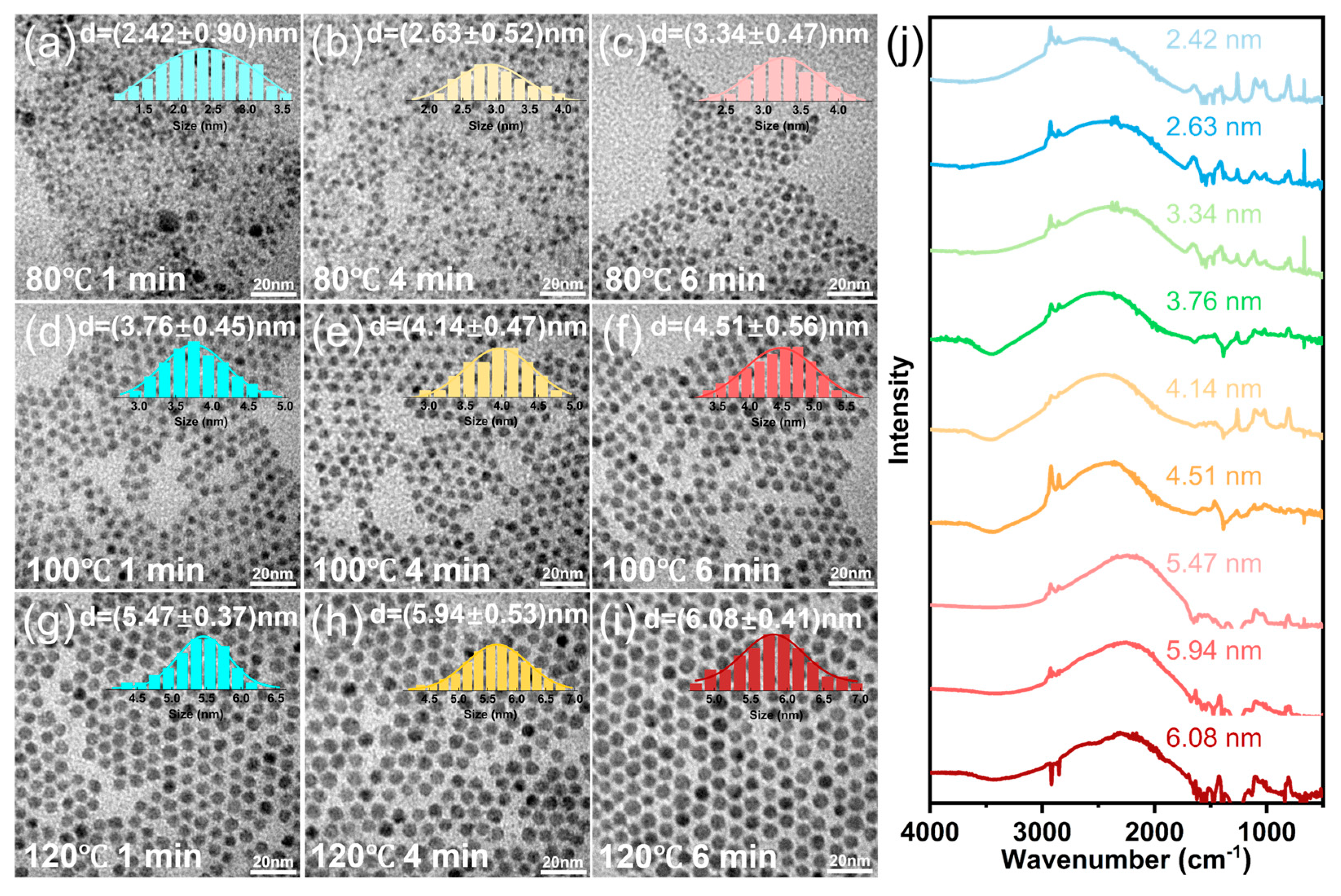
Initially, the reaction temperature and growth time were meticulously regulated to ascertain their impact on the HgSe QDs. The reaction temperatures were set at 80 °C, 100 °C, and 120 °C, while the reaction times were set at 1 min, 4 min, and 6 min, respectively. Subsequent to the purification process, the reaction products were characterized by employing TEM and FTIR, as illustrated in Figure 2. As demonstrated by the TEM characterization and the concomitant particle size distribution images in Figure 2, at a constant reaction temperature, the particle size of the products exhibited a gradual increase with an increase in reaction time. Additionally, at equivalent reaction times, the mean particle size increased with rising temperatures. Through the combined regulation of reaction temperature and growth time, we obtained HgSe QDs with a continuous size distribution ranging from 2.42 nm to 6.08 nm and excellent monodispersity. Furthermore, the intraband absorption of HgSe QDs has been observed to exhibit a progressive red shift with increasing temperature and reaction time, extending into the mid-infrared spectral range (Figure 2j).
It is noteworthy that under reaction conditions at 120 °C, the sizes of HgSe QDs synthesized for 4 min and 6 min exhibited minimal variation (5.94 nm, 6.08 nm). This finding suggests that the enhancing effect of reaction time on particle size gradually diminishes. Prolonging the reaction time beyond this point may not only fail to further increase the product size but could also compromise its dispersion, as demonstrated in Figure S2 (120 °C, 12 min). When attempting to increase particle size by further raising the temperature, it was unfortunately observed that excessively high temperatures significantly darken the color of the selenourea-oleylamine precursor. This finding suggests the possibility of structural degradation, which renders the substance unsuitable for use as a precursor. Consequently, a reaction time of six minutes at 120 °C is deemed to be the optimal balance between temperature and reaction duration for the effective control of the synthesis reaction.

3.2. The Regulation of the Hg:Se Stoichiometric Ratio

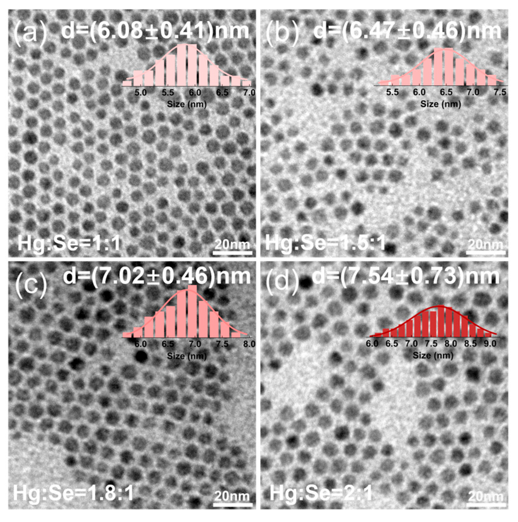
In order to achieve a continued increase in the particle size of HgSe QDs, further investigations centered on the effect of the Hg:Se stoichiometric ratio on the product under conditions of 120 °C for 6 min. In the above experiments, the Hg:Se ratio was established at 1:1. Given the established relationship between excess Se and reduced particle size [27], the present study systematically increased the proportion of Hg to ascertain the effect on the product’s particle size. As illustrated in Figure 3a–d, the alterations in the dimensions of the particles are evident, as the Hg:Se ratios are varied from 1 to 2. It has been observed that as the Hg stoichiometric ratio increases, the particle size of HgSe QDs gradually increases from 6.08 nm to 7.54 nm while maintaining excellent monodispersity. However, the subsequent increase in the Hg proportion yielded negligible outcomes.

3.3. Phase Transfer and Film Characterization

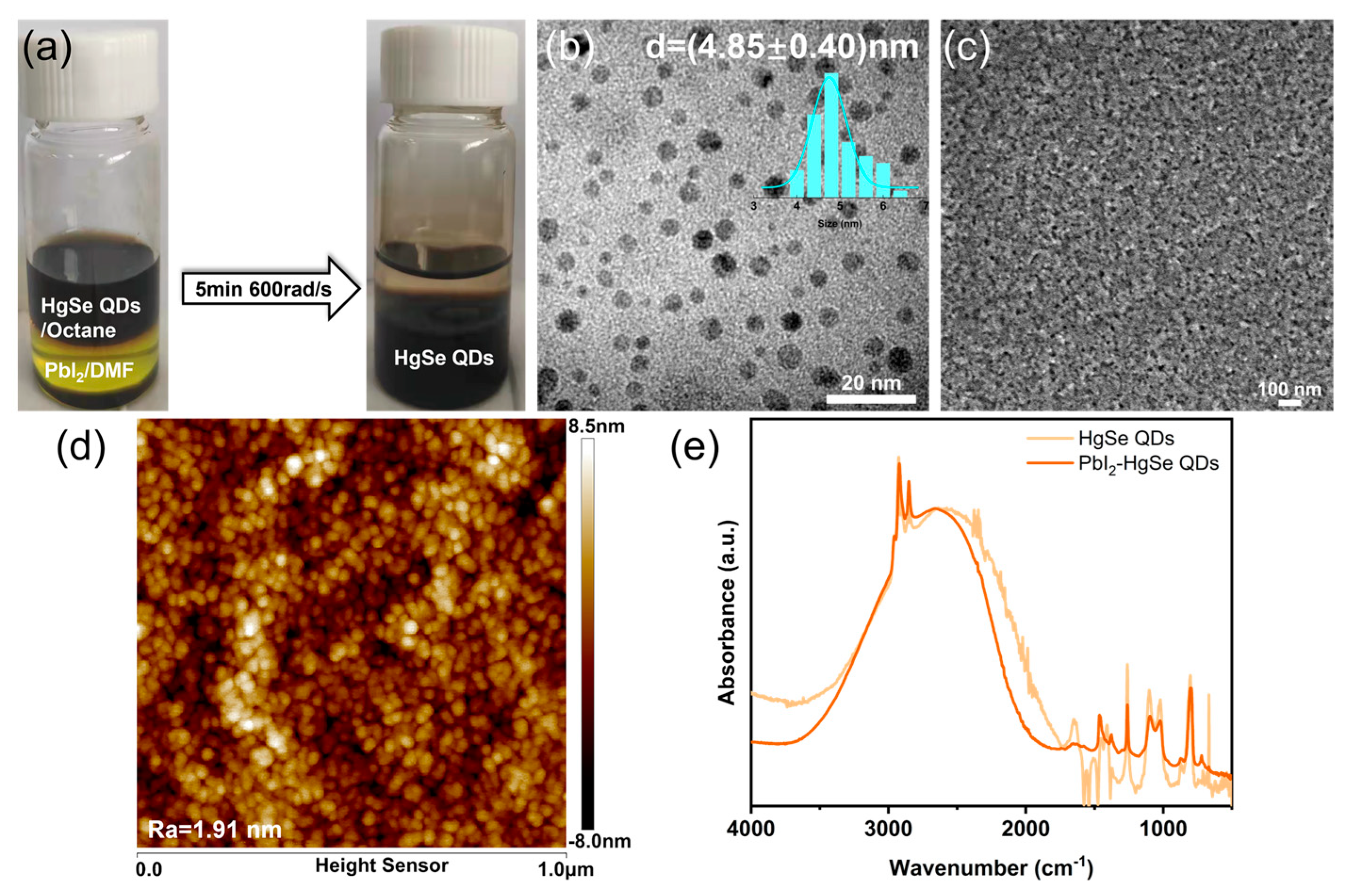
It is well established that quantum dots undergo a ligand exchange process during thin-film device fabrication in order to reduce inter-dot distances, thereby enhancing their charge transport capabilities and improving device performance [28,29,30]. The present study has therefore investigated the ligand exchange and film-forming processes of HgSe QDs. PbI2 is a widely used inorganic ligand material, with extensive application in various quantum dot devices [28,29,30]. Consequently, the PbI2-DMF solution was selected for ligand exchange with HgSe QDs. As illustrated in Figure 4a, the HgSe QDs, which were initially present in the upper layer of the n-octane solvent, underwent a significant migration to the lower layer of the DMF solvent after the ligand exchange process. The effectiveness of the ligand exchange was further confirmed by comparing the signal peaks of Pb and I elements before and after the process. As demonstrated in the Figure S3, no signal peaks corresponding to Pb or I elements were detected prior to ligand exchange. However, the exchanged sample exhibited distinct Pb 4f and I 3d signals. Subsequent TEM characterization confirmed that the HgSe QDs exhibited good dispersion after ligand exchange (Figure 4b). Thereafter, the HgSe QDs were prepared into films via a spin-coating process. The results of the SEM (Figure 4c) and AFM (Figure 4d) analyses indicate that the HgSe QD films exhibit excellent uniformity and flatness. Intraband absorption tests demonstrate that ligand exchange exerts negligible influence on the absorption positions of HgSe QDs. Consequently, it can be deduced that this HgSe QDs exhibit considerable potential for utilization in the domain of infrared detectors.

4. Conclusions

In summary, in this study, we employed selenourea-oleylamine as a precursor to achieve the preparation of high-quality HgSe QDs. Subsequently, by regulating the reaction temperature, growth period and Hg:Se stoichiometric ratio, we realized continuous and controllable scaling of the HgSe QDs size from 2.42 nm to 7.54 nm while maintaining excellent uniformity. Further investigation revealed that through ligand exchange, these HgSe QDs can form smooth and uniform thin films, rendering them promising candidates for further application in infrared detection devices.

Supplementary Materials

The following supporting information can be downloaded at: https://www.mdpi.com/article/10.3390/cryst15100872/s1, Figure S1: the effect of different Selenium Sources on the morphology of HgSe QDs, Figure S2: morphological changes in HgSe QDs after 12 min of growth at 120 °C.

Author Contributions

Conceptualization, S.Z. and L.W. (Lei Wang); methodology, L.-S.L.; validation, S.Z., and L.W. (Lingshi Wang); formal analysis, L.W. (Lei Wang) and J.S.; investigation, S.Z. and J.S.; resources, S.Z.; data curation, L.W. (Lei Wang); writing—original draft preparation, S.Z., and L.W. (Lei Wang); writing—review and editing, K.Z.; funding acquisition, K.Z. All authors have read and agreed to the published version of the manuscript.

Funding

This research was funded by Natural Science Foundation of Henan Province, 232300420154, the National Natural Science Foundation of China, 12204554, Postgraduate Education Reform and Quality Improvement Project of Henan Province, YJS2026YBGZZ19.

Data Availability Statement

Data is contained within the article or Supplementary Material.

Acknowledgments

This project is founded by the Young Backbone Teachers Training Program of Zhongyuan University of Technology, 2023XQG11, Key Project of the Natural Science Foundation of Zhongyuan University of Technology, K2026ZD011, Young Master’s Supervisor Development Program of Zhongyuan University of Technology, SD202404.

Conflicts of Interest

The authors declare no conflicts of interest.

Abbreviations

The following abbreviations are used in this manuscript:
QDsQuantum dots

References

  1. Lhuillier, E.; Guyot-Sionnest, P. Recent Progresses in Mid Infrared Nanocrystal Optoelectronics. IEEE J. Sel. Top. Quantum Electron. 2017, 23, 1–8. [Google Scholar] [CrossRef]
  2. Vatansever, F.; Hamblin, M.R. Far infrared radiation (FIR): Its biological effects and medical applications. Photonics Lasers Med. 2012, 1, 255–266. [Google Scholar] [CrossRef]
  3. Rogalski, A. Recent progress in infrared detector technologies. Infrared Phys. Techn. 2011, 54, 136–154. [Google Scholar] [CrossRef]
  4. Keuleyan, S.; Lhuillier, E.; Brajuskovic, V.; Guyot-Sionnest, P. Mid-infrared HgTe colloidal quantum dot photodetectors. Nat. Photonics 2011, 5, 489–493. [Google Scholar] [CrossRef]
  5. Guyot-Sionnest, P.; Roberts, J.A. Background limited mid-infrared photodetection with photovoltaic HgTe colloidal quantum dots. Appl. Phys. Lett. 2015, 107, 395–423. [Google Scholar] [CrossRef]
  6. Deng, Z.; Guyot-Sionnest, P. Colloidal Quantum Dots Intraband Photodetectors. ACS Nano 2014, 8, 11707–11714. [Google Scholar] [CrossRef] [PubMed]
  7. Rogalski, A. HgCdTe infrared detector material: History, status and outlook. Rep. Prog. Phys. 2005, 68, 2267–2336. [Google Scholar] [CrossRef]
  8. Deng, Z.; Guyot-Sionnest, P. Intraband Luminescence from HgSe/CdS Core/Shell Quantum Dots. ACS Nano 2016, 10, 2121–2127. [Google Scholar] [CrossRef]
  9. Zhao, X.; Mu, G.; Tang, X.; Chen, M. Mid-IR Intraband Photodetectors with Colloidal Quantum Dots. Coatings 2022, 12, 467. [Google Scholar] [CrossRef]
  10. Wang, S.; Wang, Y.; Guo, T.; Cao, S. A Long Wave-Infrared Miniatured Quantum Dot Spectrometer. Anal. Chem. 2024, 96, 14090–14098. [Google Scholar] [CrossRef]
  11. Yu, C.; Shan, Y.; Zhu, J.; Sun, D.; Zheng, X.; Zhang, N.; Hou, J.; Fang, Y.; Dai, N.; Liu, Y. Heterojunctions of Mercury Selenide Quantum Dots and Halide Perovskites with High Lattice Matching and Their Photodetection Properties. Materials 2024, 17, 1864. [Google Scholar] [CrossRef]
  12. Sevim Ünlütürk, S.; Taşcıoğlu, D.; Özçelik, S. Colloidal quantum dots as solution-based nanomaterials for infrared technologies. Nanotechnology 2024, 36, 082001. [Google Scholar] [CrossRef]
  13. Livache, C.; Martinez, B.; Goubet, N.; Gréboval, C.; Qu, J.; Chu, A.; Royer, S.; Ithurria, S.; Silly, M.G.; Dubertret, B.; et al. A colloidal quantum dot infrared photodetector and its use for intraband detection. Nat. Commun. 2019, 10, 2125. [Google Scholar] [CrossRef] [PubMed]
  14. Ramiro, I.; Özdemir, O.; Christodoulou, S.; Gupta, S.; Dalmases, M.; Torre, I.; Konstantatos, G. Mid- and Long-Wave Infrared Optoelectronics via Intraband Transitions in PbS Colloidal Quantum Dots. Nano Lett. 2020, 20, 1003–1008. [Google Scholar] [CrossRef]
  15. Li, S.S.; Xia, J.B. Intraband optical absorption in semiconductor coupled quantum dots. Phys. Rev. B 1997, 55, 15434–15437. [Google Scholar] [CrossRef]
  16. Sato, S.A.; Lucchini, M.; Volkov, M.; Schlaepfer, F.; Gallmann, L.; Keller, U.; Rubio, A. Role of intraband transitions in photocarrier generation. Phys. Rev. B 2018, 98, 035202. [Google Scholar] [CrossRef]
  17. Qu, J.; Goubet, N.; Livache, C.; Martinez, B.; Amelot, D.; Gréboval, C.; Chu, A.; Ramade, J.; Cruguel, H.; Ithurria, S.; et al. Intraband Mid-Infrared Transitions in Ag2Se Nanocrystals: Potential and Limitations for Hg-Free Low-Cost Photodetection. J. Phys. Chem. C 2018, 122, 18161–18167. [Google Scholar] [CrossRef]
  18. Park, M.; Choi, D.; Choi, Y.; Shin, H.-B.; Jeong, K.S. Mid-Infrared Intraband Transition of Metal Excess Colloidal Ag2Se Nanocrystals. ACS Photonics 2018, 5, 1907–1911. [Google Scholar] [CrossRef]
  19. Palosz, W.; Trivedi, S.; DeCuir, E.; Wijewarnasuriya, P.S.; Thon, S.M.; Cheng, Y.; Lu, C.; Jensen, J.L. Synthesis and Characterization of Large PbSe Colloidal Quantum Dots. Part. Part. Syst. Char. 2021, 38, 2000285. [Google Scholar] [CrossRef]
  20. Ponomarenko, V.P.; Popov, V.S.; Shuklov, I.A.; Ivanov, V.V.; Razumov, V.F. Photosensors based on colloidal quantum dots. Russ. Chem. Rev+ 2024, 93, RCR5113. [Google Scholar] [CrossRef]
  21. Pierini, S.; Capitani, F.; Scimeca, M.; Kozlov, S.; Pierucci, D.; Alchaar, R.; Abadie, C.; Khalili, A.; Cavallo, M.; Dang, T.H.; et al. Vanishing Confinement Regime in Terahertz HgTe Nanocrystals Studied under Extreme Conditions of Temperature and Pressure. J. Phys. Chem. Lett. 2022, 13, 6919–6926. [Google Scholar] [CrossRef]
  22. Lee, W.; Smith, A.M. Interdiffusion-enhanced cation exchange for HgSe and HgCdSe nanocrystals with infrared bandgaps. Nat. Synth. 2024, 3, 1243–1254. [Google Scholar] [CrossRef]
  23. Chen, M.; Shen, G.; Guyot-Sionnest, P. State-Resolved Mobility of 1 cm2/(Vs) with HgSe Quantum Dot Films. J. Phys. Chem. Lett. 2020, 11, 2303–2307. [Google Scholar] [CrossRef]
  24. Sokolova, D.; Dyomkin, D.V.; Katsaba, A.V.; Bocharova, S.I.; Razumov, V.F. Effect of different ligand types on sensitivity of infrared photodetectors based on colloidal HgSe quantum dots. Infrared Phys. Techn. 2022, 123, 104188. [Google Scholar] [CrossRef]
  25. Chen, M.; Shen, G.; Guyot-Sionnest, P. Size Distribution Effects on Mobility and Intraband Gap of HgSe Quantum Dots. J. Phys. Chem. C 2020, 124, 16216–16221. [Google Scholar] [CrossRef]
  26. Liu, L.; Long, Z.; Shi, K.; Zhong, H. A General Crystallization Picture of Quantum Dots: The Underlying Physical Chemistry. CCS Chem. 2025, 7, 926–949. [Google Scholar] [CrossRef]
  27. Wang, S.; Guo, T.; Cao, S. The Influence of Synthetic Parameters on HgSe QDs. ACS Omega 2023, 8, 44804–44811. [Google Scholar] [CrossRef] [PubMed]
  28. Liu, M.; Voznyy, O.; Sabatini, R.; Garcia de Arquer, F.P.; Munir, R.; Balawi, A.H.; Lan, X.; Fan, F.; Walters, G.; Kirmani, A.R.; et al. Hybrid organic-inorganic inks flatten the energy landscape in colloidal quantum dot solids. Nat. Mater. 2017, 16, 258–263. [Google Scholar] [CrossRef]
  29. Wang, L.; Wang, Y.; Jia, Y.; Liu, X.; Liu, T.; Fu, T.; Li, J.; Weng, B.; Zhang, X.; Liu, Y. Manipulation of Phase-Transfer Ligand-Exchange Dynamics of PbS Quantum Dots for Efficient Infrared Photovoltaics. J. Phys. Chem. C 2019, 123, 30137–30144. [Google Scholar] [CrossRef]
  30. Lin, Q.; Yun, H.J.; Liu, W.; Song, H.-J.; Makarov, N.S.; Isaienko, O.; Nakotte, T.; Chen, G.; Luo, H.; Klimov, V.I.; et al. Phase-Transfer Ligand Exchange of Lead Chalcogenide Quantum Dots for Direct Deposition of Thick, Highly Conductive Films. J. Am. Chem. Soc. 2017, 139, 6644–6653. [Google Scholar] [CrossRef] [PubMed]
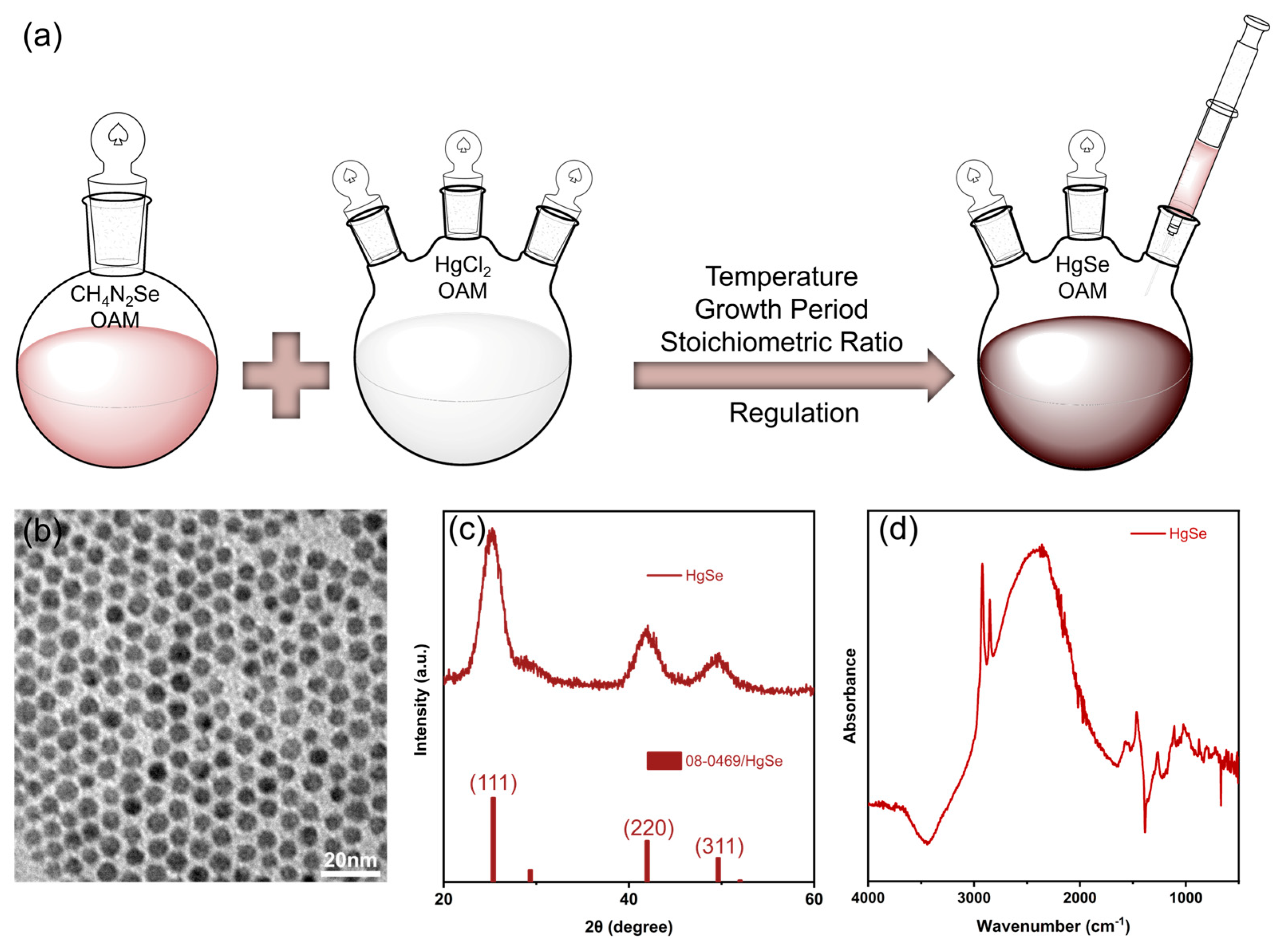
Figure 1. (a) Schematic diagram of preparing HgSe QDs via the hot-injection method, (b), (c) and (d) represent morphological, structural, and intraband absorption characterization, respectively.
Crystals 15 00872 g001
Figure 2. (ai) the morphological characterization and size distribution of HgSe QDs obtained under different conditions, (j) the intraband absorption variation in HgSe QDs with different particle sizes.
Crystals 15 00872 g002
Figure 3. The effect of Hg:Se stoichiometric ratio on HgSe QDs size, where (ad) represent 1, 1.5, 1.8, and 2, respectively.
Crystals 15 00872 g003
Figure 4. (a) Comparison images before and after ligand exchange, TEM (b), SEM (c), AFM (d) and intraband absorption (e) characteristics of HgSe QDs after ligand exchange.
Crystals 15 00872 g004
Disclaimer/Publisher’s Note: The statements, opinions and data contained in all publications are solely those of the individual author(s) and contributor(s) and not of MDPI and/or the editor(s). MDPI and/or the editor(s) disclaim responsibility for any injury to people or property resulting from any ideas, methods, instructions or products referred to in the content.