Article

Mono- and Polynuclear Hg(II) Complexes with Mixed Ligands: Nicotinamide and Oxalate, Nitrate, or Sulphate

by
Laurențiu Pricop
1,*,
Anamaria Hanganu
1,
Mihaela Ganciarov
2 and
Augustin M. Mădălan
1
1
Department of Inorganic Chemistry, Organic Chemistry, Biochemistry and Catalysis, Faculty of Chemistry, University of Bucharest, 90–92 Panduri St., 050663 Bucharest, Romania
2
Analysis Team, National Institute for Research & Development in Chemistry and Petrochemistry—ICECHIM, Splaiul Independentei No. 202, Sector 6, 060021 Bucharest, Romania
*
Author to whom correspondence should be addressed.
Crystals 2025, 15(10), 835; https://doi.org/10.3390/cryst15100835
Submission received: 8 August 2025 / Revised: 23 September 2025 / Accepted: 24 September 2025 / Published: 25 September 2025
(This article belongs to the Section Hybrid and Composite Crystalline Materials)

Abstract

Three new complexes of Hg(II), with the general formulas [Hg2(ox)2(NA)4]n·3nH2O (1), [Hg(NO3)2(NA)2(H2O)2]·2NA (2), and [Hg2(SO4)2(H2O)2(NA)4]·6H2O (3), where ox = oxalate and NA = nicotinamide, were synthesized and characterized by single crystal X-ray diffraction, elemental analysis, FT-IR, and fluorescence spectra. For complex (2), 13C and 1H NMR spectra were recorded. Thermogravimetric analysis was also performed for complexes (1) and (2). Single crystal X-ray diffraction shows that in the polymeric complex (1) and the binuclear complex (3), the Hg(II) ions are hexacoordinated, whereas in the mononuclear complex (2), Hg(II) is octacoordinated. In complex (1), each oxalate group acts in a µ4 coordination manner, the basal plan being made up by four oxygen atoms belonging to the two oxalate ligands, while the nicotinamide molecules occupy the axial positions. In complex (2), the nitrate groups coordinate in a bidentate chelating mode, whereas in complex (3), each sulphate ligand acts in a bidentate chelating–bis monodentate bridging manner.

Graphical Abstract

1. Introduction

Nicotinamide, a water-soluble form of vitamin B3, acts as a ligand mostly through the pyridine nitrogen atom (as a terminal ligand). So far, only a few examples of complexes containing nicotinamide as a bridging ligand have been reported [1,2,3,4,5]. In this context, a facile synthesis pathway for polynuclear complexes containing nicotinamide as a ligand involves the use of anionic bridging co-ligands.
Coordination compounds of mercury(II) are well-known, as the metal ion can adopt different coordination numbers, generating both mononuclear and various types of polynuclear complexes. Some of them show promising antimicrobial and cytotoxic activities [6,7,8,9,10,11]. The goal of this work was to study the ability of oxalate, nitrate, and sulphate anions to generate mercury polynuclear complexes in the presence of nicotinamide as a co-ligand.
The oxalate group is a recognized versatile ligand, acting in manifold coordination modes [12,13,14,15,16]. These complexes display a wide range of nuclearities, from mononuclear compounds to coordination polymers. Among them, anhydrous mercury oxalate is a polynuclear compound that crystallizes in the monoclinic system. Its structure consists of mercury atoms, each of them being surrounded by eight oxygen atoms, located at the corners of a cube. Each cube shares four edges with the neighboring coordination polyhedra [17,18]. Many complexes of divalent mercury with carboxylate anions and various molecules with heterocyclic nitrogen atoms acting as mixed ligands have also been synthesized and characterized. In this regard, notable examples include complexes with pyridine-2,3-dicarboxylic acid and 1,10-phenantroline [19], 3,3′-bipyridine and Hg(CH3COO)2 [20], 1,2-bis(4-pyridyl)ethane, 1,4-bis(4-pyridyl)-2,3-diaza-1,3-butadiene, 1,4-bis(3-pyridyl)-2,3-diaza-1,3-butadiene and mercury acetate [21], and 2,2′-bipyridine and Hg(CH3COO)2 [22]. Additionally, Hg(II) coordination compounds with picolinic acid [23,24] and pyridine-2,6-dicarboxylic acid [25] and ligands containing O- and N-heterocyclic donor atoms are noteworthy. In turn, mercury is a versatile element that lacks geometry preferences. Most of its coordination polymers are one-dimensional, with coordination numbers ranging from 3 (T-shaped geometry) to 7 and 8 (distorted pentagonal or hexagonal bipyramidal coordination spheres) [26,27]. However, in many cases, the mercury coordination number is 4.
The nitrate group can coordinate in 28 different ways, the most common being the terminal bidentate one (47%) [28]. An interesting example is a polynuclear complex of Hg(II) with 9-methyl-1-deazapurine, where two nitrate groups act in a tetradentate manner, forming a honeycomb-like chain structure. In this compound, Hg(II) ion is octacoordinated, adopting a distorted hexagonal bipyramidal geometry [29]. Additionally, previous studies have reported Zn(II) and Cd(II) complexes with nicotinamide and nitrate [30,31]. In the Zn(II) complex, nitrate acts as a counterion, while in the Cd(II) compound, the nitrate groups are coordinated in a monodentate manner.
The sulphate ligand (SO42−) can bind between 2 and 10 metal ions through 16 different coordination modes [32]. Two complexes of Ni(II) [33] and Fe(II) [34] with nicotinamide and sulphate have been reported so far. The Ni(II) complex was not characterized by means of single-crystal X-ray diffraction, while the Fe(II) complex is, in fact, a co-crystallization of ferrous sulphate with [Fe(NA)2(H2O)4]. A polymeric complex of Hg(II) with nicotinamide and thiocyanate fragments acting as bridges has also been synthesized and characterized [35]. Recently, an inorganic-organic hybrid mercury(II) complex, with a nicotinamide derivative, nicotinamide N-acetic acid, was prepared and structurally characterized by single-crystal X-ray diffraction. Its formula is (nia-N-CH2COOH)2[Hg2Br6]·2(nia-N-CH2COO), where nia = nicotinamide [36]. Apart from these compounds, no other complexes of Hg(II) with nicotinamide have been studied. However, two binuclear mercury(II) complexes with a ligand related to nicotinamide have been described, the ligand being a derivative of isonicotinamide (N-(naphthalene-1-yl) isonicotinamide) [37].
In continuation of our work on coordination compounds with nicotinamide as a co-ligand, we have synthesized and characterized three new complexes of mercury (II) with different nuclearities: a mononuclear, a binuclear, and a 1D coordination polynuclear compound. The versatility of Hg(II) and its ability to form complexes with interesting structures and unusual coordination numbers prompted us to synthesize and characterize them.

2. Materials and Methods

2.1. Materials

The reagents were purchased from local suppliers. All solvents and reagents were reagent grade and used as received without further purification.

2.2. Synthesis of Complexes

2.2.1. [Hg2(ox)2(NA)4]n·3nH2O (1): A solution with a volume of 10 mL was obtained by dissolving 2 mmol (0.244 g) of nicotinamide in distilled water. Then, we added it to a water solution (20 mL) of 1 mmol (0.525 g) mercurous nitrate. We stirred the mixture for 10 min at room temperature, and after that, we mixed it with a 20 mL volume of an ammonium oxalate (1 mmol, 0.124 g) solution. The solution obtained was refluxed for 1 h and left to slowly evaporate at room temperature. After two weeks, colorless crystals were separated. Yield: 2.85% (31.90 mg) (based on Hg2(NO3)2)

The reaction mechanism by which Hg(I) is converted into Hg(II) is disproportionation. After refluxing the mixture of reactants, we filtered off a grey precipitate. We washed it with a 20% HCl solution in order to dissolve the mercury oxalate, and then with distilled water. The precipitate took on a silver-gray appearance. It was identified as metallic mercury by amalgamation with aluminum and the subsequent reaction of the amalgam with water. We noticed the formation of gas bubbles (H2) and the deposition of a white precipitate (Al(OH)3) [38].
Hg2C28H30N8O15
Found (%): C, 30.31; H, 2.60; N, 10.17. Calculated (%): C, 30.00; H, 2.67; N, 10.00. IR/cm−1): 3500—3100(υ O-H), 3373(υas N-H), 3186(υs N-H), 1690(υ C=O), 1653(υas C=O), 1596(υ C=N + δ N-H + δ O-H), 1472(ν CC), 1424(υs CO + υ CC), 1405(υ C-C), 1309(υs CO + δ O-C=O), 1283(υs CO + δ O-C=O), 1201(υ C-NH2), 1150(υ C-N), 1128(δ C-H), 1045(δ C-H + υ C-C), 1029(γ C-H), 772(δ O-C=O + υ MO), 695(β C=O + δ N-H), 644(δ CCC), 526(δ C-NH2 + γ C=O).

2.2.2. [Hg(NO3)2(NA)2(H2O)2]·2NA (2): We added 3 mL solution of diluted nitric acid (5%) to a water solution (10 mL) of mercurous acetate (1 mmol, 0.519 g), and we gently stirred the mixture for two minutes. After this, we added a volume of 15 mL water–ethanol (1:2) nicotinamide solution (2 mmol, 0.244 g) and we refluxed the final mixture for 1h. After cooling and filtering, we left the filtrate to slowly evaporate. After several days, the appearance of colorless crystals was observed. Yield: 28.03% (238 mg) (based on Hg2(CH3COO)2)

HgC24H28N10O12
Found (%): C, 33.50; H, 3.27; N, 16.40. Calculated (%): C, 33.91; H, 3.30; N, 16.48. IR(cm−1): 3400—2800(υ O-H), 3377(υas N-H), 3203(υs N-H), 3076(υ C-H), 1673(υ C=O), 1638(υ C=N), 1595(δ N-H + δ O-H), 1549(υas N=O) 1467(δ O-H), 1387(υ C-C), 1327(υ C-N), 1295(υs NO2), 1193(υ C-NH2), 1143(υ C-N), 1128(δ C-H), 1045(δ C-H + υ C-C), 1021(γ C-H), 924(δ NO2), 843(γ C-H), 765(ρw NH2), 672(β C=O + δ N-H), 612(δ CCC), 599(ρw H2O), 490(δ C-NH2 + γ C=O).

2.2.3. [Hg2(SO4)2(H2O)2(NA)4]·6H2O (3): A water solution (10 mL) of mercurous acetate (1 mmol, 0.519 g) was mixed with 3 mL of diluted sulfuric acid solution (3%). Then, 15 mL of nicotinamide (2 mmol, 0.244 g) was added to the water–ethanol (1:2) solution, and the mixture obtained was refluxed for 1h. After cooling and filtering, we left the solution to slowly evaporate. In a few days, colorless crystals were separated. The single-crystal X-ray diffraction analysis revealed that these crystals correspond to the compound C6H7N2O+·HSO4, indicating that they are formed from protonated nicotinamide and an acid sulfate anion. Because the synthesis of the complex failed and the mercurous acetate had no role in our attempt, we tried to use the obtained compound to achieve our goal. Accordingly, a 10 mL solution (1 mmol, 0.22 g) of this compound was mixed with a 5 mL solution of mercurous acetate (0.5 mmol, 0.26 g), and the final solution was refluxed for 1h. After several days, colorless crystals appeared, and they proved to be the desired coordination compound, having the formula [Hg2(SO4)2(H2O)2(NA)4]·6H2O. Yield: 14.25% (43 mg) (based on Hg2(CH3COO)2)

HgC12H20N4O10S
Found (%): C, 23.64; H, 2.63; N, 9.30. Calculated (%): C, 23.49; H, 3.26; N, 9.13. IR(cm−1): 3529(υ O-H), 3241(υs N-H), 3072(υ C-H), 1713(υ C=O + δ O-H), 1596(δ N-H + ν C=N), 1386(υ C-C), 1325(υ C-N), 1302(υ C-N), 1124(υ3 SO4), 1039(υ3 SO4), 812(γ C-H), 747(ρw NH2), 693(β C=O + δ N-H), 618(υ4 SO4).

2.3. Single Crystal X-Ray Diffraction and Refinement

X-ray diffraction measurements were performed on a Rigaku XtaLAB Synergy-S diffractometer (Rigaku Polska, Wroclaw, Poland) operating with a Mo-Kα (λ = 0.71073 Å) micro-focus sealed X-ray tube. The structure was solved by direct methods and refined by full-matrix least squares techniques based on F2. The non-H atoms were refined with anisotropic displacement parameters. Calculations were performed using the SHELX-2018 crystallographic software package. A summary of the crystallographic data and the structure refinement is given in Table 1. CCDC reference number: 2447174–2447176.

2.4. Fourier-Transform Infrared Spectroscopy (FT-IR)

FT-IR spectra for the complexes were recorded on a Bruker Tensor 37 spectrophotometer in the 4000–400 cm−1 region. The FT-IR spectrum of the residue of complex (1) was recorded with a GX Spectrum (Perkin Elmer, Waltham, MA, USA) spectrometer, in the 4000–400 cm−1 region. The mercury(II) oxide spectrum was taken from its database.

2.5. NMR Spectroscopy

1H NMR spectra were measured in DMSO-d6 using a Bruker Advance UltraShield Puls III spectrometer (Karlsruhe, Germany), operating at 500 MHz. The chemical shifts (δ) are reported as ppm values, and the residual solvent peaks were used as an internal reference.

2.6. Photoluminescence

The fluorescence spectra were collected on powder using a JASCO FP-6500 spectrofluorometer.

2.7. Thermogravimetric Analysis (TG)

The heating curves (TG) were recorded using a Metler Toledo TGA/SDATA 851 instrument, Columbus, OH, USA. The measurements were carried out in synthetic air at a heating rate of 10 K/min.

2.8. Elemental Analysis

A FlashSmart Thermo Fisher Scientific elemental analyzer, Bremen, Germany, was used for chemical analyses (C, H, and N). Elemental analysis was performed in duplicate for each complex, and the reported values represent averages.

3. Results

3.1. Structural Characterization

A summary of the crystallographic data and the structure refinement is given in Table 1.

3.2. FT-IR Spectra

All spectra exhibit a broad band, which lies between 3500 and 3000 cm−1, attributed to O–H stretching vibrations of lattice or coordinated water molecules. The bands observed at around 3370 cm−1 and in the range of 3186–3241 cm−1 are due to the asymmetric and symmetric N–H stretching vibrations of the amide groups in the nicotinamide molecules. The N–H in-plane bending vibration appears at 1595–1596 cm−1, together with the υ(C=N) band of the pyridine ring, in complexes (1) and (3). The band appearing in complex (1) at 1201 cm−1 and in complex (2) at 1193 cm-1 could be assigned to C–NH2 valence vibration, while that from 1140 to 1150 cm−1 is specific to υ(C–N) stretching [39]. These two bands are not found in the spectrum of complex (3), probably being overlapped by the much more intense band situated at 1124 cm−1. The presence of these peaks proves the existence of nicotinamide molecules in all complexes. In complex (1), the coordination of oxalate fragments to the central atoms is suggested by the existence of the specific bands at 1690 and 1653 cm−1, due to asymmetric υas(C=O) vibrations. Additionally, the peaks at 1424, 1309, and 1283 cm−1, assigned to υs(CO) and υs(CO) + δ(O-C=O) vibrations, respectively, support this conclusion. The positions of these bands show a chelating coordination manner of the oxalate anions, through deprotonated oxygen atoms, while the other two oxygen atoms of the group are not involved in bonding with mercury ions [40,41]. In complex (2), the characteristic bands of nitrate groups are situated at 1549 (υ1), 1295 (υ5), and 924 (υ2). The separation between the first two bands is 255 cm−1, suggesting a chelating bidentate coordination mode [40]. In complex (3), the specific vibrations of the sulphate group appear at 1124, 1039, and 618 cm−1, in accordance with a chelating bidentate coordination manner [40].

3.3. Crystal Structure Descriptions

3.3.1. Complex (1)

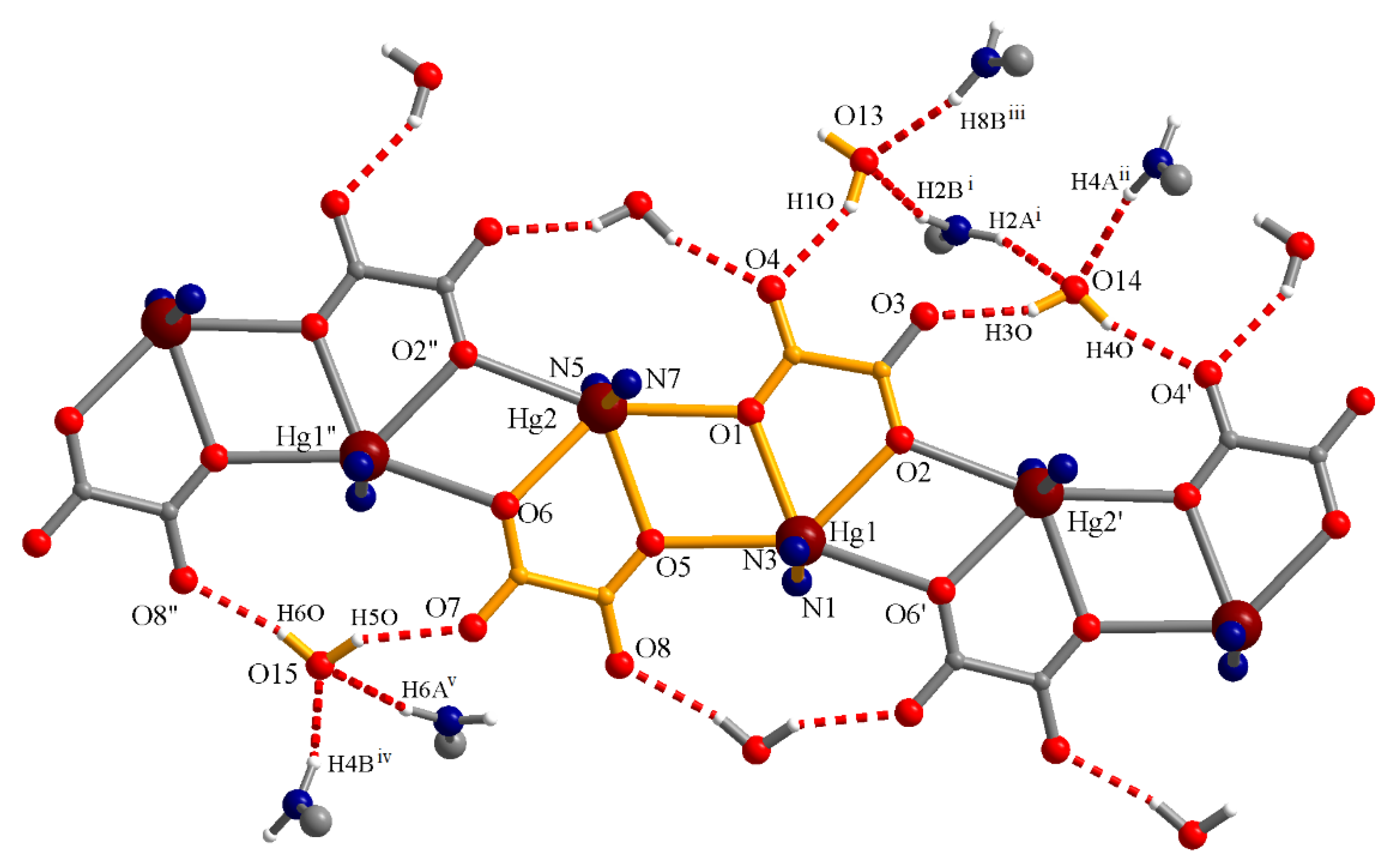
The coordination polymer [Hg2(ox)2(NA)4]n·3nH2O (1) crystallizes in the P21/c centrosymmetric monoclinic space group. The asymmetric unit consists of two metal centers, each coordinated by one oxalate anion and two nicotinamide ligands, together with three crystallization water molecules. The oxalate ions act as chelating ligands and, also, as bridging ligands. The Hg(II) ions are hexacoordinated, adopting a highly distorted triangular prism geometry, as indicated by SHAPE analysis (Supporting Information) [42]. The oxalate anions are placed in the equatorial planes, generating, together with the metal ions, a 1D coordination polymer, with a zig-zag positioning of the Hg(II) ions (Figure 1). The nicotinamide molecules coordinate via the pyridine nitrogen atoms at the axial positions of the metal ions. Selected bond distances around mercury(II) ions are presented in Table 2. According to the data in Table 3, there are two types of mercury—oxygen bonds: shorter ones (2.437–2.509 Å), involving oxygen atoms that close a five-membered cycle with the metal ions, and longer ones (2.604–2.633 Å), involving oxygen atoms that act as bridges. A similar situation occurs in HgC2O4, where two Hg–O distances are shorter (2.10–2.12 Å) and the other six are longer (2.63–2.83 Å) [17] as well as in other compounds in which oxalate acts as a bridge [43].
The geometries of the Hg(II) ions are highly irregular, with high values of the distortion parameters in SHAPE analysis, ranging from around 7 for the triangular prism to higher values for other geometries (a perfect match between the environment of the metal ions and the ideal polyhedron is indicated by a zero value). For example, the O2–Hg1–O1 angle is 65.79°, while the O2–Hg1–O5 is 135.18°, a similar situation being observed for the Hg2 atom. Also, the angles between the nitrogen atoms in the axial positions and the mercury centers deviate substantially from the 180° value: 155.55° (Hg1) and 157.51° (Hg2), respectively. At the same time, the shorter distances Hg–N in the axial plan (2.158–2.175 Å), compared with the Hg–O distances in the equatorial plane, show the compressed distortion of the octahedral geometry.
The lattice water molecules are involved in hydrogen interactions, acting as donors toward the oxalate anions of one chain and as acceptors toward the amide groups of nicotinamide belonging to neighboring chains, generating a supramolecular 3D architecture (Figure 2). Some data from these interactions are presented in Table 3.
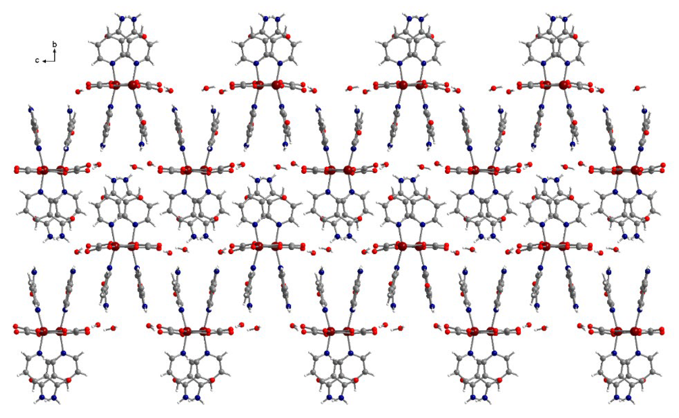
Nicotinamide molecules are also involved in π-π interactions. The nicotinamide fragments containing the nitrogen atoms N3 and N7 are involved only in intramolecular π-π interactions (3.26–3.55 Å). The nicotinamide fragments containing the nitrogen atoms N1 and N5 participate in both intra- and intermolecular π-π interactions (Figure 3). The shortest contacts for the intramolecular interactions are 2.9–3.15 Å, while for the intermolecular interactions, they are 3.35–3.45 Å.

3.3.2. Complex (2)

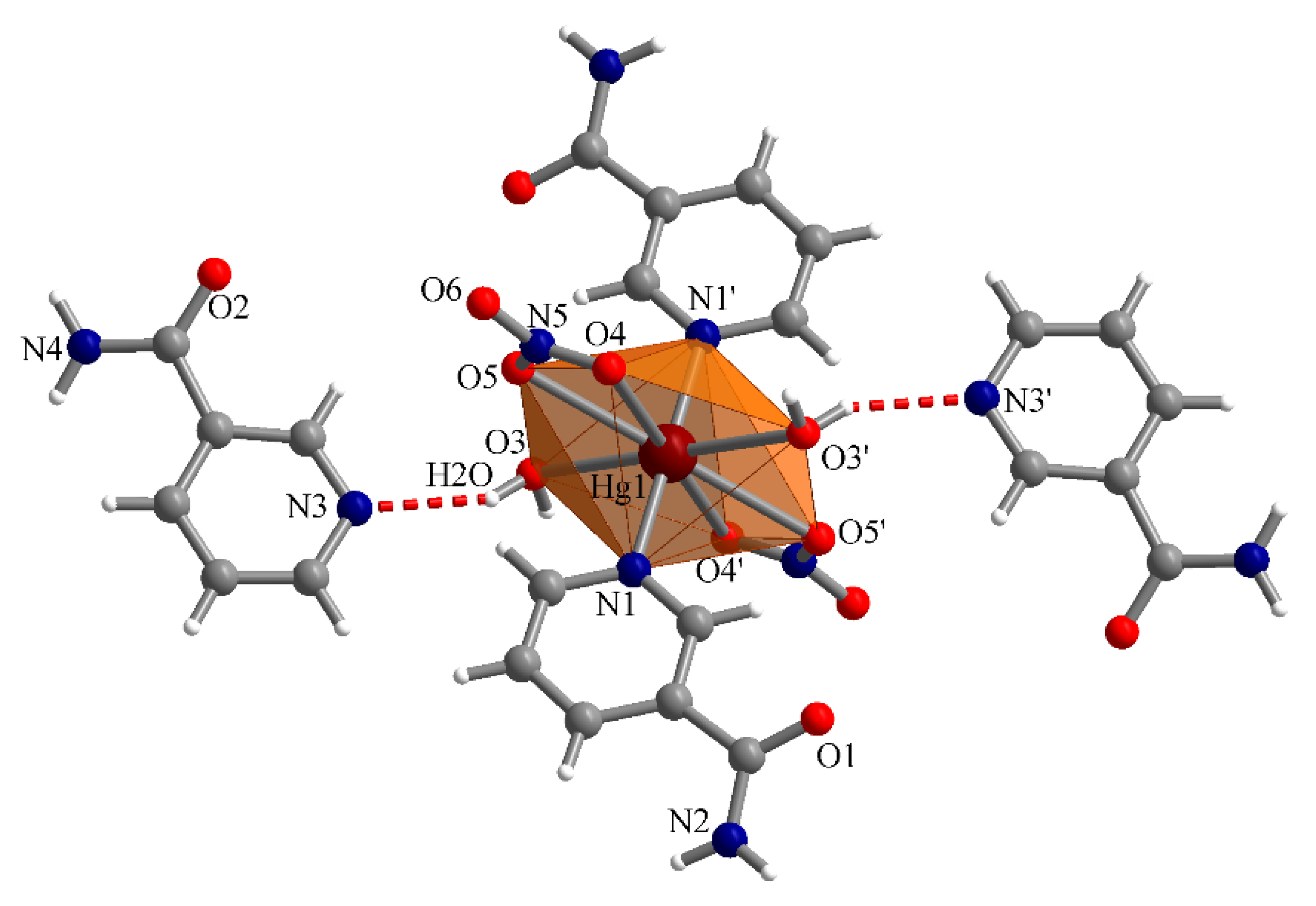
The compound [Hg(NO3)2(NA)2(H2O)2]·2NA (2) crystallizes in the centrosymmetric P21/n monoclinic space group and is a mononuclear complex, [Hg(NO3)2(NA)2(H2O)2], together with uncoordinated nicotinamide molecules (Figure 4). The metal ion adopts a distorted hexagonal bipyramidal geometry, being coordinated by two nicotinamides, two nitrate anions, and two water molecules. The basal plan consists of six oxygen atoms: two belong to the coordinated water molecules, and the other four to the bidentate chelated nitrate groups. The nicotinamide molecules occupy the axial positions and coordinate through the pyridine nitrogen atoms.
The bond lengths between Hg(II) and the pyridine nitrogen atoms are 2.127(3) Å, while those between the metal ion and the oxygen atoms of the nitrate groups are 2.739(4) Å (Hg1–O5) and 2.948(4) Å (Hg1–O4). These last values indicate a weak coordination of the nitrate oxygen atoms, resulting in an increased coordination number of Hg(II) to eight. This situation is also encountered in other mercury complexes [27]. The bond lengths between Hg(II) and the oxygen atoms of the coordinated water molecules are 2.588(3) Å. The varying bond lengths indicate a distorted geometry of the hexagonal basal plane. This conclusion is also confirmed by the angle values, which range from 43.8° (O4–Hg1–O5) to 66.6 (O3–Hg1–O5) and 70.6° (O3–Hg1–O4). Instead, the nitrogen atoms in the apical positions are aligned along an axis, the angle value N1–Hg1–N1′ being 180°. Within the coordinated nicotinamide fragments, the amido groups are twisted with respect to the plane of the pyridine rings. The dihedral angle between the mean planes of the amide fragments and the pyridine rings is 25.8°.
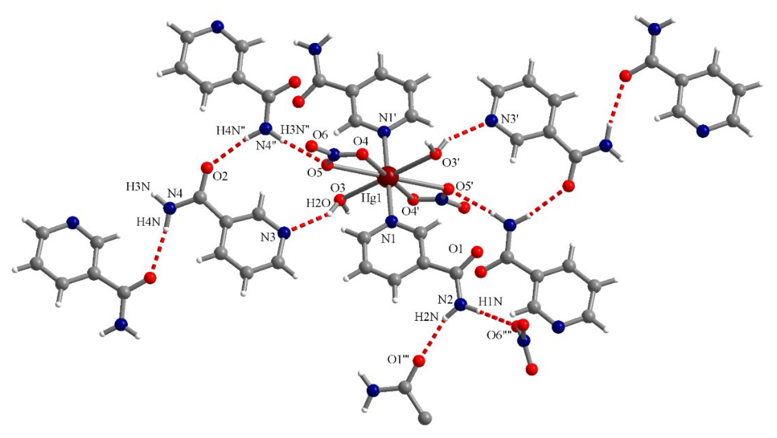
In complex (2), the nicotinamide molecules embedded in the lattice are involved in hydrogen bonds with similar neighboring molecules, as well as with nitrate groups and water molecules coordinated to the metal ions (Figure 5). The pyridine N atoms of the uncoordinated nicotinamide molecules are hydrogen-bonded to water molecules. Table 4 contains details of these hydrogen interactions.

3.3.3. Complex (3)

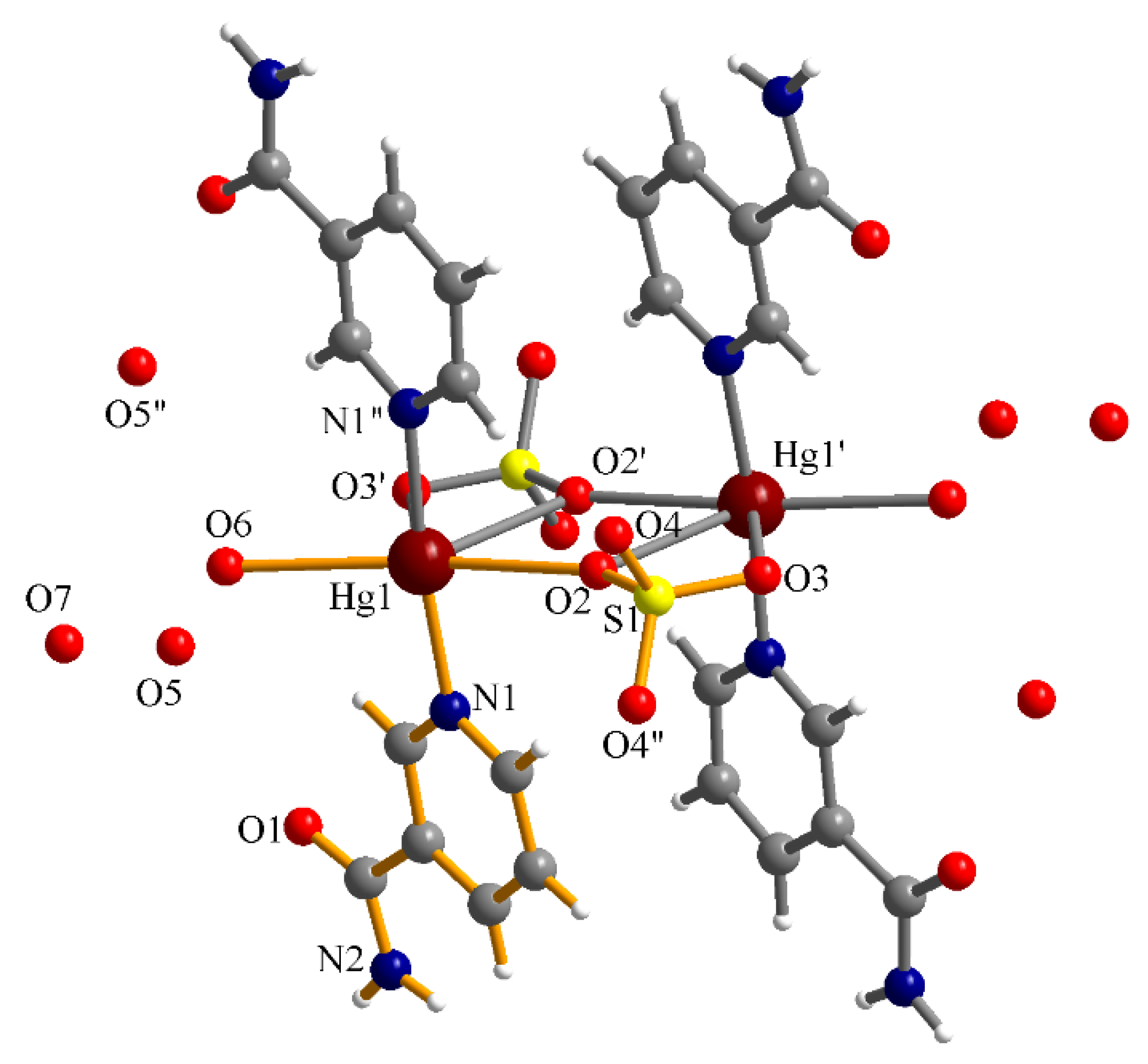
Complex (3), [Hg2(SO4)2(H2O)2(NA)4]·6H2O, crystallizes in the monoclinic system, C2/m space group. The asymmetric unit consists of one Hg(II) ion, one nicotinamide molecule, one sulphate group, one coordinated water molecule, and two lattice water molecules. The Hg(II) ions are hexacoordinated. More precisely, each ion coordinates one water molecule and one sulfate group, which acts in a bidentate chelating manner; the fourth position of the basal plane is occupied by an oxygen atom of a neighboring sulfate group. Accordingly, each sulphate group behaves in a chelating bidentate/bridging monodentate mode. In the axial positions of the metal centers, the heterocyclic nitrogen atoms of the nicotinamide ligands are coordinated (Figure 6).
Compared to ideal polyhedra, the geometry of the complex is highly distorted (the results of SHAPE analysis are presented in the Supporting Information). The bond lengths in the equatorial plane are 2.667(11) Å (Hg1–O6), 2.603(8) Å (Hg1–O2), 2.710(8) Å (Hg1–O2′), and 2.756(10) Å (Hg1–O3′), O2′ and O3′ being the oxygen atoms of the chelated sulphate group. In the axial plane, the distances Hg1–N1 are equal to 2.117(7) Å. The angles in the basal plane show significant deviation from the theoretical value of 90°. Thus, angle O6–Hg1–O2 is 168.3(4)°, the O2′–Hg1–O3′ angle, formed by the oxygen atoms of the chelated sulphate group with the central atom, is 51.2(2)°, while those between the oxygen atoms which act as bridges and Hg(II), O2–Hg–O2′, is 68.4(2)°.
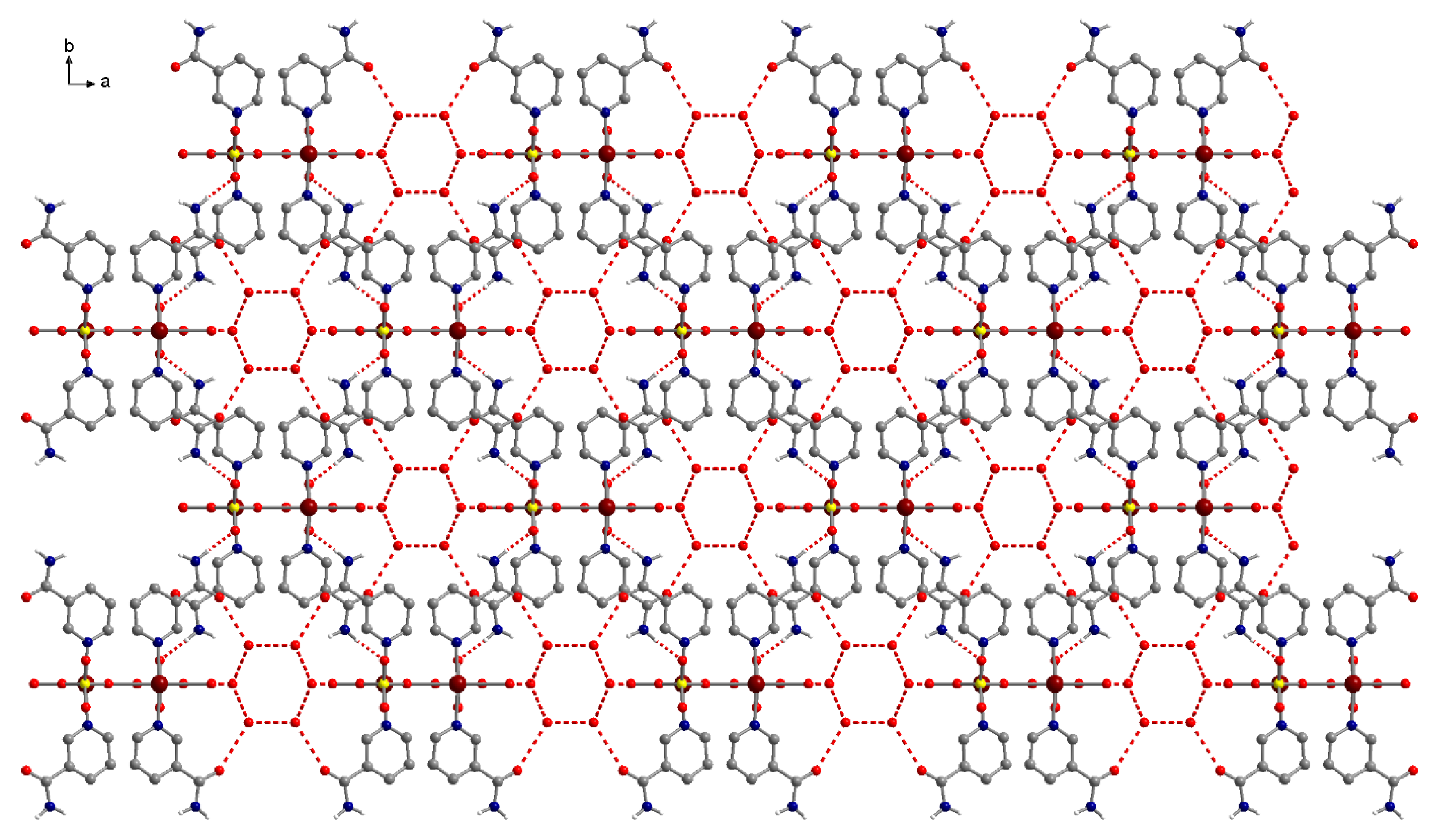
In turn, the complex (3) shows an array of contacts that ensure the stability of the crystal lattice (Figure 7). Thus, the oxygen atom (O7) of one of the lattice water molecules is bonded to two other lattice water molecules and to a sulphate oxygen atom coordinated to the metal ion (O3). The free oxygen atom of the sulphate ligand (O4) is linked to the nitrogen atoms of two neighboring nicotinamide molecules: the heterocyclic one (N1) and that of the amido group (N2). Also, the oxygen amido group (O5) is connected to one lattice water molecule. Nicotinamide molecules are involved in intra- and intermolecular π-π interactions. The intramolecular contact distances are in the range of 3.36–3.66 Å, while those for the intermolecular ones are 3.30–3.45 Å.

3.4. Solution Phase Magnetic Resonance Spectroscopy (NMR)

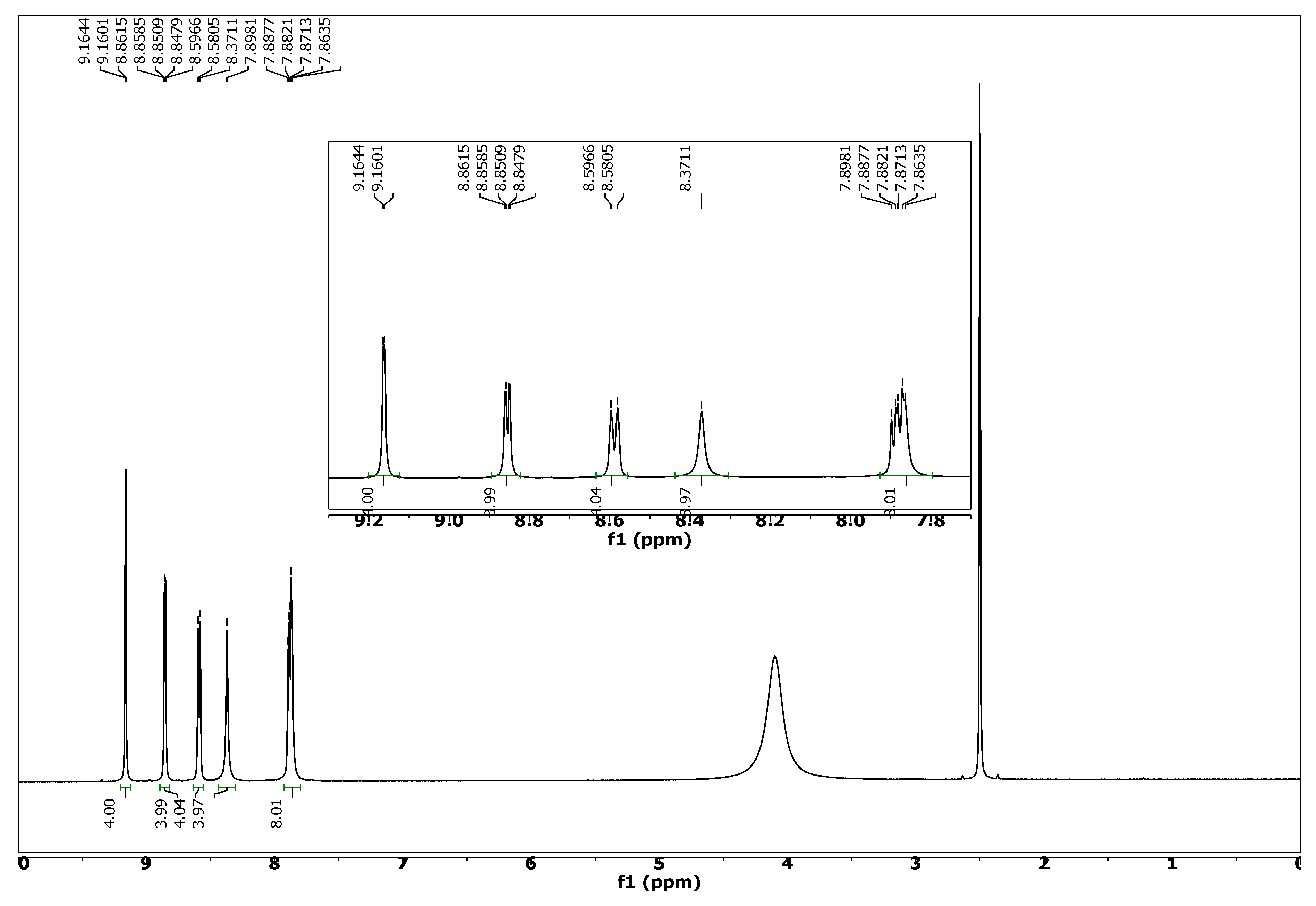
(2) 1H-NMR (500 MHz, DMSO-d6, δ ppm, J Hz): 9.16 (d, 4H, H-1, 1.6 Hz), 8.85 (dd, 4H, H-5, 1.6 Hz, 5.2 Hz), 8.59 (d, 4H, H-3, 8.0 Hz), 8.37 (s, 4H, NH2), 7.89–7.87 (m, 8H, H-4 and NH2) ppm. (Figure 1).
(2) 13C-NMR (125 MHz, DMSO-d6, δ ppm): 164.93, 149.66, 147.18, 139.30, 131.41, 125.41 ppm.
As can be seen in Figure 8, complex (2) exhibits signals in the chemical shift (δ) range of 9.16–7.87 ppm. The signals corresponding to the protons of the pyridine ring of nicotinamide appear at 9.16, 8.85, 8.59, and 7.89 ppm. Additionally, the amino group protons of the nicotinamide molecules yield signals at 8.37 and 7.87 ppm, indicating the presence of two types of this compound: coordinated and uncoordinated at the metal center.

3.5. Thermogravimetric Analysis

The TG curve of complex (1) (Figure S7) shows two major decomposition steps. The first occurs between 90°C and 150 °C and corresponds to a mass loss of 15.50%. This seems to indicate the elimination of the three lattice water molecules and one nicotinamide molecule (theoretical mass loss = 15.71%). The second step of decomposition, observed between 150 °C and 240°C, corresponds to a mass loss of 68.57%. The residue is a black powder, which was analyzed by FT-IR spectroscopy, and the spectrum was compared with that of mercury(II) oxide (Figure S8). The comparison revealed that the decomposition product most likely contains HgO, along with residual carbon.
The thermogram of complex (2) (Figure S9) shows three or even four decomposition steps. The first takes place between 74 and 143 °C and is due to the loss of the two coordinated water molecules (3.7% found/4.23% calculated). The second step occurs between 189 °C and 267 °C and appears to indicate the loss of nicotinamide fragments (59.75% found/57.48% calculated). The last two decomposition stages are likely due to the elimination of nitrate groups (267–327 °C, 13.40% experimental mass loss) and the decomposition of HgO, respectively.

3.6. Luminescent Properties

The room temperature photoluminescence of compounds (1)(3) was studied in the solid state, using different excitation wavelengths, in the 270–370 nm range. For compound (1), the emission spectrum resulting from excitation at 300 nm shows a complex band with two maxima at 468 and 494 nm. The corresponding excitation spectrum (λem = 470 nm) reveals an asymmetric band with a maximum at 354 nm (Figure 9a).
The emission spectrum of compound (2), λex = 330 nm, displays a broad asymmetric band, with a maximum at 368 nm and “a shoulder” at lower energies (Figure 9b). The corresponding excitation spectrum (λem = 470 nm) shows a maximum at 353 nm. Figure 9c presents the emission band of the compound (3), resulting from excitation at 300 nm, and showing a maximum at 384 nm.

4. Conclusions

This work presents the synthesis and characterization of three new Hg(II) complexes with oxalate, nitrate, sulphate, and nicotinamide as mixed ligands. The product obtained from the reaction between mercurous nitrate, ammonium oxalate, and nicotinamide is a polynuclear complex with a strongly distorted octahedral geometry of the metal center. Oxalate groups coordinate to Hg(II) in a chelating bidentate-bis (monodentate) fashion. The complex of Hg(II) with nicotinamide and nitrate is mononuclear, the nitrate acting in a bidentate chelating manner and the metal center being octacoordinated. The complex of Hg(II) with sulphate and nicotinamide is dinuclear, the sulphate groups behaving in a chelating bidentate-bridging monodentate way. In all complexes, nicotinamide coordinates through the heterocyclic nitrogen atom. The luminescent properties of the complexes were studied in the solid state.

Supplementary Materials

The following supporting information can be downloaded at: https://www.mdpi.com/article/10.3390/cryst15100835/s1; Figure S1: ORTEP diagram of the asymmetric unit of the compound [Hg2(ox)2(NA)4]n·3nH2O. The ellipsoids were represented with 50% probability; Figure S2: ORTEP diagram of the asymmetric unit of the compound [Hg(NO3)2(NA)2(H2O)2]·2NA. The ellipsoids were represented with 50% probability; Figure S3: ORTEP diagram of the asymmetric unit of the compound [Hg2(SO4)2(H2O)2(NA)4]·6H2O. The ellipsoids were represented with 50% probability; Figure S4: IR spectrum of the compound [Hg2(ox)2(NA)4]n·3nH2O; Figure S5: IR spectrum of the compound [Hg(NO3)2(NA)2(H2O)2]·2NA; Figure S6: IR spectrum of the compound [Hg2(SO4)2(H2O)2(NA)4]·6H2O; Figure S7: TG curve of complex (1); Figure S8: FT-IR spectra of the residue of complex (1) (blue curve) and mercury(II) oxide (black curve); Figure S9: TG curve for complex (2); Table S1: SHAPE analysis for compound [Hg2(ox)2(NA)4]n·3nH2O; Table S2: SHAPE analysis for compound [Hg(NO3)2(NA)2(H2O)2]·2NA; Table S3: SHAPE analysis for compound [Hg2(SO4)2(H2O)2(NA)4]·6H2O.

Author Contributions

Conceptualization: L.P.; methodology: L.P.; software,: L.P., A.M.M., A.H. and M.G.; validation: L.P., A.M.M., A.H. and M.G.; investigation: L.P., A.M.M., A.H. and M.G.; writing—original draft preparation: L.P. and A.M.M.; writing—review and editing: L.P. All authors have read and agreed to the published version of the manuscript.

Funding

This work was carried out through the PN 23.06 and PN 23.04 Core Program–ChemNewDeal within the National Plan for Research, Development and Innovation 2022–2027, developed with the support of the Ministry of Research, Innovation, and Digitization.

Data Availability Statement

The original contributions presented in this study are included in the article/Supplementary Material. Further inquiries can be directed to the corresponding author.

Conflicts of Interest

The authors declare no conflicts of interest.

References

  1. Bozkurt, N.; Dilek, N.; Delibaș, N.C.; Necefoğlu, H.; Hökelek, T. Di-µ-nicotinamide-k2N1:O; k2O: N1-bis[aquabis(3-chlorobenzoato-k2O,O’) cadmium]. Acta Cryst. 2013, E69, m389–m390. [Google Scholar]
  2. Zhao, X.-L.; Mak, T.C.W. Silver cages with encapsulated acetylenediide as building blocks for hydrothermal synthesis of supramolecular complexes with n-cyanopyridine and pyridine-n-carboxamide (n = 3,4). Dalton Trans. 2004, 3212–3217. [Google Scholar] [CrossRef] [PubMed]
  3. Kozlevčar, B.; Leban, I.; Turel, I.; Šegedin, P.; Petric, M.; Pohleven, F.; White, A.J.P.; Williams, D.J.; Sieler, J. Complexes of copper(II) acetate with nicotinamide: Preparation, characterization and fungicidal activity: Crystal structures of [Cu2(O2CCH3)4(nia)] and [Cu2(O2CCH3)4(nia)2]. Polyhedron 1999, 18, 755–762. [Google Scholar] [CrossRef]
  4. Monfared, H.H.; Kalantari, Z.; Kamyabi, M.-A.; Janiak, C. Synthesis, structural characterization and electrochemical studies of a nicotinamide-bridged dinuclear copper complex derived from a tridentate hydrazone Schiff base ligand. Z. Anorg. Allg. Chem. 2007, 633, 1945–1948. [Google Scholar] [CrossRef]
  5. Özbek, F.E.Ö.; Sertçelik, M.; Yüksek, M.; Elmalı, A.; Şahin, E. The superiority of the classical synthesis compared to the hydrothermal synthesis upon the structural, optical absorption and fluorescent properties of new Cd(II) 3-fluorobenzoate complexes with pyridine-3-carboxamide/pyridine-3-carboxylate. Inorg. Chim. Acta 2020, 509, 119694. [Google Scholar] [CrossRef]
  6. Malekhoseini, A.; Montazerozohori, M.; Naghiha, R.; Kokdan, E.P.; Joohari, S. Antimicrobial/antioxidant and cytotoxicity activities of some new mercury(II) complexes. Chem. Rev. Lett. 2023, 6, 166–182. [Google Scholar]
  7. Aliabadi, A.; Motieiyan, E.; Hosseinabadi, F.; Ghadermazi, M.; Abdolmaleki, S. One-pot synthesis, crystallographic characterization, evaluation as in vitro antibacterial and cytotoxic agents of two mercury(II) complexes containing pyridine dicarboxylic acid derivatives. J. Mol. Struct. 2021, 1226, 129405. [Google Scholar] [CrossRef]
  8. Maqsood, F.; Al-Rawi, S.S.; Ibrahim, A.H.; Jamil, F.; Zafar, A.; Iqbal, M.A.; Shoukat, U.S.; Asad, M.; Zia, S.U.; Ahmad, F.; et al. Recent trends in medicinal applications of mercury based organometallic and coordination compounds. Rev. Inorg. Chem. 2025, 45, 375–396. [Google Scholar] [CrossRef]
  9. Alshater, H.; Al-Sulami, A.I.; Aly, S.A.; Abdalla, E.M.; Sakr, M.A.; Hassan, S.S. Antitumor and antibacterial activity of Ni(II), Cu(II), Ag(I), and Hg(II) complexes with ligand derived from thiosemicarbazones: Characterization and theoretical studies. Molecules 2023, 28, 2590. [Google Scholar] [CrossRef]
  10. Akhtari, E.; Mohammadi, K.; Hayati, P.; Keshavarzi, S.; Tavallaei, O.; Derakhshankhah, H.; Karamveysi, H.; Mahammadpour, M.; Retailleau, P. In Vitro Anticancer and Antibacterial Activity of a New Hg(II) Pyridinedicarboxylate Coordination Supramolecular Compound. Appl. Organomet. Chem. 2025, 39, e70173. [Google Scholar] [CrossRef]
  11. Shahzad, A.; Khan, E.; Said, M.; Khan, G.S.; Sied, M.G.; Noor, A.; Zahoor, M.; Ullah, R.; Bari, A. Complexes of 1,3-diisobutyl thiourea with copper(I), zinc(II) and mercury(II): Their antioxidant and antibacterial evaluation. Crystals 2021, 11, 989. [Google Scholar] [CrossRef]
  12. Kim, J.A.; Park, H.; Kim, J.C.; Lough, A.J.; Pyun, S.Y.; Roh, J.; Lee, B.M. 1D copper(II) and Zn(II) coordination polymers containing an unusual twisted oxalate bridge. Inorg. Chim. Acta 2008, 361, 2087–2093. [Google Scholar] [CrossRef]
  13. Yesilel, O.Z.; Erer, H.; Odabasoglu, M.; Buyukgungor, O. A novel copper(II)-hydrogen oxalate coordination polymer showing a new coordination mode. J. Inorg. Organomet. Polym. Mater. 2010, 20, 78–82. [Google Scholar] [CrossRef]
  14. Hernandez-Molina, M.; Lorenzo-Luis, P.A.; Ruiz-Perez, C. A new coordination mode in oxalate-bridged polymers: Molecular and crystal structure of [Cu3(C2O4)2(C2H8N2)4(ClO4)2]n·2H2O. CrystEngComm 2001, 3, 60–63. [Google Scholar] [CrossRef]
  15. Lu, J.; Li, Y.; Zhao, K.; Xu, J.Q.; Yu, J.H.; Li, G.H.; Zhang, X.; Bie, H.Y.; Wang, T.G. Novel oxalate coordination mode and roles: Synthesis, structure and fluorescence property of [Cd25-ox)(µ3-OH)2] with 3-D structure. Inorg. Chem. Commun. 2004, 7, 1154–1156. [Google Scholar] [CrossRef]
  16. Louka, F.R.Y.; Mautner, F.A.; Vicente, R.; Massoud, S.S. µ2-oxalato-bridged tricopper(II) complex derived from 1,4,8,12-tetraazacyclopentadecane: Synthesis, structure and magnetic characterization. Inorg. Chem. Commun. 2008, 4, 438–441. [Google Scholar] [CrossRef]
  17. Christensen, A.N.; Norby, P.; Hanson, J.C. A crystal structure determination of HgC2O4 from synchrotron X-ray and neutron powder diffraction data. Z. Krystallograph. 1994, 209, 874–877. [Google Scholar] [CrossRef]
  18. Colmenero, F.; Timon, V. Mechanical anomalies in mercury oxalate and the deformation of the mercury cube coordination environment under pressure. Appl. Phys. A 2021, 127, 395. [Google Scholar] [CrossRef]
  19. Chen, Z.; Wu, X.; Qin, S.; Lei, C.; Liang, F. Structure and fluorescent properties of mercury(II) pyridine-2,3-dicarboxylate coordination polymers tuned by ancillary ligands and alkaline-earth metal ions. CrystEngComm 2011, 13, 2029–2038. [Google Scholar] [CrossRef]
  20. Khutia, A.; Sanz-Miguel, P.J.; Lippert, B. Molecular arhitectures derived from metal ions and the flexible 3,3’-bipyridine ligand: Unexpected dimer with Hg(II). Bioinorg. Chem. Appl. 2010, 2010, 169054. [Google Scholar] [CrossRef]
  21. Mahmoudi, G.; Morsali, A.; Zeller, M. Mercury(II) acetate/thiocyanate coordination polymers with n-donor ligands, spectroscopic, thermal and structural studies. Inorg. Chim. Acta 2009, 362, 217–225. [Google Scholar] [CrossRef]
  22. Ramazani, A.; Mahmoudi, G.; Dolatyari, L.; Morsali, A.; Hu, M.L. New mixed-anion mercury(II) complex, spectroscopic, thermal and structural studies of [Hg(bipy)2(CH3COO)2](SO4)0.5NaCl. J. Coord. Chem. 2007, 60, 2115–2120. [Google Scholar] [CrossRef]
  23. Soldin, Z.; Kukovec, B.M.; Matkovic-Calogovic, D.; Popovic, Z. The solvent effect on composition and dimensionality of mercury (II) complexes with picolinic acid. Molecules 2021, 26, 5002–5015. [Google Scholar] [CrossRef]
  24. Popovic, Z.; Pavlovic, G.; Soldin, Z. Iodo (picolinato-k2N, O)(picolinic acid, k2N, O) mercury(II). Acta Cryst. 2006, C62, m272–m274. [Google Scholar]
  25. Hayati, P.; Rezvani, A.R.; Morsali, A.; Retailleau, P.; Garcia-Granda, S. Influences of temperature, power ultrasound and reaction time on the morphological properties of two new mercury(II) coordination supramolecular compounds. Ultrason. Sonochem. 2017, 34, 968–977. [Google Scholar] [CrossRef] [PubMed]
  26. Morsali, A.; Masoomi, M.Y. Structure and properties of mercury(II) coordination polymers. Coord. Chem. Rev. 2009, 253, 1882–1905. [Google Scholar] [CrossRef]
  27. Yilmaz, V.T.; Yazicilar, T.K.; Andac, O.; Kutuk, H.; Bekdemir, Y.; Harrison, W.T.A. Bis(p-nitrobenzoxasulfamato) complexes of cadmium(II) and mercury(II)–synthesis, spectra and X-ray crystal structures. Z. Anorg. Allg. Chem. 2002, 628, 1908–1912. [Google Scholar] [CrossRef]
  28. Morozov, I.V.; Sherezhkin, V.N.; Troyanov, S.I. Modes of coordination of the NO3- anions in inorganic nitrates. Russ. Bull. Chem. 2008, 57, 439–450. [Google Scholar] [CrossRef]
  29. Polonius, J.M.F.-A.; Roitzsch, M. [Hg(9-methyl-1-deazapurine)2](NO3)2.H2O: A complex with a distorted hexagonal bipyramidal metal ion coordination sphere. Inorg. Chim. Acta 2005, 358, 1225–1230. [Google Scholar]
  30. Dziewulska-Kulaczkowska, A.; Mazur, L.; Ferenc, W. Thermal, spectroscopic and structural studies of zinc(II) complex with nicotinamide. J. Therm. Anal. Calorim. 2009, 96, 255–260. [Google Scholar] [CrossRef]
  31. Lian, Z.; Zhao, N.; Yang, F.; Liu, P. Crystal structure of trans-trans-trans-diaquabis(nicotinamide)-dinitratocadmium(II)-nicotinamide(1:2), Cd(H2O)2(C6H6N2O)2(NO3)2.2C6H6N2O. Z. Crystallogr. NCS 2011, 226, 289–290. [Google Scholar]
  32. Papatriantafyllopoulou, C.; Manesi, E.; Escuer, A.; Perlepes, S.P. The sulphate ligand as a promising “player” in 3d-metal cluster chemistry. Inorg. Chim. Acta 2009, 362, 634–650. [Google Scholar] [CrossRef]
  33. Gamov, G.A.; Zavalishin, M.N.; Dushina, S.V. Synthesis and thermal analysis of a Ni(II) complex of nicotinamide. Russ. J. Gen. Chem. 2017, 87, 613–618. [Google Scholar] [CrossRef]
  34. Cakir, S.; Bicer, E.; Aoki, K.; Coskun, E. Structural features of a new [Fe(nicotinamide)2(H2O)4] center dot [Fe(H2O)6] center dot (SO4)2 center dot 2H(2)O complex. Cryst. Res. Technol. 2006, 41, 314–320. [Google Scholar] [CrossRef]
  35. Dakovic, M.; Popovic, Z.; Giester, G.; Rajic-Linaric, M. Synthesis, spectroscopic and structural investigation of Zn(NCS)2(nicotinamide)2 and Hg(SCN)2(nicotinamide)]n. Polyhedron 2008, 27, 465–472. [Google Scholar] [CrossRef]
  36. Soldin, Z.; Kukovec, B.-M.; Perokovic, V.P.; Dakovic, M.; Popovic, Z. Cadmium(II) coordination compounds and mercury(II) inorganic-organic hybrid with a quaternary nicotinamide salt, 3-carbamoyl-1-carboxymethylpyridinium bromide. Inorg. Chim. Acta 2025, 582, 122662. [Google Scholar] [CrossRef]
  37. Amani, V.; Shokouhmanesh, M.; Khavasi, H.R. Binuclear mercury(II) complexes containing N-(naphthalene-1-yl) isonicotinamide ligand: X-ray studies and spectroscopic characterization. Inorg. Chem. Res. 2025, 9, 8–13. [Google Scholar]
  38. Kotai, L.; Lippart, J.; Gacs, I. Deuterium isotope separation in the chemical reaction of aluminum amalgam and water. Eur. Chem. Bull. 2012, 1, 37–38. [Google Scholar]
  39. Ramalingam, S.; Periandy, S.; Govindarajan, M.; Mohan, S. FT-IR and FT-Raman vibrational spectra and molecular structure investigation of nicotinamide: A combined experimental and theoretical study. Spectrochim. Acta A Mol. Biomol. Spectrosc. 2010, 75, 1552–1558. [Google Scholar] [CrossRef]
  40. Nakamoto, K. Infrared and Raman Spectra of Inorganic and Coordination Compounds, Part B–Applications in Coordination, Organometallic, and Bioinorganic Chemistry, 6th ed.; John Wiley & Sons, Inc.: Hoboken, NJ, USA, 2009; pp. 79–82. [Google Scholar]
  41. Andros, L.; Juric, M.; Planinic, P.; Zilic, D.; Ravkin, B.; Molcanov, K. New mononuclear oxalate complexes of copper (II) with 2D and 3D architecture: Synthesis, crystal structure and spectroscopic characterization. Polyhedron 2010, 29, 1291–1298. [Google Scholar] [CrossRef]
  42. Llunell, M.; Casanova, D.; Cirera, J.; Alemany, P.; Alvarez, S. SHAPE v 2.1; Institut de Química Teòrica i Computacional-Universitat de Barcelona: Barcelona, Spain, 2013. [Google Scholar]
  43. Escriva, E.; Folgado, J.V.; Espallargas, G.M.; Soto, L.; Sancho, A.; Perello, L.; Ortiz, R. New dinuclear copper complexes incorporating bis(imidazolyl) based ligands and bidentate-monodentate oxalate bridges. Crystal structure and magnetic properties of [Cu2(BIM)2(C2O4)2].4H2O and [Cu2(BIK)2(C2O4)2] (BIM = bis(2-imidazolyl)methane), BIK = bis(2-imidazolyl)ketone). Exploring magneto-structural correlations. Polyhedron 2016, 112, 137–144. [Google Scholar]
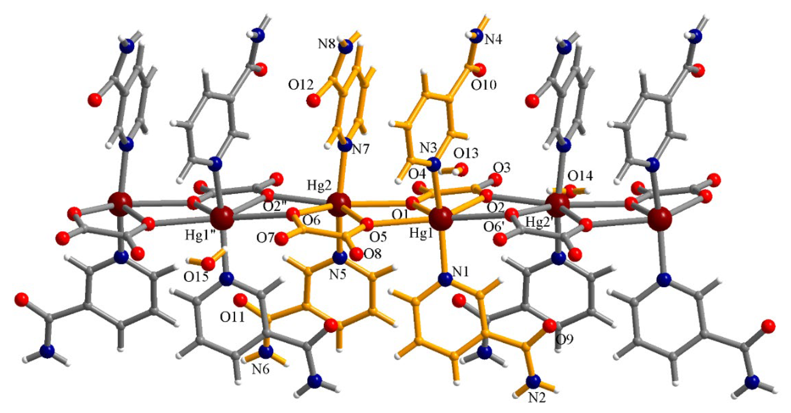
Figure 1. The structure of the 1D coordination polymer [Hg2(ox)2(NA)4]n·3nH2O. The asymmetric unit is represented in orange. Symmetry codes: ′ = 1 + x, y, z; ″ = −1 + x, y, z.
Crystals 15 00835 g001
Figure 2. Hydrogen interactions involving the lattice water molecules in the crystal [Hg2(ox)2(NA)4]n·3nH2O. Symmetry codes: ′ = 1 + x, y, z; ″ = −1 + x, y, z; i = x, 1.5 − y, 0.5 + z; ii = 2 − x, 1 − y, 1 − z; iii = 1 − x, 1 − y, 1 − z; iv = −x, 1 − y, −z; v = x, 1.5 − y, −0.5 + z.
Crystals 15 00835 g002
Figure 3. View of the packing diagram in the crystal (1) along the crystallographic a-axis.
Crystals 15 00835 g003
Figure 4. The crystal structure of the compound [Hg(NO3)2(NA)2(H2O)2]·2NA. Symmetry code: ′ = 1 − x, 1 − y, 2 − z.
Crystals 15 00835 g004
Figure 5. View of the packing diagram in the crystal (2) showing the hydrogen interactions. Symmetry codes: ′ = 1 − x, 1 − y, 2 − z; ″ = 0.5 + x, 0.5 − y, 0.5 + z; ′″ = −0.5 + x, 1.5 − y, −0.5 + z; ″″ = 1.5 − x, 0.5 + y, 1.5 − z.
Crystals 15 00835 g005
Figure 6. The crystal structure of the compound [Hg2(SO4)2(H2O)2(NA)4]·6H2O (3). The asymmetric unit of the complex is depicted in orange. Symmetry codes: ′ = 1 − x, y, 2 − z; ″ = x, 1 − y, z.
Crystals 15 00835 g006
Figure 7. View of the packing diagram along the crystallographic c-axis in the crystal (3) showing the hydrogen interactions.
Crystals 15 00835 g007
Figure 8. 1H-NMR spectrum of the compound [Hg(NO3)2(NA)2(H2O)2]·2NA.
Crystals 15 00835 g008
Figure 9. (a) Emission (right) and excitation (left) spectra for the compound (1); (b) emission (right) and excitation (left) spectra for the compound (2); and (c) emission spectrum for the compound (3).
Crystals 15 00835 g009
Table 1. Crystallographic data for complexes (1)(3).
Compound(1)(2)(3)
Chemical formulaC28H30N8O15Hg2C24H28N10O12HgC12H16N4O10SHg
M (g mol−1)1119.78849.15612.97
Temperature, (K)293(2)293(2)293(2)
Wavelength, (Å)0.710730.710730.71073
Crystal systemMonoclinicMonoclinicMonoclinic
Space groupP21/cP21/nC2/m
a (Å)7.8085(3)7.4611(4)16.5391(8)
b (Å)31.3979(11)22.4487(10)18.0007(7)
c (Å)14.2847(5)9.4543(4)6.9969(4)
α (°)909090
β (°)101.671(4)108.570(5)113.293(6)
γ (°)909090
V (Å3)3429.8(2)1501.07(13)1913.30(18)
Z424
Dc (g cm−3)2.1691.8792.128
Size of the crystal (mm)0.3 × 0.1 × 0.080.2 × 0.04 × 0.020.2 × 0.02 × 0.01
μ (mm−1)9.0255.2078.213
F(000)21368361184
Goodness-of-fit on F21.0441.0441.210
Final R1, wR2 [I > 2σ(I)]0.0283, 0.05830.0266, 0.06170.0389, 0.1054
R1, wR2 (all data)0.0400, 0.06190.0381, 0.06700.0405, 0.1061
Table 2. Selected bond distances (Å) and angles (°) for the complex [Hg2(ox)2(NA)4]n.
AtomsBond DistanceAtomsAngle
Hg1–O12.437(3)O1–Hg1–O265.79(11)
Hg1–O22.492(4)O1–Hg1–O569.66(11)
Hg1–O52.629(3)O2–Hg1–O5135.18(11)
Hg1–O62.633(3)O5–Hg1–O6158.91(12)
Hg1–N12.163(4)N3–Hg1–N1155.55(16)
Hg1–N32.158(4)N3–Hg1–O686.42(13)
Hg2–O12.604(3)N1–Hg1–O689.49(16)
Hg2–O22.626(3)O5–Hg2–O664.90(11)
Hg2–O52.509(3)O2–Hg2–O665.59(11)
Hg2–O62.469(4)O1–Hg2–O2160.44(12)
Hg2–N52.175(4)N5–Hg2–O289.26(13)
Hg2–N72.158(4)N5–Hg2–N7157.51(17)
Table 3. Geometrical details of the hydrogen interactions.
D–H∙∙∙Ad(H∙∙∙A)(Å)d(D∙∙∙A)(Å)<<(DHA)(°)
O13–H10∙∙∙O42.0222.796150.87
O14–H30∙∙∙O32.0152.825158.62
O14–H40∙∙∙O4′1.9872.809162.01
O15–H50∙∙∙O72.0402.793147.29
O15–H60∙∙∙O8″1.9992.833167.01
N8iii–H8Biii∙∙∙O132.1372.969162.62
N2i–H2Bi∙∙∙O132.1652.995162.08
N2i–H2Ai∙∙∙O142.2052.963146.86
N4–H4A∙∙∙O14v2.1542.999167.5
N4iv–H4Biv∙∙∙O152.1943.016159.6
N6–H6A∙∙∙O15iii2.2993.122160.47
Symmetry codes: ′ = 1 + x, y, z; ″ = −1 + x, y, z; i = x, 1.5 − y, 0.5 + z; ii = 2 − x, 1 − y, 1 − z; iii = 1 − x, 1 − y, 1 − z; iv = −x, 1 − y, −z; v = x, 1.5 − y, −0.5 + z.
Table 4. Hydrogen bond interactions in complex [Hg(NO3)2(NA)2(H2O)2]·2NA (2).
D–H∙∙∙Ad(H∙∙∙A)(Å)d(D∙∙∙A)(Å)<(DHA)(°)
O3–H2O∙∙∙N32.1252.901151.4
N4″–H4N″∙∙∙O22.1152.963168.6
N4″–H3N″∙∙∙O52.1813.037173.5
N2–H1N∙∙∙O6″″2.1202.956164.2
N2–H2N∙∙∙O1′″2.0712.906163.4
Symmetry codes: ″ = 0.5 + x, 0.5 − y, 0.5 + z; ′″ = −0.5 + x, 1.5 − y, −0.5 + z; ″″ = 1.5 − x, 0.5 + y, 1.5 − z.
Disclaimer/Publisher’s Note: The statements, opinions and data contained in all publications are solely those of the individual author(s) and contributor(s) and not of MDPI and/or the editor(s). MDPI and/or the editor(s) disclaim responsibility for any injury to people or property resulting from any ideas, methods, instructions or products referred to in the content.