Article

Effect of Nano-Silica on Mechanical Properties and Cytotoxicity of Calcium-Silicate-Based Root Canal Filling Materials

1
School of Mechanical and Automotive Engineering, Guangxi University of Science and Technology, Liuzhou 545616, China
2
Powder Metallurgy Research Institute, Central South University, Changsha 410083, China
3
School of Materials Science and Engineering, Xiangtan University, Xiangtan 411105, China
4
XiangYa School of Medicine, Central South University, Changsha 410013, China
*
Authors to whom correspondence should be addressed.
Crystals 2025, 15(1), 55; https://doi.org/10.3390/cryst15010055
Submission received: 11 December 2024 / Revised: 29 December 2024 / Accepted: 1 January 2025 / Published: 7 January 2025
(This article belongs to the Section Mineralogical Crystallography and Biomineralization)

Abstract

:
A study was conducted to evaluate the effect of nano-silica (NS) content on the strength and cytotoxicity of calcium-silicate-based root canal filling materials. In this experimental laboratory study, four types of calcium-silicate-based paste with different NS ratios were prepared and hydrated for seven or 28 days. The setting times, compressive strengths, and porosities were tested to determine the properties of the pastes. The residual NS and the calcium hydroxide (CH; Ca (OH)2) content were investigated to analyze the hydration products. The experiments demonstrated that NS facilitates the hydration of calcium-silicate-based materials, enhances the formation of hydration products, and ensures effective porosity filling. Consequently, these findings suggest that NS contributes to the enhancement of the early compressive strength of calcium silicate root canal filling pastes. The addition of 9% NS enhanced the early compressive strength by 41.99% compared with the NS-free samples. Additionally, it was found that the test group without NS, as well as the test groups with 3% and 9% NS, exhibited mild cytotoxicity, while the test group with 15% NS exhibited moderate cytotoxicity. The observed cytotoxicity can be attributed to the increase in the ambient pH due to the production of CH during hydration. The findings of this study demonstrate that the early compressive strength of calcium-silicate-based root canal filling materials exhibits a substantial increase in response to the incorporation of NS, and that calcium-silicate-based root canal filling materials do not manifest significant levels of toxicity. NS improved hydration product formation and made efficient pore filling possible.

1. Introduction

Single-visit root canal treatment is recognized for its effectiveness in treating pulpal diseases, with a short treatment duration and minimal side effects [1,2]. Calcium silicate bioceramics are becoming increasingly popular for vital pulp therapy, crown–root restoration, and root canal filling because of their excellent sealing properties and biological activity [3,4]. A vertical root fracture is a serious complication associated with root canal treatment [5], and it often necessitates tooth extraction. The root canal procedure, which involves the mechanical preparation of root canals [6,7], endodontic irrigation [8], and filling pressure [9,10], can diminish the tooth root’s strength by damaging the dentin. Over time, the continuous occlusal force transmits stress from the tooth crown to the compromised root apex, resulting in a root fracture [11,12]. Therefore, it is imperative to select a filling material that can reinforce the root resistance. Research has shown that the fracture resistance in an intact tooth is 923.72 N, which decreases to 499.892 N following root canal preparation [13,14]. The utilization a of traditional resin composite for filling leads to a significant 17.59% reduction in fracture resistance compared with an intact tooth. Aslan et al. conducted an extensive study on root canal fillings using resin and calcium-silicate-based sealants [15]. They assessed the flexural strength of the teeth in each group and found that all root canal sealants enhanced the flexural performance of the teeth after root canal preparation, with no significant difference between them. Uzunoglu Ozyurek et al. reported that BioRoot RCS, a calcium-silicate-based root canal filling material, achieved intact tooth fracture resistance of 80.9% [16]. Papynov et al. used spark plasma sintering (SPS) to solidify porous nanostructured wollastonite powders, obtaining composites with high mechanical strength (Young’s modulus >60 MPa) [17]. However, despite these advances, the current filling materials cannot fully restore the tooth fracture resistance lost during root canal preparation. Furthermore, several studies have demonstrated that the compressive strength of calcium-silicate-based root canal filling materials is slightly lower than that of resin-based root canal sealants [18]. Chew and Turkyilmaz et al. attributed this to the reduced permeability of the current filling materials [19,20]. Zamparini and Komabayashi et al. reported that the size of most sealer particles ranges from 2 to 10 μm, which exceeds the diameter of the dentin tubules in the apical region, typically measuring l to 2 μm [21,22]. Hachem et al. found that the BC sealer and NTS exhibited superior tubule penetration to that of the AH Plus sealer because of their small particle sizes [23]. Consequently, sealers with particle sizes of less than l μm, or even at the nanoscale, may enhance the compressive strength, which is essential in optimizing their utility in dental applications. Enhancing the compressive strength of calcium-silicate-based bioceramics is crucial in optimizing their practicality in dental applications.
Nano-silica (NS) is a type of nanoparticle known for its ability to enhance the compressive strength of cement-based materials [24]. NS reacts with calcium hydroxide (CH) during hydration to form calcium silicate hydrate (C S H). This process accelerates cement hydration, refines the pore structure, reduces the low-intensity phase, and increases the compressive strength of cement-based materials. Bai et al. examined the influence of the NS-specific surface area on the strength of ordinary Portland cement [25]. A larger NS surface area resulted in more Si-OH groups, which enhanced the reactivity, refined the pore structure, and ultimately contributed to higher compressive strength. Sun et al. studied the influence of NS on the early hydration of alitesulfoaluminate cement and showed a substantial improvement in amorphous aluminosilicate cement hydration with NS addition [26,27]. At NS content of 3%, there was a 54.09% reduction in porosity after three days, indicating a denser microstructure. Furthermore, the addition of 4% NS to microfine cement resulted in the maximum compressive strength of 78.9 MPa after a 28-day curing period [28]. Shahram et al. investigated the effects of different NS content, particle sizes, and dispersion methods on slag mortar strength [29]. The results demonstrated that the addition of 2% NS led to a 39% increase in compressive strength on the third day and a 30% increase on the seventh day. The inclusion of smaller silica particles can enhance the early strength of slag mortar. The use of nano-silica to enhance materials’ properties has gained significant attention in recent years. Nano-silica has been shown to improve the mechanical strength, durability, and biocompatibility of various materials, owing to its large surface area and unique physical and chemical properties.
Several studies have extensively investigated the effects of NS on the strength of construction cements. However, there is a significant research gap in understanding the effects of calcium silicate on the strength and cytotoxicity of calcium-silicate-based bioceramics, particularly for dental applications. This research gap is important given the crucial role of calcium-silicate-based bioceramics in dentistry, particularly in root canal treatment. The purpose of this study was to evaluate the effects of nano-silica on the mechanical properties and cytotoxicity of calcium-silicate-based root canal filling materials. It was hypothesized that the addition of nano-silica would significantly improve the mechanical strength and biocompatibility of these materials, making them more suitable for clinical applications. The aim was to explore comprehensively the mechanisms whereby NS influences the compressive strength of these materials by determining the setting time, assessing the porosity, analyzing the phase composition, quantifying the total CH generated during hydration, and observing the microscopic morphology in detail. Furthermore, considering the dental applications of these materials and the need to ensure their safety and effectiveness in biological contexts, the cytotoxicity of calcium-silicate-based root canal filling with NS was evaluated, confirming its suitability for dental applications.

2. Materials and Methods

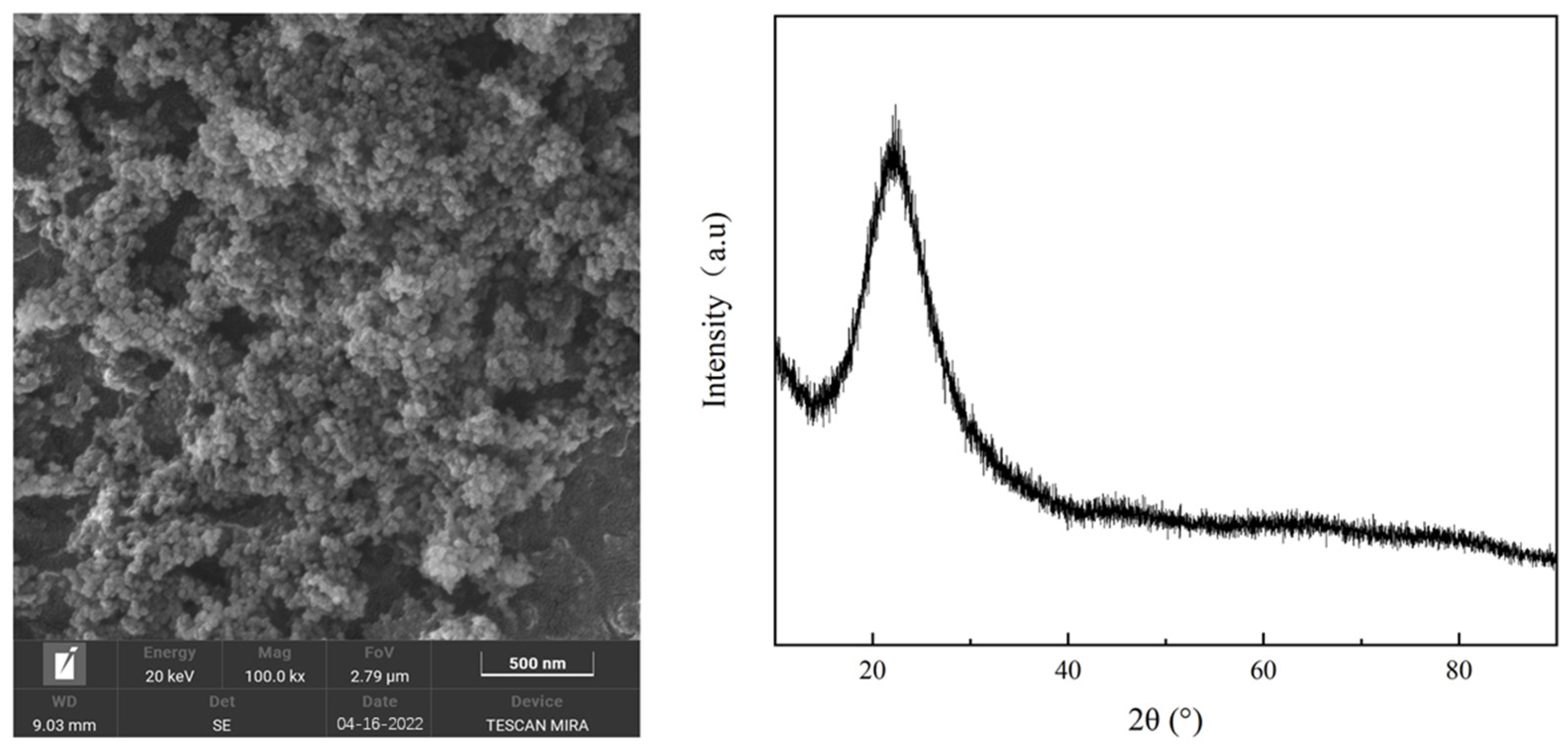
2.1. Powder Raw Materials

In the experiment, tricalcium silicate (Ca3SiO5; C3S), zirconium dioxide (ZrO2), propylene glycol (C3H8O2, average molecular weight of 400), and NS were used as the primary raw materials. Table 1 lists the most important performance parameters and manufacturer details. Figure 1 shows the NS microstructure and its X-ray pattern. The NS powder exhibited a spherical morphology with a standard deviation (SD) in the average particle size of approximately 30 ± 5 nm. The X-ray diffraction (XRD) pattern displayed a broad, short peak at 23° (2θ), without distinct, sharp diffraction peaks, confirming the amorphous nature of NS.

2.2. Experimental Method

As shown in Figure 2, the experimental methodology involved paste preparation, performance testing (including setting time, compressive strength, and porosity evaluation), hydration product analysis, and cytotoxicity assessment.

2.3. Paste Preparation and Performance Testing

In this study, four calcium-silicate-based pastes were prepared, and their formulations are listed in Table 2. The C3S, NS, ZrO2, and C3H8O2 were combined in a ball mill to obtain a uniform paste. For the curing time experiment, the resulting paste was placed in cylindrical molds with a diameter of 10 mm and a height of 1 mm to create test samples, following the guidelines of the International Organization for Standardization (ISO) standard ISO 6876-2012 [30], Chapter 5.4. For the compressive strength experiment, the paste was placed in cylindrical molds with a diameter of 6 mm and a height of 12 mm to fabricate the samples. These samples were then cured in a controlled environment with a temperature of 37 °C and a humidity level of ≥95%. After seven and 28 days of curing, the samples were removed from the curing environment and immersed in anhydrous ethanol to halt hydration, thereby completing the sample preparation process.
The setting time was assessed using the Gillmore double-needle method, following the American Society for Testing and Materials C266-21 standards [31]. This involved the use of a needle (Changsha Deyue Technology Co., Ltd., Changsha, China) with an initial mass of 113.4 g and a diameter of 2.13 mm and a needle with a final mass of 453.6 g and a diameter of 1.06 mm. The time at which no visible indentations were observed was recorded. Each sample was tested in triplicate. To determine the compressive strength, the cured bone-cement columns were polished at their ends using 600-grit sandpaper and subsequently subjected to testing using a universal testing machine (Dongguan Dongzhiri Instrument Co., Ltd., Dongguan, China) at a loading speed of 0.5 mm/min. Each group was composed of five test samples, ensuring that the strength deviations remained within 15%.
The methods of Hall and Hamilton [32] and Shen [33] were used to determine the porosity via the Archimedes drainage method. Porosity measurements were conducted using the Archimedes method, with three samples per group. Each sample was subjected to three measurements. The reported test results represent the averages of three samples. The porosity was calculated as follows:
P = m 2 m 1 m 2 m 3 × 100 %
where P (%) is the porosity, m1 (g) is the mass of the sample after drying, m2 (g) is the mass of the sample after saturation with water, and m3 (g) is the weight of the sample suspended in water.

3. Measurement and Analysis of Hydration Products

To determine the residual NS content, an analysis was conducted in accordance with the GB/T 28629-2012 detection standard [34]. A spectrophotometric method was employed to assess the free silica content, which was suitable for samples containing up to 5% free silica. The test sample was ground into a powder, dissolved in phosphoric acid, and subjected to filtration, calcination, precipitation, and other necessary procedures. Subsequently, it was fused with sodium carbonate borax. For samples with silica content exceeding 5%, the potassium carbonate fusion method was employed. The test sample was ground into a powder, dissolved in phosphoric acid, and subjected to filtration, calcination, precipitation, and similar treatments, followed by fusion with potassium carbonate. For the test groups containing 3% and 9% NS, a spectrophotometric method was employed to measure the free silica content, whereas the potassium fluorosilicate method was used for the test group containing 15% NS. Each sample was tested three times, and three samples were included in each group.
The CH content of the hydration products was analyzed using thermogravimetric (TG) analysis. The cured samples were ground into a fine powder and sifted through a 200-mesh sieve. Approximately 5 mg of a powdered sample was evenly distributed at the base of the crucible. The analysis was conducted using an STA 8000 synchronous thermal analyzer (PerkinElmer, Waltham, MA, USA) with protection provided by an argon atmosphere. The temperature of the experiment ranged from 25 to 1000 °C at a heating rate of 10 °C/min.

3.1. Cytotoxicity Measurement

Cytotoxicity testing involved six groups: four utilizing calcium-silicate-based pastes, one utilizing a 2% phenol dilution as a positive control, and one employing high-density polyethylene as a negative control. Each sample was exposed to ultraviolet light for 30 min, followed by sterilization at 121 °C for 15 min.
L-929 cells were maintained in minimum essential medium (MEM) with 10% fetal bovine serum and 1% antibiotics (100 U/mL penicillin, 100 μg/mL streptomycin) at 37 °C and 5% carbon dioxide. For cell detachment, 0.25% trypsin–ethylenediaminetetraacetate was used. After centrifugation, the cells were resuspended in the desired medium. The cell suspension was added to a six-well cell culture plate containing 2 mL of agar medium (comprising equal parts of 3% agar and 2 × MEM with 20% fetal bovine serum). The cells were cultured for 24 h to allow them to reach near-confluence.
The cells formed a monolayer, and the samples were placed on a cured agar layer, with three parallel samples per group. After 48 h of incubation, the six-well cell culture plates were discarded after the sample positions were labeled. Each culture dish received 2 mL of neutral red solution and was stained for 20 min. Excess dye was removed, followed by two washes with a phosphate-buffered saline solution. The cell morphology was observed under a microscope, and group images were processed using the ImageJ software. The cytotoxicity of the remaining groups was calculated relative to that of the negative control group, which was set at 100%. Cellular viability, in conjunction with the ISO 7405-2018 [35] Section 6 classification criteria, was employed to classify the cytotoxicity as severe (<30%), moderate (30–60%), mild (60–90%), and non-cytotoxic (>90%).

3.2. Phase and Micromorphology Analysis

For the phase composition analysis, the cured sample was ground into a powder and sieved through a 200-mesh sieve. Subsequently, the powdered samples underwent a 2 h drying process at 90 °C under vacuum conditions. The phase compositions of the hydration products of the composite materials were examined using a D8 ADVANCE XRD diffractometer (Bruker, Billerica, MA, USA). This analysis employed a copper target, a diffraction angle ranging from 10° to 80°, and a step size of 0.02°/s.
To prepare scanning electron microscopy (SEM) samples, the cured paste was first subjected to drying in a constant-temperature and -humidity box at 70 °C for 4 h. Then, it was subjected to pre-grinding treatment. The samples were subsequently embedded in resin and polished, and a conductive adhesive was applied. After gold spraying, the samples were observed using Quanta 250 FEG SEM equipment (FEI, Brno, Czech Republic). Observations were conducted at 20 kV.

4. Results

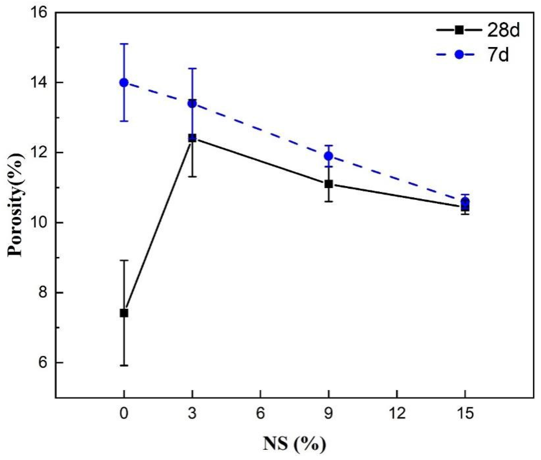
4.1. Physical Properties of the Material and Their Effects on the Compressive Strength

Figure 3 shows the compressive strength profiles of the calcium-silicate-based root canal filling adhesives with varying NS concentrations after seven and 28 days of hydration. For the samples hydrated for seven days, the compressive strength exhibited an initial increase, followed by a decrease as the NS content increased. The sample with 9% NS achieved the highest strength at 38.11 MPa, a substantial 41.99% improvement compared with the NS-free group. After 28 days of hydration, the trend reversed, and the compressive strength diminished with increasing NS content. The sample without NS exhibited the highest compressive strength, whereas that with 15% NS added had the lowest strength, measuring only 23.5 MPa. The compressive strengths of the NS-free and 3% NS samples increased after 28 days of hydration, compared with those subjected to hydration for seven days. However, the compressive strengths of the 9% and 15% NS samples remained relatively consistent after hydration for seven or 28 days.
The typical compressive strength of calcium-silicate-based bioceramics falls within the range of 30–90 MPa [36,37,38] and generally increases with increasing hydration durations. The introduction of NS influences internal factors, such as the phase composition and porosity, leading to alterations in the compressive strength of calcium-silicate-based root canal fillers. Li et al. observed that the compressive strengths of 0%, 40%, and 60% fly ash mortars increased by 9%, 50%, and 64%, respectively, at seven days for 5% nano-silica- modified cement mortars [39]. It has been observed previously that adding nano-silica at various weight percentages of cement (1%, 2%, and 3%) increased the compressive strength by 21.49%, 24.25%, and 32.45%, respectively, and there was a 15% improvement after seven days for CS with the addition of 3% NS [40,41,42]. Tests revealed that the compressive, flexural, and tensile strengths of a 3.0% NS mortar at 56 days were 19.4%, 30.3%, and 35.8% greater than the strengths measured at 28 days, respectively [43]. The present study aligns with these findings, showing an increase in compressive strength for NS content below 9% for samples hydrated for seven days. However, when the NS content exceeded 9%, the strength started to decrease. Beyond 9% NS, the strength did not exhibit a further improvement with the extended curing period. In this study, this phenomenon was investigated by examining such aspects as the setting time and porosity to provide insights.
Figure 4 shows the setting time curves of the calcium-silicate-based root canal filling materials with varying NS concentrations. The initial and final setting times gradually decreased as the NS content increased. In the absence of NS, the initial setting time was 9 h, and the final setting time was 10.5 h. With the addition of 3% NS, the initial setting time was 7.0 h, and the final setting time was 8.2 h. Those for 9% NS were 5.8 and 6.8 h, and those for 15% NS were 4.0 and 4.8 h, respectively. The addition of 3%, 9%, and 15% NS decreased the initial setting times by 2, 3.2, and 5 h, respectively, and the final setting times by 2.3, 3.7, and 5.7 h, respectively. This reveals that the inclusion of NS significantly shortened the setting time of the calcium-silicate-based root canal filling materials. This outcome aligns with the findings of Kooshafar and Madani [44], who observed a 23.08% reduction in the initial setting time and a 26.67% reduction in the final setting time upon adding 3% NS to cement. The shortened setting time can be attributed to the NS, serving as a nucleation site, reducing the induction period of cement hydration, and, thus, shortening the initial setting time of C3S hydration [24].
Figure 5 shows the porosity curves of the calcium-silicate-based root canal filling materials with varying NS concentrations after seven and 28 days of hydration. The results show that, after seven days of hydration, an increase in the NS content led to a gradual reduction in the material’s porosity. In the absence of NS, the porosity was highest, at 14.06%. When the NS content reached 15%, the porosity was the lowest, at 10.53%. At 28 days of hydration, the porosity initially increased and then gradually decreased. In the absence of NS, the porosity of the samples was 7.42%. When 3% NS was added, the porosity peaked at 12.41%. However, with a further increase in the NS content, the porosity of the hydration products decreased again. When 15% NS was added, the porosity decreased to 10.44%. Under identical compositional conditions, as the sample hydration time increased from seven to 28 days, the porosities of the sample groups without NS and with 3% NS, 9% NS, and 15% NS decreased by 6.64%, 1.01%, 0.54%, and 0.09%, respectively.
For samples hydrated for seven days, the added NS acted as a nucleation site, expediting the hydration process [26,27]. NS can react with CH to form a denser hydration gel, thereby reducing the material’s porosity. In samples hydrated for 28 days, the high-density, low-calcium–silicon-ratio C S H that formed in the early stage of hydration tightly encapsulated the NS and C3S [45], which hindered the later-stage hydration process, causing the group with 3% NS to display increased porosity. However, as the NS content increased, the excess NS filled the small pores, thereby reducing the material’s porosity. Some studies have indicated that as the hydration time increases, the porosity of ultra-high-strength concrete with added NS continues to decrease, albeit at a decreasing rate [46], which is consistent with the present findings. Wang et al. found that cement hydrated for seven days with 3% NS had porosity of 16.72%, which was 2.95% lower than that of the control group without NS [47]. This finding is consistent with the results of this study. In contrast to the present study, when the cement was hydrated for 28 days, the porosity was still 5.40% lower than that of the control group (13.26%). This difference can be attributed to the use of a high-efficiency water reducer in the cement.

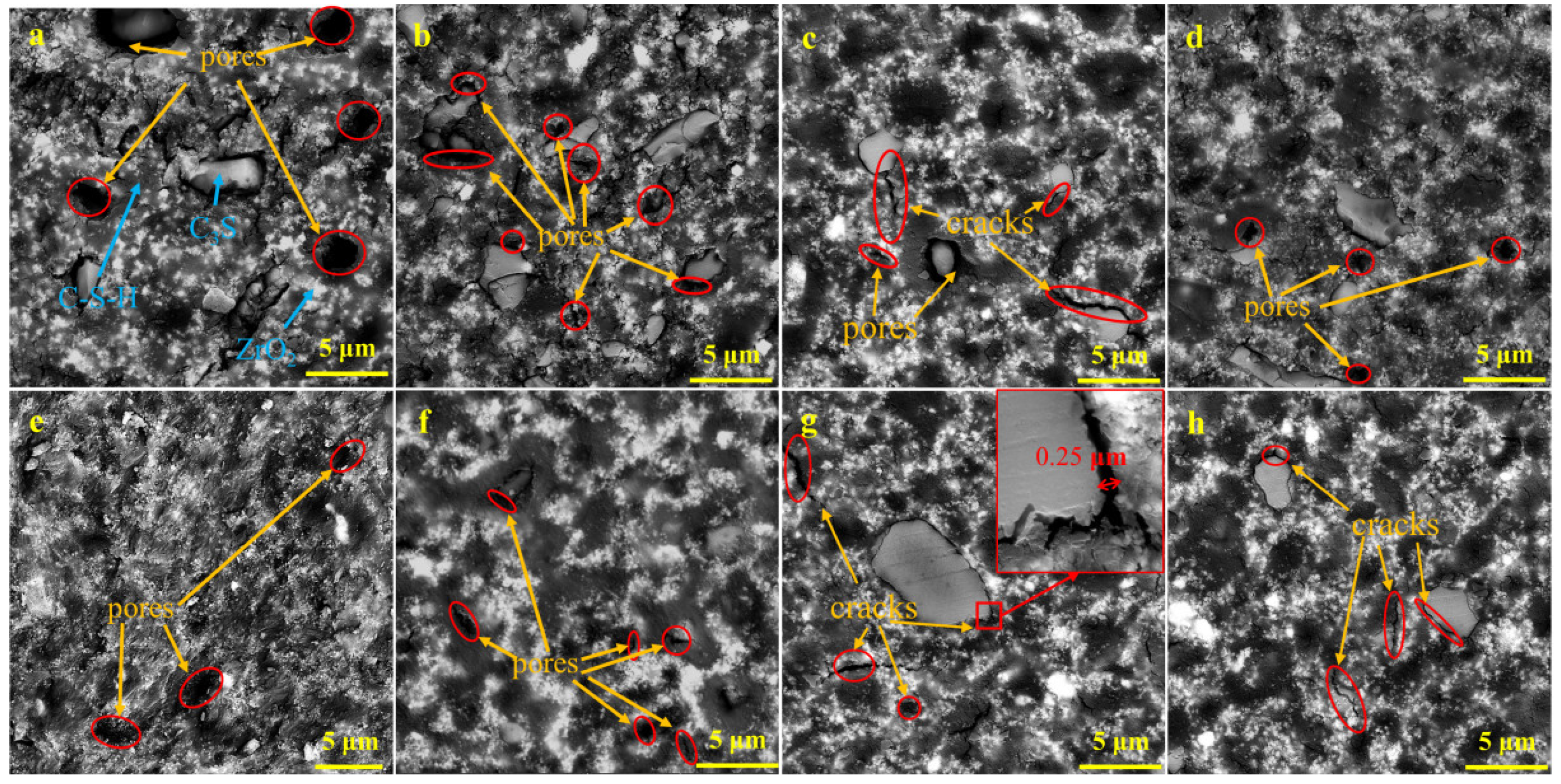
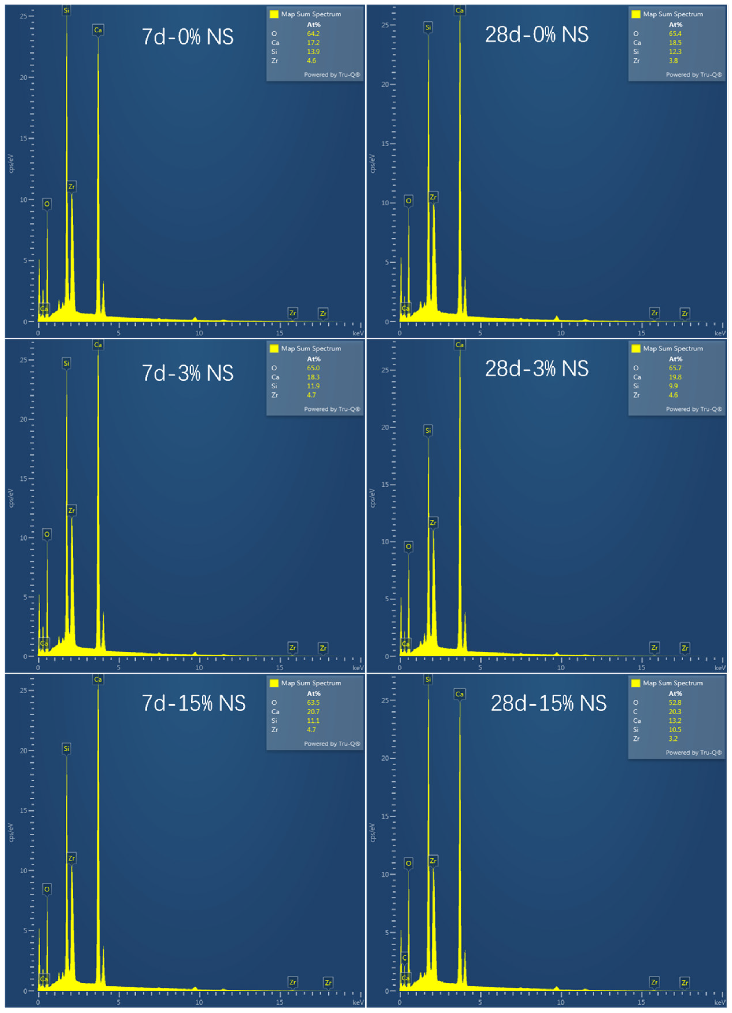
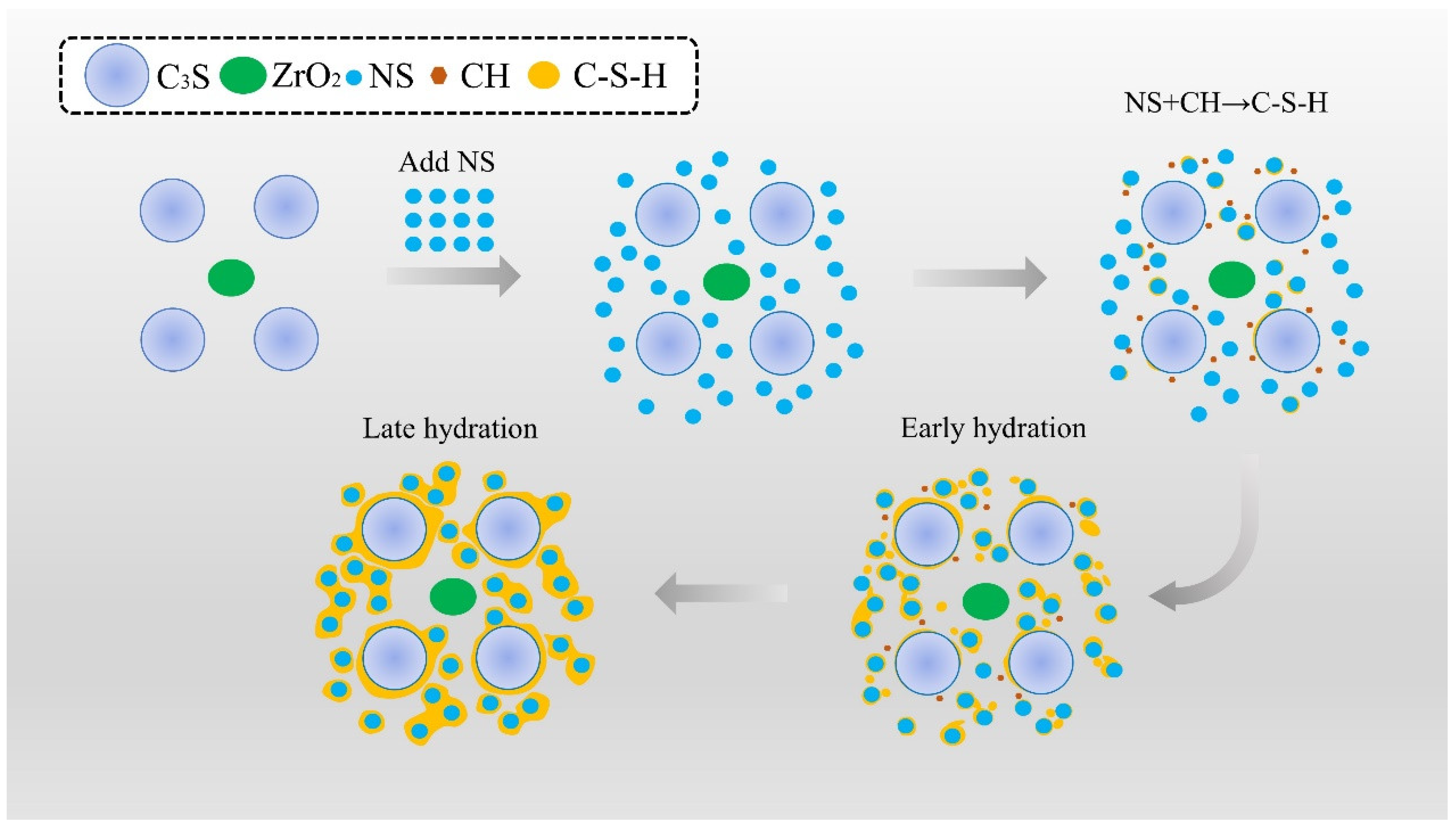
4.2. Effect of Microstructure and Composition on Compressive Strength

Figure 6 shows the XRD patterns of the different NS samples after seven and 28 days of hydration. The results indicate similar phase compositions for the samples at both time points, with ZrO2, C3S, CH, and calcium carbonate (CaCO3) present. In contrast, both the C S H gel and the NS were amorphous and could not be observed by XRD analysis. However, as the NS content increased, the diffraction peak corresponding to the CH phase (2θ = 18.08°, 28.72°, and 34.12°) gradually decreased. After the addition of 15% NS, almost no CH phase was observed after 28 days of hydration. The intensity and position of the ZrO2 diffraction peak (2θ = 28.16°, 31.40° 50.88°, and 62.64°) remained consistent, indicating inertness during the hydration process. The diffraction peaks of the C3S phase (2θ = 34.12°, 41.24°, and 62.64°) overlapped with those of the CaCO3, CH, and ZrO2 phases, making it challenging to determine whether unreacted C3S was present. An SEM analysis was performed to address this issue. The appearance of the CaCO3 phase resulted from the reaction of CH with carbon dioxide in the air.
Figure 7 shows the SEM micrographs of the samples with different NS content after hydration for seven and 28 days. Figure 8 shows the EDX plots of samples with different amounts of NS after 7 and 28 days of hydra-tion. As shown in Figure 7a, the uniformly distributed white clusters represented ZrO2. The irregularly shaped particles were unhydrated C3S. The net-like structure interwoven in the material was hydrated CaSiO3. In addition, numerous pores and microcracks were observed. After seven days of hydration, the sample without added NS (a) had large pores and a loose structure. After NS was added, the pore size decreased, forming a dense C S H gel. Unreacted C3S was observed in all sample groups. After 28 days of hydration, the sample without added NS, seen in Figure 7e, showed a more compact structure than the sample after seven days, seen in Figure 7a, and no obvious C3S was present. However, in the sample with 9% NS, shown in Figure 7g, unreacted C3S was clearly observed. In addition, there was a gap of approximately 0.25 μm between the unreacted C3S and the surrounding hydration gel.
During the C3S hydration process, CH and C S H are produced. The calculation of the CH content made it possible to assess the relative degree of hydration in each sample indirectly [24,26,27]. To understand the impact of NS addition on hydration, it is essential to analyze the total CH production resulting from the hydration process in each sample. The CH generated during sample hydration comprised three components: CH decomposed by heat via Equation (2); CH that reacted with CO2 to form CaCO3 via Equation (3); and CH that reacted with NS to create a hydration gel—see Equation (4). The first two were determined using TG analysis and calculations, whereas the latter was measured using spectrophotometry and the potassium fluorosilicate method. This made it possible to estimate the amount of reacted silica and calculate the corresponding CH content.
C a O H 2 C a O + H 2 O
C a 0 H 2 + C O 2 = C a C O 3 + H 2 O
C a O H 2 + S i O 2 = C a S i O 3 + H 2 O
The total CH content resulting from hydration was calculated using Equation (5).
W C H = 74.1 18 × m 1 + 74.1 44 × m 2 + 74.1 60 × m N S
where WCH represents the overall content of CH generated through hydration (%), m1 represents the mass loss rate resulting from CH dehydration (%), m2 represents the mass loss rate stemming from CaCO3 decomposition (%), and mNS signifies the amount of NS involved in the reaction (%). The free NS content in the sample after 28 days of hydration is shown in Table 3.
Figure 9 shows the TG analysis of the NS-modified samples after seven and 28 days of hydration. The first peak at 40–200 °C signifies C S H dehydration and free water evaporation. The second peak at 380–500 °C represents CH decomposition, and the third peak at 530–700 °C is related to CaCO3 decomposition. Over the hydration period, the CH content decreased with the addition of NS, and the 15% NS sample lacked significant CH content after 28 days. However, the CH content of the NS-free samples increased from seven to 28 days. Nevertheless, the samples with 3%, 9%, and 15% NS exhibited a decrease in CH content with increasing hydration time.
Using Equation (5), the total CH content generated after 28 days of hydration was determined for each sample group, as shown in Figure 10. The figure shows that, as the samples were hydrated for 28 days, the total CH content gradually decreased with increasing NS content. The NS-free samples exhibited the highest total CH production (21.35%). In contrast, the samples with 15% NS only generated 62.25% of the CH that the NS-free group generated. The total CH production was directly correlated with the degree of C3S hydration, implying that increasing the NS content led to a relatively reduced C3S hydration degree. This observation aligns with the SEM images in Figure 7e–h, which indicate that higher NS content resulted in more visible unreacted C3S particles.

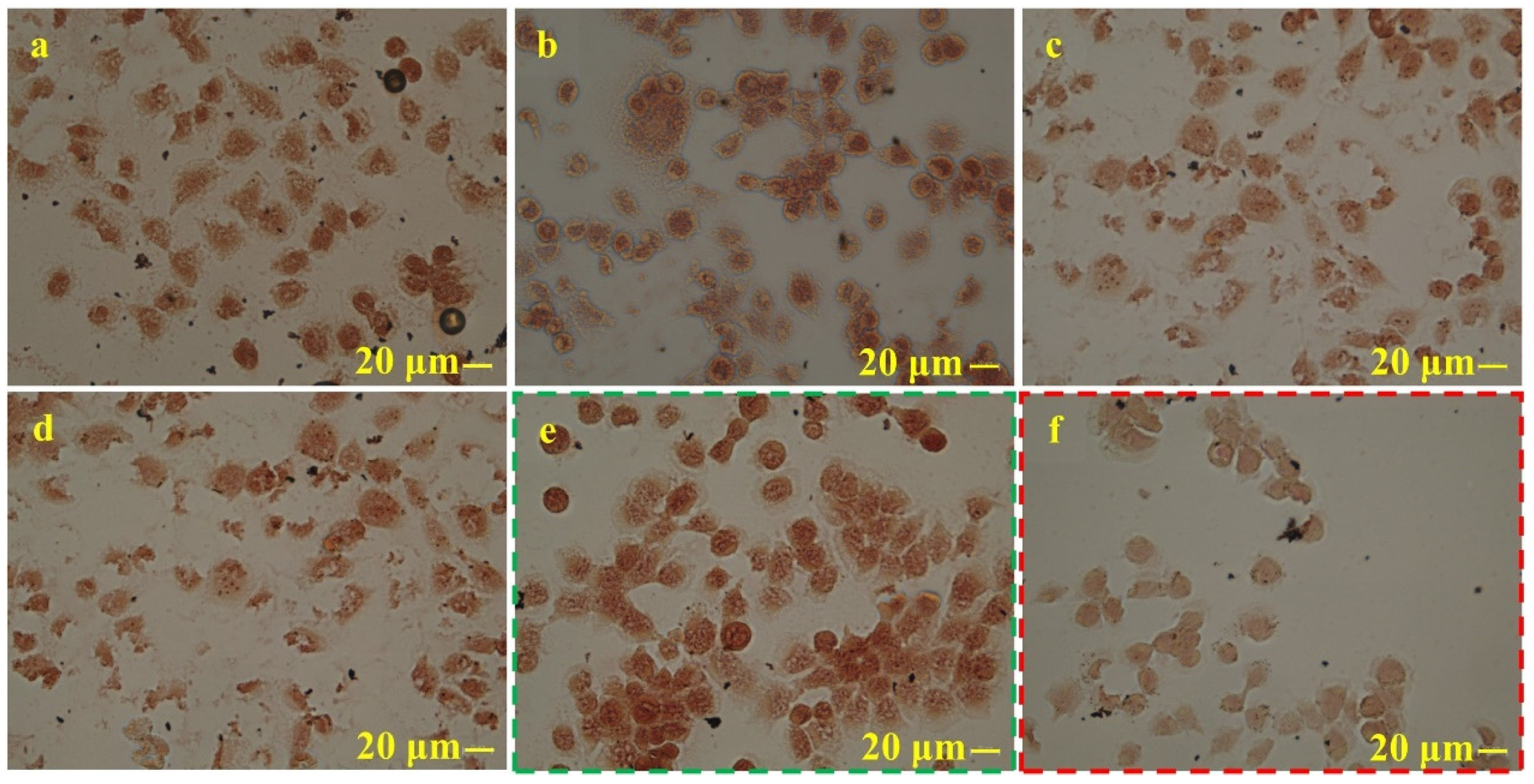
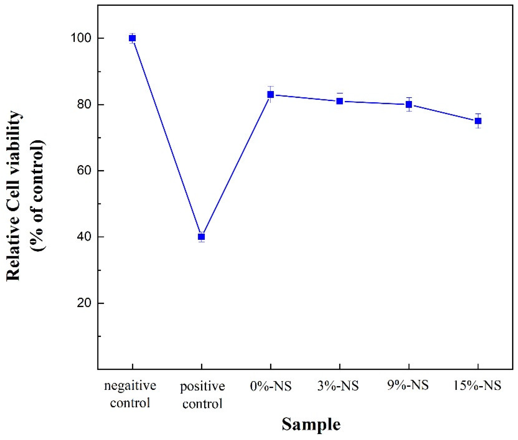
4.3. Effect of NS Content on the Cytotoxicity of the Material

Figure 11 shows the cell morphologies of the test and control groups. In the negative control group in Figure 11e, the cell morphology was complete with no malformed cells. In the positive control group in Figure 11f, cell growth was inhibited, resulting in a reduced number of cells compared with those in the negative control group, and a significant proportion of cells appeared to be malformed. The NS-free samples and those with 3% NS did not show any notable inhibition of cell growth. Furthermore, specimens with NS addition of 9%, shown in Figure 11c, and 15% NS, shown in Figure 11d, had an increased number of malformed cells compared with the specimens without NS and those with 3% added NS. The mean value of the number of cells in the negative control group was established as 100%, the images for each group were processed, and their respective cell activities were calculated. Figure 12 shows the cell viability of the test and control samples. Based on the five grades of cytotoxicity, the negative control group exhibited no cytotoxicity. The NS-free, 3% NS, and 9% NS groups displayed slight toxicity; the 15% NS group showed mild toxicity; and the positive control group exhibited moderate cytotoxicity.

5. Discussion

The findings suggest that NS has the potential to enhance the early strength of calcium-silicate-based root canal filling paste, albeit at the cost of diminishing its compressive strength during the later stages of hydration. As shown in Figure 13, the initial stages of C3S hydration produced colloidal-sized C S H and larger CH crystals that enveloped the surface of the C3S. As the hydration progressed, this unstable wrapping layer on the C3S surface broke down, resulting in the precipitation of more stable hydration crystals. The introduced NS acted as a nucleation site to accelerate C3S hydration and interacted with CH to create a dense C S H gel [26,27]. This gel, combined with CH crystals and hydrated Ca3SiO5, generated a dense structure that filled the voids [48]. Consequently, in the case of samples hydrated for seven days, the porosity decreased and the compressive strength increased in samples containing 3% and 9% NS. However, the sample containing 15% NS exhibited decreased strength. This phenomenon can be attributed to the excessive NS content, which dramatically accelerated the initial hydration of C3S, ultimately encasing it entirely within the hydration products. The observed enhancement in early hydration is consistent with findings by Vivek et al. [49], who reported that NS as a replacement for ordinary Portland cement up to 3% weight could accelerate C S H gel formation and adversely increases the resistance to water permeability of high-performance concrete. In the later stages of hydration, the dense C S H gel, acting as a diffusion barrier, tightly enveloped the surfaces of NS and C3S, preventing further interactions between the C3S and water, thereby hindering the continued hydration of C3S. Consequently, a significant number of unreacted C3S particles remained in the samples. The unreacted C3S featured a gap of 0.25 μm with the hydration gel, and these gaps were conducive to the creation of stress concentration points under external forces, ultimately reducing the compressive strength.
Cytotoxicity assessment is a crucial initial step in evaluating the biocompatibility of root canal filling materials. Examining the cytotoxicity of a calcium-silicate-based root canal filling paste revealed acceptable compatibility with human fibroblasts [50,51,52]. Fresh iRoot SP sealer displayed only mild cytotoxicity in mouse fibroblasts, with relative cell activities ranging from 80% to 100% [53]. This cytotoxicity level was lower than that of AH Plus [54] and MTA Fillapex [55,56]. Research on the BioRoot RCS and TotalFill BC sealers indicated no cytotoxicity within 24 h, with cell viability of approximately 93.41% and 90.70%, respectively. However, at 48 and 72 h, mild cytotoxicity was observed, with cell activity ranging from 60% to 72.15%. Figure 14 shows a comparison of the cytotoxicity of calcium-silicate-based root canal filling materials with NS added (this study) to various root canal sealers reported in the literature. The cytotoxicity of the calcium-silicate-based root canal filling paste with NS was comparable to that of AH Plus, endomethasone, and the pulp canal sealer and slightly higher than that of MTA Filapex, but lower than that of EndoSequence BC. As the NS content increased, there was a slight decrease in cell activity; however, it was still within the range observed for calcium-silicate-based root canal sealers. This suggests that the addition of NS has no significant impact on the cytotoxicity of calcium-silicate-based root canal filling materials. The cytotoxicity of the sample may be related to the hydration product, CH, and an increase in the environmental pH may harm cellular biofilms and DNA, affecting both bacteria and host cells [57]. Martins et al. found that, as the paste was cured and the pH stabilized, the cell damage gradually decreased [58]. With increasing NS content, the early hydration of is C3S accelerated, leading to increased CH production, which may cause the deformation of adjacent cells and medium proteins [54]. Consequently, as the NS content increased, the cytotoxicity in the test group shifted from mild to moderate. Excellent biocompatibility for NS addition was also demonstrated by Wu [59], suggesting that nano-silica not only improves the mechanical properties but also enhances the cellular viability. These results indicate that nano-silica could be a valuable additive for the development of more effective and biocompatible root canal filling materials. Future research should focus on long-term in vivo studies to confirm these findings.

6. Conclusions

The effects of the NS content on the compressive strength and cytotoxicity of a calcium-silicate-based root canal filling paste were investigated. The following conclusions were drawn based on the experimental data.
NS enhanced the early compressive strength of the calcium-silicate-based root canal filling paste by facilitating C3S hydration, increasing hydration product formation, and making efficient pore filling possible. The addition of 9% NS enhanced the early compressive strength by 41.99% compared with the NS-free samples. However, NS exerted some inhibitory effects on later-stage hydration, resulting in reduced compressive strength during this phase. Specifically, the compressive strength of samples with 15% NS in the later stage was only 23.5 ± 2.5 MPa—a 76.38% decline compared with the NS-free samples.
The inclusion of NS did not significantly affect the cytotoxicity of the calcium-silicate-based root canal filling paste. The test groups without NS and those with 3% and 9% NS exhibited mild cytotoxicity, whereas the group with 15% NS showed moderate cytotoxicity. The cytotoxicity observed can be attributed to the increase in the environmental pH resulting from CH production during the hydration process.
In this study, calcium-silicate-based slurries were prepared using different proportions of NS. The results of this study highlight that NS improves the early compressive strength of C3S by facilitating the hydration reaction, while decreasing the initial and final solidification times of the hydrated samples. In addition, NS improved the formation of hydration products and increased the pore filling efficiency. Notably, NS had no significant effect on the cytotoxicity of root canal filling materials. Nonetheless, further research is warranted to support the unrestricted clinical use of NS-enriched calcium-silicate-based root canal filling pastes.

Author Contributions

H.H.: writing—review and editing, conceptualization, validation, funding acquisition. B.H.: writing—original draft, conceptualization, resources. X.X.: validation, resources, project administration. Y.C.: writing—review and editing, data curation, formal analysis. J.L.: funding acquisition, writing—review and editing, project administration. Z.H.: writing—original draft, supervision, visualization, validation, methodology. D.L.: validation, formal analysis, methodology, data curation. Z.W.: investigation, formal analysis, methodology. J.Q.: visualization, software. All authors have read and agreed to the published version of the manuscript.

Funding

This work was supported by the National Natural Science Foundation of China (Project No.: 52164042), the Guangxi Science and Technology Program (Project No.: 2021AB16028, 2022AB25008), and the Foundation of Hunan Provincial Education Department (21B0121), as well as the General Guidance Subject Project of Hunan Provincial Health Commission (Project No. 202208025262). The views expressed are those of the authors and not related to the above projects.

Data Availability Statement

The data presented in this study are available on request from the corresponding author.

Conflicts of Interest

The authors declare no conflicts of interest.

References

  1. Olcay, K.; Eyüboglu, T.F.; Özcan, M. Clinical outcomes of non-surgical multiple-visit root canal retreatment: A retrospective cohort study. Odontology 2019, 107, 536–545. [Google Scholar] [CrossRef]
  2. Xiao, S.; Sun, G.; Huang, S.; Lin, C.; Li, Y. Nanoarchitectonics-Based Materials as a Promising Strategy in the Treatment of Endodontic Infections. Pharmaceutics 2024, 16, 759. [Google Scholar] [CrossRef]
  3. Song, W.C.; Sun, W.; Chen, L.L.; Yuan, Z.L. In vivo biocompatibility and bioactivity of calcium silicate-based bioceramics in endodontics. Front. Bioeng. Biotechnol. 2020, 8, 580954. [Google Scholar] [CrossRef] [PubMed]
  4. Primus, C.M.; Tay, F.R.; Niu, L.N. Bioactive tri/dicalcium silicate cements for treatment of pulpal and periapical tissues. Acta Biomater. 2019, 96, 35–54. [Google Scholar] [CrossRef] [PubMed]
  5. Goldberger, T.; Rosen, E.; Blau-Venezia, N.; Tamse, A.; Littner, D. Pathognomonic combination of clinical signs for diagnosis of vertical root fracture: Systematic review of the literature. Appl. Sci. 2021, 11, 10893. [Google Scholar] [CrossRef]
  6. Liao, W.C.; Chen, C.H.; Pan, Y.H.; Chang, M.C.; Jeng, J.H. Vertical Root Fracture in Non-Endodontically and Endodontically Treated Teeth: Current Understanding and Future Challenge. J. Pers. Med. 2021, 11, 1375. [Google Scholar] [CrossRef]
  7. Sairaman, S.; Solete, P.; Jeevanandan, G.; Antony, S.D.P.; Kavoor, S.; Adimulapu, H.S. Comparative analysis of novel heat-treated retreatment file system on the removal of obturating material using nano-computed tomography. J. Conserv. Dent. Endod. 2024, 27, 82–86. [Google Scholar] [CrossRef]
  8. Grinkevičiūtė, P.; Leknickė, G.; Lodienė, G.; Kriūkienė, R. Effects of irrigation solutions on root canal dentin. Int. J. Appl. Sci. Eng. Technol. 2019, 9, 4. [Google Scholar] [CrossRef]
  9. Oliveira Tavares, S.J.; Pintor, A.V.B.; Caetano, S.K.; Dos Santos, N.C.A.; Pistoia, B.M.; de Carvalho Camilo, M.R.; Scelza, P.; Scelza, M.F.Z. Is There a Relationship between Laser Therapy and Root Canal Cracks Formation? A Systematic Review. Iran. Endod. J. 2023, 18, 2–14. [Google Scholar] [PubMed]
  10. Dimitriu, B.; Suciu, I.; Amza, O.E.; Ciocârdel, M.; Bodnar, D.; Țâncu, A.M.C.; Tanase, M.; Branescu, M.S.; Chirilă, M. Optical microscopy evaluation of root canal filling removal by beginner operators in posterior teeth. J. Med. Life 2024, 17, 555–563. [Google Scholar] [PubMed]
  11. D’souza, J.M.; de Ataide, I.N.; Lambor, R. Effect of Newer Intraorifice Barriers on the Fracture Resistance of Endodontically Treated Teeth: An In Vitro Study. Cureus 2024, 16, e69463. [Google Scholar] [CrossRef] [PubMed]
  12. Sönmez, S.; Koç, C. Effect of several nickel-titanium rotary systems on stress distribution in mandibular molars under occlusal forces: A finite element analysis. Odontology 2024. Advance online publication. [Google Scholar] [CrossRef]
  13. Abdel-Maksoud, H.B.; Eid, B.M.; Hamdy, M.; Abdelaal, H.M. Optimizing fracture resistance of endodontically treated maxillary premolars restored with preheated thermos-viscous composite post-thermocycling, a comparative study. Part I. BMC Oral Health 2024, 24, 295. [Google Scholar] [CrossRef] [PubMed]
  14. Mergulhão, V.A.; de Mendonça, L.S.; de Albuquerque, M.S.; Braz, R. Fracture Resistance of Endodontically Treated Maxillary Premolars Restored with Different Methods. Oper. Dent. 2019, 44, E1–E11. [Google Scholar] [CrossRef] [PubMed]
  15. Aslan, T.; Esim, E.; Üstün, Y. Finite element evaluation of dentin stress changes following different endodontic surgical approaches. Odontology 2024, 112, 798–810. [Google Scholar] [CrossRef]
  16. Uzunoglu Ozyurek, E.; Aktemur Turker, S. Evaluation of fracture resistance of roots-filled with various root canal sealers at different time periods. Eur. Oral Res. 2019, 53, 6–11. [Google Scholar] [CrossRef]
  17. Papynov, E.K.; Shichalin, O.O.; Buravlev, I.Y.; Portnyagin, A.S.; Belov, A.A.; Maiorov, V.Y.; Skurikhina, Y.E.; Merkulov, E.B.; Glavinskaya, V.O.; Nomerovskii, A.D.; et al. Reactive Spark Plasma Synthesis of Porous Bioceramic Wollastonite. Russ. J. Inorg. Chem. 2020, 65, 263–270. [Google Scholar] [CrossRef]
  18. Issar, R.; Ranjan, S.; Saurav, S.; Kar, S.; Bera, S.; Singh, P. Comparison of Fracture Resistance of Two Resin-based Sealers to Root Canal Walls: An In Vitro Study. J. Contemp. Dent. Pract. 2020, 21, 1253–1257. [Google Scholar]
  19. Chew, S.T.; Eshak, Z.; Al-Haddad, A. Evaluation of interfacial adaptation and penetration of bioceramic-based sealers in oval root canals: A confocal laser scanning microscope study. Microsc. Res. Tech. 2023, 86, 754–761. [Google Scholar] [CrossRef]
  20. Turkyilmaz, A.; Erdemir, A. Comparison of dentin penetration ability of different root canal sealers used with different obturation methods. Microsc. Res. Tech. 2020, 83, 1544–1551. [Google Scholar] [CrossRef]
  21. Zamparini, F.; Prati, C.; Taddei, P.; Spinelli, A.; Di Foggia, M.; Gandolfi, M.G. Chemical-Physical Properties and Bioactivity of New Premixed Calcium Silicate-Bioceramic Root Canal Sealers. Int. J. Mol. Sci. 2022, 23, 13914. [Google Scholar] [CrossRef]
  22. Komabayashi, T.; Colmenar, D.; Cvach, N.; Bhat, A.; Primus, C.; Imai, Y. Comprehensive review of current endodontic sealers. Dent. Mater. J. 2020, 39, 703–720. [Google Scholar] [CrossRef] [PubMed]
  23. Hachem, R.E.; Khalil, I.; Brun, G.L.; Pellen, F.; Jeune, B.L.; Daou, M.; Osta, N.E.; Naaman, A.; Abboud, M. Dentinal tubule penetration of AH Plus, BC Sealer and a novel tricalcium silicate sealer: A confocal laser scanning microscopy study. Clin. Oral Investig. J. 2019, 23, 1871–1876. [Google Scholar] [CrossRef] [PubMed]
  24. Yang, H.B.; Monasterio, M.; Zheng, D.P.; Cui, H.Z.; Tang, W.C.; Bao, X.H.; Chen, X.S. Effects of nano silica on the properties of cement-based materials: A comprehensive review. Constr. Build. Mater. 2021, 282, 122715. [Google Scholar] [CrossRef]
  25. Bai, S.; Guan, X.C.; Li, H.; Ou, J.P. Effect of the specific surface area of nano-silica particle on the properties of cement paste. Powder Technol. 2021, 392, 680–689. [Google Scholar] [CrossRef]
  26. Zhang, N.; Zhou, H.; Hu, Y.; Wang, J.; Hou, G.; Ma, J.; Jiang, R. Effect of Stöber Nano-SiO2 Particles on the Hydration Properties of Calcined Coal Gangue-Blended Cement. Materials 2024, 17, 4218. [Google Scholar] [CrossRef]
  27. Boris, R.; Wilińska, I.; Pacewska, B.; Antonovič, V. Investigations of the Influence of Nano-Admixtures on Early Hydration and Selected Properties of Calcium Aluminate Cement Paste. Materials 2022, 15, 4958. [Google Scholar] [CrossRef]
  28. Hisham Shah, M.H.; Lee, C.C.; Zamzuri, M.Z.; Vijayan, V. Colloidal Nanosilica Effect on the Properties and Durability of Coir Reinforced Cement Brick. Adv. Mater. Sci. Eng. 2022, 2022, 7610297. [Google Scholar] [CrossRef]
  29. Shahram, M.A.; Alireza, K.; Mohammad, H.B.; Mohammad, S.T.M. Performance of polypropylene fiber reinforced concrete-filled UPVC tube columns under axial compression. Constr. Build. Mater. 2020, 231, 117049. [Google Scholar]
  30. 1SO 6876: 2012[S/OL]; Dentistry—Root Canal Sealing Materials. ISO Technical Committees: Geneva, Switzerland, 2012. Available online: https://www.iso.org/standard/45117.html (accessed on 28 December 2024).
  31. C266-21[S/OL]; Standard Test Method for Time of Setting of Hydraulic-Cement Paste by Gillmore Needles. ISO Technical Committees: Geneva, Switzerland, 2021. Available online: https://www.astm.org/standards/c266 (accessed on 28 December 2024).
  32. Hall, C.; Hamilton, A. Porosity–density relations in stone and brick materials. Mater. Struct. J. 2015, 48, 1265–1271. [Google Scholar] [CrossRef]
  33. Shen, C.C. Preparation process and application research of calcium silicate-based materials. Master’s Thesis, Tongfang CNKI (Beijing) Technology Co., Ltd., Beijing, China, 2020. [Google Scholar] [CrossRef]
  34. GB/T 28629-2012[S/OL]; Methods for Chemical Analysis of Free Silicon Dioxide for Cement Clinker. National Technical Committee for Cement Standardization: Beijing, China, 2013. Available online: https://std.samr.gov.cn/gb/search/gbDetailed?id=173829859CF91AA5E06397BE0A0AA311 (accessed on 28 December 2024).
  35. ISO 7405: 2018[S/OL]; Dentistry—Evaluation of Biocompatibility of Medical Devices Used in Dentistry. ISO Technical Committees: Geneva, Switzerland, 2018. Available online: https://www.iso.org/standard/71503.html (accessed on 28 December 2024).
  36. Jain, D.; Shetty, K.P.; Luke, A.M.; Ballal, N.V. Root canal irrigants effect on the compressive strength and calcium ion release of Biodentine. Heliyon 2024, 10, e38267. [Google Scholar] [CrossRef] [PubMed]
  37. Ruiz-Linares, M.; de Oliveira Fagundes, J.; Solana, C.; Baca, P.; Ferrer-Luque, C.M. Current status on antimicrobial activity of a tricalcium silicate cement. J. Oral Sci. 2022, 64, 113–117. [Google Scholar] [CrossRef] [PubMed]
  38. Baghdadi, I.; AbuTarboush, B.J.; Zaazou, A.; Skienhe, H.; Özcan, M.; Zakhour, M.; Salameh, Z. Investigation of the structure and compressive strength of a bioceramic root canal sealer reinforced with nanomaterials. J. Appl. Biomater. 2021, 19, 22808000211014747. [Google Scholar] [CrossRef]
  39. Li, L.G.; Zheng, J.Y.; Ng, P.L.; Zhu, J.; Kwan, A.K.H. Cementing efficiencies and synergistic roles of silica fume and nano-silica in sulphate and chloride resistance of concrete. Constr. Build. Mater. 2019, 223, 965–975. [Google Scholar] [CrossRef]
  40. Saleh, A.N.; Attar, A.A.; Ahmed, O.K.; Mustafa, S.S. Improving the thermal insulation and mechanical properties of concrete using Nano-SiO2. Results Eng. 2021, 12, 100303. [Google Scholar] [CrossRef]
  41. Vineet, S. Performance evaluation of carbonated cement paste derived from hydrated Portland cement based binders as supplementary cementitious material. CEMENT 2024, 15, 100091. [Google Scholar]
  42. Gao, C.; Huang, L.; Yan, L.B.; Jin, R.Y.; Chen, H.Z. Mechanical properties of recycled aggregate concrete modified by nano-particles. Constr. Build. Mater. 2020, 241, 118030. [Google Scholar] [CrossRef]
  43. Ma, B.G.; Li, H.N.; Mei, J.P.; Li, X.G. Influence of Nano-SiO2 addition on properties of sulphoaluminate cement based material. J. Wuhan Univ. Technol. 2017, 32, 106–112. [Google Scholar] [CrossRef]
  44. Kooshafar, M.; Madani, H. An investigation on the influence of nano silica morphology on the characteristics of cement composites. J. Build. Eng. 2020, 30, 101293. [Google Scholar] [CrossRef]
  45. Anna, M.; Andrea, C.; Francesco, B.B.; Emiliano, B.; Fabio, P.; Andrea, S.; Fabio, I.; Giovanni, I.; Peter, Z.; Giovanni, C.; et al. Exceptional thermal strain reduction by a tilting pillar architecture: Suspended Ge layers on Si (001). Mater. Design 2017, 116, 144–151. [Google Scholar]
  46. Wang, D.; Shi, C.; Wu, Z.; Wu, L.; Xiang, S.; Pan, X. Effects of nanomaterials on hardening of cement–silica fume–fly ash-based ultra-high-strength concrete. Adv. Cem. Res. 2016, 28, 555–566. [Google Scholar] [CrossRef]
  47. Wang, L.G.; Zheng, D.P.; Zhang, S.P.; Cui, H.Z.; Li, D.X. Effect of Nano-SiO2 on the hydration and microstructure of portland cement. Nanomater. 2016, 6, 241. [Google Scholar] [CrossRef] [PubMed]
  48. Hou, P.K.; Shi, J.; Prabakar, S.; Cheng, X.; Wang, K.J.; Zhou, X.M.; Surendra, P.S. Effects of mixing sequences of nanosilica on the hydration and hardening properties of cement-based materials. Constr. Build. Mater. 2020, 263, 120226. [Google Scholar] [CrossRef]
  49. Vivek, D.; Elango, K.S.; Saravanakumar, R.; Mohamed Rafek, B.; Ragavendra, P.; Kaviarasan, S.; Raguram, E. Effect of Nano-Silica in high performance concrete. Mater. Today Proc. 2021, 37, 1226–1229. [Google Scholar] [CrossRef]
  50. La Rosa, G.R.M.; Canova, F.S.; Leotta, M.L.; Pedullà, E. Effects of heating on bioceramic sealers: A scoping review of chemo-physical properties and clinical implications. Odontology 2024, 1–14. [Google Scholar] [CrossRef]
  51. Zhou, H.M.; Du, T.F.; Shen, Y.; Wang, Z.J.; Zheng, Y.F.; Haapasalo, M. In vitro cytotoxicity of calcium silicate-containing endodontic sealers. J. Endodont. 2015, 41, 56–61. [Google Scholar] [CrossRef] [PubMed]
  52. Seo, D.G.; Lee, D.; Kim, Y.M.; Song, D.; Kim, S.Y. Biocompatibility and Mineralization Activity of Three Calcium Silicate-Based Root Canal Sealers Compared to Conventional Resin-Based Sealer in Human Dental Pulp Stem Cells. Materials 2019, 12, 2482. [Google Scholar] [CrossRef] [PubMed]
  53. López-García, S.; Myong-Hyun, B.; Lozano, A.; García-Bernal, D.; Forner, L.; Llena, C.; Guerrero-Gironés, J.; Murcia, L.; Rodríguez-Lozano, F.J. Cytocompatibility, bioactivity potential, and ion release of three premixed calcium silicate-based sealers. Clin. Oral Investig. 2020, 24, 1749–1759. [Google Scholar] [CrossRef]
  54. Zhang, W.; Li, Z.; Peng, B. Ex vivo cytotoxicity of a new calcium silicate-based canal filling material. Int. Endod. J. 2010, 43, 769–774. [Google Scholar] [CrossRef]
  55. Mestieri, L.B.; Zaccara, I.M.; Pinheiro, L.S.; Barletta, F.B.; Kopper, P.M.P.; Grecca, F.S. Cytocompatibility and cell proliferation evaluation of calcium phosphate-based root canal sealers. Restor. Dent. Endod. 2019, 45, e2. [Google Scholar] [CrossRef] [PubMed]
  56. Silva, E.J.; Carvalho, N.K.; Ronconi, C.T.; Deus, G.D.; Zuolo, M.L.; Zaia, A.A. Cytotoxicity profile of endodontic sealers provided by 3D cell culture experimental model. Braz. Dent. J. 2016, 27, 652–656. [Google Scholar] [CrossRef] [PubMed]
  57. Gaudin, A.; Tolar, M.; Peters, O.A. Cytokine Production and Cytotoxicity of Calcium Silicate-based Sealers in 2- and 3-dimensional Cell Culture Models. J. Endod. 2020, 46, 818–826. [Google Scholar] [CrossRef] [PubMed]
  58. Martins, M.D.; Coelho, F.H.; Moreira, M.S.; Marques, M.M.; Pilar, E.F.S.; Palo, R.M.; Silveira, F.M.; Kopper, P.M.P. Cytotoxicity, biocompatibility and osteoinductive profile of an MTA-hydrogel-based cement: An in vitro and animal study. Int. Endod. J. 2023, 56, 955–967. [Google Scholar] [CrossRef] [PubMed]
  59. Wu, M.; Wang, T.; Zha, W.; Xiang, W.; Zheng, W.B. Effects of nano-SiO2 particles on physio-chemical properties of bioactive tricalcium silicate cements. J. Aust. Ceram. Soc. 2021, 57, 9–20. [Google Scholar] [CrossRef]
Figure 1. Morphology and XRD pattern of NS powder.
Crystals 15 00055 g001
Figure 2. Experimental method: (a) sample preparation and (b) experiment.
Crystals 15 00055 g002
Figure 3. Compressive strengths of samples after hydration for seven and 28 days (mean ± SD of compressive strength).
Crystals 15 00055 g003
Figure 4. Setting times of samples after hydration for seven days and 28 days (mean ± SD of setting time).
Crystals 15 00055 g004
Figure 5. Porosity of samples after hydration for seven days and 28 days (mean ± SD of porosity).
Crystals 15 00055 g005
Figure 6. XRD analysis of NS samples (0–15%): (a) seven days of hydration and (b) 28 days of hydration.
Crystals 15 00055 g006
Figure 7. SEM micrographs of samples with different amounts of NS after hydration for seven and 28 days: hydration for seven days with (a) 0% NS, (b) 3% NS, (c) 9% NS, and (d) 15% NS and hydration for 28 days with (e) 0% NS, (f) 3% NS, (g) 9% NS, and (h) 15% NS.
Crystals 15 00055 g007
Figure 8. EDX plots of samples with different amounts of NS after 7 and 28 days of hydration.
Crystals 15 00055 g008
Figure 9. TG analysis of NS-modified samples at seven and 28 days of hydration.
Crystals 15 00055 g009
Figure 10. Total CH content generated after 28 days of hydration: mean ± SD of total content of CH.
Crystals 15 00055 g010
Figure 11. Cell morphologies of the samples: (a) 0% NS; (b) 3% NS; (c) 9% NS; (d) 15% NS; (e) negative control group (Marked with a green dot box); (f) positive control group (Marked with a red dot box).
Crystals 15 00055 g011
Figure 12. Relative cellular viability of the test and control groups (mean ± SD of cell viability).
Crystals 15 00055 g012
Figure 13. Schematic diagram of the decrease in compressive strength in the later stage of hydration.
Crystals 15 00055 g013
Figure 14. Cytotoxicity comparison: calcium-silicate-based material with NS versus root canal sealers [54,55,56].
Crystals 15 00055 g014
Table 1. Important performance parameters and manufacturer details of powder raw materials.
MaterialDensity (g/mL)Average Particle Size
(Standard Deviation)
PurityManufacturer
Ca3SiO5
(C3S)
3.7412 ± 2 μm≥99.0%Hubei Wande Chemical Co., Ltd. Hubei, China.
NS2.6030 ± 5 nm≥99.5%Shanghai Maclin Biochemical Technology Co., Ltd. Shanghai, China.
ZrO25.8950 ± 10 nm≥99.9%Shanghai Maclin Biochemical Technology Co., Ltd. Shanghai, China.
Table 2. Adhesive proportion.
TypeSolid–Liquid RatioZrO2 Content (Solid)NS Content (Solid)
Paste 14:120%0%
Paste 24:120%3%
Paste 34:120%9%
Table 3. Content of free NS after 28 days of hydration.
NS Content (%)Detection MethodResidual NS Content (%)NS Involved in Reaction (%)
3Spectrophotometer0.262.74
9Spectrophotometer4.144.86
15Potassium fluorosilicate5.569.44
Disclaimer/Publisher’s Note: The statements, opinions and data contained in all publications are solely those of the individual author(s) and contributor(s) and not of MDPI and/or the editor(s). MDPI and/or the editor(s) disclaim responsibility for any injury to people or property resulting from any ideas, methods, instructions or products referred to in the content.

