Article

A Fluorescence Sensor Based on Biphenolic Backbone for Metal Ion Detection: Synthesis and Crystal Structure

by
Kanokporn Chantaniyomporn
1,
Kiratikarn Charoensuk
1,
Tanwawan Duangthongyou
2,
Kittipong Chainok
3 and
Boontana Wannalerse
2,*
1
Department of Chemistry, Faculty of Science, Kasetsart University, Bangkok 10900, Thailand
2
Center of Excellence for Innovation in Chemistry, Department of Chemistry, Faculty of Science, Kasetsart University, Bangkok 10900, Thailand
3
Thammasat University Research Unit in Multifunctional Crystalline Materials and Applications (TU-MCMA), Faculty of Science and Technology, Thammasat University, Pathum Thani 12121, Thailand
*
Author to whom correspondence should be addressed.
Crystals 2024, 14(11), 943; https://doi.org/10.3390/cryst14110943
Submission received: 9 October 2024 / Revised: 28 October 2024 / Accepted: 28 October 2024 / Published: 30 October 2024
(This article belongs to the Section Inorganic Crystalline Materials)

Abstract

:
2′-(hexyloxy)-[1,1′-biphenyl]-2-yl 5-(dimethylamino)naphthalene-1-sulfonate (KC1) was synthesized by using biphenol and dansyl chloride as starting materials. The KC1 was characterized via single X-ray diffraction, FTIR, HRMS and 1H and 13C-NMR. The KC1 indicates triclinic as P1 in the space group type. From the KC1, the biphenolic backbone structure is twisted at an angle of 54.48° due to connecting the dansyl unit and hexyl moiety. Upon the addition of the Fe3+ ion to the KC1 solution, the fluorescence emission at 585 nm of KC1 was quenched due to complexation between KC1 and the Fe3+ ion. The complexation ratio of KC1 and Fe3+ was determined to be a 1:1 formation via Job’s analysis. The Stern–Volmer constant (Ksv) calculated was 21,203 M−1 for the KC1 and the Fe3+ ion complex.

1. Introduction

Crystal ligands have been designed and synthesized for many applications, such as sensors and molecular tags. These ligands contain the nitrogen, oxygen and sulfur donor atoms that are important parts of binding sites for metal ion coordination complexes [1,2,3,4]. The coordinated complexes formed from ligands and specific metal ions lead to changes in optical and electronic properties. There are many coordination processes, such as cheated quenching/enhancing fluorescence, internal charge transfer and photo-induced electron transfer [5,6,7,8,9]. These mechanisms are essential for sensitivity and selectivity during metal complexation. Metal ions are found everywhere in daily life and have essential uses in biological, environmental and industrial fields. However, some metal ions are toxic and harmful to humans upon excessive intake [10,11,12,13,14,15,16]. Currently, many researchers are focused on synthesizing fluorescence sensor molecules that are specifically coordinated to metal ions. Among metal ions, iron (Fe) is an important transition element that plays many roles, such as in metabolism and oxygen carrying and catalysis. The Fe3+ ion is greater in natural abundance rather than other oxidation states due to its stability. For the human body, a lack of iron is linked to many diseases and disorders [17,18,19], and metal fluorescence detection is an easier and quicker technique than other methods to diagnose deficiencies. Moreover, the advantages of the fluorescence method include being able to detect metal ions in aqueous samples and in vivo cells [20,21]. For example, Areti et. al. (2016) [4] prepared triazole-connected quinolone based on glucopyranose binding to Zn2+ and Cd2+ producing an enhancement in fluorescence, but Cd2+ changed to quench fluorescence. Moreover, this molecule can be generated via INHIBIT and IMPLICATION logic gates for Zn2+-H2PO4 and Hg2+-CN, respectively. Zhang et. al. (2018) [15] prepared a new luminescent MOF, [Cd2(pbdc)(H2O)3], containing the 5-phosphonobenzene-1,3-dicarboxylic acid (H4pbdc) ligand as [Cd2(pbdc)(H2O)3], which linked three dimensions. With the addition of Cu2+, Al3+ and Fe3+ ions, the fluorescence intensity of [Cd2(pbdc)(H2O)3] was more obviously quenched than with other metal ions, meaning that this MOF has higher sensitivity and selectivity for Cu2+, Al3+ and Fe3+ ions in aqueous solution systems. Rasin et. al. (2022) [22] synthesized a fluorescence sensor containing benzene-1,2-diamine and pyrene moieties in DMSO:H2O (9.5/5 v/v) for Fe3+ ion detection, with the addition of the Fe3+ ion increasing the quench fluorescence when compared to other metal ions. This process occurred under photo-induced electron transfer. For this research, we described the synthesis, crystal structure and fluorescence properties of KC1 and investigated the sensitivity and selectivity for metal ions.

2. Experimental Section

2.1. Material

For all reagents in the experiment, 2,2′-biphenol was purchased from Sigma-Aldrich (St. Louis, MO, USA). Potassium carbonate was purchased from Carlo Erba Reagents S.A.S. (Val de Reuil, France). 1-bromahexane and dansyl chloride were purchased from Tokyo Chemical Industry Company Limited (Tokyo, Japan). Iron(III) nitrate nonahydrate, zinc(II) nitrate hexahydrate, copper(II) nitrate trihydrate, nickel(II) nitrate hexahydrate, lead(II) nitrate and mercury(II) nitrate monohydrate were purchased from QRëC, New Zealand. Dichloromethane, dimethyl sulfoxide, hexane and acetonitrile were purchased from RCI Labscan Limited (Bangkok, Thailand).

2.2. Apparatus

The Fourier transform infrared technique (FTIR) was used to examine the functional groups within compounds using a Spectrum Two FT-IR Perkin Elmer spectrometer, Waltham, MA, USA. An FL8500 Fluorescence Perkin Elmer spectrometer was used to investigate the binding behavior of KC1 and metal ions. 1H-13C NMR spectroscopy was utilized to study the structures of synthesized compounds using a 400 MHz NMR Bruker spectrometer, Madison, WI, USA. HRMS was utilized to investigate the parent peak using a nano-LC quadrupole time-of-flight high-resolution mass spectrophotometer.

2.3. X-Ray Crystallography

CCDC 2361572 is the crystallographic data for KC1 accessed on 10 June 2024. These data can be provided from http://www.ccdc.cam.ac.uk. Crystal data of KC1 were collected on a Bruker D8 QUEST CMOS PHOTONII, Germany, and combined with graphite monochromated Mo-Kα(λ = 0.71073Å) radiation at 296 K. In addition, the KCl structure was solved using the ShelXT [23] structure solution program with included Pattererson and dual space recycling methods, and was also performed using least squares with ShelXL [24]. All hydrogen atoms were treated by calculation [25]. The crystal data of KC1are listed in Table 1. Mercury software (http://www.ccdc.cam.ac.uk) was used to depict the KC1 molecule [26,27].

2.4. Fluorescence Titration

The solvent used in the fluorescence experiment was DMSO:H2O (7/3 v/v). The stock solution of KC1 in a concentration of 1 × 10⁻³ M was prepared. Then, the KC1 solution was diluted to 1 × 10⁻⁵ M in DMSO:H2O (7/3 v/v). Metal ions were added to prepare KC1 solution in a concentration of 0–3000 equivalents. Then, the emission spectra of fluorescence titration were recorded at 350–800 nm.

2.5. Synthesis of 2′-(Hexyloxy)-[1,1′-Biphenyl]-2-yl 5-(Dimethylamino)Naphthalene-1-Sulfonate, KC1 Exhibited in Scheme 1

2.5.1. 2′-(Hexyloxy)-[1,1′-Biphenyl]-2-ol, (C18H22O2) Compound A

2,2′-biphenol (3 g, 16.1 mmol) and potassium carbonate (K2CO3) (2.226 g, 16.1 mmol) in 30 mL CH3CN were stirred and refluxed at 130 °C under an N2 atmosphere for 1 h. 1-bromo hexane (2.259 mL, 16.1 mmol) in 50 CH3CN was added dropwise with an additional funnel. Then, the mixture reaction was refluxed for 4 h at 130 °C under N2. The reaction mixture reaction was evaporated and then extracted with dichloromethane and distilled water. The organic layer was without water after using sodium sulfate. The product was purified via column chromatography with CH2Cl2 as an eluent, affording a colorless liquid in the percentage yield of 80.6 (3.513 g). 1H-NMR (CDCl3): δ 7.41 (ddd, J = 7.7, 6.3, 1.9 Hz, 2H), 7.38–7.32 (m, 2H), 7.17 (td, J = 7.5, 1.1 Hz, 1H), 7.13–7.03 (m, 3H), 6.70 (s, 1H), 4.11 (t, J = 6.6 Hz, 2H), 1.79 (dt, J = 14.6, 6.7 Hz, 2H), 11.47–1.37 (m, 2H), 1.32 (tt, J = 5.5, 2.7 Hz, 4H), 0.96–0.87 (m, 3H). 13C-NMR(CDCl3): δ 155.09, 154.12, 132.63, 131.46, 129.23, 128.06, 126.75, 122.42, 121.01, 117.80, 113.45, 69.93, 31.50, 29.13, 25.56, 22.62, 14.08. IR (cm−1): 1226, 1497, 1572, 2858, 2930, 3382. HRMS(m/z): C18H22O2Na was calculated to be 293.1517 and found to be 293.1543 (M + Na)+.

2.5.2. 2′-(Hexyloxy)-[1,1′-Biphenyl]-2-yl 5-(Dimethylamino)Naphthalene-1-Sulfonate, C30H33NO4S (KC1)

Compound A (3.5 g, 11.9 mmol) and potassium carbonate (K2CO3) (1.789 g, 12.9 mmol) in 30 mL CH3CN were refluxed at 130 °C under an N2 atmosphere for 1 h. Then, dansyl chloride (2.259 mL, 8.37 mmol) in 50 mL CH3CN was added drop by drop using an additional funnel. Next, the reaction mixture was refluxed at 130 °C for 4 h under an N2 atmosphere. The mixture was evaporated by rotary evaporator and then extracted three times by using dichloromethane and distilled water. For extraction, the organic layer was produced without water by using sodium sulfate. Column chromatography using hexane: dichloromethane (3:7 v/v) as an eluent was added to purify the product, affording a yellow solid with a percentage yield of 42.03 (1.918 g). 1H-NMR(CDCl3): δ 8.50 (s, 1H), 8.09 (s, 1H), 7.87 (s, 1H), 7.42 (s, 1H), 7.42 (s, 1H), 7.35 (s, 1H), 7.28 (s, 1H), 7.20 (s, 2H), 7.15 (dd, J = 7.6, 0.9 Hz, 2H), 6.93–6.87 (m, 2H), 6.81 (d, 1H), 6.53 (d, J = 14.9 Hz, 1H), 6.43 (d, 1H), 2.91 (m, 6H), 3.67 (t, 2H), 1.53 (q, 2H), 1.36–1.16 (m, 6H), 1.36–1.16 (m, 1H), 0.91 (t, 3H). 13C NMR(CDCl3): δ 155.65, 132.63, 130.67, 129.95, 129.79, 128.53, 128.23, 126.61, 119.95, 119.61, 115.26, 111.13, 147.65, 132.63, 132.10, 128.53, 128.34, 125.62, 123.20, 122.78, 119.95, 115.26, 67.87, 31.48, 29.04, 25.55, 22.58, 14.02, 45.52. IR (cm−1): 1095, 1234, 1364, 1152, 1465, 1570, 2858, 2934. HRMS (m/z): C30H33NO4SNa was calculated to be 526.2028 and found to be 526.2057 (M + Na)+.

3. Results and Discussion

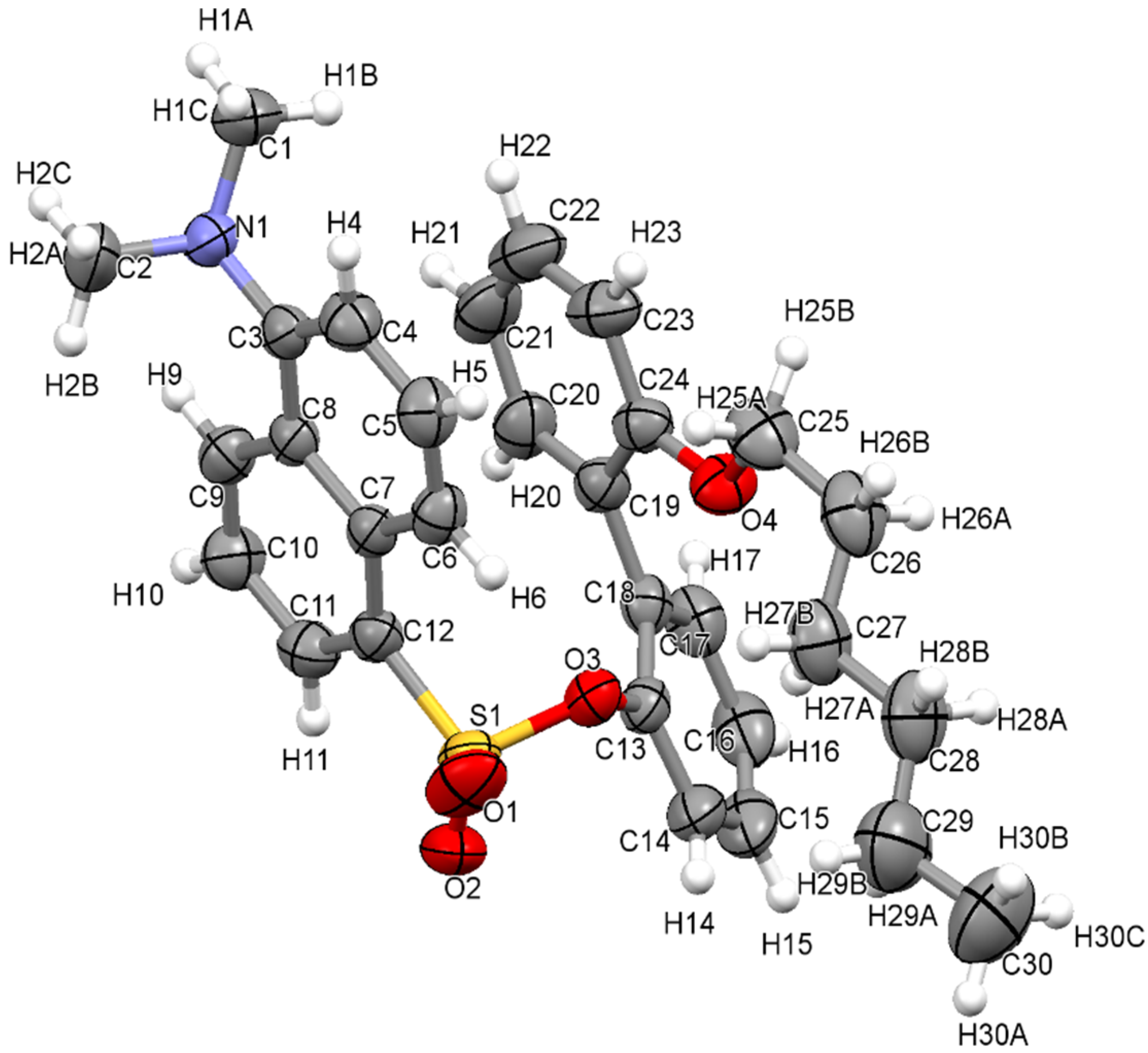
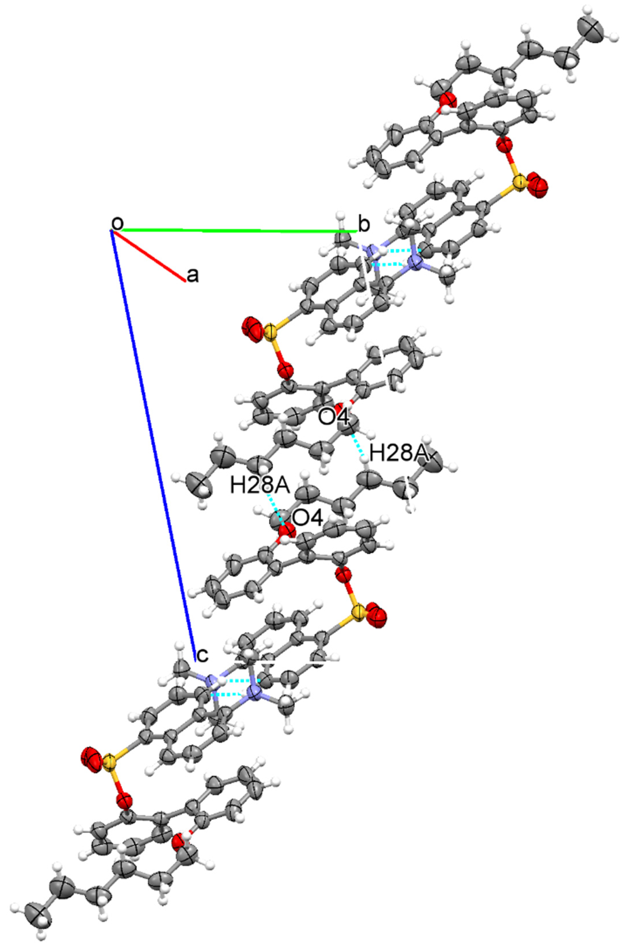
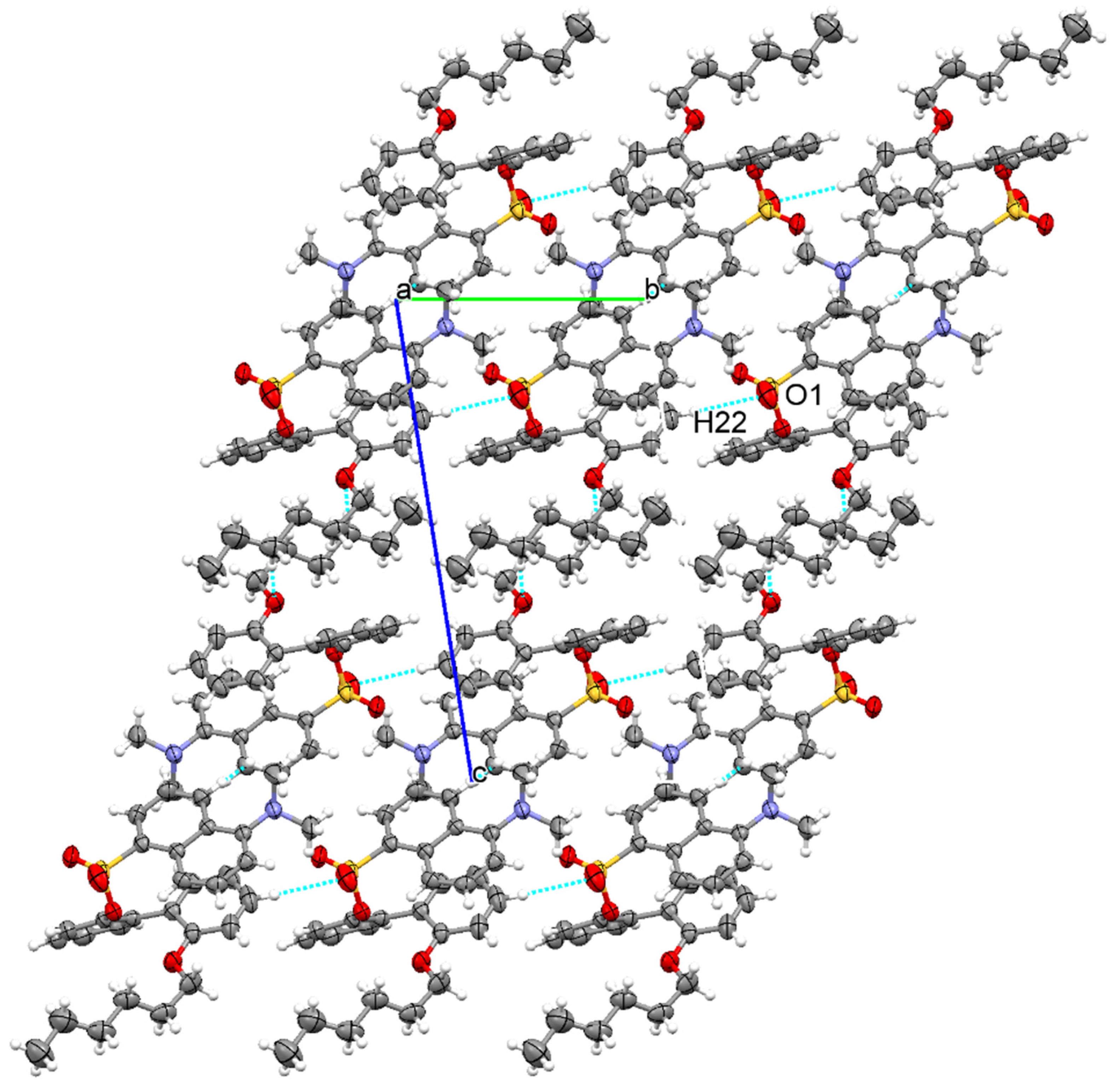
3.1. The Crystallographic Structure Detail of Sensor KC1

The KC1 in the dichloromethane solvent was grown in crystals. An asymmetric unit of KC1 consists of one molecule of KC1, biphenol with two substitutional groups on hydroxyl groups and dansyl and hexyl groups (Figure 1). The two aromatic rings of biphenyl moiety are not coplanar. The dihedral angle between the two aromatic rings is 54.48°. This may have an effect on the steric interactions of the hexyl and dansyl groups. As a result, the naphthalene plane of the dansyl group aligns in parallel with one phenyl plane. The hexyl moiety is in the zigzag form. The torsion angle between hexyl and one phenol ring is 8.37° (C23-C24-O4-C25), while that between another phenol and dansyl unit is 103.17° (C18-C13-O3-S1). The two adjacent molecules showed that the molecule structures are stabilized by a C-H···O interaction between an oxygen atom of phenol and a hydrogen atom of a hexyl moiety (O4---H28-C28) at a distance of 2.678 Å as shown in Table 2. Furthermore, these two adjacent molecules are further assembled by van der Waals forces to form a 1D structure the along diagonal of the bc plane, shown in Figure 2. In addition, the crystal structure of KC1 was stabilized by using another C-H···O interaction at a distance of 2.668 Å between a dansyl oxygen atom in the 1D structure and a phenol hydrogen atom (O1---H22-C22) in the other 1D chain to contribute a 2D structure in the bc plane, as shown in Figure 3. This 2D layer is further stabilized by van der Waals forces to support the 3D structure.

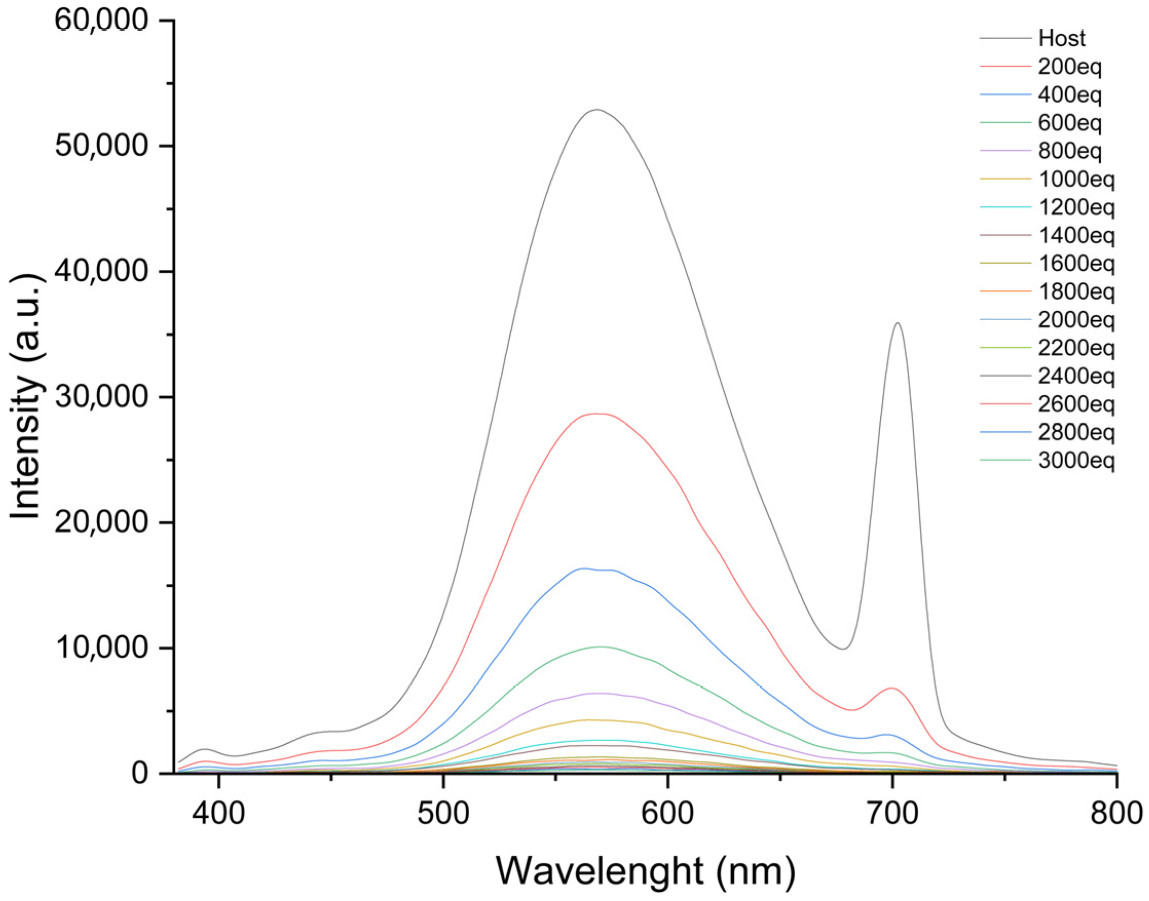
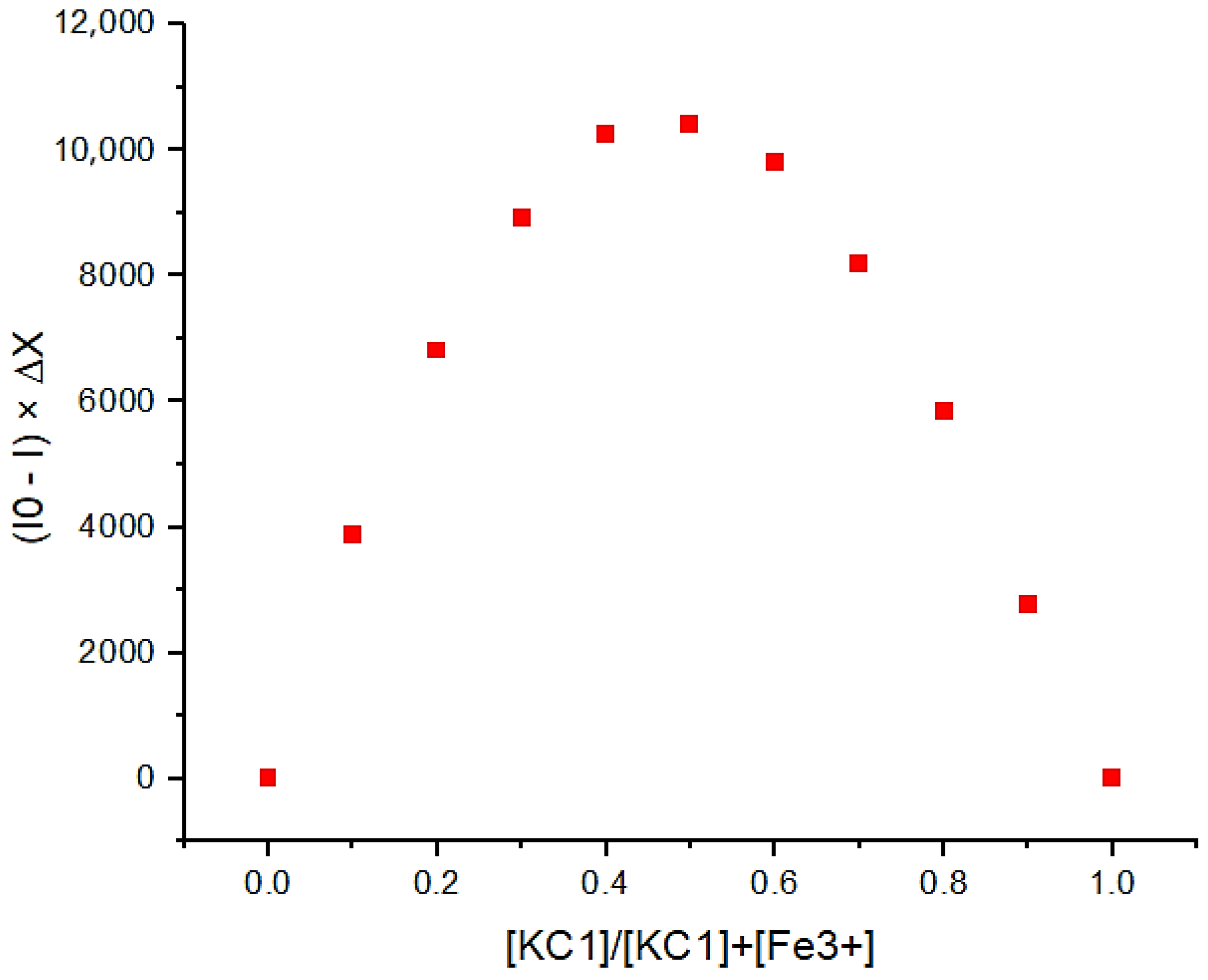
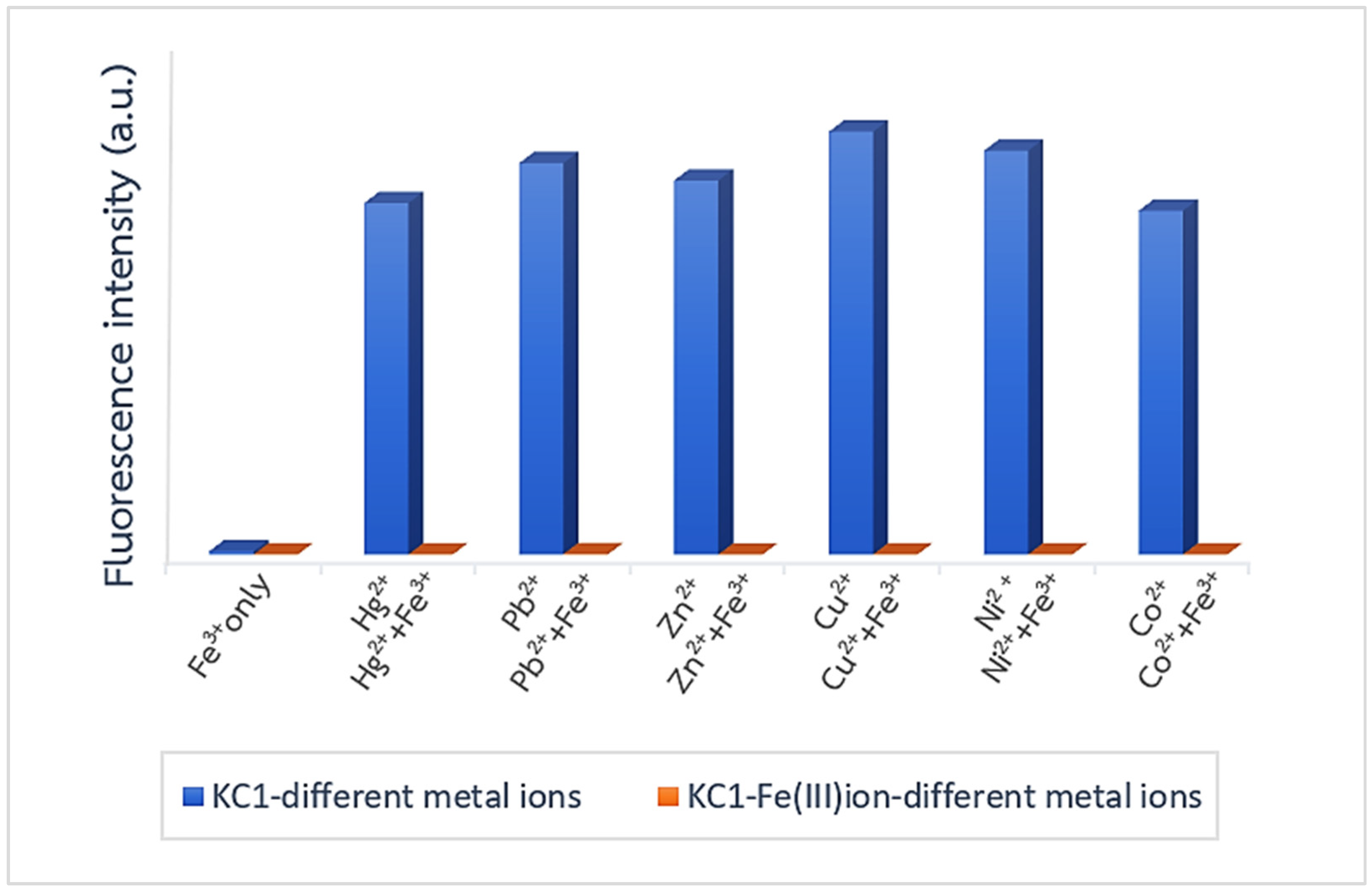
3.2. An Investigation of the Binding Behavior of KC1 and Metal Ions by Using a Fluorescence Technique

The emission wavelength of KC1 displayed a maximum at 585 nm, and there was another wavelength at 700 nm in DMSO:H2O (7:3 v/v). Upon the addition of the Fe3+ ion to the KC1 solution in the equivalent concentration range of 0–3000, the emission bands of KC1 at 585 and 700 nm are quenched due to the Fe3+ ion complex directly binding to KC1 (Figure 4) [22]. Upon the addition of different metal ions, the fluorescence emission at 585 gradually decreased, as depicted in Figure S2 (see Supplementary Materials). Results from the fluorescence titration indicated that KC1 was selective regarding the Fe3+ ion rather than the other metal ions. In the case of other metal ions, the KC1 showed a weak interaction, with the emission wavelength at 585 nm representing a small change. The maximum intensity ratio between KC1 and Fe3+ ions was shown to be 0.5 by using Job’s method, and a complex between KC1 and Fe3+ ions at a 1:1 ratio formation is shown in Figure 5 [28,29]. Moreover, there was only a color change of KC1 and Fe3+ ions under a UV lamp at 365 nm. For other metal ions, the color stayed the same, as shown in Figure 6. This result suggested that KC1 was obviously selective to the Fe3+ ion. The Stern–Volmer plot of complexation between KC1 and the Fe3+ ion is shown in Figure 7. The Stern–Volmer constant (Ksv) was evaluated as 21,203 M−1. Furthermore, the sensitivity and selectivity of the complexation between KC1 and the Fe3+ ion was investigated by analyzing the competition of various metal ions in the solution, as shown in Figure 8. It was found that KC1 and Fe3+ exhibited quench fluorescence, but KC1 and other metal ions still displayed the fluorescence’s blue bar graph. For KC1-Fe3+ and various metal ions, the orange bar graph indicated that there was no fluorescence emission at 585 nm, meaning there was no effect when adding other metal ions into the KC1-Fe3+ solution. Therefore, KC1 has the ability to detect Fe3+ while mixed with other metal ions in a solution. Furthermore, the limit of detection (LOD) between KC1 and the Fe3+ ion was evaluated as being 0.74 μM [28,29].

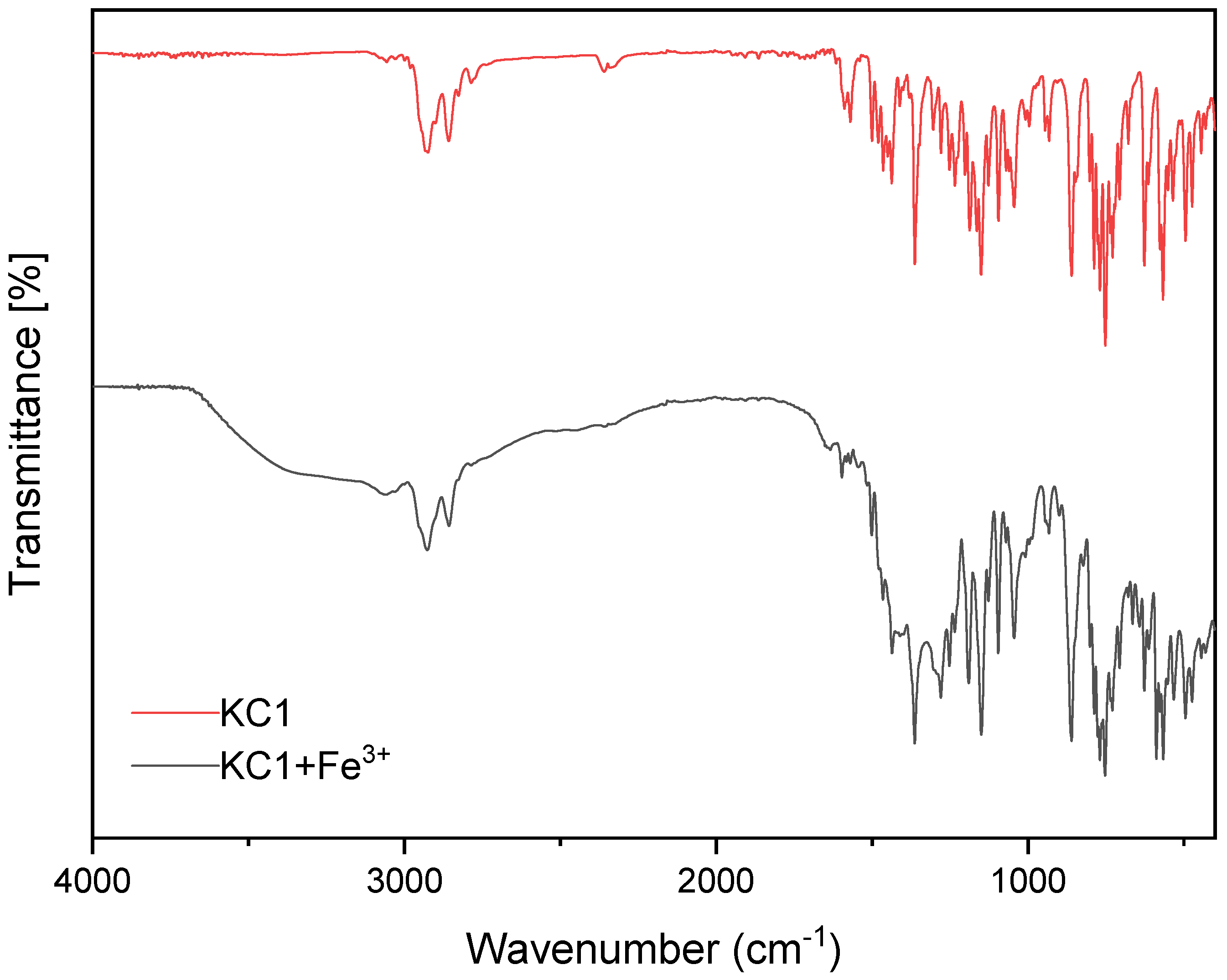
3.3. The Binding Mode Between KC1 and Fe3+ Ion via FTIR Spectroscopy

The FTIR spectra of KC1 and mixing KC1 with Fe3+ by using the ATR mode for analysis are shown in Figure 9. For the KC1 solid, the main function groups and wavenumber in units of cm−1 are represented by a red line and are also listed in Table 3.
In addition to mixing KC1 and the Fe3+ ion (black line), it was found that the strong shoulder between 3000 cm−1 and 4000 cm−1 exhibited –OH stretching of H2O belonging to Fe(NO)3.9H2O, and the wavenumber of the -S=O group at 1234 cm−1 moved to 1277 cm−1, indicating the oxygen atom position of the -S=O group and showing that KC1 was directly bound to the Fe3+ ion.

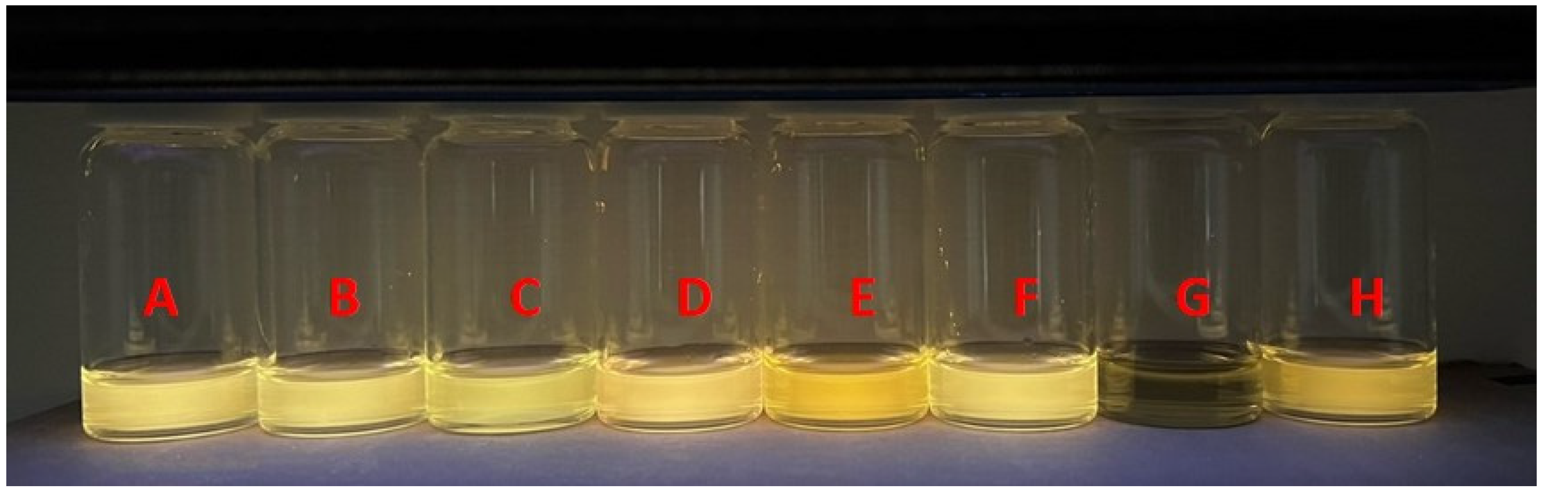
3.4. Application of KC1 and Various Concentrations of Fe3+ Ions on TLC Plates

The TLC plates with coated-KC1 were evaluated regarding the detection of Fe3+ ions in various concentrations, as shown in Figure 10. The result showed that only KC1 displayed yellow brightness on a TLC plate. The color of KC1 on TLC plates changed to a pale yellow upon the addition of the 1 and 5 equivalents, as shown in Figure 10. Furthermore, after the addition of the 10 equiv. to the coated-KC1 on the TLC plate, the yellow brightness of KC1 darkened. The TLC plates with coated-KC1 were utilized to detect the Fe3+ ion.

4. Conclusions

The KC1 sensor was successfully synthesized in two steps. From the KC1 structure, it was found that the two linked molecules are connected and stabilized by the C-H···O interaction. Upon adding the Fe3+ ion to the KC1 solution, it was found that the KC1 emission band was quenched under the complex formation between KC1 and the Fe3+ ion. IR results indicated that the KC1 binding mode at the oxygen atom (S=O group) interacted with the Fe3+ ion. Moreover, the coated-KC1 on the TLC plate has the ability to fluorescence quench for Fe3+ ion detection. Therefore, KC1 has the ability to detect the Fe3+ ion when among other metal ions.

Supplementary Materials

The following supporting information can be downloaded at: https://www.mdpi.com/article/10.3390/cryst14110943/s1, Figure S1: 1H-NMR spectra of KC1 in d-CDCl3; Figure S2: Fluorescence titration between KC1 (1 × 10⁻⁵ M) and Ni2+ ion (0–3000 eq) in DMSO:H2O (7:3 v/v).

Author Contributions

Conceptualization, B.W.; methodology, B.W.; funding, B.W.; writing draft, B.W.; writing, T.D.; methodology, K.C. (Kanokporn Chantaiyomporn), K.C. (Kiratikarn Charoensuk) and K.C (Kittipong Chainok). All authors have read and agreed to the published version of the manuscript.

Funding

The authors would like to thank the Kasetsart University Research and Development (KURDI) for financial support.

Data Availability Statement

The original contributions presented in the study are included in the article/Supplementary Material, further inquiries can be directed to the corresponding author/s.

Acknowledgments

The authors thank the Department of Chemistry, Faculty of Science, Kasetsart University, for providing equipment and facilities.

Conflicts of Interest

The authors declare no conflict of interest.

References

  1. Rao, C.P.; Mandal, S. Europium-Based Metal–Organic Framework as a Dual Luminescence Sensor for the Selective Detection of the Phosphate Anion and Fe3+ Ion in Aqueous Media. Inorg. Chem. 2018, 57, 11855–11858. [Google Scholar]
  2. Gu, Y.-N.; Lu, J.-F.; Liu, H.; Zhao, B.; Zhou, X.-H.; Zhao, Y.-Q.; Sun, Q.-Z.; Zhang, B.-G. Two Eu3+ Based Complexes Containing Uncoordinated Lewis Basic Pyridyl Sites and Chemical Sensing of 4-Nitrophenol and Fe3+ ions. Cryst. Growth Des. 2022, 22, 4874–4884. [Google Scholar] [CrossRef]
  3. He, T.-S.; Lan, Y.-L.; Li, Z.-Y.; Zhu, L.-N.; Li, X.-Z. Chiral Coordination Polymers from a New 2-Deoxy-D-ribose Derivative Linker: Syntheses, Structures, and Fe3+ Fluorescent Probe Functions. Cryst. Growth Des. 2021, 21, 2233–2242. [Google Scholar] [CrossRef]
  4. Areti, S.; Bandaru, S.; Rao, C.P. Triazole-Linked Quinoline Conjugate of Glucopyranose: Selectivity Comparison among Zn2+, Cd2+, and Hg2+ Based on Spectroscopy, Thermodynamics, and Microscopy, and Reversible Sensing of Zn2+ and the Structure of the Complex Using DFT. ACS Omega 2016, 1, 626–635. [Google Scholar] [CrossRef] [PubMed]
  5. Hou, B.-L.; Tian, D.; Liu, J.; Dong, L.-Z.; Li, S.-L.; Li, D.-S.; Lan, Y.-Q. A Water-Stable Metal–Organic Framework for Highly Sensitive and Selective Sensing of Fe3+ Ion. Inorg. Chem. 2016, 55, 10580–10586. [Google Scholar] [CrossRef]
  6. Li, Y.-W.; Li, J.; Wan, X.-Y.; Sheng, D.-F.; Yan, H.; Zhang, S.-S.; Ma, H.-Y.; Wang, S.-N.; Li, D.-C.; Gao, Z.-Y.; et al. Nanocage-Based N-Rich Metal–Organic Framework for Luminescence Sensing toward Fe3+ and Cu2+ Ions. Inorg. Chem. 2021, 60, 671–681. [Google Scholar] [CrossRef]
  7. Finelli, A.; Chabert, V.; Herault, N.; Crochet, A.; Kim, C.; Fromm, K.M. Sequential Multiple-Target Sensor: In3+, Fe2+, and Fe3+ Discrimination by an Anthracene-Based Probe. Inorg. Chem. 2019, 58, 13796–13806. [Google Scholar] [CrossRef]
  8. Khatun, M.; Ghorai, P.; Mandal, J.; Chowdhury, S.G.; Karmakar, P.; Blasco, S.; García-Espana, E.; Saha, A. Aza-phenol Based Macrocyclic Probes Design for “CHEF-on” Multi Analytes Sensor: Crystal Structure Elucidation and Application in Biological Cell Imaging. ACS Omega 2023, 8, 7479–7491. [Google Scholar] [CrossRef]
  9. Tan, S.S.; Teo, Y.N.; Kool, E.T. Selective Sensor for Silver Ions Built From Polyfluorophores on a DNA Backbone. Org. Lett. 2010, 12, 4820–4823. [Google Scholar] [CrossRef]
  10. Bazzicalupia, C.; Bianchia, A.; García-Españab, E.; Delgado-Pinar, E. Metals in Supramolecular Chemistry. Inorg. Chim. Acta 2014, 417, 3–26. [Google Scholar] [CrossRef]
  11. Xu, M.; Wang, X.; Liu, X. Detection of Heavy Metal Ions by Ratiometric Photoelectric Sensor. J. Agric. Food Chem. 2022, 70, 11468–11480. [Google Scholar] [CrossRef] [PubMed]
  12. Zhou, X.; Nie, J.; Du, B. Functionalized Ionic Microgel Sensor Array for Colorimetric Detection and Discrimination of Metal Ions. ACS Appl. Mater. Interfaces 2017, 9, 20913–20921. [Google Scholar] [CrossRef] [PubMed]
  13. Hussain, M.M.; Rahman, M.M.; Arshad, M.N.; Asiri, A.M. Hg2+ Sensor Development Based on (E)-N’-Nitrobenzylidene Benzenesulfonohydrazide (NBBSH) Derivatives Fabricated on a Glassy Carbon Electrode with a Nafion Matrix. ACS Omega 2017, 2, 420–431. [Google Scholar] [CrossRef] [PubMed]
  14. Jensen, G.C.; Janis, M.K.; Nguyen, H.N.; David, O.W.; Zastrow, M.L. Fluorescent Protein-Based Sensors for Detecting Essential Metal Ions across the Tree of Life. ACS Sens. 2024, 9, 1622–1643. [Google Scholar] [CrossRef]
  15. Zhang, C.; Shi, H.; Sun, L.; Yan, Y.; Wang, B.; Liang, Z.; Wang, L.; Li, J. Water Stable Metal–Organic Framework Based on Phosphono containing Ligand as Highly Sensitive Luminescent Sensor toward Metal Ions. Cryst. Growth Des. 2018, 18, 7683–7689. [Google Scholar] [CrossRef]
  16. George, A.; Shibu, E.S.; Maliyekkal, S.M.; Bootharaju, M.S.; Pradeep, T. Luminescent, Freestanding Composite Films of Au15 for Specific Metal Ion Sensing. ACS Appl. Mater. Interfaces 2012, 4, 639–644. [Google Scholar] [CrossRef]
  17. Cui, J.; Zhu, X.; Liu, Y.; Liang, L.; Peng, Y.; Wu, S.; YanZhao, Y. N-Doped Carbon Dots as Fluorescent “Turn-Off” Nanosensors for Ascorbic Acid and Fe3+ Detection ACS. Appl. Nano Mater. 2022, 5, 7268–7277. [Google Scholar] [CrossRef]
  18. Rajendran, S.; Ramanaiah, D.V.; Kundu, S.; Bhunia, S.K. Yellow Fluorescent Carbon Dots for Selective Recognition of As3+ and Fe3+ Ions. ACS Appl. Nano Mater. 2021, 4, 10931–10942. [Google Scholar] [CrossRef]
  19. Singh, V.K.; Mishra, H.; Ali, R.; Umrao, S.; Srivastava, R.; Abraham, S.; Misra, A.; Singh, V.N.; Mishra, H.; Tiwari, R.S.; et al. In Situ Functionalized Fluorescent WS2-QDs as Sensitive and Selective Probe for Fe3+ and a Detailed Study of Its Fluorescence Quenching. ACS Appl. Nano Mater. 2019, 2, 566–576. [Google Scholar] [CrossRef]
  20. Dong, S.; Ji, W.; Ma, Z.; Zhu, Z.; Ding, N.; Nie, J.; Du, B. Thermosensitive Fluorescent Microgels for Selective and Sensitive Detection of Fe3+ and Mn2+ in Aqueous Solutions. ACS Appl. Polym. Mater. 2020, 2, 3621–3631. [Google Scholar] [CrossRef]
  21. Das, N.K.; Ghosh, S.; Priya, A.; Datta, S.; Mukherjee, S. Luminescent Copper Nanoclusters as a Specific Cell-Imaging Probe and a Selective Metal Ion Sensor. J. Phys. Chem. C 2015, 119, 24657–24664. [Google Scholar] [CrossRef]
  22. Rasin, P.; Mathew, M.M.; Manakkadan, V.; Palakkeezhillam, V.N.V.; Sreekanth, A. A Highly Fluorescent Pyrene-Based Sensor for Selective Detection of Fe3+ Ion in Aqueous Medium: Computational Investigations. J. Fluoresc. 2022, 32, 1229–1238. [Google Scholar] [CrossRef] [PubMed]
  23. Sheldrick, G.M. SHELXT—Integrated Space-Group and Crystal-Structure Determination. Acta Cryst. Sect. A Found. Adv. 2015, 71, 3–8. [Google Scholar] [CrossRef] [PubMed]
  24. Sheldrick, G.M. Crystal Structure Refinement with SHELXL. Acta Cryst. Sect. C Found. Adv. 2015, 71, 3–8. [Google Scholar]
  25. Bruker, S. APEX3, SAINT and SADABS, 26; Bruker AXS Inc.: Madison, WI, USA, 2016. [Google Scholar]
  26. Spek, A.L. PLATON SQUEEZE: A Tool for the Calculation of the Disordered Solvent Contribution to the Calculated Structure Factors. Acta Cryst. Sect. C Struct. Chem. 2015, 71, 9–18. [Google Scholar] [CrossRef]
  27. Macrae, C.F.; Bruno, I.J.; Chisholm, J.A.; Edgington, P.R.; McCabe, P.; Pidcock, E.; Rodriguez-Monge, L.; Taylor, R.J.; van de Streek, J.; Wood, P. Mercury CSD 2.0—New Features for the Visualization and Investigation of Crystal Structures. J. Appl. Cryst. 2008, 41, 466–470. [Google Scholar] [CrossRef]
  28. Khunarj, S.; Saijaroensakul, W.; Morom, W.; Chainok, K.; Duangthongyou, T.; Pinchaipat, B.; Wannalerse, B. Synthesis, Crystal Structure and Optical Properties of 3,5-Dihydroxyphenyl-5-(dimethylamino)naphthlene-1-sulfonate as a Fluorescence Sensor for Fluoride Ion Detection. Crystals 2022, 12, 1836. [Google Scholar] [CrossRef]
  29. Buasakun, J.; Srilaoong, P.; Chainok, K.; Raksakoon, C.; Rattanakram, R.; Duangthongyou, T. Dual Luminescent Coordination Polymers Based on Flexible Aliphatic Carboxylate Ligands Supplemented by Rigid Bipyridyl Ligands for 2,4-Dinitrophenol (DNP) and Iron(III) Ion Detection. Polyhedron 2021, 204, 115265. [Google Scholar] [CrossRef]
Scheme 1. The synthetic pathway for KC1 sensor.
Crystals 14 00943 sch001
Figure 1. Labelled atoms in the asymmetric unit of KC1.
Crystals 14 00943 g001
Figure 2. The 1D structure of KC1 along the diagonal of the bc plane.
Crystals 14 00943 g002
Figure 3. The 2D structure of KC1 along the bc plane.
Crystals 14 00943 g003
Figure 4. The fluorescence titration between KC1 (1 × 10⁻⁵ M) and Fe3+ ions (0–3000 eq) in DMSO:H2O (7:3 v/v) (Host is the KC1).
Crystals 14 00943 g004
Figure 5. Job’s analysis of KC1 and Fe3+ in DMSO:H2O (7:3 v/v).
Crystals 14 00943 g005
Figure 6. The color change of KC1 (1 × 10⁻³ M) (A) with metal ions (3000 equiv.): (B) Pb2+, (C) Ni2+, (D) Zn2+, (E) Cu2+, (F) Co2+, (G) Fe3+ and (H) Hg2+ in DMSO: H2O (7:3 v/v).
Crystals 14 00943 g006
Figure 7. The plot graph of Stern–Volmer between KC1 and Fe3+ ions in DMSO:H2O (7:3 v/v).
Crystals 14 00943 g007
Figure 8. The competitive metal ions (Hg2+, Pb2+, Zn2+, Cu2+, Ni2+, Co2+); blue = KC1(1 × 10⁻⁵ M)—different metal ions (3000 equiv.) and orange = KC1(1 × 10⁻⁵ M)—Fe3+ ion and other metal ions (3000 equiv.) at 585 nm in DMSO:H2O (7:3 v/v).
Crystals 14 00943 g008
Figure 9. FTIR spectra of KC1 (red line) and mixing of KC1+Fe3+ ions (black line).
Crystals 14 00943 g009
Figure 10. The TLC plates of (a) coated-KC1 (1 × 10⁻³ M) with Fe3+ ions in (b) 1 equiv., (c) 5 equiv. and (d) 10 equiv. under a UV lamp at 365 nm in DMSO.
Crystals 14 00943 g010
Table 1. Crystal data of KC1.
CompoundCompound KC1
CCDC Number2361572
Empirical FormulaC30H33NO4S
Formula Weight (g/mol)503.67
Temperature (K)296
Wavelength (Å)0.71073
Crystal SystemTriclinic
Space GroupP1
a (Å) 8.7613 (2)
b (Å) 9.6370 (2)
c (Å) 17.5443 (5)
α (◦) 79.447 (1)
β (◦)84.094 (1)
γ (◦)66.799 (1)
Volume/Å31337.71 (6)
Z2
Density (calculated) (g/cm3)1.250
Absorption Coefficient (mm−1)0.16
F(000) 536.561
Crystal Size (mm3)0.32 × 0.32 × 0.22
Theta Range for Data Collection (◦)5 to 57.8
Index Ranges −11 ≤ h ≤ 11, −13 ≤ k ≤ 13, −23 ≤ l ≤ 23
Reflections Collected6982
Independent Reflections 5073 [Rint = 0.057]
Max. and Min. Transmission 0.7458 and 0.6903
Data/Restraints/Parameters 5073/0/328
Goodness-of-fit on F21.06
Final R Indices [I > 2sigma(I)] R1 = 0.0457, wR2 = 0.1274
R Indices (all data)R1 = 0.0706, wR2 = 0.1128
Largest Diff. Peak and Hole 0.26/−0.14 e Å−3
Table 2. Selected bond length (Å) and bond angle (°) of KC1.
Atom-AtomBond Length (Å)Atom-AtomBond Length (Å)
S1-O11.4195 (14)S1-C121.7614 (15)
C14-C151.382 (3)C16-H160.93
S1-O21.4235 (14)O3-C131.4124 (18)
C15-H150.93C16-C171.380 (3)
S1-O31.5992 (12)O4-C241.361 (2)
C15-C161.366 (3)C17-H170.93
O4-C251.422 (2)C17-C181.394 (2)
N1-C11.457 (2)C18-C191.483 (2)
N1-C21.449 (2)C19-C201.392 (2)
O2-S1-O1119.77 (8)C24-C19-C18121.14 (14)
C17-C16-C15120.29 (18)C3-N1-C2114.14 (14)
O3-S1-O1103.49 (8)C24-C19-C20118.34 (16)
C17-C16-H16119.86 (12)H1a-C1-N1109.5
O3-S1-O2109.35 (7)H20-C20-C19119.30 (11)
H17-C17-C16119.14 (12)H1b-C1-N1109.5
C12-S1-O1110.31 (8)C21-C20-C19121.4 (2)
C18-C17-C16121.72 (18)H1b-C1-H1a109.5
C12-S1-O2109.28 (8)C21-C20-H20119.30 (13)
C18-C17-H17119.14 (10)H1c-C1-N1109.5
C12-S1-O3103.25 (6)H21-C21-C20120.32 (13)
C17-C18-C13116.06 (15)H1c-C1-H1a109.5
C13-O3-S1118.79 (9)C22-C21-C20119.37 (19)
C19-C18-C13124.09 (14)H1c-C1-H1b109.5
C25-O4-C24119.31 (14)C22-C21-H21120.32 (12)
C19-C18-C17119.85 (15)H2a-C2-N1109.5
C2-N1-C1110.58 (15)H22-C22-C21119.58 (12)
C20-C19-C18120.31 (16)H2b-C2-N1109.5
C3-N1-C1115.05 (15)C23-C22-C21120.84 (19)
Table 3. The main functional groups of KC1 at different wavenumbers.
Wavenumber (cm−1)Functional Group
2858-C-H
1095-C-O
1570 and 1465-C=C
1152-C-N
1364 and 1234-S=O
Disclaimer/Publisher’s Note: The statements, opinions and data contained in all publications are solely those of the individual author(s) and contributor(s) and not of MDPI and/or the editor(s). MDPI and/or the editor(s) disclaim responsibility for any injury to people or property resulting from any ideas, methods, instructions or products referred to in the content.

