Article

Structures, Characterization and DFT Studies of Four Novel Nickel Phenanthroline Complexes

1
Department of Chemistry, College of Science, King Faisal University, Al-Ahsa 31982, Saudi Arabia
2
Umm Al-Momineen Maimouna Bint Al-Harth Intermediate, School at the National Guard, Al Khobar Education Office, General Directorate of Education Eastern Province, Ministry of Education, Al Khobar 32416, Saudi Arabia
3
Department of Chemistry, Al al-Bayt University, Al-Mafraq 25113, Jordan
4
Department of Medicinal Chemistry, Faculty of Pharmacy, University of Mansoura, Mansoura 35516, Egypt
5
Department of Chemistry, COMSATS university Islamabad, Abbottabad Campus, Abbottabad KPK 22060, Pakistan
*
Authors to whom correspondence should be addressed.
Crystals 2023, 13(5), 738; https://doi.org/10.3390/cryst13050738
Submission received: 25 March 2023 / Revised: 16 April 2023 / Accepted: 24 April 2023 / Published: 27 April 2023
(This article belongs to the Special Issue Synthesis and Characterization of Coordination Compounds)

Abstract

:
Four Ni–phen complexes were prepared and characterized by FTIR and thermal analysis. The X-ray structure determination shows that the geometry around NiII ions in complexes 1, [Ni(phen)2(H2O)(ONO2)](NO3); 2, [Ni(phen)2(H2O)Cl]Cl; 3, [Ni(phen)(CH3CN)(ONO2)(O2NO)] and 4, [Ni(phen)3](NO3)2.H2O.C2H5OH is considerably distorted octahedral. The coordination core geometries in 1, 2, 3 and 4 are NiN4O2, NiN4OCl, NiN3O3 and NiN6, respectively. The crystal packing and crystal supramolecularity analysis of the complexes reveal the importance of aryl∙∙∙aryl interactions, and both offset face-to-face (OFF) and edge-to-face (EF) motifs are dominant. Hydrogen bonding interactions in these structures reinforce aryl∙∙∙aryl interactions. FTIR proved the coordination of M-N and M-Cl as well as the presence of nitro groups in the complexes, while thermal analysis revealed that the loss of water, chloro and nitro groups occurred before the degradation of phenanthroline ligand in the complexes. DFT study indicates that a strong correlation exists among theoretical and experimentally determined geometric parameters with distorted octahedral geometry. The charge density in HOMO is localized on the metal halogen bond and nitrate group (NO3), whereas the LUMO cloud density is mainly distributed on the phen ring, which indicates the electroactive nature of the ring.

1. Introduction

Ortho-phenanthroline or 1,10-Phenanthroline (phen) is a tricyclic, aromatic, N-heterocyclic organic compound. In coordination chemistry, phen is a well-known classic bidentate ligand mostly formed from complexes of type [M(phen)3]X2 when treated with metal halides (where M is metal and X is halide) [1]. Moreover, phen is a versatile building block for the construction of ligands for various purposes [2].
The coordination properties of phen are similar to 2,2′-bpy. According to Teng, Q. and Huynh, H. [3], 2,2′-bipyridine (bipy) binds metals more tightly than phen since their chelating N-donor’s atoms are preorganized. The crystal structures of several Ni(II)-phen complexes showed six-coordinated complexes of Ni(II) with phen. In the complex [cis-Ni(phen)2Cl(H2O)]Cl.H2O.C2H5OH, a cis configuration of phen was observed. The molecule was incorporated with one chloride and one water molecule, whose positions they can interchange in or out of the coordination sphere; so, a complex of type [cis-Ni(phen)2(H2O)2]Cl2 C2H5OH is possible, as well [4]. The crystal structure of {[cis-Ni(phen)2Cl(H2O)] [cis-Ni(phen)2(H2O)2]} (PF6)3.4H2O combines two nickel complexes but with different substitution groups as an evidence of metathesis ionization isomerization. The dimeric complex [Ni(phen)2(µ-Cl)]2(PF6)2.2(C2H5)2O in which the chloride bridges two different metal ions is typical, and similar structures of the formula Ni(phen)2(µ-Cl)2M(L)x have been reported [5]. The ligation of solvent in the coordination sphere of Ni(II) is shown in the example [cis-Ni(phen)2 (AN)2](PF6)2 (where AN is acetonitrile solvent); however, the addition of phen to this complex produces [Ni(phen)3](PF6)2, which crystallizes as one and two complexes in one unit cell [4]. In general, the crystallization of Ni(II) with phen could result in one, two or three phen-containing complexes, depending on many factors, as it is in various inorganic coordination states. The ligand type, solvent, method of synthesis, etc., all are important factors in this course.
The light-blue crystals [Ni(phen)2(H2O)Br]Br.3H2O with pseudo-octahedral geometry with a cis arrangement of Br and H2O and the light-red crystals [Ni(phen)3]Br2.8H2O with disordered bromide anions were prepared and characterized [6]. The crystal structure of [Ni(1,10-phenanthroline)2(H2O)2](NO3)2 showed a remarkably complicated 3D hydrogen-bonding supramolecular assembly that simplified upon heating [7]. Other structures were synthesized with tetrahedral counter anions: [Ni(1,10-phen)2(H2O)2](ClO4)2.0.4H2O [8], [Ni(1,10-phen)2(H2O)2](SO4).5.6H2O [9] and [Ni(1,10-phen)2(H2O)2](ZnCl4).H2O [10].
Both bipy and phen are nitrogen heterocycles and are among the most widely utilized chelate ligands in coordination chemistry. Although they have a very similar structure, there is a difference in their chelating ability due to the different geometry of the free molecules [11,12]. The chelating nitrogen-donor atoms of 2,2′-bipy are more preorganized than those of phen, leading to better binding with metal ions [3].
In the complex cis-[Ni(NCS)2(bipy)2], the central Ni(II) ion is six-coordinated in a distorted octahedral environment defined by four N atoms from two chelating bipy ligands and two mutually cis-positioned N atoms from two SCN- anionic ligands [13].
An evaluation of the competitive complexation between bipy and phen with manganese ion, Mn(II), using the collision-induced dissociation (CID) mass spectroscopy method revealed a preferential elimination of bipy first, indicating its weakening ligation to this metal [14]. Reyzer et al. have examined the ability of multidentate ligands to displace other pyridyl ligands in a metal complex. The results showed that the order of binding strength in the gas phase followed the trend: 4,4′-bipy < 2,2′-bipy < phen [12]. However, a series of 4,4′-bipyridne complexes with perchlorate counter ions have been prepared: [Ni (4,4′-bipy)3 (H2O)2](ClO4)2·1.4 (4,4′-bipy)·3(H2O);[Co (4,4′-bipy)3(H2O)2](ClO4)2·1.4(4,4′-bipy)·3(H2O);[Cu (4,4′-bipy)3(DMSO)2](ClO4)2·2(4,4′-bipy) [15].
Niphen complexes, when pyrolyzed under nitrogen, are expected to act as a catalyst for the preparation of nickel graphite nanocarbon as found in the case of the pyrolysis of Ni(2,2′-bipyridine)Cl2. H2O [16]. Thus, in this present work, we have synthesized four nickel phenanthroline complexes by using the three-layer diffusion method with varying nickel salt, solvent nature and a mole ratio of nickel ion to phen ligand. The octahedral complexes of the four crystals, compounds [Ni(phen)2(H2O)(ONO2)](NO3) 1, [Ni(phen)2(H2O)Cl]Cl 2, [Ni(phen)(CH3CN)(ONO2)(O2NO)] 3 and [Ni(phen)3](NO3)2.H2O.C2H5OH 4 were characterized by single X-ray diffraction and supported by thermal analysis, FTIR and DFT computation.
Thus, the combination of nickel metal ions and phenanthroline ligand in the four new complexes with the formula 1, 2, 3 and 4 can be used as a catalyst for the preparation of nickel graphite nanocarbon since cobalt phen sulfate [Co(phen)(H2O)4]SO4.2H2O complex [17] behaved as a catalyst in the pyrolysis experiment to prepare cobalt graphitic nanocarbon. Further support is that Co(tph)(2,2-bipy)·4H2O [18] and Co(2,20-bipy)Cl2 [19] were used as a precursor catalyst in the pyrolytic preparation of cobalt hierarchical nanocarbon with an application as an electrochemical sensor for urea and fructose, respectively.
In addition, nickel graphitic nanocarbons that can be obtained from the pyrolysis of Niphen complexes can act as a catalyst for the hydrogenation of nitrophenol. This is supported by a recent work where cobalt hierarchical graphitic nanocarbon prepared from the pyrolysis of [Co(phen)2Cl2]1.5CH3CN complex acted as an efficient reduction catalyst of p-nitrophenol [20]. In addition, nickel nanoparticle carbon microspheres were successfully applied for the reduction of p-nitrophenol [21].

2. Experimental

2.1. Synthesis

All chemicals used are of high quality and used as purchased from Sigma-Aldrich.
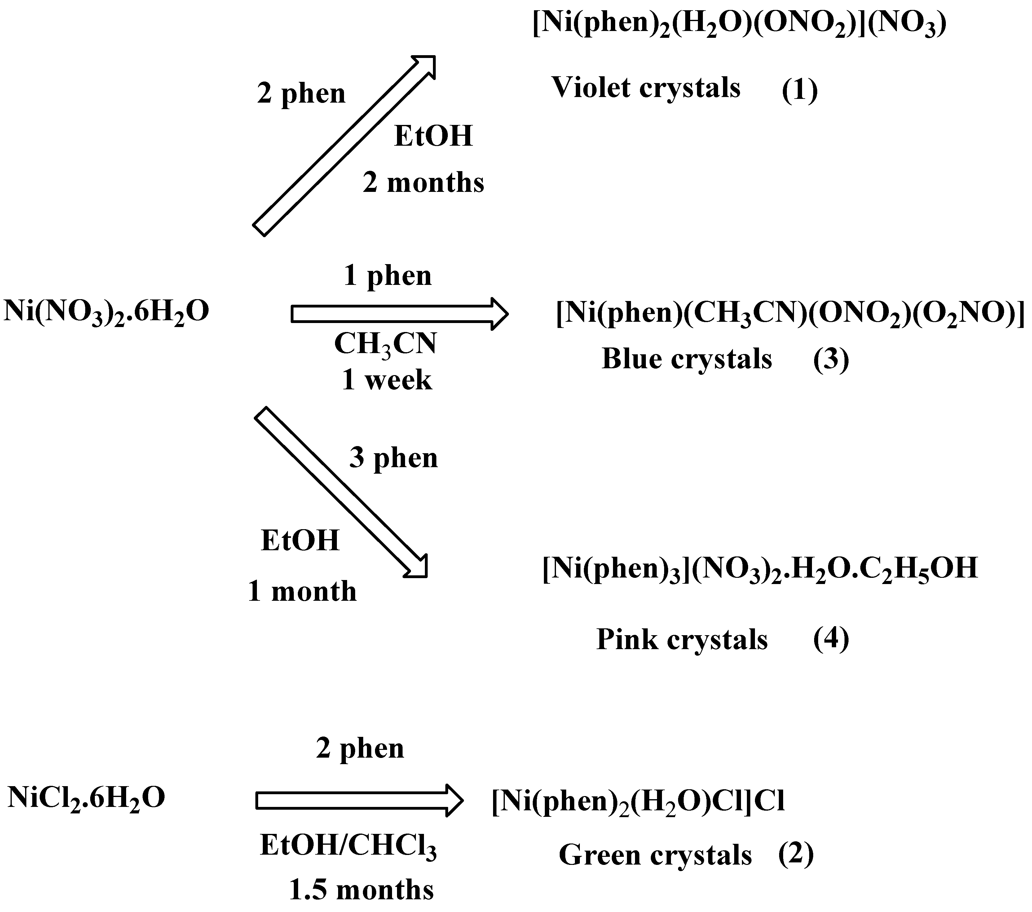
A diffusion method with three layers in a test tube was employed for the preparation of four complexes (14). The three layers were prepared as shown in the following Table 1. Layer 1 consisted of nickel salt (1 mmoL) dissolved in 5 mL of the appropriate solvent. Then, 5 mL of solvent (layer 2) was carefully added dropwise on the top of layer 1. Phenanthroline ligand (phen) (x mmol) dissolved in 7 mL of the solvent was added, carefully forming layer 3. The tube was then covered with paraffin paper and left for slow diffusion and the reaction of solutes. Crystals of complexes 1 (violet), 2 (green) and 4 (pink) were obtained after 2 months, 1.5 months and 1 month, respectively, while blue crystals of complex 3 were obtained after only one week.
The infrared spectra of 14 were recorded on a Shimadzu 8300 FTIR spectrophotometer in the range of 200–4000 cm−1.

2.2. FTIR Vibration cm−1

Complex 1: 3284.2, 3141.5, 3069.4, 1635.4, 1585.2, 1511.9, 1415.5, 1388.5, 1294.9, 1140.7, 1098.1, 1028.0, 842.5, 720.6, 642.7, 580.1, 530.3
Complex 2: 3744.1, 3047.0, 2296.9, 2357.7, 2225.5, 2154.1, 1997.9, 1773.0, 1659.2, 1569.2, 1508.0, 1420.4, 1104.6, 1047.2, 939.2, 852.7, 769.5, 726.5, 672.6, 553.5, 534.5, 526.5
Complex 3: 3502.7, 3228.3, 3070.2, 2331.6, 1633.1, 1589.1, 1455.7, 1300.4, 1141.4, 1027.9, 847.2, 722.4, 561.7
Complex 4: 3307.4, 3396.1, 3052.8, 3020.4, 3006.5, 2355.1, 1586.4, 1514.1, 1415.9, 1326.8, 1278.6, 1215.1, 1139.8, 1080.5, 1041.0, 977.7, 842.3, 800.5, 718.9, 641.9, 603.1, 565.7
Differential Thermal Analysis (TG-DTA) of the complexes 14 was conducted using a Setaram Labsys Thermal Analyzer within the 30–1100 temperature range under dry nitrogen gas flow at a rate of 10 °C/min in a chamber with a cooling water flow rate of 10 dm3.

2.3. X-ray Structure Determination

Single crystals of compounds 1, 2, 3 and 4 were obtained from the appropriate reaction mixture at room temperature using the three-layer diffusion method. Data were collected on a Bruker APEX-II D8 Venture area diffractometer equipped with graphite monochromatic Mo Kα radiation, λ = 0.71073 Å at 293 (2) K. Cell refinement and data reduction were carried out by Bruker SAINT. SHELXT [22,23] was used to solve the structure. The final refinement was carried out by full-matrix least-squares techniques with anisotropic thermal data for non-hydrogen atoms on F. CCDC 1870131, 1860200, 1860201 and 1870142 contain the supplementary crystallographic data for crystals 1, 2, 3 and 4, respectively, which can be obtained free of charge from the Cambridge Crystallographic Data Centre via www.ccdc.cam.ac.uk/data_request/cif (accessed on 25 March 2023). Crystallographic data and structure refinement details for the determination of 1, 2, 3 and 4 are summarized in Table 2.

3. Results and Discussion

3.1. Synthesis of Crystals

Because of the high stability constant of the Niphen complex, the synthesis of these complexes from nickel salts and phenanthroline results in fast precipitation of the product as powder, and thus it is difficult to obtain single crystals. We have adopted in this present work a three-layer method for the preparation of complexes that can provide the slow diffusion of ligand and metal salts from two different layers to form the complex in an intermediate layer.
The four crystals, compounds 1, [Ni(phen)2(H2O)(ONO2)](NO3); 2, [Ni(phen)2(H2O)Cl]Cl; 3, [Ni(phen)(CH3CN)(ONO2)(O2NO)] and 4, [Ni(phen)3](NO3)2.H2O.C2H5OH were grown from reaction medium, as described in the experimental section (Table 1) and presented in Scheme 1, into adequate sizes for single crystal X-ray diffraction analysis (SCXRD). Crystals were slowly grown from the three layers containing Ni(NO3)2.6H2O or NiCl2.6H2O solution/solvent/Phen ligand. Variation in the solvents EtOH, CHCl3 and CH3CN, and the molar ratio of the two components Ni ion and phen ligand, afforded various crystals (14). The duration of crystal growth ranged from 1 week to 2 months.

3.2. Molecular Structure of Crystals

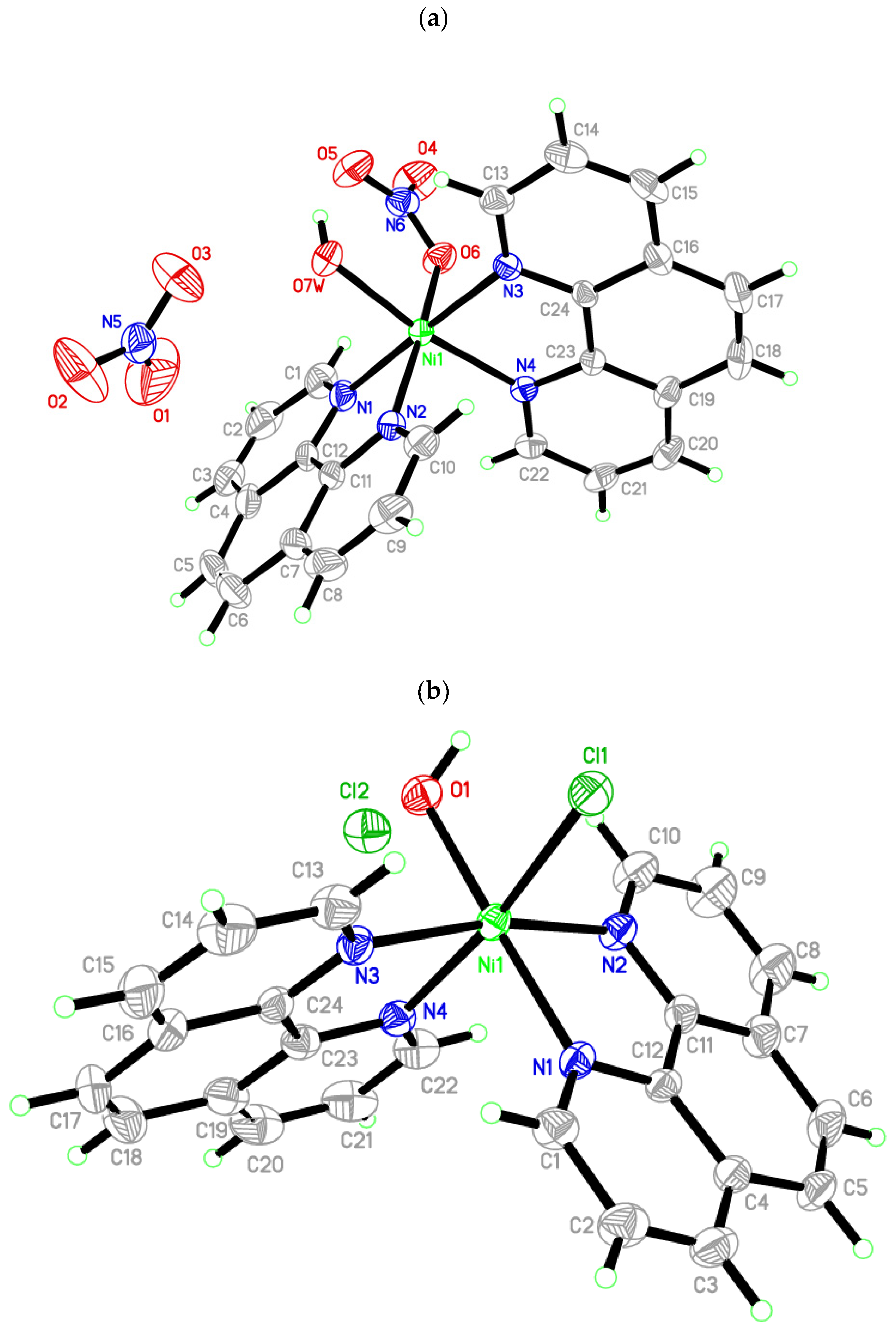
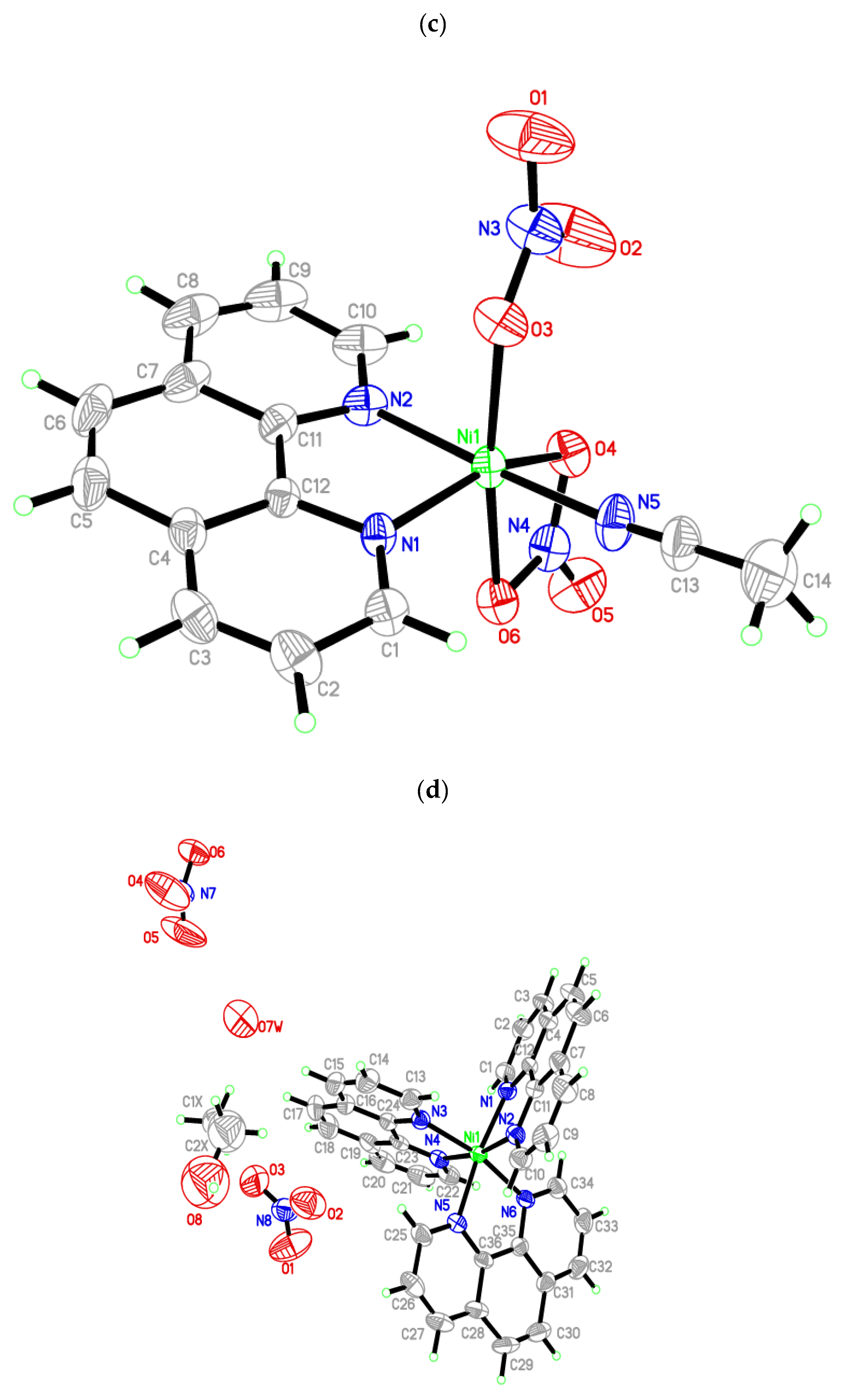
The molecular structure of compounds 1, [Ni(phen)2(H2O)(ONO2)](NO3); 2, [Ni(phen)2(H2O)Cl]Cl and 4, [Ni(phen)3](NO3)2.H2O.C2H5OH are depicted in Figure 1. It contains one mono cation NiII complex, one counter anion (nitrate in 1, chloride in 2) and two nitrate counter anions in 4, while complex 3, [Ni(phen)(CH3CN)(ONO2)(O2NO)] is constructed of one neutral complex. The geometry of the NiII atom is distorted octahedral with d8 electronic configuration in all complexes. In 1, the coordination geometry is NiN4O2, which is constructed from two bidentate phenanthroline (phen) ligands—one water molecule and one nitrato ligand—whereby the nitrato ligand acts as a monodentate ligand (η1-ONO2). Compound 2, [Ni(phen)2(H2O)Cl]Cl consists of two bidentate phen ligands— one water and one chloro ligand in a NiN4OCl core. In Complex 3, Ni ions are bonded to one bidentate phen and one each of acetonitrile, bidentate (η2-O2NO) and monodentate (η1-ONO2) nitrato ligands in NiN3O3 coordanation geometry, while in 4, three bidentate phen ligands are bonded to Ni ions with a NiN6 core. Selected bond lengths and angles are gathered in Table S1 (see supporting information). The Ni―N(phen) and Ni―O(NO3) bond distances are typical values of related Ni―phen [24,25,26] and Ni―N(NO3) [27] complexes.

3.3. Supramolecular Features

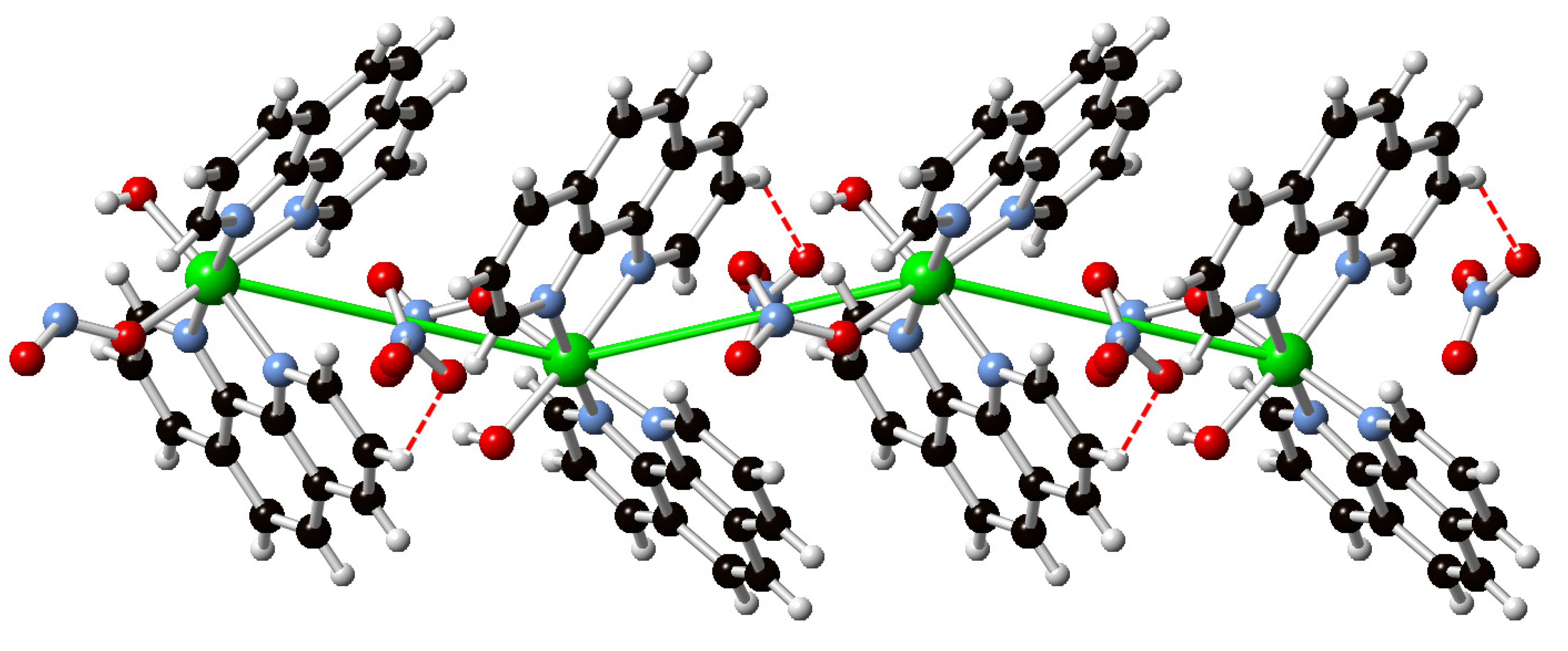
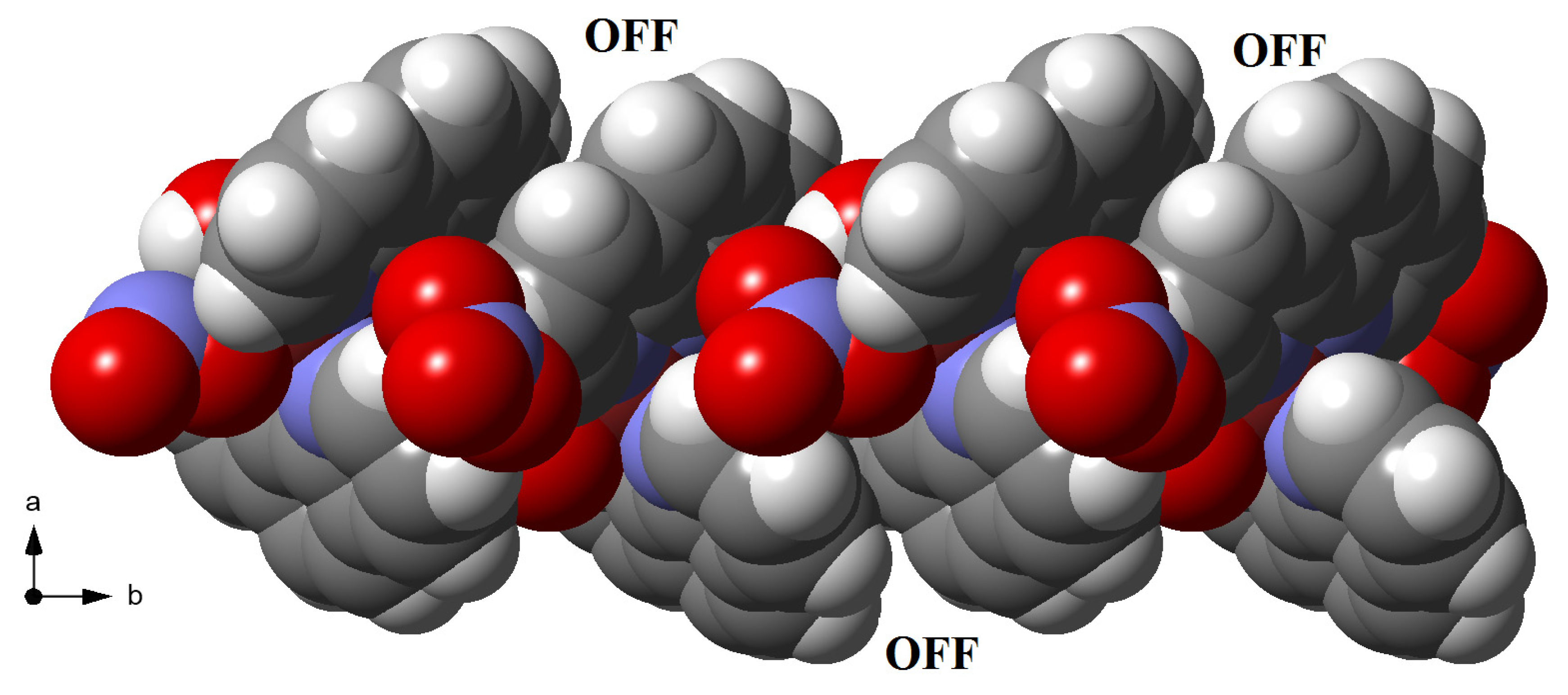
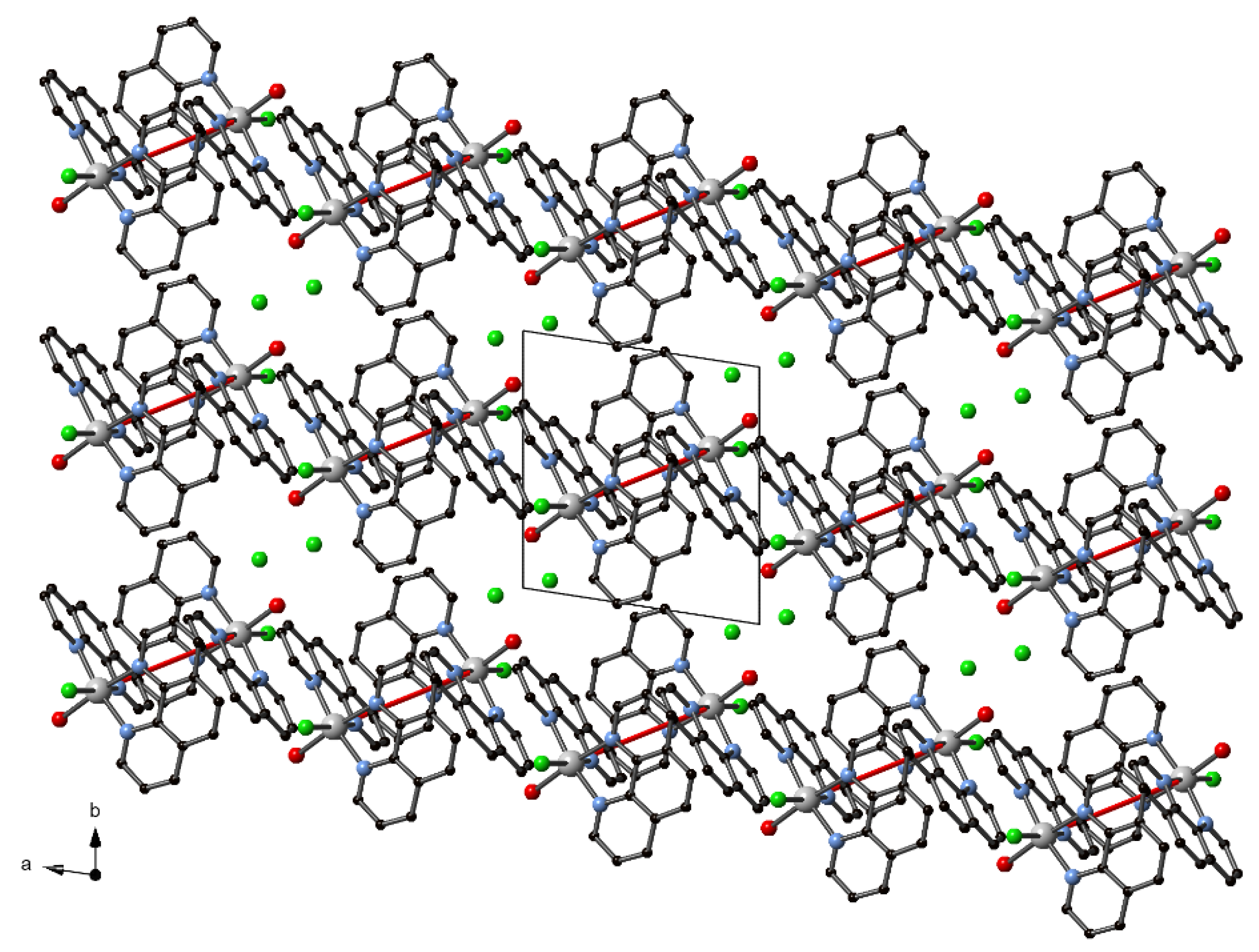
The crystal lattice of 1 is columnar along the b-axis, as illustrated in Figure 2. The [Ni(phen)2(H2O)(ONO2)]+ ions occur in chains along the b-axis, forming cavities that are occupied by the nitrate counter anions. Within each chain, the molecules are assembled, along the b-axis, through offset face-to-face (OFF) aryl∙∙∙aryl interactions of phen ligands (Figure 3). The OFF motifs are evident by the centroids’ separation distance of the rings (N2/C7–C11) Cg1∙∙∙Cg2 (N3/C13–C16/C24) of 3.760 Å. The cavities formed in the chains are occupied the nitrate counter anions. The nitrate ions add extra stability by hydrogen bonding to the hydrogen atoms of phen (π)C―H groups, forming (π)C―H∙∙∙ONO2 interactions, as shown in Figure 3. Hydrogen-bonding geometrical details are listed in Table 3.
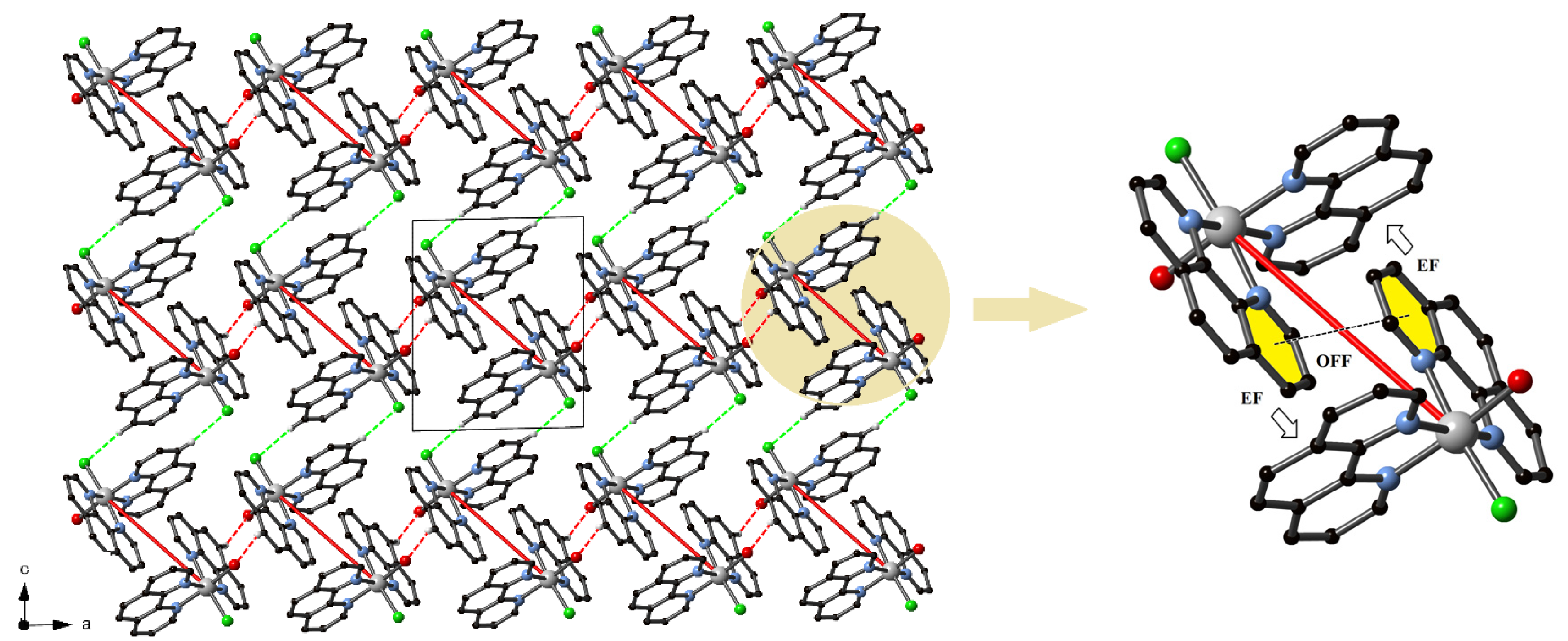
The packing of complex 2 contains alternating slabs of [Ni(phen)2(H2O)Cl]+ cations and chloride counter anions (Figure 4). Each slab of cations is constructed of embraced cations, whereby a pair of cations is involved in the parallel fourfold aryl embrace (P4AE) [28,29] parallel to the a-axis (Figure 5). The P4AE motif (Ni∙∙∙Ni distance of 8.949 Å) is constructed from one OFF aryl interaction with the centroids’ separation distance of ring (N4/C19–C23; C1g), and the symmetry-related ring (N4/C19–C23, C1g) being C1g∙∙∙C1g = 3.696 Å and two EF (edge-to-face) aryl interactions (Figure 5). Each P4AE motif of embraced cationic complexes is further connected with the next one, parallel to the a-axis, by OFF π∙∙∙π stacking through phen ligands in which the centroids of (C16–19/C23C24) C2g∙∙∙C3g (C16–19/C23C24) and (C16–19/C23C24) C3g∙∙∙C4g (N3/C13–C16/C24) are separated by 3.670 and 3.954 Å, respectively, with Ni∙∙∙Ni distances of 9.196 Å. These stacking interactions are supported by two inversion-related C―H···O interactions of 2.634 Å, leading to infinite chains parallel to the a-axis. The chains parallel to the a-axis interact with the next ones along the c-axis through complementary OFF aryl stacking interactions in which the phen ligands are connected via the symmetry-related rings (N1/C1–C4/C12) Cg5∙∙∙Cg5 and the rings Cg5∙∙∙Cg6 (C4–C7/C11C12) with the centroids separated by 3.623 and 3.847 Å, respectively. Other than these OFF interactions, the molecules possess additional symmetry related to two C―H···Cl interactions with a contact distance of 2.738 Å, as shown in Figure 5. The crystal lattice is further stabilized by additional hydrogen-bonding interactions in which the counter chloride anion interacts with bonded water and phen ligands in O—H···Cl2 and C—H···Cl2 interactions. Details of hydrogen bonding interactions are listed in Table 3.
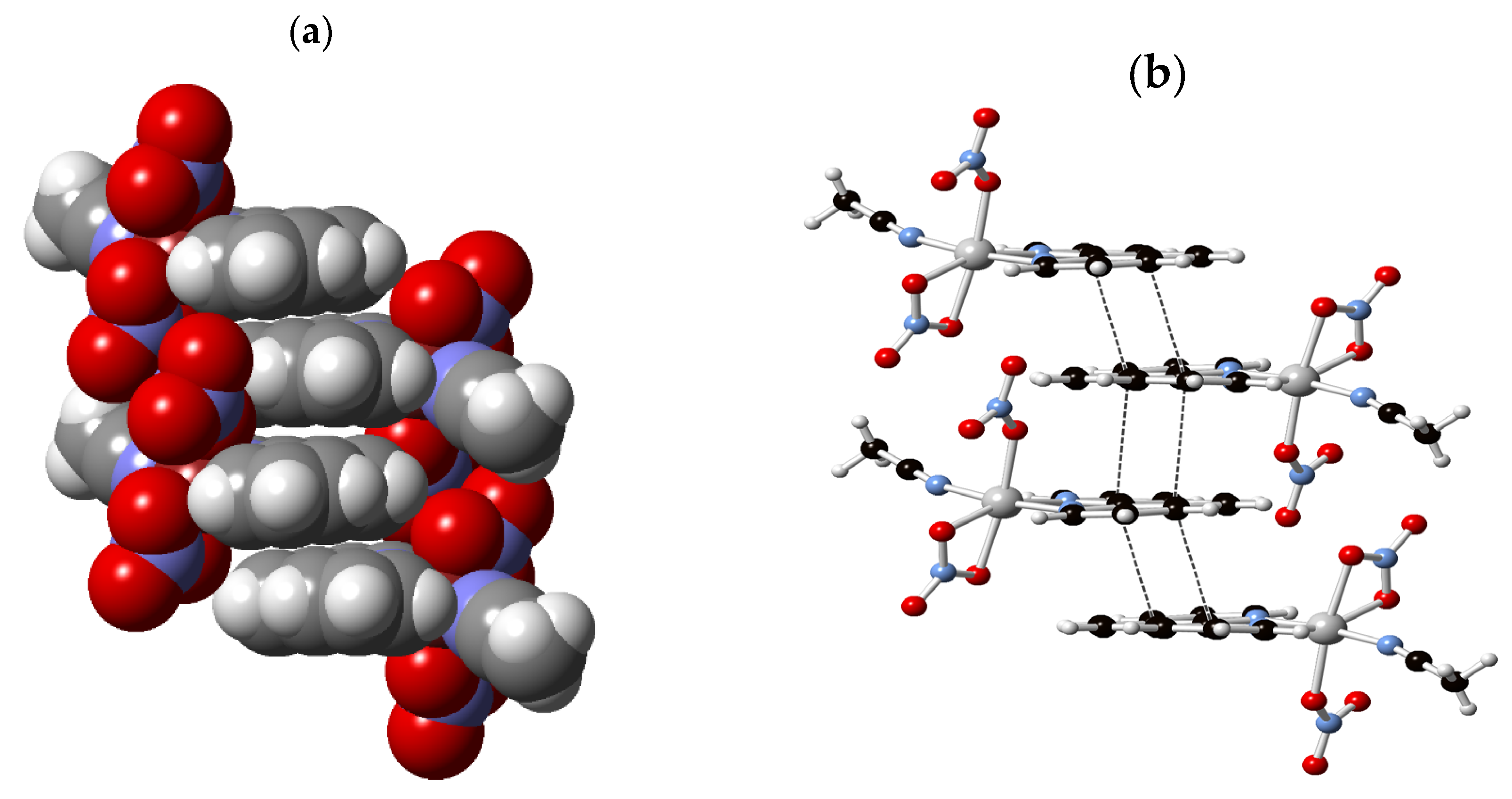
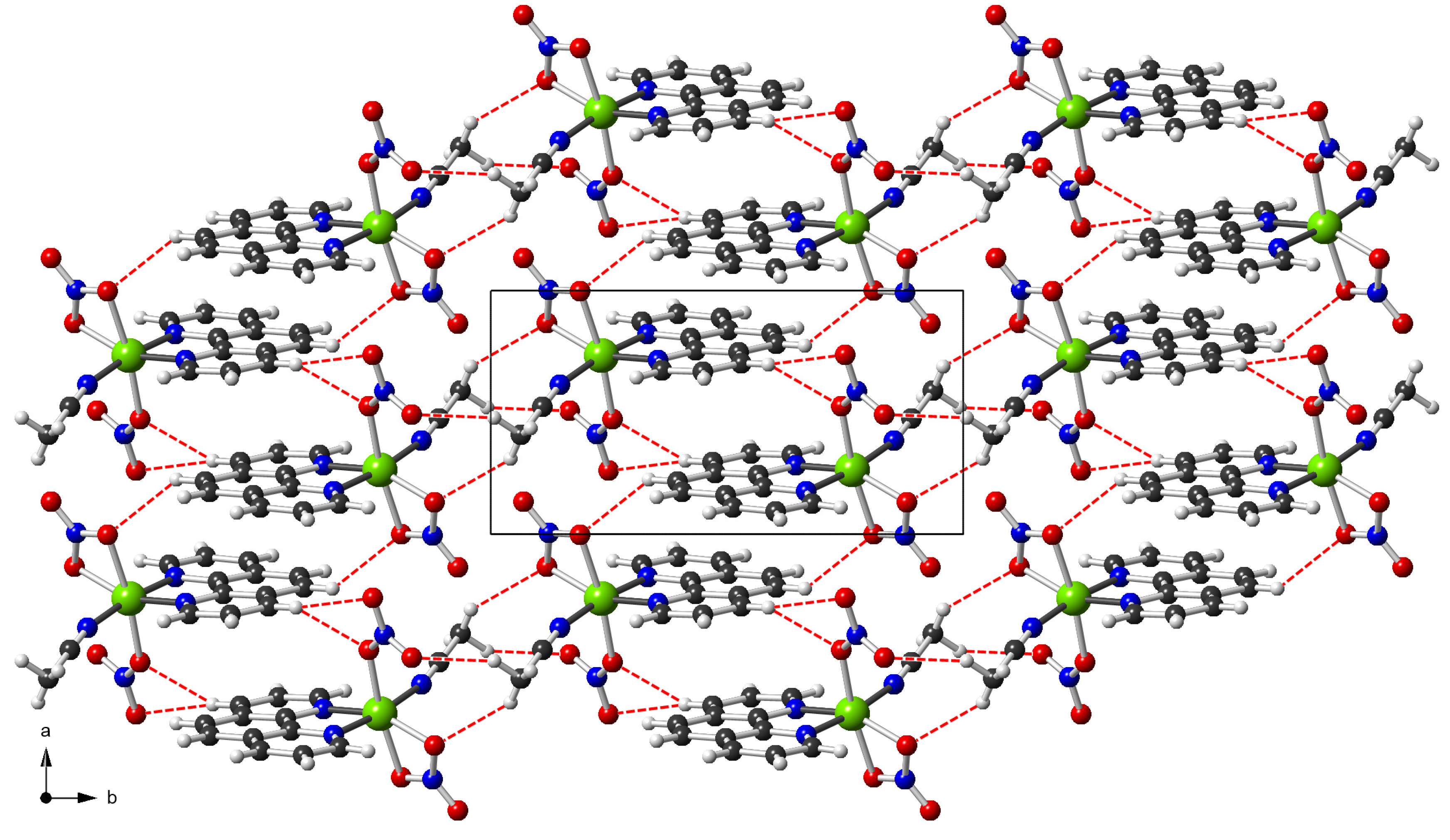
The crystal structure of 3 is columnar along the a-axis, as illustrated in Figure 6. The complex species occur in stacks along the a-axis. The stacks occur in an approximately fourfold array whereby each stack of complex species is surrounded by four other stacks. The molecules are assembled in each stack along the a-axis through OFF aryl∙∙∙aryl interactions of phen ligands (Figure 7). These are evident by the centroids’ separation distances of the symmetry-related phen rings (N1/C1–C4/C12) Cg1∙∙∙Cg2 (C4–C7/C11C12) (2−x, 1−y, 1−z) of 3.704 Å and the rings (N1/C1–C4/C12) Cg1∙∙∙Cg1 (C4–C7/C11C12) (1−x, 1−y, 1−z) of 3.717 Å (Figure 7). The stacks of complexes are further connected in the b-direction via (Me)C—H∙∙∙ONO2 contact interactions to generate layers in the ab plane (Figure 8). Along the c-axis, the layers are connected by additional (π)C―H∙∙∙ONO2 interactions (Figure 8; Table 3). The (Me)C—H∙∙∙ONO2 and (π)C―H∙∙∙ONO2 hydrogen bonds on the edges reinforce the stack. Details of hydrogen-bonding interactions along with symmetry operations are listed in Table 3.
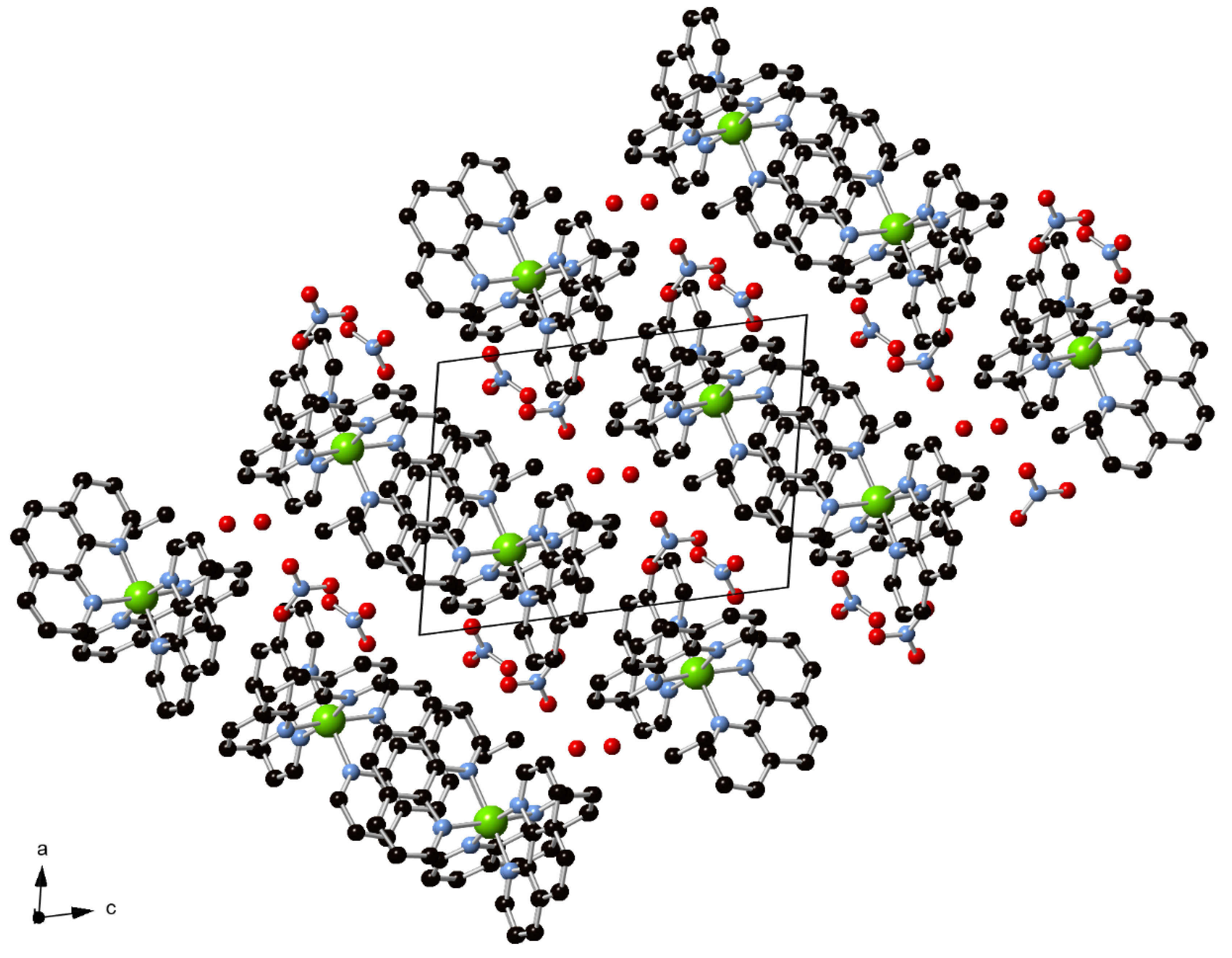
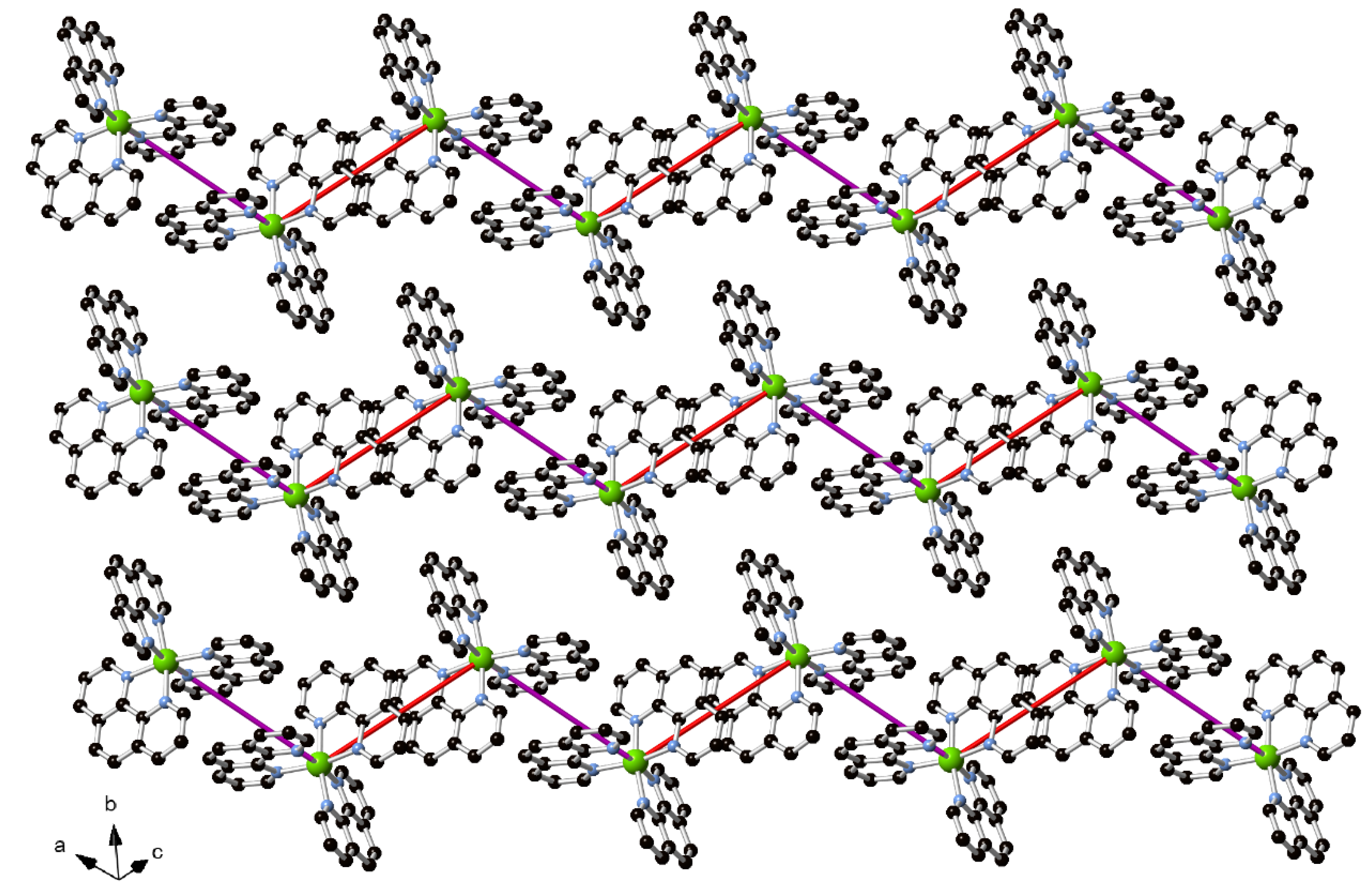
The crystal packing of complex 4 contains alternating blocks of [Ni(phen)3]2+ cations and nitrates, oxygen atoms of water and ethanol solvent (Figure 9). Each block of cations is constructed of embraced cations, whereby cations are involved in two alternating motifs of P4AE. Within the blocks of cations, the dominant motif is the infinite P4AE chain, in which each [Ni(phen)3]2+ participates in two alternating P4AE motifs. The chains down the a-axis with the adjacent Ni∙∙∙Ni separation distance of 9.320 Å (Figure 10a) are further interconnected along the c-axis with a Ni∙∙∙Ni separation distance of 9.215 Å (Figure 10b) to form layers in the ac plane (Figure 11). The zigzag chains down the a-axis are built by OFF π∙∙∙π stacking interactions of phen ligands in which the centroids of rings (N5/C25–C28/C36) Cg1∙∙∙Cg2 (C28–C31/C35–C36) are separated by 3.795 Å (Ni∙∙∙Ni is 9.320 Å) and EF interactions, as shown in Figure 10a. The chains down the c-axis are also involved in OFF π∙∙∙π interactions where the centroid∙∙∙centroid of rings (N3/C13–C16/C24) Cg3∙∙∙Cg4 (C16–C19/C23–C24) are separated by 3.827 Å (Ni∙∙∙Ni is 9.215 Å) along with EF interactions, as shown in Figure 10b. As shown in Figure 11, the zigzag P4AE chains are aligned side by side into the blocks of [Ni(phen)3]2+ cations. It is worth mentioning that the strands of ethanol molecules, water oxygen atoms and nitrate anions separating the blocks of cations exhibit extensive hydrogen-bonding interactions within the strands and with the blocks of cations. Hydrogen-bonding geometrical parameters are tabulated in Table 3.

3.4. Thermal Analysis

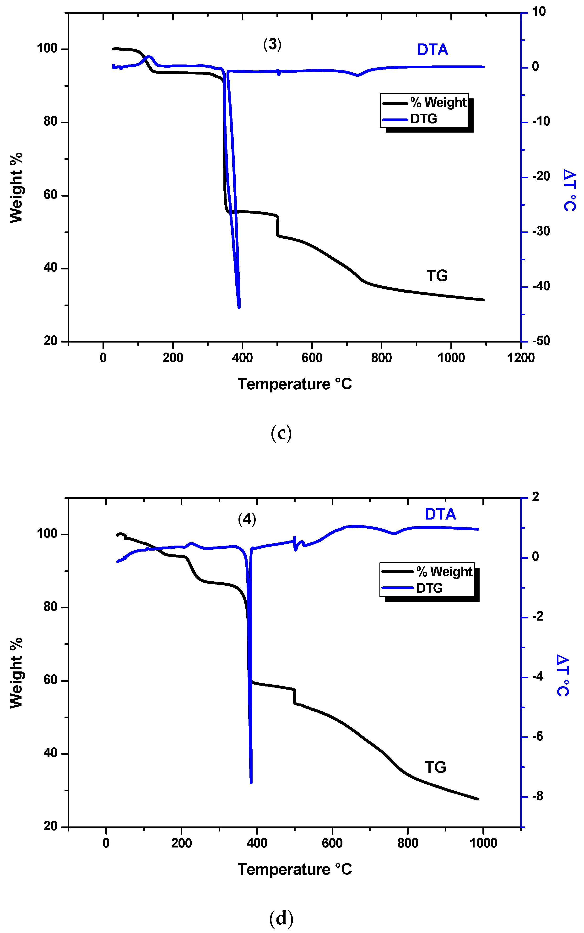
Thermogravimetric analysis (TGA) is an appropriate method for the estimation of thermal stability as well as the confirmation of the structural composition of complexes under investigation. It can provide information about physical and chemical phenomenon occurred upon gradual temperature increases such as degradation. These characteristics can be identified by, for example, the determination of the changes in complex weight as a function of change in temperature or time [30,31,32,33]. The thermal analysis for the complexes were performed at the temperature range of 30–1100 °C under nitrogen. The thermograms in Figure 12a–d show the decomposition behaviors of the four complexes (14). The thermograms indicate the multistep degradation of the complexes. The registered steps of the thermal decomposition of the complexes, the experimental mass loss as a function of change in temperature, matched with theoretical mass loss of the assigned fragment, were shown in Table 4. By investigation of the decomposition steps, it can be concluded that the number of steps is mostly in consistence with the number of ligand groups that are possibly lost stepwise. It was noticed from Table 4 that the water molecule and the anion (chloride and nitrate) were lost at a lower temperature than the degradation or loss of organic molecule phenanthroline ligand, [34,35,36,37,38]. Most of the decomposition steps of the complexes were endothermic. However, complex 2 showed two major exothermic processes at 119.2 and 472.1 °C. The expected residue materials obtained at higher temperature are nickel or nickel/carbon composites. The % mass of residues obtained after the degradation of complexes 1, 2, 3 and 4 were 26.9%, 42.2%, 31.6% and 27.5%, respectively.
Finally, a thermal analysis of Niphen complexes provided the temperature at which major decomposition of phen ligands occurred (about 400 °C). This temperature can be used as the pyrolysis set temperature to prepare nickel graphite nanocarbon by pyrolysis of the Niphen complex in a nitrogen gas furnace.

3.5. DFT Studies

3.5.1. Computational Details

The Gaussian 09 program suite was used to compute all the quantum chemical calculations [39]. The visualization of optimized structures was achieved through Chemcraft and GaussView 5.0 [40]. The geometries of considered complexes were optimized at M06-2X/6-31G (d) level of theory. The geometries from crystal structures (CIF files) were taken as input for geometry optimization. Several functionals were evaluated for reproducing experimental geometries before concluding that M06-2X is the functional of choice. The frequency analysis was also computed at an M06-2X/6-31G (d) level of theory to validate that the optimized structures are at true minimum; i.e., there was an absence of imaginary frequency. Furthermore, the simulated infrared (IR) spectrum was also analyzed from frequency parameters. UV-visible absorption spectrum was simulated using the TD-DFT method at the CAM-B3LYP/6-31G (d, p) level of theory. CAM-B3LYP has well known accuracy for predicting the UV-visible absorption properties of a variety of different classes of compounds. Frontier molecular orbital (FMO) analysis was also performed at the M062X/6-31G (d) level of DFT. The highest occupied molecular orbitals (HOMOs) and lowest unoccupied molecular orbitals (LUMOs) of a molecule have association with its reactivity. Energy gap (∆E) is defined as:
∆E = EHOMO − ELUMO

3.5.2. Geometry Optimization

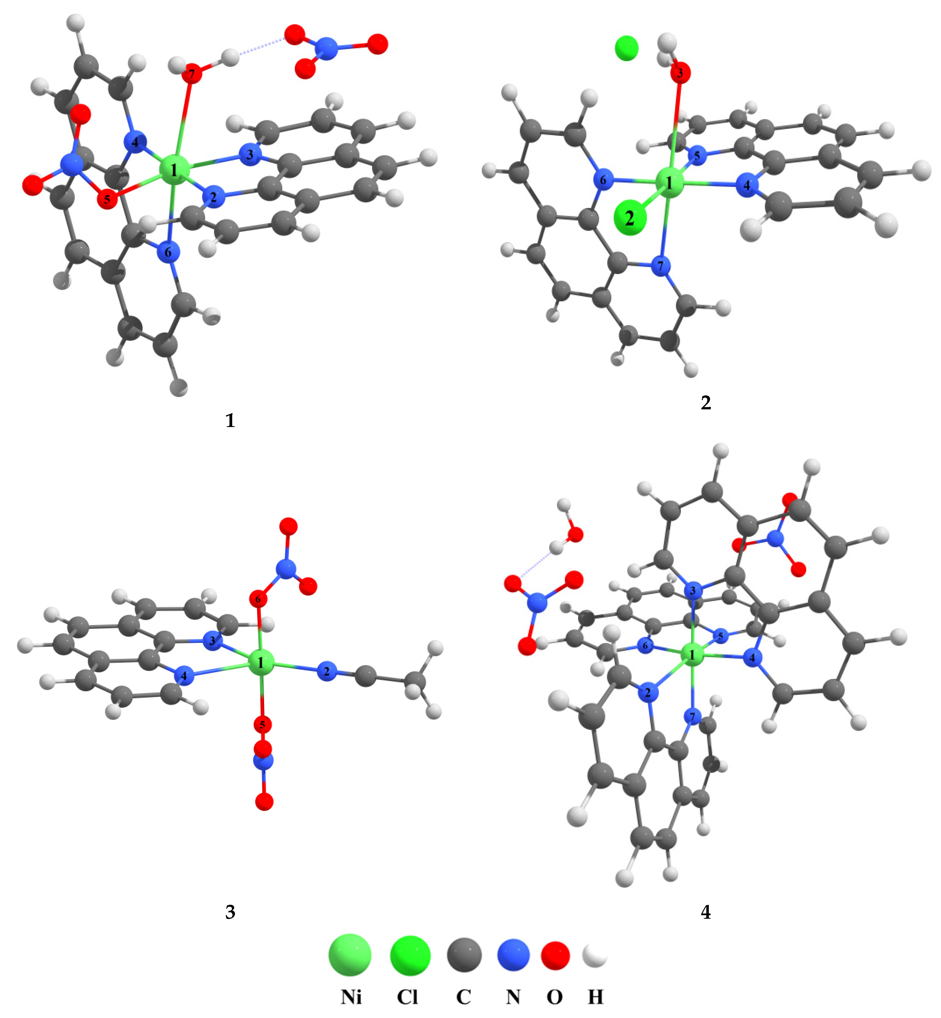
The structures for the optimization of considered Ni crystals were carried out from the experimental (atomic position) X-ray crystallographic information file (CIF). All other DFT computations were performed on the resultant optimized geometric parameters at the M062X/6-31G (d) level of theory. The structural parameters of the optimized geometries were compared with the experimentally obtained values, as given in Table 5. The optimized structures of Ni crystal phenanthroline-based complexes are given in Figure 13. With a few exceptions, theoretically obtained geometric parameters show nice agreement with the experimentally obtained geometric parameters.
The calculated coordination bond lengths of Ni—N for complex 2 are observed in the range of 2.00 Å to 2.19 Å, which is in accordance with the experimentally obtained results (2.09 Å to 2.12 Å). Due to a higher atomic radius of chlorine, the Ni—Cl bond length (2.24 Å) is higher as compared to the rest of the bond lengths. Similarly, most calculated angles show good correlation with the experimental values (see Table 5). Additionally, in complex 4, all Ni coordinated bonds are Ni—N, and the calculated bond distances are in the range of 1.96 Å to 2.27 Å. On the other hand, calculated bond angles show good agreement with the X-ray crystallographic data, except for the angle (N2—Ni1—N3), which shows a higher deviation (numbering from Figure 13).
In complex 3, two types of coordination bonds were observed; three are Ni—N and two are Ni—O bonds. The calculated bond lengths are in the range of 1.97 Å to 2.06 Å, consistent with the X-ray crystal data. Likewise, four Ni—N and two Ni—O coordination bonds were observed in complex 1. The calculated bond lengths are in accordance with the experimentally determined values. It is quite obvious from the figures that the octahedral geometry is retained in the 1, 2, 3 and 4 complexes.

3.5.3. Vibrational Spectral Analysis

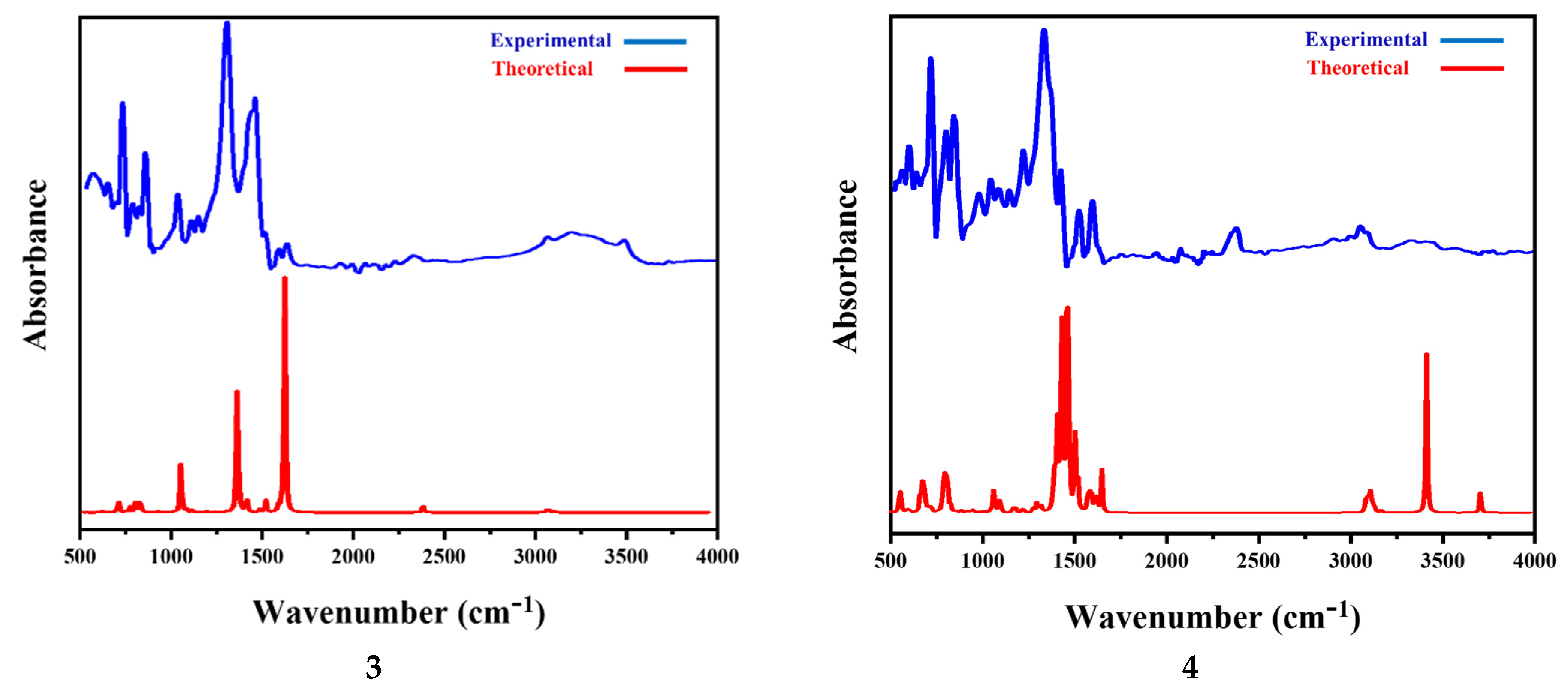
Vibrational analysis is an important and powerful tool in both theoretical and experimental chemistry. Experimental FTIR stretching vibrations in complex 1 were observed for OH (water) at 3141.5 and 3284.2 cm−1, while in complex 2, they were observed at 3307.4 and 3396.1 for OH (water and ethanol). Stretching for CH aromatic occurred at 3069.4, 3047.0, 3070.2 and 3020.9 cm−1 for complexes 1, 2, 3 and 4, respectively.
The CN stretch of coordinated acetonitrile appeared at 2331.6 cm−1 in complex 3.
The strong absorption bands at 1400–1500 cm−1 and medium intensity absorption peaks at 1550–1650 cm−1 are due to the stretching vibration of aromatic C–C and C–N bonds [41,42].
NO3 stretches occurred at 842.5, 1294.9 cm−1 for 1; 847.2, 1300.4 cm−1 in 3 and 842.3, 1326.8 cm−1 in 4. The peak at 1215.1 cm−1 in complex 4 is due to C–O stretch of ethanol. Out-of-plane aromatic bend occurred in the range of 950 to 700 cm−1 for all the complexes.
According to references [43,44], Ni–N bonding and strong IR spectrum is found at 530.3 and 580.1 cm−1 in complex 1, 526.5 and 553.5 cm−1 in 2, 561.7 cm−1 in 3 and 565.7 cm−1 in 4. The strong intensity absorption peaks at 672.6 cm−1 are due to the stretching vibration of the Ni–Cl bond in complex 2.
DFT-based computational methods predict the almost precise vibrational spectra and molecular structure with reasonable computational cost. The optimization of studied complexes corresponds to energy and vibrational frequencies, which are calculated at the M06-2X/6-31G (d) level of theory to aid the explanation of the experimental infrared (IR) data.
The DFT calculated and experimental vibrational spectra of the representative complex 4 is presented in Figure 14, while the spectra of the rest of the considered Ni–phenanthroline-based complexes 1–3 are given in supporting information (Table S1). Usually, the simulated vibrational spectra are overestimated because of harmonic models; thus, a scaling factor of (0.9613) was applied to obtain the scaled frequencies of simulated frequencies. The theoretically calculated vibrational peak at 3030–3100 cm−1 is attributed to the C–H stretching vibration in complex 4. A band in the similar region (of low intensity) in the experimental IR spectrum is associated with C–H stretching. The peak around 1650 cm−1 is attributed to the aromatic C–H bending mode of the phenanthroline ligand confirmed through both theoretical and experimental spectra. Similarly, the peak at around 1500–1650 cm−1 is assigned to the C–C stretching of the conjugated phenanthroline skeleton, which originates from the sp2 hybridized carbon atoms at 1530–1580 cm−1 and is confirmed experimentally [45]. Both simulated and experimental spectra show peaks at 1380 cm−1 and 800 cm−1, which are attributed to the asymmetric stretching of NO3 and symmetric stretching of NO3, are supported well by the literature [46,47].
Similarly, the theoretical IR spectra of complexes 1–3 possess peak vibrations in the region of 3000–3200 cm−1, which are mainly attributed to the stretching vibrations of the C—H bond. In simulated spectra, prominent C—C stretching vibrations were observed in the range of 1412–1687 cm−1 for complexes 1–3, whereas the vibrational C—H bending mode was seen at around 1460–1515 cm−1. In the case of complex 1, the asymmetric stretching of NO3 was observed at 715 cm 1, whereas the symmetric stretching of NO3 appeared at 1102 cm−1. In the case of complex 2, a clear bending (scissoring) mode of water appeared at around 1700 cm 1. Similarly, the Ni—N stretching mode in complex 2 appeared at 881 cm−1. In the case of complex 3, the asymmetric stretching of NO3 was observed at 785 cm 1, whereas the symmetric stretching of NO3 appeared at 1068 cm−1. Similarly, the Ni—N stretching mode in complex 3 appeared at 888 cm−1. In simulated spectra, a good correlation exists in complexes 1, 3 and 4 due to the similarity of the nature of ligands attached to Ni–metal.

3.5.4. UV-Visible Spectroscopic Study

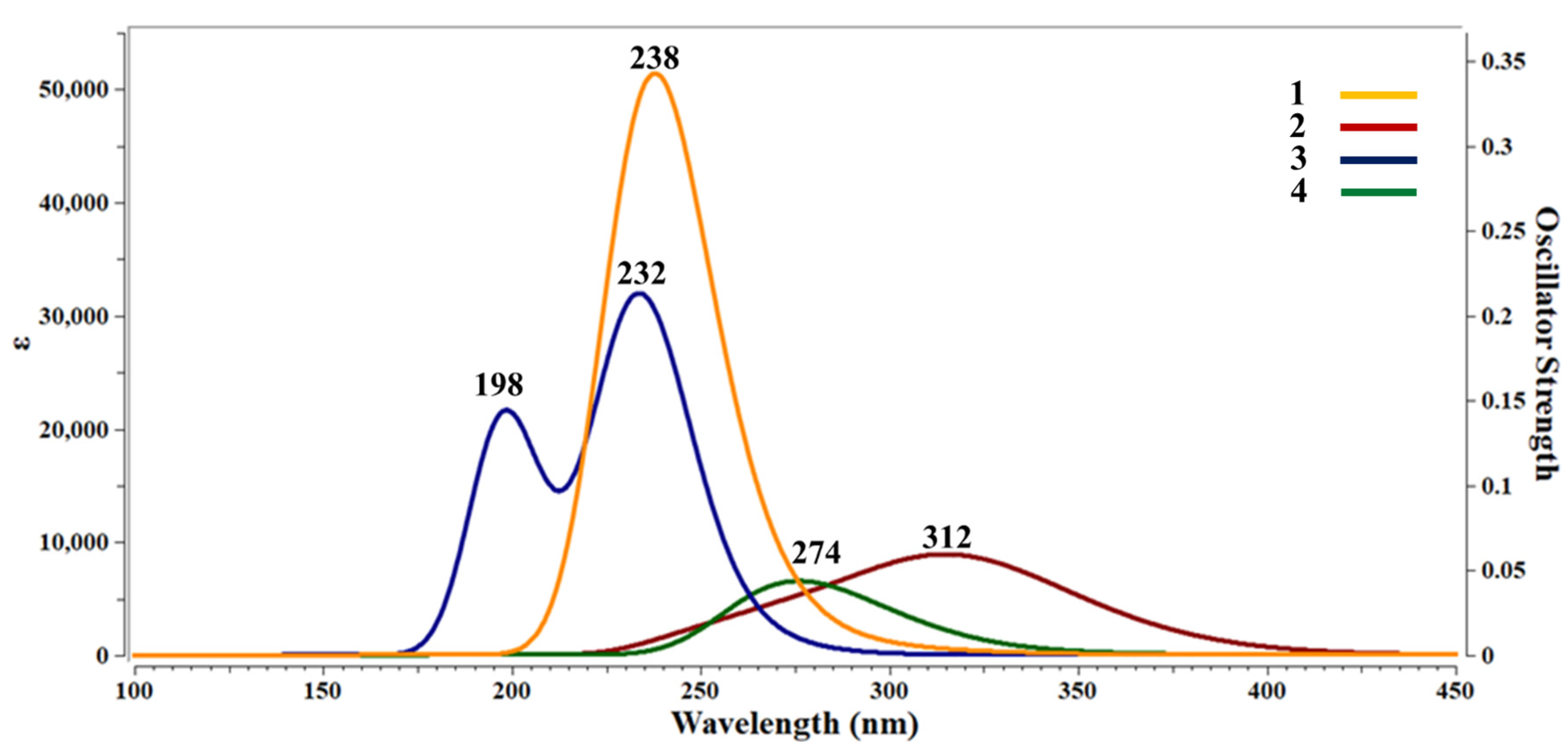
TD-DFT calculations were performed for the analysis of the UV-visible spectra of Ni crystal phenanthroline complexes (Figure 15). In the UV-visible spectra, the excitation energies are plotted against intensity as well as oscillator strength. Absorption spectra of Ni crystal phenanthroline complexes (1, 2, 3 and 4) were computed at ambient temperature. All the studied complexes showed main absorption peaks in the ultraviolet (UV) region. Therefore, all the studied Ni–phenanthroline-based complexes were UV-active. In the case of complex 3, two absorption bands were observed at 198 and 232 nm. An intense absorption peak was observed at 238 nm for complex 1, whereas for complexes 2 and 4, absorption peaks were observed at 274 and 312 nm with low absorptivity.
According to frontier molecular orbital analysis, the electronic cloud of HOMOs is mainly distributed on ligands. If the LUMOs of these complexes accept electrons, then these electrons will be mostly localized over ligands. Therefore, the type of electron transfer is ligand–ligand charge transfer. The key transitions responsible for UV-visible spectra occur from the HOMO to LUMO or from HOMO-1 to LUMO+1, and these are associated with the π-π* transitions. However, in the case of complex 2, where the HOMO is mainly distributed over Cl- ion and LUMO is on phenanthroline ring, the key transition observed is associated with the n-π* transition. Overall, the absorption spectra show that for studied organometallic complexes n-π * and π-π *, transitions may appear.
In the case of complexes 1, 3 and 4, where the nitrate group is attached along with phenanthroline rings, the absorption maxima were observed at 238nm, 232 nm and 274 nm, respectively, whereas for complex 4, where the Cl- ion ligand was attached instead of the nitrate group, absorption maxima is observed at longer wavelength of 312 nm. The results declare that by changing the ligand, the absorption maximum is shifted towards longer wavelength, hence there might be applications in optoelectronic devices.

3.5.5. Frontier Molecular Orbitals (FMO) Analysis

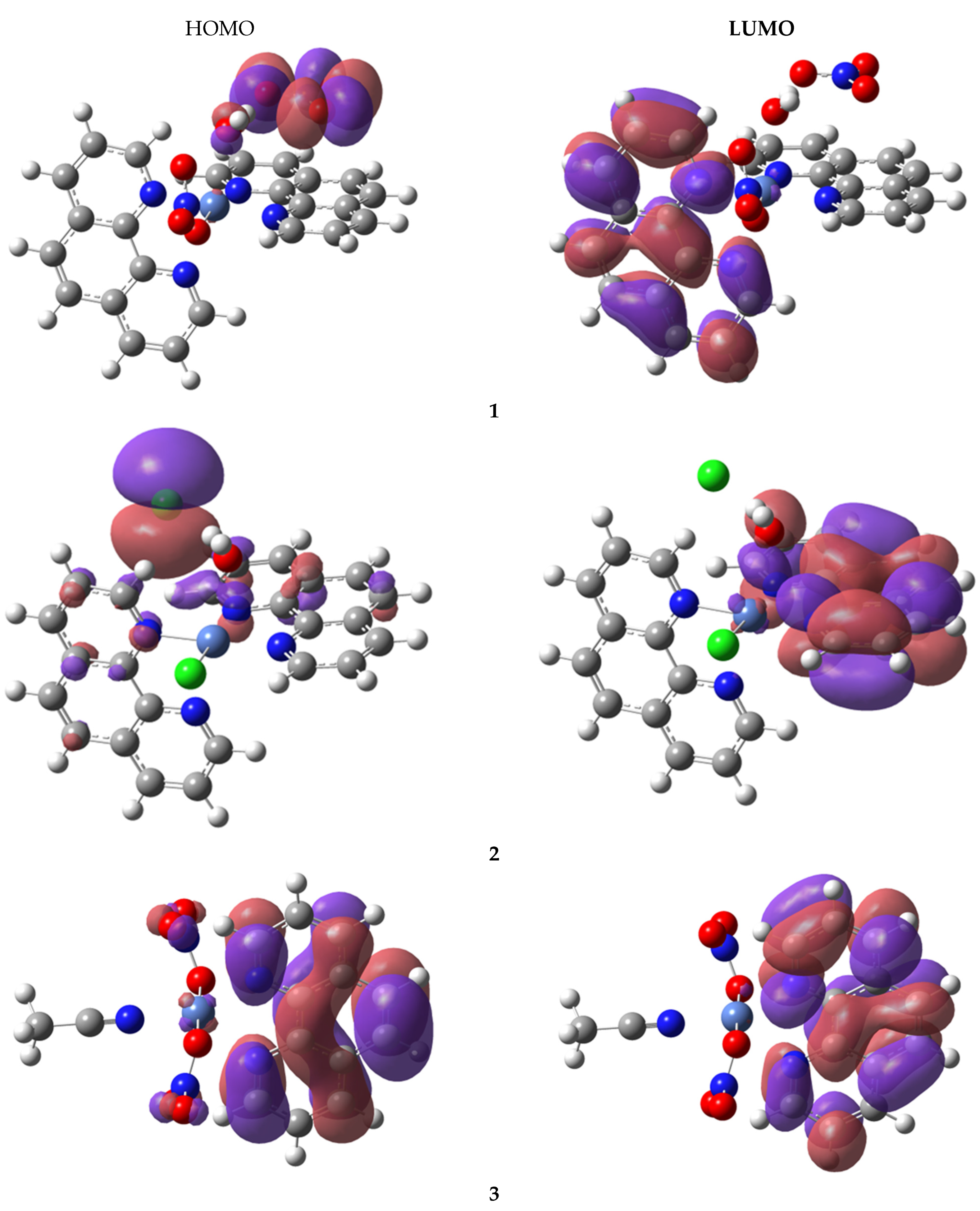
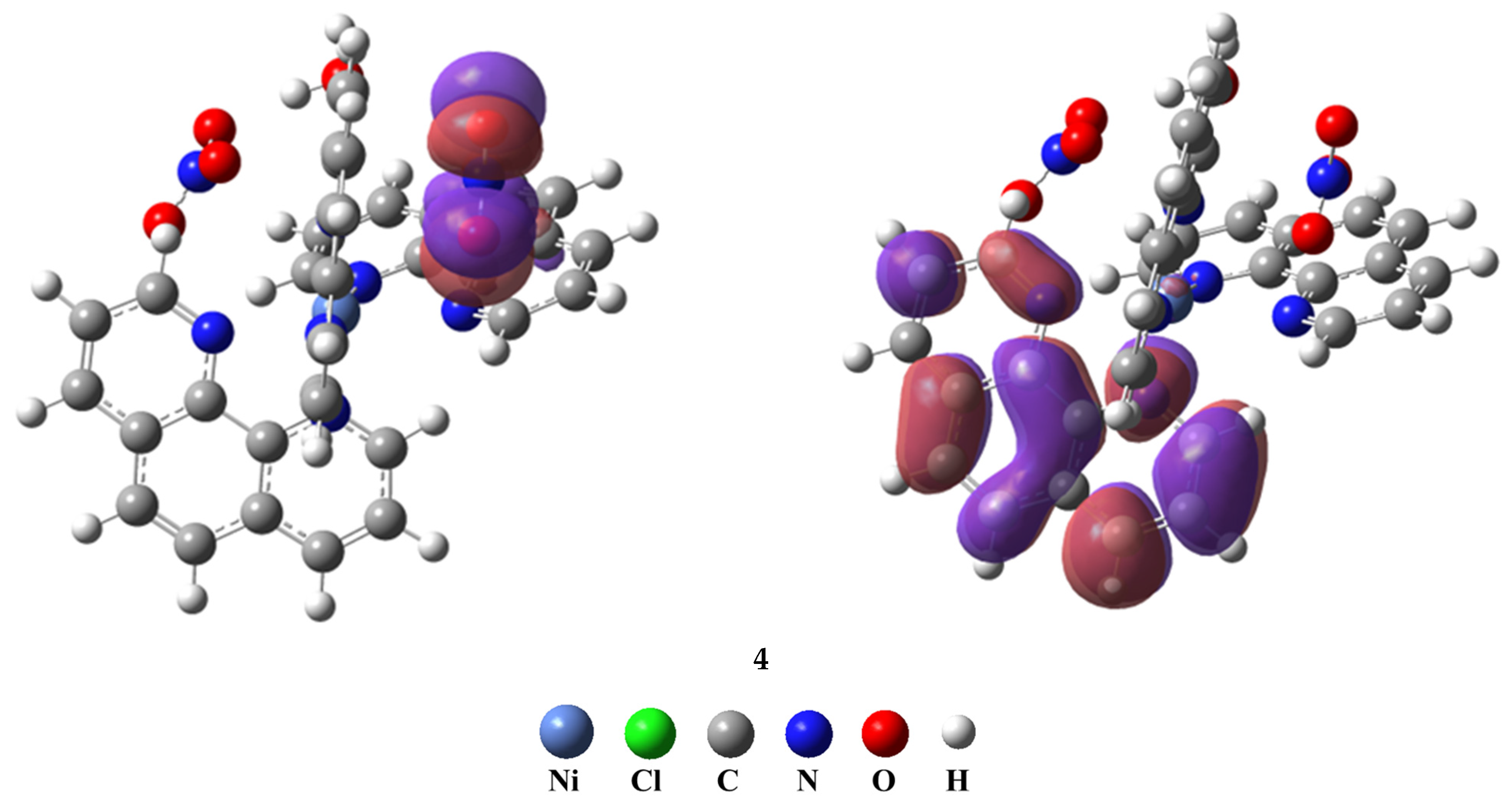
The highest occupied molecular orbitals (HOMOs) and lowest unoccupied molecular orbitals (LUMOs) are those orbitals that directly interact with other reacting species; therefore, they are known as the frontier molecular orbitals (FMOs). These FMOs are crucial in the optical and electronic properties, as well as for studying chemical reactions [48]. The energies of HOMO (EHOMO), LUMO (ELUMO) and their energy gaps (∆E) are given in Table 6, while the distribution of the iso-densities of these FMOs are shown in Figure 16.
Molecular orbitals generally give clues regarding the nature of reactivity, lone pairs, conjugation and some physical properties of reacting species. The electron-donating ability is characterized through HOMO energy, and the electron-accepting capacity is characterized through LUMO energy, whereas the energy gap (∆E) characterizes the electrical transport ability and chemical stability of molecules [49]. The HOMO energies of studied complexes are in the range of −6.44 eV to −8.11 eV, while LUMO energies are in the range of −1.25 eV to −1.70 eV. The energy gaps (∆E) observed for 1, 2, 3 and 4 are 5.63 eV, 5.18 eV, 6.94 eV and 4.74 eV, respectively. The lowest energy gap is estimated for complex 4. In the case of complex 2, the HOMO cloud density is mainly distributed on Cl atom, whereas for 1 and 4 complexes, the HOMO cloud is mostly distributed over the nitrate group (NO3−). However, the LUMO cloud density in all studied complexes is distributed on one of the phenanthroline rings, which indicates the electroactive or reactive nature of the ring.
The transition that occurs from the HOMO to the LUMO is associated with the π-π * transitions of the nitrate group (NO3−) and phenanthroline ring, respectively. However, in the case of complex 2, the HOMO is mainly distributed over the Cl- ion and LUMO on the phenanthroline ring, which is associated with the n-π * transition. The next frontier molecular orbitals (HOMO−1 and LUMO+1) and (HOMO-2 and LUMO+2) almost show the same type of transitions/excitations trend with slight differences in the distribution pattern of orbital densities. The overall charge transfer occurs from ligand to ligand or intramolecular charge transfer in all studied complexes.

4. Conclusions

Four distorted octahedral Ni–phen complexes were prepared and characterized by FTIR, thermal and X-ray analysis: 1, [Ni(phen)2(H2O)(ONO2)](NO3); 2, [Ni(phen)2(H2O)Cl]Cl; 3, [Ni(phen)(CH3CN)(ONO2)(O2NO)] and 4, [Ni(phen)3](NO3)2.H2O.C2H5OH. FTIR proved the coordination of M-N and M-Cl as well as the presence of nitro groups in the complexes, while thermal analysis revealed that the loss of chloro and nitro groups occurred before the degradation of the phenanthroline ligand. We found that the complex degradation using thermal analysis shows a different pattern depending on the number of coordinated phen ligands and the type of co-ligand nitrate, chloride, acetonitrile or water in the complex.
The X-ray structure of all complexes contains NiII in a distorted octahedral geometry. In the structures, aryl∙∙∙aryl interactions within phen ligands dictate the packing, and extend to parallel fourfold aryl embrace (P4AE) in the cases of 2 and 4. Hydrogen-bonding interactions, namely (π)C―H∙∙∙ONO2 in 1; C―H···O, C―H···Cl and O—H···Cl in 2 and (Me)C—H∙∙∙ONO2 and (π)C―H∙∙∙ONO2 in 3, reinforce aryl∙∙∙aryl interactions and add extra supra-molecularity in the structures.
Moreover, DFT investigation was also performed on studied Ni–phen complexes to correlate their geometric parameters with the experimental outcomes, and a strong agreement was observed. For electronic characteristics, FMO analysis and TD-DFT UV calculations were performed which reveal the ligand–ligand charge transfer in studied Ni–phen complexes.

Supplementary Materials

The following supporting information can be downloaded at: https://www.mdpi.com/article/10.3390/cryst13050738/s1, Table S1: IR simulated and experimental spectra of complex 1, 2 and 3: the theoretical IR spectra are simulated at M062X/6-31G (d) level of theory.

Author Contributions

Conceptualization, H.H.H., B.F.A. and K.A.; methodology, H.H.H., K.A. and S.S.; software, H.A.G., B.F.A. and S.S.; validation, H.H.H. and K.A.; investigation, S.A.A. and S.S.; resources, K.A., H.H.H. and S.A.A.; writing—original draft preparation, H.H.H. and S.A.A.; writing—review and editing, H.H.H., B.F.A. and K.A.; supervision, H.H.H. and K.A.; project administration, K.A. and H.H.H.; funding acquisition, H.H.H. and S.A.A. All authors have read and agreed to the published version of the manuscript.

Funding

This work was supported by the Deanship of Scientific Research, Vice Presidency for Graduate Studies and Scientific Research, King Faisal University Saudi Arabia [Grant No. 3267].

Data Availability Statement

All data are available in the main text or the Electronic Supplementary Information (ESI).

Acknowledgments

The authors would like to express their gratitude to the Deanship of Scientific Research, Vice Presidency for Graduate Studies and Scientific Research, King Faisal University Saudi Arabia [Grant No. 3267] for the funding support.

Conflicts of Interest

The authors declare that they have no known competing financial interests or personal relationships that could have appeared to influence the work reported in this paper.

References

  1. Preston, H.; Kennard, C. Stereochemistry of rigid chelate–metal complexes. Part II. Crystal structure of di-µ-chloro-sym-trans-dichloro-bis-(2,9-dimethyl-1,10-phenanthroline) dinickel (II)–2-chloroform. J. Chem. Soc. A Inorg. Phys. Theor. 1969, 2682–2685. [Google Scholar] [CrossRef]
  2. Bencini, A.; Lippolis, V. 1, 10-Phenanthroline: A versatile building block for the construction of ligands for various purposes. Coord. Chem. Rev. 2010, 254, 2096–2180. [Google Scholar] [CrossRef]
  3. Teng, Q.; Huynh, H.V. A unified ligand electronic parameter based on 13CNMR spectroscopy of N-heterocyclic carbene complexes. Dalton Trans. 2017, 46, 614–627. [Google Scholar] [CrossRef]
  4. Brewer, B.; Brooks, N.R.; Abdul-Halim, S.; Sykes, A.G. Differential metathesis reactions of 2,2′-bipyridine and 1,10-phenanthroline complexes of cobalt (II) and nickel (II): Cocrystallization of ionization isomers {[cis-Ni (phen)2(H2O)2][cis-Ni(phen)2 (H2O)Cl]}(PF6)3·4.5 H2O, and a synthetic route to asymmetric tris-substituted complexes. J. Chem. Crystallogr. 2003, 33, 651–662. [Google Scholar]
  5. Chesnut, D.J.; Haushalter, R.C.; Zubieta, J. The hydrothermal synthesis and structural characterization of a new class of compounds: Nickel organoamine-halocadmates. Inorg. Chim. Acta 1999, 292, 41–51. [Google Scholar] [CrossRef]
  6. Norman, R.E.; Xie, M. Nickel(II) 1,10-Phenanthroline complexes: Cis-[Aqua(Bromo) bis (1,10-Phenanthroline)Nickel(II)] Bromide Trihydrate and (tris (1,10-Phenanthroline)Nickel(II)] Bromide Octahydrate. J. Coord. Chem. 2004, 57, 425–434. [Google Scholar] [CrossRef]
  7. Prior, T.J.; Rujiwatra, A.; Chimupala, Y. [Ni(1,10-phenanthroline)2(H2O)2](NO3)2: A Simple Coordination Complex with a Remarkably Complicated Structure that Simplifies on Heating. Crystals 2011, 1, 178–194. [Google Scholar] [CrossRef]
  8. Tang, X.Y.; Qiu, Y.C.; Sun, F.; Yue, S.T. Diaquabis(1,10-phenanthroline-k2N,N′)nickel(II) diperchlorate 0.4-hydrate. Acta Crystallogr. 2007, E63, m2515. [Google Scholar]
  9. Zhu, L.; Zhang, L.H.; Liu, B.Y.; An, Z. Crystal structure of diaquabis(1,10-phenanthroline)nickel(II) sulfate hydrate (1:5.6), [Ni(H2O)2(C12H8N2)2][SO4]·5.6H2O. Z. Kristallogr.-New Cryst. Struct. 2006, 221, 451–452. [Google Scholar]
  10. Ferbinteanu, M.; Cimpoesu, F.; Mariusandruh, A.; Rochon, F.D. Solid-state chemistry of [Ni(AA)3][PdCl4]·nH2O complexes (AA = bipy, phen) and crystal structures of cis-diaquabis(phenanthroline)nickel(II) tetrachlorozincate and cis-dichloro-bis(bipyridine)nickel(II). Polyhedron 1998, 17, 3671–3679. [Google Scholar] [CrossRef]
  11. Oresmaa, L.; Haukka, M.; Vainiotalo, P.; Pakkanen, T.A. Ab Initio Calculations and Mass Spectrometric Determination of the Gas-Phase Proton Affinities of 4,4′-Disubstituted 2,2′-Bipyridines. J. Org. Chem. 2002, 67, 8216–8219. [Google Scholar] [CrossRef]
  12. Reyzer, M.; Brodbelt, J. Ligand displacement reactions of dimer and trimer pyridyl ligand/transition metal complexes1. Int. J. Mass Spectrom. 1999, 182, 311–322. [Google Scholar] [CrossRef]
  13. Ha, K. Crystal structure of cis-bis (2,2′-bipyridine-κ2N,N′) bis (thiocyanato-κN) nickel (II), C22H16N6NiS2. Z. Für Krist.-New Cryst. Struct. 2017, 232, 151–152. [Google Scholar]
  14. Tsierkezos, N.G.; Diefenbach, M.; Roithová, J.; Schröder, D.; Schwarz, H. Competitive Complexation of Gaseous MnII by 1, 10-Phenanthroline, 2,2′-Bipyridine, and 4, 5-Diazafluorene. Inorg. Chem. 2005, 44, 4969–4978. [Google Scholar] [CrossRef]
  15. Woodward, J.D.; Backov, R.; Abboud, K.A.; Ohnuki, H.; Meisel, M.W.; Talham, D.R. Structural, thermal, and magnetic properties of three transition metal-4,4′-bipyridine coordination polymers:[Ni(4,4′-bipy) 3(H2O)2](ClO4)2 1.4(4,4′-bipy)·3(H2O);[Co(4,4′-bipy)3(H2O)2](ClO4)2·1.4(4,4′-bipy)· 3(H2O);[Cu(4,4′-bipy)3(DMSO)2](ClO4)2 2(4,4′-bipy). Polyhedron 2003, 22, 2821–2830. [Google Scholar]
  16. Alramadhan, S.A.; Hammud, H.H. Graphene Nickel Silica Supported Nanocomposites, Application in Water Treatment. Appl. Nanosci. 2020, 11, 273–291. [Google Scholar] [CrossRef]
  17. Hammud, H.H.; Karnati, R.K.; Alotaibi, N.; Hussain, S.G. Thirumurugan Prakasam, Cobalt–carbon silica nanoparticles for uptake of cationic and anionic dye from polluted water. Molecules 2021, 26, 7489. [Google Scholar] [CrossRef]
  18. Alotaibi, N.; Hammud, H.H.; Al Otaibi, N. Electrocatalytic Properties of 3D Hierarchical Graphitic Carbon Cobalt Nanoparticles for Urea Oxidation. ACS Omega 2020, 5, 26038–26048. [Google Scholar] [CrossRef]
  19. Hammud, H.H.; Alotaibi, N.; Al Otaibi, N.; Aljaafari, A.; Ahmed, F.; Azam, A.; Prakasam, T. Hierarchical Porous Carbon Cobalt Nanocomposites based Sensor for Fructose. Chemosensors 2021, 9, 6. [Google Scholar] [CrossRef]
  20. Hammud, H.H.; Traboulsi, H.; Karnati, R.K.; Hussain, S.G.; Bakir, E. Hierarchical Graphitic Carbon-Encapsulating Cobalt Nanoparticles for Catalytic Hydrogenation of 2,4-Dinitrophenol. Catalysts 2022, 12, 39. [Google Scholar] [CrossRef]
  21. Krebsz, M.; Kótai, L.; Sajó, I.E.; Váczi, T.; Pasinszki, T. Carbon Microsphere-Supported Metallic Nickel Nanoparticles as Novel Heterogeneous Catalysts and Their Application for the Reduction of Nitrophenol. Molecules 2021, 26, 5680. [Google Scholar] [CrossRef]
  22. Sheldrick, G.M. A short history of SHELX. Acta Cryst. 2008, A64, 112–122. [Google Scholar] [CrossRef] [PubMed]
  23. Sheldrick, G. SHELXTL-PC (Version 5.1); Siemens analytical instruments, Inc.: Madison, WI, USA, 1997. [Google Scholar]
  24. Li, S.-D.; Su, F.; Zhou, C.-Y.; Hu, Q.-L.; Li, Y.-Q.; Wang, Z.-J. Syntheses, structures and magnetic properties of two CoII/NiII isostructural coordination polymers based on an asymmetric semirigid tricarboxylate ligand. Acta Cryst. 2022, C78, 23–29. [Google Scholar] [CrossRef]
  25. Nath, H.; Sharma, P.; Frontera, A.; Barcelo-Oliver, M.; Verma, A.K.; Das, J.; Bhattacharyya, M.K. Phenanthroline-based Ni(II) coordination compounds involving unconventional discrete fumarate-water-nitrate clusters and energetically significant cooperative ternary π-stacked assemblies: Antiproliferative evaluation and theoretical studies. J. Mol. Struct. 2022, 1248, 131424. [Google Scholar] [CrossRef]
  26. Ren, X.-C.; Li, J.; Wang, W.-H.; Chen, X.; Zhang, B.; Li, L.-Z. [NH4][Ni(phen)3]BiI6: Synthesis, structure, photocatalytic property and theoretical study of a discrete iodobismuthate. Inorg. Chem. Commun 2021, 130, 108714. [Google Scholar] [CrossRef]
  27. Matsia, S.; Kaoulla, A.; Menelaou, M.; Hatzidimitriou, A.; Papadopoulos, T.; Reimann, M.K.; Pöttgen, R.; Salifoglou, A. Structural speciation in chemical reactivity profiling of binary-ternary systems of Ni(II) with iminodialcohol and aromatic chelators. Polyhedron 2022, 212, 115577. [Google Scholar] [CrossRef]
  28. Russell, V.; Craig, D.; Scudder, M.; Dance, I. Crystal packing motifs for mixed oxalate/phenanthroline metal complexes. CrystEngComm 2001, 3, 96–106. [Google Scholar] [CrossRef]
  29. Dance, I.; Scudder, M. Molecules embracing in crystals. CrystEngComm 2009, 11, 2233–2247. [Google Scholar] [CrossRef]
  30. Shaabani, B.; Mirtamizdoust, B.; Viterbo, D.; Croce, G.; Hammud, H.; Hojati-Lalemi, P.; Khandar, A. Sonochemical synthesis of a novel nano-scale lead(II) coordination polymers: Synthesis, crystal structure, thermal properties and DFT calculations of [Pb(dmp)(μ-N3)(μ-NO3)]n with the novel Pb2-(μ-N3)2-(μ-NO3)2 unit. ZAAC J. Inorg. Gen. Chem. 2011, 637, 713–719. [Google Scholar]
  31. Mirtamizdoust, B.; Trávníček, Z.; Hanifehpour, Y.; Talemi, P.; Hammud, H.; Joo, S.W. Synthesis and characterization of nano-peanuts of lead(II) coordination polymer [Pb(qcnh)(NO3)2]n with ultrasonic assistance: A new precursor for the preparation of pure-phase nano-sized PbO. Ultrason. Sonochem. 2017, 34, 255–261. [Google Scholar] [CrossRef]
  32. Zaworotko, M.J.; Hammud, H.H.; Kabbani, A.; McManus, G.J.; Ghannoum, A.M.; Masoud, M.S. Synthesis and characterization of some transition metal complexes with mixed adenine and acetylacetonate ligands. Crystal structures of solvated complex {[Cu(acac)2(adenine)]•EtOH} and {[Cu(acac)2(adenine)]•DMF•H2O. J. Chem. Crystallogr. 2009, 39, 853–863. [Google Scholar] [CrossRef]
  33. Hammud, H.H.; Nemer, G.; Sawma, W.; Touma, J.; Barnabe, P.; Bou-Mouglabey, Y.; Ghannoum, A.; El-Hajjar, J.; Usta, J. Copper–adenine complex, a compound, with multi-biochemical targets and potential anti-cancer effect. Chem.-Biol. Interact. 2008, 173, 84–96. [Google Scholar] [CrossRef] [PubMed]
  34. Zaworotko, M.J.; Hammud, H.H.; Kravtsov, V.C. The co-crystal of iron(II) complex hydrate with hydroxybenzoic acid: [Fe(Phen)3]Cl(p-hydroxybenzoate).2(p-hydroxybenzoic acid).7H2O. J. Chem. Crystallogr. 2007, 37, 219–231. [Google Scholar] [CrossRef]
  35. Hammud, H.H.; El Hamaoui, B.; Noubani, N.H.; Feng, X.; Wu, Z.-S.; Müllen, K.; Ayub, K. Carbon-Cobalt Nanostructures as an Efficient Adsorbent of Malachite Green. Nanosci. Nanotechnol.-Asia 2018, 8, 263–280. [Google Scholar] [CrossRef]
  36. Mroueh, M.; Daher, C.; Hariri, E.; Demirdjian, S.; Isber, S.; Choi, E.S.; Mirtamizdoust, B.; Hammud, H.H. Magnetic property, DFT calculation, and biological activity of bis[(μ2-chloro)chloro(1,10-phenanthroline)copper(II)] complex. Chem.-Biol. Interact. 2015, 231, 53–60. [Google Scholar] [CrossRef]
  37. Hammud, H.H.; Kortz, U.; Bhattacharya, S.; Demirdjian, S.; Hariri, E.; Isber, S.; Choi, E.S.; Mirtamizdoust, B.; Mroueh, M.; Daher, C.F. Structure, DFT studies, Magnetism and Biological activity of Bis[(µ2-azido)-chloro-(1,10-phenanthroline)-copper(II)] complex. Inorg. Chim. Acta 2020, 506, 119533. [Google Scholar] [CrossRef]
  38. Hammud, H.H.; Zaworotko, M.J.; McManus, G.J.; Tabesh, R.N.; Ibrahim, H.I.M.; Ayub, K.; Ludwig, R. The co-crystal of copper(II) phenanthroline chloride complex hydrate with p-aminobenzoic acid: Structure, cytotoxicity, thermal analysis and DFT calculation. Chem. Mon. 2021, 152, 323–336. [Google Scholar] [CrossRef]
  39. Frisch, M.; Clemente, F. Gaussian 09, Revision A. 01; Frisch, M.J., Trucks, G.W., Schlegel, H.B., Scuseria, G.E., Robb, M.A., Cheeseman, J.R., Scalmani, G., Barone, V., Mennucci, B., Petersson, G.A., Eds.; Gaussian, Inc.: Wallingford, CT, USA, 2009. [Google Scholar]
  40. Khan, S.; Yar, M.; Kosar, N.; Ayub, K.; Arshad, M.; Zahid, M.N.; Mahmood, T. First-principles study for exploring the adsorption behavior of G-series nerve agents on graphdyine surface. Comput. Theor. Chem. 2020, 1191, 113043. [Google Scholar] [CrossRef]
  41. Roy, T.G.; Hazari, S.K.S.; Dey, B.K.; Miah, H.A.; Olbrich, F.; Rehder, D. Synthesis and antimicrobial activities of isomers of N(4),N(11)-dimethyl-3, 5,7,7,10,12,14,14-octamethyl-1,4,8,11-tetraazacyclotetradecane and their nickel(II) complexes. Inorg. Chem. 2007, 46, 5372–5380. [Google Scholar] [CrossRef]
  42. Roy, T.G.; Hazari, S.K.S.; Dey, B.K.; Meah, H.A.; Rahman, S.; Kim, D.I.; Park, Y.C. Synthesis, electrolytic behaviour and antimicrobial activities of cadmium complexes of isomers of 3,10-C-meso-3,5,7,7,10,12,14,14-octamethyl-1,4,8,11-tetraazacyclotetradecane. J. Coord. Chem. 2007, 60, 1567–1578. [Google Scholar] [CrossRef]
  43. Affan, M.A.; Foo, S.W.; Jusoh, I.; Hanapi, S.; Tiekink, E.R.T. Synthesis, characterization and biological studies of organotin(IV) complexes with hydrazone ligand. Inorg. Chim. Acta 2009, 362, 5031–5037. [Google Scholar] [CrossRef]
  44. Rapheal, P.F.; Manoj, E.; Kurup, M.R.P.; Suresh, E. Structural and spectral studies of novel Co(III) complexes of N(4)-substituted thiosemicarbazones derived from pyridine-2-carbaldehyde. Polyhedron 2007, 26, 607–616. [Google Scholar] [CrossRef]
  45. Barrios, V.A.E.; Méndez, J.R.R.; Aguilar, N.V.P.; Espinosa, G.A.; Rodríguez, J.L.D. FTIR–An Essential Characterization Technique for Polymeric Materials; InTech: Singapore, 2012; Volume 4. [Google Scholar]
  46. Vicente-Martínez, Y.; Caravaca, M.; Soto-Meca, A.; Martín-Pereira, M.; García-Onsurbe, M.d.C. Adsorption studies on magnetic nanoparticles functionalized with silver to remove nitrates from waters. Water 2021, 13, 1757. [Google Scholar]
  47. Trivedi, M.K.; Branton, A.; Trivedi, D.; Nayak, G.; Bairwa, K.; Jana, S. Spectroscopic characterization of disodium hydrogen orthophosphate and sodium nitrate after biofield treatment. J. Chromatogr. Sep. Tech. 2015, 6. [Google Scholar] [CrossRef]
  48. Parvathy, G.; Kaliammal, R.; Maheshwaran, G.; Devendran, P.; Kumar, M.K.; Sudhahar, S. Experimental and theoretical studies on 4-hydroxy-3-methoxybenzaldehyde nicotinamide organic co-crystal for third harmonic nonlinear optical applications. J. Mater. Sci. Mater. Electron. 2020, 31, 18937–18953. [Google Scholar] [CrossRef]
  49. Xavier, R.J.; Dinesh, P. Spectroscopic (FTIR, FT-Raman, 13C and 1H NMR) investigation, molecular electrostatic potential, polarizability and first-order hyperpolarizability, FMO and NBO analysis of 1-methyl-2-imidazolethiol. Spectrochim. Acta Part A Mol. Biomol. Spectrosc. 2014, 118, 999–1011. [Google Scholar] [CrossRef] [PubMed]
Scheme 1. Synthesis of the complexes 1, 2, 3 and 4.
Crystals 13 00738 sch001
Figure 1. ORTEP diagram of complexes: (a) 1; (b) 2; (c) 3 and (d) 4. Displacement ellipsoids are plotted at the 40% probability level for non-H atoms.
Crystals 13 00738 g001aCrystals 13 00738 g001b
Figure 2. A down view of the b-axis showing the crystal packing of 1, in which the cations are arranged in chains of [Ni(H2O)(ONO2)(phen)2]+ ions (one chain is highlighted) parallel to the b-axis. The cavities between chains are occupied by nitrate counter anions green circles. Hydrogen atoms not involved in interactions are omitted for clarity.
Crystals 13 00738 g002
Figure 3. One chain of [Ni(phen)2(H2O)(ONO2)]+ ions in 1, parallel to the b-axis. Top view shows ball and stick presentation of the OFF motifs, which are indicated by Ni∙∙∙Ni green rods (7.141 Å). The nitrate counter anions are located in the cavities and hydrogen bonded to cationic complexes (red dashed lines). Down view shows the same view of the chain but in space-filling presentation.
Crystals 13 00738 g003aCrystals 13 00738 g003b
Figure 4. Packing diagram of complex 2 showing slabs of cationic complexes (parallel to the a-axis) alternating with chloride anions. Hydrogen atoms omitted for clarity.
Crystals 13 00738 g004
Figure 5. The chains of embraced complexes (parallel to the a-axis), shown as Ni∙∙∙Ni (8.949 Å), red rods connected with C―H···O; red dashed lines, interactions (parallel to a-axis) and C―H···Cl; green dashed lines, interactions (parallel to c-axis). The P4AE motif (highlighted) was constructed from one OFF (C1g∙∙∙C1g = 3.696 Å) and two EF aryl interactions. Hydrogen atoms not involved in interactions omitted for clarity.
Crystals 13 00738 g005
Figure 6. The crystal packing of 3, projected along the a-axis: Ni, green; N, blue; C, black; H not shown. The structure contains stacks of complex species ions parallel to the view direction. Highlighted stacks show that each one is surrounded by four others. Note that the Ni∙∙∙Ni vectors of the complexes within each stack are aligned to the projection axis in the crystal. Hydrogen atoms omitted for clarity.
Crystals 13 00738 g006
Figure 7. OFF stack motif: (a) space-filling representation; (b) ball and stitch presentation in [Ni(phen)(CH3CN)(ONO2)(O2NO)], 3. Centroids’ separations in OFF stacking is shown in (b) as dashed lines. In the complex, hydrogen bonds between nitrate and acetonitrile molecules on the edges reinforce the stack.
Crystals 13 00738 g007
Figure 8. The layers in the ab plane in [Ni(phen)(CH3CN)(ONO2)(O2NO)], 3 show the edges of OFF-stacked molecules connected via (π)C―H∙∙∙ONO2 and (Me)C—H∙∙∙ONO2 hydrogen bonding. The interactions on the edges reinforce the stack.
Crystals 13 00738 g008
Figure 9. Packing diagram of complex 4 showing the alternating blocks of the cationic complex and the strands of ethanol molecules, water oxygen atoms and nitrate anions. Hydrogen atoms omitted for clarity.
Crystals 13 00738 g009
Figure 10. The slightly different P4AE of embraced cations in 4 down the (a) a-axis; (b) c-axis.
Crystals 13 00738 g010
Figure 11. The layer of parallel zigzag P4AE chains in [Ni(phen)3](NO3)2.C2H5OH∙O, 4. The P4AE motifs are indicated by Ni∙∙∙Ni purple (9.320 Å) and red (9.215 Å) rods. The ethanol molecules, water oxygen atoms and nitrate anions (deleted for clarity) are located between the layers. Hydrogen atoms omitted for clarity.
Crystals 13 00738 g011
Figure 12. Thermal analysis of complexes 1 (a), 2 (b), 3 (c) and 4 (d) using TG and DTA mode, and showing % mass loss and the temperature change of the process DT °C.
Crystals 13 00738 g012aCrystals 13 00738 g012b
Figure 13. Optimized geometries of studied Ni crystal phenanthroline-based complexes at the M062X/6-31G (d) level of theory.
Crystals 13 00738 g013
Figure 14. The experimental and simulated vibrational spectra of all studied complexes 1, 2, 3 and 4: the theoretical IR spectrum is simulated at M06-2X/6-31G (d) level of theory.
Crystals 13 00738 g014aCrystals 13 00738 g014b
Figure 15. UV-visible absorption spectra of studied Ni crystal complexes (1, 2, 3 and 4).
Crystals 13 00738 g015
Figure 16. HOMO–LUMO orbital densities of Ni crystals 1–4 (isovalue 0.02). Colored balls represent the corresponding atom.
Crystals 13 00738 g016aCrystals 13 00738 g016b
Table 1. Preparation of the listed complexes 1–4 by a three-layer diffusion method. Amounts of reagents and volume of solvents.
Complex No.Layers
Lower MiddleUpper
1Ni(NO3)2.6H2O (1 mmol)
EtOH (5 mL)
EtOH (5 mL)Phen (2 mmol)
EtOH (7 mL)
2NiCl2.6H2O (1 mmol)
[EtOH + CHCl3 (1:1)]
(5 mL)
[EtOH + CHCl3 (1:1)]
(5 mL)
Phen (2 mmol)
[EtOH + CHCl3 (1:1)]
(7 mL)
3Ni(NO3)2.6H2O (1 mmol)
CH3CN (5 mL)
CH3CN (5 mL)Phen (2 mmol)
CH3CN (7 mL)
4Ni(NO3)2.6H2O
EtOH (5 mL)
EtOH (5 mL)Phen (3 mmol)
EtOH (7 mL)
Table 2. Crystallographic data and details of the structure determination of 1, 2, 3 and 4.
Identification Code1234
CCDC Number1870131186020018602011870142
Chemical formulaC24H17N6NiO7C24H17Cl2N4NiOC14H11N5NiO6C38H30N8NiO8
Mr560.14507.02403.99785.39
Crystal system, space groupMonoclinic, P21/nTriclinic, P-1Monoclinic, P21/cTriclinic, P-1
Temperature (K)293293293293
a (Å)12.8433 (12)9.6283 (9), 7.3081 (5)10.9832 (10)
b (Å)12.7849 (10)11.3616 (12)14.1152 (9)12.9050 (12),
c (Å)15.3264 (14)12.8315 (13)15.5505 (10)13.9599 (14)
α (°) 65.101 (3) 81.982 (3)
β (°)112.344 (3)84.546 (3)95.725 (2)76.578 (3)
γ (°) 79.794 (3) 67.807 (3)
V (Å3)2327.6 (4)1252.7 (2)1596.12 (18)1779.1 (3)
Z4242
Radiation typeMo KαMo KαMo KαMo Kα
µ (mm−1)0.891.011.260.61
Crystal size (mm)0.49 × 0.21 × 0.130.18 × 0.14 × 0.110.62 × 0.19 × 0.150.23 × 0.17 × 0.15
Tmin, Tmax0.858, 0.9060.785, 0.8530.834, 0.8670.834, 0.876
No. of measured reflections66913342264183822519
Independent reflections 8530914277708011
Observed reflections [I > 2σ(I)]4055435833604461
Rint0.1700.1210.1310.083
R [F2 > 2σ(F2)]0.0690.0880.0750.064
wR (F2)0.1550.2930.1580.195
S1.021.091.011.00
No. of reflections8530914277708011
No. of parameters347293247482
No. of restraints0000
H-atom treatmenta mixture of independent and constrainedconstrained
Δρmax, Δρmin (e Å−3)0.51, −0.721.77, −0.770.52, −0.550.65, −0.53
Table 3. Hydrogen bond parameters (Å, °) in complexes 1–4.
Complex 1
D—H···AD—HH···AD···AD—H···A
O7W—H1OW···O50.65 (4)2.09 (4)2.693 (4)156 (5)
C21—H21A···O3i0.93002.53003.230 (5)132.00
Symmetry codes: (i) x – ½, −y + 3/2, z – ½.
Complex 2
D—H···AD—HH···AD···AD—H···A
O1—H1O1···Cl2i0.85 (10)2.37 (10)3.166 (5)157 (7)
C3—H3A···Cl1ii0.93002.74003.595 (6)153.00
C15—H15A···Cl2iii0.93002.82003.714 (7)161.00
Symmetry codes: (i) – x, −y + 2, −z + 1; (ii) −x + 1, −y + 1, −z + 2; (iii) −x, −y + 1, −z + 1.
Complex 3
D—H···AD—HH···AD···AD—H···A
C1—H1A···O2i0.93002.52003.064 (4)117.00
C2—H2A···O2i0.93002.30002.949 (5)126.00
C3—H3A···O3ii0.93002.55003.258 (3)133.00
C8—H8A···O4iii0.93002.51003.332 (4)148.00
Symmetry codes: (i) x, −y – 3/2, z – ½; (ii) −x + 1, −y – 1, −z – 1; (iii) −x + 2, y + ½, −z – ½.
Complex 4
D—H···AD—HH···AD···AD—H···A
C2X—H2XA···O7W0.98001.94002.814 (8)147.00
C1—H1A···O7Wi0.95002.52003.310 (7)141.00
C2X—H2XC···O30.98001.92002.806 (9)149.00
C3—H3A···O5ii0.95002.27003.148 (9)153.00
C5—H5A···O4ii0.95002.59003.498 (8)160.00
C8—H8A···O1iii0.95002.57003.457 (9)155.00
C9—H9A···O1iv0.95002.52003.463 (8)174.00
C13—H13A···O6v0.95002.50003.223 (7)133.00
C18—H18A···O4vi0.95002.44003.319 (9)153.00
C20—H20A···O7Wvi0.95002.54003.379 (9)147.00
C22—H22A···O5i0.95002.38003.068 (9)129.00
C22—H22A···O6i0.95002.58003.349 (7)138.00
C27—H27A···O6vii0.95002.43003.256 (6)145.00
C30—H30A···O1viii0.95002.48003.345 (9)152.00
C33—H33A···O3ii0.95002.56003.407 (7)149.00
Symmetry codes: (i) −x + 2, −y + 1, −z + 1; (ii) x + 1, y – 1, z; (iii) x, y – 1, z; (iv) −x + 2, −y + 1, −z; (v) −x + 1, −y + 1, −z + 1;
(vi) x + 1, y, z; (vii) x + 1, y, z – 1; (viii) −x + 3, −y + 1, −z.
Table 4. Thermal analysis data TGA and DTA for crystals 1, 2, 3 and 4 under N2.
Crystal Degr. StepsTGA
(°C)
DTA
Peak (°C)
(Process)
Mass Loss Exp. (%)Mass-Loss Calc.
(%)
Assig. Process, Product
1130–106.7
(75.1)
92.9
(exo)
12.914.28NO3 + H2O
2106.7–158.5 (333.9)131.5 (exo)
3283.7–342.5 (762.2)351.2 (endo)34.932.2phen
4342.5–517.5502.3 (endo)10.811.06NO3
5517.5–789.7713.2 (endo)9.79.5C3H3N
residue Ni, NiO, carbonaceous products (26.9%)
2130–190.4119.2 (exo)14.513.982Cl
2190.4–557.6472.1 (exo)2726.2C8H5N, H2O
3557.6—870786.9 (endo)13.512.8C4H3N
residue Ni, NiO, carbonaceous products (42.2%)
3130–149.7125.9 (exo)67.67CH3, ½ O2
2294.7–400.0390.4 (endo)37.941.1C12H8N
3400–518.4505.7 (endo)7.06.4CN
4518.4–830.2732.8 (endo)14.013.1C3H3N
residue Ni, NiO, carbonaceous products (31.6%)
4130–190.4-6.25.9C2H5OH
2190.4–237.7233.0 (exo)7.17.89NO3
3237.7–422.4384.3 (endo)27.525.2Phen + H2O
4422.4–517.4502.4 (endo)6.25.9NO2
5517.4–850.2767.1 (endo)21.422.9phen
residue Ni, NiO, carbonaceous products (27.5%)
Table 5. Calculated and experimental geometric parameters of studied Ni–phenanthroline 1–4-based complexes (all bond lengths are in Å, and bond angles in degree).
12
Bond Lengths/AnglesExpM06-2XBond Lengths/AnglesExpM06-2X
Ni1---N22.071.96Ni1---Cl22.362.24
Ni1---N32.081.99Ni1---O32.092.37
Ni1---N42.081.99Ni1---N42.091.98
Ni1---O52.071.89Ni1---N52.122.00
Ni1---N62.082.20Ni1---N62.092.16
Ni1---O72.112.22Ni1---N72.092.19
N2—Ni1—N39884Cl2—Ni1—O390102
N2—Ni1—O59190Cl2—Ni1—N49394
N2—Ni1—O79089Cl2—Ni1—N69590
N2—Ni1—N678106Cl2—Ni1—N79497
O7—Ni1—N38677O3—Ni1—N49589
O7—Ni1—N49190O3—Ni1—N58674
O7—Ni1—O593113O3—Ni1—N69296
34
Bond lengths/anglesExpM06-2XBond lengths/anglesExpM06-2X
Ni1---N21.941.98Ni1---N22.102.27
Ni1---N32.032.06Ni1---N32.081.96
Ni1---N41.972.03Ni1---N42.101.98
Ni1---O52.191.97Ni1---N52.092.26
Ni1---O62.061.98Ni1---N62.081.96
N2—Ni1—N399154Ni1---N72.102.01
N2—Ni1—N4172130N2—Ni1—N393102
N3—Ni1—N47776N2—Ni1—N49292
N3—Ni1—O59489N2—Ni1—N69597
N4—Ni1—O58392N2—Ni1—N77975
N2—Ni1—O59192N3—Ni1—N47984
N2—Ni1—O68690N3—Ni1—N59488
------N3—Ni1—N69291
Table 6. FMO analysis of studied Ni crystal complexes 1–4 (all values are in eV).
ComplexesEHOMO (eV)ELUMO (eV)Egap (eV)
1−7.11−1.475.63
2−6.51−1.335.18
3−8.19−1.256.94
4−6.44−1.704.74
Disclaimer/Publisher’s Note: The statements, opinions and data contained in all publications are solely those of the individual author(s) and contributor(s) and not of MDPI and/or the editor(s). MDPI and/or the editor(s) disclaim responsibility for any injury to people or property resulting from any ideas, methods, instructions or products referred to in the content.