Article

Performance Comparison of Eichhornia crassipes and Salvinia natans on Azo-Dye (Eriochrome Black T) Phytoremediation

1
Environmental Science Department, Sapientia Hungarian University of Transylvania, Calea Turzii. no. 4, 400193 Cluj-Napoca, Romania
2
Department of Genetics, Microbiology and Biotechnology, Szent István University, Páter Károly no. 1, H-2100 Gödöllő, Hungary
3
Faculty of Chemistry and Chemical Engineering, Babeș-Bolyai University of Cluj-Napoca, 11 Arany János no. 11, 400028 Cluj-Napoca, Romania
4
Faculty of Sciences, Institute of Chemistry, University of Pécs, Ifjúság no. 6, H-7624 Pécs, Hungary
5
Department of Bioengineering, Faculty of Economics, Socio-Human Sciences and Engineering, Sapientia Hungarian University of Transylvania, Piaţa Libertăţii. no.1, 530104 Miercurea-Ciuc, Romania
6
Nanostructured Materials and Bio-Nano-Interfaces Centre, Interdisciplinary Research Institute on Bio-Nano-Sciences, Babeș-Bolyai University, Treboniu Laurian 42, 400271 Cluj-Napoca, Romania
7
Institute of Environmental Science and Technology, University of Szeged, Tisza Lajos no.103, 6720 Szeged, Hungary
*
Author to whom correspondence should be addressed.
Crystals 2020, 10(7), 565; https://doi.org/10.3390/cryst10070565
Submission received: 29 May 2020 / Revised: 24 June 2020 / Accepted: 29 June 2020 / Published: 1 July 2020
(This article belongs to the Special Issue Structure and Properties of Organic Dyes in Solid State)

Abstract

:
Organic pollutants, such as dyes, have a negative effect on the aqueous environment, therefore, their elimination from water bodies is a high priority. In this work, Eichhornia crassipes and Salvinia natans, both model plants with high phytoremediation efficiency, were exposed to various concentrations (Ci = 50–500 mg/L) of Eriochrome Black T (EBT). Their capacity to assimilate EBT was studied for 16 days of exposure, similar to natural conditions and by spectrophotometric monitoring of the dye concentration (EE. crassipes; 150 mg/L = 33%; ES. natans; 150 mg/L = 71.5%). The changes of the experimental parameters (pH—equalised by day 5, temperature, humidity, light intensity) were followed, and plant growth and biochemical responses to toxic stress effects (photosynthetic pigments, Energy-dispersive X-ray spectroscopy (EDX)—decreased effect of P, Mg, Ca, S and K, Scanning electron microscopy (SEM), defense enzyme) were examined. Furthermore, changes in oxidative- and photo-degradation of EBT in time and the solid-state properties (SEM, EDX, Fourier-transform infrared spectroscopy-FTIR) of the dye were investigated. Our results demonstrate that, despite the toxic stress, both species succeeded in reducing the dye-concentration of the water and S. natans proved to be more efficient in binding and removing organic dyes. With our findings, we proved that both plants alleviated the abiotic stress of dye contamination.

Graphical Abstract

1. Introduction

The textile industry is one of the most water-consuming and polluting industries. The increasing demand for non-fading coloured textile dyes has led to excessive use of azo-dyes (the most abundant colourant group, with over 3000 different varieties, which accounts for 60–70% of the dyes used in the textile industry) in cotton fabrics, which are ultimately discharged into the effluents, posing a severe health threat [1,2,3].
Eriochrome Black T (EBT) (C20H12N3NaO7S, Molecular Weight = 461.39 g/mol), containing a -N=N- group in its molecule, belongs to the group of organic azo-dyes. The pre-treated dye with chromium salts is used for colouring silk, wool, and nylon fibers, as the pure form is an indicator for determining Ca2+, Mg2+, and Zn2+ ions in complexometric titration [4,5]. It is used as a model compound of azo-dyes, which represent more than 50% of the global dye production [6]. The dye is hazardous not only because, when released into natural waters, it can significantly influence the photosynthetic activity of the aquatic environment, but also because its degradation products such as naphthoquinone can be carcinogenic. Thus, its presence in drinking and surface water can be lethal [4,7,8]. The dye’s segregation from water bodies is a high priority. However, it is challenging to withdraw EBT from water even in low concentrations because of its high resistance to heat, light, water, chemical agents, and bacteria, therefore, an alternative, effective technology is needed for its removal [9].
The phytoremediation process is based on the unique ability of plants to absorb some of the impurities in the water with their nutrients without simultaneously affecting the metabolism of the plant, thus it is a cost-effective technique for water remediation. Plants have already been proven to accumulate heavy metals, synthetic dyes, pesticides, solvents, and polycyclic aromatic compounds in their various tissues (mainly their roots, leaves, and stems) [10,11]. According to the literature, freely floating aquatic plants have higher efficiency in removing organic and inorganic impurities from natural waters than terrestrial, rooted plants. Free-floating plants can cover a larger area than their root-suspended counterparts. Accordingly, absorption takes place on a more substantial area [12].
Even though several studies have discussed clean-up techniques designed for both Eichhornia crassipes (E. crassipes: water hyacinth (Mart.) Solms) and Salvinia natans (S. natans: floating fern (Linnaeus) Allioni) with different kinds of pollutants, to the best of our knowledge, only a few have attempted to address the removal and characterisation of EBT organic dye, to link the morphological, physiological (biochemical) responses, and to study the changes induced by abiotic stress condition caused by the highly toxic EBT dye.
The investigated aquatic model plants can be highly invasive weeds (mostly in tropical and subtropical areas) and are classified into different taxonomic categories (E. crassipes. is a free-floating perennial aquatic plant, a family of aquatic hyacinths, Pontederiaceae, and S. natans. is a real ferns class, Salviniaceae family). They are ideal plants for phytoremediation because both plants can proliferate to a large extent, and they are known to develop rapidly and in an invasive manner, covering the surface of the water to prevent sunlight from entering the lower layers of water, thereby preventing the spread of photosynthetic plants in the lower layers [13,14,15,16,17]. E. crassipes is considered as the ideal water plant for rhizofiltration of noxious elements and is accepted worldwide in treating wastewaters, being infamous for its fast, uncontrolled growth, its high tolerance to pollutions, and its capability to uptake heavy metals and persistent organic pollutants together with nutrients [14,18,19]. As researchers have shown, Salvinia sp. is prominent in sewage wastewater treatment due to its ability to accumulate minerals on heavily polluted water and to its preference for high levels of nitrogen (because of this, S. natans is promising in EBT elimination because it is an azo-dye) [20].
Even though there are many studies regarding the topic of phytoremediation, no in-depth investigations were conducted towards the removal and characterisation of EBT organic dye together with linking the morphological, physiological (biochemical) responses, and changes induced by abiotic stress condition caused by the highly toxic EBT dye. Taking into account the aspects mentioned above, in the present research, we search for a viable and effective solution for removing EBT dye from aqueous solution using the phytoremediation ability of two model plants compared with the (photo)degradation efficiencies of a commercial TiO2-based photocatalyst (P25). Moreover, besides investigation of the decolourisation process, the detoxifying response of the plants was analysed by studying the morphological changes, monitoring the dye solution pH, and using elemental analyses (EDX), scanning electron microscopy (SEM), photosynthetic pigments, and enzyme activity assay of polyphenol-oxidase to characterise the solid-state dyes’ structure and composition. Regarding the novelty of the present research, it can also be emhpasised that it covers a niche in solid-state dye characterisation, offering different pathways (photocatalytic processes and phytoremediation) for the efficient removal of this pollutant from the water, focusing on the ecotoxicological effect on the seedlings.

2. Materials and Methods

2.1. Materials

Eriochrome Black T (C.I. 14645) dye was purchased from Merck KGaA, Darmstadt, Germany, and was further used without any purifications, or physical or chemical changes. The stock solution (1 g/L) was prepared by dissolving 1 g of EBT in 1 L distilled water. Subsequently, the required concentrations were achieved using a dilution of the stock solution. The special characteristics and the molecular, chemical structure can be seen in Table 1 and Figure 1.

2.2. Phytoremediation Studies

After purchase, the plants underwent a seven-day acclimatisation period in Hoagland solution to provide all the necessary nutrients [21]. Following this, plants of uniform length and weight (these morphological characteristics were recorded) of both species were put in 1 L of solutions with different dye concentrations (50–500 mg/L). The experiments were performed under natural conditions, and the pH of the plant solutions, light intensity (KLx), air temperature (°C), and humidity (%) were recorded daily. Experimental parameters were: 24.8 ± 1.8 °C, 53.5 ± 7.0%, and 4.2 ± 2.9 KLx. To avoid dye concentration/thickening due to transpiration of the plants and volume reduction during water evaporation, the amount of water in the storage vessels was kept constant at 1 L, filled with deionised water if necessary, with daily monitoring.
Phytoremediation experiments lasted for 16 days until the concentration of the solution decreased to equilibrium. The absorbance (λ = 530 nm) of the solution was measured daily with a CaryVin60 UV-VIS spectrophotometer (Cluj-Napoca, Romania), from which the concentration of the dye was calculated from a pre-prepared calibration curve (R2/n = 0.999/18). During the phytoremediation process, the binding efficiency, the relative growth rate of the plants, the decolourisation of the dye, and the average decolourisation rate were calculated.
After the 16-day experiment, the plants were harvested, and the morphological features were recorded as before the experiment. Furthermore, the leaves and roots were separated and put in the freezer at −80 °C until further analyses, except for the samples for photosynthetic pigment determination, which were measured on day 16.

2.3. Photocatalytic Investigations

In order to determine the photocatalytic decomposition of the dye, degradation investigations were performed. The photocatalytic activity was determined in a way similar to Baia et al. [22], using UV irradiation (6 × 6 W) (λmax = 365 nm), with the commercial TiO2-based catalyst P25 (composed of anatase and rutile crystal phases, with a reported ratio usually being around 80:20) concentration of 1 g/L. Samples (≈1.0 mL) were taken at specific time intervals (in the first hour, every 10 minutes, in the second hour, at 20 minutes), then centrifuged at 8000 rpm, and filtered with a 0.02 μm pore size Whatman filter [22]. The changes in the concentration of EBT were followed by the spectrophotometric method, using a JASCO-V650 (Tokyo, Japan) spectrophotometer (from 400–800 nm). The spectra were studied by overlapping absorption maxima peaks using Origin 2016 software. A deconvolution procedure completed the separation of the peaks after a 2000 fitting iteration cycle.
The photolysis of EBT was studied in order to verify whether the concentration of the dye is changed by irradiation with visible light. A control sample contained 1 L of 500 mg/L dye without plants. The sample’s concentration was monitored daily.

2.4. Scanning Electron Microscopy and Elemental Analysis

The surface and elemental composition of the E. crassipes and the S. natans was examined by Scanning Jeol JEM 5510 JV SEM and Oxford Instruments EDS Analysis System Inca 300 (U.K.) in Cluj-Napoca, Romania. Samples were lyophilised (freeze-dried, dewatered) to preserve the biological activity, while the plants’ properties did not change after regeneration. They were coated with a layer of gold under vacuum (1.33 × 10⁻⁶ mBar) to increase electrical conductivity, therefore improving the imaging quality.
EDX analysis was used to determine phytotoxicity symptoms in plants (Cluj-Napoca, Romania). It evaluates the proportion of the qualitative and quantitative composition of the elements in the plant before and after contamination.

2.5. Photosynthetic Pigment Determination

After the experiment, the leaves of the plants faded, which is why the photosynthetic pigments (chlorophylls, carotenoids) were determined. Samples taken on the last day of the experiment were analysed and control plants were compared with plants in 50 and 500 mg/L solutions. 1 g of fresh plants was smashed in the presence of 95% ethanol. The resulting mass was centrifuged at 5000 rpm for 10 minutes. Quantitative data were determined using a spectrophotometer. The absorbance of the solutions was measured at three wavelengths (664,648,470 nm) and the amount of each pigment was calculated based on the known Lichtenthaler equations [21,23].

2.6. Enzyme Activity Assay of Polyphenol-Oxidase (PPO)

Enzyme measurements were performed for S. natans, where total protein was extracted from 1 g of frozen (−80 °C) plant sample pulverised in a mortar and mixed with 2–3 ml Q.B. buffer (that consists of: 2 M KHPO4 (pH 7.8), 0.5 M EDTA- ethylenediaminetetraacetic acid, Triton X-100, 80% glycerol, distilled H2O, 1 M DTT- dithiothreitol). Samples were centrifuged at 14,000 rpm for 15 minutes at 4 °C. Then, the supernatant was transferred into a new Eppendorf tube and stored at −20 °C. Protein concentration was determined by the BSA (bovine serum albumin) standard curve method. In the case of plant samples, 100 µL of the sample was added to 1000 µL Bradford, and absorbance was read at 595 nm.
The activity of polyphenol oxidase (PPO) was determined using a reaction mixture as follows: 650 µL of phosphate buffer (pH 7.5), 100 µL 50 mM 3-methylcatechol (substrate), and 150 µL crude protein extract. The plant protein extract was added prior to the measurement. The absorbance was measured for 15 minutes, at every minute, at a wavelength of 400 nm, and enzyme activity was determined [24]. One unit of PPO activity was defined as the amount of enzymes producing 1 μmol of quinone per minute. Specific activity was expressed in U/µg protein [25].

2.7. Dye Toxicity

The effect of dye on seedling growth was studied using the Hungarian standard testing method MSZ21978/8-85 [26]. This method was used in previous research, where the effects of four indicator dyes on lettuce seed within the concentration range of 10–1000 mg/L were examined [27,28]. During the experiment, the effect of EBT at different concentrations, Ci = 50–500 mg/L, on lettuce (Lactuca sativa) was studied.

3. Results

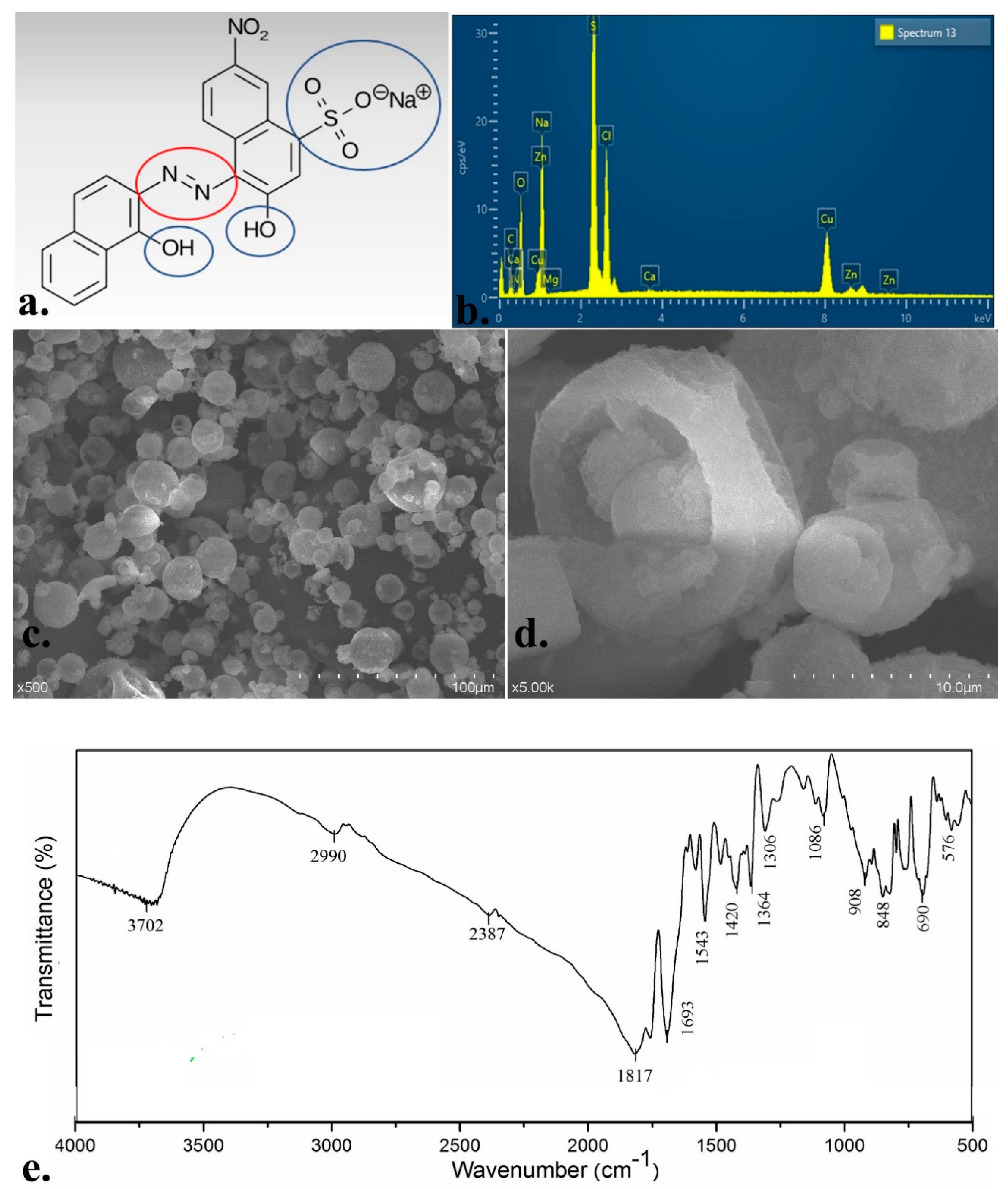
3.1. EBT Dye Solid-State Characteristics

EBT is not only used as a fabric or plastic paint but also for water hardness determination and rare earth metal detection, being a metallochromic indicator. As with most azo-dyes, EBT is hazardous. It may constitute a direct threat to health, causing blindness, skin diseases, kidney failure, and different kinds of cancers [29]. The dye has non-biodegradable components, but its intermediate degradation product, naphthoquinone, is even more dangerous. The removal of EBT from wastewater is a prodigious task aiming to prevent damage to aquatic and human life. With this knowledge in mind, we investigated the structure of the dye, its removal from the aqueous medium by the phytoremediation method, and the stress effects on the used E. crassipes and S. natans plants.
As it can be seen in Figure 1a, EBT is a mono-azo-dye. This means that it contains a −N=N− bond attached to two groups, both aromatic (usually, azo-dyes have at least one aromatic ring). According to the literature [30], this azo bond is in transform with an angle of 120° and both N2 atoms are sp2-hybridised. The two aromatic naphthalene rings point to a carbocyclic azodye. This chemical structure and arrangement results in a larger and much stronger, more stable conjugated π system [31]. Research demonstrates that complexation of the dye with calcium and magnesium affects colour fading and organic matter removal [31].
EBT has chromophores and auxochromes that provide its colour of blue and red respectively, in protonated form or after complex formation (Figure 1a). The chromophores of a dye are usually the functional groups of readily decomposed bonds (double or triple). It is not a proven fact, but they are usually situated at the centre of the dye structure. On the other hand, auxochromes are the saturated simple bonded functional groups, and they appear at the corners of the molecule. In our case, they are shown in Figure 1a [32]. Figure 1c contains the SEM images of the dye at 500 and 5 k magnifications. According to this figure, the dye has an irregular size, mostly rounded but cellular shaped form, with small (< 10 µm) particles on the surface.
The EDX spectra of the dye is presented in Figure 1b. The elemental composition results are the means and standard deviations of three parallel measurements. EBT mostly contains C (wt% = 61.1 ± 2.6), O (wt% = 37.8 ± 2.8), S (wt% = 0.15 ± 0.02), and other elements in trace amounts.
Fourier-transform infrared spectroscopy (FTIR) data were obtained using a JASCO 615 FTIR spectrophotometer (Cluj-Napoca, Romania) in the wavelength range of 500 to 4000 cm−1, in order to determine the functional groups of EBT [28,33]. After measurements, specific bands that represent the functional groups of the dye molecule appeared. Vibrations above 2990 cm−1 are characteristic peaks of strong or weak signs of phenolic O–H bonds, secondary amine N–H, and aromatic C–H. Prominent naphthalene peaks [34,35] appear at 1693 and 1364 cm−1, whereas specific azo bond vibration can be identified at 1543 cm−1, that can be N–O nitro compounds as well [36]. According to Veerakumar, peaks of EBT dye around 3432, 1401, 1332, 1215, and 1085 cm−1 correspond to −OH, −N=N−, −NO2, and −SO3 groups; also, 1100−700 cm−1 could be attributed to C−O and C−O−C groups [37].

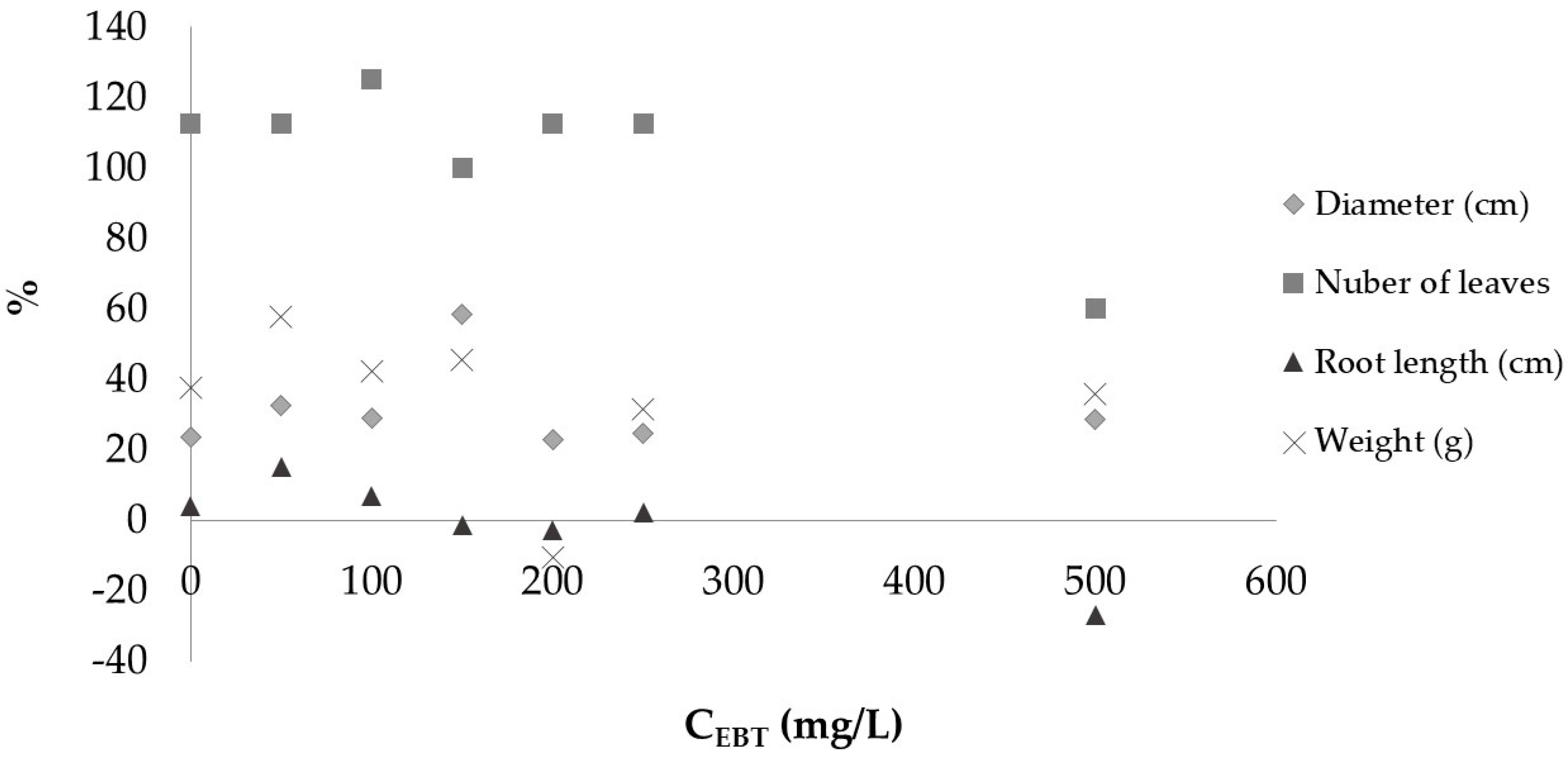
3.2. Seedling Growth Test

The lettuce root-growth inhibition test was carried out for EBT dye for six different concentration values (50–500 mg/L) that were previously tested for phytoremediation. The results listed in Table 2 show that, at a low concentration (50 mg/L), the dye stimulated the growth of the root. This can be explained by the presence of sulphur and nitrogen in the dye solution. As the concentration increased, the root length decreased after a 2-day experiment. At high dye concentrations, we reached 32% inhibition. Although the seeds were germinated in large quantities at the end of the experiment, they all became discoloured and browned. The colour of the dye faded in the seed inhibition zone, and the concentration of EBT in the solution was reduced.

3.3. Photocatalytic Decomposition and Photolysis

The photocatalytic decomposition was performed with 50 mg P25 catalyst, a commercial TiO2-based photocatalyst that is widely used because of its relatively high levels of activity in many photocatalytic systems. Having its “full name” as Degussa (Evonik) P25 or Aeroxide TiO2 P25, it is composed of anatase and rutile crystal phases, with a reported ratio usually being around 80:20 [38]. As described in Section 2.3., results of the decay curve showed that, during the first minutes (10 minutes, in case the irradiation was not started, showed as the time before zero in Figure 2), strong adsorption of the EBT could be observed on the P25, when ≈ 88% of the colouring agent bound to the surface of the catalyst. This finding is similar to the results presented by Behnajady et al., where nearly ≈ 70% of the azo-dye Acid Red 27 was adsorbed on the surface of the catalyst [39]. In the next part of the investigation, the EBT was removed with high efficiency from the water using UV irradiation, and it was decomposed with high efficiency (93.6% of the total amount of EBT). After 2 hours, the initially dark solution became translucent, like drinking water. Moreover, it was observed that the colour of the titania-based photocatalyst had not suffered a significant colour change.
Regarding the photolysis of the colourant, it can be affirmed that the concentration of the control sample did not change during the whole experiment, remaining constant (≈500 mg/L) in the limit of experimental errors.

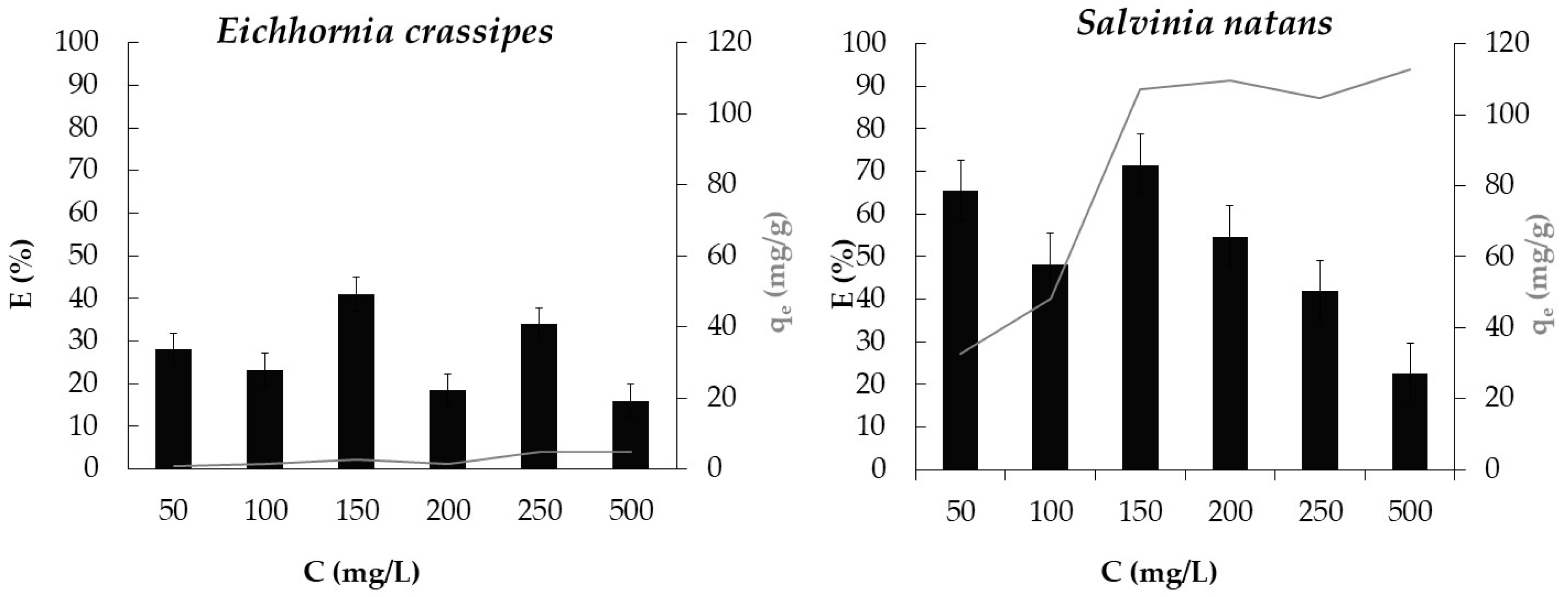
3.4. Effect of Dye Concentration and of Plant Species

As it was recently recognised by the scientific society, over the passage of time, aquatic and sessile plants are genetically adapting their defense mechanism to survive and grow in heavily polluted environments by metabolizing and detoxifying inorganic and organic pollutants. The types of pollutants determines the phytoremediation method a plant uses, organic materials (such as pesticides, drug residues, and dyes) are mostly reduced/detoxified/removed by phytotransformation, photodegradation, or phytovolatilisation. During phytotransformation, the uptake of the contaminant occurs through the plant roots or metabolism within the root zone, where the mechanism of the process is the absorption by the root system, where the contaminant undergoes a metabolic or enzymatic transformation, whereas, rhizodegradation means the degradation of pollutants in the rhizosphere simultaneously with microbial activity. All in all, the transformation occurs within the rhizosphere, the root zone, where the root exudates or enzymes are secreted around root zones; furthermore, xenobiotics are microbially degraded. Under the term of phytovolatilisation, we understand the transformation of water-soluble pollutants into volatile form, with the mechanism of pollutant modification during the vascular translocation of these organic materials from the root to the leaves [40].
Efficiency and capacity of phytoremediation of E. crassipes and S. natans were studied with varying initial concentrations (Figure 3).
In Figure 3, quantity in equilibrium (qe) and efficiency (E) were plotted separately for the two plants. It can be observed that for both species, the plants in the 150 mg/L solution were the most effective. The efficiency of S. natans (71.5%) is more than double that of E. crassipes (33%). The highest removal ratio was obtained for S. natans (Ci = 500 mg/L, qe = 112.9 mg/g). Results show that the active parts of the surface of the plants and the uptake are affected by the saturation of the plants, whereas the efficiency of the process decreases at high concentrations. It was found that there is a limit beyond which higher dye concentration does not imply greater binding capacity [23]. We can see from our results that azo-dye decolourisation is species-dependent.
Presumably, to the best of our knowledge, there are four possible bio-degradation mechanisms that occur during phytoremediation: (1) in the wastewater bodies, the growth of the microorganisms is increased by rhyzo-deposition by releasing 10% of photosynthetic carbon, (2) enzymes such as lignin, peroxidase, manganese-dependent peroxidase, and laccase (exuded through the root system) are reported to decolourise dyes, (3) in the rhizosphere, an anaerobic and aerobic microenvironment is formed, that helps in azo-dye degradation (this phenomenon is gained after the roots pump oxygen into this narrow zone of the rhizosphere, where other extracted chemicals can also exert their effects), and (4) through adsorption on the surface of roots, leaves, and in water shoots, where there will be greater exposure to water microorganisms [41]. In order to give a clear answer as to the mechanism, additional measurements are needed for both the aqueous solution and the plants. More detailed physiological, morphological, biochemical, and molecular changes should be investigated in the plants used during the research. Table 3 shows some phytoremediation efficiency results of different plants for different dyes.

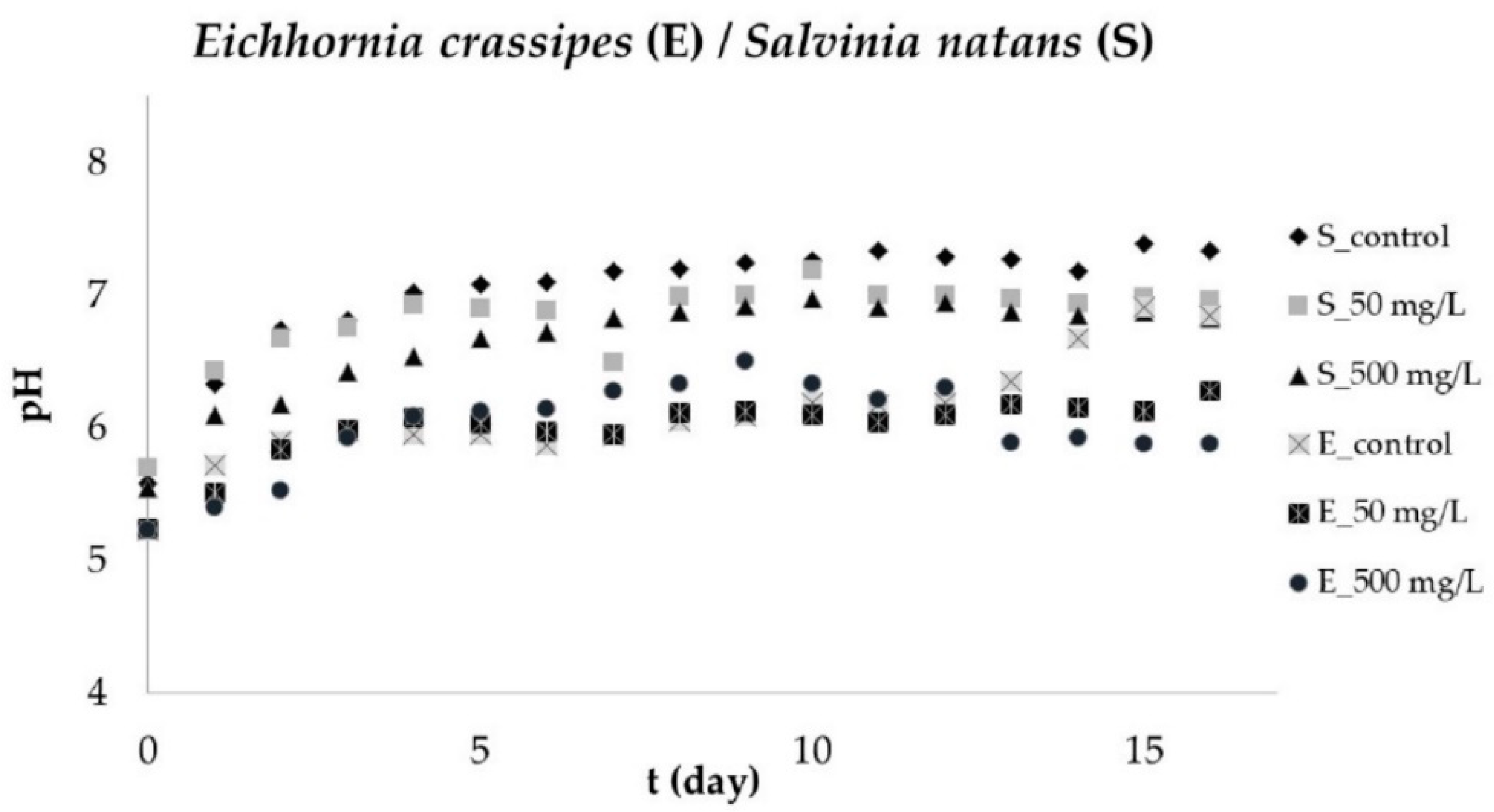
3.5. pH Changes of Aqueous Solution

External factors such as water pH, light intensity, temperature, and the quality and quantity of nutrients, influence plant metabolism. The used plants prefer a medium between pH 6 and 8. If the values are outside this interval, the plant can adjust the pH of the aqueous medium by its growth. pH between 3.2 and 4.2 can be toxic to the metabolism of the plant. pH in the range of 4.2–4.3 has an inhibitory effect, and the same is probably the case between 4.3 and 4.5 [24]. During the phytoremediation studies, the pH of the water solution was monitored every day. We observed that the plants set the initial near-acidic medium (pH = 5) at approximately neutral pH = 7 within 5 days (Figure 4).

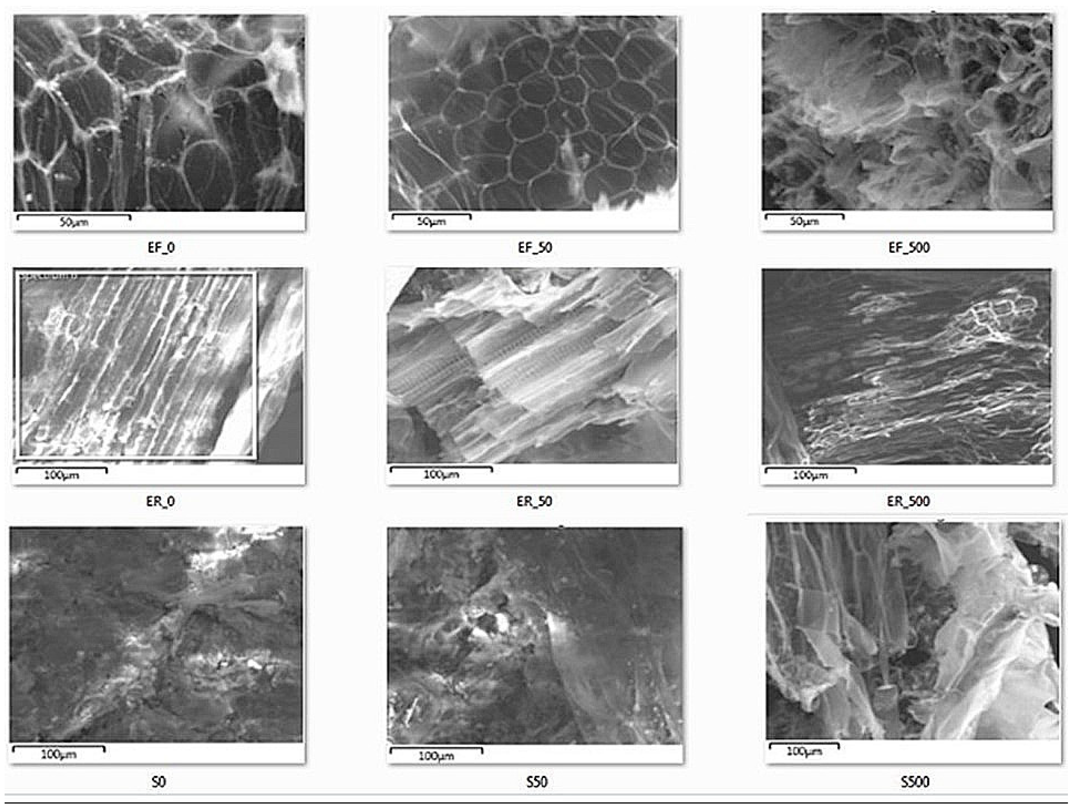
3.6. Morphology Studies

The presence of EBT as an abiotic stress factor, an extreme, harsh external environment condition, can lead to alterations in the growth and development patterns of plants. Therefore, on the first and last days of the experiment, morphological parameters were recorded. Only the initial and final weight was measured for S. natans, because of small leaves and roots. Plants gained weight in each dye solution from 8.5 ± 0.6 to 10.9 ± 0.8 g.
In contrast, in the case of E. crassipes, the diameter of the rosette of the plant, the length, the weight of the root, and the number of leaves and airbags were measured. Figure 5 shows the percentage of morphological changes of the E. crassipes as a function of the different EBT concentrations. According to the results, plants’ weight increased with one exception (plant in 200 mg/L solution), and the number of leaves increased in all cases. Except for the plant in the 500 mg/L solution, the initial number of leaves was doubled. Moreover, at high concentrations, the plant was exposed to higher stress. Therefore, the root length was reduced by 27% compared to the initial length. Plant rosettes also increased, but not significantly.
The relative growth rate (RGR) provides information regarding plant productivity, or, how much the plant’s dry matter increased over time [44]. E. crassipes grows at an extraordinary rate. Some calculations show that plant biomass increases by 400 to 700 tonnes/hectare/day. According to the results, the relative growth rate is 4–6% [45]. In our case, the RGR for most dye concentrations exceeded 2%, RGR500 mg/L EBT; E. crassipes = 1.9%. Owing to this rapid reproduction rate, it was included in the IUCN list (The International Union for Conservation of Nature’s Red List of Threatened Species) of the 100 most dangerous invasive species.
Previous investigations showed that under optimal conditions, S. natans grows 0.012 g/day [46]. In our experimental conditions, EBT helped the relative growth of S. natans, compared to research conducted by Netten et al. [46]. We received higher values (0.07–0.25 g/day) for each concentration (RGR500mg/L EBT; S. natans = 1.5%).
The morphological study indicates that the dye contamination had no adverse effects on the growth of the plant, as the yield (gain in mass) of the plant increased. It is important to note that the colour of the new leaf shoots was much lighter in green, white, or black, and many leaves and airbags died or showed brownish discolouration. In the case of S. natans, the leaves of the plants in the solutions with higher concentrations were almost whitened.
Figure 6 shows the micrographs of E. crassipes and S. natans before and after treatment. The explanation of the captions is as follows: E—E. crassipes, S—S. natans, F—leaf, R—root, 0—control plant, 50—plant in 50 mg/L, and 500—plant in 500 mg/L EBT solution. It can be observed from the recordings that there were no significant differences between control plants (S. natans and E. crassipes) and those used for remediation.

3.7. Elemental Analyses

Energy-dispersive X-ray spectroscopy (EDX) measurements were performed on the control leaves and on the ones in the lowest and highest concentration solutions. The calculated enrichment factor (E.F.) results obtained in the assay can be found in Table 4 for E. crassipes and S. natans.
Based on the calculated E.F., a large percentage of decrease was observed by dye concentration growth for vital elements such as P, Mg, Ca, S, and K. These elements are commonly known to play an important/critical role in the metabolism, growth, use of light energy of plants, and in stress condition (biotic, abiotic) help in survival [47,48,49,50].
EDX shows that the amount of phosphorus decreased under the optimal (0.3–0.5%) growth value [51]. Lack of phosphorus may lead to a reduction in the length of the root, and the leaves may become dark green or purple [52]. These effects can be seen in photos taken after the experiment (Figure 7).
Lack of potassium or its decrease (75% for E. crassipes, 66.5% for S. natans, in 500 mg/L EBT) prevents plant growth and development, inhibits pH stabilisation, and affects scorching old leaves (effects shown in Figure 7) and weakly developed roots [53]. Moreover, in the case of E. crassipes, root length was reduced by 27% (Figure 4).
The amount of sulphur decreased by 66% in the case of E. crassipes (CEBT = 500 mg/L) and above 60% in the case of S. natans, which could have a negative effect in chlorophyll production [54].
The absence of calcium may cause chlorosis, necrosis in young leaves (Figure 7), and may lead to biochemical and physiological disorders [55]. In our experimental conditions, the required minimum amount (5 mg/L) of Ca [56] was not reached (E. crassipes: wt50mg/L EBT% = 0.9; wt500mg/L EBT% = 0.3 and S. natans: wt50mg/L EBT% = 0.4; wt500mg/L EBT% = 0.1), and after phytoremediation, the percentage of Ca decreased by 68% in E. crassipes and 89% in S. natans.
Magnesium plays a vital role in photosynthesis, one of the basic elements of chlorophylls, and the lack of it or its decrease (E. crassipes: 81%, S. natans: 75%) can lead to the fading of leaves (Figure 7). The decrease of Mg and Ca could be explained by the fact that the EBT forms a complex with Ca and Mg. That explains why EBT is primarily used to determine water hardness as an indicator.

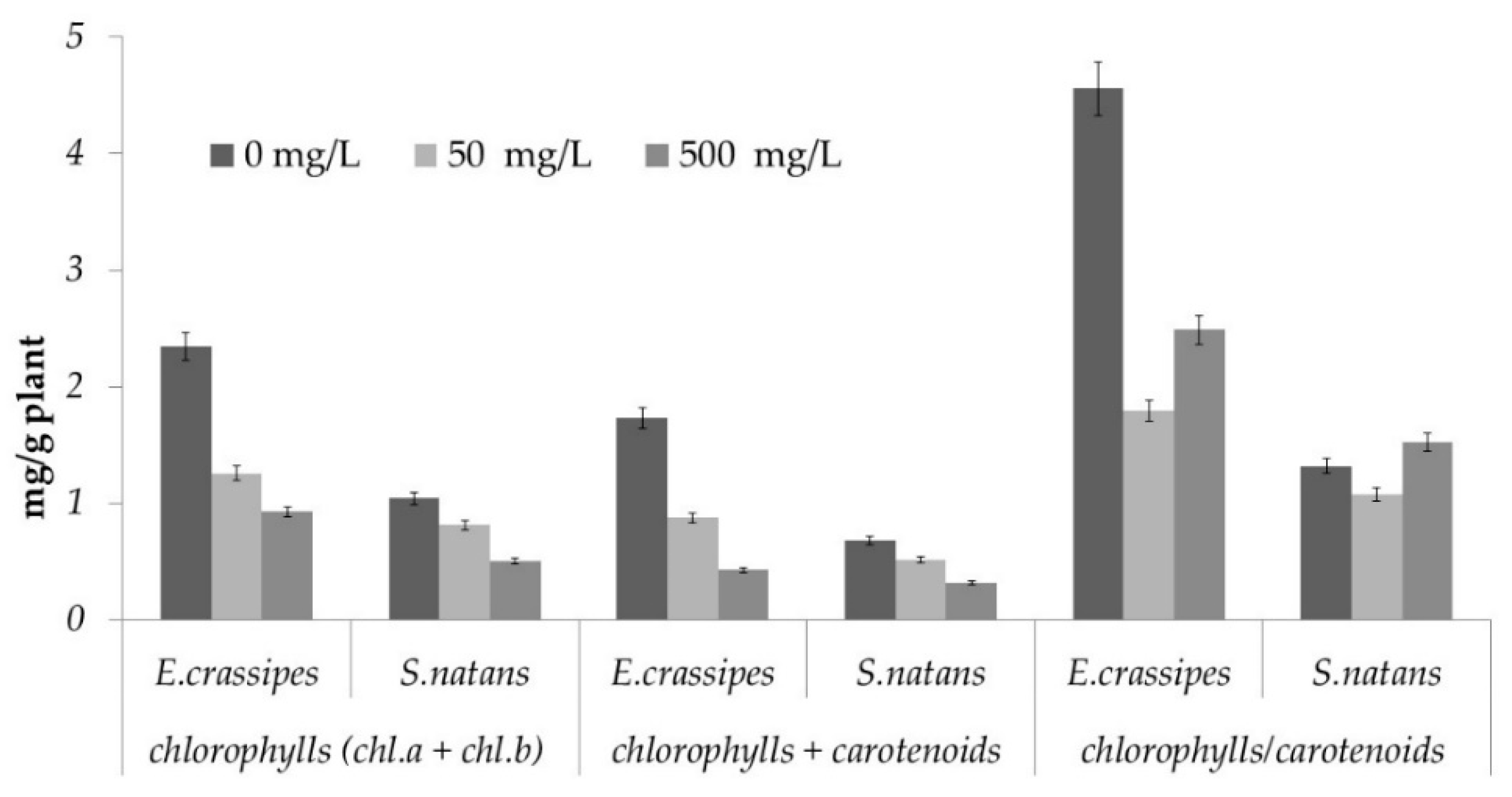
3.8. Photosynthetic Pigment Determination

The amount of photosynthetic pigments in the leaves of the plant was examined, as it was one of the most visible effects of stress condition, to clarify the effect of the dye on one of the plant’s life functions. Figure 8 is a graphical representation of the proportions of each pigment and the change in total pigments. The amount of chlorophylls in the plant decreases with the increase of the dye concentration (the decrease of the amount of Mg, which is the main element of chlorophylls, was evaluated by EDX measurements). The total amount of photosynthetic pigments shows a decreasing trend. Quantitative ratios indicate adaptation to individual light conditions and environmental changes. The ratio of chlorophylls to carotenoids decreased in response to the stress on the plant, having a protective effect based on the carotenoid function of the excess. Under strong light conditions, the amount of carotenoids increases; therefore, more ATP (adenosine triphosphate) is synthesised, which promotes the growth of plants.

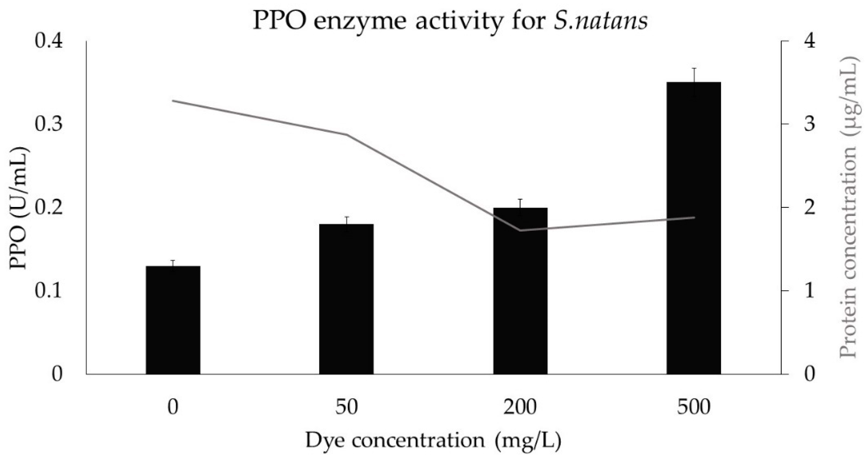
3.9. Stress Protein Assay

PPO (polyphenol oxidase) activity depends to a large extent on the pH, because it affects the binding of substances and the catalysis. The optimal pH of PPOs ranges between 4 and 8. In our experimental studies, the aqueous solution of S. natans was adjusted to neutral pH. Several studies report on the positive correlation between PPO expression and resistance/tolerance to stress. In the case of S. natans (Figure 9), the effects of abiotic stress can be observed, because the polyphenol oxidase enzyme activity increased with the increasing amount of dye.

4. Conclusions

As an outcome of this study, we have confirmed our hypothesis that EBT dye removal by phytoremediation with E. crassipes and S. natans is a promising method. S. natans has proven to be more effective (71.5%) than E. crassipes (33%). Moreover, the highest removal ratio obtained was 112.9 mg/g (S. natans; Ci = 500 mg/L); therefore, the method can be used with good results in binding and removing organic dyes.
The solid-state characteristics of EBD dye were analysed using SEM, EDX, and FTIR analytical methods, where the specific elemental composition functional groups of the dye were described. The mono-azo EBT dye has chromophores and auxochromes that provide its colour in the protonated form (blue) or after complex formation (red). With the help of SEM images at 500 and 5 k magnifications, we were able to see the irregularly sized, mostly rounded, but cellular shaped dyes. EDX measurements quantitatively determined the presence of O and C and qualitatively determined the S and trace elements that are also present in the molecular formula of the dye. This structure was further studied by FTIR analyses, where the detected peaks show the strong or weak bonds present in the dye molecule, for instance: phenolic O–H, secondary amine N–H, aromatic C–H, naphthalene, and azo bond vibration. When studying the toxic effect of EBT on seedling growth, test results showed that at a low concentration (50 mg/L), the dye stimulated the growth of the root, whereas, at high dye concentrations, we reached 32% inhibition. It was also shown that the photocatalytic degradation using TiO2 (P25) could be an ideal “assistant” for the phytoremediation to deal with organic dyes having higher concentration, as it showed an increased activity toward the model pollutant, removing more than 90% of it.
Despite this abiotic stress, both plants showed a rapid reproduction rate: 1.9% and 1.5% RGR was reached for E. crassipes and S. natans respectively, when they were put in 500 mg/L dye solution. However, a large decrease (more than 66% for both plants in 500 mg/L dye solution) of vital elements (P, Mg, Ca, S, K) was observed by EDX measurements. At the end of the experiment week, damaged roots, whose length was reduced even by 27%, purple or dark green old leaves, and chlorosis and necrosis in young leaves remained. According to our studies, the ratio of chlorophyll–carotenoids suggested that the change in photosynthetic pigments was triggered by the stress in the plant. Owing to a positive correlation between PPO emission and tolerance/resistance to abiotic stresses, we can emphasise that S. natans defends against stress because the amount of PPO increases with increasing concentration.
All in all, based on the experimental data obtained from the present study, the potentially used phytoremediation with E. crassipes and S. natans holds a great promise for application in the removal of EBT dye from wastewaters.

Author Contributions

E.R., A.C., K.P., and S.T. designed the work, carried out the experiments and analyses, interpreted all data received, and wrote the manuscript; B.É.V., G.M., and I.H. performed measurements for PPO analyses and helped in data interpretation; G.K. performed the photocatalytic analyses and results. All authors have read and agreed to the published version of the manuscript.

Funding

This research was funded by the Sapientia Hungarian University of Transylvania (Romania), Márton Áron Research Program of the Hungarian Ministry of Foreign Affairs and Trade and by Higher Education Institutional Excellence Program (NKFIH-1159-6/2019), awarded by the Ministry of Human Capacities within the framework of water-related researches of Szent István University (Hungary). G.K. acknowledges the financial support by National Research, Development, and Innovation, project number NKFI PD-125311.

Acknowledgments

Special thanks to Erzsébet Buta for providing the plants grown in the Agronomy Institute of the University of Babeș-Bolyai.

Conflicts of Interest

The authors declare no conflict of interest.

References

  1. Adki, V.S.; Jadhav, J.P.; Bapat, V.A. Exploring the Phytoremediation Potential of Cactus (nopalea Cochenillifera Salm. Dyck.) Cell Cultures for Textile Dye Degradation. Int. J. Phytoremediat. 2012, 14, 554–569. [Google Scholar] [CrossRef] [PubMed]
  2. Chang, J.-S.; Chou, C.; Lin, Y.-C.; Lin, P.-J.; Ho, J.-Y.; Lee Hu, T. Kinetic characteristics of bacterial azo-dye decolorization by Pseudomonas luteola. Water Res. 2001, 35, 2841–2850. [Google Scholar] [CrossRef]
  3. Saratale, R.G.; Saratale, G.D.; Chang, J.S.; Govindwar, S.P. Bacterial decolorization and degradation of azo dyes: A review. J. Taiwan Inst. Chem. Eng. 2011, 42, 138–157. [Google Scholar] [CrossRef]
  4. Dave, P.; Kaur, S.; Khosla, E. Removal of Eriochrome black-T by adsorption on to eucalyptus bark using green technology. Indian J. Chem. Technol. 2011, 18, 53–60. [Google Scholar]
  5. Moeinpour, F.; Alimoradi, A.; Kazemi, M. Efficient removal of Eriochrome black-T from aqueous solution using NiFe2O4 magnetic nanoparticles. J. Environ. Health Sci. Eng. 2014, 12, 112. [Google Scholar] [CrossRef] [PubMed] [Green Version]
  6. Khalid, A.; Zubair, M. A Comparative Study on the Adsorption of Eriochrome Black T Dye from Aqueous Solution on Graphene and Acid-Modified Graphene. Arab. J. Sci. Eng. 2018, 43, 2167–2179. [Google Scholar] [CrossRef]
  7. El-Dars, F.; Ibrahim, H.M.; Farag, H.A.; Zakaria Abdelwahhab, M.; Shalabi, M. Adsorption Kinetics of Bromophenol Blue and Eriochrome Black T using Bentonite Carbon Composite Material. Int. J. Sci. Eng. Res. 2015, 6, 679–688. [Google Scholar]
  8. Ghosh, A.; Dastidar, M.G.; Sreekrishnan, T.R. Response surface modeling of bioremediation of acid black 52 dye using Aspergillus flavus. Water Sci. Technol. 2017, 75, 2864–2874. [Google Scholar] [CrossRef]
  9. Barka, N.; Abdennouri, M.; Makhfouk, M.E. Removal of Methylene Blue and Eriochrome Black T from aqueous solutions by biosorption on Scolymus hispanicus L.: Kinetics, equilibrium and thermodynamics. J. Taiwan Inst. Chem. Eng. 2011, 42, 320–326. [Google Scholar] [CrossRef]
  10. Simon, L.; Vincze, G.; Varga, C.; Szabó, B.; Koncz, J. Passive phytoextraction of toxic elements from sewage sludge compost by Salix viminalis energy plants. Acta Phytopathol. Entomol. Hung. 2012, 47, 285–291. [Google Scholar] [CrossRef]
  11. Simon, L.; Széles, É.; Kovács, B.; Prokisch, J.; Győri, Z. Phytoextraction of selenium from contaminated soils with Indian mustard, fodder radish and alfalfa. Trace elements in the food chain. In Proceedings of the International Symposium on Trace Elements in the Food Chain, Budapest, Hungary, 25–27 May 2006; pp. 40–44. [Google Scholar]
  12. Kumari, M.; Tripathi, B.D. Effect of aeration and mixed culture of Eichhornia crassipes and Salvinia natans on removal of wastewater pollutants. Ecol. Eng. 2014, 62, 48–53. [Google Scholar] [CrossRef]
  13. Saha, P.; Mondal, A.; Sarkar, S. Phytoremediation of cyanide containing steel industrial wastewater by Eichhornia crassipes. Int. J. Phytoremediat. 2018, 20, 1205–1214. [Google Scholar] [CrossRef] [PubMed]
  14. Tabinda, A.B.; Arif, R.A.; Yasar, A.; Baqir, M.; Rasheed, R.; Mahmood, A.; Iqbal, A. Treatment of textile effluents with Pistia stratiotes, Eichhornia crassipes and Oedogonium sp. Int. J. Phytoremediat. 2019, 21, 939–943. [Google Scholar] [CrossRef] [PubMed]
  15. Dhir, B.; Sharmila, P.; Saradhi, P.P. Photosynthetic performance of Salvinia natans exposed to chromium and zinc rich wastewater. Braz. J. Plant Physiol. 2008, 20, 61–70. [Google Scholar] [CrossRef]
  16. Schneller, J.J. Salviniaceae. In Pteridophytes and Gymnosperms; Kramer, K.U., Green, P.S., Eds.; Springer: Berlin, Heidelberg, 1990; pp. 256–258. [Google Scholar]
  17. Sculthorpe, C.D. The Biology of Aquatic Vascular Plants; Edward Arnold: London, UK, 1967. [Google Scholar]
  18. Malik, A. Environmental challenge vis a vis opportunity: The case of water hyacinth. Environ. Int. 2007, 33, 122–138. [Google Scholar] [CrossRef]
  19. Tan, K.A.; Morad, N.; Ooi, J.Q. Phytoremediation of Methylene Blue and Methyl Orange Using Eichhornia crassipes. IJESD 2016, 7, 724–728. [Google Scholar] [CrossRef]
  20. Ng, Y.S.; Chan, D.J.C. Wastewater phytoremediation by Salvinia molesta. J. Water Process Eng. 2017, 15, 107–115. [Google Scholar] [CrossRef]
  21. Török, A.; Buta, E.; Indolean, C.; Tonk, S.; Silaghi-Dumitrescu, L.; Majdik, C. Biological removal of triphenylmethane dyes from aqueous solution by Lemna minor. Acta Chim. Slov. 2015, 62, 452–461. [Google Scholar] [CrossRef] [Green Version]
  22. Baia, L.; Orbán, E.; Fodor, S.; Hampel, B.; Kedves, E.Z.; Saszet, K.; Székely, I.; Karácsonyi, É.; Réti, B.; Berki, P.; et al. Preparation of TiO2/WO3 composite photocatalysts by the adjustment of the semiconductors’ surface charge. Mater. Sci. Semicond. Process. 2016, 42, 66–71. [Google Scholar] [CrossRef]
  23. Lichtenthaler, H.K. Chlorophylls and carotenoids: Pigments of photosynthetic biomembranes. In Methods in Enzymology; Plant Cell Membranes; Academic Press: Cambridge, MA, USA, 1987; Volume 148, pp. 350–382. [Google Scholar]
  24. Cheema, S.; Sommerhalter, M. Characterization of polyphenol oxidase activity in Ataulfo mango. Food Chem. 2015, 171, 382–387. [Google Scholar] [CrossRef]
  25. Vincze, E.B.; Salamon, R.V.; Kovacs, E.; Mara, G. Effect of metal tolerant plant growth promoting rhizobacteria on bean growth, cadmium and zinc uptake and stress responses. Environ. Eng. Manag. J. 2018, 17, 803–811. [Google Scholar]
  26. Veszélyes Hulladékok Vizsgálata | KÖRnyezetvédelmi INFOrmáció. Available online: https://enfo.hu/etanfolyam/5491 (accessed on 6 October 2019).
  27. Rápó, E.; Szép, R.; Keresztesi, Á.; Suciu, M.; Tonk, S. Adsorptive Removal of Cationic and Anionic Dyes from Aqueous Solutions by Using Eggshell Household Waste as Biosorbent. Acta Chim. Slov. 2018, 65, 709–717. [Google Scholar] [CrossRef]
  28. Rápó, E.; Aradi, L.E.; Szabó, Á.; Posta, K.; Robert, S.; Szende, T. Adsorption of Remazol Brilliant Violet-5R Textile Dye from Aqueous Solutions by Using Eggshell Waste Biosorbent. Sci. Rep. 2020, 10, 8385. [Google Scholar]
  29. Sahoo, J.K.; Konar, M.; Rath, J.; Kumar, D.; Sahoo, H. Magnetic hydroxyapatite nanocomposite: Impact on eriochrome black-T removal and antibacterial activity. J. Mol. Liq. 2019, 294, 111596. [Google Scholar] [CrossRef]
  30. Hunger, K.; Gregory, P.; Miederer, P.; Berneth, H.; Heid, C.; Mennicke, W. Important Chemical Chromophores of Dye Classes. In Industrial Dyes; John Wiley & Sons, Ltd.: Weinheim, Germany, 2004; pp. 13–112. ISBN 978-3-527-60201-8. [Google Scholar]
  31. da Silva, Á.R.L.; Jhones dos Santos, A.; dos Santos, E.V.; da Silva, D.R.; Martínez-Huitle, C.A. Theoretical and experimental study of the influence of cation–Eriochrome complexes on the BDD anodic oxidation of Eriochrome Black T solutions. Electrochem. Commun. 2020, 112, 106668. [Google Scholar] [CrossRef]
  32. Zollinger, H. Color Chemistry: Syntheses, Properties, and Applications of Organic Dyes and Pigments; John Wiley & Sons: Zürich, Switzerland, 2003; ISBN 978-3-906390-23-9. [Google Scholar]
  33. Rápó, E.; Posta, K.; Suciu, M.; Szép, R.; Tonk, S. Adsorptive Removal of Remazol Brilliant Violet-5R Dye from Aqueous Solutions using Calcined Eggshell as Biosorbent. Acta Chim. Slov. 2019, 66, 648–658. [Google Scholar] [CrossRef]
  34. Naphthalene. Available online: https://webbook.nist.gov/cgi/cbook.cgi?ID=C91203&Type=IR-SPEC&Index=1 (accessed on 5 May 2020).
  35. 1,5-Dihydroxy Naphthalene(83-56-7) IR2. Available online: https://www.chemicalbook.com/SpectrumEN_83-56-7_ir2.htm (accessed on 15 May 2020).
  36. Ahmed, F.; Dewani, R.; Pervez, M.K.; Mahboob, S.J.; Soomro, S.A. Non-destructive FT-IR analysis of mono azo dyes. Bulg. Chem. Commun. 2016, 48, 71–77. [Google Scholar]
  37. Veerakumar, P.; Jeyapragasam, T.; Surabhi; Salamalai, K.; Maiyalagan, T.; Lin, K.-C. Functionalized Mesoporous Carbon Nanostructures for Efficient Removal of Eriochrome Black-T from Aqueous Solution. J. Chem. Eng. Data 2019, 64, 1305–1321. [Google Scholar] [CrossRef]
  38. Ohtani, B.; Prieto-Mahaney, O.O.; Li, D.; Abe, R. What is Degussa (Evonik) P25? Crystalline composition analysis, reconstruction from isolated pure particles and photocatalytic activity test. J. Photochem. Photobiol. A Chem. 2010, 216, 179–182. [Google Scholar] [CrossRef] [Green Version]
  39. Behnajady, M.A.; Yavari, S.; Modirshahla, N. Investigation on adsorption capacity of TiO2-P25 nanoparticles in the removal of a mono-azo dye from aqueous solution: A comprehensive isotherm analysis. Chem. Ind. Chem. Eng. Q. 2014, 20, 97–107. [Google Scholar] [CrossRef] [Green Version]
  40. Tahir, U.; Yasmin, A.; Khan, U.H. Phytoremediation: Potential flora for synthetic dyestuff metabolism. J. King Saud Univ. Sci. 2016, 28, 119–130. [Google Scholar] [CrossRef] [Green Version]
  41. Zhou, X.; Xiang, X. Effect of different plants on azo-dye wastewater bio-decolorization. Procedia Environ. Sci. 2013, 18, 540–546. [Google Scholar] [CrossRef] [Green Version]
  42. Muthunarayanan, V.; Santhiya, M.; Swabna, V.; Geetha, A. Phytodegradation of textile dyes by Water Hyacinth (Eichhornia crassipes) from aqueous dye solutions. Int. J. Environ. Sci. 2011, 1, 1702–1717.41. [Google Scholar]
  43. Torbati, S.; Khataee, A.R.; Movafeghi, A. Application of watercress (Nasturtium officinale R. Br.) for biotreatment of a textile dye: Investigation of some physiological responses and effects of operational parameters. Chem. Eng. Res. Des. 2014, 92, 1934–1941. [Google Scholar] [CrossRef]
  44. Valipour, A.; Raman, V.K.; Ahn, Y.-H. Effectiveness of Domestic Wastewater Treatment Using a Bio-Hedge Water Hyacinth Wetland System. Water 2015, 7, 329–347. [Google Scholar] [CrossRef] [Green Version]
  45. Téllez, T.R.; López, E.; Granado, G.; Pérez, E.; López, R.; Guzmán, J. The Water Hyacinth, Eichhornia crassipes: An invasive plant in the Guadiana River Basin (Spain). Aquat. Invasions 2008, 3, 42–53. [Google Scholar] [CrossRef]
  46. Netten, J.J.C.; Arts, G.H.P.; Gylstra, R.; van Nes, E.H.; Scheffer, M.; Roijackers, R.M.M. Effect of temperature and nutrients on the competition between free-floating Salvinia natans and submerged Elodea nuttallii mesocosms. Fundam. App. Lim. 2010, 177, 125–132. [Google Scholar] [CrossRef]
  47. Wang, M.; Zheng, Q.; Shen, Q.; Guo, S. The critical role of potassium in plant stress response. Int. J. Mol. Sci. 2013, 14, 7370–7390. [Google Scholar] [CrossRef] [Green Version]
  48. Wang, M.; Shen, Q.; Xu, G.; Guo, S. New insight into the strategy for nitrogen metabolism in plant cells. Int. Rev. Cell Mol. Biol. 2014, 310, 1–37. [Google Scholar]
  49. Zhao, D.; Reddy, K.R.; Kakani, V.G.; Reddy, V.R. Nitrogen deficiency effects on plant growth, leaf photosynthesis, and hyperspectral reflectance properties of sorghum. Eur. J. Agron. 2005, 22, 391–403. [Google Scholar] [CrossRef]
  50. Amtmann, A.; Troufflard, S.; Armengaud, P. The effect of potassium nutrition on pest and disease resistance in plants. Physiol. Plant 2008, 133, 682–691. [Google Scholar] [CrossRef] [PubMed]
  51. Lambers, H.; Brundrett, M.C.; Raven, J.A.; Hopper, S.D. Plant mineral nutrition in ancient landscapes: High plant species diversity on infertile soils is linked to functional diversity for nutritional strategies. Plant Soil 2010, 348, 7. [Google Scholar] [CrossRef]
  52. Allen, B.; David, P. Handbook of Plant Nutrition; CRC Press Taylor & Francis Group: Boca Raton, FL, USA, 2015; ISBN 978-1-4398-8197-2. [Google Scholar]
  53. Fageria, N.K. Nutrient Uptake in Crop Plants; CRC Press: Boca Raton, FL, USA, 2019; ISBN 978-0-367-38688-7. [Google Scholar]
  54. Mills, H.A.; Jones, J.B. Plant Analysis Handbook II: A Practical Sampling, Preparation, Analysis, and Interpretation Guide; Micro-Macro Pub.: Athens, GA, USA, 1996; ISBN 978-1-878148-05-6. [Google Scholar]
  55. White, P.J.; Broadley, M.R. Calcium in plants. Ann. Bot. 2003, 92, 487–511. [Google Scholar] [CrossRef] [PubMed]
  56. Oki, Y.; Ito, M.; Ueki, K. Studies on the Growth and Reproduction of Water hyacinth, Eichhornia crassipes (Mart.) Solms. J. Weed Sci. Technol. 1978, 23, 120–125. [Google Scholar] [CrossRef]
Figure 1. Dye-related characteristics, (a) chemical structure, where the red circle is the chromophore group, and blue are the auxochromes, (b) EDX spectra of the dye, (c) scanning eclectron microscopy (SEM) images of EBT at 500 magnification, (d) SEM images of EBT at 5 k magnification, (e) Fourier-transform infrared spectroscopy (FTIR) spectra of EBT dye.
Crystals 10 00565 g001
Figure 2. Photocatalytic decomposition of EBT dye.
Crystals 10 00565 g002
Figure 3. This Influence of the initial EBT dye concentration on the phytoremediation capacity and removal efficiency for (a) E. crassipes and (b) S. natans plants; Ci = 50–500 mg/L, T = 20 ± 0.5 °C.
Crystals 10 00565 g003
Figure 4. Dye solution pH change.
Crystals 10 00565 g004
Figure 5. Percentage of morphological changes of E. crassipes.
Crystals 10 00565 g005
Figure 6. SEM images of E. crassipes and S. natans plants, where EF_0: E. crassipes leaf in control sample, EF_50: E. crassipes leaf in 50 mg/L EBT dye sample, EF_500: E. crassipes leaf in 500 mg/L EBT dye sample, ER_0: E. crassipes root in control sample, ER_50: E. crassipes root in 50 mg/L EBT dye sample, ER_500: E. crassipes root in 500 mg/L EBT dye sample, S_0: S. natans leaf in control sample, S_50: S. natans leaf in 50 mg/L EBT dye sample, S_500: S. natans leaf in 500 mg/L EBT dye sample.
Crystals 10 00565 g006
Figure 7. Photos of phytoremediation: (a,f) plants before remediation, (be) E. crassipes after adsorption, (g) control S. natans after remediation, (h) S. natans after remediation in 500 mg/L dye solution, (i) leaves of S. natans after remediation.
Crystals 10 00565 g007
Figure 8. Photosynthetic pigment determination for plants in 0, 50, and 500 mg/L dye solution.
Crystals 10 00565 g008
Figure 9. PPO enzyme activity and concentration change of protein for S. natans at different initial dye concentrations after phytoremediation.
Crystals 10 00565 g009
Table 1. Characteristics of Eriochrome Black T (EBT) dye.
Properties of the Dye
Dye namesEriochrome Black T, Mordant Black 11
Molecular formulaC20H12N3O7SNa
M. weight461.381 g/mol
Max wavelength530 nm
Colour index14645
ColourPowder: black, deep purple
Aqueous solution after protonated form: blue
Aqueous solution after making complex with C2+ and Mg 2+: red
CharacteristicsMono-azo
Table 2. Seedling growth inhibition test.
Sample
(mg/L EBT)
Lettuce Seed
Root Length (cm)Number of Germinated SeedsRoot Growth Inhibition (%)
01.219.5
501.519.5−23.7
1001.215.54.4
1501.017.021.7
2000.916.030.2
2500.918.030.4
5000.818.032.2
Table 3. Comparison of phytoremediation efficiency.
PlantDye Name (Colour Index Number)Initial Concentration (mg/L)Efficiency (%)Reference
Lemna minorMalachite Green (C.I. 11294)4098.0[23]
Lemna minorCristal Violet (C.I. 42555)4096.0[23]
Sesbania cannabinaAcid red B (C.I. 14680)10044.23[41]
Sesbania cannabinaAcid red B (C.I. 14680)50022.58[41]
Sesbania cannabinaAcid red B (C.I. 14680)100012.23[41]
Sesbania cannabinaAcid scarlet GR (C.I. 27290)10008.66[41]
Eichhornia crassipesReactive Black 5 (C.I. 20505)10 99.5[42]
Eichhornia crassipesReactive Red 198 (C.I. 18221)1095.0[42]
Nasturtium officinaleAcid Blue 92 (C.I. 13390)579.1[43]
Eichhornia crassipesEriochrome Black T (C.I. 14645)15033.0Present study
Salvinia natansEriochrome Black T (C.I. 14645)15077.5Present study
Table 4. Bioconcentration factors calculated from 10 replicate EDX measurements for E. crassipes and S. natans.
Bioconcentration Factors (%)
Eichhornia crassipesSalvinia natans
Plant in
CEBT = 50 mg/L
Plant in
CEBT = 500 mg/L
Plant in
CEBT = 50 mg/L
Plant in
CEBT = 500 mg/L
Mg−40.5−81.2−55.6−75.2
P−65−78.6−39.5−69
S−38.1−66.3−63−68.9
K7−75−48.4−66.5
Ca17.5−68.4−64.2−88.8

