1. Introduction
The advancement of composite scintillators (CSs) and thermoluminescent (TL) detectors for the registration of the components of mixed ionization fluxes is now an actual subject of luminescent materials engineering. The basis for such novel engineering are the latest decisions in creating bulk single crystal (SC) and single crystalline film (SCF) scintillators [
1,
2] as well as the technologies of their production using the Czochralski method [
3,
4] and the liquid phase epitaxy (LPE) growth technique, respectively [
5,
6,
7,
8,
9,
10,
11]. Namely, in our previous work, we have shown the possibility of simultaneous registration of α-particles and γ-quanta using the separation of the scintillation decay kinetics of the film (SCF) and crystal (SC) parts of composite scintillators, based on the epitaxial structures of different garnet compounds [
12,
13,
14,
15,
16].
The application of composite scintillators presupposes the active in situ mode of the registration of incoming ionization fluxes. Meanwhile, such a mode of registration is not always possible to use in the case of low doses of radiation and a long-time exposition of radiation. Namely, particles and quanta with different energies cannot be registered using the separation between scintillation decay kinetics curves. There are also restrictions in application of the above-mentioned materials regarding the analysis of liquid and gaseous radioactive materials or the registration of high-dose sources.
The problems described above stimulated us to developing new approaches in the production of composite ionizing radiation detectors. One solution is to develop TL detectors for the simultaneous registration of different components of mixed ionization fluxes using the differences in the thermoluminescence (TL) glow curves. Particularly, such differences can be described by ΔT = T
F − T
S parameter, e.g., the difference between the position of the main TL peaks, recording from the film (T
F) and substrate (T
S) parts of the composite detector (
Figure 1a).
The possibility of the creation of such types of composite TL detectors for the simultaneous registration of α- and β-particle excitation was shown firstly by us in [
17]. This work is a continuation of the research in this direction and dedicated to the development of new types of composite TL detectors for the simultaneous registration of the different components of mixed ionization fluxes based on the SCFs of Ce
3+-doped Lu
3−xGd
xAl
5O
12:Ce (x = 0–1.5) garnet and Y
3Al
5O
12:Ce (YAG:Ce) SC substrates using the LPE growth method.
Recently, the SCFs of Lu
3−xGd
xAl
5O
12:Ce garnet with the Gd content, x, from 0 up to 2.5 has been successfully crystallized by the LPE method onto undoped YAG substrates from the melt-solutions based on PbO-B
2O
3 flux, and their optical properties have been investigated as well [
12]. In the present work, we used the LPE-grown epitaxial structures, containing Lu
3−xGd
xAl
5O
12:Ce SCFs with different Gd concentrations in the x = 0–1.5 range, and Ce
3+-doped YAG:Ce substrates for the simultaneous registration of α- and β-particles in the mixed ionization fluxes. For this purpose, the TL properties of the epitaxial structures were examined under excitation by α- and β-particles from
242Am and
90Sr-
90Y sources, respectively. We expected that the cation engineering of SCF content would result in more significant separation of the TL glow curves of SCFs and substrates in comparison with the prototype of such composite detectors based on the LuAG:Ce SCF/YAG:Ce SC epitaxial structure [
17].
2. Growth of Composite Detectors
Four sets of composite detectors, based on the epitaxial structures containing Lu
3−xGd
xAl
5O
12:Ce SCFs with Gd content x = 0, 0.5, 1, and 1.5 and YAG:Ce substrates with the (110) orientation and a thickness of 0.5 mm, were crystallized using the LPE method from the melt-solution based on PbO-B
2O
3 flux (
Figure 1). The conditions of the growth of these structures are presented in
Table 1.
Due to the fact that the segregation coefficient of Gd
3+ ions in the LPE growth of LuAG-based SCF onto YAG substrates is equal to 0.95–1.05 [
18], the content of SCFs was close to a nominal content of garnets in the melt-solution. The concentration of Ce
3+ ions in SCFs and YAG:Ce substrates was in the 0.25–0.5 at % range.
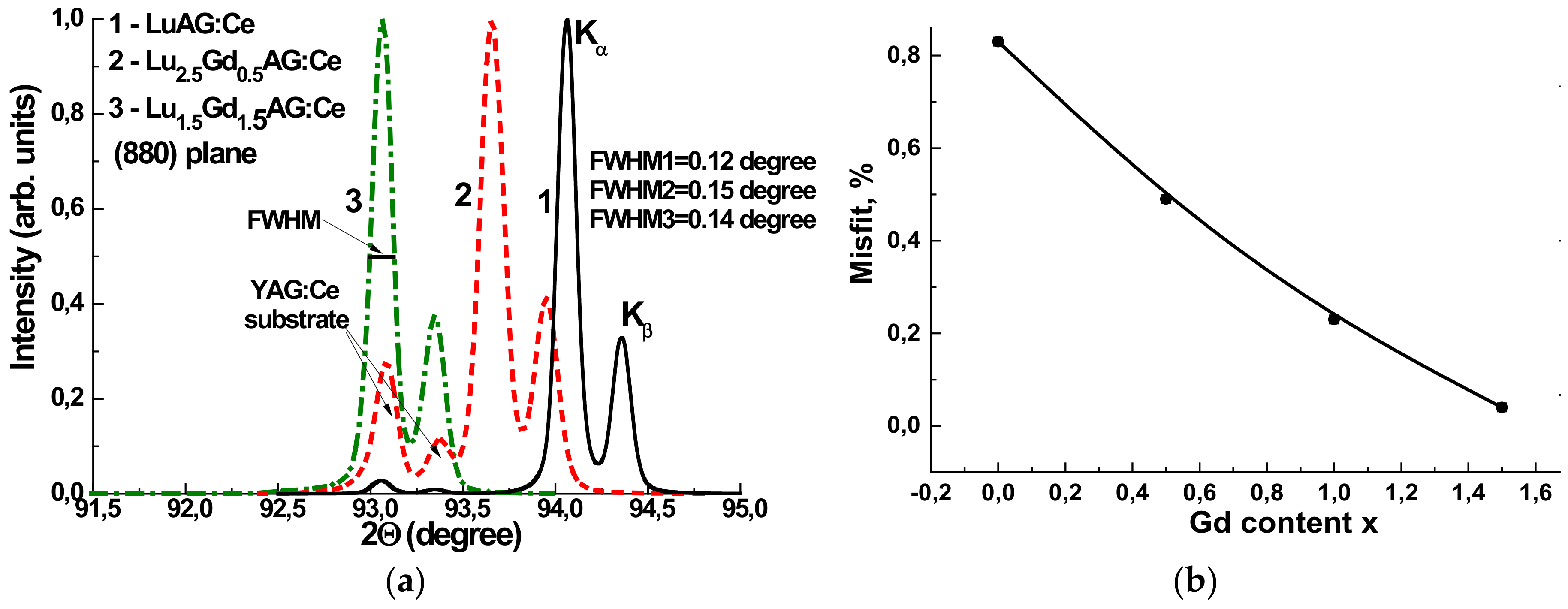
XRD measurements were used for the characterization of the structural quality of Lu
3−xGd
x Al
5O
12:Ce SCF samples with different Gd content and determination of the SCF lattice constants and SCF/substrate misfit m (
Figure 2).
From the respective XRD patterns of the Lu3−xGdxAl5O12:Ce SCFs at x value ranging from 0 to 1.5 we also calculated the lattice constant of the different garnet compositions and estimated the misfit between the lattice constants of SCF and YAG substrate
Δa = (aSCF − asub/asub) × 100% (
Table 1). We found that the lattice constant of Lu3−xGdxAl5O12:Ce SCF and the misfit value
m steeply depends on the Gd content in accordance with Vegard’s law. Namely, the lattice constant changed from 11.902 Ȧ for LuAG SCFs to 11.998 Ȧ for Lu
1.5Gd
1.5Al
5O
12:Ce SCFs. The value of misfit
m varies from −0.83% for LuAG SCF to 0.04% for Lu
1.5Gd
1.5Al
5O
12:Ce SCF (
Figure 1b), i.e., the last sample was grown practically without SCF/substrate misfit.
The full width at half maximum (FWHM) of the XRD peaks of the garnet samples under study was also determined from the respective patterns. As can be seen from this figure, the structural quality of Lu2.5Gd0.5AG:Ce and Lu1.5Gd1.5AG:Ce SCF samples, proportional to the FHWM of the respective XRD peaks (0.15 and 0.14 degree, respectively), is very close to that of LuAG:Ce SC (0.12 degree).
3. Experimental Technique
For the characterization of the luminescent properties of the Lu3−xGdxAl5O12:Ce SCF/YAG:Ce SC structures, we used absorption spectra, cathodoluminescence (CL), and thermoluminescent (TL) spectra. The absorption spectra were measured using a Jasco 760 UV-Vis spectrometer (Jasco Int. Co. Ltd., Tokyo, Japan) in the 200–1100 nm range. The CL spectra were measured at room temperature (RT) using a SEM JEOL JSM-820 electron microscope (JEOL, Tokyo, Japan) additionally equipped with a spectrometer Stellar Net with a TE-cooled Charge Coupled Device (CCD) detector working in the 200–925 nm range (StellarNet Inc, Tampa, FL, USA).
The TL glow curves were measured under the excitation by α- and β-particles from 241Am and 90Sr + 90Y sources. For measuring the TL in a Risø TL/OSL-DA-20 reader (Risø DTU, Roskilde, Denmark) we used the triangular fragment of samples with 3.75 mm × 3.75 mm × 5.25mm dimensions.
It is worth to note here that the mechanism of TL in the SCF samples under study was connected with the electron liberation from deeper electron traps and their subsequent recombination with the holes localized around Ce
3+ ions [
17,
18]. Therefore, for the registration of the TL glow curves, a “green” Schott BG 39 filter was used. The transmittance range of this filter, extending from 350 to 700 nm, was well matched with the emission range of the Ce
3+ luminescence in the SCF samples.
4. Optical Properties of Lu3−xGdxAl5O12:Ce SCFs/YAG:Ce Epitaxial Structures
4.1. Absorption Spectra
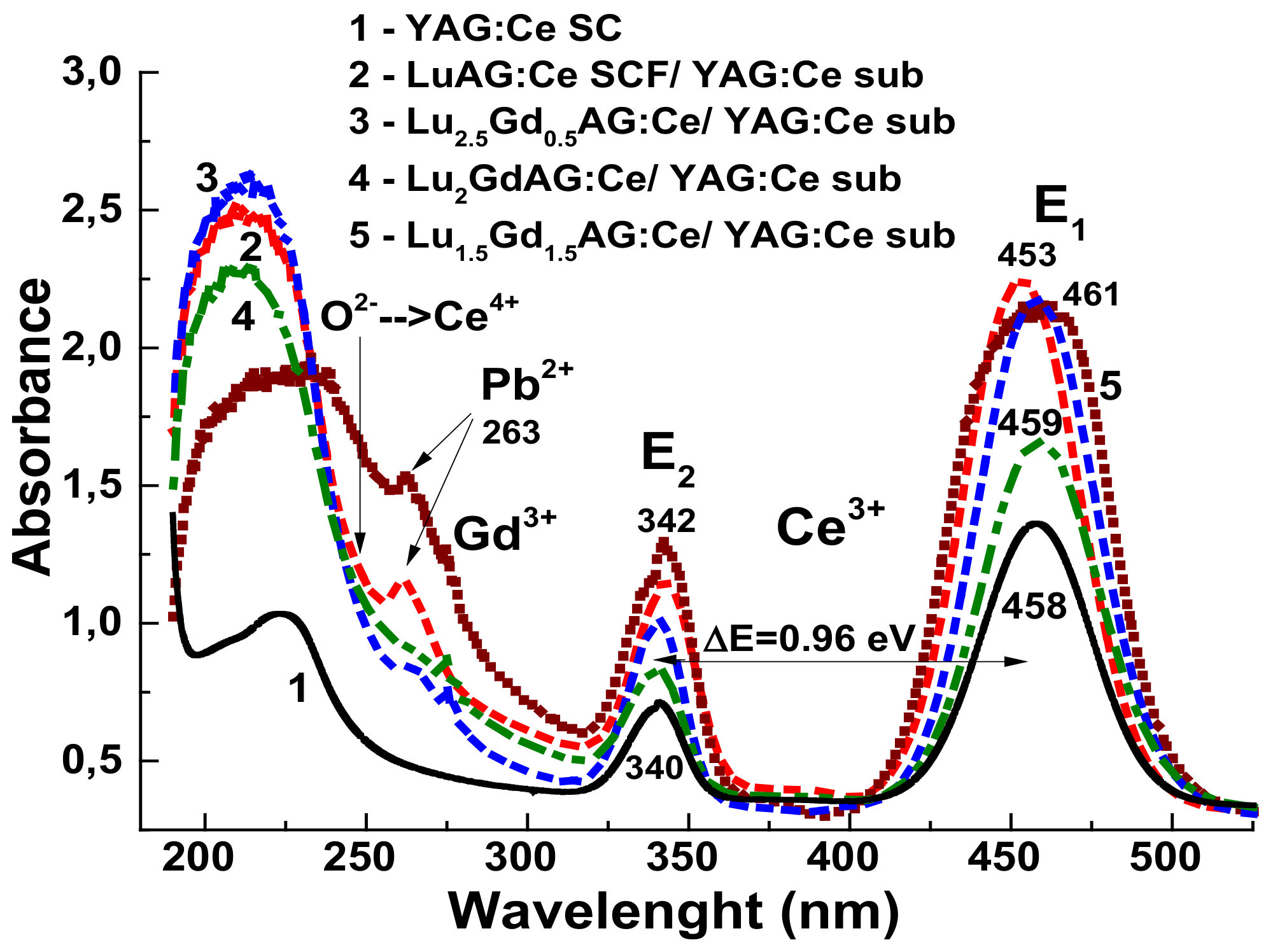
The absorption spectra of Lu
3−xGd
xAl
5O
12:Ce SCF/YAG:Ce SC epitaxial structures in comparison with the absorption spectrum of the YAG:Ce substrate are shown in
Figure 3. It is worth noting that the absorption spectra of the epitaxial structures represent the mixes of the spectra of the YAG:Ce substrate and the respective SCF sample with a total thickness in the 32–100 μm range (see
Table 1).
The absorption bands peaked within 453–461 nm and at the 340–342 nm range in the spectra of all the composite detector samples and YAG:Ce substrates were related to the 4f(
2F
5/2)→5d
1,2(
2E) transitions of Ce
3+ ions E
1 and E
2 bands, respectively. The ΔE values, proportional to the crystal field strength on the dodecahedral position of the garnet lattice [
18,
19], were equal approximately to 1 eV for these samples.
The bands that peaked around 263 nm correspond to the absorption of Pb
2+ flux-related impurity in Lu
3−xGd
xAl
5O
12:Ce SCFs and caused by the
1S
0→
3P
1 transitions of these ions [
18]. The intensity of this band increased with raising the Gd content in SCFs. Such behavior was connected with increasing the lattice constant of the garnet and respective dimension of the dodecahedral sites for the localization of the Pb
2+ ions with large ionic radii (1.29 Å) in comparison with Lu
3+ (0.997 Å) and Gd
3+ (1.078 Å) cations. Meanwhile, the concentration of this impurity even in the highly Gd-doped SCFs was still below the detection limit (100 ppm) of a SEM JEOL JSM-820 electron microscope used for the microanalyses of the samples’ content and only the intensity of Pb
2+-related absorption bands (
Figure 3) could be used for the estimation of the concentration of these ions.
The large Pb
2+ ion content in Gd-doped samples could also be contributed to the decrease of the scintillation LY of these SCFs (see
Table 1). A negative influence of Pb
2+ on the LY of SCF scintillators was recently observed for many types of SCF scintillators [
6,
9,
18]. Such influence can also be connected with the possible creation of Pb
2+-Ce
4+ pairs with local charge and local compensation at relatively large concentration of lead ions in SCF samples. Indeed, the absorption spectrum of Lu
1.5Gd
1.5Al
5O
12:Ce SCF (
Figure 2, curve 5) shows the presence of an additional broad band peak at approximately 255 nm. Most probably, this band was related to the O
2+→Ce
4+ charge transfer transitions (CTT). The confirmation of this conclusion was a close position of these bands in the mentioned SCF sample to the similar O
2+→Ce
4+ CTT band in Ca
2+- and Mg
2+-doped LuAG:Ce SCs [
20,
21].
4.2. Cathodoluminescence Spectra
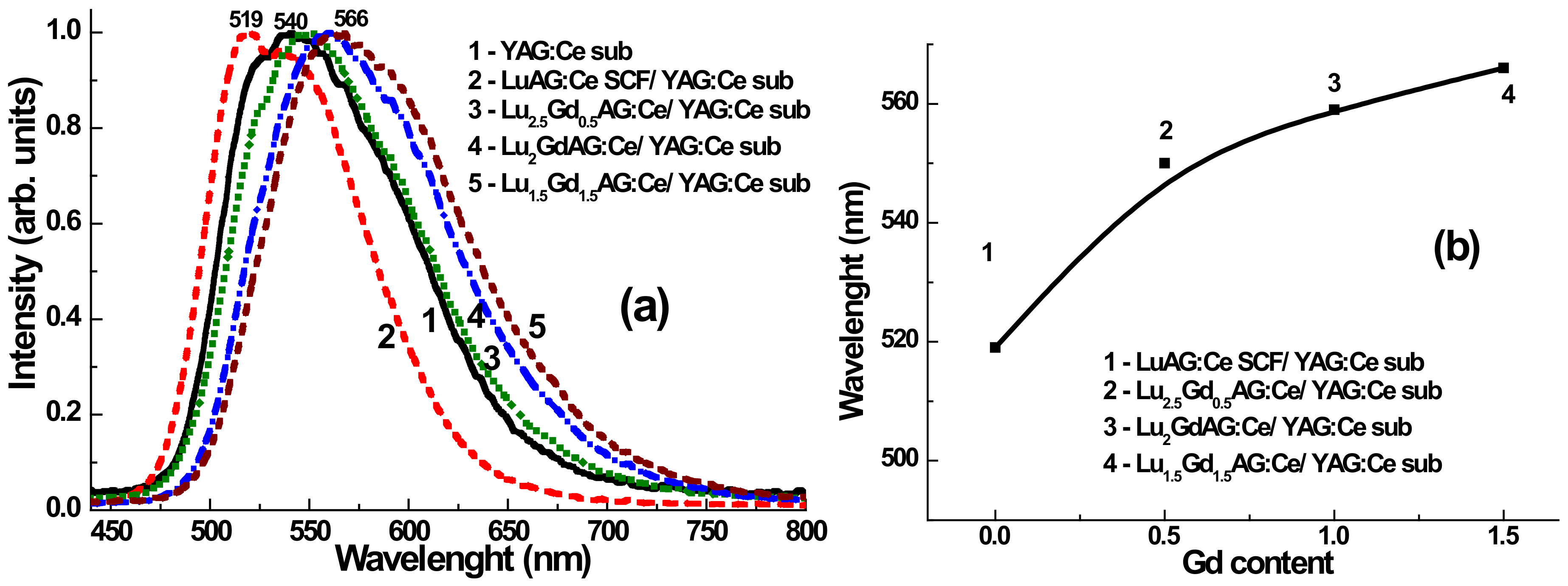
The normalized CL spectra of Lu
3−xGd
xAl
5O
12:Ce SCFs at x = 0, 0.5, and 1.5 in comparison with the CL spectrum of YAG:Ce substrate are shown in
Figure 4. The maximum of Ce
3+ emission bands was shifted (see
Figure 4b) to the red range with increasing Gd content in the SCF samples due to increasing crystal field strength in the dodecahedral position of the garnet lattice, see also [
18,
19].
4.3. Thermoluminescence Spectra
The first attempt to create a composite TL detector based on the LuAG:Ce SCF/YAG:Ce epitaxial structures was described in our previous work [
17]. The observed difference (about 80 degrees) between the position of the main TSL peaks of the SC and SCF parts of the composite detector under α- and β-particle excitation enabled the simultaneous registration of these particles in the mixed ionization fluxes. Meanwhile, the optimization of the TSL properties of such a type of composite detector is also possible and was considered in this work.
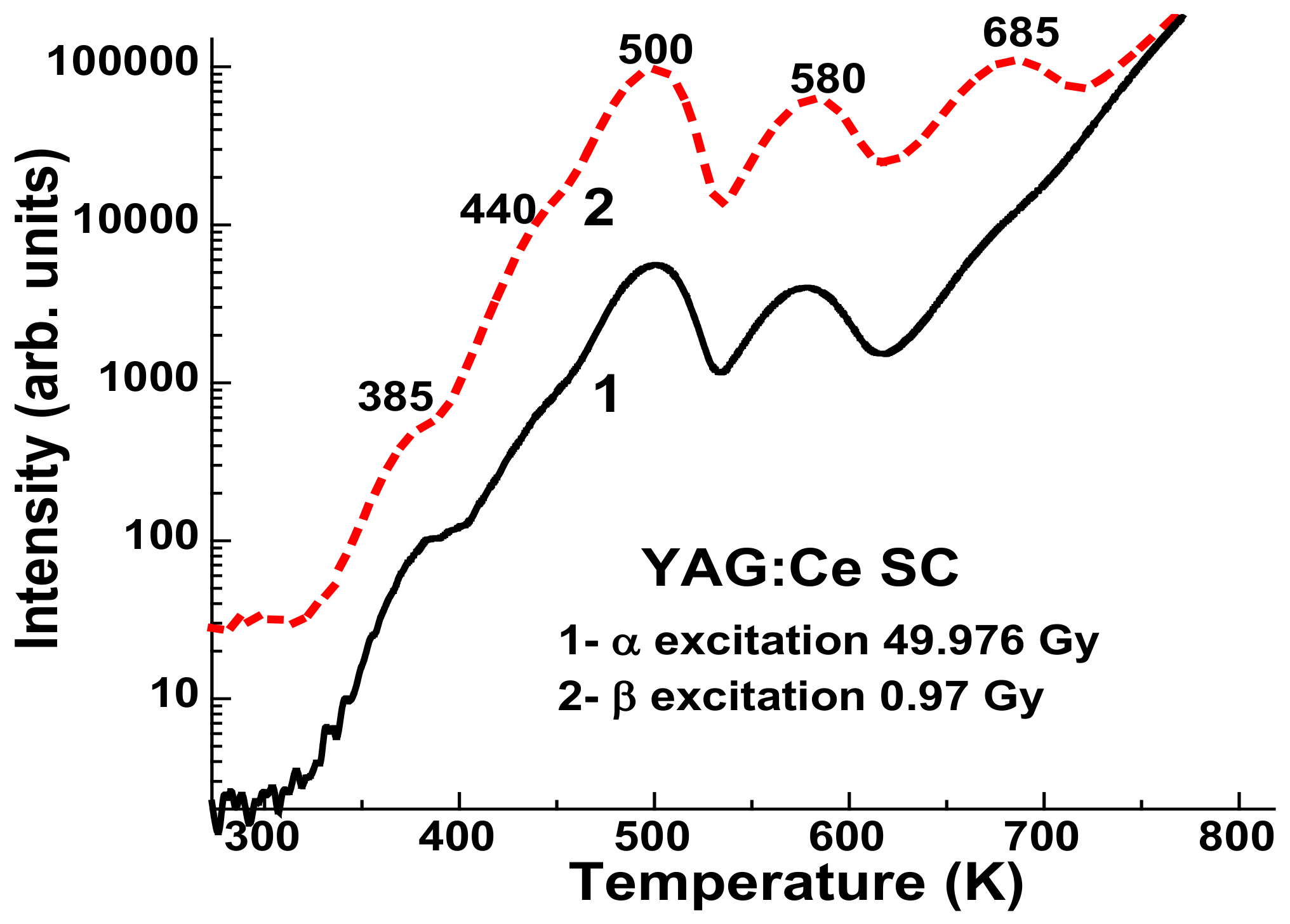
In the first stage, we analyzed the thermoluminescent curves for substrate.
Figure 5 shows the similar TSL properties under excitation by α- and β-particles, but the integral intensity of the TSL peaks is strongly determined by the absorbed dose of radiation (curves 1 and 2, respectively). The TSL glow curves of Lu
3−xGd
xAl
5O
12:Ce SCFs/YAG:Ce SC epitaxial structures with different Gd content x under excitation by α- and β-particles from
241Am and
90Sr +
90Y sources, respectively, are shown in
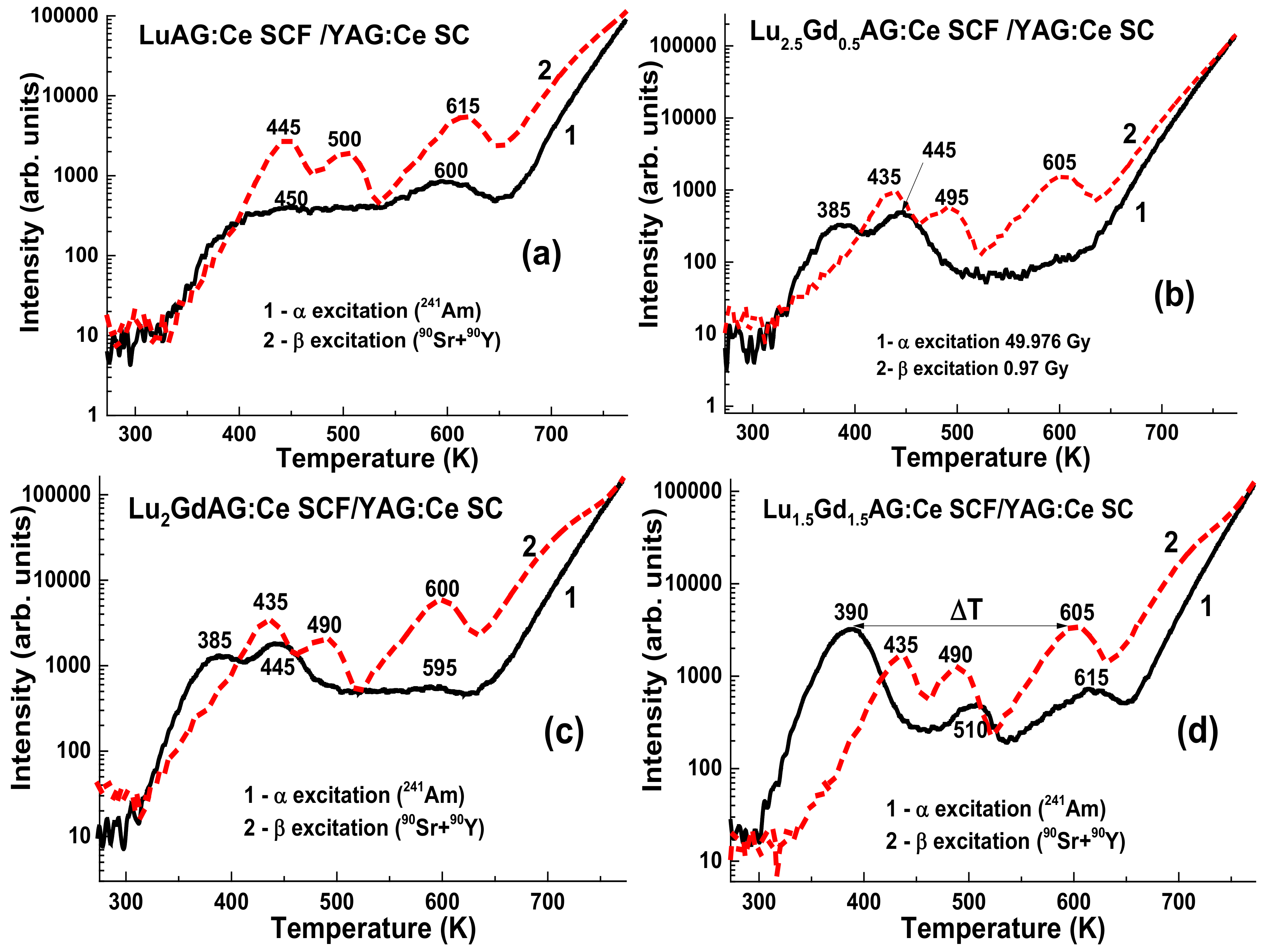
Figure 6.
As mentioned above, the α-particles from an
241Am (5.5 MeV) source with a typical passway in the LuAG of 12–15 μm were fully stopped in the SCF parts of a composite detector with a thickness of several tens of microns, when β-particles from a
90Sr +
90Y source with an average energy of 1.1 MeV could penetrate in the whole substrates. Therefore, due to the high thickness of the SCF samples in the 16–51 μm range (
Table 1), the TSL glow curves under α-particle excitation correspond exclusively to SCFs.
As can be seen from
Figure 6a, after α-particle irradiation, the main peak of the LuAG:Ce SCF/YAG:Ce SC structure was observed at 600 K. This result is consistent with the results of our previous work [
17]. In accordance with the results of the work in [
18], increasing the Gd content in the Lu
3−xGd
x Al
5O
12:Ce SCFs led to the shift of position of the main TSL peaks to the low temperature range, to 390 K in the Lu
1.5Gd
1.5Al
5O
12:Ce SCF sample (
Table 2 and
Figure 6d). Meanwhile, the position of the main TL peaks in the Lu
3−xGd
xAl
5O
12:Ce SCF/YAG:Ce SC structure after β-particle irradiation did not change notably (
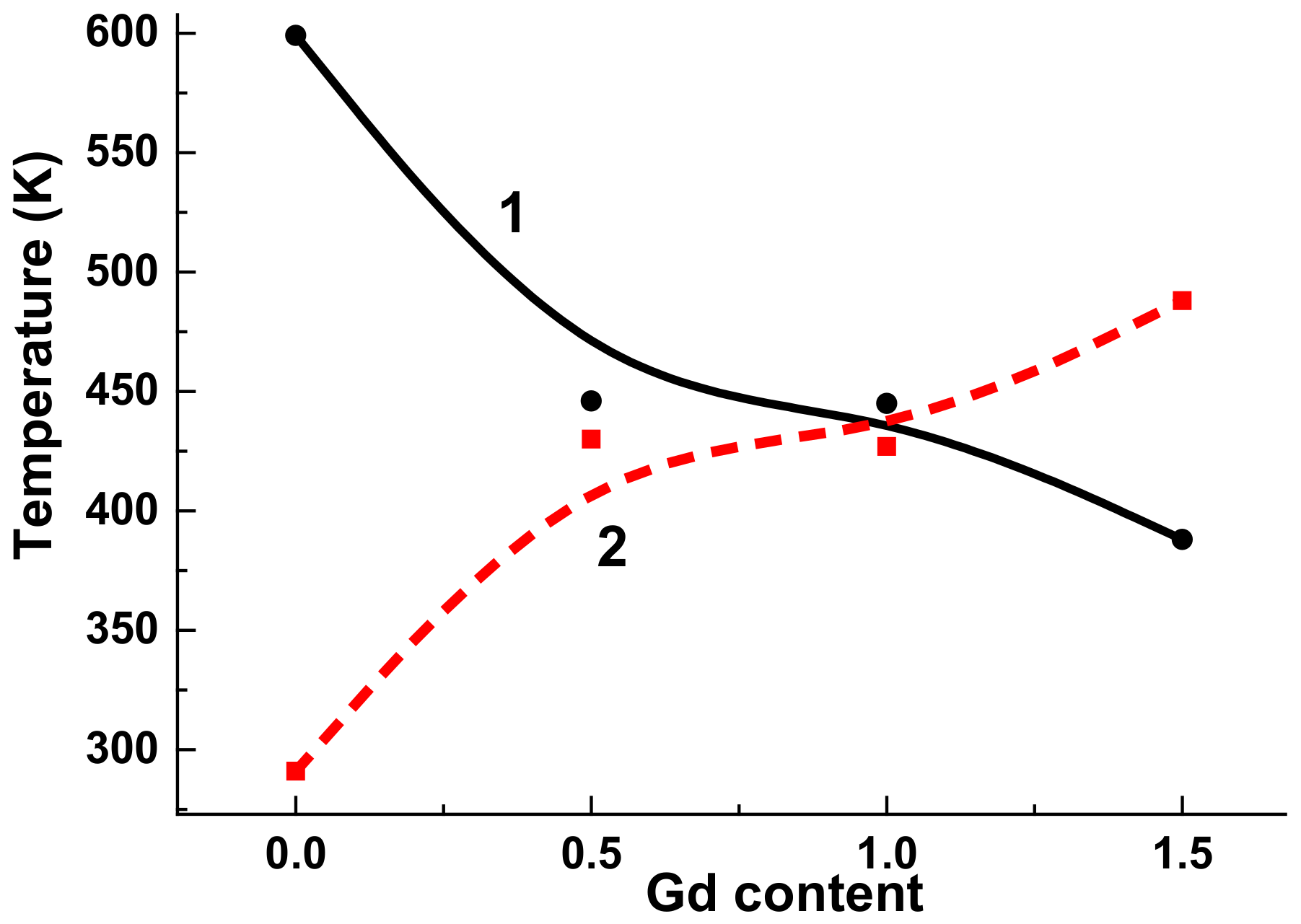
Table 1) due to the fact that the β-particles a were mainly absorbed by the YAG:Ce substrate. Therefore, the difference ΔT between the main TL peaks of SCF and substrate parts of a composite detector, corresponding to the registration of α- and β-particles, increased steeply with an increasing Gd content x in the Lu
3−xGd
xAl
5O
12:Ce SCFs. Namely, ΔT value is equal of 215 degrees for Lu
1.5Gd
1.5Al
5O
12:Ce SCFs (
Figure 6d and
Figure 7, curve 2).
It is important to note that due to the large feeding in the case of the TL peak positions in the low temperature range, of the most optimal was the location of the TL peaks of Lu3−xGdxAl5O12:Ce SCFs in the range above 150 K. For this reason, the creation of a composite detector based on the Lu2GdAl5O12:Ce SCF/YAG:Ce SC epitaxial structure was the optimal option. For such a type of composite detector the ΔT value is equal to 155 degrees, which is completely enough for the simultaneous registration of TL signals coming from the SCF and SC parts of a composite detector in the case of the registration of α- and β-particles, respectively.
5. Conclusions
The creation of novel types of composite TL detectors based on the Lu3−xGdxAl5O12:Ce SCF/YAG:Ce SC epitaxial structures for simultaneous registration of the mixed ionization fluxes, containing α- and β-particles, is reported in this work. The detectors, consisting of Lu3−xGdxAl5O12:Ce SCFs with Gd content in the x = 0–1.5 range, were fabricated using the liquid phase epitaxy (LPE) growth method onto YAG:Ce substrates. The registration of α- and β-particles by the SCF and SC components of composite detectors was performed using the differences between TL glow curves of these components of composite detectors.
The results of testing of Lu3−xGdxAl5O12:Ce SCF/YAG:Ce SC epitaxial structures are encouraging. In the Lu3−xGdxAl5O12:Ce SCF/YAG:Ce SC detector, the difference between the positions of the main TL peaks of the glow curves of the SCF and SC components after α- and β-particle irradiation, ΔT, increases steeply from 15 to 215 degrees with an increasing Gd content in the 0–1.5 range. Therefore, such a ΔT value is significantly larger than the 80 degrees obtained recently for an LuAG:Ce SCF/YAG:Ce SC epitaxial structure. Meanwhile, due to feeding influence, the optimal garnet combination for the creation of a composite detector is the Lu2GdAl5O12:Ce SCF/YAG:Ce SC epitaxial structure with the ΔT value above 150 degrees.
The obtained results confirm the assumption that the cation engineering of SCF content enables the significant improvement of the TL properties of composite detectors. Namely, the application of such an approach leads to an increase in the difference between the TL glow curves of Lu
2–1.5Gd
1–1.5Al
5O
12:Ce SCFs and YAG:Ce substrates up to 150–215 degrees in comparison with a respective value of 80 degrees for the prototype of a composite detector based on LuAG:Ce/YAG:Ce epitaxial structures [
17].
Author Contributions
S.W.-L. collected and analyzed the SCF structural and optical properties and participated in writing and preparation of the paper; V.G. performed SCF growth experiments and wrote the growth part of the paper; A.F. measured the XRD patterns of the SCF samples; T.Z. performed and analyzed the absorption and cathodoluminescence measurements; P.B. and A.M. performed the TSL measurements of the SCF samples; and Y.Z. analyzed all of the experimental materials and wrote the introduction, third part, and conclusion of the paper. All authors have read and agreed to the published version of the manuscript.
Funding
The work was supported by the Polish NCN Opus 2018/31/B/ST8/03390 and Miniatura 2018/02/X/ST8/02686 projects.
Acknowledgments
The work was supported by the Miniatura NCN 2018/02/X/ST8/02686 and Polish NCN 2018/31/Z/ST8/00007 projects.
Conflicts of Interest
The authors declare no conflict of interest.
References
- Nikl, M. Nanocomposite, Ceramic, and Thin Film Scintillators; Jenny Stanford Publishing: New York, NY, USA, 2017. [Google Scholar]
- Nikl, M.; Yoshikawa, A. Recent R&D Trends in Inorganic Single-Crystal Scintillator Materials for Radiation detection. Adv. Opt. Mater. 2015, 3, 463–481. [Google Scholar] [CrossRef]
- Nikl, M.; Kamada, K.; Babin, V.; Pejchal, J.; Pilarova, K.; Mihokova, E.; Beitlerova, A.; Bartosiewicz, K.; Kurosawa, S.; Yoshikawa, A. Defect engineering in Ce-doped aluminum garnet single crystal scintillators. Cryst. Growth Des. 2014, 14, 4827–4833. [Google Scholar] [CrossRef]
- Dujardin, C.; Auffray, E.; Bourret-Courchesne, E.; Dorenbos, P.; Lecoq, P.; Nikl, M.; Vasil’ev, A.N.; Yoshikawa, A.; Zhu, R.-Y. Needs, trends, and advances in inorganic scintillators. IEEE Trans. Nucl. Sci. 2018, 65, 1977–1997. [Google Scholar] [CrossRef]
- Robertson, J.M.; Van Tol, M.W. Cathodoluminescent garnet layers. Thin Solid Film. 1984, 114, 221–240. [Google Scholar] [CrossRef]
- Zorenko, Y.V.; Novosad, S.S.; Pashkovskii, M.V.; Lyskovich, A.B.; Savitskii, V.G.; Batenchuk, M.M.; Malyutenkov, P.S.; Patsagan, N.I.; Nazar, I.V.; Gorbenko, V.I. Epitaxial structures of garnets as scintillation detectors of ionizing radiation. J. Appl. Spectrosc. 1990, 52, 645–649. [Google Scholar] [CrossRef]
- Ferrand, B.; Chambaz, B.; Couchaud, M. Liquid phase epitaxy: A versatile technique for the development of miniature optical components in single crystal dielectric media. Opt. Mater. 1991, 11, 101–114. [Google Scholar] [CrossRef]
- Molva, E. Microchip lasers and their applications in optical microsystems. Opt. Mater. 1999, 11, 289–299. [Google Scholar] [CrossRef]
- Zorenko, Y.; Konstankevych, I.; Globus, M.; Grynyov, B.; Lyubinskij, V. New scintillation detectors based on oxide single crystal films for biological microtomography. Nucl. Instrum. Methods Phys. Res. A 2003, 486, 93–96. [Google Scholar] [CrossRef]
- Klimczak, M.; Malinowski, M.; Sarnecki, J.; Piramidowicz, R.J. Luminescence properties in the visible of Dy:YAG/YAG planar waveguides. J. Lumin. 2009, 129, 1869–1873. [Google Scholar] [CrossRef]
- Martin, T.; Koch, A. Recent developments in X-ray imaging with micrometer spatial resolution. J. Synchrotron Radiat. 2006, 13, 180–194. [Google Scholar] [CrossRef] [PubMed]
- Witkiewicz-Lukaszek, S.; Gorbenko, V.; Zorenko, T.; Sidletskiy, O.; Gerasymov, I.; Fedorov, A.; Yoshikawa, A.; Mares, J.A.; Nikl, M.; Zorenko, Y. Development of composite scintillators based on single crystalline films and crystals of Ce3+-doped (Lu,Gd)3(Al,Ga)5O12 mixed garnet compounds. Cryst. Growth Des. 2018, 18, 1834–1842. [Google Scholar] [CrossRef]
- Witkiewicz-Lukaszek, S.; Gorbenko, V.; Zorenko, T.; Paprocki, K.; Sidletskiy, O.; Fedorov, A.; Mares, J.A.; Kucerkova, R.; Nikl, M.; Zorenko, Y. Epitaxial growth of composite scintillators based on Tb3Al5O12:Ce single crystalline films and Gd3Al2.5Ga2.5O12:Ce crystal substrates. CrystEngComm 2018, 20, 3994–4002. [Google Scholar] [CrossRef]
- Witkiewicz-Lukaszek, S.; Gorbenko, V.; Zorenko, T.; Paprocki, K.; Sidletskiy, O.; Gerasymov, I.; Mares, J.A.; Kucerkova, R.; Nikl, M.; Zorenko, Y. Novel all-solid-state composite scintillators based on the epitaxial structures of LuAG garnet doped with Pr, Sc, and Ce Ions. IEEE Trans. Nucl. Sci. 2018, 65, 2114–2119. [Google Scholar] [CrossRef]
- Witkiewicz-Lukaszek, S.; Gorbenko, V.; Zorenko, T.; Paprocki, K.; Sidletskiy, O.; Gerasymov, I.; Mares, J.A.; Kucerkova, R.; Nikl, M.; Zorenko, Y. Composite scintillators based on the crystals and single crystalline films of LuAG garnet doped with Ce3+, Pr3+ and Sc3+ ions. Opt. Mater. 2018, 84, 593–599. [Google Scholar] [CrossRef]
- Mares, A.; Witkiewicz-Lukaszek, S.; Gorbenko, V.; Zorenko, T.; Kucerkova, R.; Beitlerova, A.; D’Ambrosio, C.; Dlouhy, J.; Nikl, M.; Zorenko, Y. Alpha and gamma spectroscopy of composite scintillators based on the LuAG:Pr crystals and single crystalline films of LuAG:Ce and (Lu,Gd,Tb)AG: Ce garnets. Opt. Mater. 2019, 96, 109268. [Google Scholar] [CrossRef]
- Witkiewicz-Lukaszek, S.; Gorbenko, V.; Zorenko, T.; Zorenko, Y.; Gieszczyk, W.; Mrozik, A.; Bilski, P. Composite thermoluminescent detectors based on the Ce3+ doped LuAG/YAG and YAG/LuAG epitaxial structures. Radiat. Meas. 2019, 128, 106–124. [Google Scholar] [CrossRef]
- Zorenko, Y.; Gorbenko, V.; Vasylkiv, J.; Zelenyj, A.; Fedorov, A.; Kucerkova, R.; Mares, J.A.; Nikl, M.; Bilski, P.; Twardak, A. Growth and luminescent properties of scintillators based on the single crystalline films of Lu3−xGdxAl5O12: Ce garnet. Mater. Res. Bull. 2015, 64, 355–363. [Google Scholar] [CrossRef]
- Wu, J.L.; Gundiah, G.; Cheetham, A.K. Structure–property correlations in Ce-doped garnet phosphors for use in solid state lighting. Chem. Phys. Lett. 2007, 441, 250. [Google Scholar] [CrossRef]
- Tyagi, M.; Meng, F.; Koschan, M.; Donnald, S.B.; Rothfuss, H.; Melcher, C.L. Effect of codoping on scintillation and optical properties of a Ce-doped Gd3Ga3Al2O12 scintillator. J. Phys. D Appl. Phys. 2013, 46, 475302. [Google Scholar] [CrossRef]
- Wu, Y.; Meng, F.; Li, Q.; Koschan, M.; Melcher, C.L. Role of Ce4+ in the scintillation mechanism of codoped Gd3Ga3Al2O12Ce. Phys. Rev. Appl. 2014, 2, 044009. [Google Scholar] [CrossRef]
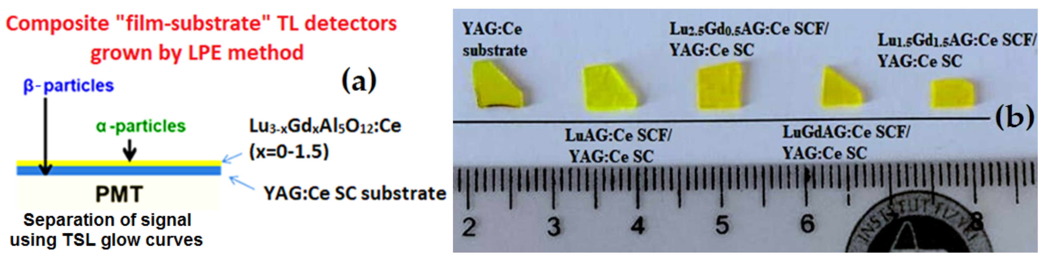
Figure 1.
(a) principal scheme of the composite thermoluminescent (TL) detector, based on the Lu3−xGdxAG:Ce SCF/Y3Al5O12:Ce (YAG:Ce) single crystal (SC) epitaxial structure with single crystalline film (SCF) and SC thickness in the 12-15 μm and 0.9-1.0 mm ranges, respectively, and photomultiplier (PMT); (b) the set of TL detectors based on the liquid phase epitaxy (LPE)-grown epitaxial structures containing Lu3−xGdx Al5O12:Ce SCFs at x = 0–1.5 and YAG:Ce substrates. The irregular shapes of the samples result from cutting the fragment of these samples with a 3.75 mm × 3. 5 mm × 5.25 mm size for TL measurements using a Risø TL/OSL-DA-20 reader.
Figure 2.
(a) XRD patterns of (880) planes of Lu3−xGdxAl5O12:Ce SCFs at x = 0, 0.5, and 1.5, grown onto YAG:Ce substrates; (b) the dependence of the misfit between the lattice constants of Lu3−x GdxAl5O12:Ce SCF and YAG substrate on Gd content x.
Figure 2.
(a) XRD patterns of (880) planes of Lu3−xGdxAl5O12:Ce SCFs at x = 0, 0.5, and 1.5, grown onto YAG:Ce substrates; (b) the dependence of the misfit between the lattice constants of Lu3−x GdxAl5O12:Ce SCF and YAG substrate on Gd content x.
Figure 3.
Absorption spectra of LuAG:Ce SCF/YAG:Ce SC, Lu2.5Gd0.5AG:CeSCF/YAG:Ce, LuGdAG:Ce SCF/YAG:Ce SC, and Lu1.5Gd1.5AG:Ce SCF/YAG:Ce composite detectors in comparison with YAG:Ce substrate.
Figure 3.
Absorption spectra of LuAG:Ce SCF/YAG:Ce SC, Lu2.5Gd0.5AG:CeSCF/YAG:Ce, LuGdAG:Ce SCF/YAG:Ce SC, and Lu1.5Gd1.5AG:Ce SCF/YAG:Ce composite detectors in comparison with YAG:Ce substrate.
Figure 4.
CL spectra (a) of LuAG:Ce, Lu2.5Gd0.5AG:Ce, LuGdAG:Ce, Lu1.5Gd1.5AG:Ce SCFs in comparison with YAG:Ce substrate and (b) shift of emission band.
Figure 4.
CL spectra (a) of LuAG:Ce, Lu2.5Gd0.5AG:Ce, LuGdAG:Ce, Lu1.5Gd1.5AG:Ce SCFs in comparison with YAG:Ce substrate and (b) shift of emission band.
Figure 5.
Glow curves of YAG:Ce SC after irradiation by α- (1) and β- (2) particles, respectively.
Figure 5.
Glow curves of YAG:Ce SC after irradiation by α- (1) and β- (2) particles, respectively.
Figure 6.
TL glow curves of LuAG:Ce SCF/YAG:Ce SC (a), Lu2.5Gd0.5AG:Ce SCF/YAG:Ce SC (b), Lu2GdAG:Ce SCF/YAG:Ce SC (c), and Lu1.5Gd1.5AG:Ce SCF/YAG:Ce SC (d) composite detectors after irradiation by α- (1) and β- (2) particles, respectively.
Figure 6.
TL glow curves of LuAG:Ce SCF/YAG:Ce SC (a), Lu2.5Gd0.5AG:Ce SCF/YAG:Ce SC (b), Lu2GdAG:Ce SCF/YAG:Ce SC (c), and Lu1.5Gd1.5AG:Ce SCF/YAG:Ce SC (d) composite detectors after irradiation by α- (1) and β- (2) particles, respectively.
Figure 7.
Dependence of position of the main TL peaks in Lu3−xGdx Al5O12:Ce SCF/YAG:Ce SC epitaxial structures under α-particle (241Am) excitation (1) and the difference between the main TSL peaks of SCFs and SC parts of Lu3−xGdxAl5O12:Ce SCF/YAG:Ce SC composite detectors under α-particle (241Am) and β-particle (90Sr + 90Y) excitation (2).
Table 1.
Conditions of the LPE growth of Lu3−xGdxAl5O12:Ce SCFs at x = 0–1.5 onto YAG:Ce substrates. H—SCF thickness; f and T—velocity and temperature of the SCFs growth; a—SCF lattice constants, m—SCF/substrate misfit, LY—relative photoelectron light yield (LY) of SCFs under α-particle excitation in comparison with the LY of YAG:Ce SCF standard samples with a LY of 2.65 ph/KeV.
| Nominal Content of SCFs in Melt-Solution | Substrate | m, % | h, µm | T, °C | f, μm/min | LY, % |
|---|
| YAG:Ce SC | YAG | | | | | 128 |
| LuAG:Ce SCF | YAG:Ce | 0.83 | 37 | 973 | 0.43 | 98 |
| Lu2.5Gd0.5AG:Ce SCF | YAG:Ce | 0.49 | 51 | 970 | 0.73 | 63 |
| Lu2GdAG:Ce SCF | YAG:Ce | | 16 | 970 | 0.53 | 54 |
| Lu1.5Gd1.5AG:Ce SCF | YAG:Ce | 0.04 | 20 | 975 | 0.4 | 45 |
Table 2.
Position of the main TL peaks in Lu3−xGdxAl5O12:Ce SCF/YAG:Ce SC composite structures after irradiation by α- and β-particles. * the main TL peaks.
Table 2.
Position of the main TL peaks in Lu3−xGdxAl5O12:Ce SCF/YAG:Ce SC composite structures after irradiation by α- and β-particles. * the main TL peaks.
| SC and SCF Content | α-Particles (241Am) | β-Particles (90Sr + 90Y) |
|---|
| LuAG:Ce SCF/YAG:Ce SC | 450, 600 * | 445, 500, 615 * |
| Lu2.5Gd0.5AG:Ce SCF/YAG:Ce SC | 385, 445 * | 435, 495, 605 * |
| Lu2GdAG:Ce SCF/YAG:Ce SC | 385, 445 *, 593 | 435, 490, 600 * |
| Lu1.5Gd1.5AG:Ce SCF/YAG:Ce SC | 390 *, 510, 615 | 435, 490, 605 * |
© 2020 by the authors. Licensee MDPI, Basel, Switzerland. This article is an open access article distributed under the terms and conditions of the Creative Commons Attribution (CC BY) license (http://creativecommons.org/licenses/by/4.0/).

 ,
, 


