Review

Predatory and Defensive Strategies in Cone Snails

by
Zahrmina Ratibou
1,
Nicolas Inguimbert
1,* and
Sébastien Dutertre
2,*
1
CRIOBE, UAR CNRS-EPHE-UPVD 3278, University of Perpignan Via Domitia, 58 Avenue Paul Alduy, 66860 Perpignan, France
2
IBMM, University of Montpellier, CNRS, ENSCM, 34093 Montpellier, France
*
Authors to whom correspondence should be addressed.
Toxins 2024, 16(2), 94; https://doi.org/10.3390/toxins16020094
Submission received: 17 January 2024 / Revised: 31 January 2024 / Accepted: 3 February 2024 / Published: 7 February 2024

Abstract

:
Cone snails are carnivorous marine animals that prey on fish (piscivorous), worms (vermivorous), or other mollusks (molluscivorous). They produce a complex venom mostly made of disulfide-rich conotoxins and conopeptides in a compartmentalized venom gland. The pharmacology of cone snail venom has been increasingly investigated over more than half a century. The rising interest in cone snails was initiated by the surprising high human lethality rate caused by the defensive stings of some species. Although a vast amount of information has been uncovered on their venom composition, pharmacological targets, and mode of action of conotoxins, the venom–ecology relationships are still poorly understood for many lineages. This is especially important given the relatively recent discovery that some species can use different venoms to achieve rapid prey capture and efficient deterrence of aggressors. Indeed, via an unknown mechanism, only a selected subset of conotoxins is injected depending on the intended purpose. Some of these remarkable venom variations have been characterized, often using a combination of mass spectrometry and transcriptomic methods. In this review, we present the current knowledge on such specific predatory and defensive venoms gathered from sixteen different cone snail species that belong to eight subgenera: Pionoconus, Chelyconus, Gastridium, Cylinder, Conus, Stephanoconus, Rhizoconus, and Vituliconus. Further studies are needed to help close the gap in our understanding of the evolved ecological roles of many cone snail venom peptides.
Key Contribution: This review summarizes the current knowledge on injected venom composition for some cone snail species in relation to their predatory and defensive behaviors.

1. Introduction

Cone snails are specialized carnivorous marine mollusks that can be found in coral reef areas, from shallow intertidal to deeper waters, and spread across the tropical Indian, Pacific, and Atlantic Oceans [1]. They are classified as gastropods within the Conidae family, which feature hollow radular teeth and venom glands [2]. They use a complex venom mixture to paralyze and hunt fish, mollusks, and worms [3]. This venom is secreted through epithelial cells lining the cone’s venom gland, which is a long and thin tubular duct [4]. A singular radular tooth, analogous to a hypodermic needle, is then moved into the proboscis through which the rapid-acting venom is injected. The venom is acknowledged as a rich source of potent pharmacological components, raising high interest in the drug development field [5].
This venom consists primarily of biologically active peptides, generally characterized as conotoxins or conopeptides. They can be classified into two groups: conotoxins, which are cysteine-rich conopeptides consisting of 10 to 30 amino acids, while conopeptides are cysteine-poor, meaning 1 or no disulfide bond [4,5,6]. Moreover, conotoxins are highly structured and often show high affinity and selectivity toward membrane receptors, ion channels, and other transmembrane proteins of the nervous and non-nervous systems [4]. Conopeptides include several types of cysteine-poor peptides, such as contulakins, conantokins, conorfamides, conolysins, conophans, conomarphins, contryphans, conopressins, and more recently, hormone-like conopeptides, such as elevenins or prohormones [5,7]. Conopeptides are usually minor in comparison to conotoxins in the venom mixture and each presents a selective type of target [7]. These small peptides can work as ligands, which induce a physiological reaction by interacting with a given receptor [4]. Conotoxins and conopeptides are secreted as peptide precursors, which can be portioned into three characteristic sections: a highly conserved signal peptide, representative of the gene superfamily from which it was translated, a pro-peptide section, and a highly diversified mature peptide (Figure 1). The mature peptide is the active sequence portion, which is enzymatically cleaved and then modified into a highly stable structure within the injected venom [8].
The cysteine pattern within the conotoxin sequence is designated with roman numerals and it directs the tridimensional structure, which in turn also influences their biological activity. So far, although only few conotoxins have been fully characterized pharmacologically, more than 20 pharmacological targets have been identified. Some of the biological targets involve, for the most part, ion channels, but also some G-protein-coupled receptors and transporters [3]. Conotoxins are classified according to their targets into pharmacological families, defined by Greek letters, such as α, δ, μ, ω, κ, γ, etc. (Figure 2) [3]. For instance, ω-conotoxins are antagonists of voltage-gated calcium channels, and some are effective against neuropathic pain [3]. Such activity was the basis for the development of the first marine-based drug isolated from a cone snail, known as Prialt®. This drug is a synthetic version of the ω-conotoxin MVIIA isolated from the piscivorous species, Pionoconus magus [9]. Likewise, some α-conotoxins have been characterized as nicotinic acetylcholine receptors (nAChRs) antagonists, with some of them having potential in the treatment of pain, cognitive, cardiovascular, and other disorders [9]. For the past three decades, research in the field has been mainly focused on finding new ligands for known targets, with a strong emphasis on modulators of pain receptors [9].
The ~800 species of cone snails can be categorized into three main groups according to their diet. Piscivorous species hunt fish, molluscivorous species prey upon mollusks, and vermivorous species feed upon worms (Figure 3). The type of radula tooth seems to be directly correlated to the diet, and this criterion has been used to support the classification of species [11]. Based on molecular phylogenetic studies, cone snails have been classified into a single large family, Conidae, which can then be divided into four genera: Conus, Conasprella, Profundiconus, and Californiconus [2]. The genus Conus constitutes more than 85% of all cone snail species, which can then be further classified into 57 subgenera or ‘clades’ of Conus species, which represent a clear subgrouping within the genera [2]. These classifications can provide a better understanding of the “biotic interactions” within Conus species [4]. Unfortunately, rather than being tested on biologically relevant animal models, cone snail venoms have almost exclusively been investigated using mammalian bioassays. As a result, the conclusions drawn from these assays should be interpreted with caution when extrapolated to the biology of cone snails.

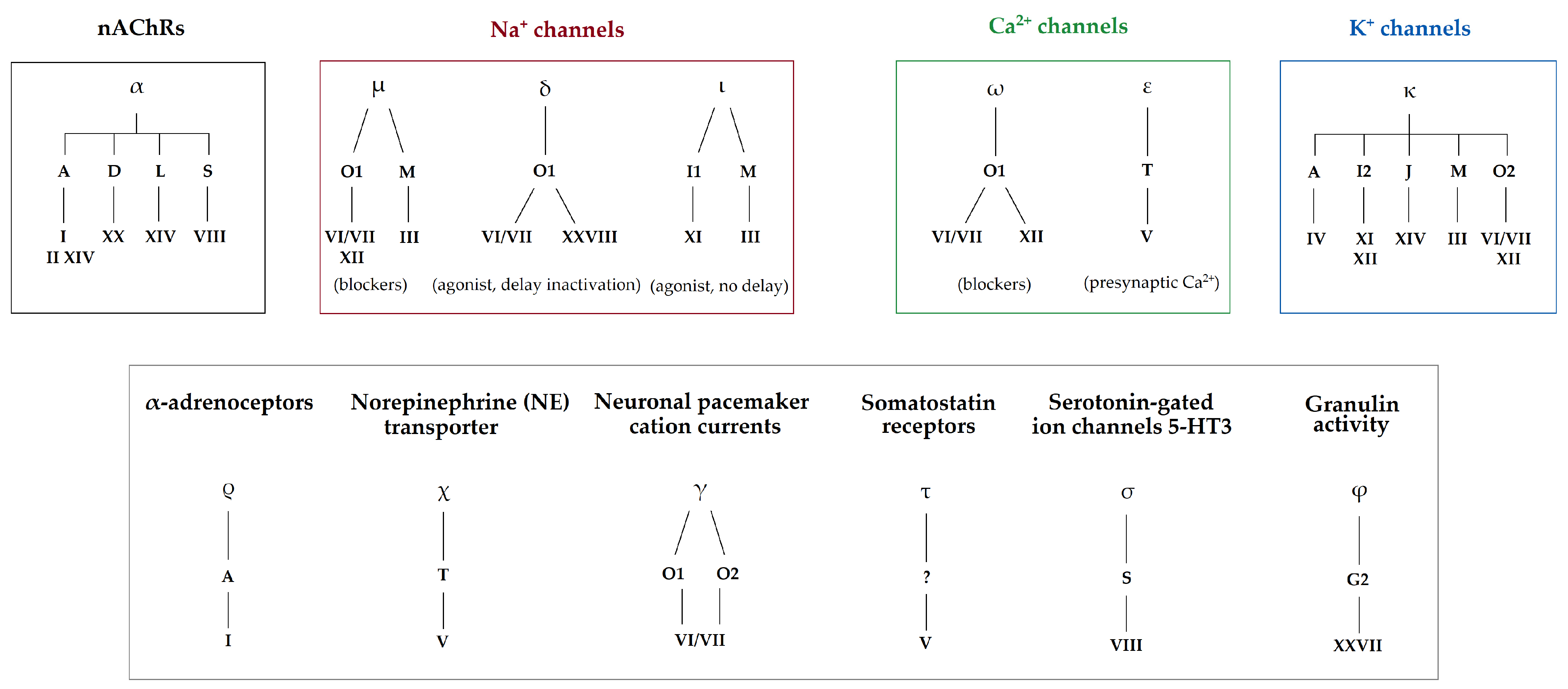
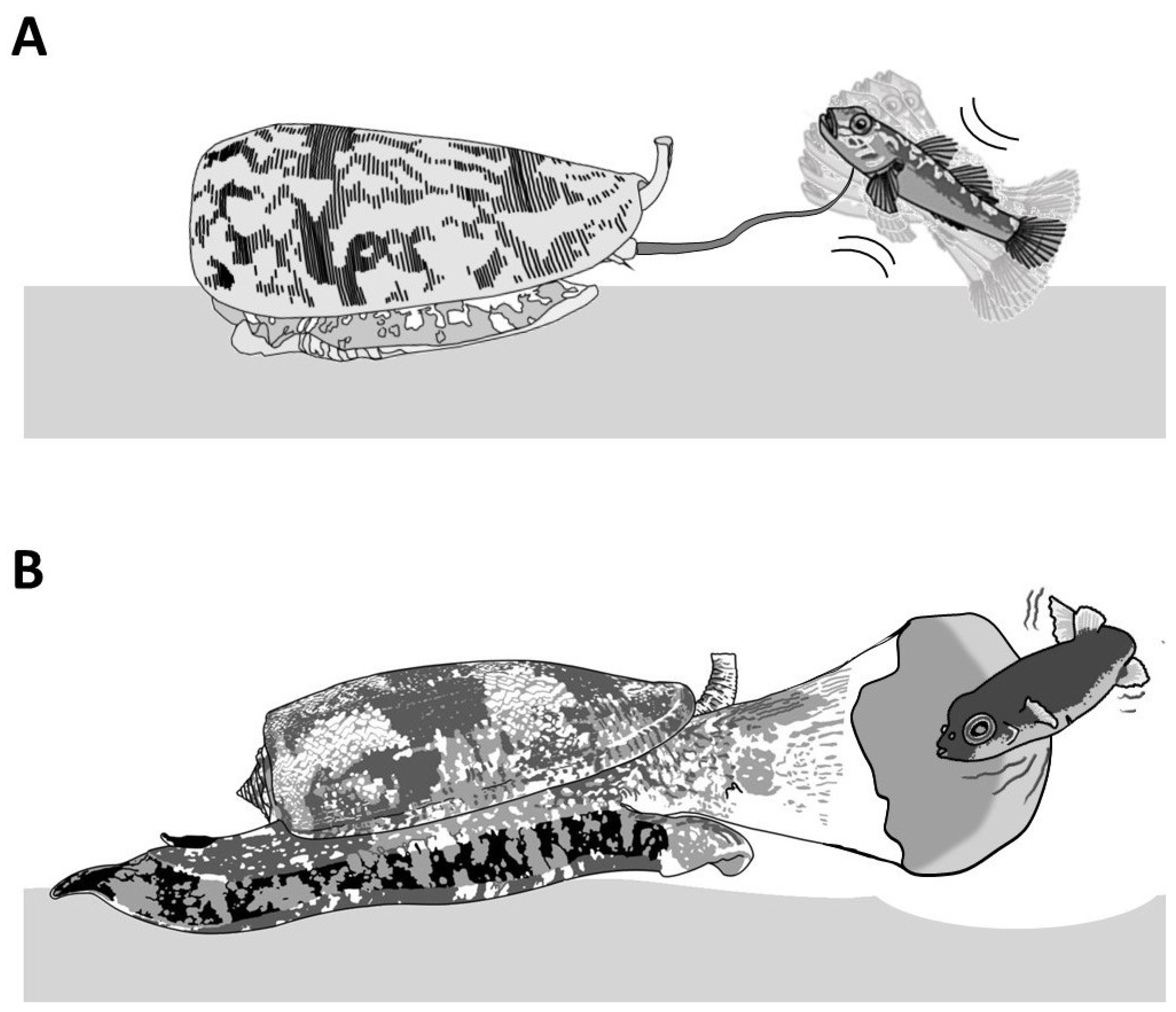
1.1. Envenomation Strategies in Cone Snails

Piscivorous cone snails exhibit varying types of hunting behaviors. For instance, upon the detection of a prey, first through chemosensory cues [12], some cone snails extend their proboscis in order to inject a paralytic venom (Figure 4A). The venom is injected via a radula tooth that is comparable to a miniature harpoon that the cone snail uses to sting and tether the prey to avoid its escape [4,13]. Upon the strike, the prey often displays an immediate tetanic paralysis. The cone snail then retracts its proboscis to drag its victim toward its enlarged rostrum to engulf it [13]. The archetype of this behavior is the ‘taser-and-tether’ strategy employed by the majority of piscivorous species from the Pionoconus, Textilia, and Chelyconus clades, where injection of venom first produces an immediate paralysis (“taser”), followed by the reeling back of the tethered fish into the rostrum via the contraction of the proboscis, which is still tightly grasping the base of the radula tooth [4].
Remarkably, some other cone snails have been observed to catch their prey without prior sting. In this case, the cone snail is hypothesized to release a set of toxins in the water, which places the prey into a sedative-sleepy state (Figure 4B). The cone snail then opens its rostrum to engulf it and may proceed to envenomate and predigest the prey [13]. Thus, cone snails that use this strategy, named as ‘net-hunting’, would supposedly release venom components in the water and inject paralytic peptides, which induces an irreversible neuromuscular paralysis of the captured prey. Lastly, the “strike-and-stalk” envenomation strategy is a variation of the taser-and-tether strategy, where the cone snail strikes a prey without tethering it and engulfs it after immobilization has occurred. The latter strategy remains less studied in terms of the neurobiological mechanism involved [13].
In non-piscivorous cone snails, the hunting behaviors have been much less investigated. For most molluscivorous species observed in captivity or in the wild, the predatory strategy involves actively chasing the prey and injecting, multiple times, fine, arrow-like radula teeth into the foot of the prey [14]. The firing of the radula tooth is usually accompanied by vigorous pumping of copious amount of venom, which can be seen, when injected in excess, as a whitish cloud escaping out the tip of the proboscis and/or out of the base of the tooth from back pressure [15]. In the case of the mass spectrometry (MS) analysis of successive stings by Cylinder textile, modest variations in the venom composition were described [16]. The first injection usually stops or slows down the prey but does not completely incapacitate it; therefore, it was suggested that a second, third, or more injections, possibly with different peptides, were needed to eventually overcome the prey.
Hunting behaviors for vermivorous species are even more elusive, except for only a few species. Both Stephanoconus imperialis and Stephanoconus regius prey almost exclusively on amphinomid worms (“fireworms”). These two species use a prey capture strategy reminiscent of the “taser-and-tether” strategy employed by many piscivorous species. Indeed, the targeted worm is first detected by the chemosensory organs, inducing the extension of a reddish proboscis. The short radula tooth (1–1.5 mm) is then fired and embedded into the worm’s body, forcefully pushing through a remarkable quantity of a greening venom (Figure 3C) [9]. As described for the fish-hunters, the envenomated prey shows immediate involuntary contractions, leading to incapacitation, and is reeled back into the rostrum. Our personal observations on other vermivorous species often reveal, surprisingly, an apparent venom-less strategy, where the snail directly attempts to swallow the worm through its extended rostrum without prior stinging via the proboscis. One of the most mysterious prey strategies relates to the vermivorous species hunting tube worms, as there is no description in the literature.

1.2. Reality Check on the Concept of Cabals

Early pharmacological characterization of conotoxins from venom gland extracts revealed a variety of targets and modes of action. From the pharmacological effects obtained mostly on mammals, extrapolations were made to explain the effects observed on prey, and this is how the concept of cabals was first crafted. The cabals are defined as a group of (artificially put together) conotoxins, which seem to modulate the same physiological target or may act synergistically. Thus, the “lightening-strike cabal” is defined as a set of κ- and δ-conotoxins, as well as conkunitzins, which would together elicit an excitatory state on the prey [17,18]. This reaction is due, respectively, to the inhibition of K+ channels, as well as a delayed inactivation of Na+ channels [4].
Meanwhile, the “nirvana cabal” is highly speculative, but could include the release in the surrounding water of a mixture of B1-conotoxins [19] and hormone-like peptides that would induce a “hypoactivity in sensory neuronal circuity” [4,20,21]. Although prey capture observations of net-hunting species seem to corroborate this hypothesis, there is currently no direct evidence to support any release of venom into the water. Lastly, an additional “motor cabal” was proposed to be responsible for the final flaccid paralysis that prevents the prey from recovering the initial excitatory shock. The latter involves α-, µ-, and ω-conotoxins that interfere with the neuromuscular junction [18].
Although these cabals were logically formulated, do they actually correspond to the reality of the predatory strategies employed by cone snails to defeat their prey? Nearly thirty years ago, an ingenious procedure, now commonly referred to as “milking”, was devised that allows for the collection of the injected venom, providing a direct means of interrogating the conotoxin cocktail used for prey capture [22]. Using a live prey to arouse the cone snail and trigger a predatory behavior, a microcentrifuge tube covered with parafilm, and a piece of the prey’s tissue, is presented to the tip of the extended proboscis. Sensory cilia at the tip of the proboscis identify the tissue as “prey” and instantaneously trigger the injection of venom through the radula tooth. Such recovered “milked venoms” can now be analyzed, and the composition revealed. Over the last two decades, the more milked venoms were investigated, the less obvious the role of the conotoxins described in these cabals was for prey capture [23].
Overall, in all cases investigated, milked venoms appear significantly less complex compared to dissected gland extracts. For instance, in Pionoconus species, the predatory venom is usually dominated by one class of conotoxins (sometimes the only conotoxins seemingly injected), the κA-conotoxins [24,25,26]. Therefore, it appears that κA-conotoxins are responsible for the immediate “taser” effect in this clade, not a combination of κ- and δ-conotoxins, as originally described for the lightning-strike cabal. Indeed, injection of κA-conotoxins alone into fish recapitulates the tetanic paralysis observed during prey capture [27]. However, it has to be noted that intraspecific variations in the injected venom can be dramatic and, occasionally, the paralytic peptides from the “motor cabal” are detected, suggesting that they could play a significant role in prey capture [23]. Although not fully explained at the time, one aspect of this diversification was later attributed, at least in part, to the unsuspected ability of some cone snails to produce two types of venoms [28].

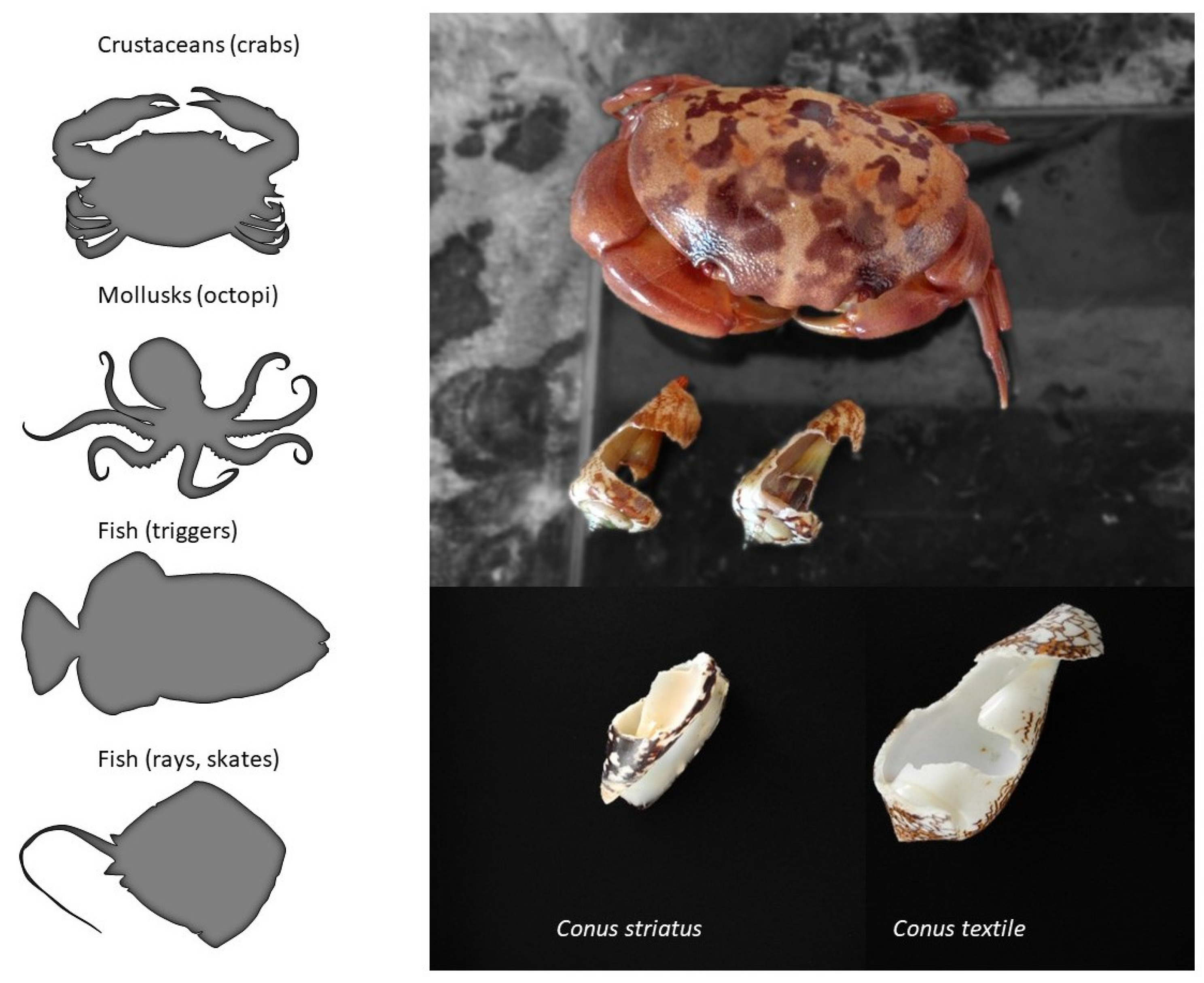
1.3. Defensive Strategies

From the three dozen human deaths reported, it has long been known that cone snails can also inject their venom defensively [29]. In the literature, there is only anecdotal information on the natural predators of cone snails, but fish, mollusks (octopi), and some crustaceans are known to prey on them (Figure 5). For instance, a rare species of deep-water cone snail was first described only from a shell recovered from the stomach content of a large fish (personal communication). The defensive use of venom provides an obvious evolutionary advantage. Indeed, avoiding being eaten is one of the most important fitness-related criterion for the survival of a species, together with being able to feed and reproduce. In fact, some venomous animals only use their venom defensively (some hymenopterans, fish, etc.), whereas the reverse is not true, suggesting that the defensive use of venom may actually have a stronger evolutionary role than anticipated, possibly more than predation in some cases [30].
Thanks to their capacity to defend themselves, some species of cone snails have evolved some unique behaviors. However, for most species, the first line of defense is usually to retract deeply into the shell, which offers a strong and often inviolable fortress (Figure 6C). Others will respond aggressively to any threat by extending their proboscis (Figure 6D). If the threat intensifies, the cone snail will inject venom into the aggressor, but there are also reports of cone snails squirting venom (personal observations). Additional behavioral studies are needed to fully decipher the complex defensive responses displayed by cone snails.
The most dangerous species to humans, Gastridium geographus, displays an unusually aggressive behavior, and will readily use its venom defensively when handled. There seems to be a striking relationship between the fragility of the shell (as in the case of Gastridium geographus) and the propensity to use venom defensively. Typically, large vermivorous species will often be unfazed by any threat, being protected by heavily built shells and narrow apertures [28]. However, many species were reported to inflict injuries to humans, regardless of their diet, with varied degrees of consequences. From the known human Conus envenomation, various levels of severity were distinguished, from fatal to minor effects, comparable to bee stings, and the most adverse symptoms were attributed to piscivorous cone snails, especially Gastridium geographus [29].
The first investigation of a defense-evoked venom uncovered an unsuspected twist in cone snail biology [28]. Indeed, the defensive venom of Gastridium geographus was highly complex and contained massive amounts of paralytic conotoxins from the “motor cabal”, explaining the lethal symptoms in humans, as opposed to the predatory venom, which was devoid of these and instead contained prey-specific conotoxins with no activity on human receptors. Therefore, in this iconic species, paralytic conotoxins directed to the neuromuscular junction are essentially defensive weapons, not part of the prey capture strategy, a result in conflict with the cabal narrative. From this initial discovery, more data on different species were needed to evaluate how widespread this separate evolution of predatory and defensive venoms is among cone snail species. Triggering and collecting defensive venom can be achieved through different means, including using a natural predator (i.e., a molluscivorous species, such as Conus marmoreus or Cylinder textile), applying pressure to the shell, or pinching the foot of the cone (Figure 6D) [28].
Overall, the remarkable ability of cone snails to purposefully modify their venom composition upon different triggering stimuli (predatory or defensive) offers novel and unprecedented research opportunities. Indeed, separately collecting each venom type will allow unambiguous interpretation of the ecological and evolutionary roles of each conotoxin. In this review, we will describe the reported predatory- and defense-evoked venoms of 16 species belonging to eight clades of the Conus genus, with three being piscivorous (Pionoconus, Chelyconus, and Gastridium), two molluscivorous (Cylinder and Conus), and three vermivorous (Stephanoconus, Rhizoconus, and Vituliconus), and discuss the work that remains in order to better understand venom–ecology relationships in cone snails.

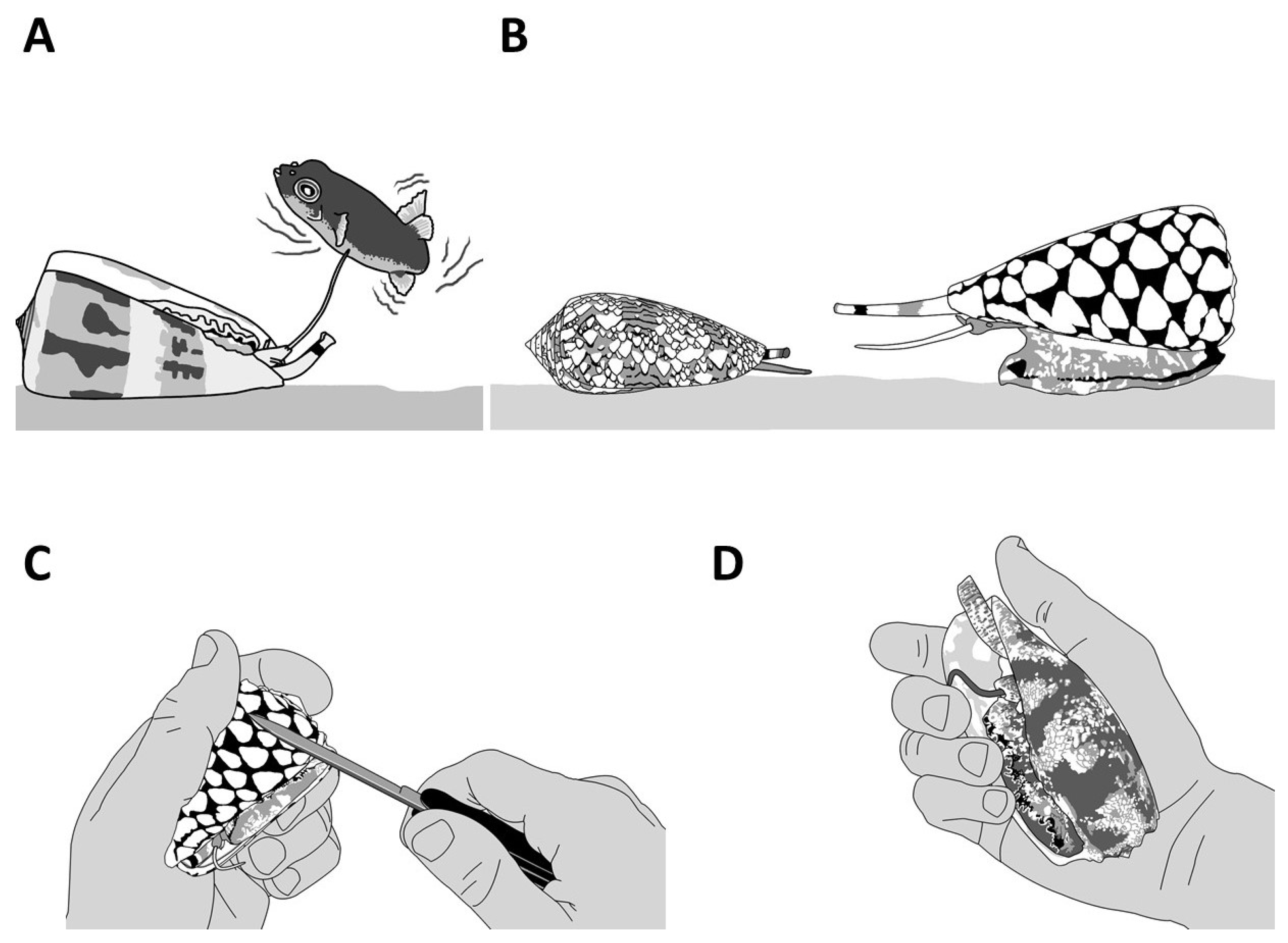
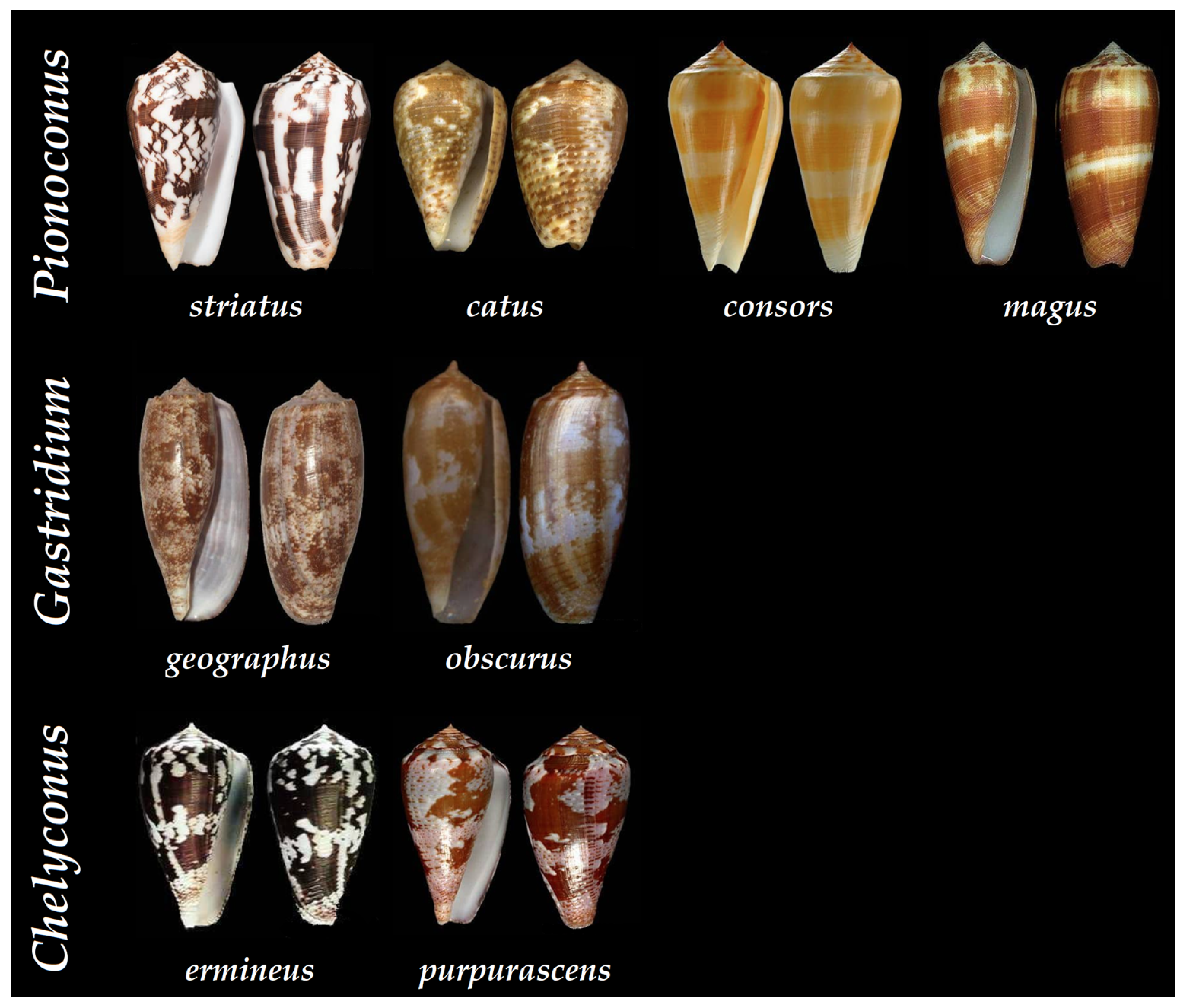
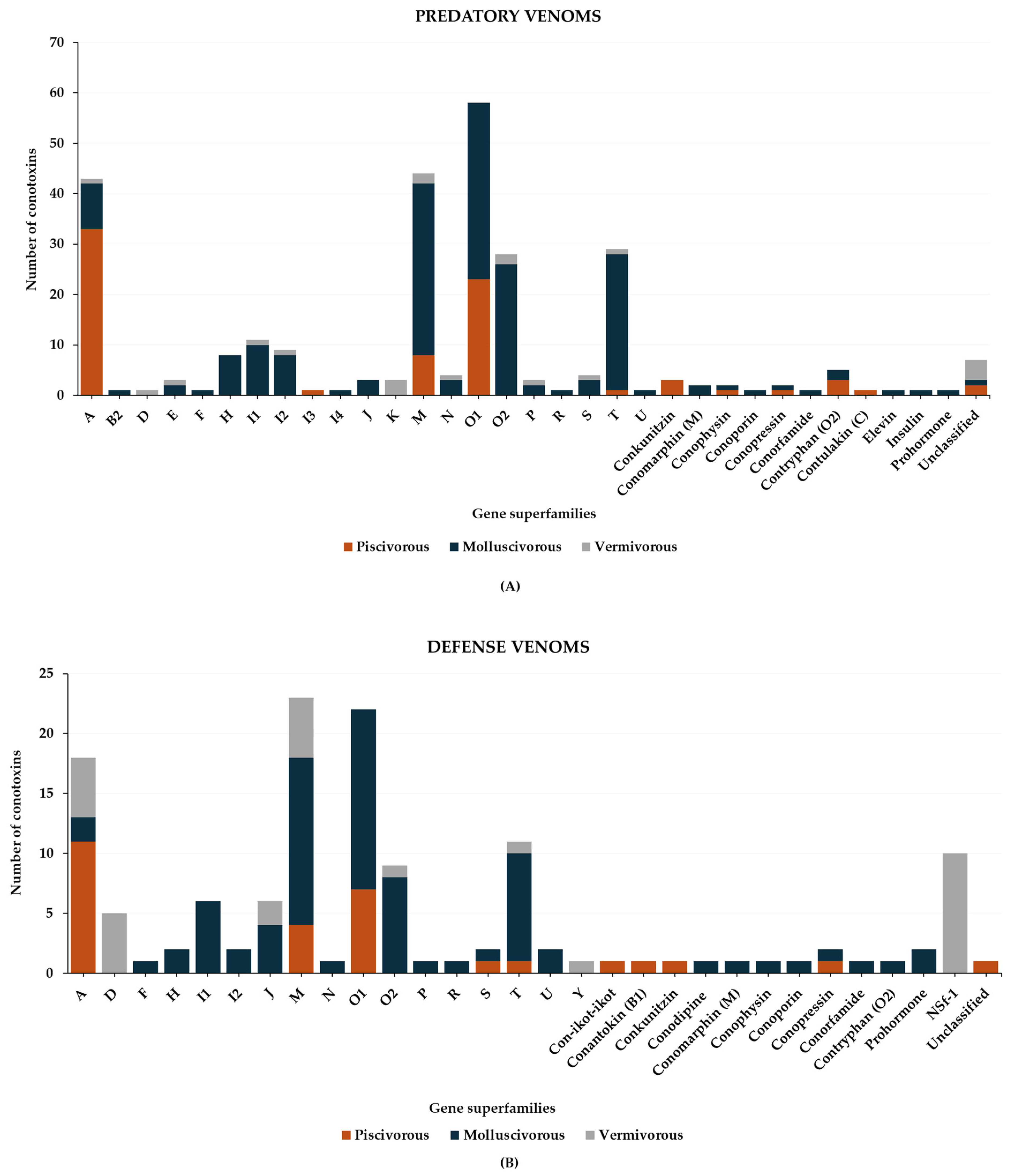
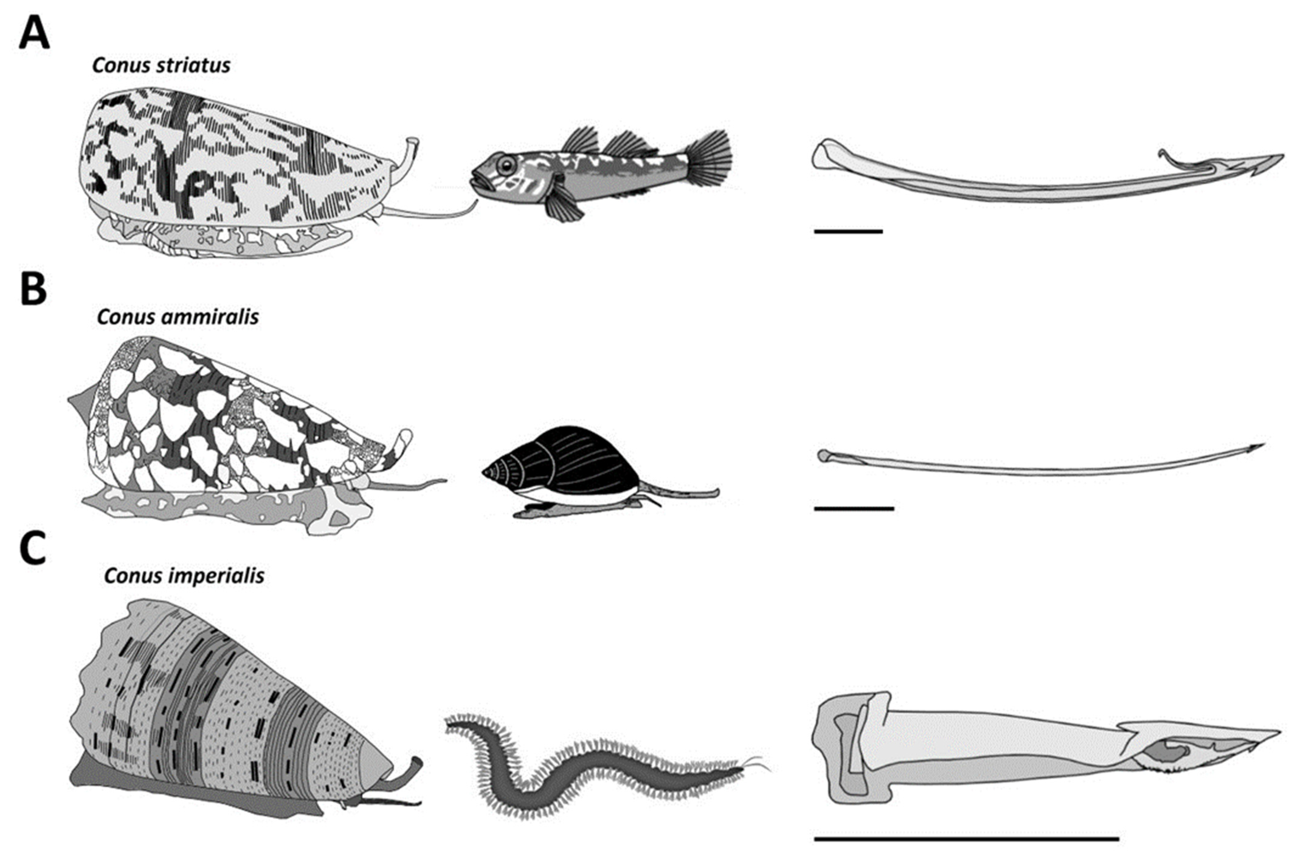
2. Piscivorous Cone Snails

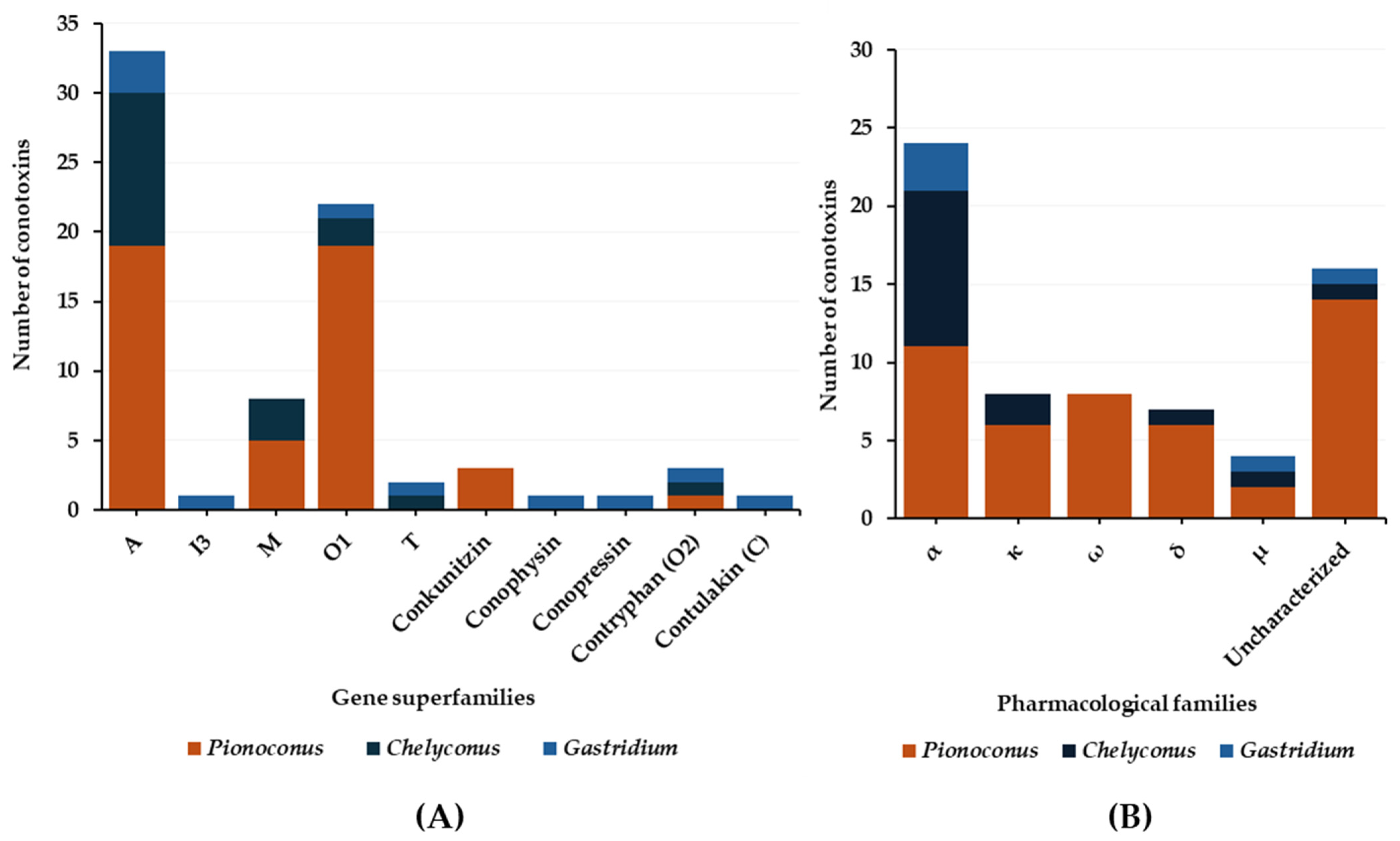
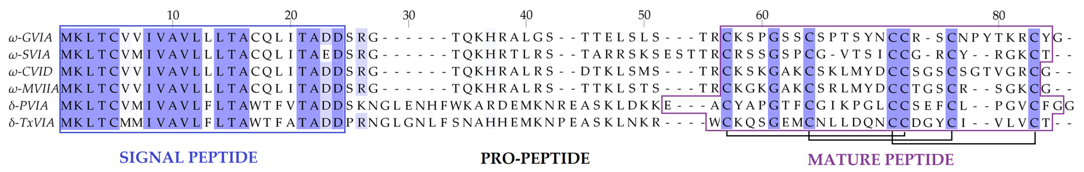
2.1. Predatory Venom

Thus far, the Conus genus counts around 800 different species, representing about 70% of vermivorous, 20% piscivorous, and 10% of molluscivorous cone snails [31]. Over a hundred piscivorous cone snails have been classified into the following clades: Afonsoconus, Asprella, Chelyconus, Embrikena, Gastridium, Phasmoconus, Pionoconus, and Textilia, although the piscivorous diet requires confirmation for the Afonsoconus, Asprella, and Embrikena clades [13]. In comparison, the venoms of fish-hunting cone snails, such as Pionoconus striatus, Gastridium geographus, and Chelyconus purpurascens, have been extensively characterized against the prevailing vermivorous species (Figure 7) [28,32,33].
In general, the predatory venoms of fish-hunting cone snails show major contributions of small disulfide-rich conotoxins over larger ones and cysteine-poor conopeptides, such as conophysins [28], conopressins [28,33], and contryphans [32,35] (Figure 8A). The conotoxins identified are scattered into a dozen gene superfamilies, dominated by A-, O1- and M-conotoxins. The rest are attributed to the I3, C, O2, T, and B1 superfamilies [23,29,33]. Most conotoxins that were identified in the predatory venoms of fish-hunting cone snails were previously biologically characterized from venom gland extracts. The α-, κA-, δ-, κ-, μ-, and ω-conotoxins constitute the major pharmacological families identified in the predatory venom (Figure 8B). κA-conotoxins are the most abundant (relative contribution to the injected venom) (Table 1), but α-conotoxins are the most prevalent (in terms of number of sequences identified) in the predatory venoms of fish-hunting cone snails (Table 2). These conotoxins are especially represented in the Pionoconus and Chelyconus clades, and less in the Gastridium clade.
As mentioned, the superfamily A is the most represented in the predatory venom thanks to κA-conotoxins and α-conotoxins. κA-conotoxins were first discovered in the predatory venom of Pionoconus striatus, with κA-SIVA and κA-SIVB [32], and their short and non-glycosylated equivalent κA-PIVE and κA-PIVF were identified in Chelyconus purpurascens (Table 1) [17,38]. Exhaustive investigation of the predatory venom of Pionoconus consors also revealed the importance of κA-conotoxins, with the abundant injection of κA-CcTx and the sequencing of a series of CcTx variants [24,37,50]. Although not confirmed, the major compounds found in the predatory venom of another Pionoconus species, Pionoconus magus, were determined within the mass range of κA-conotoxins [40]. More recently, these κA-conotoxins were identified abundantly in both predatory and defensive venom of Pionoconus striatus [32] and in the predatory venom of Pionoconus catus [25]. Interestingly, a recent study has identified a variant of the glycosylated κA-conotoxins (κA-SIVC) in the predatory venom of specimens of Pionoconus striatus from Mayotte (France), suggesting that geographical variations can be population-specific [36]. κA-conotoxins were initially characterized as excitatory peptides that block K+ channels, yet controversy remains over the molecular target since Na+ channels were also suggested as the targeted receptor [27]. Although they uphold the same IV cysteine framework as certain αA-conotoxins, such as α-OIVA (Table 2), their activities are different: the firsts are excitatory while the seconds are not [41]. Generally, these κA-conotoxins appear as the major and most abundant component in the predatory venom of Pionoconus species and are likely solely responsible for the rapid immobilization of prey.
Next, α-conotoxins (targets are the nAChRs) are the most prevalent pharmacological family in terms of number of sequences identified in the predatory venoms of fish-hunting cone snails. These α-conotoxins, although not systematically injected, are especially represented in the Pionoconus and Chelyconus clades, and less in the Gastridium clades [2]. For instance, some of the α-conotoxins identified in the Pionoconus clade include α-SI (Pionoconus striatus) [23,32,39], α-CnIB (Pionoconus consors) [37,51], α-MI (Pionoconus magus) [40], and α-CIB (Pionoconus catus) [25]. Similarly, α-PIB (Chelyconus purpurascens) and α-EIIA (Chelyconus ermineus) [52] are found in the Chelyconus clade. Finally, the Gastridium subgenus shows the least amount of identified α-conotoxins in their predatory venom, with only α-GIC (Gastridium geographus) [33] and α-OIVA (Gastridium obscurus) confirmed so far [41]. When injected into a prey, some of these α-conotoxins operate as slow (several minutes) paralytics analogous to the snake α-neurotoxins, as they selectively target and inhibit the muscle type of nAChRs (Figure 2) [10,53]. Interestingly, the subtle variations in length and amino acid residues determine the targeted site or nAChR subtype [10]. Usually, small 3/5 (3 and 5 correspond to the number of residues in inter-cysteine loops) α-conotoxins, such as α-SI, α-MI, and α-CnIB, selectively target muscle-type nAChRs [5], whereas larger 4/7 α-conotoxins, such as α-MII and α-GIC, target neuronal nAChRs, and their role in prey capture is less understood [43]. Considering their potentially useful paralytic capacities, and according to the “motor cabal” hypothesis, their presence in the predatory venoms would seem compulsory, but analysis of individual predatory venom rather than pool of collected venom suggests otherwise [10].
Exceptionally, δ-conotoxins were also detected in the predatory venom of some fish-hunters of the Pionoconus and Chelyconus clades (Table 3). This family of conotoxins was characterized as voltage-gated sodium channel (VGSC) modulators. Indeed, δ-conotoxins activate Nav channels via a delay in the inactivation mechanism (Figure 2) [54]. δ-Conotoxins, like the κ-conotoxins, are defined as excitatory peptides, which induce the rapid tetanic paralysis in their prey [32]. For example, the δ-PVIA isolated from Chelyconus purpurascens was characterized as the “lock-jaw peptide” because it causes a rigid paralysis of the prey, particularly visible around the mouth musculature [55]. Again, considering their critical role in the “lightning-strike cabal”, these conotoxins are expected to be always injected for prey capture, but it is almost never the case.
Very few μ-conotoxins have been found in the predatory venom of three piscivorous clades: Pionoconus consors, Gastridium geographus, and Chelyconus purpurascens (Table 4). The molecular targets of μ-conotoxins are VGSCs, more precisely Nav channels, which play an important role in the central and peripheral nervous system (CNS and PNS) (Figure 2). However, contrary to δ-conotoxins, they act as blockers of the channel, instead of delaying its inactivation. The μ-conotoxin-GS is the only μ-conotoxin identified from Gastridium geographus predatory venom, and is highly potent on fish, but less on mammalian Nav channels [57]. Another example is the μ-CnIIIC, isolated from the venom of Pionoconus consors, from which a synthetic version was commercialized as a cosmetic to smoothen facial lines (XEPTM-018) [58]. The μ-CnIIIB was shown more specifically to block tetrodotoxin-resistant (TTX-R) Na channels, where the blockade was observed as “slow and reversible” [59]. Another type of μ-conotoxins, the μO-Conotoxins, restrict channel opening, which has been associated with many side effects upon intravenous application, such as paralysis and death of models in inflammatory and neuropathic pain, leading to a discouragement of further research as therapeutic agents [10].
Trace amounts of ω-conotoxins known as “shaker” peptides have been detected in the predatory venoms of some fish-hunters [60]. ω-Conotoxins belong to the O1 gene superfamily with a VI/VII framework (Table 5). They are able to selectively block N-type Ca2+ channels (at the nerve terminals) and prevent the release of important neurotransmitters (such as glutamate, GABA, acetylcholine, dopamine, etc.) [61]. They are pore blockers, which means that they can physically block the influx of Ca2+ ions [62]. Upon intracerebral injection, they produce persistent shaking in mice [60]. ω-Conotoxins were extensively investigated for their potential in pain treatments, including ω-MVIIA, which is the only FDA-approved conotoxin drug (from Pionoconus magus, known as Ziconotide), as well as ω-CVID (Leconotide) isolated from Pionoconus catus, which is also under development for pain treatment [63]. Few ω-conotoxins have been detected in the predatory venom of Pionoconus cone snails, such as Pionoconus striatus (ω-SVIA and ω-SVIB) [39,64], Pionoconus consors (ω-CnVIIA) [37,51], and Pionoconus catus (ω-CVIA) [25,61].
Finally, some conopeptides, larger conotoxins, and proteins were also isolated from predatory venoms of fish-hunting cone snails (Table 6). These include contryphans, detected in Gastridium geographus, Chelyconus purpurascens, and Pionoconus striatus. Contryphans are known as Ca2+ channel modulators. For instance, contryphan-P (Chelyconus purpurascens) was associated with the “stiff-tail” syndrome when injected in mice [54]. Contryphan-S was found in the venom of Pionoconus striatus but was annotated as contryphan-G, as they present identical sequences [32]. Both contryphan-S and -G seem to be used for preying purposes by Pionoconus striatus and Gastridium geographus [28]. Moreover, Gastridium geographus injects non-paralytic compounds, including conopressin, conophysin, contulakin, and conantokin [28,33]. Conopressins and conophysins have been shown to have agonist/antagonist activity against vasopressin receptors [65], while conantokin-T was deemed responsible for the sleep-like state of the prey prior to being engulfed, especially for the “net-hunting” cone snails (Figure 4), which is caused by the inhibition of NMDA (N-methyl-D-aspartate) receptors [54,66]. Contulakin-G, on the other hand, is a glycopeptide identified as an agonist of neurotensin receptors, which has shown analgesic properties [67]. Interestingly, Pionoconus striatus also injects larger peptides, such as the conkunitzins, which have been characterized as voltage-gated K+ channel blockers, affiliated with the Shaker potassium channels [68]. Lastly, large polypeptides, such as p21a (Chelyconus purpurascens) [69], or con-ikot-ikot (Pionoconus striatus) [70] that inhibits AMPA (α-amino-3-hydroxy-5-methyl-4-isoxazole propionic acid) receptors, as well as hyaluronidases (Pionoconus consors and Chelyconus purpurascens) [37,71], phospholipases A2 (Chelyconus purpurascens) [72], and proteases (Chelyconus purpurascens and Chelyconus ermineus) [73], have been identified in predatory venoms. More high-molecular-weight proteins, such as metalloproteases, are likely to be present, but their role in prey capture remains unclear [74].

2.2. Defensive Venom

The data on defensive venom are more scarce compared to predatory venoms. Indeed, so far only Pionoconus striatus and Gastridium geographus, as well as Gastridium obscurus, have been investigated regarding their defense strategies [32,33]. Six gene superfamilies and three conopeptide classes (Figure 9A) have been identified in the defense venoms. Most of them are A-, O1-, and M-conotoxins found in both Pionoconus and Gastridium clades. In addition, B1-, S-, and T-conotoxins are found exclusively in Gastridium cone snails, as well as conophysins. Moreover, Pionoconus striatus also injected conkunitzin and con-ikot-ikot (Table 6).
Overall, five pharmacological families were characterized from the defensive venom of these cone snails: α, ω, μ, κ, and σ (Figure 9B). The α-conotoxins α-SI and α-SIA from Pionoconus striatus and α-OIVA and α-OIVB from Gastridium obscurus are used, both in the predatory and defense venoms. However, Gastridium geographus injects a specific set of α-conotoxins, including α-GIA, α-GII, and α-GID, all with a I cysteine framework [33]. Targeting neuronal rather than muscle-type nAChRs, α-GID has an unusual 4-residue N-terminal tail, which appears critical for activity at the α4β2 nAChR subtype but not the others (α3β2 or α7) [75]. In the defense venom of Pionoconus species, κA-conotoxins, such as κA-SIVA and κA-SIVB in Pionoconus striatus, are found abundantly [32].
In contrast to the predatory venom, we observe that Gastridium geographus uses large amounts of μ-conotoxins μ-GIIIA, μ-GIIIB, and μ-GIIIC [28,33] for defense purposes, but not the prey-specific μ-conotoxin-GS (Table 4). Moreover, Pionoconus striatus, which lacked μ-conotoxin in the predatory strategy, injects one in the defensive venom (μ-S3-G02) [32]. Defense venoms also include ω-conotoxins (Table 5). In addition to ω-SVIA and ω-SVIB, Pionoconus striatus injects two O1-conotoxins, SO4 and SO5, in rather high amounts [32]. Similarly, Gastridium geographus injects three potent and paralytic ω-conotoxins, ω-GVIA, ω-GVIIA, and ω-GVIIB [28,33].
An unusual conotoxin, σ-GVIIIA, was identified in the defense venom of Gastridium geographus (Table 7) [28,33]. This toxin is a σS-conotoxin presenting a VIII cysteine framework and has been characterized as a blocker of the 5-HT3 serotonin receptor (“involved in the inhibition of neurotransmitter release at motor and sensory synapses“) [76]. Additional conopeptides, such as conophysin-G [28], were also identified in the defensive strategies. Interestingly, Gastridium geographus also employs two unclassified conotoxins G5.1 and a so-called “scratcher peptide”, which was named from the “scratching” symptoms observed in mice [77]. No data are available for the defensive strategies of other piscivorous clades, including Chelyconus and Textilia.

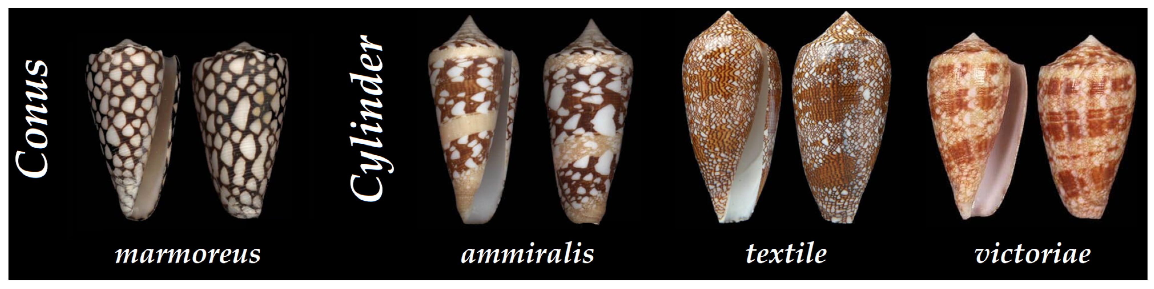
3. Molluscivorous Cone Snails

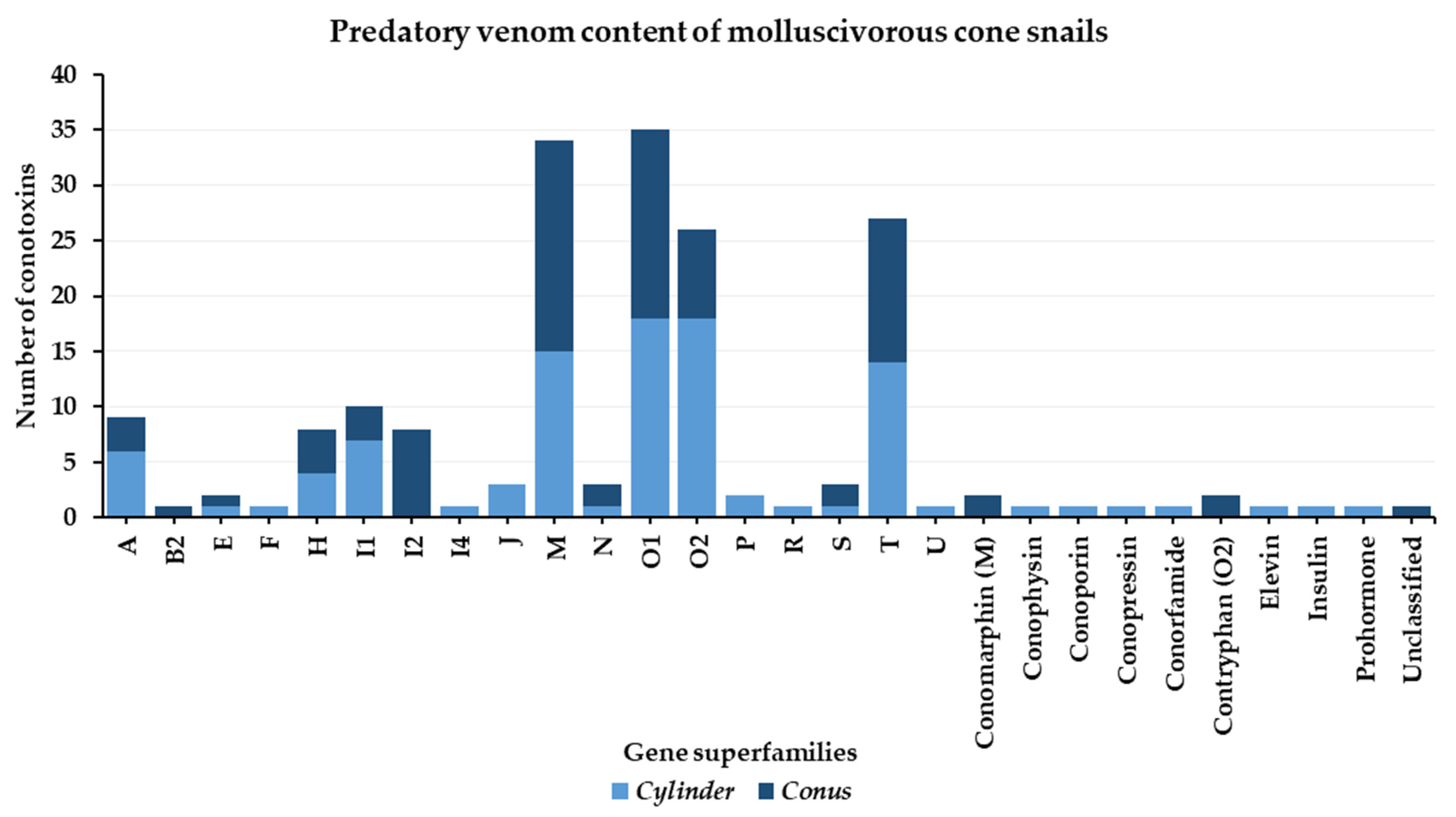
3.1. Predatory Venom
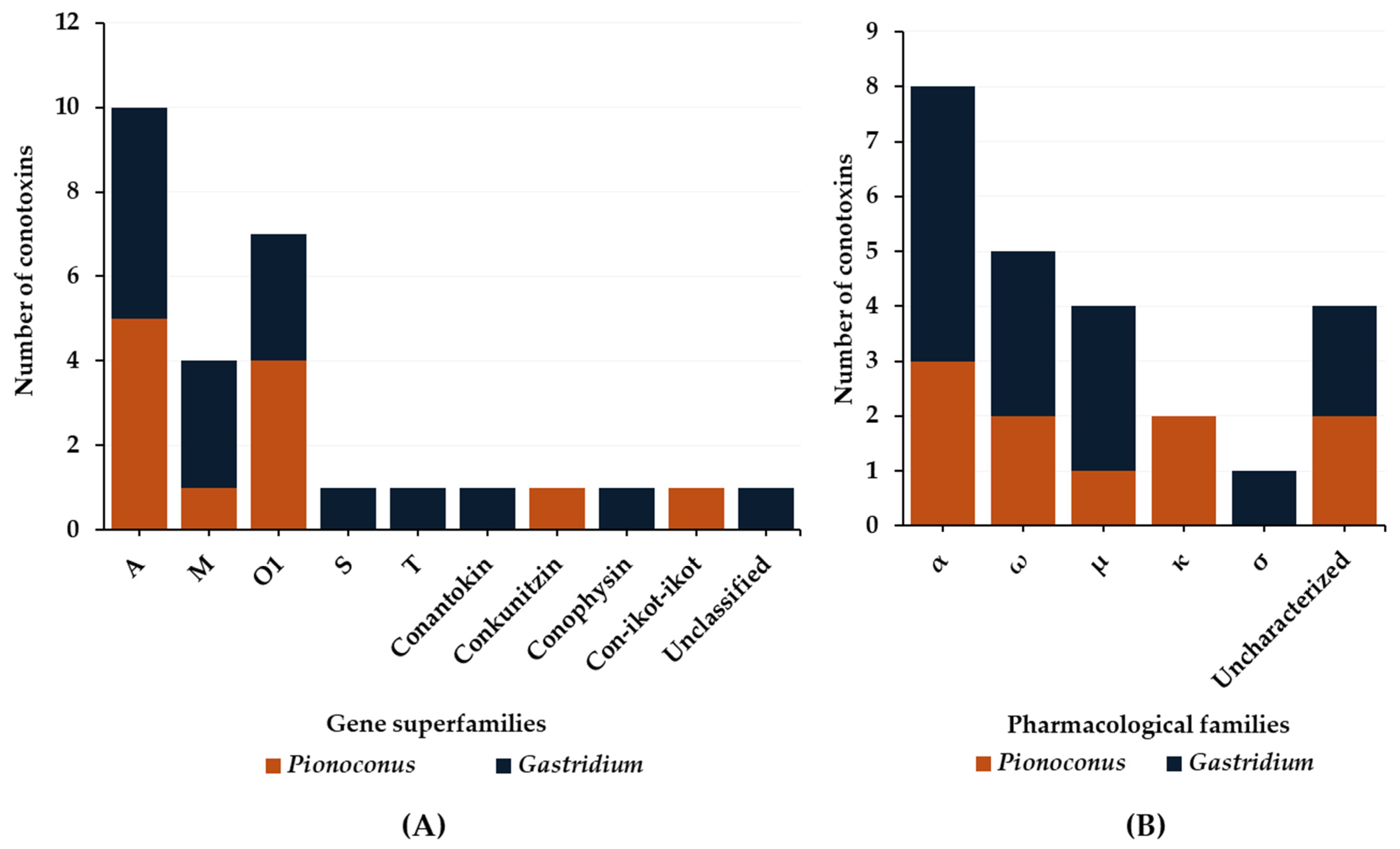
A little over 60 cone snails have been identified as mollusc-hunters, which represents a smaller portion over the fish- and worm-hunting species [31]. It is important to note that some cone snails do not exclusively prey on a single type of mollusc prey, but have adapted to more than one [31]. Although a complete genome assembly from mollusc-hunters is still lacking, it is fairly possible that mollusc-hunting cone snails have originated from a single root in opposition to piscivorous cone snails, which have been shown to be polytopic [31]. From Puillandre et al.’s phylogenetic classification, these mollusc-eating cone snails have been classified into six different clades: Calibanus, Conus, Cylinder, Darioconus, Eugeniconus, and Leptoconus [2].
The Cylinder and Conus clades represent the most studied in general, as they regroup some of the larger cone snails, such as the common Cylinder textile and Conus marmoreus. Fewer data are found in comparison to the extensively studied piscivorous cone snails, and even less on the predatory and defensive differentiation of venom for mollusc-hunters, limited so far to three species from the Cylinder clade (ammiralis, textile, and victoriae), as well as one from the Conus clades (marmoreus) (Figure 10).
Compared to piscivorous species, the predatory-evoked venom of mollusc-hunters shows high complexity. Analyses of Conus and Cylinder species have permitted the identification of 17 gene superfamilies, 7 conopeptide classes, and a few unclassified toxins (Figure 11). Within this high diversity of conotoxins injected, M-, O1, T, and O2 superfamilies dominate the predatory venoms in general, followed by I1, A, I2, and H superfamilies. Additionally, a few conopeptide classes, such as insulins, contryphans, conophysins/conopressins, and conorfamides, were also identified, as well as less common classes, such as conomarphins, elevin, and even prohormones (Figure 11).
Although the venom of a single cone snail from the Conus clade was characterized (Conus marmoreus), it provided almost half of the knowledge gathered on the predatory strategy of mollusc-hunters. Indeed, the Conus clade predatory venom comprises 14 out of a total of 24 gene superfamilies. Meanwhile, the 3 Cylinder cone snails together yielded only slightly more diversity in their predatory venoms, with 20 gene superfamilies. From these, two gene superfamilies and types of conopeptides are found exclusively in the predatory venom of Conus marmoreus (respectively, B2 and I2, and conomarphins and contryphans), while Cylinder cone snails present exclusively five gene superfamilies and five types of conopeptide (F, P, R, elevin, and prohormone in Cylinder victoriae [28,78,79], and I4, J, conophysin/conopressin, conorfamide, and insulin in Cylinder ammiralis [80]) (Figure 11).
Superfamily A conotoxins are found in both molluscivorous clades investigated but they appear not as prevalent as in the piscivorous cone snails (Table 8). Some of these conotoxins have a similar structure and function as the α-conotoxins from fish-hunters, such as α-Mr1.1 (Conus marmoreus) [81] and α-VcIA (Cylinder victoriae) [28]. A very recent proteomic study has been conducted to uncover the predatory and defensive venoms of Cylinder ammiralis, which allowed the characterization of new sets of conotoxins and conopeptides, with some still unknown [80]. The predatory venom of Cylinder ammiralis shows α-like conotoxins, Ai1.2 and Ai1.2, but they have not been characterized yet [80].
As mentioned, M-conotoxins are of the most abundant in predatory venoms as well as O1- and T-conotoxins (Figure 11). M-conotoxins (Table 9), such as MrIIIB (Conus marmoreus) and TxIIIC (Cylinder textile), seemed to induce several symptoms in mice upon intracranial injection, such as scratching, hyperactivity, and circular motion [82], while Mr1e induced excitatory effects [83]. However, it is unknown if these effects reported in mammalian animal models can be translated to mollusc prey physiology.
O1-Conotoxins are also highly produced in the predatory venom of both clades. Most of them uphold the classical VI/VII framework (Table 10). Among them, we find μ-conotoxins such as μ-MrVIA and μ-MrVIB [81] from Conus marmoreus, which block Na+ currents of VGSCs [84]. The sequence homology can be clearly seen between M-conotoxins produced by the different species of mollusk-hunters, for example between TxO1 (Cylinder textile) [16] and P_019 (Cylinder ammiralis), with this conservation indicating a likely important ecological role [80].
A few O2-conotoxins were identified from the predatory venom of both Cylinder ammiralis and Conus marmoreus. These include a majority of almost 30 amino-acid long conotoxins, upholding a VI/VII cysteine framework. In addition, a contryphan-like conopeptide (P_163) was also identified in the venom of Cylinder ammiralis (Table 11) [80].
One other major family identified in the predation of molluscivorous cone snails is the T superfamily, which is absent from piscivorous species (Table 12). The T-conotoxins are smaller peptides that uphold a V or X framework made of four cysteine residues. Only a few χ-conotoxins have been identified, some of which inhibit the antidepressant binding site of the NE transporter (dependent on Na+), such as χ-MrIA [81], in Conus marmoreus. Interestingly, χ-MrIA was identified as an analgesic, efficacious in managing neuropathic pain in mice experiments [81].
Finally, in addition to the conotoxins, a few conopeptides were also identified, which include contryphans, conomarphins, a conoporin, a conorfamide, conopressin/conophysin, elevin, prohormone, insulin, and other unidentified toxins (Table 13). The contryphan-M was identified as having similar activity to ω-conotoxins [54,85].
Pharmacological characterization of conotoxins from mollusc-hunting cone snails remains far behind compared to piscivorous species, rendering the interpretation of their predatory strategies more difficult. Nonetheless, a few have been characterized, such as α-conotoxins α-Mr1.1 [80] and α-VcIA [28], μO-conotoxins such as μ-MrVIA and μ-MrVIB, and the δ-conotoxin, δ-TxVIA (Table 10). Unusual pharmacological families are also characterized in some predatory venoms, such as χ- and ε-conotoxins, including χ-MrIA [80], χ-MrIA [81], χ/λ-CMrX [82], and ε-TxVA [83] from the T-superfamily (Table 12), as well as the χ/λ-CMrVIA [82] and the ω-contryphan M [80] (Table 13).

3.2. Defense Venom

The defensive venom of molluscivorous cone snails has only been described for three species: Conus marmoreus, Cylinder ammiralis, and Cylinder victoriae. Similar to the predatory venoms, defensive venoms are complex, and a high diversity of conotoxin gene superfamilies are detected. A total of 21 gene superfamilies were identified: A, H, F, I1, I2, J, M, N, O1, O2, P, R, S, T, U, contryphan, conoporin, conodipine, conopressin/conophysin, conorfamide, and prohormones (Figure 12). The majority of the conotoxins recovered belong to M, O1, O2, and T superfamilies, which is reminiscent of the predatory venoms, although the nature of the individual conotoxin sequences is often different (Figure 11).
M-Conotoxins included a few toxins that were used in both predatory and defense venoms (Table 9), such as Mr1e, Mr3.8, MrIIIB, MrIIID, MrIIIE, MrIIIF, and MrIIIG in Conus marmoreus, as well as P_147 and P_063 in Cylinder ammiralis (Table 9). In the same way, O1- and T-conotoxins included toxins that are shared in both strategies, such as the μO-conotoxins, μ-MrVIA and μ-MrVIB, and VcVIB (Table 10), and the T-conotoxins, χ-MrIA and VcVA (Table 12). Some O2-conotoxins, only identified from the predatory venom of Cylinder ammiralis, included contryphan-like toxins, such as D_054 and P_163, which show similarities with contryphan-M (Table 11) [80]. Conopeptides are also found in the defensive venom, which include contryphan-M, and conoporin, conorfamide, conodipine, conopressin/conophysin, and prohormones (Table 13).

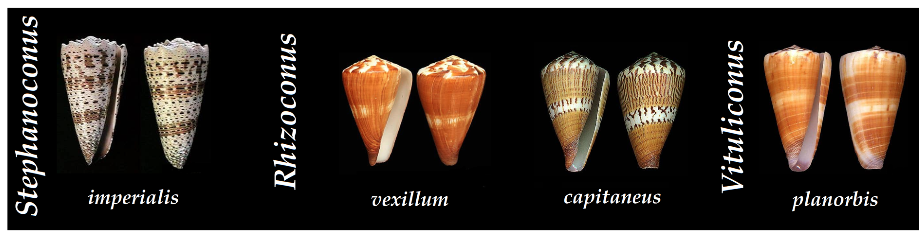
4. Vermivorous Cone Snails

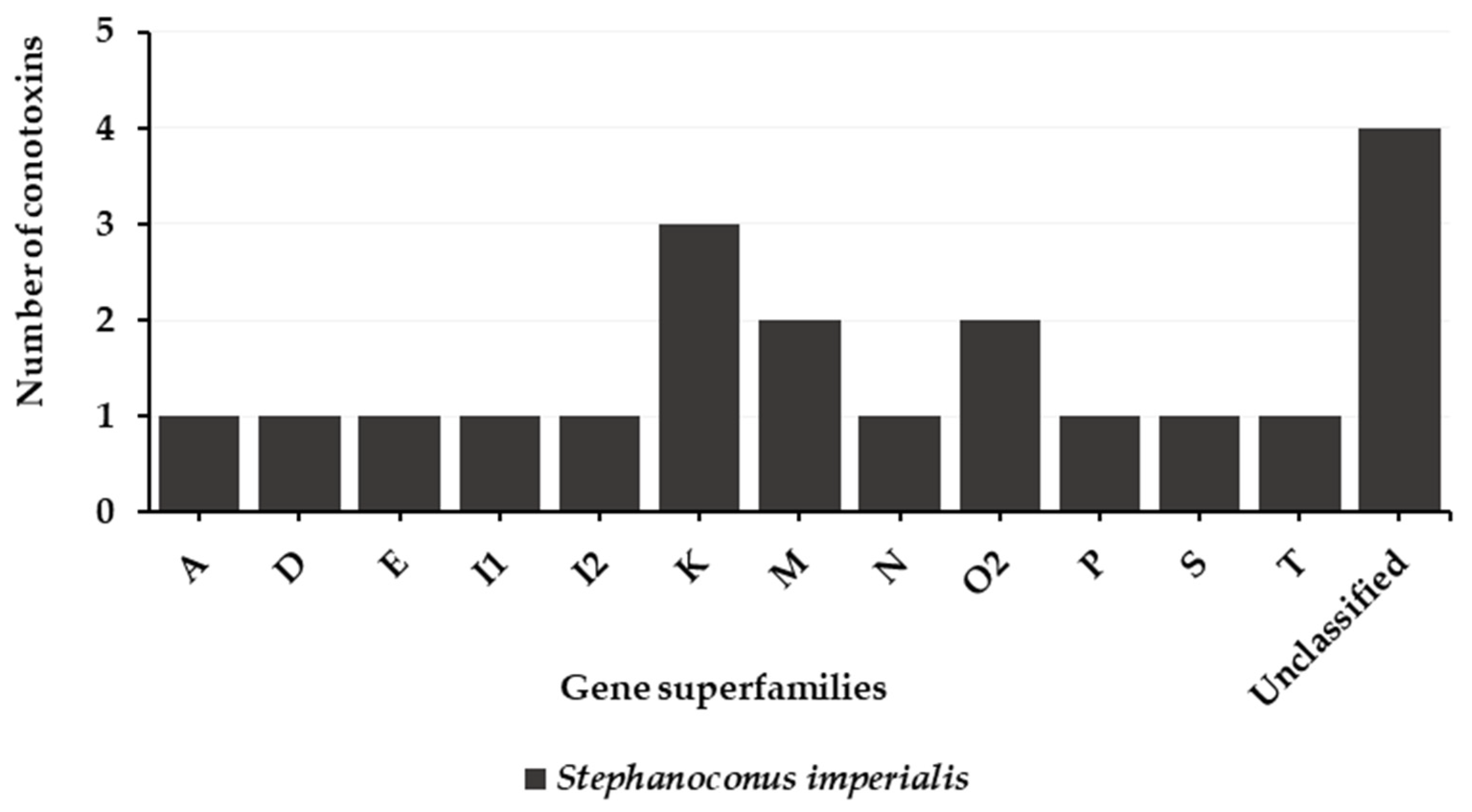
4.1. Predatory Venom

While they present the highest number of species within the Conidae, vermivorous cone snails have the least amount of published data on their predatory and defensive venoms. Most likely because of the difficulty in collecting the injected venom (small radula tooth, specific type of prey worms, etc.), the investigation of vermivorous cone snails is lagging behind [31]. Consequently, this lack of investigation impacted the study of their predatory and defensive venoms. A few cone snails have been studied with that intent, such as the Stephanoconus/Rhombiconus cone snail, Stephanoconus imperialis [9], as well as two Rhizoconus cone snails, Rhizoconus vexillum and Rhizoconus capitaneus [86], and a single Vituliconus cone, Vituliconus planorbis [87] (Figure 13).
So far, from the worm-hunters, only the predatory venom of Stephanoconus imperialis could be collected. Its analysis has revealed a moderately complex venom, which presents 12 gene superfamilies (Figure 14) [9]. Interestingly, we observe conotoxins with different types of cysteine frameworks, which differ from the other types of cone snails (Table 14). Overall, the K-conotoxins were the most abundant in the venom of Stephanoconus imperialis. Indeed, three K-conotoxins, Im23a, Im23b, and Im23.4, were identified with a XXIII framework and a structure containing two helices [9]. Their biological activity is unknown. An α-conotoxin, Im1.1, was also identified.

4.2. Defense Venom

Defense venoms have been recorded from vermivorous Rhizoconus and Vituliconus cone snails (Figure 15). First, defense venom of the Vituliconus cone snail, Vituliconus planorbis, presented a set of A-, J-, M-, O2-, T-, Y-, and a new superfamily-1 (NSf-1). The most represented were NSf-1, A, and M gene superfamilies (Table 14) [87]. Rhizoconus worm-hunting cone snails appear to use massively and almost exclusively homodimeric αD-conotoxins for their defense strategies (Table 15). Indeed, αD-conotoxins, presenting 10 cysteines arranged in a unique XX framework, were discovered in the defensive-evoked venom of both Rhizoconus vexillum and Rhizoconus capitaneus [86]. They can be detected in their dimeric form between 10 and 12 kDa. Similar to some defense-related αA-conotoxins, they potently inhibit the α7 nAChR subtype in mammalian assays [86], but more information is needed to understand their defensive role [86].

5. Discussion

When first discovered, the remarkable ability of cone snails to produce different venoms for specific purposes (predation or defense) was totally unexpected. For the first time, within the same animal, was shown that a defensive injection was not simply equivalent to a predatory sting, and vice versa. While this novel paradigm would have immediate implications for the management of envenomation victims for instance, or for our understanding of cone snail biology, it also argues for the future need to characterize individual conotoxins on the correct animal model to avoid erroneous conclusions on their true ecological role. In the case of vertebrate venomous animals, such as snakes, both preys (often small vertebrate mammals, but also reptiles and birds) and predators (mostly higher vertebrates) share a very conserved physiology, explaining how by a fortunate coincidence, the same toxins would be effective both in capturing prey and in defending against a predator [88]. To the contrary, cone snails had to deal with more complicated venom uses (very diverse vertebrate/invertebrate preys and predators), stimulating the evolution of distinct strategies [89]. We, therefore, encourage future studies to investigate predatory venoms on laboratory animals more closely related to prey types, such as zebrafish (piscivorous), Lymnaea or Aplysia snails (molluscivorous), and any worms (vermivorous), including Caenorhabditis elegans, although annelids would be preferable over nematodes.
The controlled and selective injection of different conotoxin cocktails suggest that the evolution of cone snail venom is not only under the direct influence of one but at least two, potentially equally important, driving forces: predation and defense. Therefore, it provides a unique opportunity to study venom–ecology relationships in unprecedented details, on the condition that both predatory and defensive venoms can be collected and analyzed separately. From the current literature, it appears that injected venom can theoretically be collected from all cone snail diet groups. However, this review exposes a clear bias toward fish-hunting species in comparison to the other feeding groups. This bias likely has to do with the ease of “milking”, as piscivorous species tend to use the large and strong radula tooth, which facilitates the collection procedure. In addition, a fish prey can be conveniently obtained in most laboratories, as piscivorous cone snails will happily accept freshwater fish (goldfish) or zebrafish (with the caveat of ethical considerations around using vertebrate animals). Molluscivorous species also use a large radula tooth, which are thinner and more flexible, rendering the milking more technical but still relatively simple. The real challenge comes with the tiny radula tooth of most worm-hunting species (often <1 mm), which rarely pierces through both the prey tissue and the parafilm for successful milking (personal experiences).
An important note relates to the description of injected venoms prior to 2014 and the knowledge about distinct predatory and defensive venoms in cone snails. Indeed, some of the variations observed in venom composition in “milked venoms” could be a posteriori attributed to the non-discrimination between predatory and defensive behaviors. Another area of discrepancy concerns the study of individual vs. pooled injected venoms. From personal experience, pooling large batches of injected venom could also “falsely” increase venom complexity (accumulating all subtle individual variations in one complex lot) and, more importantly, could also lead to inadvertent mixing of defensive and predatory venoms. Therefore, for pharmacological characterization of conotoxins, larger amounts of venom are required, and pooling can be acceptable in this case, but investigation of precise envenomation strategies should be limited to individual milking. Lastly, the use of proteomic softwares that allow the automated interpretation of MS/MS spectra and identification of peptides and proteins from sequence databases may generate substantial amounts of false positives if not carefully curated [90]. Therefore, these could also artificially inflate the number of conotoxin sequences identified in injected venoms.
With these considerations and limitations in mind, a high diversity of conotoxin gene superfamilies was identified in the composition of predatory and defensive venoms to date (summarized in Figure 16). However, it is puzzling that particular species can inject a very restricted set of conotoxins selected from the complex repertoire present in their venom duct. For instance, the piscivorous species of the Pionoconus clade often rely on a very simple predatory venom composition to subdue fish prey, with sometimes only one class of conotoxin injected (κA-conotoxins). Similarly, some vermivorous species of the Rhizoconus clade have evolved a defensive strategy almost exclusively centered around the injection of αD-conotoxins. The conotoxin selection mechanisms are not understood, but likely under the control of the nervous and/or hormonal systems. Importantly, some species from more basal clades show a non-differentiated venom duct, suggesting that they may not have the ability to produce two types of venom, although this remains to be experimentally demonstrated [91].
Ideally, further works on cone snails should systematically include the characterization of predatory and defense venoms when possible. The more data collected on different clades and diet types, the closer we will come to truly understand the intended evolved venom use and the ecological role of each conotoxin. For instance, no data are available for the predatory venom of any Textilia species, which may seem surprising, considering the piscivorous diet, large radula tooth, and relatively wide geographical distribution of some species in this clade, such as Textilia bullatus. Moreover, the characterization of the defense venoms is still lacking for many cone snail clades, especially for common and large molluscivorous, such as Cylinder textile, episcopatus, aulicus, or canonicus, and other members of the molluscivorous Conus clade (e.g., Conus bandanus or Conus araneosus). Another future line of research should investigate the possible use of small molecules in either predatory or defensive (or both) venoms [92]. Indeed, recent publications have focused on the characterization of non-peptidic components in the venom of cone snails [93]. Some of these small molecules were even ascertained to have a direct role in the prey-capture strategy of a vermivorous species on the basis of their structural and functional resemblance to polychaete mating pheromones [94]. However, critically, the role of small molecules in the ecology of cone snails can only be considered relative to their presence (or absence) in the injected venoms.
With the advent of more and more sophisticated tools to investigate venom, including those in the field of mass spectrometry, peptide synthesis, and high-throughput sequencing and screenings, novel details about the predatory and defensive strategies in cone snails are likely to be revealed in the near future [95]. This has also allowed the uprising of multi-omics strategies known as “venomics” by integrating genomics, proteomics, and transcriptomics to accelerate the characterization of complex venoms [56]. For instance, it might be possible to test the hypothesis of the release of venom components in the water in the case of piscivorous net-hunting species. Furthermore, new AI (artificial intelligence)-based bioinformatic tools may also provide assistance in the interpretation of “big data”, in visualizing conotoxin–receptor interactions at atomic levels, or in developing novel hypotheses. However, the main challenge will remain uncovering the pharmacology of conotoxins through prey- and predator-relevant bioassays.

Supplementary Materials

The following supporting information can be downloaded at: https://www.mdpi.com/article/10.3390/toxins16020094/s1, Table S1: Predatory/defensive venom.

Author Contributions

Z.R.; writing—original draft preparation, S.D. and N.I.; writing—review and editing, S.D. and N.I.; supervision, S.D. and N.I.; project administration, S.D. and N.I.; funding acquisition, S.D. and N.I. All authors have read and agreed to the published version of the manuscript.

Funding

This research was funded by the Departmental Council of Mayotte through the projects VENCOME and “Chercheuse d’avenir”, promoting Mahoran women in research (PhD thesis funding for Z.R.). Contribution to the funding: Dhahabia Chanfi (direction of school politics and universities) and Fahoulia Mohamadi (representative of the research and innovation at the teaching authority, ‘rectorat de Mayotte’).

Institutional Review Board Statement

Not applicable.

Informed Consent Statement

Not applicable.

Data Availability Statement

All data used in this study were retrieved from publicly available databases.

Conflicts of Interest

The authors declare no conflicts of interest.

References

  1. Kohn, A.J. Microhabitats, Abundance and Food of Conus on Atoll Reefs in the Maldive and Chagos Islands. Ecology 1968, 49, 1046–1062. [Google Scholar] [CrossRef]
  2. Puillandre, N.; Duda, T.F.; Meyer, C.; Olivera, B.M.; Bouchet, P. One, Four or 100 Genera? A New Classification of the Cone Snails. J. Molluscan Stud. 2015, 81, 1–23. [Google Scholar] [CrossRef] [PubMed]
  3. Gao, B.; Peng, C.; Yang, J.; Yi, Y.; Zhang, J.; Shi, Q. Cone Snails: A Big Store of Conotoxins for Novel Drug Discovery. Toxins 2017, 9, 397. [Google Scholar] [CrossRef]
  4. Olivera, B.M. “Conus” Venom Peptides: Reflections from the Biology of Clades and Species. Annu. Rev. Ecol. Syst. 2002, 33, 25–47. [Google Scholar] [CrossRef]
  5. Akondi, K.B.; Muttenthaler, M.; Dutertre, S.; Kaas, Q.; Craik, D.J.; Lewis, R.J.; Alewood, P.F. Discovery, Synthesis, and Structure–Activity Relationships of Conotoxins. Chem. Rev. 2014, 114, 5815–5847. [Google Scholar] [CrossRef] [PubMed]
  6. Puillandre, N.; Koua, D.; Favreau, P.; Olivera, B.M.; Stöcklin, R. Molecular Phylogeny, Classification and Evolution of Conopeptides. J. Mol. Evol. 2012, 74, 297–309. [Google Scholar] [CrossRef] [PubMed]
  7. Lebbe, E.K.M.; Tytgat, J. In the Picture: Disulfide-Poor Conopeptides, a Class of Pharmacologically Interesting Compounds. J. Venom. Anim. Toxins Trop. Dis. 2016, 22, 30. [Google Scholar] [CrossRef]
  8. Kaas, Q.; Westermann, J.-C.; Craik, D.J. Conopeptide Characterization and Classifications: An Analysis Using ConoServer. Toxicon 2010, 55, 1491–1509. [Google Scholar] [CrossRef]
  9. Jin, A.-H.; Dutertre, S.; Dutt, M.; Lavergne, V.; Jones, A.; Lewis, R.; Alewood, P. Transcriptomic-Proteomic Correlation in the Predation-Evoked Venom of the Cone Snail, Conus Imperialis. Mar. Drugs 2019, 17, 177. [Google Scholar] [CrossRef]
  10. Jin, A.-H.; Muttenthaler, M.; Dutertre, S.; Himaya, S.W.A.; Kaas, Q.; Craik, D.J.; Lewis, R.J.; Alewood, P.F. Conotoxins: Chemistry and Biology. Chem. Rev. 2019, 119, 11510–11549. [Google Scholar] [CrossRef]
  11. Kohn, A.J.; Nishi, M.; Pernet, B. Snail Spears and Scimitars: A Character Analysis of Conus Radular Teeth. J. Molluscan Stud. 1999, 65, 461–481. [Google Scholar] [CrossRef]
  12. Kohn, A.J. Piscivorous Gastropods of the Genus Conus. Proc. Natl. Acad. Sci. USA 1956, 42, 168–171. [Google Scholar] [CrossRef] [PubMed]
  13. Olivera, B.M.; Seger, J.; Horvath, M.P.; Fedosov, A.E. Prey-Capture Strategies of Fish-Hunting Cone Snails: Behavior, Neurobiology and Evolution. Brain Behav. Evol. 2015, 86, 58–74. [Google Scholar] [CrossRef] [PubMed]
  14. Kohn, M. Biology of Conus on Shores of the Dampier Archipelago, Northwestern Australia; University of Washington Seattle: Seattle, WA, USA, 2003. [Google Scholar]
  15. Salisbury, S.M.; Martin, G.G.; Kier, W.M.; Schulz, J.R. Venom Kinematics during Prey Capture in Conus: The Biomechanics of a Rapid Injection System. J. Exp. Biol. 2010, 213, 673–682. [Google Scholar] [CrossRef] [PubMed]
  16. Prator, C.A.; Murayama, K.M.; Schulz, J.R. Venom Variation during Prey Capture by the Cone Snail, Conus textile. PLoS ONE 2014, 9, e98991. [Google Scholar] [CrossRef] [PubMed]
  17. Himaya, S.W.A.; Marí, F.; Lewis, R.J. Accelerated Proteomic Visualization of Individual Predatory Venoms of Conus Purpurascens Reveals Separately Evolved Predation-Evoked Venom Cabals. Sci. Rep. 2018, 8, 330. [Google Scholar] [CrossRef] [PubMed]
  18. Abalde, S.; Tenorio, M.J.; Afonso, C.M.L.; Zardoya, R. Conotoxin Diversity in Chelyconus ermineus (Born, 1778) and the Convergent Origin of Piscivory in the Atlantic and Indo-Pacific Cones. Genome Biol. Evol. 2018, 10, 2643–2662. [Google Scholar] [CrossRef] [PubMed]
  19. Hu, H.; Bandyopadhyay, P.K.; Olivera, B.M.; Yandell, M. Elucidation of the Molecular Envenomation Strategy of the Cone Snail Conus Geographus through Transcriptome Sequencing of Its Venom Duct. BMC Genom. 2012, 13, 284. [Google Scholar] [CrossRef]
  20. Safavi-Hemami, H.; Gajewiak, J.; Karanth, S.; Robinson, S.D.; Ueberheide, B.; Douglass, A.D.; Schlegel, A.; Imperial, J.S.; Watkins, M.; Bandyopadhyay, P.K.; et al. Specialized Insulin Is Used for Chemical Warfare by Fish-Hunting Cone Snails. Proc. Natl. Acad. Sci. USA 2015, 112, 1743–1748. [Google Scholar] [CrossRef]
  21. Robinson, S.D.; Li, Q.; Bandyopadhyay, P.K.; Gajewiak, J.; Yandell, M.; Papenfuss, A.T.; Purcell, A.W.; Norton, R.S.; Safavi-Hemami, H. Hormone-like Peptides in the Venoms of Marine Cone Snails. Gen. Comp. Endocrinol. 2017, 244, 11–18. [Google Scholar] [CrossRef]
  22. Hopkins, C.; Grilley, M.; Miller, C.; Shon, K.-J.; Cruz, L.J.; Gray, W.R.; Dykert, J.; Rivier, J.; Yoshikami, D.; Olivera, B.M. A New Family of Conus Peptides Targeted to the Nicotinic Acetylcholine Receptor (*). J. Biol. Chem. 1995, 270, 22361–22367. [Google Scholar] [CrossRef]
  23. Jakubowski, J.A.; Kelley, W.P.; Sweedler, J.V.; Gilly, W.F.; Schulz, J.R. Intraspecific Variation of Venom Injected by Fish-Hunting Conus Snails. J. Exp. Biol. 2005, 208, 2873–2883. [Google Scholar] [CrossRef]
  24. Dutertre, S.; Biass, D.; Stöcklin, R.; Favreau, P. Dramatic Intraspecimen Variations within the Injected Venom of Conus consors: An Unsuspected Contribution to Venom Diversity. Toxicon 2010, 55, 1453–1462. [Google Scholar] [CrossRef]
  25. Himaya, S.W.A.; Jin, A.-H.; Dutertre, S.; Giacomotto, J.; Mohialdeen, H.; Vetter, I.; Alewood, P.F.; Lewis, R.J. Comparative Venomics Reveals the Complex Prey Capture Strategy of the Piscivorous Cone Snail Conus catus. J. Proteome Res. 2015, 14, 4372–4381. [Google Scholar] [CrossRef]
  26. Kelley, W.P.; Schulz, J.R.; Jakubowski, J.A.; Gilly, W.F.; Sweedler, J.V. Two Toxins from Conus striatus That Individually Induce Tetanic Paralysis. Biochemistry 2006, 45, 14212–14222. [Google Scholar] [CrossRef]
  27. Le Gall, F.; Favreau, P.; Benoit, E.; Mattei, C.; Bouet, F.; Menou, J.-L.; Ménez, A.; Letourneux, Y.; Molgó, J. A New Conotoxin Isolated from Conus Consors Venom Acting Selectively on Axons and Motor Nerve Terminals through a Na+-Dependent Mechanism. Eur. J. Neurosci. 1999, 11, 3134–3142. [Google Scholar] [CrossRef]
  28. Dutertre, S.; Jin, A.-H.; Vetter, I.; Hamilton, B.; Sunagar, K.; Lavergne, V.; Dutertre, V.; Fry, B.G.; Antunes, A.; Venter, D.J.; et al. Evolution of Separate Predation- and Defence-Evoked Venoms in Carnivorous Cone Snails. Nat. Commun. 2014, 5, 3521. [Google Scholar] [CrossRef]
  29. Kohn, A.J. Conus Envenomation of Humans: In Fact and Fiction. Toxins 2019, 11, 10. [Google Scholar] [CrossRef]
  30. Casewell, N.R.; Wüster, W.; Vonk, F.J.; Harrison, R.A.; Fry, B.G. Complex Cocktails: The Evolutionary Novelty of Venoms. Trends Ecol. Evol. 2013, 28, 219–229. [Google Scholar] [CrossRef]
  31. Zhao, Y.; Antunes, A. Biomedical Potential of the Neglected Molluscivorous and Vermivorous Conus Species. Mar. Drugs 2022, 20, 105. [Google Scholar] [CrossRef]
  32. Himaya, S.W.A.; Jin, A.-H.; Hamilton, B.; Rai, S.K.; Alewood, P.; Lewis, R.J. Venom Duct Origins of Prey Capture and Defensive Conotoxins in Piscivorous Conus striatus. Sci. Rep. 2021, 11, 13282. [Google Scholar] [CrossRef]
  33. Dutertre, S.; Jin, A.-H.; Alewood, P.F.; Lewis, R.J. Intraspecific Variations in Conus geographus Defence-Evoked Venom and Estimation of the Human Lethal Dose. Toxicon 2014, 91, 135–144. [Google Scholar] [CrossRef]
  34. Shells For Sale|The Largest Official Seashells Website|Conchology. Available online: https://www.conchology.be/ (accessed on 18 July 2023).
  35. Gajewiak, J.; Azam, L.; Imperial, J.; Walewska, A.; Green, B.R.; Bandyopadhyay, P.K.; Raghuraman, S.; Ueberheide, B.; Bern, M.; Zhou, H.M.; et al. A Disulfide Tether Stabilizes the Block of Sodium Channels by the Conotoxin μO§-GVIIJ. Proc. Natl. Acad. Sci. USA 2014, 111, 2758–2763. [Google Scholar] [CrossRef]
  36. Teichert, R.W.; Jacobsen, R.; Terlau, H.; Yoshikami, D.; Olivera, B.M. Discovery and Characterization of the Short κA-Conotoxins: A Novel Subfamily of Excitatory Conotoxins. Toxicon 2007, 49, 318–328. [Google Scholar] [CrossRef]
  37. Biass, D.; Dutertre, S.; Gerbault, A.; Menou, J.-L.; Offord, R.; Favreau, P.; Stöcklin, R. Comparative Proteomic Study of the Venom of the Piscivorous Cone Snail Conus consors. J. Proteom. 2009, 72, 210–218. [Google Scholar] [CrossRef]
  38. Violette, A.; Biass, D.; Dutertre, S.; Koua, D.; Piquemal, D.; Pierrat, F.; Stöcklin, R.; Favreau, P. Large-Scale Discovery of Conopeptides and Conoproteins in the Injectable Venom of a Fish-Hunting Cone Snail Using a Combined Proteomic and Transcriptomic Approach. J. Proteom. 2012, 75, 5215–5225. [Google Scholar] [CrossRef]
  39. Kapono, C.A.; Thapa, P.; Cabalteja, C.C.; Guendisch, D.; Collier, A.C.; Bingham, J.-P. Conotoxin Truncation as a Post-Translational Modification to Increase the Pharmacological Diversity within the Milked Venom of Conus magus. Toxicon 2013, 70, 170–178. [Google Scholar] [CrossRef]
  40. Saintmont, F.; Cazals, G.; Bich, C.; Dutertre, S. Proteomic Analysis of the Predatory Venom of Conus striatus Reveals Novel and Population-Specific κA-Conotoxin SIVC. Toxins 2022, 14, 799. [Google Scholar] [CrossRef]
  41. Teichert, R.W.; Rivier, J.; Dykert, J.; Cervini, L.; Gulyas, J.; Bulaj, G.; Ellison, M.; Olivera, B.M. αA-Conotoxin OIVA Defines a New αA-Conotoxin Subfamily of Nicotinic Acetylcholine Receptor Inhibitors. Toxicon 2004, 44, 207–214. [Google Scholar] [CrossRef]
  42. Ramilo, C.A.; Zafaralla, G.C.; Nadasdi, L.; Hammerland, L.G.; Yoshikami, D.; Gray, W.R.; Kristipati, R.; Ramachandran, J.; Miljanich, G. Novel Alpha- and Omega-Conotoxins and Conus striatus Venom. Biochemistry 1992, 31, 9919–9926. [Google Scholar] [CrossRef]
  43. Biass, D.; Violette, A.; Hulo, N.; Lisacek, F.; Favreau, P.; Stöcklin, R. Uncovering Intense Protein Diversification in a Cone Snail Venom Gland Using an Integrative Venomics Approach. J. Proteome Res. 2015, 14, 628–638. [Google Scholar] [CrossRef] [PubMed]
  44. Quinton, L.; Servent, D.; Girard, E.; Molgó, J.; Le Caer, J.-P.; Malosse, C.; Haidar, E.A.; Lecoq, A.; Gilles, N.; Chamot-Rooke, J. Identification and Functional Characterization of a Novel α-Conotoxin (EIIA) from Conus ermineus. Anal. Bioanal. Chem. 2013, 405, 5341–5351. [Google Scholar] [CrossRef] [PubMed]
  45. Terlau, H.; Olivera, B.M. Conus Venoms: A Rich Source of Novel Ion Channel-Targeted Peptides. Physiol. Rev. 2004, 84, 41–68. [Google Scholar] [CrossRef] [PubMed]
  46. López-Vera, E.; Jacobsen, R.B.; Ellison, M.; Olivera, B.M.; Teichert, R.W. A Novel Alpha Conotoxin (α-PIB) Isolated from C. purpurascens Is Selective for Skeletal Muscle Nicotinic Acetylcholine Receptors. Toxicon 2007, 49, 1193–1199. [Google Scholar] [CrossRef] [PubMed]
  47. Teichert, R.W.; Rivier, J.; Torres, J.; Dykert, J.; Miller, C.; Olivera, B.M. A Uniquely Selective Inhibitor of the Mammalian Fetal Neuromuscular Nicotinic Acetylcholine Receptor. J. Neurosci. 2005, 25, 732–736. [Google Scholar] [CrossRef] [PubMed]
  48. Hoggard, M.F.; Rodriguez, A.M.; Cano, H.; Clark, E.; Tae, H.-S.; Adams, D.J.; Godenschwege, T.A.; Marí, F. In Vivo and In Vitro Testing of Native α-Conotoxins from the Injected Venom of Conus purpurascens. Neuropharmacology 2017, 127, 253–259. [Google Scholar] [CrossRef] [PubMed]
  49. Van Wagoner, R.M.; Jacobsen, R.B.; Olivera, B.M.; Ireland, C.M. Characterization and Three-Dimensional Structure Determination of ψ-Conotoxin P IIIF, a Novel Noncompetitive Antagonist of Nicotinic Acetylcholine Receptors. Biochemistry 2003, 42, 6353–6362. [Google Scholar] [CrossRef]
  50. Jacobsen, R.; Yoshikami, D.; Ellison, M.; Martinez, J.; Gray, W.R.; Cartier, G.E.; Shon, K.-J.; Groebe, D.R.; Abramson, S.N.; Olivera, B.M.; et al. Differential Targeting of Nicotinic Acetylcholine Receptors by Novel αA-Conotoxins. J. Biol. Chem. 1997, 272, 22531–22537. [Google Scholar] [CrossRef]
  51. Rivera-Ortiz, J.A.; Cano, H.; Marí, F. Intraspecies Variability and Conopeptide Profiling of the Injected Venom of Conus ermineus. Peptides 2011, 32, 306–316. [Google Scholar] [CrossRef]
  52. Echterbille, J.; Gilles, N.; Araóz, R.; Mourier, G.; Amar, M.; Servent, D.; De Pauw, E.; Quinton, L. Discovery and Characterization of EIIB, a New α-Conotoxin from Conus Ermineus Venom by nAChRs Affinity Capture Monitored by MALDI-TOF/TOF Mass Spectrometry. Toxicon 2017, 130, 1–10. [Google Scholar] [CrossRef]
  53. Martinez, J.S.; Olivera, B.M.; Gray, W.R.; Craig, A.G.; Groebe, D.R.; Abramson, S.N.; McIntosh, J.M. Alpha-Conotoxin EI, A New Nicotinic Acetylcholine Receptor Antagonist with Novel Selectivity. Biochemistry 1995, 34, 14519–14526. [Google Scholar] [CrossRef]
  54. Robinson, S.D.; Norton, R.S. Conotoxin Gene Superfamilies. Mar. Drugs 2014, 12, 6058–6101. [Google Scholar] [CrossRef]
  55. Shon, K.-J.; Grilley, M.M.; Marsh, M.; Yoshikami, D.; Hall, A.R.; Kurz, B.; Gray, W.R.; Imperial, J.S.; Hillyard, D.R.; Olivera, B.M. Purification, Characterization, Synthesis, and Cloning of the Lockjaw Peptide from Conus Purpurascens Venom. Biochemistry 1995, 34, 4913–4918. [Google Scholar] [CrossRef]
  56. Himaya, S.; Lewis, R. Venomics-Accelerated Cone Snail Venom Peptide Discovery. Int. J. Mol. Sci. 2018, 19, 788. [Google Scholar] [CrossRef] [PubMed]
  57. Yanagawa, Y.; Abe, T.; Satake, M.; Odani, S.; Suzuki, J.; Ishikawa, K. A Novel Sodium Channel Inhibitor from Conus geographus: Purification, Structure, and Pharmacological Properties. Biochemistry 1988, 27, 6256–6262. [Google Scholar] [CrossRef] [PubMed]
  58. Del Río-Sancho, S.; Cros, C.; Coutaz, B.; Cuendet, M.; Kalia, Y.N. Cutaneous Iontophoresis of μ-Conotoxin CnIIIC-A Potent NaV1.4 Antagonist with Analgesic, Anaesthetic and Myorelaxant Properties. Int. J. Pharm. 2017, 518, 59–65. [Google Scholar] [CrossRef]
  59. Zhang, M.-M.; Fiedler, B.; Green, B.R.; Catlin, P.; Watkins, M.; Garrett, J.E.; Smith, B.J.; Yoshikami, D.; Olivera, B.M.; Bulaj, G. Structural and Functional Diversities among μ-Conotoxins Targeting TTX-Resistant Sodium Channels. Biochemistry 2006, 45, 3723–3732. [Google Scholar] [CrossRef]
  60. Olivera, B.M.; McIntosh, J.M.; Curz, L.J.; Luque, F.A.; Gray, W.R. Purification and Sequence of a Presynaptic Peptide Toxin from Conus geographus Venom. Biochemistry 1984, 23, 5087–5090. [Google Scholar] [CrossRef]
  61. Lewis, R.J.; Nielsen, K.J.; Craik, D.J.; Loughnan, M.L.; Adams, D.A.; Sharpe, I.A.; Luchian, T.; Adams, D.J.; Bond, T.; Thomas, L.; et al. Novel ω-Conotoxins from Conus Catus Discriminate among Neuronal Calcium Channel Subtypes. J. Biol. Chem. 2000, 275, 35335–35344. [Google Scholar] [CrossRef] [PubMed]
  62. Zhou, K.; Luo, W.; Liu, T.; Ni, Y.; Qin, Z. Neurotoxins Acting at Synaptic Sites: A Brief Review on Mechanisms and Clinical Applications. Toxins 2023, 15, 18. [Google Scholar] [CrossRef]
  63. Safavi-Hemami, H.; Brogan, S.E.; Olivera, B.M. Pain Therapeutics from Cone Snail Venoms: From Ziconotide to Novel Non-Opioid Pathways. J. Proteom. 2019, 190, 12–20. [Google Scholar] [CrossRef]
  64. Zafaralla, G.C.; Ramilo, C.; Gray, W.R.; Karlstrom, R.; Olivera, B.M.; Cruz, L.J. Phylogenetic Specificity of Cholinergic Ligands:.Alpha.-Conotoxin SI. Biochemistry 1988, 27, 7102–7105. [Google Scholar] [CrossRef]
  65. Dutertre, S.; Croker, D.; Daly, N.L.; Andersson, Å.; Muttenthaler, M.; Lumsden, N.G.; Craik, D.J.; Alewood, P.F.; Guillon, G.; Lewis, R.J. Conopressin-T from Conus tulipa Reveals an Antagonist Switch in Vasopressin-like Peptides. J. Biol. Chem. 2008, 283, 7100–7108. [Google Scholar] [CrossRef]
  66. Haack, J.A.; Rivier, J.; Parks, T.N.; Mena, E.E.; Cruz, L.J.; Olivera, B.M. Conantokin-T. A Gamma-Carboxyglutamate Containing Peptide with N-Methyl-d-Aspartate Antagonist Activity. J. Biol. Chem. 1990, 265, 6025–6029. [Google Scholar] [CrossRef]
  67. Craig, A.G.; Norberg, T.; Griffin, D.; Hoeger, C.; Akhtar, M.; Schmidt, K.; Low, W.; Dykert, J.; Richelson, E.; Navarro, V.; et al. Contulakin-G, an O-Glycosylated Invertebrate Neurotensin. J. Biol. Chem. 1999, 274, 13752–13759. [Google Scholar] [CrossRef] [PubMed]
  68. Bayrhuber, M.; Vijayan, V.; Ferber, M.; Graf, R.; Korukottu, J.; Imperial, J.; Garrett, J.E.; Olivera, B.M.; Terlau, H.; Zweckstetter, M.; et al. Conkunitzin-S1 Is the First Member of a New Kunitz-Type Neurotoxin Family. J. Biol. Chem. 2005, 280, 23766–23770. [Google Scholar] [CrossRef] [PubMed]
  69. Möller, C.; Marí, F. 9.3 KDa Components of the Injected Venom of Conus purpurascens Define a New Five-Disulfide Conotoxin Framework. Pept. Sci. 2011, 96, 158–165. [Google Scholar] [CrossRef] [PubMed]
  70. Walker, C.S.; Jensen, S.; Ellison, M.; Matta, J.A.; Lee, W.Y.; Imperial, J.S.; Duclos, N.; Brockie, P.J.; Madsen, D.M.; Isaac, J.T.R.; et al. A Novel Conus Snail Polypeptide Causes Excitotoxicity by Blocking Desensitization of AMPA Receptors. Curr. Biol. 2009, 19, 900–908. [Google Scholar] [CrossRef]
  71. Möller, C.; Clark, E.; Safavi-Hemami, H.; DeCaprio, A.; Marí, F. Isolation and Characterization of Conohyal-P1, a Hyaluronidase from the Injected Venom of Conus purpurascens. J. Proteom. 2017, 164, 73–84. [Google Scholar] [CrossRef] [PubMed]
  72. Möller, C.; Davis, W.C.; Clark, E.; DeCaprio, A.; Marí, F. Conodipine-P1-3, the First Phospholipases A2 Characterized from Injected Cone Snail Venom. Mol. Cell. Proteom. MCP 2019, 18, 876–891. [Google Scholar] [CrossRef] [PubMed]
  73. Möller, C.; Vanderweit, N.; Bubis, J.; Marí, F. Comparative Analysis of Proteases in the Injected and Dissected Venom of Cone Snail Species. Toxicon 2013, 65, 59–67. [Google Scholar] [CrossRef]
  74. Safavi-Hemami, H.; Möller, C.; Marí, F.; Purcell, A.W. High Molecular Weight Components of the Injected Venom of Fish-Hunting Cone Snails Target the Vascular System. J. Proteom. 2013, 91, 97–105. [Google Scholar] [CrossRef]
  75. Nicke, A.; Loughnan, M.L.; Millard, E.L.; Alewood, P.F.; Adams, D.J.; Daly, N.L.; Craik, D.J.; Lewis, R.J. Isolation, Structure, and Activity of GID, a Novel A4/7-Conotoxin with an Extended N-Terminal Sequence. J. Biol. Chem. 2003, 278, 3137–3144. [Google Scholar] [CrossRef] [PubMed]
  76. England, L.J.; Imperial, J.; Jacobsen, R.; Craig, A.G.; Gulyas, J.; Akhtar, M.; Rivier, J.; Julius, D.; Olivera, B.M. Inactivation of a Serotonin-Gated Ion Channel by a Polypeptide Toxin from Marine Snails. Science 1998, 281, 575–578. [Google Scholar] [CrossRef]
  77. Diversity of Conus Neuropeptides. Available online: https://www.science.org/doi/10.1126/science.2165278 (accessed on 14 December 2023).
  78. Jakubowski, J.A.; Sweedler, J.V. Sequencing and Mass Profiling Highly Modified Conotoxins Using Global Reduction/Alkylation Followed by Mass Spectrometry. Anal. Chem. 2004, 76, 6541–6547. [Google Scholar] [CrossRef] [PubMed]
  79. Jakubowski, J.A.; Kelley, W.P.; Sweedler, J.V. Screening for Post-Translational Modifications in Conotoxins Using Liquid Chromatography/Mass Spectrometry: An Important Component of Conotoxin Discovery. Toxicon 2006, 47, 688–699. [Google Scholar] [CrossRef] [PubMed]
  80. Abalde, S.; Dutertre, S.; Zardoya, R. A Combined Transcriptomics and Proteomics Approach Reveals the Differences in the Predatory and Defensive Venoms of the Molluscivorous Cone Snail Cylinder ammiralis (Caenogastropoda: Conidae). Toxins 2021, 13, 642. [Google Scholar] [CrossRef] [PubMed]
  81. Dutertre, S.; Jin, A.; Kaas, Q.; Jones, A.; Alewood, P.F.; Lewis, R.J. Deep Venomics Reveals the Mechanism for Expanded Peptide Diversity in Cone Snail Venom. Mol. Cell. Proteom. 2013, 12, 312–329. [Google Scholar] [CrossRef] [PubMed]
  82. Corpuz, G.P.; Jacobsen, R.B.; Jimenez, E.C.; Watkins, M.; Walker, C.; Colledge, C.; Garrett, J.E.; McDougal, O.; Li, W.; Gray, W.R.; et al. Definition of the M-Conotoxin Superfamily:  Characterization of Novel Peptides from Molluscivorous Conus Venoms. Biochemistry 2005, 44, 8176–8186. [Google Scholar] [CrossRef] [PubMed]
  83. Wang, Y.; Shao, X.; Li, M.; Wang, S.; Chi, C.; Wang, C. Mr1e, a Conotoxin from Conus Marmoreus with a Novel Disulfide Pattern. Acta Biochim. Biophys. Sin. 2008, 40, 391–396. [Google Scholar] [CrossRef]
  84. McIntosh, J.M.; Hasson, A.; Spira, M.E.; Gray, W.R.; Li, W.; Marsh, M.; Hillyard, D.R.; Olivera, B.M. A New Family of Conotoxins That Blocks Voltage-Gated Sodium Channels. J. Biol. Chem. 1995, 270, 16796–16802. [Google Scholar] [CrossRef]
  85. Grant, M.A.; Hansson, K.; Furie, B.C.; Furie, B.; Stenflo, J.; Rigby, A.C. The Metal-Free and Calcium-Bound Structures of a γ-Carboxyglutamic Acid-Containing Contryphan from Conus Marmoreus, Glacontryphan-M. J. Biol. Chem. 2004, 279, 32464–32473. [Google Scholar] [CrossRef]
  86. Prashanth, J.R.; Dutertre, S.; Jin, A.H.; Lavergne, V.; Hamilton, B.; Cardoso, F.C.; Griffin, J.; Venter, D.J.; Alewood, P.F.; Lewis, R.J. The Role of Defensive Ecological Interactions in the Evolution of Conotoxins. Mol. Ecol. 2016, 25, 598–615. [Google Scholar] [CrossRef]
  87. Jin, A.-H.; Vetter, I.; Himaya, S.W.A.; Alewood, P.F.; Lewis, R.J.; Dutertre, S. Transcriptome and Proteome of Conus Planorbis Identify the Nicotinic Receptors as Primary Target for the Defensive Venom. Proteomics 2015, 15, 4030–4040. [Google Scholar] [CrossRef]
  88. Casewell, N.R.; Jackson, T.N.W.; Laustsen, A.H.; Sunagar, K. Causes and Consequences of Snake Venom Variation. Trends Pharmacol. Sci. 2020, 41, 570–581. [Google Scholar] [CrossRef] [PubMed]
  89. Rogalski, A.; Himaya, S.W.A.; Lewis, R.J. Coordinated Adaptations Define the Ontogenetic Shift from Worm- to Fish-Hunting in a Venomous Cone Snail. Nat. Commun. 2023, 14, 3287. [Google Scholar] [CrossRef] [PubMed]
  90. Prunier, G.; Cherkaoui, M.; Lysiak, A.; Langella, O.; Blein-Nicolas, M.; Lollier, V.; Benoist, E.; Jean, G.; Fertin, G.; Rogniaux, H.; et al. Fast Alignment of Mass Spectra in Large Proteomics Datasets, Capturing Dissimilarities Arising from Multiple Complex Modifications of Peptides. BMC Bioinform. 2023, 24, 421. [Google Scholar] [CrossRef] [PubMed]
  91. Prashanth, J.R.; Dutertre, S.; Rai, S.K.; Lewis, R.J. Venomics Reveals a Non-Compartmentalised Venom Gland in the Early Diverged Vermivorous Conus Distans. Toxins 2022, 14, 226. [Google Scholar] [CrossRef] [PubMed]
  92. Lin, Z.; Torres, J.P.; Watkins, M.; Paguigan, N.; Niu, C.; Imperial, J.S.; Tun, J.; Safavi-Hemami, H.; Finol-Urdaneta, R.K.; Neves, J.L.B.; et al. Non-Peptidic Small Molecule Components from Cone Snail Venoms. Front. Pharmacol. 2021, 12, 655981. [Google Scholar] [CrossRef] [PubMed]
  93. Neves, J.L.B.; Lin, Z.; Imperial, J.S.; Antunes, A.; Vasconcelos, V.; Olivera, B.M.; Schmidt, E.W. Small Molecules in the Cone Snail Arsenal. Org. Lett. 2015, 17, 4933–4935. [Google Scholar] [CrossRef] [PubMed]
  94. Torres, J.P.; Lin, Z.; Watkins, M.; Salcedo, P.F.; Baskin, R.P.; Elhabian, S.; Safavi-Hemami, H.; Taylor, D.; Tun, J.; Concepcion, G.P.; et al. Small-Molecule Mimicry Hunting Strategy in the Imperial Cone Snail, Conus imperialis. Sci. Adv. 2021, 7, eabf2704. [Google Scholar] [CrossRef] [PubMed]
  95. Walker, A.A.; Robinson, S.D.; Hamilton, B.F.; Undheim, E.A.B.; King, G.F. Deadly Proteomes: A Practical Guide to Proteotranscriptomics of Animal Venoms. Proteomics 2020, 20, 1900324. [Google Scholar] [CrossRef] [PubMed]
Figure 1. Conotoxin precursors. An alignment of six conotoxins belonging to the same gene superfamily (O1). The signal region (framed in blue) presents a sequence of highly conserved residues, mainly hydrophobic, while the mature region (framed in purple) presents more diversity of sequence and a greater number of cysteine residues. Conotoxin precursors: ω-GVIA (Gastridium geographus), ω-SVIA (Pionoconus striatus), ω-CVID (Pionoconus catus), ω-MVIIA (Pionoconus magus), δ-PVIA (Chelyconus purpurascens), and δ-TxVIA (Cylinder textile). The conotoxin precursors were aligned, amino acid residues were highlighted (in purple) according to the conservation, and disulfide bonds are represented with black lines.
Toxins 16 00094 g001
Figure 2. Pharmacological classification of conotoxins according to their gene superfamilies and cysteine framework. Pharmacological families are defined by Greek letters (α, μ, δ, ι, ω, ε, κ, ρ, χ, γ, τ, σ, and φ), gene superfamilies by Arabic capital letters (A, B, C, D, E, F, G, H, I, J, etc.), and cysteine frameworks by roman numbers (I, II, III, IV, V, VI, etc.). Identified biological targets may be linked to one or several pharmacological families (i.e., voltage-gated Na+ channels are targeted by µ-, δ-, and ι-conotoxins) [3,5,10].
Toxins 16 00094 g002
Figure 3. Major diet types observed in cone snails. (A) The piscivorous diet is represented here with a Pionoconus striatus specimen, which uses a “taser-and-tether” strategy to subdue its fish prey. The radula tooth is modified into a mini-harpoon. (B) Cylinder ammiralis is a molluscivorous species that injects thick venom multiple times through fine and long arrow-like radula teeth to incapacitate its gastropod prey. (C) Stephanoconus imperialis, which preys exclusively on amphinomid worms, uses a short and stout radula tooth to forcefully inject its greenish venom in large quantities. Horizontal bars indicate 1 mm.
Toxins 16 00094 g003
Figure 4. Piscivorous “taser-and-tether” and “net-hunting” strategies. (A) Pionoconus striatus is the prototypical species that uses a “taser-and-tether” strategy. The extended proboscis is reminiscent of a fish line and the radula tooth modified into a mini-harpoon to tether a prey. (B) The net-hunting strategy of a Gastridium geographus implies the extension of its rostrum in order to engulf a school of fish, which are already dazed by the hypothetical release of sedative compounds in the water.
Toxins 16 00094 g004
Figure 5. Natural predators of cone snails. The left panel shows the known predators of cone snails, whereas on the right is an example of the damages caused by a crab that was held in captivity together with various mollusks, including cone snails.
Toxins 16 00094 g005
Figure 6. The defensive behaviors of cone snails. A defensive reaction can be triggered by different means, including using a natural predator (A,B) or aggravating the animal by directly interacting with it (C) or applying pressure to the shell (D). Live cone snails should not be handled.
Toxins 16 00094 g006
Figure 7. Shells of some of the piscivorous cone snails that have been characterized at the peptide level according to their clade. Pionoconus striatus, Pionoconus catus, Pionoconus consors, Pionoconus magus, Gastridium geographus, Gastridium obscurus, Chelyconus purpurascens, and Chelyconus ermineus [34].
Toxins 16 00094 g007
Figure 8. Gene superfamilies (A) and pharmacological families (B) identified within the predatory-evoked venoms of piscivorous cone snails.
Toxins 16 00094 g008
Figure 9. Gene superfamilies (A) and pharmacological families (B) identified within defense-evoked venoms of piscivorous cone snails. Cone snails include Pionoconus striatus, Gastridium geographus, and Gastridium obscurus.
Toxins 16 00094 g009
Figure 10. Shells of some of the molluscivorous cone snails that have been characterized at the peptide level according to their clade. Conus marmoreus, Cylinder ammiralis, Cylinder textile, and Cylinder victoriae [34].
Toxins 16 00094 g010
Figure 11. Gene superfamilies identified within predatory-evoked venoms of molluscivorous cone snails (Conus marmoreus, Cylinder ammiralis, Cylinder textile, and Cylinder victoriae).
Toxins 16 00094 g011
Figure 12. Gene superfamilies identified within defense-evoked venoms of molluscivorous cone snails (Conus marmoreus, Cylinder ammiralis, and Cylinder victoriae). No defense venoms were described from Cylinder textile.
Toxins 16 00094 g012
Figure 13. Shells of vermivorous cone snails that have been characterized at the peptide level according to their clade. Stephanoconus imperialis, Rhizoconus vexillum, Rhizoconus capitaneus, and Vituliconus planorbis [34].
Toxins 16 00094 g013
Figure 14. Gene superfamilies identified within predatory-evoked venoms of a vermivorous cone snail (Conus imperialis (Stephanoconus)) [9]. So far, this is the only predatory venom isolated from a worm-hunting cone snail.
Toxins 16 00094 g014
Figure 15. Gene superfamilies identified within defense-evoked venoms of vermivorous cone snails. Conus vexillum, Conus capitaneus (Rhizoconus) [86], and Conus planorbis (Vituliconus) [87]. NSf-1: new superfamily-1, a new gene superfamily that was identified in the venom of Vituliconus planorbis [87].
Toxins 16 00094 g015
Figure 16. Summary of the gene superfamilies identified in the predatory (A) and defense venoms (B) of piscivorous, molluscivorous, and vermivorous cone snails belonging to different clades: Pionoconus, Chelyconus, Gastridium, Cylinder, Conus, Stephanoconus, Rhizoconus, and Vituliconus.
Toxins 16 00094 g016
Table 1. κ-Conotoxins identified from predatory and defense venoms of fish-hunting cone snails. Presented here are conotoxins found exclusively in the predation-evoked or in both venoms. Each conotoxin is characterized by its Conus clade, the Conus species in which it was detected, the given name, its sequence, its classification within the gene superfamilies, and the cysteine framework. Cysteine residues are highlighted in red.
CladesConus SpeciesConotoxinsMature SequenceGene
Superfamily
Cysteine FrameworkReferences
PionoconusstriatusκA-SIVAZKSLVP(gSr)VITTCCGYDOGTMCOOCRCTNSCXAIV[32]
κA-SIVBZKELVP(gSr)VITTCCGYDOGTMCOOCRCTNSCOTKOKKOXAIV[32]
κA-SIVCAOAL(I)VVTATTNCCGYTGOACHOCL(I)CTQTC IV[36]
catusC4.41QKELVPSTITTCCGHEPGTMCPKCMCDNTCPPQKEEKTRPQAIV[25]
C1.5QKELVPSTITTCCGNGTGDNVDPKCMCDNTSSPKKKKRPAI[25]
consorsκA-CcTxAOWLVP(gSr)QITTCCGYNOGTMCOSCMCTNTCAIV[24,37]
ChelyconuspurpurascensκA-PIVEDCCGVKLEMCHPCLCDNSCKNYGKXAIV[17,38]
κA-PIVFDCCGVKLEMCHPCLCDNSCKKSGKXAIV[17,38]
Table 2. α-Conotoxins identified from predatory and defense venoms of fish-hunting cone snails. Presented here are conotoxins found exclusively in the predation-evoked, the defense-evoked, or in both venoms. Each conotoxin is characterized by its Conus clade, the Conus species in which it was detected, the given name, its sequence, its classification within the gene superfamilies, and the cysteine framework. Cysteine residues are highlighted in red.
CladesConus SpeciesConotoxinsMature SequenceGene
Superfamily
Cysteine FrameworkReferences
Pionoconusstriatusα-SIICCNPACGPKYSCXAI[23,32,39]
α-SIAYCCHPACGKNFDCXAI[23,32]
α-SIIGCCCNPACGPNYGCGTSCSAII[23,32,39]
consorsα-CnIBCCHPACGKYYSCXAI[24,37]
α-CnIAGRCCHPACGKYYSCXAI[24,37]
CnIGCCHPACGKYFKCX I[37]
CnIJGRCCHPACGGKYFKCXAI[37]
CnIHNGRCCHPACGKHFSCXAI[37]
CnIKNGRCCHPACGKYYSCXAI[37]
CnILDGRCCHPACGKYYSCXAI[37]
catusα-C4.3NGRCCHPACGKHFSCAI[25]
α-CIBGCCSNPVCHLEHPNACXAI[25]
α-C1.3GCCSNPVCHLEHSNLCXAI[25]
magusα-MIGRCCHPACGKNYSCXAI[40]
α-MIIGCCSNPVCHLEHSNLCXAI[40]
α-MICCCHPACGKNYSCXAI[40]
Gastridiumgeographusα-GICGCCSHPACAGNNQHICXAI[28,33]
α-GIAECCHPACGRHYSCGKAI[28,33]
α-GIIECCHPACGKHFSCXAI[28,33]
α-GIDIRD(Gla)CCSNPACRVNNPHVCAI[28,33]
obscurusα-OIVACCGVONAACHOCVCKNTCXAIV[28,41]
α-OIVBCCGVONAACPOCVCNKTCGXAIV[28,42]
Chelyconuspurpurascensα-PIBZSOGCCWNPACVKNRCXAI[17,43]
α-PICSGCCKHOACGKNRCAI[17,44]
α-PIVAGCCGSYONAACHOCSCKDROSYCGQXAIV[17]
α-PIIIEHOOCCLYGKCRRYPGCSSASCCQRX MIII[17]
α-PIIIFGOOCCLYGSCROFOGCYNALCCRKXMIII[17,45]
ermineusα-EIVAGCCGPYONAACHOCGCKVGROOYCDROSGGXAIV[46,47]
α-EIVBGCCGKYONAACHOCGCTVGROOYCDROSGGXAIV[46,47]
α-EIIAZTOGCCWNPACVKNRCXAI[47,48]
α-EIIBZTOGCCWHPACGKNRCXAI[47,48]
α-EIRDOCCYHPTCNMSNPQICXAI[47,49]
Table 3. δ-Conotoxins identified from predatory and defense venoms of fish-hunting cone snails. Presented here are conotoxins found exclusively in the predation-evoked venoms. Each conotoxin is characterized by its Conus clade, the Conus species in which it was detected, the given name, its sequence, its classification within the gene superfamilies, and the cysteine framework. Cysteine residues are highlighted in red.
CladesConus SpeciesConotoxinsMature SequenceGene
Superfamily
Cysteine FrameworkReferences
Pionoconusconsorsδ-CnVIAYECYSTGTFCGINGGLCCSNLCLFFVCLTFSO1VI/VII[37]
δ-CnVIBDECFSOGTFCGTKOGLCCSARCFSFFCISLEFXO1VI/VII[37]
δ-CnVICDECFSOGTFCGIKOGLCCSARCFSFFCISLEFXO1VI/VII[37]
striatusδ-SVIEDGCSSGGTFCGIHOGLCCSEFCFLWCITFIDO1VI/VII[32]
catusδ-CVIE-2YGCSNAGAFCGIHOGLCCSELCLVWCTO1VI/VII[25]
δ-C6.2DGCYNAGTFCGIROGLCCSEFCFLWCITFVDSXO1VI/VII[25]
Chelyconuspurpurascensδ-PVIAEACYAPGTFCGIKPGLCCSEFCLPGVCFGXO1VI/VII[55,56]
Table 4. μ-Conotoxins identified from predatory and defense venoms of fish-hunting cone snails. Presented here are conotoxins found exclusively in the predation-evoked or the defense-evoked venoms. Each conotoxin is characterized by its Conus clade, the Conus species in which it was detected, the given name, its sequence, its classification within the gene superfamilies, and the cysteine framework. Cysteine residues are highlighted in red.
CladesConus SpeciesConotoxinsMature SequenceGene
Superfamily
Cysteine FrameworkReferences
Pionoconusconsorsμ-CnIIIBZGCCGEPNLCFTRWCRNNARCCRQQM III[24,37]
μ-CnIIICZGCCNGPKGCSSKWCRDHARCCXMIII[24,37]
striatusμ-S3-G02QKCCGEGSSCPKYFKNNFICGCCMIII[32]
Chelyconuspurpurascensμ-PIIIAZRLCCGFOKSCRSRQCKOHRCCMIII[56]
Gastridiumgeographusμ-Conotoxin-GSACSGRGSRCOOQCCMGLRCGRGNPQKCIGAH(Gla)DVO1VI/VII[28]
μ-GIIIARDCCTOOKKCKDRQCKOQRCCAXMIII[28,33]
μ-GIIIBRDCCTOORKCKDRRCKOMKCCAXMIII[33]
μ-GIIICRDCCTOOKKCKDRRCKOLKCCAMIII[28,33]
Table 5. ω-Conotoxins identified from predatory and defense venoms of fish-hunting cone snails. Presented here are conotoxins found exclusively in the predation-evoked, the defense-evoked, or in both venoms. Each conotoxin is characterized by its Conus clade, the Conus species in which it was detected, the given name, its sequence, its classification within the gene superfamilies, and the cysteine framework. Cysteine residues are highlighted in red.
CladesConus SpeciesConotoxinsMature SequenceGene
Superfamily
Cysteine FrameworkReferences
Pionoconusstriatusω-SVIA CRSSGSOCGVTSICCGRCYRGKCTXO1VI/VII[39,64]
ω-SVIBCKLKGQSCRKTSYDCCSGSCGRSGKCXO1VI/VII[39,64]
SO4ATDCIEAGNYCGPTVMKICCGFCSPYSKICMNYPKNO1VI/VII[23,32]
SO5STSCMEAGSYCGSTTRICCGYCAYFGKKCIDYPSNO1VI/VII[23,32]
consorsω-CnVIIACKGKGAOCTRL(Mox)YDCCHGSCSSSKGRCXO1VI/VII[24,37,51]
magusω-MVIIACKGKGAKCSRLMYDCCTGSCRSGKCXO1VI/VII[40]
ω-MVIIBCKGKGASCHRTSYDCCTGSCNRGKCXO1VI/VII[40]
catusω-Catus-C2CQGRGASCRKTMYNCCSGSCNRGRCO1VI/VII[25]
ω-CVIACKSTGASCRRTSYDCCTGSCRSGRCXO1VI/VII[25,61]
ω-CVIDCKSKGAKCSKLMYDCCSGSCSGTVGRCXO1VI/VII[25,61]
Gastridiumgeographusω-GVIACKSOGSSCSOTSYNCCRSCNOYTKRCYO1VI/VII[28,33]
ω-GVIIACKSOGTOCSRGMRDCCTSCLLYSNKCRRYO1VI/VII[28,33]
ω-GVIIBCKSOGTOCSRGMRDCCTSCLSYSNKCRRYO1VI/VII[28,33]
Table 6. Conopeptides identified from predatory and defense venoms of fish-hunting cone snails. Presented here are conotoxins found exclusively in the predation-evoked, the defense-evoked, or in both venoms. Each conotoxin is characterized by its Conus clade, the Conus species in which it was detected, the given name, its sequence, its classification within the gene superfamilies, and the cysteine framework. Cysteine residues are highlighted in red.
CladesConus SpeciesConotoxinsMature SequenceGene
Superfamily
Cysteine FrameworkReferences
PionoconusstriatusConkunitzin S1KDRPSLCDLPADSGSGTKAEKRIYYNSARKQCLRFDYTGQGGNENNFRRTYDCQRTCLYTConkunitzinXIV[32]
Str54PSYCNLPADSGSGTKPEQRIYYNSAKKQCVTFTYNGKGGNGNNFSRTNDCRQTCQYPLYACISGCRCETConkunitzin [32]
Con-ikot-ikot SSGPADCCRMKECCTDRVNECLQRYSGREDKFVSFCYQEATVTCGSFNEIVGCCYGYQMCMIRVVKPNSLSGAHEACKTVSCGNPCACon-ikot-ikot [32]
Conkunitzin S2ARPKDRPSYCNLPADSGSGTKPEQRIYYNSAKKQCVTFTYNGKGGNGNNFSRTNDCRQTCQYPVGConkunitzinXIV[32]
ChelyconuspurpurascensContryphan-PGCVLLPWCO2 [35]
GastridiumgeographusContryphan-GGCPWEPWCO2 [32]
Conopressin-GCFIRNCPKGXConopressin [28,33]
Contulakin-GQSEEGGSNATKKPYILC [33]
G5.1QGWCCKENIACCVTV[28,33]
Scratcher peptideKFLSGGFKIVCHRYCAKGIAKEFCNCPD XIV[28,33]
Conantokin-GGE(Gla)(Gla)LQ(Gla)NQ(Gla)LIR(Gla)KSNB1 [28,33]
Conophysin-GTHPCMSCSFGQCVGPQICCGLGGCEMGTAEANKCIEEDDDQTPCQVLGDHCDLNNLDIEGHCVADGICCVDDTCAIHSSCConophysin [28]
Table 7. σ-Conotoxin identified from predatory venoms of a fish-hunting cone snail (Gastridium geographus). Presented here are conotoxins found exclusively in the defense-evoked venom. Each conotoxin is characterized by its Conus clade, the Conus species in which it was detected, the given name, its sequence, its classification within the gene superfamilies, and the cysteine framework. Cysteine residues are highlighted in red.
CladesConus SpeciesConotoxinsMature SequenceGene
Superfamily
Cysteine FrameworkReferences
Gastridiumgeographusσ-GVIIIAGCTRTCGGOKCTGTCTCTNSSKCGCRYNVHPSG(Btr)GCGCACSXSVIII[28,33]
Table 8. A-conotoxins identified from predatory and defense venoms of mollusc-hunting cone snails. Presented here are conotoxins found exclusively in the predation-evoked or in both venoms. Each conotoxin is characterized by its Conus clade, the Conus species in which it was detected, the given name, its sequence, and the cysteine framework. Cysteine residues are highlighted in red.
CladesConus SpeciesConotoxinsMature SequenceCysteine FrameworkReferences
Conusmarmoreusα-Mr1.1GCCSHPACSVNNPDICXI[81]
Mr1.8aECCTHPACHVSNPELCXI[81]
Mr1.8 ROECCTHOACHVSNPELCSI[81]
Cylindervictoriaeα-VcIAGCCSDPRCNYDHPEICXI[28]
ammiralisAi1.2PECCSDPRCNSTHPELCGI[80]
Ai1.1QECCSYPACNLDHPELCI[80]
P_113AGINDVCKSWRDCPQGADCYVDVGLRCRWPSDHSCTANNQCSVDSCINGICKANIGGRCLSDRDCPKGATCKSQEVIII[80]
P_165DCPVTGGPNPYHHCMIACMADGTKEYCRCHYCKDCVDSNGDKPACXXII[80]
P_114AGINDVCKSWRDCPQGADCYVDVGLRCRWPSDHSCTANNQCSVDSCINGICKANIGGRCLSNKDCPEGATCKSQGWLNFEKKCETXXXIII[80]
Table 9. M-conotoxins identified from predatory and defense venoms of mollusc-hunting cone snails. Presented here are conotoxins found exclusively in the predation-evoked or in both predatory and defense venoms. Cysteine residues are highlighted in red. The complete table can be found in the Supplementary Table S1.
CladesConus SpeciesConotoxinsMature SequenceCysteine FrameworkReferences
ConusmarmoreusMr1.9VCCPFGGCHELCTADDXI[81]
Mr3.11CCRIACNLKCNOCCXIII[81]
Mr3.15VCCPHGGCHQICQCCGCIII[81]
Mr3.18CCHRNWCDHLCSCCGSIII[81]
MrIIIFVCCPFGGCHELCLCCDXIII[81]
Mr1eCCHSSWCKHLCI[28,33,81]
Mr3.8CCHWNWCDHLCSCCGSIII[81]
MrIIIBSKQCCHLAACRFGCTOCCWIII[81]
MrIIIDCCRLSCGLGCHOCCXIII[81]
MrIIIEVCCPFGGCHELCYCCDXIII[81]
MrIIIGDCCOLPACPFGCNOCCXIII[81]
CylindertextileTxIIICCCRTCFGCTOCCXIII[16]
Tx3fRCCKFPCPDSCRYLCCXIII[16]
Tx3aCCSWDVCDHPSCTCCGXIII[16]
Tx3hKFCCDSNWCHISDCECCYXIII[16]
ammiralisP_148RCCRFPCPDTCRHLCCIII[80]
P_099FCCDSDWCHLPECLCCNIII[80]
P_147CCMTCFGCTPCCIII[80]
P_063CCSWDVCDHPSCTCCSIII[80]
P_122CCNDSECDYSCWPCCIFSIII[80]
P_143CCSWDVCDHPSCTCCIII[80]
P_149CCNAGFCRFGCTPCCWMTSFVIAASSSVIII[80]
P_151VCCPFGGCHELCQCCEIII[80]
P_170GILLPALRKFCCDSNWCHISDCECCYIII[80]
Table 10. O1-conotoxins identified from predatory-evoked venoms of mollusc-hunting cone snails. Presented here are conotoxins found exclusively in the predation-evoked, the defense-evoked, or in both venoms. Each conotoxin is characterized by its Conus clade, the Conus species in which it was detected, the given name, its sequence, and the cysteine framework. Cysteine residues are highlighted in red. The complete table can be found in the Supplementary Table S1.
CladesConus SpeciesConotoxinsMature SequenceCysteine FrameworkReferences
ConusmarmoreusMr6.22CIDGGEMCDPFSSDCCSGWCIFFFCTVI/VII[81]
Mr6.8CIDGGEICDIFFPNCCSGWCIILVCAVI/VII[81]
MaIr332CLDGGEICGILFPSCCSGWCIVLVCAVI/VII[81]
MaIr34ECLEADYYCVLPFVGNGMCCSGICVFVCIAQKYVI/VII[81]
MaIr137DDECEPPGDFCGFFKIGPPCCSGWCFLWCAVI/VII[81]
Mr6.17ACRQKWEYCIVPILGFVYCCPGLICGPFVCVVI/VII[81]
μ-MrVIAACRKKWEYCIVPIIGFIYCCPGLICGPFVCVVI/VII[81]
μ-MrVIBACSKKWEYCIVPILGFVYCCPGLICGPFVCVVI/VII[81]
CylindertextileTxO1CLDAGEVCDIFFPTCCGYCILLFCAVI/VII[16]
δ-TxVIAWCKQSGEMCNLLDQNCCDGYCIVLVCTVI/VII[16]
TxO4YDCEPPGNFCGMIKIGPOCCSG(Btr)CFFACAVI/VII[16]
victoriaeVcVIBGKPCHEEGQLCDPFLQNCCLGWNCVFVCIVI/VII[28]
ammiralisP_020CVDQFDPCDMIRHTCCVGVCFLMACIVI/VII[80]
D_045CKQADEPCSILSLDQCCSGVCFGICIVI/VII[80]
D_050ECQEKWDYCPIPFFGSRYCCYGLFCTLFFCAVI/VII[80]
D_047WCKQSGEMCNFTDQNCCDGYCILLFCTVI/VII[80]
P_019CTQSGELCDVIDPDCCNKFCIIFFCIVI/VII[80]
P_162CYDGGTSCNTGNQCCSGWCIFVCLVI/VII[80]
P_076VKPCRKEGQLCDPIFQNCCRGWNCVFVCIVI/VII[80]
P_121DDCEPPGNFCGMIKIGPPCCSGWCFFACAVI/VII[80]
Ai6.1WCKQSGEMCNLLDQNCCEGYCIVLVCTVI/VII[80]
Table 11. O2-conotoxins identified from predatory and defense venoms of mollusc-hunting cone snails. Presented here are conotoxins found exclusively in the predation-evoked, the defense-evoked, or in both venoms. Cysteine residues are highlighted in red. The complete table can be found in the Supplementary Table S1.
CladesConus SpeciesConotoxinsMature SequenceCysteine FrameworkReferences
ConusmarmoreusMr6.13DCLPIGSLCHSSEQCCSGWCSPKRVCVI/VII[28,81]
Mr6.14SCDQTGEPCVLNEQCCYGWCTNHGTCYVI/VII[28,81]
Mr6.15SCVPIGRPCASNEQCCTRWCTPRRICVI/VII[81]
Mr6.12GCKATWMSCSSGWECCSMSCDMYCXVI/VII[81]
MaI51QCEDVWMPCTSNWECCSLDCEMYCTQIXVI/VII[81]
CylinderammiralisP_053LCPDYTEPCSHAHECCSWNCHNGHCTVI/VII[80]
P_167NCSDDWQYCESPSDCCSWDCDVVCSVI/VII[80]
D_054CRMTPLC [80]
P_163ECPWKPWC [80]
P_067WWDGDCRTWNAPCNPGVECCFGRCSHRRCVFWVI/VII[80]
Table 12. T-conotoxins identified from predatory and defense venoms of mollusc-hunting cone snails. Presented here are conotoxins found exclusively in the predation-evoked or both predatory- and defense-evoked venoms. Cysteine residues are highlighted in red. The complete table can be found in the Supplementary Table S1.
CladesConus SpeciesConotoxinsMature SequenceCysteine FrameworkReferences
ConusmarmoreusMrVANACCIVRQCCV[81]
Mr5.6NGCCRAGDCCSV[81]
χ/λ-CMrXGICCGVSFCYPC [81]
Mr10.2ACCVYKICYPCX[81]
Gla-MrIIIFCCRTQ(Gla)VCC(Gla)AIKNXV[81]
Mr5.1bCCPGWELCC(Gla)WDDGWV[28,81]
Mr5.4aCCQVMPQCCEWNV[81]
Mr5.4bCCQIVPQCCEWNV[81]
Mr5.8CCQIVPQCCEWVSDV[81]
χ-MrIANGVCCGYKLCHPCX[81]
CylindertextileTx-D0111QCCWYFDISCCITVV[16]
TxXIIIATSDCCFYHNCCCV[16]
victoriaeVcVACCPGKPCCRIXV[28]
Vc5.3VNCCGIDESCCSV[28]
ammiralisP_175RCCSIHDNSCCGLV[80]
P_071NMCCGFKPYCCNWV[80]
P_038HDMPLASFHGNAMRILQMLSNNRYCCIFDHSCCLWPV[80]
P_159SGCCVIDRNCCV[80]
P_070TSDCCFYHNCCCV[80]
P_166GCCSYFDVSCCLWPV[80]
P_037PCCSIHDSSCCGLV[80]
P_039FCCRPVTPCCAV[80]
P_072QACCGFKMCVPCI[80]
P_097NLQILCCKHTLSCCTV[80]
Table 13. Conopeptides identified from predatory and defense venoms of mollusc-hunting cone snails. Presented here are conotoxins found exclusively in the predation-evoked, the defense-evoked, or in both venoms. Cysteine residues are highlighted in red. The complete table can be found in the Supplementary Table S1.
CladesConus
Species
Conotoxin/
Conopeptide
Mature SequenceConopeptide ClassCysteine FrameworkReferences
Conusmarmoreusχ/λ-CMrVIAVCCGYKLCHPC X[81]
Contryphan-M2ESECPWHPWCXContryphan [81]
ω-Contryphan-M NESECPWHPWCXContryphan [81]
Conomarphin-Mr1DWEYHAHPKPNSFWTConomarphin [81]
Conomarphin-Mr2DWVNHAHPQPNSIWSConomarphin [81]
CylindertextileTextile Convulsant Peptide (TCP)NCPYCVVYCCPPAYCEASGCRPPX O1[16]
victoriaeElevenin-Vc1RRIDCKVFVFAPICRGVAAElevenin [21]
Prohormone-4-Vc1IGFPGFSTPPRProhormone [21]
P_088TTVEKNKPGVLDIPVKSNSDDDSIFRYGRRDMQSPLLSERLRFConorfamide [80]
P_087SFGGEHVCWLGDPNHPQGVCGPQVANIVEIRCEEKEAEQGGANNARANTGRTSSLMKRRGFLSLLKKRGKRDEGSPLQRSGRGIVCECCKHHCTKEEFTEYCHInsulinXII[80]
ammiralisD_086HSGILLAWSGPRNRFVRFGConorfamide [80]
D_002AQDYSTAELCRINSNDCSVPFSWIPCQQHFLAACDRHDTCYLCGAHFNFTQDDCDNAFLRDMTALCANGTDDEGFCLQConodipineXXXIII[80]
D_030ASCPENSCDFASPFQCGEMQTCIRLFQVCDGLWHCENGFDEDLAVCAAVLRPLECAIWEFLEEQSDWILPELFNNADSDLVAPVLHGAYSMGDLQSILNLTAQNIENIRNSTRGAIEGDERPLLALGMPEGAWNDVRYLLEELYKLGLDVWTEProhormone-4XXII[80]
ammiralisP_016INCKVFVYAPICRGVAAConoporin [80]
P_158HPTKACMNCTFGQCVGPQVCCGAGGCEMGTAEANRCSEEDEDPIPCLVIGAHCSLNNPGNIHGNCVAHGICCVDDTCAIHFGCLConophysin [80]
CFIRNCPSGGConopressin [80]
Table 14. Various conotoxins identified from predatory and defense venoms of worm-hunting cone snails. Presented here are conotoxins found exclusively in the predation-evoked or the defense-evoked venoms. Cysteine residues are highlighted in red. The complete table can be found in the Supplementary Table S1.
CladesConus
Species
ConotoxinsMature SequenceGene
Superfamily
Cysteine FrameworkReferences
StephanoconusimperialisIm1.1DYCCHRGPCMVWCAI[9]
Im22.1NCKKNILRTYCSNKICGEATKNTNGELQCTMYCRCANGCFRGQYIDWPNQQTNLLFCEXXIII[9]
Im11.8CSDNIGATCSDRFDCCGSMCCIGGQCVVTFAECSI1XI[9]
Im11.9CHMDCSKMTCCSGICCFYCGRPMCPGTI2XI[9]
Im23bIPYCGQTGAECYSWCIKQDLSKDWCCDFVKTIARLPPAHICSQKXXIII[9]
Im23.4VPYCGQTGAECYSWCKEQHLIRCCDFVKYVGMNPPADKCKXXIII[9]
Im23aIPYCGQTGAECYSWCIKQDLSKDWCCDFVKDIRMNPPADKCPKXXIII[9]
VituliconusplanorbisPl1.1GIRGNCCMFHTCPIDYSRFYCPAI[87]
Pl169TVIMHNCCTRSFCKRIYPDLCSAI[87]
Pl170GIGGSCCVIRSCAIKFSTLCGAI[87]
Pl172TCYGVCLEDKKPEEHCWEEVTKTVRGEPGDVQFCA [87]
α/κ-PlXIVAFPRPRICNLACRAGIGHKYPFCHCRXJXIV[87]
Pl058FPRPRICNLACRAGIGYKYPFCHCRJ [87]
Pl022SFDCCPQYDYCCWT [87]
κ-Y-Pl1ARFLHPFQYYTLYRYLTRFLHRYPIYYIRYConopeptide Y [87]
Pl069FQSWPLTNPDLKAAFVKGSAQRVAHGYGNSf-1 [87]
Pl074AFKQYNWQRMPYGTNSf-1 [87]
Table 15. αD-conotoxins identified from predatory and defense venoms of worm-hunting cone snails. Presented here are conotoxins found exclusively in the predation-evoked or the defense-evoked venoms. Cysteine residues are highlighted in red. The complete table can be found in the Supplementary Table S1.
CladesConus SpeciesConotoxinsMature SequenceGene
Superfamily
Cysteine FrameworkReferences
StephanoconusimperialisIm28.1LHCHEISDLTPWILCSPEPLCGGKGCCAQEVCDCSGPVCTCPPCLDXXVIII[9]
Rhizoconusvexillumα-VxXXADVQDCQVSTPGSKWGRCCLNRVCGPMCCPASHCYCVYHRGRGHGCSCDXX[86]
α-VxXXBDDESECIINTRDSPWGRCCRTRMCGSMCCPRNGCTCVYHWRRGHGCSCPGDXX[86]
α-VxXXCDLRQCTRNAPGSTWGRCCLNPMCGNFCCPRSGCTCAYNWRRGIYCSCDXX[86]
capitaneusCp20.3EVQECQVDTPGSSWGKCCMTRMCGTMCCSRSVCTCVYHWRRGHGCSCPGDXX[86]
Cp20.5DNEAECQIDTPGSSWGKCCMTRMCGTMCCSRSVCTCVYHWRRGHGCSCPGDXX[86]
Disclaimer/Publisher’s Note: The statements, opinions and data contained in all publications are solely those of the individual author(s) and contributor(s) and not of MDPI and/or the editor(s). MDPI and/or the editor(s) disclaim responsibility for any injury to people or property resulting from any ideas, methods, instructions or products referred to in the content.

